
Identification of Protor as a novel Rictor-binding component of mTOR complex-2
10
Biochem. J. (2007) 405, 513–522 (Printed in Great Britain) doi:10.1042/BJ20070540 513 Identification of Protor as a novel Rictor-binding component of mTOR complex-2 Laura R. PEARCE* 1,2 , Xu HUANG* 1 , J´ erˆ ome BOUDEAU*, Rafal PAWLOWSKI*, Stephan WULLSCHLEGER*, Maria DEAK*, Adel F. M. IBRAHIM*, Robert GOURLAY*, Mark A. MAGNUSON† and Dario R. ALESSI* *MRC Protein Phosphorylation Unit, School of Life Sciences, MSI/WTB Complex, University of Dundee, Dundee DD1 5EH, Scotland, U.K., and †Department of Molecular Physiology and Biophysics and Centre for Stem Cell Biology, Vanderbilt University School of Medicine, Nashville, TN 37232, U.S.A. The mTOR (mammalian target of rapamycin) protein kinase is an important regulator of cell growth. Two complexes of mTOR have been identified: complex 1, consisting of mTOR–Raptor (re- gulatory associated protein of mTOR)–mLST8 (termed mT- ORC1), and complex 2, comprising mTOR–Rictor (rapamycin- insensitive companion of mTOR)–mLST8–Sin1 (termed mTORC2). mTORC1 phosphorylates the p70 ribosomal S6K (S6 kinase) at its hydrophobic motif (Thr 389 ), whereas mTORC2 phosphorylates PKB (protein kinase B) at its hydrophobic motif (Ser 473 ). In the present study, we report that widely expressed iso- forms of unstudied proteins termed Protor-1 (pr otein o bserved with Rictor -1) and Protor-2 interact with Rictor and are com- ponents of mTORC2. We demonstrate that immunoprecipitation of Protor-1 or Protor-2 results in the co-immunoprecipitation of other mTORC2 subunits, but not Raptor, a specific component of mTORC1. We show that detergents such as Triton X-100 or n- octylglucoside dissociate mTOR and mLST8 from a complex of Protor-1, Sin1 and Rictor. We also provide evidence that Rictor regulates the expression of Protor-1, and that Protor-1 is not required for the assembly of other mTORC2 subunits into a complex. Protor-1 is a novel Rictor-binding subunit of mTORC2, but further work is required to establish its role. Key words: cancer, mammalian target of rapamycin (mTOR), mTOR complex-2 (mTORC2), proline-rich repeat protein-5 (PRR-5), protein kinase, rapamycin-insensitive companion of mTOR (Rictor), protein observed with Rictor-1 (Protor). INTRODUCTION mTOR (mammalian target of rapamycin) plays a vital role in coupling cell growth to signalling pathways, availability of nutrients and cellular energy supplies [1]. Recent studies in yeast and mammals indicate that mTOR exists in two different complexes which have been termed mTORC1 (mTOR complex 1) and mTORC2 (mTOR complex 2) (reviewed in [1,2]). mTORC1 consists of mTOR, Raptor (regulatory associated protein of mTOR) and mLST8 (previously known as Gβ L) and is acutely inhibited by the macrolide rapamycin [3–5]. mTORC1 is activated by growth factors via a PI3K (phosphoinositide 3-kinase)- regulated pathway, involving PKB (protein kinase B)-mediated phosphorylation of TCS2 (tuberous sclerosis complex 2) protein and activation of the Rheb GTPase (reviewed in [6]). The activity of mTORC1 is also stimulated by amino acids through a poorly characterized network, which might involve the Vps34 PI3K [7,8] and a Ste20-related protein kinase termed MAP4K-3 (mitogen- activated protein kinase kinase kinase kinase-3) [9]. A key cellular substrate for mTORC1 is the ribosomal p70 S6K (S6 kinase), which is phosphorylated by mTORC1 at its hydrophobic motif residue (Thr 389 ) [4,5], thereby promoting S6K activation by PDK1 (phosphoinositide-dependent kinase 1) [10,11]. mTORC2 consists of mTOR, Rictor (rapamycin-insensitive companion of mTOR; also known as mAVO3), Sin1 (also known as mSin1 or MIP1) and mLST8 [12–17]. Unlike mTORC1, mTORC2 is insensitive to acute rapamycin treatment, although prolonged treatment disrupts mTORC2 assembly in certain cell lines [18]. mTORC2 is thought to be activated by PI3K through an unknown mechanism, but, unlike mTORC1, its activity is not regulated by amino acids [12,13,15,19]. mTORC2 phosphorylates PKB at its hydrophobic motif (Ser 473 ) [19,20], which, together with phosphorylation of the T-loop of PKB (Thr 308 ) by PDK1, is required for maximal activation of PKB [21]. In the present study, we identify a novel Rictor-binding component of mTORC2 as a previously unstudied protein that we have termed Protor-1 (pr otein o bserved with Rictor -1). Moreover, we demonstrate that an isoform of this protein, termed Protor-2, also interacts with mTORC2. MATERIALS AND METHODS Materials Protein G–Sepharose, calmodulin–Sepharose 4B and gluta- thione–Sepharose were purchased from Amersham Bioscience; Colloidal Blue, protease-inhibitor cocktail tablets, precast SDS polyacrylamide Bis-Tris gels and oligofectamine were from Invit- rogen; Tween 20, rabbit IgG–agarose and dimethyl pimelimidate were from Sigma, and CHAPS was from Calbiochem. The hexa- histidine-tagged TEV (tobacco etch virus) protease was expressed in Escherichia coli by Gursant Kular (MRC Protein Phos- phorylation Unit, School of Life Sciences, University of Dundee, Abbreviations used: DTT, dithiothreitol; EST, expressed sequence tag; HEK-293 cells, human embryonic kidney cells; HRP, horseradish peroxidase; IGF-1, insulin-like growth factor-1; I.M.A.G.E., Integrated Molecular Analysis of Genomes and their Expression; mTOR, mammalian target of rapamycin; mTORC, mTOR complex; NCBI, National Center for Biotechnology Information; PDK1, phosphoinositide-dependent kinase 1; PI3K, phosphoinositide 3- kinase; PKB, protein kinase B; Raptor, regulatory associated protein of mTOR; Rictor, rapamycin-insensitive companion of mTOR; Protor, protein observed with Rictor; S6K, S6 kinase; siRNA, small interfering RNA; SPAK, Ste20/SPS1-related proline/alanine-rich kinase; TAP, tandem affinity purification. 1 These authors made similar contributions to the study. 2 To whom correspondence should be addressed (email [email protected]). c The Authors Journal compilation c 2007 Biochemical Society www.biochemj.org Biochemical Journal
Transcript of Identification of Protor as a novel Rictor-binding component of mTOR complex-2

Biochem. J. (2007) 405, 513–522 (Printed in Great Britain) doi:10.1042/BJ20070540 513
Identification of Protor as a novel Rictor-binding component of mTORcomplex-2Laura R. PEARCE*1,2, Xu HUANG*1, Jerome BOUDEAU*, Rafał PAWŁOWSKI*, Stephan WULLSCHLEGER*, Maria DEAK*,Adel F. M. IBRAHIM*, Robert GOURLAY*, Mark A. MAGNUSON† and Dario R. ALESSI**MRC Protein Phosphorylation Unit, School of Life Sciences, MSI/WTB Complex, University of Dundee, Dundee DD1 5EH, Scotland, U.K., and †Department of Molecular Physiologyand Biophysics and Centre for Stem Cell Biology, Vanderbilt University School of Medicine, Nashville, TN 37232, U.S.A.
The mTOR (mammalian target of rapamycin) protein kinase isan important regulator of cell growth. Two complexes of mTORhave been identified: complex 1, consisting of mTOR–Raptor (re-gulatory associated protein of mTOR)–mLST8 (termed mT-ORC1), and complex 2, comprising mTOR–Rictor (rapamycin-insensitive companion of mTOR)–mLST8–Sin1 (termedmTORC2). mTORC1 phosphorylates the p70 ribosomal S6K(S6 kinase) at its hydrophobic motif (Thr389), whereas mTORC2phosphorylates PKB (protein kinase B) at its hydrophobic motif(Ser473). In the present study, we report that widely expressed iso-forms of unstudied proteins termed Protor-1 (protein observedwith Rictor-1) and Protor-2 interact with Rictor and are com-ponents of mTORC2. We demonstrate that immunoprecipitationof Protor-1 or Protor-2 results in the co-immunoprecipitation of
other mTORC2 subunits, but not Raptor, a specific component ofmTORC1. We show that detergents such as Triton X-100 or n-octylglucoside dissociate mTOR and mLST8 from a complexof Protor-1, Sin1 and Rictor. We also provide evidence thatRictor regulates the expression of Protor-1, and that Protor-1 isnot required for the assembly of other mTORC2 subunits into acomplex. Protor-1 is a novel Rictor-binding subunit of mTORC2,but further work is required to establish its role.
Key words: cancer, mammalian target of rapamycin (mTOR),mTOR complex-2 (mTORC2), proline-rich repeat protein-5(PRR-5), protein kinase, rapamycin-insensitive companion ofmTOR (Rictor), protein observed with Rictor-1 (Protor).
INTRODUCTION
mTOR (mammalian target of rapamycin) plays a vital rolein coupling cell growth to signalling pathways, availability ofnutrients and cellular energy supplies [1]. Recent studies inyeast and mammals indicate that mTOR exists in two differentcomplexes which have been termed mTORC1 (mTOR complex 1)and mTORC2 (mTOR complex 2) (reviewed in [1,2]). mTORC1consists of mTOR, Raptor (regulatory associated protein ofmTOR) and mLST8 (previously known as GβL) and is acutelyinhibited by the macrolide rapamycin [3–5]. mTORC1 is activatedby growth factors via a PI3K (phosphoinositide 3-kinase)-regulated pathway, involving PKB (protein kinase B)-mediatedphosphorylation of TCS2 (tuberous sclerosis complex 2) proteinand activation of the Rheb GTPase (reviewed in [6]). The activityof mTORC1 is also stimulated by amino acids through a poorlycharacterized network, which might involve the Vps34 PI3K [7,8]and a Ste20-related protein kinase termed MAP4K-3 (mitogen-activated protein kinase kinase kinase kinase-3) [9]. A key cellularsubstrate for mTORC1 is the ribosomal p70 S6K (S6 kinase),which is phosphorylated by mTORC1 at its hydrophobic motifresidue (Thr389) [4,5], thereby promoting S6K activation by PDK1(phosphoinositide-dependent kinase 1) [10,11].
mTORC2 consists of mTOR, Rictor (rapamycin-insensitivecompanion of mTOR; also known as mAVO3), Sin1 (also knownas mSin1 or MIP1) and mLST8 [12–17]. Unlike mTORC1,mTORC2 is insensitive to acute rapamycin treatment, although
prolonged treatment disrupts mTORC2 assembly in certain celllines [18]. mTORC2 is thought to be activated by PI3K throughan unknown mechanism, but, unlike mTORC1, its activity is notregulated by amino acids [12,13,15,19]. mTORC2 phosphorylatesPKB at its hydrophobic motif (Ser473) [19,20], which, togetherwith phosphorylation of the T-loop of PKB (Thr308) by PDK1,is required for maximal activation of PKB [21]. In the presentstudy, we identify a novel Rictor-binding component of mTORC2as a previously unstudied protein that we have termed Protor-1(protein observed with Rictor-1). Moreover, we demonstrate thatan isoform of this protein, termed Protor-2, also interacts withmTORC2.
MATERIALS AND METHODS
Materials
Protein G–Sepharose, calmodulin–Sepharose 4B and gluta-thione–Sepharose were purchased from Amersham Bioscience;Colloidal Blue, protease-inhibitor cocktail tablets, precast SDSpolyacrylamide Bis-Tris gels and oligofectamine were from Invit-rogen; Tween 20, rabbit IgG–agarose and dimethyl pimelimidatewere from Sigma, and CHAPS was from Calbiochem. The hexa-histidine-tagged TEV (tobacco etch virus) protease was expressedin Escherichia coli by Gursant Kular (MRC Protein Phos-phorylation Unit, School of Life Sciences, University of Dundee,
Abbreviations used: DTT, dithiothreitol; EST, expressed sequence tag; HEK-293 cells, human embryonic kidney cells; HRP, horseradish peroxidase;IGF-1, insulin-like growth factor-1; I.M.A.G.E., Integrated Molecular Analysis of Genomes and their Expression; mTOR, mammalian target of rapamycin;mTORC, mTOR complex; NCBI, National Center for Biotechnology Information; PDK1, phosphoinositide-dependent kinase 1; PI3K, phosphoinositide 3-kinase; PKB, protein kinase B; Raptor, regulatory associated protein of mTOR; Rictor, rapamycin-insensitive companion of mTOR; Protor, protein observedwith Rictor; S6K, S6 kinase; siRNA, small interfering RNA; SPAK, Ste20/SPS1-related proline/alanine-rich kinase; TAP, tandem affinity purification.
1 These authors made similar contributions to the study.2 To whom correspondence should be addressed (email [email protected]).
c© The Authors Journal compilation c© 2007 Biochemical Society
www.biochemj.org
Bio
chem
ical
Jo
urn
al

514 L. R. Pearce and others
Dundee, Scotland, U.K.), using a construct kindly provided byDavid Barford (Institute of Cancer Research, Chester BeattyLaboratories, London, U.K.), and was purified using nickel–agarose affinity chromatography and gel filtration.
Antibodies
The following antibodies were raised in sheep and affinity-purifiedon the appropriate antigen: anti-mLST8 was raised against thehuman full-length mLST8 protein expressed in E. coli (used forimmunoblotting), anti-mTOR [residues 2–20 of human mTOR(LGTGPAAATTAATTSSNVS), and used for immunoblottingand immunoprecipitation in HEK-293 cells (human embryonickidney cells)], anti-(Protor-1) was raised against the humanfull-length Protor-1 protein expressed in E. coli (used forimmunoblotting and immunoprecipitation), anti-Raptor [residues1–20 of human Raptor (MESEMLQSPLLGLGEEDEAD), andused for immunoblotting and immunoprecipitation], anti-Rictor[residues 6–20 of human Rictor (RGRSLKNLRVRGRND), andused for immunoblotting and immunoprecipitation], anti-Sin1was raised against the human full-length Sin1 protein expressed inE. coli (used for immunoblotting and immunoprecipitation), andanti-SPAK (Ste20/SPS1-related proline/alanine-rich kinase) wasraised against the human full-length SPAK protein expressed inE. coli (used for immunoblotting and immunoprecipitation). Theanti-mTOR antibody used for immunoblotting of mouse mTOR inmouse fibroblasts was purchased from Santa Cruz Biotechnology(#sc-1549). The anti-(Filamin A) monoclonal antibody raisedagainst a peptide near the N-terminus was purchased fromSanta Cruz Biotechnology (#sc-17749), and was used forimmunoblotting and immunoprecipitation. The monoclonalantibody recognizing the FLAG epitope tag was purchased fromSigma (#F1804), and secondary antibodies coupled with HRP(horseradish peroxidase) used for immunoblotting were obtainedfrom Pierce.
General methods
Tissue culture, immunoblotting, restriction enzyme digests, DNAligations and other recombinant DNA procedures were performedusing standard protocols. DNA constructs used for transfectionwere purified from E. coli DH5α using Qiagen plasmid Megaor Maxi kits, according to the manufacturer’s protocol, andtransfection studies were carried out using polyethyleneimine,as described previously [22], unless otherwise stated. AllDNA constructs were verified by DNA sequencing, whichwas performed by The Sequencing Service, School of LifeSciences, University of Dundee, Dundee, Scotland, U.K. usingDYEnamic ET terminator chemistry (Amersham Biosciences)on Applied Biosystems automated DNA sequencers. Mouseembryonic fibroblasts were cultured with additional non-essentialamino acids and 1% sodium pyruvate solution.
Buffers
The following buffers were used: CHAPS lysis buffer [50 mMTris/HCl (pH 7.5), 1 mM EGTA, 1 mM EDTA, 0.3% (w/v)CHAPS, 1 mM sodium orthovanadate, 10 mM sodium β-glycero-phosphate, 50 mM sodium fluoride, 5 mM sodium pyrophosphate,0.27 M sucrose, 0.15 M NaCl, 0.1 % (v/v) 2-mercaptoethanoland either 1 mM benzamidine and 0.1 mM PMSF or completeprotease-inhibitor cocktail (one tablet/50 ml)], Triton lysis buffer[this was identical with CHAPS lysis buffer, except that 1%(v/v) Triton X-100 replaced the CHAPS detergent], buffer A[50 mM Tris/HCl (pH 7.5), 0.1mM EGTA and 1 mM DTT(dithiothreitol)], buffer B [50 mM Tris/HCl (pH 7.5), 0.15MNaCl, 0.27M sucrose, 0.3% (w/v) CHAPS and 1mM DTT],
buffer C [50 mM Tris/HCl (pH 7.5), 0.15 M NaCl, 1 mM MgCl2,1 mM imidazole, 2 mM CaCl2, 0.27 M sucrose and 1 mM DTT],buffer D [10 mM Tris/HCl (pH 7.5), 1 mM imidazole, 20 mMEGTA and 1 mM DTT] and TBS-Tween buffer [50mM Tris/HCl(pH 7.5), 0.15M NaCl and 0.25% Tween 20].
Plasmids
All I.M.A.G.E. (Integrated Molecular Analysis of Genomes andtheir Expression) Consortium clones were purchased fromGeneservice Ltd. A Sin1 cDNA (GenBank® accession numberNM 001006617) I.M.A.G.E. Consortium clone 2823015 en-coding for a splice variant termed mSin1.2 [15], lacking 36amino acids between residues 320–356, was obtained. StandardPCR methods were used to create a clone that was identical insequence with the longest Sin1 isoform (mSin1.1 [15]; GenBank®
accession number NM 001006617), which was subcloned intoStrataclone blunt PCR vectors (Stratagene) and subcloned furtheras a Bgl2/Not1 fragment into different expression vectors(pGEX6p, FLAG-pCMV and pEGB2T). A full-length clone ofProtor-1α [NCBI (National Center for Biotechnology Infor-mation) Entrez Protein accession number NP 851850] I.M.A.G.E. Consortium clone 5013578 was obtained, amplified andsubcloned into Strataclone blunt PCR vectors and then subclonedfurther into different expression vectors (pGEX6p, FLAG-pCMV and pEGB2T) as Bgl2/Not1 fragments. This cDNA wasutilized to produce by PCR the Protor-1β (NCBI Entrez Proteinaccession number NP 056181) and Protor-1γ (NCBI EntrezProtein accession number NP 001017530) splice variants. A full-length clone of Protor-2 (NCBI Entrez Protein accessionnumber CAE45978) was obtained from RZPD German ResourceCentre for Genome Research [EST (expressed sequence tag)DKFZp686N03132]. The open reading frame was amplified,the PCR product was subcloned into Strataclone blunt PCRvectors and then subcloned further into different expressionvectors (pGEX6p, FLAG-pCMV and pEGB2T) as a BamH1/Not1fragment. Full-length cDNA of human mTOR (NCBI EntrezProtein accession number NP 004949) was obtained from theI.M.A.G.E. Consortium (clone 40125717). N-terminal FLAG-and HA (haemagglutinin)-tagged forms of mTOR were createdby PCR and subcloned as a Not1/Not1 fragment into vectors(FLAG-pCMV, HA-pCMV and pEGB2T). A pRL62-3 constructencoding for mouse Rictor (GenBank® accession numberAY497009) was kindly provided by M. Hall and R. Loewith(Biozentrum, University of Basel, Basel, Switzerland). Thisconstruct was used as a PCR template to subclone Rictorinto the pSC-A vector (Stratagene). Rictor was subsequentlysubcloned as a Bcl1/Bcl1 fragment into several expression vectors(FLAG-pCMV, HA-pCMV and pEGB2T, and to pEBGFP-C2-TAP [23]). A human mLST8-coding sequence (Genbank®
accession number BC020499) was PCR-amplified from theI.M.A.G.E. Consortium EST clone 3903638, cloned intothe pGEM-T Easy vector (Promega) and, subsequently, sub-cloned as a BamHI/NotI fragment into different expressionvectors (pGEX6p, FLAG-pCMV5, HA-pCMV5 and pEBG2T).The Raptor cDNA (NCBI Entrez Protein accession numberNP 065812) was amplified from placental mRNA (usingSuperscript III kit ; Invitrogen) with the following primers 5′-AT-GCTGCAATCGCCTCTTCTGGGCCTG-3′ and 5′-CTATCTG-ACACGCTTCTCCACCGAGT-3′. The resulting PCR productwas ligated into PCR 2.1 TOPO vector (Invitrogen), sequencedand subcloned as an EcoR1/EcoR1 or BamH1/Not1 fragmentinto different expression vectors (FLAG-pCMV5, HA-pCMV5and pEBG2T).
c© The Authors Journal compilation c© 2007 Biochemical Society

Role of Protor isoforms 515
Immunoprecipitation of endogenous Rictor
Rictor and pre-immune IgG antibodies were covalently coupledto Protein G–Sepharose at a ratio of 1 mg of antibody to 1 mlof resin using a dimethyl pimelimidate cross-linking procedure[24]. A total of 250 mg of HEK-293 cell suspension celllysate was pre-cleared by incubation with 0.5 ml of Protein G–Sepharose for 30 min at 4 ◦C. The supernatant was incubated with0.5 ml of Rictor or pre-immune IgG antibodies conjugatedwith Protein G–Sepharose for 1.5 h at 4 ◦C on a rolling shaker.The immunoprecipitates were washed four times with CHAPSlysis buffer and three times with buffer A. The resin wasresuspended in 0.6 ml of 1/80-diluted NuPAGE® LDS samplebuffer (Invitrogen) for 30 min at 25 ◦C, the resin was removedby filtration through a 0.45 µm Spin-X filter and the eluate wasconcentrated by a Speed-Vac to approx. 20 µl. DTT was addedto a concentration of 10 mM to the sample, which was heatedfor 1 min at 95 ◦C and allowed to cool to 25 ◦C. Iodoacetamidewas added to a final concentration of 50 mM in order to alkylatecysteine residues. After incubation in the dark for 30 min at25 ◦C, samples were electrophoresed on a precast SDS/4–12%polyacrylamide gel, which was stained with Colloidal Blue andphotographed. The bands labelled in Figure 1(A) were excised,washed and digested with trypsin, as described previously [25].Peptides were analysed by combined MALDI–TOF, MALDI–TOF/TOF (matrix-assisted laser-desorption ionization–tandemtime-of-flight) MS analysis on an Applied Biosystems 4700TOF/TOF Proteomics Analyser using 5 mg/ml α-cyanocinnamicacid in 10 mM ammonium phosphate as the matrix, or by LC–MSon an Applied Biosystems 4000 Q-TRAP. The Celera DiscoverySystem (Applied Biosystems) human database was searchedusing the Mascot search algorithm (http:www.matrixscience.com;[26]).
Generation of stable cell line
HEK-293 cells were cultured in 10-cm-diameter dishes to 30–50% confluence and transfected with 2 µg of the pEGFP-TAPconstruct encoding human full-length Rictor using Fugene 6reagent (Roche), according to the manufacturer’s instructions.After 48 h, cells were split into five 10-cm-diameter dishes. G418was added to the medium to a final concentration of 3 mg/mlafter another 24 h allowing cells to recover, and then the mediumwas changed every 24 h containing fresh G418 for further anti-biotic selection. After 14–20 days, individual surviving coloniesexpressing moderate levels of GFP (green flourescent protein)were selected and expanded. FACS analysis was also performedto ensure uniform expression of GFP in the selected cell lines. Inaddition, anti-Rictor immunoblotting analysis of lysed cells wasperformed to ensure that the expressed protein migrated at theexpected molecular mass. For TAP (tandem affinity purification),we selected the stable cell lines that expressed low levels of GFP–TAP–Rictor to maximize the proportion of purified enzyme thatis bound to an endogenous binding partner.
TAP
The purification method was adapted from the TAP protocoldescribed previously [23,27]. For each TAP, we cultured ten75 ml flasks of the confluent cell line, trypsinized and resuspendedthe cells in 500 ml of HEK-293 cell suspension culture medium(Pro-293 DMEM; Cambrez) with 3% (v/v) fetal bovine serum.When the cell density reached approx. 3 × 10⁶ cells/ml, cells wereharvested by centrifugation at 150 g for 5 min, washed twice withice-cold PBS and lysed in 30 ml of ice-cold CHAPS lysis buffer.The lysates were centrifuged at 26000 g for 30 min at 4 ◦C, and
the supernatant was incubated with 0.5 ml of rabbit IgG–agarosebeads for 1 h at 4 ◦C. The IgG–agarose was washed three timeswith CHAPS lysis buffer, followed by two washes in buffer B,prior to incubation with 2 ml of buffer B containing 100 µg ofTEV protease for 3.5 h at 4 ◦C. The cleaved TAP-tagged Rictorprotein was eluted with 7.5 ml of buffer C containing 7.5 µl of1 M CaCl2 and was incubated with 0.25 ml of rabbit calmodulin–Sepharose equilibrated in buffer C for 1 h at 4 ◦C. The calmod-ulin–Sepharose beads were washed twice with buffer C and thentwice with buffer D. The samples were eluted from the beadsin NuPAGE® LDS sample buffer, electrophoresed and analysedfollowing digestion with trypsin and MS, as described above.
Immunoprecipitation of FLAG–Protor-1α for MS analysis
A total of 15 10-cm-diameter dishes of HEK-293 cells at 30–50% confluence were transiently transfected with 5 µg of FLAG–Protor-1α DNA per dish. At 36 h post-transfection, cells werelysed in 0.5 ml of CHAPS lysis buffer, clarified by centrifugation,and 60 µl of anti-FLAG antibody covalently attached to ProteinG–Sepharose was incubated with 12 mg of cell lysate for 1 hat 4 ◦C on a vibrating platform. The immunoprecipitates werewashed four times with CHAPS lysis buffer and twice with bufferA. The resin was resuspended in 60 µl of 1/8-diluted NuPAGE®
LDS sample buffer, and the eluted proteins were concentrated,electrophoresed on a polyacrylamide gel and analysed followingdigestion with trypsin and MS, as described above.
Immunoprecipitation of forms of FLAG–Protor for immunoblottinganalysis
In order to generate samples for immunoblotting, 3 mg of celllysate derived from transfected cells was incubated with 10 µl ofanti-FLAG antibody covalently attached to protein G–Sepharosefor 1 h at 4 ◦C on a vibrating platform. The immunoprecipitateswere washed six times with CHAPS lysis buffer in which 2-mercaptoethanol was omitted, followed by two washes withbuffer A in which DTT was omitted. The immunoprecipitateswere resuspended in 30 µl of NuPAGE® LDS sample buffer (notcontaining DTT) and filtered through a 0.45 µm Spin-X filter toremove the Sepharose resin. DTT was added to a concentrationof 10 mM to the eluted samples. These were then subjected toelectrophoresis and immunobloting analysis, as described below.
Immunoprecipitation of endogenous mTOR complexesfor immunoblotting analysis
HEK-293 cells were lysed in CHAPS lysis buffer, and mousetissues were homogenized in CHAPS lysis buffer. Debris wereremoved from cell lysates/tissue extracts by centrifugation at12000 g for 20 min. A total of 1 mg of lysate or tissue extractwas pre-cleared by incubating with 5 µl of Protein G–Sepharose.The lysates or tissue extracts were then incubated with 5 µlof Protein G–Sepharose conjugated with 5 µg of the indicatedantibodies. Except in the case of Filamin-A, all of the other anti-bodies were covalently conjugated with Protein G–Sepharose.Immunoprecipitations were carried out for 1 h at 4 ◦C on avibrating platform. The immunoprecipitates were washed sixtimes with CHAPS lysis buffer or Triton lysis buffer in which2-mercaptoethanol was omitted, followed by two washes withbuffer A in which DTT was omitted. The immunoprecipitateswere resuspended in 30 µl of NuPAGE® LDS sample buffer (notcontaining DTT) and filtered through a 0.45 µm Spin-X filter.DTT was added to a concentration of 10 mM, and the sampleswere subjected to electrophoresis and immunoblot analysis, asdescribed below.
c© The Authors Journal compilation c© 2007 Biochemical Society
516 L. R. Pearce and others
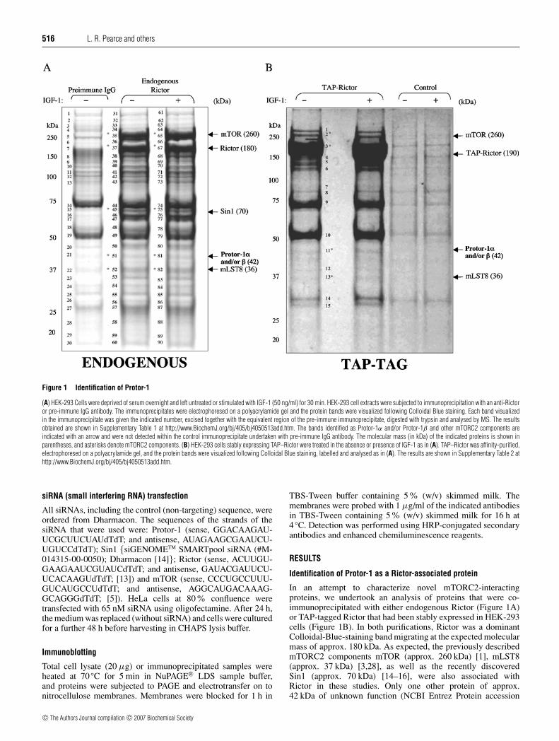
Figure 1 Identification of Protor-1
(A) HEK-293 Cells were deprived of serum overnight and left untreated or stimulated with IGF-1 (50 ng/ml) for 30 min. HEK-293 cell extracts were subjected to immunoprecipitation with an anti-Rictoror pre-immune IgG antibody. The immunoprecipitates were electrophoresed on a polyacrylamide gel and the protein bands were visualized following Colloidal Blue staining. Each band visualizedin the immunoprecipitate was given the indicated number, excised together with the equivalent region of the pre-immune immunoprecipitate, digested with trypsin and analysed by MS. The resultsobtained are shown in Supplementary Table 1 at http://www.BiochemJ.org/bj/405/bj4050513add.htm. The bands identified as Protor-1α and/or Protor-1β and other mTORC2 components areindicated with an arrow and were not detected within the control immunoprecipitate undertaken with pre-immune IgG antibody. The molecular mass (in kDa) of the indicated proteins is shown inparentheses, and asterisks denote mTORC2 components. (B) HEK-293 cells stably expressing TAP–Rictor were treated in the absence or presence of IGF-1 as in (A). TAP–Rictor was affinity-purified,electrophoresed on a polyacrylamide gel, and the protein bands were visualized following Colloidal Blue staining, labelled and analysed as in (A). The results are shown in Supplementary Table 2 athttp://www.BiochemJ.org/bj/405/bj4050513add.htm.
siRNA (small interfering RNA) transfection
All siRNAs, including the control (non-targeting) sequence, wereordered from Dharmacon. The sequences of the strands of thesiRNA that were used were: Protor-1 (sense, GGACAAGAU-UCGCUUCUAUdTdT; and antisense, AUAGAAGCGAAUCU-UGUCCdTdT); Sin1 {siGENOMETM SMARTpool siRNA (#M-014315-00-0050); Dharmacon [14]}; Rictor (sense, ACUUGU-GAAGAAUCGUAUCdTdT; and antisense, GAUACGAUUCU-UCACAAGUdTdT; [13]) and mTOR (sense, CCCUGCCUUU-GUCAUGCCUdTdT; and antisense, AGGCAUGACAAAG-GCAGGGdTdT; [5]). HeLa cells at 80 % confluence weretransfected with 65 nM siRNA using oligofectamine. After 24 h,the medium was replaced (without siRNA) and cells were culturedfor a further 48 h before harvesting in CHAPS lysis buffer.
Immunoblotting
Total cell lysate (20 µg) or immunoprecipitated samples wereheated at 70 ◦C for 5 min in NuPAGE® LDS sample buffer,and proteins were subjected to PAGE and electrotransfer on tonitrocellulose membranes. Membranes were blocked for 1 h in
TBS-Tween buffer containing 5% (w/v) skimmed milk. Themembranes were probed with 1 µg/ml of the indicated antibodiesin TBS-Tween containing 5% (w/v) skimmed milk for 16 h at4 ◦C. Detection was performed using HRP-conjugated secondaryantibodies and enhanced chemiluminescence reagents.
RESULTS
Identification of Protor-1 as a Rictor-associated protein
In an attempt to characterize novel mTORC2-interactingproteins, we undertook an analysis of proteins that were co-immunoprecipitated with either endogenous Rictor (Figure 1A)or TAP-tagged Rictor that had been stably expressed in HEK-293cells (Figure 1B). In both purifications, Rictor was a dominantColloidal-Blue-staining band migrating at the expected molecularmass of approx. 180 kDa. As expected, the previously describedmTORC2 components mTOR (approx. 260 kDa) [1], mLST8(approx. 37 kDa) [3,28], as well as the recently discoveredSin1 (approx. 70 kDa) [14–16], were also associated withRictor in these studies. Only one other protein of approx.42 kDa of unknown function (NCBI Entrez Protein accession
c© The Authors Journal compilation c© 2007 Biochemical Society
Role of Protor isoforms 517
no. NP 851850), which we have named Protor-1 (protein observedwith Rictor-1), was also present in both preparations of Rictor(Figure 1, and Supplementary Tables 1 and 2 at http://www.BiochemJ.org/bj/405/bj4050513add.htm). In one study, the geneencoding this protein was called PRR-5 (proline-rich repeatprotein-5) [29]. We also observed that stimulation of HEK-293cells with IGF-1 (insulin-like growth factor-1) did not alter theamounts of Protor-1 or mTOR, Sin1 and mLST8 associated withRictor (Figure 1).
Protor-1 mRNA is reportedly expressed as three splicevariants [29], which we have termed Protor-1α (residues 1–388;NCBI Entrez Protein accession number NP 851850), Protor-1β (residues 10–388; NCBI Entrez Protein accession numberNP 056181) and Protor-1γ (residues 97–388; NCBI EntrezProtein accession number NP 001017530). Protor-1 possessesno recognizable functional domains and has not been studiedpreviously at the protein level. The apparent molecular mass withwhich the Protor-1 protein migrated on an SDS/polyacrylamidegel (approx. 42 kDa; Figure 1) and MS analysis of the peptidesequences suggested that the splice variant of Protor associatedwith Rictor in the studies shown in Figure 1 was either Protor-1αor Protor-1β, which only differ by the presence of nine additionalresidues at the N-terminus (which we failed to detect in the MSanalysis; Figure 1 and Supplementary Tables 1 and 2). Databaseanalysis suggested that there is a second isoform of human Protor,encoded by an unstudied gene, that we have termed Protor-2 (NCBI Entrez Protein accession number CAE45978). Pro-tor-2 encodes a protein of 368 residues displaying 39%identity in amino acid sequence with Protor-1α (SupplementaryFigure 1 at http://www.BiochemJ.org/bj/405/bj4050513add.htm).The highest region of conservation between Protor-1 and Protor-2 encompasses residues 42–225 of Protor-1α (SupplementaryFigure 1).
A number of other proteins were associated with immuno-precipitated endogenous Rictor (Figure 1A), which were notidentified in the purification of the stably expressed TAP-tagged Rictor (Figure 1, and Supplementary Tables 1 and 2).Further investigation is required to determine whether any ofthese proteins interact with Rictor or whether these were non-specifically immunoprecipitated with the Rictor antibody used.Two of these proteins, MLEL1 and CDYL, which we have testedthus far, failed to interact when co-expressed with Rictor in HEK-293 cells (L.R. Pearce, unpublished work). We also found thatthree isoforms of 14-3-3 interacted with TAP–Rictor (Figure 1B,and Supplementary Table 2), but these adaptor proteins werenot detected in the MS analysis of the endogenous Rictorimmunoprecipitates (Figure 1A, and Supplementary Table 1). Toour knowledge, neither Rictor nor other components of mTORcomplexes have been identified in large-scale proteomic analysisof 14-3-3-interacting proteins.
Protor-1 interacts with mTORC2
We next expressed FLAG–Protor-1α in HEK-293 cells andinvestigated by MS which endogenously expressed proteins co-immunoprecipitated with it. Consistent with the notion thatProtor-1 might comprise a component of mTORC2, mTOR,Rictor and Sin1 were readily detected in the FLAG–Protor-1αimmunoprecipitates (Figure 2A). Although MS analysis did notdetect mLST8 within the FLAG–Protor-1α immunoprecipitates,its presence was confirmed by immunoblotting analysis(Figure 2D). Raptor was not detected in immunoprecipitates ofFLAG–Protor-1α by either MS (Figure 2A) or immunoblotting(Figure 2D). We also co-expressed FLAG–Protor-1α with GST–Rictor or six other GST fusion proteins in HEK-293 cells and
observed that Protor-1α only interacted with Rictor, suggestingthat the interaction was specific (Supplementary Figure 2 athttp://www.BiochemJ.org/bj/405/bj4050513add.htm).
Forms of Protor that interact with mTORC2
We next investigated whether Protor-1 splice variants as well asProtor-2 were capable of interacting with endogenous mTORC2components when overexpressed in HEK-293 cells. The resultsof these studies demonstrate that Protor-1β and Protor-2 bindto mTORC2 components to the same extent as Protor-1α(Figure 2D). Interestingly, Protor-1γ that lacks the N-terminalpart of the region conserved between Protor-1 and Protor-2(Supplementary Figure 1) failed to interact with any componentof mTORC2, despite being expressed at the same level as Protor-2in HEK-293 cells (Figure 2D).
Endogenous Protor-1 interacts with mTORC2 components
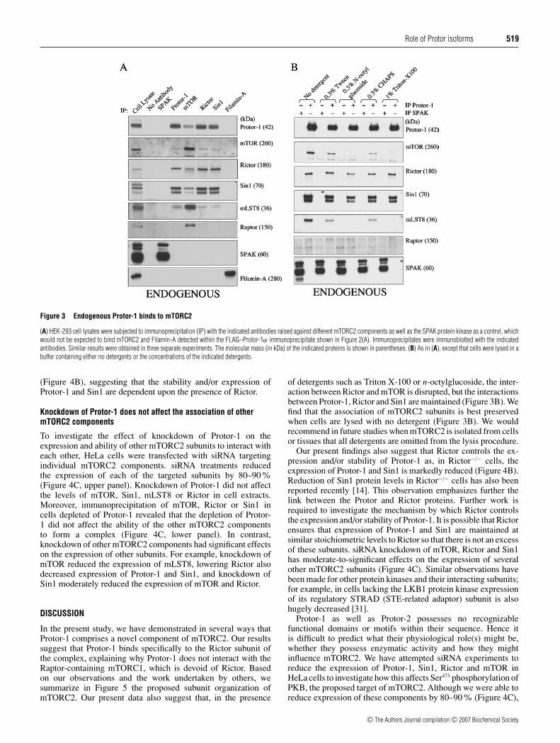
Using an antibody raised against Protor-1, immunoprecipitationof endogenous Protor-1 in HEK-293 cells resulted in the co-immunoprecipitation of endogenous mTOR, Rictor, Sin1 andmLST8 (Figure 3A). Furthermore, Protor-1 was clearly detectedwithin immunoprecipitates of endogenous mTOR, Rictor andSin1 (Figure 3A). Consistent with the view that Protor-1 associateswith mTORC2, rather than mTORC1, Raptor was not de-tected within immunoprecipitates of endogenous Protor-1. As ex-pected, Raptor was detected within immunoprecipitates ofmTOR, but not within immunoprecipitates of Rictor or Sin1(Figure 3A). We noticed that the cytoskeletal protein Filamin-A co-immunoprecipitated with FLAG–Protor-1α (Figure 2A).However, as we have observed Filamin-A to be a contaminantof other FLAG immunoprecipitations we have undertaken (D. R.Alessi, unpublished work), we were concerned that this was anon-specific interaction. Consistent with this, we failed to observeco-immunoprecipitation of endogenous forms of Filamin-A witheither Protor-1α or other mTORC2 components (Figure 3A).
Effects of detergent on the association of Protor-1with mTORC2 components
Previous studies have demonstrated that the integrity of mTORC2is disrupted when cells are lysed with 1 % (v/v) Triton X-100detergent, but not with 0.3% (w/v) CHAPS [13]. In the previousexperiments undertaken in the present study to demonstrate anassociation of Protor-1 with mTORC2, 0.3 % (w/v) CHAPSdetergent was employed. To investigate how detergents affectedthe association of Protor-1 with mTORC2, we lysed HEK-293cells either in the absence of detergent or in the presence of oneof four different detergents. None of the detergents employedaffected the ability of Protor-1 to associate with Rictor and Sin1,suggesting that the interaction between these proteins is detergent-insensitive. Strikingly, when cells were lysed with Triton X-100or n-octylglucoside, neither mTOR nor mLST8 associated withProtor-1 (Figure 3B). When cells were lysed with Tween 20,a similar association of Protor-1 with mTOR and mLST8 wasobserved as found with CHAPS (Figure 3B). The highest levelsof mTOR and mLST8 associated with Protor-1 were seen whencells were lysed with no detergent.
Protor-1 interacts specifically with Rictor
In order to establish which components of mTORC2 Protor-1αinteracts with, mTORC2 subunits and Protor-1 were co-expressedin HEK-293 cells. Immunoprecipitation of Protor-1α resulted inthe co-immunoprecipitation of mTOR, Rictor and Sin1 asexpected (Figure 4A). Omitting individual subunits suggested
c© The Authors Journal compilation c© 2007 Biochemical Society

518 L. R. Pearce and others
Figure 2 Protor-1 binds mTORC2
(A) HEK-293 cells were transfected with a DNA construct encoding FLAG–Protor-1α. At 36 h post-transfection, cells were lysed, and the FLAG–Protor-1α was immunoprecipitated (IP) andelectrophoresed on a polyacrylamide gel. The major protein bands were visualized following Colloidal Blue staining and labelled as indicated. The molecular mass (in kDa) of the indicated proteins isshown in parentheses, and asterisks denote mTORC2 components. (B) The labelled Colloidal-Blue-stained bands identified in (A) were excised from the gel, digested with trypsin and their identitiesdetermined by tryptic peptide mass-spectral fingerprint, as described in the Materials and methods section. Accession numbers are for the NCBI Entrez Protein database. †Mascot protein score, where avalue >67 is considered significant (P < 0.05). N.P.D., no significant protein identity determined. (C) Schematic representation of the Protor-1 isoforms and Protor-2. The conserved region is shadedand numbering of residues is based on the human sequence. For a sequence alignment of these proteins, see Supplementary Figure 1 at http://www.BiochemJ.org/bj/405/bj4050513add.htm. (D) Asin (A), except that HEK-293 cells were transfected with the indicated forms of Protor and a control FLAG-epitope-tagged protein (TD2; Genbank® accession number NM 014779). Immunoprecipitateswere immunoblotted (IB) with the indicated antibodies. Similar results were obtained in three separate experiments.
that Protor-1α is principally binding to mTORC2 by virtue ofits capability to bind Rictor, as, in the absence of Rictor, Protor-1α failed to interact with co-expressed mTOR or Sin1. In theabsence of mTOR, Protor-1 still associated with Rictor and Sin1(Figure 4A, middle panel). In the absence of Sin1, Protor-1 stillinteracted with Rictor. Consistent with previous reports [14–16],mTOR failed to interact with Rictor in the absence of Sin1, as Sin1is required for the stable binding of Rictor to mTOR (Figure 4A,middle panel). Further evidence that Protor-1 does not interactwith Sin1 is that, in the absence of Rictor, immunoprecipitation
of Sin1 did not result in the co-immunoprecipitation of Protor-1(Figure 4A, lower panel).
Rictor regulates the expression of Protor-1 and Sin1
To investigate further the importance of Rictor in enablingProtor-1 to interact with mTORC2 components, we utilizedRictor−/− knockout mouse embryonic fibroblast cells that havebeen generated recently [30]. Strikingly, both Protor-1 and Sin1expression was greatly decreased in the absence of Rictor
c© The Authors Journal compilation c© 2007 Biochemical Society
Role of Protor isoforms 519
Figure 3 Endogenous Protor-1 binds to mTORC2
(A) HEK-293 cell lysates were subjected to immunoprecipitation (IP) with the indicated antibodies raised against different mTORC2 components as well as the SPAK protein kinase as a control, whichwould not be expected to bind mTORC2 and Filamin-A detected within the FLAG–Protor-1α immunoprecipitate shown in Figure 2(A). Immunoprecipitates were immunoblotted with the indicatedantibodies. Similar results were obtained in three separate experiments. The molecular mass (in kDa) of the indicated proteins is shown in parentheses. (B) As in (A), except that cells were lysed in abuffer containing either no detergents or the concentrations of the indicated detergents.
(Figure 4B), suggesting that the stability and/or expression ofProtor-1 and Sin1 are dependent upon the presence of Rictor.
Knockdown of Protor-1 does not affect the association of othermTORC2 components
To investigate the effect of knockdown of Protor-1 on theexpression and ability of other mTORC2 subunits to interact witheach other, HeLa cells were transfected with siRNA targetingindividual mTORC2 components. siRNA treatments reducedthe expression of each of the targeted subunits by 80–90%(Figure 4C, upper panel). Knockdown of Protor-1 did not affectthe levels of mTOR, Sin1, mLST8 or Rictor in cell extracts.Moreover, immunoprecipitation of mTOR, Rictor or Sin1 incells depleted of Protor-1 revealed that the depletion of Protor-1 did not affect the ability of the other mTORC2 componentsto form a complex (Figure 4C, lower panel). In contrast,knockdown of other mTORC2 components had significant effectson the expression of other subunits. For example, knockdown ofmTOR reduced the expression of mLST8, lowering Rictor alsodecreased expression of Protor-1 and Sin1, and knockdown ofSin1 moderately reduced the expression of mTOR and Rictor.
DISCUSSION
In the present study, we have demonstrated in several ways thatProtor-1 comprises a novel component of mTORC2. Our resultssuggest that Protor-1 binds specifically to the Rictor subunit ofthe complex, explaining why Protor-1 does not interact with theRaptor-containing mTORC1, which is devoid of Rictor. Basedon our observations and the work undertaken by others, wesummarize in Figure 5 the proposed subunit organization ofmTORC2. Our present data also suggest that, in the presence
of detergents such as Triton X-100 or n-octylglucoside, the inter-action between Rictor and mTOR is disrupted, but the interactionsbetween Protor-1, Rictor and Sin1 are maintained (Figure 3B). Wefind that the association of mTORC2 subunits is best preservedwhen cells are lysed with no detergent (Figure 3B). We wouldrecommend in future studies when mTORC2 is isolated from cellsor tissues that all detergents are omitted from the lysis procedure.
Our present findings also suggest that Rictor controls the ex-pression and/or stability of Protor-1 as, in Rictor−/− cells, theexpression of Protor-1 and Sin1 is markedly reduced (Figure 4B).Reduction of Sin1 protein levels in Rictor−/− cells has also beenreported recently [14]. This observation emphasizes further thelink between the Protor and Rictor proteins. Further work isrequired to investigate the mechanism by which Rictor controlsthe expression and/or stability of Protor-1. It is possible that Rictorensures that expression of Protor-1 and Sin1 are maintained atsimilar stoichiometric levels to Rictor so that there is not an excessof these subunits. siRNA knockdown of mTOR, Rictor and Sin1has moderate-to-significant effects on the expression of severalother mTORC2 subunits (Figure 4C). Similar observations havebeen made for other protein kinases and their interacting subunits;for example, in cells lacking the LKB1 protein kinase expressionof its regulatory STRAD (STE-related adaptor) subunit is alsohugely decreased [31].
Protor-1 as well as Protor-2 possesses no recognizablefunctional domains or motifs within their sequence. Hence itis difficult to predict what their physiological role(s) might be,whether they possess enzymatic activity and how they mightinfluence mTORC2. We have attempted siRNA experiments toreduce the expression of Protor-1, Sin1, Rictor and mTOR inHeLa cells to investigate how this affects Ser473 phosphorylation ofPKB, the proposed target of mTORC2. Although we were able toreduce expression of these components by 80–90% (Figure 4C),
c© The Authors Journal compilation c© 2007 Biochemical Society

520 L. R. Pearce and others
Figure 4 Rictor interacts with Protor-1 and regulates its expression
(A) HEK-293 cells were co-transfected with the indicated DNA constructs encoding the indicated mTORC2 components. At 36 h post-transfection, cells were lysed and Protor-1 and Sin1 wereimmunoprecipitated (IP). The cell lysates (upper panel), the Protor-1 immunoprecipitates (middle panel) and Sin1 immunoprecipitates (lower panel) were immunoblotted with the indicated antibodies.Similar results were obtained in two separate experiments. The molecular mass (in kDa) of the indicated proteins is shown in parentheses. (B) Cell lysates derived from control wild-type Rictor+/+ andknockout Rictor−/− mouse embryonic fibroblast cells [30] were subjected to immunoblot analysis with the indicated antibodies. To confirm the marked reduction in Protor-1 expression, endogenousProtor-1 was immunoprecipitated (IP) and immunoblotted (lower panel). (C) HeLa cells were transfected with the indicated siRNAs (sequences as described in the Materials and methods section).At 72 h post-transfection, cells were lysed and cell lysates (upper panel) and the immunoprecipitates (lower panel) were immunoblotted with the indicated antibodies. Similar results were obtainedin two separate experiments. The molecular mass (in kDa) of the indicated proteins is shown in parentheses.
Figure 5 Model of the subunit organization of mTORC2
The dotted line indicates the detergent-sensitive interaction between mTOR/mLST8 andRictor/Sin1/Protor. ? indicates that direct interaction of these subunits is uncertain.
this reduction of protein was not sufficient to significantly reducephosphorylation of PKB at Ser473 (L. R. Pearce, unpublishedwork). Sabatini and co-workers [19] have elegantly demonstratedthat it is essential to completely ablate expression of mTORand Rictor in order to reduce phosphorylation of Ser473 of PKB,conditions that we have so far been unable to recapitulate in ourlaboratory. We are currently generating Protor knockout miceto address the physiological roles of this subunit in regulatingmTORC2 function.
Sequence alignments of Protor-1 and Protor-2 and Protor-1homologues found in other vertebrates (Supplementary Fig-ure 3 at http://www.BiochemJ.org/bj/405/bj4050513add.htm)also highlight that the N-terminal region of Protor encompassingresidues 42–225 of human Protor-1 is the most highly conservedand may therefore form a functional domain. Protor-1γ that lacksthe first 96 residues, hence part of the conserved domain, was notcapable of binding Rictor (Figure 2D). Further work is required todelineate the residues of Protor isoforms and Rictor that directlyinteract with each other, but it is likely that the Rictor-binding
c© The Authors Journal compilation c© 2007 Biochemical Society
Role of Protor isoforms 521
domain is within the conserved region on Protor. In addition, itremains to be investigated whether Protor-2 plays the same roleas Protor-1. In our present MS studies, we failed to detect anassociation of Protor-2 with endogenous Rictor or TAP–Rictor;however, in the absence of a specific anti-(Protor-2) antibody, it iscurrently unknown whether Protor-2 is expressed in the HEK-293cells utilized in our experiments.
Analysis of the tissue distribution patterns of mRNAexpression of Protor-1 and Protor-2 in humans using the NCBIUniGene database (http://www.ncbi.nlm.nih.gov/entrez/query.fcgi?db=unigene) suggests that these proteins, similar to the otherknown components of mTOR complexes, are widely expressed inall tissues. Northern blot analysis of Protor-1 mRNA in 12 humantissues also supported this conclusion [29]. We have analysed theexpression levels of the Protor-1 protein in nine different mousetissue extracts and found that Protor-1 was broadly expressed,with highest levels in adipose, kidney, spleen and testis andlowest levels in skeletal muscle (Supplementary Figure 4 athttp://www.BiochemJ.org/bj/405/bj4050513add.htm). In the onlyprevious study in which the Protor-1 gene was investigated, it wasreported that Protor-1 mRNA was overexpressed in 13 out of 16colorectal tumours studied [29]. It was also reported that, in humanmetastatic breast cancer, Protor-1 mRNA was overexpressed intwo out of eight tumours, whilst being down-regulated in fiveout the eight cancers [29]. No somatic mutations in the exonsencoding for the Protor-1 gene in human breast or colorectaltumours have been identified yet.
Surprisingly, we have not been able to detect any obvioushomologues of Protor-1 or Protor-2 in lower eukaryotic species,including yeast and Drosophila, frequently used for geneticanalysis of mTOR pathways. In Saccharomyces cerevisiae,two TOR complexes termed TORC1 and TORC2, believed tocomprise functional homologues of mTORC1 and mTORC2, havebeen characterized [3,32]. S. cerevisiae homologues of Rictor(AVO3), Sin1 (AVO1) and mLST8 (LST8) have been identifiedin TORC2 isolated from yeast extracts [3,32]. Two other S.cerevisiae proteins, namely AVO2 [3] and BIT61 [33], associatewith purified TORC2. To our knowledge, no homologues of theseproteins have yet been reported in mammals, and neither do AVO2nor BIT61 possess evident identity with Protor-1/Protor-2. A clearhomologue of Protor is found in frogs and fish (SupplementaryFigure 3).
In conclusion, we have identified Protor-1 and potentiallyProtor-2 as novel Rictor-binding components of mTORC2.Further work is required to define the role that Protor plays inregulating the functions of mTORC2. In particular, it would becrucial to define whether Protor-1 is required to enable mTORcomplexes to couple cell growth to the many signalling networksand environmental cues that mTOR complexes respond to [1].
We thank the Sequencing Service (School of Life Sciences, University of Dundee, Dundee,Scotland, U.K.) for DNA sequencing, the Post Genomics and Molecular InteractionsCentre (School of Life Sciences, University of Dundee, Dundee, Scotland, U.K.) for MSfacilities, and the protein production and antibody purification teams (Division of SignalTransduction Therapy, University of Dundee, Dundee, Scotland, U.K.), co-ordinated byHilary McLauchlan and James Hastie, for generation and purification of antibodies. L. R. P.is supported by a U.K. MRC (Medical Research Council) studentship. We thank theAssociation for International Cancer Research, Diabetes UK, MRC, the Moffat CharitableTrust and the pharmaceutical companies supporting the Division of Signal TransductionTherapy Unit (AstraZeneca, Boehringer-Ingelheim, GlaxoSmithKline, Merck & Co. Inc,Merck KgaA and Pfizer) for financial support.
REFERENCES
1 Wullschleger, S., Loewith, R. and Hall, M. N. (2006) TOR signaling in growth andmetabolism. Cell 124, 471–484
2 Sarbassov, D. D., Ali, S. M. and Sabatini, D. M. (2005) Growing roles for the mTORpathway. Curr. Opin. Cell. Biol. 17, 596–603
3 Loewith, R., Jacinto, E., Wullschleger, S., Lorberg, A., Crespo, J. L., Bonenfant, D.,Oppliger, W., Jenoe, P. and Hall, M. N. (2002) Two TOR complexes, only one of which israpamycin sensitive, have distinct roles in cell growth control. Mol. Cell 10, 457–468
4 Hara, K., Maruki, Y., Long, X., Yoshino, K., Oshiro, N., Hidayat, S., Tokunaga, C.,Avruch, J. and Yonezawa, K. (2002) Raptor, a binding partner of target of rapamycin(TOR), mediates TOR action. Cell 110, 177–189
5 Kim, D. H., Sarbassov, D. D., Ali, S. M., King, J. E., Latek, R. R., Erdjument-Bromage, H.,Tempst, P. and Sabatini, D. M. (2002) mTOR interacts with Raptor to form anutrient-sensitive complex that signals to the cell growth machinery. Cell 110, 163–175
6 Li, Y., Corradetti, M. N., Inoki, K. and Guan, K. L. (2004) TSC2: filling the GAP in themTOR signaling pathway. Trends Biochem. Sci. 29, 32–38
7 Byfield, M. P., Murray, J. T. and Backer, J. M. (2005) hVps34 is a nutrient-regulated lipidkinase required for activation of p70 S6 kinase. J. Biol. Chem. 280, 33076–33082
8 Nobukuni, T., Joaquin, M., Roccio, M., Dann, S. G., Kim, S. Y., Gulati, P., Byfield, M. P.,Backer, J. M., Natt, F., Bos, J. L. et al. (2005) Amino acids mediate mTOR/Raptorsignaling through activation of class 3 phosphatidylinositol 3OH-kinase. Proc. Natl.Acad. Sci. U.S.A. 102, 14238–14243
9 Findlay, G. M., Yan, L., Procter, J., Mieulet, V. and Lamb, R. F. (2007) A MAP4 kinaserelated to Ste20 is a nutrient-sensitive regulator of mTOR signalling. Biochem. J. 403,13–20
10 Biondi, R. M., Kieloch, A., Currie, R. A., Deak, M. and Alessi, D. R. (2001) ThePIF-binding pocket in PDK1 is essential for activation of S6K and SGK, but not PKB.EMBO J. 20, 4380–4390
11 Collins, B. J., Deak, M., Arthur, J. S., Armit, L. J. and Alessi, D. R. (2003) In vivo role ofthe PIF-binding docking site of PDK1 defined by knock-in mutation. EMBO J. 22,4202–4211
12 Jacinto, E., Loewith, R., Schmidt, A., Lin, S., Ruegg, M. A., Hall, A. and Hall, M. N. (2004)Mammalian TOR complex 2 controls the actin cytoskeleton and is rapamycin insensitive.Nat. Cell Biol. 6, 1122–1128
13 Sarbassov, D. D., Ali, S. M., Kim, D. H., Guertin, D. A., Latek, R. R.,Erdjument-Bromage, H., Tempst, P. and Sabatini, D. M. (2004) Rictor, a novel bindingpartner of mTOR, defines a rapamycin-insensitive and Raptor-independent pathway thatregulates the cytoskeleton. Curr. Biol. 14, 1296–1302
14 Yang, Q., Inoki, K., Ikenoue, T. and Guan, K. L. (2006) Identification of Sin1 as anessential TORC2 component required for complex formation and kinase activity.Genes Dev. 20, 2820–2832
15 Frias, M. A., Thoreen, C. C., Jaffe, J. D., Schroder, W., Sculley, T., Carr, S. A. and Sabatini,D. M. (2006) mSin1 is necessary for Akt/PKB phosphorylation, and its isoforms definethree distinct mTORC2s. Curr. Biol. 16, 1865–1870
16 Jacinto, E., Facchinetti, V., Liu, D., Soto, N., Wei, S., Jung, S. Y., Huang, Q., Qin, J. andSu, B. (2006) SIN1/MIP1 maintains Rictor-mTOR complex integrity and regulates Aktphosphorylation and substrate specificity. Cell 127, 125–137
17 Guertin, D. A., Stevens, D. M., Thoreen, C. C., Burds, A. A., Kalaany, N. Y., Moffat, J.,Brown, M., Fitzgerald, K. J. and Sabatini, D. M. (2006) Ablation in mice of the mTORCcomponents Raptor, Rictor, or mLST8 reveals that mTORC2 is required for signaling toAkt-FOXO and PKCα, but not S6K1. Dev. Cell 11, 859–871
18 Sarbassov, D. D., Ali, S. M., Sengupta, S., Sheen, J. H., Hsu, P. P., Bagley, A. F.,Markhard, A. L. and Sabatini, D. M. (2006) Prolonged rapamycin treatment inhibitsmTORC2 assembly and Akt/PKB. Mol. Cell 22, 159–168
19 Sarbassov, D. D., Guertin, D. A., Ali, S. M. and Sabatini, D. M. (2005) Phosphorylationand regulation of Akt/PKB by the Rictor-mTOR complex. Science 307, 1098–1101
20 Hresko, R. C. and Mueckler, M. (2005) mTOR.RICTOR is the Ser473 kinase forAkt/protein kinase B in 3T3-L1 adipocytes. J. Biol. Chem. 280, 40406–40416
21 Mora, A., Komander, D., Van Aalten, D. M. and Alessi, D. R. (2004) PDK1, the masterregulator of AGC kinase signal transduction. Semin. Cell. Dev. Biol. 15, 161–170
22 Durocher, Y., Perret, S. and Kamen, A. (2002) High-level and high-throughputrecombinant protein production by transient transfection of suspension-growing human293-EBNA1 cells. Nucleic Acids Res. 30, E9
23 Al-Hakim, A. K., Goransson, O., Deak, M., Toth, R., Campbell, D. G., Morrice, N. A.,Prescott, A. R. and Alessi, D. R. (2005) 14-13-3 cooperates with LKB1 to regulate theactivity and localization of QSK and SIK. J. Cell Sci. 118, 5661–5673
24 Harlow, E. and Lane, D. (1988) Antibodies: a Laboratory Manual, Cold Spring HarborLaboratory Publications, New York
25 Woods, Y. L., Rena, G., Morrice, N., Barthel, A., Becker, W., Guo, S., Unterman, T. G. andCohen, P. (2001) The kinase DYRK1A phosphorylates the transcription factor FKHR atSer329 in vitro, a novel in vivo phosphorylation site. Biochem. J. 355, 597–607
26 Perkins, D. N., Pappin, D. J., Creasy, D. M. and Cottrell, J. S. (1999) Probability-basedprotein identification by searching sequence databases using mass spectrometry data.Electrophoresis 20, 3551–3567
c© The Authors Journal compilation c© 2007 Biochemical Society

522 L. R. Pearce and others
27 Rigaut, G., Shevchenko, A., Rutz, B., Wilm, M., Mann, M. and Seraphin, B. (1999) Ageneric protein purification method for protein complex characterization and proteomeexploration. Nat. Biotechnol. 17, 1030–1032
28 Kim, D. H., Sarbassov, D. D., Ali, S. M., Latek, R. R., Guntur, K. V.,Erdjument-Bromage, H., Tempst, P. and Sabatini, D. M. (2003) GβL, a positive regulatorof the rapamycin-sensitive pathway required for the nutrient-sensitive interaction betweenRaptor and mTOR. Mol. Cell 11, 895–904
29 Johnstone, C. N., Castellvi-Bel, S., Chang, L. M., Sung, R. K., Bowser, M. J.,Pique, J. M., Castells, A. and Rustgi, A. K. (2005) PRR5 encodes a conservedproline-rich protein predominant in kidney: analysis of genomic organization,expression, and mutation status in breast and colorectal carcinomas. Genomics 85,338–351
30 Shiota, C., Woo, J. T., Lindner, J., Shelton, K. D. and Magnuson, M. A. (2006) Multiallelicdisruption of the Rictor gene in mice reveals that mTOR complex 2 is essential for fetalgrowth and viability. Dev. Cell 11, 583–589
31 Hawley, S. A., Boudeau, J., Reid, J. L., Mustard, K. J., Udd, L., Makela, T. P., Alessi, D. R.and Hardie, D. G. (2003) Complexes between the LKB1 tumor suppressor, STRADα/βand MO25α/β are upstream kinases in the AMP-activated protein kinase cascade.J. Biol. 2, 28
32 Wullschleger, S., Loewith, R., Oppliger, W. and Hall, M. N. (2005) Molecular organizationof target of rapamycin complex 2. J. Biol. Chem. 280, 30697–30704
33 Wedaman, K. P., Reinke, A., Anderson, S., Yates, 3rd, J., McCaffery, J. M. and Powers, T.(2003) Tor kinases are in distinct membrane-associated protein complexes inSaccharomyces cerevisiae. Mol. Biol. Cell 14, 1204–1220
Received 23 April 2007; accepted 27 April 2007Published as BJ Immediate Publication 27 April 2007, doi:10.1042/BJ20070540
c© The Authors Journal compilation c© 2007 Biochemical Society