Induced Plant Resistance to Herbivory - Aula Virtual - FCAyF
450
-
Upload
khangminh22 -
Category
Documents
-
view
1 -
download
0
Transcript of Induced Plant Resistance to Herbivory - Aula Virtual - FCAyF


INDUCED PLANT RESISTANCE TO HERBIVORY

Induced PlantResistance
to Herbivory
Edited by
Andreas SchallerUniversity of Hohenheim,
Stuttgart, Germany

Editor
Andreas SchallerUniversity of HohenheimStuttgart, Germany
ISBN: 978-1-4020-8181-1 e-ISBN: 978-1-4020-8182-8
Library of Congress Control Number: 2007941936
c© 2008 Springer Science+Business Media B.V.No part of this work may be reproduced, stored in a retrieval system, or transmittedin any form or by any means, electronic, mechanical, photocopying, microfilming, recordingor otherwise, without written permission from the Publisher, with the exceptionof any material supplied specifically for the purpose of being enteredand executed on a computer system, for exclusive use by the purchaser of the work.
Cover pictures showing Pieris brassicae caterpillars, the parasitic wasp Cotesia glomerata,and a parasitized Manduca sexta larva were taken by Hans Smid and Tibor Bukovinszky(http: www.bugsinthepicture.com/), and Johannes Stratmann (University of South Carolina).
Printed on acid-free paper
9 8 7 6 5 4 3 2 1
springer.com
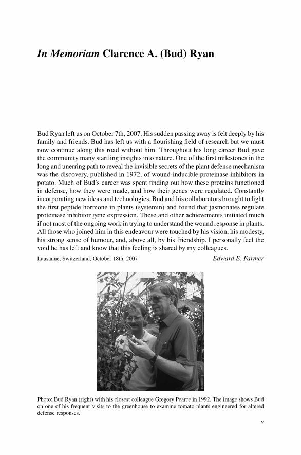
In Memoriam Clarence A. (Bud) Ryan
Bud Ryan left us on October 7th, 2007. His sudden passing away is felt deeply by hisfamily and friends. Bud has left us with a flourishing field of research but we mustnow continue along this road without him. Throughout his long career Bud gavethe community many startling insights into nature. One of the first milestones in thelong and unerring path to reveal the invisible secrets of the plant defense mechanismwas the discovery, published in 1972, of wound-inducible proteinase inhibitors inpotato. Much of Bud’s career was spent finding out how these proteins functionedin defense, how they were made, and how their genes were regulated. Constantlyincorporating new ideas and technologies, Bud and his collaborators brought to lightthe first peptide hormone in plants (systemin) and found that jasmonates regulateproteinase inhibitor gene expression. These and other achievements initiated muchif not most of the ongoing work in trying to understand the wound response in plants.All those who joined him in this endeavour were touched by his vision, his modesty,his strong sense of humour, and, above all, by his friendship. I personally feel thevoid he has left and know that this feeling is shared by my colleagues.
Lausanne, Switzerland, October 18th, 2007 Edward E. Farmer
Photo: Bud Ryan (right) with his closest colleague Gregory Pearce in 1992. The image shows Budon one of his frequent visits to the greenhouse to examine tomato plants engineered for altereddefense responses.
v
Contents
In Memoriam Clarence A. (Bud) Ryan . . . . . . . . . . . . . . . . . . . . . . . . . . . . . . . . v
Introduction . . . . . . . . . . . . . . . . . . . . . . . . . . . . . . . . . . . . . . . . . . . . . . . . . . . . . . . 1
Section I Basic Concepts of Plant Defense Against Insect Herbivores
1 Direct Defenses in Plants and Their Induction by Wounding andInsect Herbivores . . . . . . . . . . . . . . . . . . . . . . . . . . . . . . . . . . . . . . . . . . . . . . 7Gregg A. Howe and Andreas Schaller
2 Herbivore-Induced Indirect Defense: From Induction Mechanismsto Community Ecology . . . . . . . . . . . . . . . . . . . . . . . . . . . . . . . . . . . . . . . . . 31Maaike Bruinsma and Marcel Dicke
3 Induced Defenses and the Cost-Benefit Paradigm . . . . . . . . . . . . . . . . . . 61Anke Steppuhn and Ian T. Baldwin
Section II Induced Direct Defenses
Part A Anatomical Defenses
4 Leaf Trichome Formation and Plant Resistance to Herbivory . . . . . . . 89Peter Dalin, Jon Agren, Christer Bjorkman, Piritta Huttunenand Katri Karkkainen
5 Resistance at the Plant Cuticle . . . . . . . . . . . . . . . . . . . . . . . . . . . . . . . . . . 107Caroline Muller
vii
viii Contents
6 Wound-Periderm Formation . . . . . . . . . . . . . . . . . . . . . . . . . . . . . . . . . . . . 131Idit Ginzberg
7 Traumatic Resin Ducts and Polyphenolic Parenchyma Cells inConifers . . . . . . . . . . . . . . . . . . . . . . . . . . . . . . . . . . . . . . . . . . . . . . . . . . . . . . 147Paal Krokene, Nina Elisabeth Nagy and Trygve Krekling
Part B Production of Secondary Metabolites
8 Insect-Induced Terpenoid Defenses in Spruce . . . . . . . . . . . . . . . . . . . . . 173Jorg Bohlmann
9 Phenylpropanoid Metabolism Inducedby Wounding and Insect Herbivory . . . . . . . . . . . . . . . . . . . . . . . . . . . . . . 189Mark A. Bernards and Lars Bastrup-Spohr
10 Defense by Pyrrolizidine Alkaloids: Developed by Plants andRecruited by Insects . . . . . . . . . . . . . . . . . . . . . . . . . . . . . . . . . . . . . . . . . . . 213Thomas Hartmann and Dietrich Ober
Part C Anti-nutritional Enzymes and Proteins
11 Plant Protease Inhibitors: Functional Evolution for Defense . . . . . . . . 235Maarten A. Jongsma and Jules Beekwilder
12 Defensive Roles of Polyphenol Oxidase in Plants . . . . . . . . . . . . . . . . . . 253C. Peter Constabel and Raymond Barbehenn
13 Action of Plant Defensive Enzymes in the Insect Midgut . . . . . . . . . . . 271Hui Chen, Eliana Gonzales-Vigil and Gregg A. Howe
14 Plant Lectins as Part of the Plant Defense System Against Insects . . . 285Els J.M. Van Damme
Section III Defense Signaling
Part A Activation of Plant Defenses
15 Systemins and AtPeps: Defense-Related Peptide Signals . . . . . . . . . . . . 313Javier Narvaez-Vasquez and Martha L. Orozco-Cardenas
Contents ix
16 MAP Kinases in Plant Responses to Herbivory . . . . . . . . . . . . . . . . . . . . 329Johannes Stratmann
17 Jasmonate Biosynthesis and Signaling for Induced Plant Defenseagainst Herbivory . . . . . . . . . . . . . . . . . . . . . . . . . . . . . . . . . . . . . . . . . . . . . 349Andreas Schaller and Annick Stintzi
Part B Signals Between Plants and Insects
18 Caterpillar Secretions and Induced Plant Responses . . . . . . . . . . . . . . . 369Gary W. Felton
19 Fatty Acid-Derived Signals that Induce or Regulate Plant DefensesAgainst Herbivory . . . . . . . . . . . . . . . . . . . . . . . . . . . . . . . . . . . . . . . . . . . . . 389James H. Tumlinson and Juergen Engelberth
20 Aromatic Volatiles and Their Involvement in Plant Defense . . . . . . . . . 409Anthony V. Qualley and Natalia Dudareva
21 Ecological Roles of Vegetative Terpene Volatiles . . . . . . . . . . . . . . . . . . . 433Jorg Degenhardt
Abbreviations . . . . . . . . . . . . . . . . . . . . . . . . . . . . . . . . . . . . . . . . . . . . . . . . . . . . . . 443
Subject Index . . . . . . . . . . . . . . . . . . . . . . . . . . . . . . . . . . . . . . . . . . . . . . . . . . . . . . 449
Taxonomic Index . . . . . . . . . . . . . . . . . . . . . . . . . . . . . . . . . . . . . . . . . . . . . . . . . . . 457

Contributors
Jon AgrenDepartment of Ecology and Evolution, Evolutionary Biology Centre, UppsalaUniversity, SE-752 36 Uppsala, [email protected]
Ian T. BaldwinMax Planck Institute for Chemical Ecology, Department of Molecular Ecology,D-07745 Jena, [email protected]
Raymond BarbehennDepartment of Molecular, Cellular, and Developmental Biology, University ofMichigan, Ann Arbor, MI 48105, [email protected]
Lars Bastrup-SpohrFreshwater Biological Laboratory, University of Copenhagen, DK-3400 Hillerød,[email protected]
Jules BeekwilderPlant Research International B.V., Wageningen University and Research Center,6700 AA Wageningen, The [email protected]
Mark A. BernardsDepartment of Biology, The University of Western Ontario, London, ON, CanadaN6A [email protected]
Christer BjorkmanDepartment of Ecology, Swedish University of Agricultural Sciences, SE-750 07Uppsala, [email protected]
xi

xii Contributors
Jorg BohlmannMichael Smith Laboratories, University of British Columbia, Vancouver, BC,Canada V6T [email protected]
Maaike BruinsmaLaboratory of Entomology, Wageningen University, 6709 PD Wageningen,The [email protected]
Hui ChenDOE Plant Research Laboratory, Michigan State University, East Lansing, MI48824, [email protected]
C. Peter ConstabelCentre for Forest Biology and Department of Biology, University of Victoria,Victoria, BC, Canada V8W [email protected]
Peter DalinMarine Science Institute, University of California at Santa Barbara, CA93106-6150, [email protected]
Jorg DegenhardtDepartment of Biochemistry, Max Planck Institute for Chemical Ecology, D-07745Jena, [email protected]
Marcel DickeLaboratory of Entomology, Wageningen University, 6709 PD Wageningen, [email protected]
Natalia DudarevaDepartment of Horticulture and Landscape Architecture, Purdue University, WestLafayette, IN 47907, [email protected]
Juergen EngelberthDepartment of Biology, University of Texas at San Antonio, San Antonio, TX78249, [email protected]
Gary W. FeltonDepartment of Entomology, Pennsylvania State University, University Park, PA16802, [email protected]

Contributors xiii
Idit GinzbergAgricultural Research Organization, The Volcani Center, Bet Dagan 50250, [email protected]
Eliana Gonzales-VigilDOE Plant Research Laboratory, Michigan State University, East Lansing, MI48824, [email protected]
Thomas HartmannInstitute of Pharmaceutical Biology, Technical University of Braunschweig,D-38106 Braunschweig, [email protected]
Gregg A. HoweDOE Plant Research Laboratory, Michigan State University, East Lansing, MI48824, [email protected]
Piritta HuttunenDepartment of Biological and Environmental Sciences, University of Jyvaskyla,40350 Jyvaskyla, [email protected]
Maarten A. JongsmaPlant Research International B.V., Wageningen University and Research Center,6700 AA Wageningen, The [email protected]
Katri KarkkainenThe Finnish Forest Research Institute, Muhos Research Unit, 91500 Muhos,[email protected]
Trygve KreklingDepartment of Plant and Environmental Sciences, Norwegian University of LifeSciences, N-1432 As, [email protected]
Paal KrokeneNorwegian Forest and Landscape Institute, N-1432 As, [email protected]
Caroline MullerDepartment of Chemical Ecology, University of Bielefeld, D-33615 Bielefeld,[email protected]

xiv Contributors
Nina Elisabeth NagyNorwegian Forest and Landscape Institute, N-1432 As, [email protected]
Javier Narvaez-VasquezDepartment of Botany and Plant Sciences, University of California Riverside,Riverside, CA 92521, [email protected]
Dietrich OberInstitute of Botany, University of Kiel, D-24098 Kiel, [email protected]
Martha L. Orozco-CardenasPlant Transformation Research Center, University of California Riverside,Riverside, CA 92521, [email protected]
Anthony V. QualleyDepartment of Horticulture and Landscape Architecture, Purdue University, WestLafayette, IN 47907, [email protected]
Andreas SchallerUniversity of Hohenheim, Institute of Plant Physiology and Biotechnology,D-70599 Stuttgart, [email protected]
Anke SteppuhnMax Planck Institute for Chemical Ecology, Department of Molecular Ecology,D-07745 Jena, [email protected]
Annick StintziInstitute of Plant Physiology and Biotechnology, University of Hohenheim,D-70599 Stuttgart, [email protected]
Johannes StratmannDepartment of Biological Sciences, University of South Carolina, Columbia, SC29208, [email protected]
James H. TumlinsonCenter for Chemical Ecology, Department of Entomology, PennsylvaniaState University, University Park, PA 16802, [email protected]

Contributors xv
Els J.M. Van DammeLaboratory of Biochemistry and Glycobiology, Department of MolecularBiotechnology, Ghent University, 9000 Gent, [email protected]
Introduction
The class Insecta with more than one million described species is the most diversegroup of animals on Earth and outnumbers all other forms of life. Almost half ofall existing insect species are herbivores, i.e. they feed on living plants. The abil-itity of plants to persist in such a hostile environment relies on evolved resistancesystems allowing them to escape herbivores in time or in space, to confront her-bivores directly by affecting host plant preference or reproductive success, or tofight herbivores indirectly by association with other species. Plant defenses againstinsect herbivores were long known to change in evolutionary time and may evenchange during the lifetime of an individual plant. Until recently however, plant de-fenses were generally assumed to be constitutively expressed, i.e. independent fromherbivore attack. This view changed in 1972 when Green and Ryan reported thatwounding by Colorado potato beetles causes the rapid accumulation of proteinaseinhibitors in potato and tomato leaves. As inducible resistance factors which inter-fere with the digestive system of leaf-consuming insects, the proteinase inhibitorswere suggested to defend the plants against herbivores.
In the 35 years following the initial discovery by Green and Ryan, many moretraits have been identified that are induced by wounding or herbivory. The fullbreadth of induced responses became apparent with the advent of novel techniquesfor transcriptome analysis, revealing large-scale changes in gene expression in re-sponse to herbivory. In fact, many of this book’s chapters focus on herbivore-induced responses including the results of transcriptome analyses, and how theyrelate to plant defense. At this point, a cautionary note is required: Inducibility ofa certain gene (or any given trait) is per se not sufficient evidence for a functionin resistance or plant defense. Adopting the definitions advocated by R. KarbanJ.H. Myers, A.A. Agrawal, and I.T. Baldwin, the term ‘induced resistance’ refers toinduced changes in preference, performance, or reproductive success of the attacker.Therefore, to test for a role in resistance, it is necessary to compare herbivore pref-erence and/or performance on plants that differ in the trait of interest, which caneasily be done in a laboratory setting. Induced resistance is thus defined from thepoint of view of the herbivore, and it does not necessarily benefit the plant. Forexample, the investment in induced resistance may be larger than the benefit formreduced herbivore damage, or induced herbivore resistance may render the plant
A. Schaller (ed.), Induced Plant Resistance to Herbivory, 1C© Springer Science+Business Media B.V. 2008
2 Introduction
more susceptible to other stresses. Defense, on the other hand is defined from theplant’s perspective, and it implies a benefit for the plant. Only when they result ina gain in plant fitness are induced traits considered to contribute to plant defense.Therefore, to demonstrate a role in plant defense, field studies are required that re-veal differences in fitness for plants that differ in the trait of interest. Only very fewherbivore-induced responses have actually been shown to result in a fitness benefitfor the plant. Nevertheless, the term ‘defense’ is frequently used by researchers inthe field and also by the authors of this book to describe induced changes in plantarchitecture, metabolism, or physiology that are assumed to minimize the negativeimpact of herbivory.
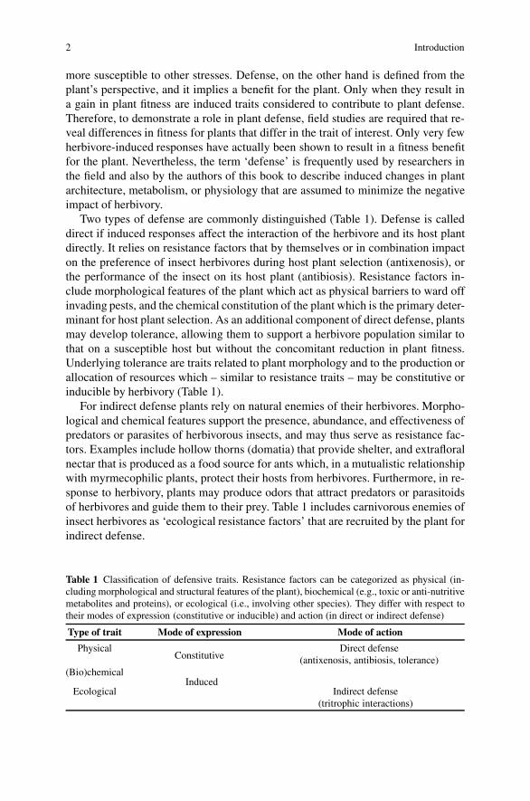
Two types of defense are commonly distinguished (Table 1). Defense is calleddirect if induced responses affect the interaction of the herbivore and its host plantdirectly. It relies on resistance factors that by themselves or in combination impacton the preference of insect herbivores during host plant selection (antixenosis), orthe performance of the insect on its host plant (antibiosis). Resistance factors in-clude morphological features of the plant which act as physical barriers to ward offinvading pests, and the chemical constitution of the plant which is the primary deter-minant for host plant selection. As an additional component of direct defense, plantsmay develop tolerance, allowing them to support a herbivore population similar tothat on a susceptible host but without the concomitant reduction in plant fitness.Underlying tolerance are traits related to plant morphology and to the production orallocation of resources which – similar to resistance traits – may be constitutive orinducible by herbivory (Table 1).
For indirect defense plants rely on natural enemies of their herbivores. Morpho-logical and chemical features support the presence, abundance, and effectiveness ofpredators or parasites of herbivorous insects, and may thus serve as resistance fac-tors. Examples include hollow thorns (domatia) that provide shelter, and extrafloralnectar that is produced as a food source for ants which, in a mutualistic relationshipwith myrmecophilic plants, protect their hosts from herbivores. Furthermore, in re-sponse to herbivory, plants may produce odors that attract predators or parasitoidsof herbivores and guide them to their prey. Table 1 includes carnivorous enemies ofinsect herbivores as ‘ecological resistance factors’ that are recruited by the plant forindirect defense.
Table 1 Classification of defensive traits. Resistance factors can be categorized as physical (in-cluding morphological and structural features of the plant), biochemical (e.g., toxic or anti-nutritivemetabolites and proteins), or ecological (i.e., involving other species). They differ with respect totheir modes of expression (constitutive or inducible) and action (in direct or indirect defense)
Type of trait Mode of expression Mode of action
PhysicalConstitutive
Direct defense(antixenosis, antibiosis, tolerance)
(Bio)chemical
EcologicalInduced
Indirect defense(tritrophic interactions)

Introduction 3
Direct and indirect mechanisms of plant defense against herbivores, and theraison d’etre for inducibility as opposed to constitutive expression of defenseare introduced in Section I of this book. These introductory chapters provide thebackground for the subsequent more focussed discussion of individual resistancefactors in Section II, and the signals and signaling mechanisms for the induction ofdirect and indirect defenses in Section III. With emphasis on plant responses thatare induced by wounding or herbivory, the progress of research is summarized in afield that continues to be highly dynamic, even 35 years after the initial discoveryof proteinase inhibitor accumulation as a wound-induced anti-nutritive defense byRyan in 1972.

Section IBasic Concepts of Plant Defense Against
Insect Herbivores

Chapter 1Direct Defenses in Plants and Their Inductionby Wounding and Insect Herbivores
Gregg A. Howe and Andreas Schaller
Resistance factors for direct plant defense against herbivorous insects compriseplant traits that negatively affect insect preference (host plant selection, oviposition,feeding behavior) or performance (growth rate, development, reproductive success)resulting in increased plant fitness in a hostile environment. Such traits include mor-phological features for physical defense, like thorns, spines, and trichomes, epicu-ticular wax films and wax crystals, tissue toughness, as well as secretory structuresand conduits for latices or resins. They also include compounds for chemical de-fense, like secondary metabolites, digestibility reducing proteins, and antinutritiveenzymes. All these traits may be expressed constitutively as preformed resistancefactors, or they may be inducible and deployed only after attack by insect herbivores.The induction of defensive traits is not restricted to the site of attack but extends tonon-infested healthy parts of the plants. The systemic nature of plant responses toherbivore attack necessitates a long-distance signaling system capable of generat-ing, transporting, and interpreting alarm signals produced at the plant–herbivoreinterface. Much of the research on the signaling events triggered by herbivory hasfocused on tomato and other solanaceous plants. In this model system, the peptidesystemin acts at or near the wound site to amplify the production of jasmonic acid.Jasmonic acid or its metabolites serve as phloem-mobile long-distance signals, andinduce the expression of defense genes in distal parts of the plant. In this chapter,we will provide an overview of physical and chemical defense traits, and reviewthe signaling mechanisms that account for their inducible expression after insectattack.
1.1 Introduction
Plants, flowering plants in particular, exhibit a tremendous diversity in size andshape, ranging from just a few millimeters in the tiny duckweeds to almost 100
A. SchallerUniversity of Hohenheim, Institute of Plant Physiology and Biotechnology, D-70599 Stuttgart,Germanye-mail: [email protected]
A. Schaller (ed.), Induced Plant Resistance to Herbivory, 7C© Springer Science+Business Media B.V. 2008
8 G.A. Howe, A. Schaller
meters in giant eucalyptus trees. Some may complete their life cycle in a fewweeks, while others live thousands of years. The amazing diversity results fromthe adaptation to different, oftentimes hostile environments, as exemplified by theearly evolution of land plants. The colonization of land by plants, dating back some480 million years according to fossil records (Kenrick and Crane 1997), marks thebeginning of an evolutionary success story, with flowering plants now occupyingevery habitat on Earth except the regions surrounding the poles, the highest moun-taintops, and the deepest oceans (Soltis and Soltis 2004). The colonization of landwas a major event in the history of plant life, and at the same time, paved the wayfor the explosive evolution of terrestrial ecosystems. Despite the vulnerability ofplants as sessile organisms to adverse biotic and abiotic conditions, they actuallydominate over much of the land surface. This apparent success of flowering plantsrelies on the evolved ability to persist in unfavorable and variable environments byvirtue of effective resistance systems that are based on a combination of physical,chemical, and developmental features (Schoonhoven et al. 2005). It was recognizedby Stahl in 1888 that the great diversity of mechanical and chemical ‘means ofprotection of plants were acquired in their struggle for existence within the animalworld’ leading to the conclusion that ‘the animal world [. . .] deeply influenced notonly their morphology but also their chemistry’ (Stahl 1888; Fraenkel 1959). Hence,not only thorns and spines as morphological resistance traits, but also the bewilder-ing variety of plant secondary chemicals attest to the selective pressure exerted byphytophagous animals (Fraenkel 1959; Ehrlich and Raven 1964).
It was later discovered that induced expression of resistance traits increases plantfitness in environments that harbor a variety of plant parasites. The inducibility ofplant resistance was first reported for fungal and bacterial pathogens in the early1900s (Karban and Kuc 1999) and, much later, inducible defenses were shownto exist also against insect herbivores. In their seminal paper of 1972, Green andRyan demonstrated that tomato and potato plants accumulate inhibitors of trypsinand chymotrypsin-like serine proteinases throughout their aerial tissues, as a di-rect consequence of insect-mediated damage or mechanical wounding (Green andRyan 1972). Proteinase inhibitors are present constitutively in high concentrationsin plant storage organs, and a possible function as protective agents against insectswas discussed at that time (Lipke et al. 1954; Applebaum and Konijn 1966). Greenand Ryan suggested that the expression of proteinase inhibitors may be regulated inleaves to make the plant less palatable and perhaps lethal to invading insects. Theaccumulation of proteinase inhibitors in aerial tissues was proposed to constitutean inducible defense system, directly affecting the performance of leaf-consuminginsects by starving them of nutrients, thus resulting in enhanced plant resistanceagainst herbivory (Green and Ryan 1972). It is now clear that the nutritional qualityof the foliage is an important determinant of herbivore growth and development(Painter 1936; Berenbaum 1995; Schoonhoven et al. 2005) and anti-nutritional de-fense as part of the plant’s arsenal for induced resistance is well accepted (Rhoadesand Cates 1976; Felton 2005).
Thirty-five years of research following the initial discovery by Ryan and co-workers established plant resistance against insect herbivores as a highly dynamic
1 Herbivore-Induced Direct Defense 9
process. In addition to the proteinase inhibitors, many more inducible factors havebeen identified which contribute to direct defense and which have the potential toenhance host plant fitness after herbivore attack. These are aspects that will be in-troduced in this chapter to provide the background for a more detailed discussion ofthe defensive role of individual proteins in the subsequent, more focused chaptersof this volume. Another aspect of induced resistance that has fascinated researcherssince the seminal Green-and-Ryan-paper is the systemic nature of the response:defense proteins accumulate not only at the site of wounding but also systemicallyin unwounded tissues of the infested plant. Obviously, a signal must be generatedlocally as a consequence of insect feeding which is then propagated throughout theplant, and able to induce the expression of defense proteins at distant sites (Greenand Ryan 1972; Ryan and Moura 2002). Our current understanding of systemicwound signaling for direct defense will also be summarized here.
1.2 Inducible Resistance Factors for Direct Defense
Since the initial observation of proteinase inhibitor accumulation in wounded tomatoand potato plants, inducibility by herbivory has been shown for a large number ofother potential resistance factors (Walling 2000; Gatehouse 2002). In the light ofrecent studies analyzing induced responses at the level of the entire transcriptome,we now begin to appreciate the full breadth and highly dynamic nature of plant-insect interactions. Numerous studies have shown that herbivory causes large-scalechanges in gene expression (Cheong et al. 2002; Delessert et al. 2004; Reymondet al. 2004; Smith et al. 2004; Voelckel and Baldwin 2004; Zhu-Salzman et al. 2004;De Vos et al. 2005; Schmidt et al. 2005; Ralph et al. 2006b; Thompson and Goggin2006; Broekgaarden et al. 2007). In hybrid poplar, for example, it is estimatedthat 11% of the transcriptome is differentially regulated by insect feeding (Ralphet al. 2006a). However, inducibility of a certain gene or enzyme per se is not suf-ficient evidence for a function in plant defense. Whereas the potential contributionof a given trait to plant resistance can be readily tested in a laboratory setting bycomparing herbivore preference and/or performance on plants that differ in thetrait of interest, a role in plant defense implies that expression of the resistancetrait is associated with a gain in plant fitness; such associations must ultimately bedemonstrated in field experiments that simulate ‘real world’ conditions (Karban andMyers 1989).
A further requisite for the evolution of inducible defense systems is heritablevariation in the degree of inducibility (Karban and Myers 1989; Agrawal 1999).Genetic variation has frequently been observed in natural populations, e.g., for phys-ical (trichome density) or chemical (glucosinolate content) resistance characters inArabidopsis, and both traits are associated with fitness costs (Mauricio 1998). Forinduced resistance traits, however, a fitness benefit has been demonstrated in onlya few cases. One example is radish plants that were induced to accumulate higherlevels of glucosinolates and to produce trichomes at increased density. Compared tocontrol plants, these induced plants exhibited both increased resistance to herbivory

10 G.A. Howe, A. Schaller
and increased seed mass (a correlate of lifetime fitness). This experiment confirmeda role in direct defense for trichomes and glucosinolates as inducible physical andchemical resistance factors, respectively (Agrawal 1998, 1999). Likewise, in Nico-tiana attenuata, the induced production of nicotine as a chemical resistance factorwas associated with metabolic costs, but provided a fitness benefit when plants wereunder attack by herbivores (Baldwin 1998; see also Steppuhn and Baldwin this vol-ume). Although these findings should not be generalized and a defensive role shouldnot be assumed for all plant responses to wounding and herbivory, the prevalenceof inducible resistance traits in present day plant-herbivore systems implies thatsuch responses are likely the result of natural selection imposed by insect herbivoresduring evolution.
Any plant trait that interferes with host plant selection, oviposition, or feedingof an insect herbivore is a potential resistance factor and may further contribute toplant defense. Most prominent among these traits are morphological features andthe chemical composition of the plant, both of which have long been recognizedas constitutive resistance characters (Stahl 1888; Fraenkel 1959), and were also thefocus of initial studies on inducible resistance to insect herbivores. In this chapter,we provide a brief overview of inducible factors that lead to enhanced resistancethrough direct effects on insect preference or performance. The traditional distinc-tion between plant defense traits that are either morphological or chemical is usedthroughout this volume (see also Table 1 on page 2 in the Introduction). It is im-portant to realize, however, that this classification is often arbitrary because anymorphological feature is the manifestation of a genetically regulated biochemicalprocess and, therefore, also chemical at its very basis.
1.2.1 Morphological Features for Physical Defense
Insect herbivores from all feeding guilds must make contact with the plant surfacein order to establish themselves on the host plant. It is therefore not surprising thatphysical and chemical features of the plant surface are important determinants ofresistance. Epicuticular wax films and crystals cover the cuticle of most vascularplants. In addition to their important role in desiccation tolerance, they also increaseslipperiness, which impedes the ability of many non-specialized insects to populateleaf surfaces. The physical properties of the wax layer as well as its chemical composi-tion are important factors of preformed resistance (see Muller this volume). Whereasinduced changes in wax production and surface chemistry have been observed, ev-idence for a role of the cuticle and epicuticular waxes in induced resistance is stillscant. Wax biosynthesis and composition areknown to vary during plantdevelopment,and the physico-chemical properties of the cuticle respond to changes in season andtemperature (Muller this volume). Considering the ingenuity of plants in dealing withtheir offenders, it would thus be surprising if regulated production of wax on theleaf surface were not adopted to influence the outcome of plant-insect interactions.
Other components of the plant surface that serve a role in constitutive de-fense include thorns and spines directed against mammalian herbivores, and hairs

1 Herbivore-Induced Direct Defense 11
(trichomes) which are effective against insects (Myers and Bazely 1991;Schoonhoven et al. 2005). Non-glandular trichomes may serve as structural resis-tance factors preventing small insects from contacting the leaf surface or limitingtheir movement. Morphological and chemical resistance factors are combined inglandular trichomes. Glands produce substances which may repel insect herbivoresor deter them from feeding (antixenosis), or immobilize them on the leaf surface.Quite interestingly, trichome density in some plant species increases in response toinsect feeding, and therefore constitutes an inducible resistance trait. The defensiverole of trichomes is discussed in more detail by Dalin et al. (this volume).
Leaf toughness is an important physical factor for plant resistance, as it affectsthe penetration of plant tissues by mouthparts of piercing-sucking insects, and alsoincreases mandibular wear in biting-chewing herbivores (Schoonhoven et al. 2005).Leaf toughness is frequently correlated with insect resistance and is a good pre-dictor of herbivory rates (Bergvinson et al. 1995; Coley and Barone 1996; Howlettet al. 2001). Although leaf toughness is typically regarded as a physical character,this trait exemplifies the general difficulty in drawing clear distinctions betweenphysical and chemical resistance factors. Cell wall reinforcement for enhanced leaftoughness results from the deposition of ‘chemicals’, including macromoleculessuch as lignin, cellulose, suberin, and callose, small organic molecules (e.g., pheno-lics), and even inorganic silica particles (Schoonhoven et al. 2005). Enhanced syn-thesis and/or deposition of these chemicals after wounding leads to induced physicalresistance (McNaughton and Tarrants 1983; Bernards this volume; Ginzberg thisvolume).
Another anatomical defense found in plants of diverse phyolgenetic origin is anetwork of canals such as lacticifers (latex-containing living cells) or resin ducts(resin-filled intercellular spaces) that store latex or resins under internal pressure.When the canal system is severed, the contents are exuded and may entrap oreven poison the herbivore. Out of more than 50 plant families for which such de-fense systems have been described, the well-studied milkweeds (genus Asclepiasin the family Asclepiadacea) may serve as an example. Milkweed latices coagulateupon exposure to air and immobilize small insect larvae. As an additional chem-ical resistance factor, the latex may contain large amounts of toxic cardenolides(Dussourd and Hoyle 2000; Agrawal 2004). Fascinatingly, many specialist herbi-vores that feed on milkweed or other latex-producing plants employ feeding strate-gies that block the flow of latex to intended feeding sites. Such feeding behaviorhas evolved independently in several phylogenetic lineages, and can be viewed asa counteradaptation of herbivores to circumvent latex-based plant defenses (Carrolland Hoffman 1980; Dussourd and Eisner 1987; Dussourd and Denno 1994).
A widely appreciated and well-established form of anatomical protection are theresin-based defenses in conifers (Berryman 1972). The resin, which is a mixtureof monoterpenes, sesquiterpenes, and diterpene resin acids, accumulates in resinducts and related secretory structures. Stem-boring bark beetles and other insectsthat breach the resin duct system are expelled (‘pitched out’) from the bore hole byresin flow. Upon exposure to air, the highly volatile monoterpene fraction evapo-rates, leaving the insects trapped in the solidifying resin acids and the wound site

12 G.A. Howe, A. Schaller
sealed (Phillips and Croteau 1999; Trapp and Croteau 2001). Although this complexresin-based defense system in conifers is preformed, it is further induced in responseto wounding. Among the inducible components of the system are terpene biosyn-thesis (Bohlmann this volume) and the formation of new resin ducts (Krokene et al.this volume).
Finally, the wound healing process itself can be considered as a wound-inducedanatomical trait for enhanced resistance. Efficient sealing of the wound is importantto prevent water loss and opportunistic infections by bacterial and fungal pathogensat the site of tissue damage. Wound closure may involve extensive cell division andformation of wound callus (e.g., Guariguata and Gilbert 1996). In the case of plantswith resin- and latex-based defenses, coagulation of the exudates may efficientlyseal the wound site. More generally, a sealing cell layer is formed by infusionof antimicrobial and water-impermeable substances, including lignin and suberin(Rittinger et al. 1987). This may be followed by the induction of cell division andthe formation of a periderm as a protective tissue that is impermeable to water andresistant to pathogens. Wound periderm formation and its potential contribution toplant defense are discussed in greater detail by Ginzberg (this volume).
1.2.2 Metabolites and Enzymes for Chemical Defense
Plant chemicals that play a role in direct defense impair herbivore performance byone of two general mechanisms: these chemicals may reduce the nutritional valueof plant food, or they may act as feeding deterrents or toxins. There has been con-siderable debate as to which of these two strategies is more important for host plantselection and insect resistance. An important part of this debate concerns the extentto which variation in the levels of primary and secondary metabolites has evolvedas a plant defense (Berenbaum, 1995). Plant primary metabolism, which is sharedwith insects and other living organisms, provides carbohydrates, amino acids, andlipids as essential nutrients for the insect. Food quality is largely determined by theavailability of these nutrients, and its importance for longevity, size, fecundity, anddeath rates in herbivorous insects has been recognized early on by Painter (1936).In addition, more than 100,000 plant compounds (i.e., secondary metabolites) havebeen identified with no apparent role in primary metabolism, and many of thesehave been regarded as expendable metabolic waste products. While many secondarymetabolites are in fact expendable for primary metabolism, it is now widely ac-cepted that they serve important ecological functions in the interaction of plantswith their biotic and abiotic environment.
According to the paradigm put forward by Fraenkel in his seminal paper in1959, secondary metabolites in a given plant species may act both as repellents forgeneralist (polyphagous) insects and as attractants for specialist (monophagous) in-sects, and may thus be largely responsible for host range restriction (Fraenkel 1959).In addition to these allelochemical functions, secondary metabolites also act in mul-tiple ways as toxins, feeding deterrents, as digestibility reducers or antinutritives,
1 Herbivore-Induced Direct Defense 13
as precursors for physical defense, and as volatiles in indirect defense (Bennett andWallsgrove 1994; Karban and Baldwin 1997). Despite their diversity in structure,activity, and distribution in the plant kingdom, all secondary compounds are de-rived from universally available intermediates of primary metabolism, includingsugar phosphates (erythrose 4-phosphate), acetyl-coenzyme A, and amino acids,and are conveniently classified according to their biosynthetic pathways as pheno-lics, terpenoids, and alkaloids. Each of these classes of compounds and their rolein induced resistance are the focus of subsequent chapters of this volume. Whilethe importance of secondary metabolites in plant defense remained undisputed fordecades following Fraenkel’s landmark paper (Fraenkel 1959), the realization thatsome secondary metabolites (e.g., tannins and phenolics) exert anti-nutritive activitybrought greater attention to the idea that food quality, nutritional value, and variationin primary metabolism may have evolved as a plant defense (Feeny 1970; Rhoadesand Cates 1976; Berenbaum 1995). The relevance of nutritional quality as a resis-tance trait was further supported by Ryan and coworkers (Green and Ryan 1972)who showed that induced expression of serine proteinase inhibitors contributes toplant defense by interfering with the insect’s digestive processes, thus limiting theavailability of essential amino acids.
Following the landmark study of Green and Ryan, many workers reported thatthe overall chemical composition of the plant is greatly influenced by developmen-tal and environmental parameters, including herbivory. Induced changes in plantchemistry involve the biosynthesis of a wide variety of secondary metabolites, in-cluding phenolics, terpenoids, alkaloids, cyanogenic glucosides, and glucosinolates(Karban and Baldwin 1997; Constabel 1999; Chapters 8–10, this volume). It wasfurther shown that the induction of anti-nutritional proteins is not limited to ser-ine proteinase inhibitors, but includes inhibitors of other classes of proteases, ox-idative enzymes, amino acid-metabolizing enzymes, and lectins (Constabel 1999;Felton 2005). Such examples of protein-based defenses are further discussed inChapters 11–14 of this volume.
1.2.3 Metabolic Reconfiguration to Shift from a Growth-to a Defense-Oriented State
The numerous anatomical and chemical changes associated with induced resistancerequire massive reprogramming of gene expression. For the quantitative analysisof large-scale changes in gene expression, novel techniques have been developedin recent years. Most notable among these approaches are microarray technolo-gies for the identification of differentially expressed transcripts, and even morerecently, techniques for high-throughput proteomic analysis (Kessler and Baldwin2002; Kuhn and Schaller 2004; Giri et al. 2006; Lippert et al. 2007). With theadvent of these techniques, it is now possible to obtain a relatively unbiased ac-count of the plant’s response to herbivory. Many of the genes required for the ex-pression of known resistance traits were in fact shown to be upregulated during
14 G.A. Howe, A. Schaller
plant-insect interaction. Consistent with the activation of structural defenses, genesof general phenylpropanoid metabolism and monolignol biosynthesis, lignin poly-merization, and cell wall fortification are induced by wounding or herbivory in hy-brid poplar (Smith et al. 2004; Lawrence et al. 2006; Major and Constabel 2006;Ralph et al. 2006a), Sitka spruce (Ralph et al. 2006b), and Arabidopsis (Cheonget al. 2002; Delessert et al. 2004; Reymond et al. 2004). Likewise, the activation ofchemical defenses is accompanied by the induction of genes involved in secondarymetabolism, including phenolics, polyamine, and alkaloid biosynthesis in N. attenu-ata (Voelckel and Baldwin 2004; Schmidt et al. 2005; Giri et al. 2006), the genes forthe formation of phenolics and terpenes in spruce and poplar (Ralph et al. 2006a, b),and phenolic metabolism and glucosinolate biosynthesis in Arabidopsis (Cheonget al. 2002; Reymond et al. 2004). These studies also confirmed the activationof genes for antidigestive and antinutritional defenses (e.g., proteinase inhibitors,oxidative enzymes, lectins), and for the signaling of the resistance response (e.g.,jasmonic acid and ethylene biosynthesis, transcription factors). Notably, however,these genes represent only a fraction of the total wound-induced changes in geneactivity. The insect-responsive transcriptome was estimated to comprise 10% of alltranscripts, suggesting that massive reprogramming of gene expression is requiredto bring about a shift from growth-oriented to defense-oriented plant metabolism(Hui et al. 2003; Ralph et al. 2006a). The latter state involves the activation of genesfor general stress responses (oxidative stress, dehydration stress, heat-shock pro-teins), protein turnover (e.g., proteases), and transport processes (e.g., aquaporins,lipid transfer proteins, ABC transporters, sugar and peptide transporters), as wellas modulation of primary metabolism (carbohydrate and lipid metabolism, nitro-gen assimilation), and downregulation of photosynthesis and chloroplast function.These changes in gene expression may reflect the herbivore-induced reallocation ofresources from primary processes to defense (Voelckel and Baldwin 2004; Ralphet al. 2006b).
Efficient mobilization of plant resources is likely to facilitate the expression ofcostly resistance traits, including the accumulation of defense proteins, the synthesisof secondary metabolites, and the formation of structural defenses. On the otherhand, mobilization of resources may also contribute to plant tolerance of herbivory.Unlike resistance, tolerance does not affect herbivore preference or performance, butrather allows the host plant to minimize the fitness consequences of tissue loss. Tol-erance and resistance are therefore viewed as alternative and complementary strate-gies for plant defense against insect herbivores (Karban and Myers 1989; Mauricio2000; Weis and Franks 2006). Whereas tolerance is still not well-understood at themolecular level, it may include the mobilization of leaf carbon and nitrogen thatis threatened by herbivory, and temporary storage of these resources for later re-growth. The induction of protein turnover, lipid and carbohydrate metabolism, andtransport functions observed in microarray studies (see above) may thus be equallyrelevant for both tolerance and induced expression of resistance traits. Temporarystorage of resources occurs in organs that are less susceptible to herbivory, e.g., theroot system. Indeed, a change in sink-source relations was observed in N. attenuataafter simulated herbivore attack, resulting in increased allocation of sugars to roots

1 Herbivore-Induced Direct Defense 15
and enhanced tolerance (Schwachtje et al. 2006). Likewise, the induction of vegeta-tive storage proteins frequently observed in response to wounding (Staswick 1994;Christopher et al. 2004; Reymond et al. 2004; Major and Constabel 2006) may allowthe plant to buffer mobilized resources for later use in re-growth. Remarkably, sucha role as interim storage or temporary protein depot had already been suggested forproteinase inhibitor I in tomato and potato plants, the first protein shown to be sys-temically induced by herbivory (Ryan and Huisman 1969; Green and Ryan 1972).
1.3 Systemic Signaling for Induced Direct Defense
An important feature of many wound-induced direct defense responses is theiroccurrence in undamaged tissues located far from the site of wounding. Wound-inducible serine proteinase inhibitors (PIs) represent one of the best examples of asystemically induced defense response. In tomato plants, PI genes are expressed indistal leaves within 1–2 hrs after insect attack or mechanical wounding (Ryan 2000;Strassner et al. 2002). The rapid and systemic nature of this response is analogousto vertebrate immune responses in which endocrine signals are delivered to targettissues via the circulatory system (Bergey et al. 1996). However, because plantslack mobile defender cells, systemic signals must be transmitted long distances viamechanisms that are specific to plants (Malone 1996; Leon et al. 2001; Schilmillerand Howe 2005). Ryan’s pioneering work on systemic wound signaling inspiredgenerations of plant biologists to investigate the underlying mechanisms of this fas-cinating response.
The widespread occurrence of systemic defense responses in the plant kingdomimplies the existence of common mechanisms to generate, transport, and perceivealarm signals that are generated at the site of tissue damage. Wound-inducible PIsin tomato and other solanaceous plants have been widely used as a model systemin which to study the molecular mechanism of systemic wound signaling. Greenand Ryan (1972) proposed that chemical signals produced at the wound site travelthrough the plant and activate PI expression in undamaged leaves. Identification ofthese signaling compounds was facilitated by a simple bioassay in which test solu-tions (e.g., containing an elicitor) are supplied to tomato seedlings through the cutstem, followed by measurement of PI accumulation in the leaves. Extensive use ofthis assay led to the discovery of several distinct classes of PI-inducing compounds,including cell-wall-derived oligogalacturonides (OGAs), systemin, jasmonic acid(JA), and hydrogen peroxide (Ryan 2000; Gatehouse 2002). Physical signals (e.g.,hydraulic forces and electrical signals) generated by tissue damage have also beenimplicated in the systemic signaling process (Wildon et al. 1992; Malone 1996).Currently, a major challenge is to determine how these diverse signals interact withone another to promote intercellular communication across long distances.
Farmer and Ryan (1992) established the current paradigm that extracellularsignals such as OGAs and systemin (so-called primary wound signals), gener-ated in response to wounding, trigger the intracellular production of JA via the

16 G.A. Howe, A. Schaller
octadecanoid pathway and that JA, in turn, activates the expression of defensivegenes. Wound-induced production of OGAs is catalyzed by a family of polygalac-turonases (PGs) that are expressed in various plant tissues (Bergey et al. 1999).OGAs are relatively immobile in the plant vascular system and thus are thoughtto act as local mediators. However, because PG activity is induced systemically inresponse to wounding, OGAs could also amplify defense responses in undamagedleaves (Ryan 2000). OGA-mediated signal transduction may result from direct phys-ical effects of these compounds on the plasma membrane or may involve specificreceptors (Navazio et al. 2002).
Systemin was the first bioactive peptide discovered in plants (Pearce et al. 1991).This 18-amino-acid peptide is derived from proteolytic cleavage of a larger pre-cursor protein, prosystemin. When used in the tomato seedling bioassay, sys-temin is >10, 000-fold more active than OGAs in inducing PI expression. Sev-eral lines of evidence indicate that systemin serves a key role in induced de-fense responses in tomato. For example, transgenic plants expressing an anti-sense prosystemin (Prosys) cDNA are deficient in wound-induced systemic ex-pression of PIs and, as a consequence, are more susceptible to insect herbivores(McGurl et al. 1992; Orozco-Cardenas et al. 1993). Overexpression of prosys-temin from a 35S::Prosys transgene constitutively activates PI expression in theabsence of wounding, thereby conferring enhanced resistance to herbivores (McGurlet al. 1994; Li et al. 2002; Chen et al. 2005). Forward genetic analysis has shown thatgenes required for systemin-mediated signaling are essential for wound-induced ex-pression of PI and other defense-related genes (Howe and Ryan 1999; Howe 2004).Thus, wounding and systemin activate defense genes through a common signalingpathway.
Transcriptional activation of defense genes in response to systemin requires thebiosynthesis and subsequent action of JA (Farmer and Ryan 1992; Howe, 2004). Thesystemin signaling pathway is initiated upon binding of the peptide to a 160-kDaplasma membrane-bound receptor (SR160) that was identified as a member ofthe leucine-rich repeat (LRR) receptor-like kinase family of proteins (Scheer andRyan 1999, 2002). Binding of systemin to the cell surface is associated with sev-eral rapid signaling events, including increased cytosolic Ca2+ levels, membranedepolarization, and activation of a MAP kinase cascade (Felix and Boller 1995;Stratmann and Ryan 1997; Moyen et al. 1998; Schaller and Oecking 1999). Theprecise mechanism by which systemin activates JA synthesis remains to be de-termined. There is evidence indicating that a systemin-regulated phospholipaseA2 activity in tomato leaves releases linolenic acid, a JA precursor, from lipidsin the plasma membrane (Farmer and Ryan 1992; Narvaez-Vasquez et al. 1999).Alternatively, the role of a chloroplast-localized phospholipase A1 in JA biosyn-thesis (Ishiguro et al. 2001) raises the possibility that systemin perception at theplasma membrane is coupled to the activation of a similar lipase in the chloro-plast. JA synthesized in response to systemin, OGAs, and wounding acts in concertwith ethylene (O’Donnell et al. 1996) and hydrogen peroxide (Orozco-Cardenaset al. 2001; Sagi et al. 2004) to positively regulate the expression of downstreamtarget genes.
1 Herbivore-Induced Direct Defense 17
1.3.1 Genetic Analysis of the Wound Response Pathway in Tomato
Genetic analysis provides a powerful approach to identify components of the sys-temic wound response pathway. The robust nature of wound-induced PI expres-sion in tomato, together with facile assays for PIs and other biochemical markers(e.g., polyphenol oxidase) of the response, has been exploited for this purpose.Forward genetic screens identified mutants that are defective in PI expression inresponse to mechanical wounding or treatment with methyl-JA (MeJA) (Howe 2004;Li et al. 2004). Additional screens have been conducted to identify mutations thatsuppress the inductive effects of the 35S::Prosys transgene (Howe and Ryan 1999).These screens have yielded numerous mutants that are deficient in wound-inducedsystemic expression of defensive genes. That most of these mutants display alteredresistance to arthropod herbivores and various pathogens demonstrates the impor-tance of induced responses to plant protection (Howe 2004).
Map-based cloning and candidate gene approaches were used to identify genesdefined by forward genetic analysis. The spr2 and acx1 mutants, which were gen-erated by ethylmethane sulfonate mutagenesis, are defective in genes required forJA biosynthesis. Spr2 encodes a plastidic �-3 fatty acid desaturase that convertslinoleic acid to the JA precursor linolenic acid (Li et al. 2003). ACX1 encodes aperoxisomal acyl-CoA oxidase that catalyzes the first step in the �-oxidation stageof JA synthesis (Li et al. 2005). The jasmonate insensitive1 ( jai1) mutant harborsa deletion in the tomato ortholog of the Arabidopsis Coronatine insensitive1 (Coi1)gene (Li et al. 2004). Coi1 encodes an F-box protein that is essential for expressionof jasmonate-responsive genes, including many wound-responsive genes involvedin anti-insect defense (Xie et al. 1998). Reverse genetic strategies identified sev-eral additional wound response mutants of tomato. For example, transgenic linesspecifically engineered for defects in JA biosynthesis (Stenzel et al. 2003), ethylenesynthesis (O’Donnell et al. 1996), ABA signaling (Carrera and Prat 1998), and ROSproduction (Sagi et al. 2004) are impaired in wound-induced systemic PI expressionand other defense responses.
1.3.2 Jasmonate Performs a Key Role in Systemic Wound Signaling
Despite significant progress in the identification of genes that regulate systemicdefense responses, relatively little is known about the specific role of these com-ponents in the long-distance signaling pathway. In theory, genes required for thesystemic response could play a role in production of the mobile signal, transloca-tion of the signal from damaged to undamaged leaves, signal perception by tar-get cells in distal leaves, or subsequent signaling steps leading to expression oftarget genes. Classical grafting techniques provide a powerful approach to deter-mine whether a particular mutant is defective in the production of the systemic (i.e.,graft-transmissible) wound signal or the recognition of that signal in respondingleaves (Li et al. 2002). Reciprocal grafting experiments performed with the

18 G.A. Howe, A. Schaller
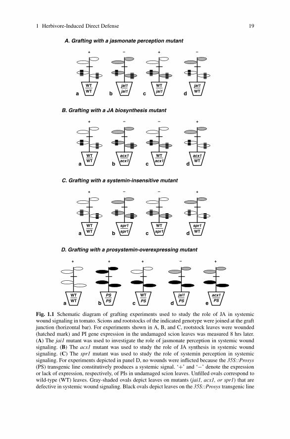
JA-insensitive jai1 mutant showed that jasmonate perception (i.e., COI1) is essentialfor recognition of the mobile signal in distal responding leaves (Fig. 1.1Ad). Thesestudies also suggest that the mobile signal is produced in the absence of COI1(Fig. 1.1Ac). Experiments conducted with JA biosynthetic mutants (e.g., acx1)showed that production of the graft-transmissible signal depends on JA biosynthesisin wounded tissues (Fig. 1.1Bc). The ability of JA-deficient scions to express PIsin response to a signal emanating from wild-type rootstock leaves further indicatedthat de novo JA synthesis is likely not necessary for recognition of the mobile sig-nal in the responding leaves (Fig. 1.1Bd). Based on these collective studies, it wasproposed that JA (or a JA derivative) is a critical component of the systemic signal(Schilmiller and Howe 2005). These findings are also consistent with DNA microar-ray studies showing that local and systemic tissues undergo distinct signaling events(Strassner et al. 2002).
The plant vascular system is involved in long-distance trafficking of a wide rangeof signaling compounds (Lucas and Lee 2004). Recent studies provide direct evi-dence that jasmonates are transported in the phloem (Fig. 1.2). For example, severalJA biosynthetic enzymes are located in the companion cell-sieve element complexof the vascular bundle (Hause et al. 2003; Wasternack 2007). This observation issupported by the occurrence of JA in phloem bundles from Plantago major (Hauseet al. 2003) and the preferential accumulation of jasmonates in the tomato leafmidrib (Stenzel et al. 2003). The hypothesis that the systemic signal is translo-cated in the phloem is further supported by the fact that wound-induced systemicresponses are strongly enhanced by the strength of vascular connections betweenwounded and responding leaves (Davis et al. 1991; Schittko and Baldwin 2003). Therate of movement of the endogenous signal in tomato plants is estimated between1 and 5 cm/hr (Schilmiller and Howe 2005). The ability of the phloem to transportsmall molecules at rates up to 40 cm/hr (Fisher 1990) could readily accommodatesuch a signal. Because systemic PI expression is mediated by a signal travelingwithin the plant rather than a signal diffusing through the atmosphere (Farmer andRyan 1992), it is unlikely that volatile MeJA released at the wound site is a causalfactor for systemic PI expression in tomato.
The idea that JA (or a JA derivative) functions as a mobile wound signal impliesthat JA synthesized in damaged leaves is transported to distal undamaged leaves.In tomato and other dicots, however, systemic increases in JA levels in response tomechanical damage are generally very low (i.e., <10% of that in damaged leaves) ornot significant (Strassner et al. 2002). In those cases where systemic increases in JAlevels have been reported, it was not determined whether accumulation of the signalresults from de novo synthesis in undamaged leaves or JA transport from woundedsource leaves. Grafting experiments (see above) support the latter possibility, as doesthe phloem mobility and systemic signaling activity of exogenous JA (Farmer andRyan 1992; Zhang and Baldwin 1997). Low levels of wound-induced JA in systemicleaves may reflect sequestration of the signal in specific cell types of the vasculature.An alternative, though not mutually exclusive, possibility is that the phloem-mobilepool of JA is rapidly metabolized to another bioactive derivative. JA derivativesproduced by methylation, glycosylation, hydroxylation, sulfonation, amino acid
1 Herbivore-Induced Direct Defense 19
WTWT
acx1acx1
WTacx1
acx1WT
WTWT
jai1jai1
WTjai1
jai1WT
+ – –+
A. Grafting with a jasmonate perception mutant
+ – – +
B. Grafting with a JA biosynthesis mutant
D. Grafting with a prosystemin-overexpressing mutant
WTWT
PSPS
WTPS
jai1PS
+ + –
WTWT
spr1spr1
WTspr1
spr1WT
+ – – +
C. Grafting with a systemin-insensitive mutant
acx1PS
+
a b c d
a b c d
a b c d
a b c d e
+
Fig. 1.1 Schematic diagram of grafting experiments used to study the role of JA in systemicwound signaling in tomato. Scions and rootstocks of the indicated genotype were joined at the graftjunction (horizontal bar). For experiments shown in A, B, and C, rootstock leaves were wounded(hatched mark) and PI gene expression in the undamaged scion leaves was measured 8 hrs later.(A) The jai1 mutant was used to investigate the role of jasmonate perception in systemic woundsignaling. (B) The acx1 mutant was used to study the role of JA synthesis in systemic woundsignaling. (C) The spr1 mutant was used to study the role of systemin perception in systemicsignaling. For experiments depicted in panel D, no wounds were inflicted because the 35S::Prosys(PS) transgenic line constitutively produces a systemic signal. ‘+’ and ‘−’ denote the expressionor lack of expression, respectively, of PIs in undamaged scion leaves. Unfilled ovals correspond towild-type (WT) leaves. Gray-shaded ovals depict leaves on mutants (jai1, acx1, or spr1) that aredefective in systemic wound signaling. Black ovals depict leaves on the 35S::Prosys transgenic line

20 G.A. Howe, A. Schaller
Companion cell Sieve element
SR160
OPDA
18:3
JA JA
Target cell
JA-x
Nucleus
COI1
Plastid Targetgenes
Plastid
Perox
JA-xPerox
Esterase Esterase
Exogenous MeJA
Systemin
Fig. 1.2 Schematic model showing the role of JA in systemic wound signaling. Chloroplastic(Plastid) and peroxisomal (Perox) JA biosynthetic enzymes are located in vascular bundles of theleaf. Binding of systemin to its receptor (SR160) activates JA accumulation. JA synthesis in tomatoleaves is also activated by systemin-independent pathways (not shown; Lee and Howe 2003). JAproduced in the companion cell-sieve element complex is transported in the phloem via plasmod-esmata connections between cells. JA, or a covalently modified form of JA (JA-x; such as JA-Ile),activates target gene expression in distal undamaged leaves through COI1. Esterases may convertexogenous MeJA to JA upon diffusion of MeJA across membranes
conjugation, and decarboxylation have been described (Wasternack 2007). One ormore of these modifications could conceivably alter the transport, stability, or inter-action of JA with target molecules (Fig. 1.2).
MeJA and certain jasmonoyl-amino acid conjugates (e.g., JA-Ile) are potentelicitors of defense gene expression (Wasternack et al. 1998). The dependence ofMeJA- and JA-Ile-induced responses on COI1 indicates that both compounds arecandidates for signals in the systemic wound response. Analysis of mutants that failto produce MeJA or JA-Ile provides a powerful approach to test this hypothesis.Conversion of JA to MeJA is mediated by JA carboxyl methyltransferase (JMT),whereas conversion of JA to JA-Ile is catalyzed by the ATP-dependent adenylate-forming enzyme JAR1 (Seo et al. 2001; Staswick and Tiryaki 2004). Although theeffect of loss of JMT function on wound-induced defense responses is not known,it is firmly established that JAR1-mediated production of JA-Ile plays a critical rolein numerous jasmonate-signaled processes (Staswick and Tiryaki 2004). Moreover,recent studies have shown that JAR1 homologs in N. attenuata are required forwound-induced defense responses to insect attack (Kang et al. 2006). JA-Ile’s keyrole in induced defense raises the possibility that biological responses previouslyattributed to JA/MeJA are in fact mediated by JA-Ile or other amino acid conjugatesof JA. Consistent with this notion, physical interaction between COI1 and repressorsof jasmonate-dependent gene expression, which results in proteasome-dependentdegradation of the repressor proteins, was recently shown to be promoted by JA-Ilebut not by JA or MeJA (Thines et al. 2007). The potency of exogenous MeJA as

1 Herbivore-Induced Direct Defense 21
an elicitor of gene expression may reflect its ability to readily penetrate cellularmembranes (Fig. 1.2). Once inside the cell, MeJA is likely converted to JA by spe-cific or non-specific esterases (Stuhlfelder et al. 2004), followed by conversion toJA-Ile by JAR1 (Staswick and Tiryaki 2004). The use of jar mutants in graftingexperiments, together with direct measurement of JA-Ile levels in phloem exudatesand wounded tissues, promises to provide additional insight into the role of thisbioactive conjugate in the wound signaling pathway.
1.3.3 Amplification of the Jasmonate Signal by Systemin
Activation of PI expression by systemin requires the synthesis and subsequent actionof JA (Schilmiller and Howe 2005; Wasternack 2007). In the context of long-distance wound signaling, this role for systemin can be reconciled with the above-mentioned grafting studies if it is postulated that systemin activates JA synthesis ator near the site of tissue damage (Li et al. 2002; Ryan and Moura 2002). This modelis consistent with grafting studies showing that a 35S::Prosys transgenic rootstockconstitutively generates a systemic signal that activates PI expression in wild-typescion leaves (Fig. 1.1Dc) (McGurl et al. 1994). Recognition of the 35S::Prosys-derived signal in scion leaves is blocked by jai1 but not by mutations such as acx1that disrupt JA biosynthesis (Fig. 1.1Dd-e) (Li et al. 2002). These findings suggestthat 35S::Prosys-expressing tissues constitutively synthesize JA, which is then mo-bilized to scion leaves where it initiates COI1-dependent responses in target cells.This model is consistent with the observation that 35S::Prosys plants accumulateincreased JA levels in the absence of wounding (Chen et al. 2006). Activation of PIexpression in JA-deficient scions (Fig. 1.1De) indicates that the long-distance signalproduced by 35S::Prosys rootstocks is likely not systemin, but rather a signal thatactivates PI expression in the absence of de novo JA synthesis.
A role for systemin in localized JA production is also in agreement with re-sults obtained from analysis of the systemin-insensitive mutant spr1 (Howe andRyan 1999; Lee and Howe 2003). spr1 mutants express PI genes in response toelicitation by OGA and JA, but not in response to systemin and prosystemin. Spr1 ispresumably required for a signaling step that links systemin perception at the plasmamembrane to activation of JA synthesis in the chloroplast. Interestingly, systemicPI expression in spr1 plants is impaired much more than the local response (Leeand Howe 2003). This phenotype is very similar to that of Prosys antisense plants(Orozco-Cardenas et al. 1993), and provides evidence that (pro)systemin functionsmainly in the long-distance response. Grafting experiments provided evidence thatSpr1 function (i.e., systemin perception) is involved primarily in the generation ofthe systemic signal in wounded leaves and is not required for recognition of thesignal in undamaged responding leaves (Fig. 1.1C). The most straightforward in-terpretation of these results is that (pro)systemin acts at or near the wound site toamplify JA accumulation and the strength of the systemic response.

22 G.A. Howe, A. Schaller
It thus appears that numerous signals, including JA, systemin, and H2O2, in-teract through a positive feedback loop to propagate the long-distance signal viathe phloem (Ryan 2000; Ryan and Moura 2002; Schilmiller and Howe 2005;Wasternack 2007). Future work is needed to understand how these signals interactwith one another to promote the systemic wound response, and to determine whichof these signals are functionally conserved in other plant species. The absence ofProsys gene homologs outside the Solanaceae suggests that systemin may haveevolved in a narrow range of plants, perhaps as a mechanism to amplify systemicdefense responses to insect attack (Howe 2004). The notion that systemin functionis rapidly evolving is supported by recent studies indicating that a systemin homologin Solanum nigrum is not involved in wound-induced direct defense responses(Schmidt and Baldwin 2006). Jasmonate-based signaling, on the other hand, appearsto play a central role in regulating responses to biotic stress in all plants. Increasingevidence indicates that the role of jasmonates in promoting systemic defense maybe more general than previously realized (Truman et al. 2007). These collectivefindings validate Ryan’s original concept that chemical alarm signals produced atthe plant-pest interface mediate systemic immunity to biotic stress.
1.4 Perspectives
Since the initial discovery by Ryan and coworkers of digestibility-reducing pro-teinase inhibitors as an inducible defense in the Solanaceae 35 years ago, in-ducible mechanisms for direct defense against insect herbivores have been identifiedthroughout the plant kingdom, from unicellular green alga (Hessen and van Donk1993; van Donk and Hessen 1993; Lampert et al. 1994) to trees (Bohlmann thisvolume). A plethora of inducible morphological and chemical resistance factorshave been identified that reduce the availability of nutrients (e.g., incorporationof silica as structural reinforcement, antinutritive secondary metabolites and pro-teins), or are outright toxic to the herbivore (e.g., secondary metabolites includingterpenoids, phenolics, and alkaloids). Numerous microarray studies aimed at an-alyzing global changes in gene expression after herbivory have confirmed the in-duced expression of many defensive genes. Moreover, the massive reprogrammingof gene expression observed in these studies suggests that herbivory results in a shiftfrom growth-oriented to defense-oriented plant metabolism (Hui et al. 2003; Ralphet al. 2006a, b). The number of herbivore-induced genes appears to greatly exceedthe requirements for known resistance traits, suggesting that additional componentsof induced defense reamin to be discovered. Indeed, in addition to interfering di-rectly with herbivore behavior or physiology, plants may use ‘scorched earth’ or‘escape strategies’ as complementary defense measures. Valuable C and N resourcesare mobilized in organs threatened by herbivory, and are either used for the synthesisof resistance factors, or stored out of reach of the herbivore. Presumably, the result-ing nutrient-deprived plant organs will poorly support the growth and developmentof attacking herbivores. Re-allocation of mobilized resources to temporary storage
1 Herbivore-Induced Direct Defense 23
proteins (vegetative storage proteins and proteinase inhibitors) and/or undergroundstorage organs (bulbs and tubers) supports later re-growth, and may allow plants toescape herbivory in time. Indeed, enhanced carbon allocation to roots in responseto herbivory was recently observed in N. attenuata, resulting in delayed senescenceand a prolonged reproductive phase. Sucrose transport to roots was found to becontrolled by SNRK1, a protein kinase that was rapidly downregulated in leavesafter attack by Manduca sexta (Schwachtje et al. 2006). Such ‘civilian defenses’(Karban and Baldwin 1997) leading to enhanced tolerance of herbivory are stillpoorly understood at the molecular level and will be an important field for futureresearch.
Tremendous progress has also been made with respect to the signaling eventsthat lead to the systemic expression of defensive traits in response to herbivory. Thisincludes the discovery of systemin as the first peptide with hormone-like activityin plants, which is now thought to act in the vicinity of the wound site to amplifythe production of a long-distance signal in the vasculature. Although the systemicsignal molecule remains to be identified, recent evidence suggests that JA or a JAmetabolite – possibly JA-Ile – may act as a phloem-mobile signal. The perception ofjasmonates and activation of defense genes in target tissues was shown to depend onCOI1, which is part of an E3 ubiquitin ligase (SCFCOI1) that was predicted to tag arepressor of JA signaling for degradation by the ubiquitin-proteasome pathway. Sev-eral members of the JAZ (Jasmonate ZIM domain) family of proteins have recentlybeen identified as targets of SCFCOI1 in tomato and Arabidopsis. At least two JAZproteins are known to act as negative regulators of jasmonate-dependent transcrip-tion, and the COI1/JAZ1 complex was suggested to be the site of JA-Ile perception(Chini et al. 2007; Thines et al. 2007). Despite these exciting findings, there are stillimportant questions to be resolved with respect to systemic wound signaling. This isparticularly true for the early events in signal transduction that couple tissue damageto the activation of the octadecanoid pathway for JA production. Most notably, theevents following systemin perception at the cell surface and the subsequent releaseof polyunsaturated fatty acids for oxylipin biosynthesis in chloroplasts remain to beelucidated. The tomato spr1 mutant is impaired in this process and the identificationof the genetic defect in spr1 may turn out to be an important step in this direction.
Acknowledgments This work was supported in part by the National Institutes of Health GrantR01GM57795 and the US Department of Energy Grant DE-FG02-91ER20021 (G.A.H). A.S.gratefully acknowledges support from the German Research Foundation (DFG).
References
Agrawal AA (1998) Induced responses to herbivory and increased plant performance. Science279:1201–1202
Agrawal AA (1999). Induced plant defense: evolution of induction and adaptive phenotypic plas-ticity. In: Agrawal AA, Tuzun S, Bent E (eds) Induced plant defenses against pathogens andherbivores: biochemistry ecology and agriculture. APS Press, Minnesota, pp 251–268

24 G.A. Howe, A. Schaller
Agrawal AA (2004) Resistance and susceptibility of milkweed: competition, root herbivory, andplant genetic variation. Ecology 85:2118–2133
Applebaum SW, Konijn AM (1966) The presence of a tribolium-protease inhibitor in wheat. JInsect Physiol 12:665–669
Baldwin IT (1998) Jasmonate-induced responses are costly but benefit plants under attack in nativepopulations. Proc Natl Acad Sci USA 95:8113–8118
Bennett RN, Wallsgrove RM (1994) Secondary metabolites in plant defence mechanisms. NewPhytol 127:617–633
Berenbaum MR (1995) Turnabout is fair play: secondary roles for primary compounds. J ChemEcol 21:925–940
Bergey DR, Howe GA, Ryan CA (1996) Polypeptide signaling for plant defensive genes exhibitsanalogies to defense signaling in animals. Proc Natl Acad Sci USA 93:12053–12058
Bergey DR, Orozco-Cardenas M, de Moura DS, Ryan CA (1999) A wound- and systemin- in-ducible polygalacturonase in tomato leaves. Proc Natl Acad Sci USA 96:1756–1760
Bergvinson DJ, Hamilton RI, Arnason JT (1995) Leaf profile of maize resistance factors toeuropean corn borer, Ostrinia nubilalis. J Chem Ecol 21:343–354
Berryman AA (1972) Resistance of conifers to invasion by bark beetle-fungus associations. Bio-Science 22:598–602
Broekgaarden C, Poelman E, Steenhuis G, Voorrips R, Dicke M, Vosman B (2007) Genotypicvariation in genome-wide transcription profiles induced by insect feeding: brassica oleracea –Pieris rapae interactions. BMC Genomics 8:239
Carrera E, Prat S (1998) Expression of the Arabidopsis abi1-1 mutant allele inhibits proteinaseinhibitor wound-induction in tomato. Plant J 15:765–771
Carroll CR, Hoffman CA (1980) Chemical feeding deterrent mobilized in response to insect her-bivory and counteradaptation by Epilachna tredecimnotata. Science 209:414–416
Chen H, Jones AD, Howe GA (2006) Constitutive activation of the jasmonate signal-ing pathway enhances the production of secondary metabolites in tomato. FEBS Lett580:2540–2546
Chen H, Wilkerson CG, Kuchar JA, Phinney BS, Howe GA (2005) Jasmonate-inducible plantenzymes degrade essential amino acids in the herbivore midgut. Proc Natl Acad Sci USA102:19237–19242
Cheong YH, Chang H-S, Gupta R, Wang X, Zhu T, Luan S (2002) Transcriptional profiling re-veals novel interactions between wounding, pathogen, abiotic stress, and hormonal responsesin Arabidopsis. Plant Physiol 129:661–677
Chini A, Fonseca S, Fernandez G, Adie B, Chico JM, Lorenzo O, Garcıa-Casado G, Lopez-Vidriero I, Lozano FM, Ponce MR, Micol JL, Solano R (2007) The JAZ family of repressors isthe missing link in jasmonate signalling. Nature 448:666–671
Christopher ME, Miranda M, Major IT, Constabel CP (2004) Gene expression profiling of system-ically wound-induced defenses in hybrid poplar. Planta 219:936–947
Coley PD, Barone JA (1996) Herbivory and plant defenses in tropical forests. Annu Rev Ecol Syst27:305–335
Constabel CP (1999) A survey of herbivory-inducible defensive proteins and phytochemicals. In:Agrawal AA, Tuzun S, Bent E (eds) Induced plant defenses against pathogens and herbivores.Biochemistry, ecology, and agriculture. APS Press, St. Paul, Minesota, pp 137–166
Davis JM, Gordon MP, Smit BA (1991) Assimilate movement dictates remote sites of wound-induced gene expression in poplar leaves. Proc Natl Acad Sci USA 88:2393–2396
De Vos M, Van Oosten VR, Van Poecke RMP, Van Pelt JA, Pozo MJ, Mueller MJ, Buchala AJ,Metraux JP, Van Loon LC, Dicke M et al (2005) Signal signature and transcriptome changes ofArabidopsis during pathogen and insect attack. Mol Plant Microbe Int 18:923–937
Delessert C, Wilson IW, Van der Straeten D, Dennis ES, Dolferus R (2004) Spatial and temporalanalysis of the local response to wounding in Arabidopsis leaves. Plant Mol Biol 55:165–181
Dussourd DE, Denno RF (1994) Host-range of generalist caterpillars – trenching permits feedingon plants with secretory canals. Ecology 75:69–78

1 Herbivore-Induced Direct Defense 25
Dussourd DE, Eisner T (1987) Vein-cutting behavior: insect counterploy to the latex defense ofplants. Science 237:898–901
Dussourd DE, Hoyle AM (2000) Poisoned plusiines: toxicity of milkweed latex and cardenolidesto some generalist caterpillars. Chemoecology 10:11–16
Ehrlich PR, Raven PH (1964) Butterflies and plants: a study in coevolution. Ecology 18:586–608Farmer EE, Ryan CA (1992) Octadecanoid precursors of jasmonic acid activate the synthesis of
wound-inducible proteinase inhibitors. Plant Cell 4:129–134Feeny P (1970) Seasonal change in oak leaf tannins and nutrients as a cause of spring feeding by
winter moth caterpillars. Ecology 51:565–581Felix G, Boller T (1995) Systemin induces rapid ion fluxes and ethylene biosynthesis in Lycoper-
sicon peruvianum cells. Plant J 7:381–389Felton GW (2005) Indigestion is a plant’s best defense. Proc Natl Acad Sci USA 102:18771–18772Fisher DB (1990) Measurement of phloem transport rates by an indicator-dilution technique. Plant
Physiol Biochem 94:455–462Fraenkel GS (1959) The raison d’etre of secondary plant substances: these odd chemicals
arose as a means of protecting plants from insects and now guide insects to food. Science129:1466–1470
Gatehouse JA (2002) Plant resistance towards insect herbivores: a dynamic interaction. New Phytol156:145–169
Giri AP, Wunsche H, Mitra S, Zavala JA, Muck A, Svatos A, Baldwin IT (2006) Molecularinteractions between the specialist herbivore Manduca sexta (Lepidoptera, Sphingidae) andits natural host Nicotiana attenuata. VII. Changes in the plant’s proteome. Plant Physiol142:1621–1641
Green TR, Ryan CA (1972) Wound-induced proteinase inhibitor in plant leaves: a possible defensemechanism against insects. Science 175:776–777
Guariguata MR, Gilbert GS (1996) Interspecific variation in rates of trunk wound closure in apanamanian lowland forest. Biotropica 28:23–29
Hause B, Hause G, Kutter C, Miersch O, Wasternack C (2003) Enzymes of jasmonate biosynthesisoccur in tomato sieve elements. Plant Cell Physiol 44:643–648
Hessen DO, van Donk E (1993) Morphological changes in Scenedesmus induced by substancesreleased from Daphnia. Arch Hydrobiol 127:129–140
Howe GA (2004) Jasmonates as signals in the wound response. J Plant Growth Regul 23:223–237Howe GA, Ryan CA (1999) Suppressors of systemin signaling identify genes in the tomato wound
response pathway. Genetics 153:1411–1421Howlett BG, Clarke AR, Madden JL (2001) The influence of leaf age on the oviposition preference
of Chrysophtharta bimaculata (Olivier) and the establishment of neonates. Agric For Entomol3:121–127
Hui D, Iqbal J, Lehmann K, Gase K, Saluz HP, Baldwin IT (2003) Molecular interactions betweenthe specialist herbivore Manduca sexta (Lepidoptera, Sphingidae) and its natural host Nico-tiana attenuata: V. Microarray analysis and further charaterization of large-scale changes inherbivore-induced mRNAs. Plant Physiol 131:1877–1893
Ishiguro S, Kawai-Oda A, Nishida I, Okada K (2001) The defective in anther dehiscence1 geneencodes a novel phospholipase a1 catalyzing the initial step of jasmonic acid biosynthesis,which synchronizes pollen maturation, anther dehiscence, and flower opening in Arabidopsis.Plant Cell 13:2191–2209
Kang J-H, Wang L, Giri A, Baldwin IT (2006) Silencing threonine deaminase and JAR4 inNicotiana attenuata impairs jasmonic acid-isoleucine-mediated defenses against Manducasexta. Plant Cell 18:3303–3320
Karban R, Baldwin IT (1997) Induced responses to herbivory. University of Chicago Press,Chicago
Karban R, Myers JH (1989) Induced plant responses to herbivory. Ann Rev Ecol Sys 20:331–348Karban R, Kuc J (1999). Induced resistance against pathogens and herbivores: an overview. In:
Agrawal AA, Tuzun S, Bent E (eds) Induced plant defenses against pathogens and herbivores.Biochemistry, ecology, and agriculture, APS Press, St. Paul, Minnesota, pp 1–16

26 G.A. Howe, A. Schaller
Kenrick P, Crane PR (1997) The origin and early evolution of plants on land. Nature389:33–39
Kessler A, Baldwin IT (2002) Plant responses to insect herbivory: the emerging molecular analysis.Ann Rev Plant Biol 53:299–328
Kuhn E, Schaller A (2004). DNA microarrays: methodology, data evaluation, and application inthe analysis of plant defense signaling. In: Setlow JK (ed) Genetic engineering, principles andmethods. Kluwer Academic, Plenum, pp 49–84
Lampert W, Rothhaupt KO, von Elert E (1994) Chemical induction of colony formation in a greenalga (Scenedesmus acutus) by grazers (Daphnia). Limnol Oceanogr 39:1543–1550
Lawrence SD, Dervinis C, Novak N, Davis JM (2006) Wound and insect herbivory responsivegenes in poplar. Biotechnol Lett 28:1493–1501
Lee GI, Howe GA (2003) The tomato mutant spr1 is defective in systemin perceptionand the production of a systemic wound signal for defense gene expression. Plant J33:567–576
Leon J, Rojo E, Sanchez-Serrano JJ (2001) Wound signalling in plants. J Exp Bot 52:1–9Li L, Li C, Lee GI, Howe GA (2002) Distinct roles for jasmonate synthesis and action in the
systemic wound response of tomato. Proc Natl Acad Sci USA 99:6416–6421Li C, Liu G, Xu C, Lee GI, Bauer P, Ling HQ, Ganal MW, Howe GA (2003) The tomato suppres-
sor of prosystemin-mediated responses2 gene encodes a fatty acid desaturase required for thebiosynthesis of jasmonic acid and the production of a systemic wound signal for defense geneexpression. Plant Cell 15:1646–1661
Li CY, Schilmiller AL, Liu GH, Lee GI, Jayanty S, Sageman C, Vrebalov J, Giovannoni JJ, Yagi K,Kobayashi Y et al (2005) Role of beta-oxidation in jasmonate biosynthesis and systemic woundsignaling in tomato. Plant Cell 17:971–986
Li L, Zhao Y, McCaig BC, Wingerd BA, Wang J, Whalon ME, Pichersky E, Howe GA (2004)The tomato homolog of coronatine-insensitive1 is required for the maternal control of seedmaturation, jasmonate-signaled defense responses, and glandular trichome development. PlantCell 16:126–143
Lipke H, Fraenkel GS, Liener IE (1954) Effect of soybean inhibitors on growth of Tribolium con-fusum. J Agr Food Chem 2:410–414
Lippert D, Chowrira S, Ralph SG, Zhuang J, Aeschliman D, Ritland C, Ritland K, Bohlmann J(2007) Conifer defense against insects: proteome analysis of sitka spruce (Picea sitchensis)bark induced by mechanical wounding or feeding by white pine weevils (Pissodes strobi).Proteomics 7:248–270
Lucas WJ, Lee JY (2004) Plant cell biology – plasmodesmata as a supracellular control networkin plants. Nat Rev Mol Cell Bio 5:712–726
Major IT, Constabel CP (2006) Molecular analysis of poplar defense against herbivory: comparisonof wound- and insect elicitor-induced gene expression. New Phytol 172:617–635
Malone M (1996). Rapid, long-distance signal transmission in higher plants. Advances in botanicalresearch. Academic Press, San Diego, pp 163–228
Mauricio R (1998) Costs of resistance to natural enemies in field populations of the annual plantArabidopsis thaliana. Am Nat 151:20–28
Mauricio R (2000) Natural selection and the joint evolution of tolerance and resistance as plantdefenses. Evol Ecol 14:491–507
McGurl B, Orozco-Cardenas M, Pearce G, Ryan CA (1994) Overexpression of the prosystemingene in transgenic tomato plants generates a systemic signal that constitutively induces pro-teinase inhibitor synthesis. Proc Natl Acad Sci USA 91:9799–9802
McGurl B, Pearce G, Orozco-Cardenas M, Ryan CA (1992) Structure, expression and antisenseinhibition of the systemin precursor gene. Science 255:1570–1573
McNaughton SJ, Tarrants JL (1983) Grass leaf silicification: natural selection for an inducibledefense against herbivores. Proc Natl Acad Sci USA 80:790–791
Moyen C, Hammond-Kosack KE, Jones J, Knight MR, Johannes E (1998) Systemin triggers anincrease of cytoplasmic calcium in tomato mesophyll cells: Ca2+ mobilization from intra- andextracellular compartments. Plant Cell Environ 21:1101–1111

1 Herbivore-Induced Direct Defense 27
Myers JH, Bazely D (1991). Thorns, spines, prickles, and hairs: are they stimulated by herbivoryand do they deter herbivores? In: Tallamy DW Raupp MJ (eds) Phytochemical induction byherbivores. John Wiley & Sons, New York, pp 325–344
Narvaez-Vasquez J, Florin-Christensen J, Ryan CA (1999) Positional specificity of a phospholi-pase a activity induced by wounding, systemin, and oligosaccharide elicitors in tomato leaves.Plant Cell 11:2249–2260
Navazio L, Moscatiello R, Bellincampi D, Baldan B, Meggio F, Brini M, Bowler C, Mariani P(2002) The role of calcium in oligogalacturonide-activated signalling in soybean cells. Planta215:596–605
O’Donnell PJ, Calvert C, Atzorn R, Wasternack C, Leyser HMO, Bowles DJ (1996) Ethylene as asignal mediating the wound response of tomato plants. Science 274:1914–1917
Orozco-Cardenas M, McGurl B, Ryan CA (1993) Expression of an antisense prosystemin gene intomato plants reduces the resistance toward Manduca sexta larvae. Proc Natl Acad Sci USA90:8273–8276
Orozco-Cardenas ML, Narvaez-Vasquez J, Ryan CA (2001) Hydrogen peroxide acts as a secondmessenger for the induction of defense genes in tomato plants in response to wounding, sys-temin, and methyl jasmonate. Plant Cell 13:179–191
Painter RH (1936) The food of insects and its relation to resistance of plants to insect attack. AmNat 70:547–566
Pearce G, Strydom D, Johnson S, Ryan CA (1991) A polypeptide from tomato leaves induceswound-inducible proteinase inhibitor proteins. Science 253:895–898
Phillips MA, Croteau RB (1999) Resin-based defenses in conifers. Trends Plant Sci 4:184–190Ralph S, Oddy C, Cooper D, Yueh H, Jancsik S, Kolosova N, Philippe RN, Aeschliman D, White R,
Huber D et al. (2006a) Genomics of hybrid poplar (Populus trichocarpa x deltoides) interactingwith forest tent caterpillars (Malacosoma disstria): normalized and full-length cDNA libraries,expressed sequence tags, and a cDNA microarray for the study of insect-induced defences inpoplar. Mol Ecol 15:1275–1297
Ralph SG, Yueh H, Friedmann M, Aeschliman D, Zeznik JA, Nelson CC, Butterfield YSN,Kirkpatrick R, Liu J, Jones SJM et al (2006b) Conifer defence against insects: microarraygene expression profiling of sitka spruce (Picea sitchensis) induced by mechanical wound-ing or feeding by spruce budworms (Choristoneura occidentalis) or white pine weevils(Pissodes strobi) reveals large-scale changes of the host transcriptome. Plant Cell Environ29:1545–1570
Reymond P, Bodenhausen N, Van Poecke RM, Krishnamurthy V, Dicke M, Farmer EE (2004) Aconserved transcript pattern in response to a specialist and a generalist herbivore. Plant Cell16:3132–3147
Rhoades DF, Cates RG (1976). Towards a general theory of plant antiherbivore chemistry. In:Wallace JW Mansell RL (eds) Biochemical interaction between plants and insects. PlenumPress, New York, pp 168–213
Rittinger PA, Biggs AR, Peirson DR (1987) Histochemistry of lignin and suberin deposition inboundary layers formed after wounding in various plant species and organs. Can J Bot 65:1886–1892
Ryan CA (2000) The systemin signaling pathway: differential activation of plant defensive genes.Biochim Biophys Acta 1477:112–121
Ryan CA, Huisman W (1969) The regulation of synthesis and storage of chymotrypsin inhibitor Iin leaves of potato and tomato plants. Plant Physiol 45:484–489
Ryan CA, Moura DS (2002) Systemic wound signaling in plants: a new perception. Proc Natl AcadSci USA 99:6519–6520
Sagi M, Davydov O, Orazova S, Yesbergenova Z, Ophir R, Stratmann JW, Fluhr R (2004) Plantrespiratory burst oxidase homologs impinge on wound responsiveness and development in Ly-copersicon esculentum. Plant Cell 16:616–628
Schaller A, Oecking C (1999) Modulation of plasma membrane H+-ATPase activity differ-entially activates wound and pathogen defense responses in tomato plants. Plant Cell 11:263–272

28 G.A. Howe, A. Schaller
Scheer JM, Ryan CA (1999) A 160 kda systemin receptor on the cell surface of Lycopersiconperuvianum suspension cultured cells: kinetic analyses, induction by methyl jasmonate andphotoaffinity labeling. Plant Cell 11:1525–1535
Scheer JM, Ryan CA (2002) The systemin receptor SR160 from Lycopersicon peruvianum is amember of the LRR receptor kinase family. Proc Natl Acad Sci USA 99:9585–9590
Schilmiller AL, Howe GA (2005) Systemic signaling in the wound response. Curr Opin Plant Biol8:369–377
Schittko U, Baldwin IT (2003) Constraints to herbivore-induced systemic responses: bidirectionalsignaling along orthostichies in Nicotiana attenuata. J Chem Ecol 29:763–770
Schmidt S, Baldwin IT (2006) Systemin in Solanum nigrum. The tomato-homologous polypeptidedoes not mediate direct defense responses. Plant Physiol Biochem 142:1751–1758
Schmidt DD, Voelckel C, Hartl M, Schmidt S, Baldwin IT (2005) Specificity in ecological inter-actions. Attack from the same lepidopteran herbivore results in species-specific transcriptionalresponses in two solanaceous host plants. Plant Physiol 138:1763–1773
Schoonhoven LM, Van Loon JJA, Dicke M (2005) Insect-plant biology, Oxford University Press,Oxford
Schwachtje J, Minchin PEH, Jahnke S, van Dongen JT, Schittko U, Baldwin IT (2006) SNF1-related kinases allow plants to tolerate herbivory by allocating carbon to roots. Proc Natl AcadSci USA 103:12935–12940
Seo HS, Song JT, Cheong J-J, Lee J-H, Lee Y-W, Hwang I, Lee JS, Choi YD (2001) Jasmonic acidcarboxyl methyltransferase: a key enzyme for jasmonate-regulated plant responses. Proc NatlAcad Sci USA 98:4788–4793
Smith CM, Rodriguez-Buey M, Karlsson J, Campbell MM (2004) The response of the poplartranscriptome to wounding and subsequent infection by a viral pathogen. New Phytologist164:123–136
Soltis PS, Soltis DE (2004) The origin and diversification of angiosperms. Am J Bot 91:1614–1626Stahl E (1888) Pflanzen und Schnecken: Biologische Studie uber die Schutzmittel der Pflanzen
gegen Schneckenfrass. Jena Z Naturwiss 22:557–684Staswick PE (1994) Storage proteins of vegetative plant tissue. Ann Rev Plant Physiol Plant Mol
Biol 45:303–322Staswick PE, Tiryaki I (2004) The oxylipin signal jasmonic acid is activated by an enzyme that
conjugates it to isoleucine in Arabidopsis. Plant Cell 16:2117–2127Stenzel I, Hause B, Maucher H, Pitzschke A, Miersch O, Ziegler J, Ryan CA, Wasternack C (2003)
Allene oxide cyclase dependence of the wound response and vascular bundle-specific genera-tion of jasmonates in tomato – amplification in wound signalling. Plant J 33:577–589
Strassner J, Schaller F, Frick UB, Howe GA, Weiler EW, Amrhein NA, Macheroux P, Schaller A(2002) Characterization and cdna-microarray expression analysis of 12-oxophytodienoate re-ductases reveals differential roles for octadecanoid biosynthesis in the local versus the systemicwound response. Plant J 32:585–601
Stratmann JW, Ryan CA (1997) Myelin basic protein kinase activity in tomato leaves is inducedsystemically by wounding and increases in response to systemin and oligosaccharide elicitors.Proc Natl Acad Sci USA 94:11085–11089
Stuhlfelder C, Mueller MJ, Warzecha H (2004) Cloning and expression of a tomato cDNA encod-ing a methyl jasmonate cleaving esterase. Eur J Biochem 271:2976–2983
Thines B, Katsir L, Melotto M, Niu Y, Mandaokar A, Liu G, Nomura K, He SY, Howe GA,Browse J (2007) JAZ repressor proteins are targets of the SCFCOI1 complex during jasmonatesignalling. Nature 448:661–665
Thompson GA, Goggin FL (2006) Transcriptomics and functional genomics of plant defence in-duction by phloem-feeding insects. J Exp Bot 57:755–766
Trapp S, Croteau R (2001) Defensive resin biosynthesis in conifers. Ann Rev Plant Physiol PlantMol Biol 52:689–724
Truman W, Bennettt MH, Kubigsteltig I, Turnbull C, Grant M (2007) Arabidopsis systemicimmunity uses conserved defense signaling pathways and is mediated by jasmonates. ProcNatl Acad Sci USA 104:1075–1080

1 Herbivore-Induced Direct Defense 29
van Donk E, Hessen DO (1993) Grazing resistance in nutrient-stressed phytoplankton. Oecologia93:508–511
Voelckel C, Baldwin IT (2004) Herbivore-induced plant vaccination. Part II. Array-studies revealthe transience of herbivore-specific transcriptional imprints and a distinct imprint from stresscombinations. Plant J 38:650–663
Walling LL (2000) The myriad plant responses to herbivores. J Plant Growth Regul 19:195–216Wasternack C (2007) Jasmonates: an update on biosynthesis, signal transduction and action in
plant stress response, growth and development. Ann Bot, 1–17Wasternack C, Ortel B, Miersch O, Kramell R, Beale M, Greulich F, Feussner I, Hause B,
Krumm T, Boland W et al (1998) Diversity in octadecanoid-induced gene expression of tomato.J Plant Physiol 152:345–352
Weis AE, Franks SJ (2006) Herbivory tolerance and coevolution: an alternative to the arms race?New Phytol 170:423–425
Wildon DC, Thain JF, Minchin PEH, Gubb IR, Reilly AJ, Skipper YD, Doherty HM, O’Donnell PJ,Bowles DJ (1992) Electrical signalling and systemic proteinase inhibitor induction in thewounded plant. Nature 360:362–65
Xie D-X, Feys BF, James S, Nieto-Rostro M, Turner JG (1998) Coi1: an Arabidopsis gene requiredfor jasmonate-regulated defense and fertility. Science 280:1091–1094
Zhang ZP, Baldwin IT (1997) Transport of [2-C-14] jasmonic acid from leaves to roots mimicswound-induced changes in endogenous jasmonic acid pools in Nicotiana sylvestris. Planta203:436–441
Zhu-Salzman K, Salzman RA, Ahn JE, Koiwa H (2004) Transcriptional regulation of sorghumdefense determinants against a phloem-feeding aphid. Plant Physiol 134:420–431
Chapter 2Herbivore-Induced Indirect Defense: FromInduction Mechanisms to Community Ecology
Maaike Bruinsma and Marcel Dicke
Herbivory may induce plant defenses that promote the activity of natural enemies ofthe herbivores. This so-called induced indirect defense may involve the productionof plant volatiles that attract carnivorous arthropods or extrafloral nectar that is ex-ploited as alternative food by carnivorous arthropods. Induced indirect plant defenseis mediated by different signal-transduction pathways, such as the jasmonic acid,the salicylic acid, and the ethylene pathways and may involve large-scale transcrip-tomic re-arrangements. Induced indirect plant defense responses result in an alteredphenotype and thus can affect the interactions of the plant with various communitymembers: attackers can be deterred, natural enemies attracted (both above- and be-lowground), pollinators may change flower visitation, and neighboring plants canexploit the information from the attacked plants to initiate defense responses as well.We discuss several approaches that are commonly used in molecular, chemical andecological studies of induced indirect plant defenses and identify some remainingknowledge gaps and directions for future research. Integrating a mechanistic ap-proach with a community ecological approach will provide important progress inunderstanding the selective pressures and dynamics of ecological interactions that aremediated by induced indirect plant defenses, as well as the underlying mechanisms.
2.1 Introduction
Plants face many challenges during their lifetime. Drought, flooding, high and lowtemperatures, and attacks by all kinds of organisms, like pathogens, nibbling insectsor mammals devouring whole plants. To be able to reproduce in spite of all thesedifficulties plants have developed defense mechanisms to protect themselves, otherthan running away. The defenses of plants against herbivorous arthropods can beclassified as ‘direct defenses’ that affect the physiology of the attacker (Howe andSchaller this volume) or ‘indirect defenses’ that promote the effectiveness of natural
M. DickeLaboratory of Entomology, Wageningen University, 6709 PD Wageningen, The Netherlandse-mail: [email protected]
A. Schaller (ed.), Induced Plant Resistance to Herbivory, 31C© Springer Science+Business Media B.V. 2008

32 M. Bruinsma, M. Dicke
enemies of herbivores (Fig. 2.1). Indirect defense comprises (a) the provision ofshelter, such as hollow thorns that are used by ants for nesting (b) the productionof alternative food, such as extrafloral nectar that is used by carnivorous arthro-pods such as ants and parasitic wasps or (c) the emission of herbivore-inducedplant volatiles that guide carnivorous arthropods such as predators or parasitoidsto their herbivorous victims. All of these support the presence and abundance ofcarnivorous enemies of herbivorous arthropods and consequently the reduction ofherbivore presence.
Indirect defense
Direct defense
Cotesia glomerata
Pieris rapae
Brassica oleracea
Fig. 2.1 Direct defense of a plant has a direct negative effect on the attacker of the plant. Indirectdefense of a plant maintains or attracts carnivores that consume or parasitize the attacker of theplant, thereby exerting a negative effect on the attacker, which contributes to plant defense. Pho-tographs were taken by Hans M. Smid (C. glomerata), Nelly Cardinel (B. oleracea) and MaaikeBruinsma (P. rapae)

2 Herbivore-Induced Indirect Defense 33
Here we will address indirect defense, especially induced indirect defense.Herbivory can induce the production of extrafloral nectar (Wackers et al. 2001) orplant volatiles (Van Poecke and Dicke 2004) and as a result the plant’s phenotypechanges. The extrafloral nectar can serve as food to various animals in the com-munity and the volatiles can be used by animals to localize the plant. Throughoutthe plant kingdom many species have been reported to produce herbivore-inducedinfochemicals that attract carnivorous enemies of the herbivorous arthropods (Dicke1999). The effect of these phenotypic changes on carnivorous enemies of herbivoreshas received most attention (Van Poecke and Dicke 2004; Turlings and Ton 2006).For instance, Lima bean plants that are infested with spider mites start to pro-duce a range of volatiles, including terpenoids and methyl salicylate, several ofwhich attract predatory mites that consume the herbivorous spider mites (Dickeet al. 1990b, 1999). Moreover, herbivory induces the production of extrafloral nectarin Lima bean plants and this results in increased numbers and duration of visits bye.g. ants and wasps (Kost and Heil 2005). The increased visitation of the plant bycarnivorous arthropods results in a reduced amount of leaf damage (Heil 2004).
However, plants are at the basis of most terrestrial food webs and consequentlythey are members of complex communities, both below and aboveground, with amultitude of dynamic, ecological interactions (Price et al. 1980; Dicke and Vet 1999;Van Zandt and Agrawal 2004, Bezemer and van Dam 2005). Ecological interactionsmay involve direct interactions such as predator–prey interactions or competitionamong herbivores, as well as indirect interactions such as apparent competition andtrait-mediated indirect effects (Holt 1977; Wootton 1994; Van Veen et al. 2006;White and Andow 2006). In many studies species interactions are considered tobe fixed: all individuals within a population are considered to have the same char-acteristics and interact in the same way with other organisms. However, becauseof phenotypic plasticity, interactions within communities are context-dependent(Agrawal 2001), which implies that they are not only influenced by genotype, butalso by e.g. physiological state, resource availability and interactions with com-munity members. As a result, understanding the effects of phenotypic plasticity isimportant to understand community dynamics (Fig. 2.2; Agrawal 2001).
Although a phenotypic change may affect many interactions in a community,herbivore-induced indirect plant defenses have mostly been studied in a tritrophiccontext in simple food chains, without taking into account the effects they mighthave on other community members. Investigating the effects of phenotypic changeson community processes is one of the major challenges that ecologists face in the re-search on herbivore-inducedplantdefenses (DickeandVet1999;Kessler and Baldwin2001). Understanding the selection pressures and the dynamics of ecological interac-tions is important for understanding the ecology and evolution of communities.
To investigate the community consequences of induced indirect plant defenses, athorough understanding of the underlying mechanisms is essential, as mechanisticknowledge allows the development of manipulative tools. Manipulative experimentsare important to address the effects of induced defense on a range of individualinteractions or on the total set of interactions (Kessler and Baldwin 2001; Dicke andHilker 2003).

34 M. Bruinsma, M. Dicke
Genes Proteins Metabolites IndividualsPhenotypes Population Community
Mechanisms Ecological functions
Fig. 2.2 Induced indirect defense can be investigated at different levels of biological organization:by investigating mechanisms of induction at the levels of genes up to individuals, or by investigat-ing ecological functions at the levels of individuals up to the community. An integration of thesetwo approaches proved to be most rewarding. Phenotypic plasticity plays a central role in under-standing both, the underlying mechanisms of induced defense, and the consequences of induceddefense for community dynamics
In this chapter we will address the mechanisms of induced indirect plant defensesand how information on these can be used to investigate the ecological consequencesof these defenses at the level of multiple ecological interactions and the community.
2.2 Induction of Indirect Plant Defense
Herbivores can cause many types of damage to plants, according to the feeding guildto which they belong. For example, caterpillars ingest small sections of the leaves,while others feed on specific parts of the leaf material: leaf-mining insects feed onparenchymal tissue and aphids ingest phloem. Different types of damage may resultin diverse defense responses in the plant. In this section we will discuss the plantresponses and the various mechanisms of their induction.
2.2.1 Response of the Plant
In response to arthropod herbivory the plant may activate several major signal-transduction pathways involved in the defense response: the JA (jasmonic acid),SA (salicylic acid) and ET (ethylene) pathways (Dicke and Van Poecke 2002).These signaling pathways are differentially induced by different feeding guilds orartificial damage (Ozawa et al. 2000; Walling 2000; Dicke and Van Poecke 2002;De Vos et al. 2005; Zheng et al. 2007). The three signal-transduction pathways alsointeract: JA can inhibit the effect of SA, and SA can interfere with JA-mediatedinduction (Pena-Cortes et al. 1993; Sano and Ohashi 1995). Similarly, JA and ETsynergistically affect induction of defense gene expression in tomato (O’Donnellet al. 1996), while ET inhibits the effect of JA on nicotine induction in tobacco(Kahl et al. 2000).
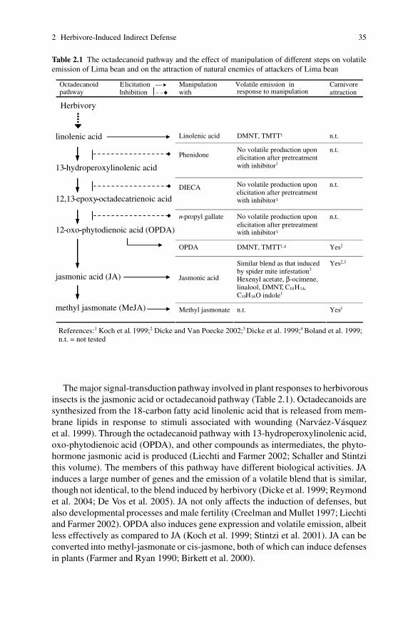
2 Herbivore-Induced Indirect Defense 35
Table 2.1 The octadecanoid pathway and the effect of manipulation of different steps on volatileemission of Lima bean and on the attraction of natural enemies of attackers of Lima bean
Octadecanoidpathway
Elicitation —Inhibition ⏐– –♦
Manipulation with
Volatile emission in response to manipulation
Carnivoreattraction
Linolenic acid DMNT, TMTT1 n.t.
PhenidoneNo volatile production upon elicitation after pretreatment with inhibitor1
n.t.
DIECA No volatile production upon
with inhibitor1
n.t.
n-propyl gallate No volatile production upon
with inhibitor1
n.t.
OPDA DMNT, TMTT1,4 Yes2
Jasmonic acid
Similar blend as that induced by spider mite infestation3
Hexenyl acetate, β-ocimene,linalool, DMNT, C10H14, C10H16O indole1
Yes2,3
Methyl jasmonate n.t. Yes3
Herbivory
linolenic acid
13-hydroperoxylinolenic acid
12,13-epoxy-octadecatrienoic acid
12-oxo-phytodienoic acid (OPDA)
jasmonic acid (JA)
methyl jasmonate (MeJA)
-
-
-
References:1 Koch et al. 1999;2 Dicke and Van Poecke 2002;3 Dicke et al. 1999;4 Boland et al. 1999;n.t. = not tested
elicitation after pretreatment
elicitation after pretreatment
The major signal-transduction pathway involved in plant responses to herbivorousinsects is the jasmonic acid or octadecanoid pathway (Table 2.1). Octadecanoids aresynthesized from the 18-carbon fatty acid linolenic acid that is released from mem-brane lipids in response to stimuli associated with wounding (Narvaez-Vasquezet al. 1999). Through the octadecanoid pathway with 13-hydroperoxylinolenic acid,oxo-phytodienoic acid (OPDA), and other compounds as intermediates, the phyto-hormone jasmonic acid is produced (Liechti and Farmer 2002; Schaller and Stintzithis volume). The members of this pathway have different biological activities. JAinduces a large number of genes and the emission of a volatile blend that is similar,though not identical, to the blend induced by herbivory (Dicke et al. 1999; Reymondet al. 2004; De Vos et al. 2005). JA not only affects the induction of defenses, butalso developmental processes and male fertility (Creelman and Mullet 1997; Liechtiand Farmer 2002). OPDA also induces gene expression and volatile emission, albeitless effectively as compared to JA (Koch et al. 1999; Stintzi et al. 2001). JA can beconverted into methyl-jasmonate or cis-jasmone, both of which can induce defensesin plants (Farmer and Ryan 1990; Birkett et al. 2000).

36 M. Bruinsma, M. Dicke
From 13-hydroperoxylinolenic acid there is a side branch of the octadecanoidpathway that leads to the production of so-called green leaf volatiles (GLVs) suchas C6-aldehydes, C6-alcohols, and their acetates (Visser and Ave 1978; Hatanakaet al. 1987). These compounds can attract both herbivorous and carnivorous arthro-pods on the one hand (Whitman and Eller 1990; Shiojiri et al. 2006), and primeneighboring plants for the induction of defenses on the other (Engelberth et al. 2004;Ruther and Furstenau 2005). In addition to the octadecanoid pathway, woundingcan also induce the hexadecanoid pathway, starting from 7(Z ),10(Z ),13(Z ) hexade-catrienoic acid (Weber et al. 1997; Stintzi et al. 2001) that also leads to JA. Theinduction of both pathways may result in specific ‘oxylipin signatures’ that allowplants to fine-tune their responses to wounding or herbivory (Weber et al. 1997). Theactivation of the signal-transduction pathways can result in the induction of directas well as indirect defenses. Many types of secondary metabolites involved in directdefense are produced upon herbivore damage, for example non-volatile compoundslike proteinase inhibitors, glucosinolates, and alkaloids (see Howe and Schaller thisvolume). As indirect defense response, plants can produce volatiles such as alcohols,esters, and terpenoids, or alternative food, such as extrafloral nectar. Induced plantscan produce either the same volatiles as non-induced plants but in different amountsor ratios, or they can produce new volatiles that are not emitted by intact plants(Dicke et al. 1999). However, during both types of responses plants produce themetabolites de novo; indicating an active investment in defense, rather than just apassive release of compounds (Donath and Boland 1994; Pare and Tumlinson 1997;Mercke et al. 2004).
The contribution of the JA pathway to indirect defense has been well studied inthe context of tritrophic interactions. JA in itself is not attractive or deterring, butinduces biosynthetic processes in the plant (e.g., the production of volatile info-chemicals), that cause behavioral responses of the insects (Avdiushko et al. 1997;Thaler et al. 2002; Bruinsma et al. 2007). JA-induced defense reactions may eitherdeter herbivores, or attract their natural enemies (Gols et al. 1999; Thaler 1999;Lou et al. 2005; Bruinsma et al. 2007). In addition to JA signaling, tritrophic inter-actions have been shown to also involve the SA pathway. The production of methylsalicylate (MeSA) is induced by spider mite infestation in e.g. Lima bean and tomato(Dicke et al. 1990b, 1998; Ozawa et al. 2000), and attracts the natural enemies ofthe spider mites (De Boer and Dicke 2004).
Another indirect defense mechanism involving JA signaling is the production ofalternative food, such as extrafloral nectar (EFN) that can be used by the members ofthe third trophic level (Van Rijn and Tanigoshi 1999; Kost and Heil 2005; Wackersand Van Rijn 2005). EFN is secreted from nectaries outside the flowers, which mayoccur on the petioles (Koptur 2005). The nectar composition can differ dramaticallybetween floral and extrafloral nectaries of one plant (Koptur 2005). The produc-tion of EFN increases upon herbivory (Heil et al. 2001; Wackers et al. 2001). Itis highest in leaves where the herbivores are feeding, but can also be increased insystemic leaves (Wackers et al. 2001). Wounding and JA application increase EFNproduction. Moreover, EFN secretion is reduced by phenidone, an inhibitor of anearly step in the octadecanoid pathway (Table 2.1) supporting a role for JA in the
2 Herbivore-Induced Indirect Defense 37
induction of EFN secretion (Heil et al. 2001). EFN, serving as an alternative foodsource, may increase the duration of predator visits to plants. Predators dispersemore slowly from plants with more EFN as compared to plants with little EFN(Choh et al. 2006), and plants may benefit from the presence of predators.
2.2.2 Induction by Herbivores
When herbivores are feeding on a plant, either by leaf chewing, phloem ingestion,or cell content feeding, they induce phytohormone signaling pathways and conse-quently elicit a plant response. The induced phytohormone signatures are attacker-specific: qualitatively, quantitatively and temporally (De Vos et al. 2005). More andmore studies also show the attacker specificity at the level of global gene expres-sion (Voelckel and Baldwin 2004; Voelckel et al. 2004; De Vos et al. 2005), al-though other studies recorded quite similar transcriptional responses after attack bydifferent herbivores (Reymond et al. 2004). Herbivore-induced plant volatiles canbe specific for herbivore species, and even herbivore instar, feeding on the plant(De Moraes et al. 1998; Takabayashi et al. 2006).
The main groups of plant volatiles induced by herbivory are green leaf volatiles,terpenes, and phenolics. The induction of volatile emission is mediated by the in-duction of the three main signal-transduction pathways – the JA, SA, and ET path-ways – which may be differentially induced by insects from different feeding guilds(Walling 2000). In Lima bean for example, JA signaling is responsible for the in-duced production of volatiles in response to caterpillar damage, while both SA andJA mediate the response to spider mite damage (Ozawa et al. 2000). In anotherplant species, Medicago truncatula, caterpillars and spider mites induced qualita-tively and quantitatively different volatile patterns. Both JA and SA accumulatedin response to damage, but the accumulation differed between the attack by thechewing and by piercing–sucking insects; SA accumulation was higher in responseto spider-mite as compared to caterpillar damage, and JA accumulated differently intime for the two modes of attack (Leitner et al. 2005). Likewise, induced changes ingene expression in Arabidopsis thaliana show differences between feeding guilds.Five attackers with different modes of attack, ranging from leaf-chewing herbivoresto pathogens causing necrotic lesions, showed different degrees of relative inductionof the three important signal-transduction pathways (De Vos et al. 2005). In general,it seems that the plant response to phloem-feeding herbivores is more similar to theresponse to pathogen attack, while leaf-chewing herbivores induce pathways alsoactivated by wounding (Walling 2000).
2.2.3 Mechanical Wounding Versus Herbivory
When herbivores attack the plant, they inflict physical damage which in itself issufficient to elicit a subset of the responses to herbivory. Water loss at the wound

38 M. Bruinsma, M. Dicke
site may result in osmotic stress and therefore, there is considerable overlap betweenplant responses to wounding and dehydration. In addition, the plant respondsto herbivore-derived compounds present in oral secretions. Physical damage andherbivore-derived elicitors are both responsible for part of the herbivore-inducedresponse of the plant. In many plant species, however, the response to mechani-cal damage differs from the one elicited by herbivory (Schoonhoven et al. 2005).This may be partly due to technical difficulties in accurately mimicking herbivory.Mechanical damage differs from herbivore-inflicted damage in the amount of re-moved tissue, age of tissue, spatial pattern of damage, and timing (Baldwin 1990).The temporal pattern of mechanical damage was in fact shown to be an importantfactor influencing its effect (Mithofer et al. 2005). Yet, when herbivore regurgitantis applied onto mechanically damaged leaves, the plant response can be similar tothe response to herbivory (Turlings et al. 1990; Halitschke et al. 2001). For exam-ple, mechanical damage with subsequent regurgitant application induces herbivore-specific plant responses in Nicotiana attenuata (Halitschke et al. 2001). Severalactive compounds have been identified in the oral secretions of feeding insects. Forexample, the enzyme �-glucosidase and volicitin (a fatty acid–amino acid conjugate,FAC) induce volatile production when added to mechanically damaged cabbageand maize plants, respectively (Mattiacci et al. 1995; Alborn et al. 1997; Schmelzet al. 2001; Tumlinson and Engelberth this volume). FACs have been found in alllepidopteran larvae studied to date (Voelckel and Baldwin 2004). Plants can notonly differentiate between mechanical wounding and herbivory, but also betweenherbivore species, even when they are from the same feeding guild. This has beenshown for N. attenuata: the plant response to a specialist herbivore differed fromthe response to two generalist herbivores, and the different responses were corre-lated with the FAC composition of the regurgitant of the herbivores (Voelckel andBaldwin 2004). The role of oral secretions in induction of plant defense responsesis discussed in more detail by Felton (this volume).
The difference between induction after mechanical damage and caterpillar feed-ing has been shown at the gene expression level in A. thaliana. Mechanical damageand caterpillar feeding both induce jasmonate-responsive genes and lead to accu-mulation of JA. However, mechanical damage induces expression of a jasmonate-responsive marker gene PDF1.2, while caterpillar feeding suppresses theinduction of this gene. PDF1.2 defense gene induction was suppressed also whencaterpillar regurgitant was added to mechanically damaged leaves, indicating a rolefor caterpillar-derived elicitors in the downregulation of plant defense responses(De Vos 2006). Three other JA-responsive genes that are induced by caterpillarfeeding are not induced by mechanical wounding, demonstrating how herbivoryand wounding differentially induce expression of specific genes (De Vos 2006). InN. attenuata plants the endogenous JA levels increase after wounding, and increaseeven more when oral secretion is applied to the wounds. The same pattern can beobserved for ethylene emission in these plants: punctured plants treated with oralsecretion temporarily emit more ethylene than do punctured plants treated withwater, while ethylene emission upon herbivory increases for as long as herbivorycontinues (Kahl et al. 2000).

2 Herbivore-Induced Indirect Defense 39
2.2.4 Priming
Apart from direct upregulation of defense signaling cascades or gene expression,the ability of the plant to rapidly activate cellular defense responses can be en-hanced, a process that is called priming (Conrath et al. 2002). When exposed toa priming stimulus the plant does not respond with the immediate production ofdefense compounds, but rather enters a sensitized state allowing it to respond fasteror stronger to subsequent challenges in the future (Turlings and Ton 2006). Priminghas originally been demonstrated for plant-pathogen (Conrath et al. 2002, 2006)and plant-rhizosphere bacteria interactions (Verhagen et al. 2004), and appears tobe advantageous with respect to defense-associated metabolic costs.
Priming can occur in response to different stimuli, such as plant volatiles,pathogen infestation, or herbivory. Priming of indirect defenses has been shownfor induced volatile emission as well as EFN secretion. Exposure to green leafvolatiles from neighboring damaged plants results in higher endogenous JA levelsand higher emission of volatiles upon herbivory or mechanical damage comparedto non-exposed plants (Engelberth et al. 2004). Also exogenously applied singlecompounds, such as (Z )-3-hexenal, (Z )-3-hexen-1-ol, and (Z )-3-hexenyl acetate,can prime plants for responses to herbivore attack (Engelberth et al. 2004). Re-cently, a field study demonstrated the priming of EFN secretion. Lima bean plantswere exposed to an artificial volatile blend mimicking the volatile emission fromherbivore-induced Lima bean plants. Upon wounding, these plants secreted moreEFN as compared to non-exposed controls indicating a priming effect of the expo-sure to volatiles (Heil and Kost 2006).
2.3 Responses of Community Members to Induced IndirectPlant Defense
Induced infochemicals (sensu Dicke and Sabelis 1988), once released from theplant, can be exploited by any of its community members, including neighboringplants, herbivores, predators, parasitoids, pollinators and other community mem-bers, both above- and belowground (Fig. 2.3). The infochemicals can function asa direct defense by repelling herbivores. For example, butterflies avoid oviposi-tion on plants that are induced by either the presence of eggs or feeding damage(Rothschild and Schoonhoven 1977; Landolt 1993; De Moraes et al. 2001). Highlevels of direct defense may deter herbivores from feeding or ovipositing on a plant,and may also slow down development of larvae, rendering them more vulnerable tonatural enemies (Rothschild and Schoonhoven 1977; Stout and Duffey 1996; Thaleret al. 1996). However, direct and indirect defense mechanisms can act antagonisti-cally. Plant toxins that are ingested by the herbivore may be sequestered to affectthe development of carnivores, or negatively influence carnivore fitness due to com-promised host size or quality (Ode 2006). Natural populations of Senecio jacobaeaexhibit genetic variation of pyrrolizidine alkaloid (PA) concentration. Plants infested
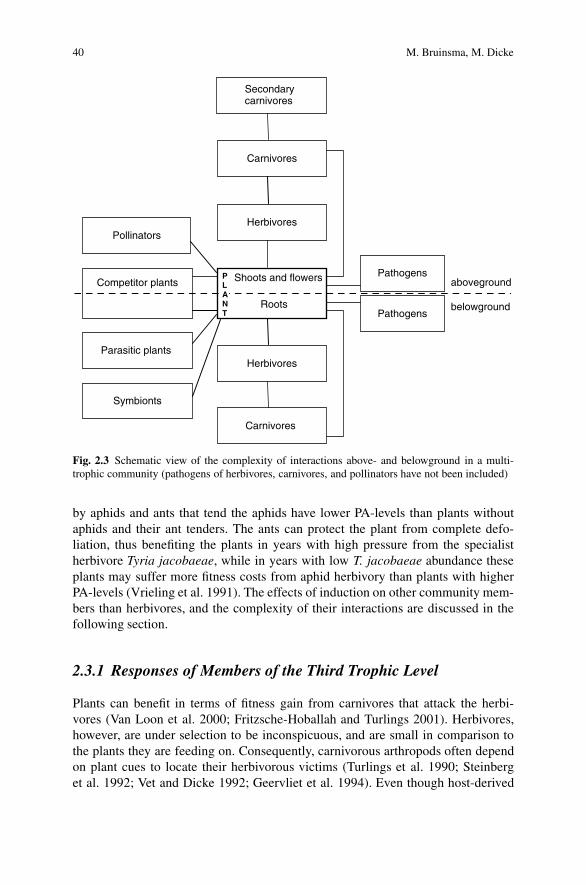
40 M. Bruinsma, M. Dicke
Shoots and flowers
Roots
aboveground
belowground
Carnivores
Herbivores
Herbivores
Carnivores
Pollinators
Pathogens
Symbionts
Competitor plants
Secondarycarnivores
Pathogens
Parasitic plants
PLANT
Fig. 2.3 Schematic view of the complexity of interactions above- and belowground in a multi-trophic community (pathogens of herbivores, carnivores, and pollinators have not been included)
by aphids and ants that tend the aphids have lower PA-levels than plants withoutaphids and their ant tenders. The ants can protect the plant from complete defo-liation, thus benefiting the plants in years with high pressure from the specialistherbivore Tyria jacobaeae, while in years with low T. jacobaeae abundance theseplants may suffer more fitness costs from aphid herbivory than plants with higherPA-levels (Vrieling et al. 1991). The effects of induction on other community mem-bers than herbivores, and the complexity of their interactions are discussed in thefollowing section.
2.3.1 Responses of Members of the Third Trophic Level
Plants can benefit in terms of fitness gain from carnivores that attack the herbi-vores (Van Loon et al. 2000; Fritzsche-Hoballah and Turlings 2001). Herbivores,however, are under selection to be inconspicuous, and are small in comparison tothe plants they are feeding on. Consequently, carnivorous arthropods often dependon plant cues to locate their herbivorous victims (Turlings et al. 1990; Steinberget al. 1992; Vet and Dicke 1992; Geervliet et al. 1994). Even though host-derived

2 Herbivore-Induced Indirect Defense 41
stimuli are potentially more reliable for host location, their use is often limitedby low detectability, especially at longer distances (Vet and Dicke 1992). There-fore, carnivorous arthropods are usually more strongly attracted by plant-derivedvolatiles as compared to volatiles derived from their herbivorous victims (Turlingset al. 1990, 1991; Steinberg et al. 1993; Geervliet et al. 1994; Dicke 1999). At-traction by volatiles from host-infested plants and by EFN was shown for egg-as well as larval parasitoids and predators (e.g., Blaakmeer et al. 1994; Geervlietet al. 1997; Lou et al. 2005; Hilker and Meiners 2006; Choh et al. 2006; Mumm andHilker 2006). Induced levels of EFN also increase the abundance of ants, wasps andflies (Kost and Heil 2005), and reduce the amount of leaf damage (Heil 2004).
The major signal-transduction pathway involved in attraction of natural enemiesseems to be the JA-pathway (Dicke and Van Poecke 2002). The involvement of JA ininduced attraction of members of the third trophic level has been demonstrated bothby manipulation of JA on the level of the plant’s phenotype and the plant’s geno-type. JA-treated plants attract natural enemies of herbivorous arthropods and haveincreased parasitism rates in field (Gols et al. 1999; Thaler 1999; Ozawa et al. 2004).Moreover, jasmonate-deficient plants are less attractive to natural enemies than con-trol plants when attacked by herbivores (Thaler et al. 2002).
2.3.2 Responses of Members of Higher Trophic Levels
The enemies of herbivores can in turn fall victim to members from higher trophiclevels, for example to hymenopterous hyperparasitoids, also called secondary para-sitoids (Brodeur 2000). Induced changes in plant chemistry affect not only the devel-opment and survival of herbivores and their parasitoids, but also that of secondaryparasitoids (Harvey et al. 2003). Performance of Lysibia nana, a hyperparasitoidwith a broad host range, was shown to be negatively affected by high levels ofdefensive toxins in the host plant (Harvey et al. 2003; Soler et al. 2005). Secondaryparasitoids are often compared to primary parasitoids, since both share commonlife-history strategies (Brodeur 2000). However, as compared to primary parasitoids,little is known about their host searching strategies, and whether or not they useplant volatiles to locate (parasitized) herbivores (Harvey et al. 2003; Buitenhuiset al. 2005). Plant-derived cues may be of limited value for hyperparasitoids for sev-eral reasons: firstly, although herbivore-induced plant volatiles may be reliable cuesfor primary parasitoids, they do not guarantee the presence of a parasitized host forthe secondary parasitoid. Secondly, primary parasitoids are often more specializedthan secondary parasitoids (Buitenhuis et al. 2005). Yet, a recent study demonstratedthat primary parasitoids are in fact able to discriminate between herbivore-inducedplant volatiles emitted from plants damaged by unparasitized or parasitized cater-pillars (Fatouros et al. 2005), suggesting that reliable plant cues may be available tosecondary parasitoids as well.
Several studies have been conducted on host searching behavior of secondaryparasitoids with varying results. A specialized ectoparasitoid Euneura augarus re-lies on plant volatiles for long range searching (Volkl and Sullivan 2000). However,

42 M. Bruinsma, M. Dicke
E. augarus does not distinguish between plants with and without host mummies forlong range searching. The two hyperparasitoid species Alloxysta victrix and Dendro-cerus carpenteri were attracted to herbivore-induced volatiles (oat plants infestedwith Sitobion avenae aphids; Siri 1993), while in another study with another plant-host-primary parasitoid system (potato infested with Macrosiphum euphorbiae) theywere not (Buitenhuis et al. 2005). Buitenhuis et al. (2005) did find analogies inforaging behavior of primary and secondary parasitoids in the use of contact cueswhile searching on a plant. Host searching behavior of hyperparasitoids may dependmore on contact cues and less on plant volatiles compared to primary parasitoids(Buitenhuis et al. 2005).
2.3.3 Responses of Pollinators
Sexual reproduction of many plants species depends on pollination by honey bees,bumble bees, solitary bees, syrphid flies or moths (Klein et al. 2006). Herbivoryin early stages of plant growth reduces the photosynthetic area of the plant, andmay result in smaller plants and a shorter flowering period. This is possibly due toallocation of resources to defenses, rather than growth and reproduction (Povedaet al. 2003). Herbivory may affect the production of pollen and nectar, the qual-ity of nectar, morphology of flowers and may reduce seed production (Lehtila andStrauss 1997; Hamback 2001; Poveda et al. 2003). Both nectar quality and quantityare parameters that determine the number and type of pollinators that are attracted tothe plants (Potts et al. 2003). While extrafloral nectar is known to increase after her-bivory, it remains unknown whether herbivory also affects floral nectar production(Adler et al. 2006). Just a handful of studies have addressed the effect of herbivoryon floral chemistry. Leaf herbivory on tobacco plants increased alkaloid concen-tration in the nectar (Adler et al. 2006), while other studies have shown increasesin defense compounds such as nicotine and glucosinolates in flower tissues (Eulerand Baldwin 1996; Ohnmeiss and Baldwin 2000; Strauss et al. 2004; Smallegangeet al. 2007). It is interesting to note that in tobacco plants nicotine levels in thecorolla are lower during the scotophase, when moths are attracted for pollination(Euler and Baldwin 1996).
However, as yet there is little knowledge on the influence of herbivory on pol-lination. Several studies have reported an indirect effect of herbivory on pollina-tion. Herbivory may affect flowering traits, for example reducing the number offlowers, flower size or plant height. This in turn, can affect flower visitation bypollinators (Lehtila and Strauss 1997; Adler et al. 2001; Hamback 2001). Lehtilaand Strauss (1997) showed that bees prefer undamaged radish plants over dam-aged plants, but this difference could be explained by reduced flower size andnumber. However, syrphid flies preferred undamaged plants over damaged ones,even when the plants were controlled for flower size and number; indicating achemical basis for syrphid fly attraction. While in this case pollinator attractiondecreased, herbivory can also enhance pollinator attraction. Root herbivory for

2 Herbivore-Induced Indirect Defense 43
example, can increase flower visitation, as demonstrated in mustard plants byPoveda et al. (2003, 2005). Although these examples demonstrate the influence ofherbivory on pollination, the mechanism behind this phenomenon is not yet eluci-dated. Possibly, the effect on plant growth, number of flowers, plant volatiles or achange in nectar quantity or quality, determine the changes in pollinator attraction.
2.3.4 Responses of Neighboring Plants
Plants may gain a fitness benefit from responding to herbivore-induced volatilesfrom neighboring plants that are being attacked. Such a signal is indicative of theimminent danger, and the receiving plant may profit from exploiting this informationby readying its defense (Dicke and Bruin 2001). Herbivore-induced plant signalscan be exploited by neighboring plants of the same and of different species alike(Engelberth et al. 2004; Baldwin et al. 2006). Strong signals may immediately ac-tivate plant defenses, while lower (more common in nature) concentrations of sig-naling molecules can prime the plant for attack (Turlings and Ton 2006). This wasnicely shown in a field study by Kessler et al. (2006) reporting that plant volatilesfrom damaged sagebrush plants can prime the induction of proteinase inhibitorsin nearby tobacco plants. The primed tobacco plants received less damage fromsubsequent herbivore attack as compared to non-exposed control plants. Not onlyvolatile emission, but also EFN secretion can be primed by exposure to volatilesfrom damaged plants. Plants that have been exposed to volatiles from herbivore-infested conspecific plants produce more EFN when they are attacked themselves(Heil and Kost 2006), although this effect was only observed during the early stagesof herbivore attack (Choh and Takabayashi 2006).
2.3.5 Above- and Belowground Interactions
Most studies on induced defenses have focused on aboveground interactions. How-ever, belowground interactions can have an important effect on aboveground pro-cesses. For example, root herbivory and belowground plant mutualists, such asarbuscular mycorrhizal fungi, affect pollination (Poveda et al. 2003, 2005; Wolfeet al. 2005).
Processes similar to those reported to occur aboveground do also take place be-lowground. Plants can release volatiles in response to root herbivory that attractnatural enemies of the root herbivores. This was first reported for a coniferoushost plant, Thuja occidentalis, that when exposed to weevil larvae, releases chem-icals attractive to nematodes that can parasitize the larvae (Van Tol et al. 2001).Rasmann et al. (2005) showed the same phenomenon for maize plants, and identi-fied a chemical attracting the entomopathogenic nematode Heterorhabditis megidis,a natural enemy of the attacking beetle. Plant-to-plant signaling also takes placebelowground. Plants emit belowground signals that are exploited by neighboring

44 M. Bruinsma, M. Dicke
plants for the attraction of predators (Chamberlain et al. 2001; Dicke and Dijkman2001), or by parasitic plants to induce germination (Bouwmeester et al. 2003;Runyon et al. 2006).
In addition to belowground interactions, it is important to consider the inter-actions between above- and belowground communities. Cotton plants, for exam-ple, increase aboveground extrafloral nectar production upon attack by root-feedingwireworms (Wackers and Bezemer 2003). Root herbivory, as well as arbuscular my-corrhizal fungi, can also change aboveground volatile emission, and subsequentlyincrease the number of visits by parasitoids of aboveground herbivores (Masterset al. 2001; Neveu et al. 2002; Guerrieri et al. 2004; Soler et al. 2007). Theperformance of the aboveground multitrophic community associated with a plantdiffers between plants with and without root herbivory (Soler et al. 2005). Root her-bivory changed plant quality, which in turn negatively affected the performance ofan aboveground herbivore, a primary parasitoid and secondary parasitoid. Bezemeret al. (2005) found that the performance of aphids was reduced by the presenceof nematodes or micro-organisms, while the primary aphid parasitoids were posi-tively affected by these belowground community members. These studies illustratethat soil communities can influence interactions and performance of abovegroundspecies at multiple trophic levels. To our knowledge, no such studies have beenconducted to investigate possible effects of aboveground herbivory on attraction andperformance of belowground carnivores.
2.4 Indirect Defense in a Complex and Variable World
Induced plant volatile blends can be very specific: parasitoids and predators candistinguish between induced blends from different plant species, herbivore species(or even herbivore instars), between feeding- and egg-induced responses, and be-tween local and systemic damage (Table 2.2). While most studies have examinedsimple tritrophic systems consisting of one plant, one herbivore and one of itsnatural enemies, in the field an organism interacts with many more species. Hostplants are frequently infested by more than one herbivore (e.g., Vos et al. 2001).For community dynamics it is important whether the organisms receiving plant-emitted signals are able to distinguish between signals indicating the presence oftheir host from non-host signals and other background odors. Shiojiri et al. (2002)compared the host searching behavior of two species of parasitoid wasps on hostplants infested with one or two herbivores. One of the parasitoids, Cotesia plutel-lae, was more strongly attracted to plants infested solely with its host, while theother parasitoid, Cotesia glomerata, preferred plants with both herbivores (Shiojiriet al. 2001). Consequently, a plant infested with both herbivores provides an enemy-free space for C. plutellae’s host (i.e., Plutella xylostella), and an enemy-dense spacefor C. glomerata’s host (i.e., Pieris rapae). The oviposition preference of the adultherbivores corresponded to this pattern; while P. xylostella preferred plants withboth herbivores, P. rapae did not show any preference (Shiojiri et al. 2002). In this
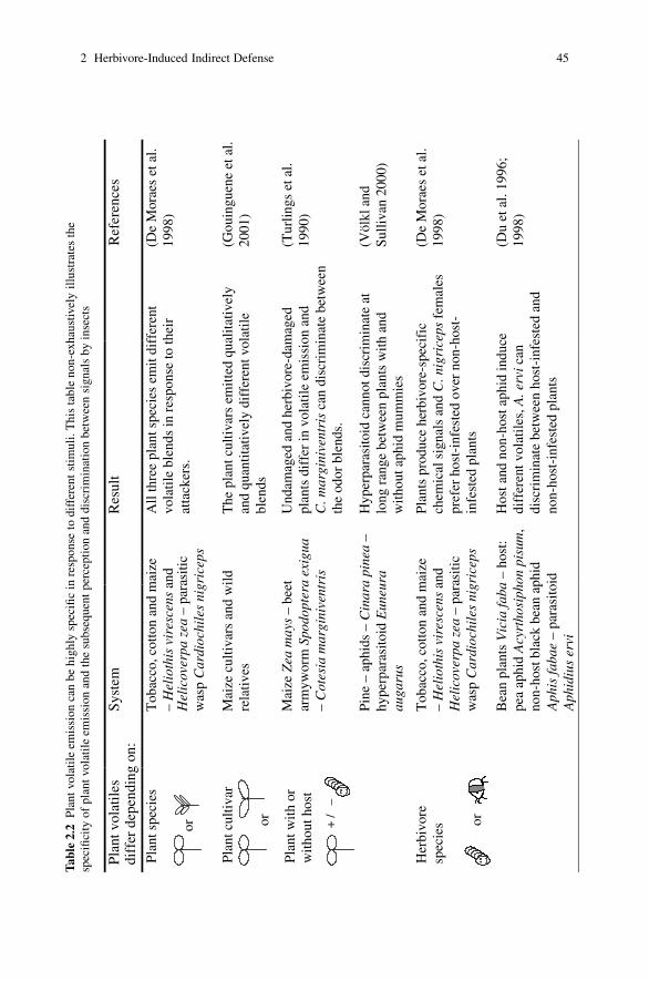
2 Herbivore-Induced Indirect Defense 45T
able
2.2
Plan
tvol
atile
emis
sion
can
behi
ghly
spec
ific
inre
spon
seto
diff
eren
tst
imul
i.T
his
tabl
eno
n-ex
haus
tivel
yill
ustr
ates
the
spec
ifici
tyof
plan
tvol
atil
eem
issi
onan
dth
esu
bseq
uent
perc
epti
onan
ddi
scri
min
atio
nbe
twee
nsi
gnal
sby
inse
cts
Pla
nt v
olat
iles
diff
er d
epen
ding
on:
Syst
emR
esul
tR
efer
ence
s
Pla
nt s
peci
es
or
Tob
acco
, cot
ton
and
mai
ze
– H
elio
this
vir
esce
ns a
nd
Hel
icov
erpa
zea
– p
aras
itic
w
asp
Car
dioc
hile
s ni
gric
eps
All
thre
e pl
ant s
peci
es e
mit
diff
eren
t vo
lati
le b
lend
s in
res
pons
e to
thei
r at
tack
ers.
(De
Mor
aes
et a
l. 19
98)
Pla
nt c
ultiv
ar
or
Mai
ze c
ulti
vars
and
wil
d re
lativ
esT
he p
lant
cul
tiva
rs e
mit
ted
qual
itativ
ely
and
quan
titat
ivel
y di
ffer
ent v
olat
ile
blen
ds
(Gou
ingu
ene
et a
l. 20
01)
Pla
nt w
ith o
r w
ithou
t hos
t
+ /
–
Mai
ze Z
ea m
ays
– be
et
arm
ywor
m S
podo
pter
a ex
igua
– C
otes
ia m
argi
nive
ntri
s
Pin
e –
aphi
ds –
Cin
ara
pine
a –
hype
rpar
asito
id E
uneu
ra
auga
rus
Und
amag
ed a
nd h
erbi
vore
-dam
aged
pl
ants
dif
fer
in v
olat
ile
emis
sion
and
C. m
argi
nive
ntri
s ca
n di
scri
min
ate
betw
een
the
odor
ble
nds.
Hyp
erpa
rasi
toid
can
not d
iscr
imin
ate
at
long
ran
ge b
etw
een
plan
ts w
ith a
nd
with
out a
phid
mum
mie
s
(Tur
lings
et a
l. 19
90)
(Völ
kl a
nd
Sul
livan
200
0)
Her
bivo
re
spec
ies or
Tob
acco
, cot
ton
and
mai
ze
– H
elio
this
vir
esce
ns a
nd
Hel
icov
erpa
zea
– p
aras
itic
w
asp
Car
dioc
hile
s ni
gric
eps
Bea
n pl
ants
Vic
ia fa
ba –
hos
t: pe
a ap
hid
Acy
rtho
siph
on p
isum
, no
n-ho
st b
lack
bea
n ap
hid
Aph
is fa
bae
– pa
rasi
toid
A
phid
ius
ervi
Pla
nts
prod
uce
herb
ivor
e-sp
ecif
ic
chem
ical
sig
nals
and
C. n
igri
ceps
fem
ales
pr
efer
hos
t-in
fest
ed o
ver
non-
host
-in
fest
ed p
lant
s
Hos
t and
non
-hos
t aph
id in
duce
di
ffer
ent v
olat
iles
, A. e
rvi c
an
disc
rim
inat
e be
twee
n ho
st-i
nfes
ted
and
non-
host
-inf
este
d pl
ants
(De
Mor
aes
et a
l. 19
98)
(Du
et a
l. 19
96;
1998
)
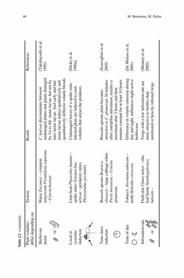
46 M. Bruinsma, M. DickeT
able
2.2
(con
tinu
ed)
Her
bivo
re
inst
ar
or
Mai
ze Z
ea m
ays
– co
mm
on
arm
ywor
m P
seud
alet
ia s
epar
ata
– C
otes
ia k
ariy
ai
C. k
ariy
ai d
iscr
imin
ates
bet
wee
n un
dam
aged
pla
nts
and
plan
ts d
amag
ed
by 1
st to
4th
ins
tar
larv
ae, b
ut n
ot b
y la
ter
inst
ar la
rvae
. And
ear
ly a
nd la
te
inst
ar la
rvae
indu
ce q
ualit
ativ
ely
and
quan
titat
ivel
y di
ffer
ent v
olat
ile b
lend
s
(Tak
abay
ashi
et a
l. 19
95)
Loc
al o
r sy
stem
icin
duct
ion
Lim
a be
an P
hase
olus
luna
tus
–sp
ider
mit
es T
etra
nych
us
urti
cae
– pr
edat
ory
mite
s P
hyto
seiu
lus
pers
imil
is
Und
amag
ed le
aves
of
a sp
ider
mite
-in
fest
ed p
lant
are
indu
ced
to e
mit
vola
tiles
that
attr
act t
he p
reda
tors
(Dic
ke e
t al.
1990
a)
Tim
e si
nce
indu
ctio
nB
russ
els
spro
uts
Bra
ssic
a ol
erac
ea –
larg
e ca
bbag
e w
hite
P
ieri
s br
assi
cae
– C
otes
ia
glom
erat
a
Bru
ssel
s sp
rout
s pl
ant b
ecom
es
attr
activ
e to
C. g
lom
erat
a 30
min
utes
afte
r ca
terp
illa
r in
fest
atio
n, r
each
es a
m
axim
um a
fter
3 h
ours
and
then
re
mai
ns c
onst
ant f
or a
t lea
st 1
4 ho
urs
(Sca
scig
hini
et a
l. 20
05)
Tim
e of
day
Tob
acco
Nic
otia
na ta
bacu
m –
mot
h H
elio
this
vir
esce
nsD
iffe
renc
e in
vol
atile
em
issi
on d
urin
g da
y an
d ni
ght,
infl
uenc
es n
ight
-act
ive
herb
ivor
es
(De
Mor
aes
et a
l. 20
01)
Infe
stat
ion
rate
or
Fiel
d el
m U
lmus
min
or –
elm
le
af b
eetle
Xan
thog
aler
uca
lutt
eola
–
Tw
igs
with
a lo
w in
fest
atio
n ra
te a
re
mor
e at
trac
tive
to b
eetle
s th
an
unin
fest
ed o
r he
avily
infe
sted
twig
s
(Mei
ners
et a
l. 20
05)
Pla
nt v
olat
iles
di
ffer
dep
endi
ng o
n:Sy
stem
Res
ult
Ref
eren
ces
2 Herbivore-Induced Indirect Defense 47
example, P. xylostella profits from a positive indirect effect (associational resistance)through the presence of P. rapae (White and Andow 2006). Defense responses toherbivores may also interfere with other interactions in the community, such as pol-lination. For tobacco plants there is a trade-off between repelling herbivores andattracting pollinators. As briefly mentioned in Section 2.3.3, these plants reduce thelevel of toxins and increase emission of a pollinator attractant by their flowers in theevening. This way tobacco can defend itself against herbivory during daytime andattract pollinators at night (Euler and Baldwin 1996).
While some community members are able to distinguish between differentvolatile blends, others cannot, or only after associative learning (Takabayashi et al.2006). Several arthropod species, such as Phytoseiulus persimilis, Cotesiamarginiventris and C. glomerata, can learn to associate certain odors with a reward(Geervliet et al. 1997; Turlings and Fritzsche 1999; De Boer et al. 2005). Learningto associate odors with the presence of host or prey may be a way to cope withthe variation in volatile blends. Learning new associations may take some time andmultiple experiences. In the meantime the host or prey species may profit from its‘invisibility’ to its natural enemy while the parasitoid or predator wastes time onsearching on plants without its host or prey (Shiojiri et al. 2002). The temporalpattern in learning to respond to certain blends and losing this learned responsewill affect the risk of herbivores to fall victim to their enemies. Temporary refugesmay stabilize predator–prey or parasitoid-host systems and thus affect dynamics ofarthropod communities on plants (Vos et al. 2001; Takabayashi et al. 2006). To gainmore insight into these subtle interactions and ecosystem stability, field studies inwhich one component is changed could provide an important tool to investigate theimpact on the community.
2.5 Manipulation of Indirect Plant Defense
To investigate the effect of indirect defenses and their components, a manipulativeapproach is most rewarding. Manipulation of defense responses can provide infor-mation on both the mechanisms of plant defense and the ecological consequences ofchanges in the plant’s phenotype. A range of manipulative approaches can be takenthat are explained below.
2.5.1 Perfuming with Individual Compounds
A simple change in a plant’s phenotype can be made by adding a single compoundto an undamaged plant. This method allows testing the importance of individualcompounds for attraction of, for example, predators and parasitoids to the plantagainst a background of natural odors (Dicke et al. 2006). Even though carnivo-rous arthropods are usually attracted to complex odor blends, increased attractionof carnivores due to addition of individual compounds has been recorded with this

48 M. Bruinsma, M. Dicke
method (De Boer and Dicke 2004). Also in a field study, the addition of linaloolto N. attenuata plants resulted in both increased predation of herbivore eggs andlarvae, and decreased oviposition by an herbivore (Kessler and Baldwin 2001).
2.5.2 Fractionation and Filtering
Phenotypic manipulation of the volatile blends can also be accomplished by usingfilters to collect compounds with specific chemical properties selectively(D’Alessandro and Turlings 2006), or by fractionation of the headspace and sub-sequently testing different fractions of the blend for biological activity (Turlingsand Fritzsche 1999; Van den Boom 2003). As compared to the testing of singlecompounds, these methods have the advantage of conserving the ratios of differentvolatiles as they are present in natural herbivore-induced volatile blends. Filters canbe used to remove one or more compounds, which are suspected to show biologicalactivity, from the total volatile blend. It is then possible to test their importance foranimal behavior in olfactometer experiments (D’Alessandro and Turlings 2006).With this method D’Alessandro and Turlings (2005) showed that the selective re-moval of the more polar plant volatiles using silica filters decreases the attractivenessof the volatile blend to a parasitoid wasp, C. marginiventris, but not to another para-sitoid Microplitis rufiventris. This study illustrates how filtering can be used as a toolto find the ecologically relevant volatiles for different species on all trophic levelsusing various adsorbing materials. For fractionation, the mixture of defense com-pounds can be trapped onto an absorbent material and subsequently recovered bychemical or thermal desorption. Subtractive fractionation and testing of the differentfractions for their biological activity provides a useful tool to identify compoundsinvolved in indirect defense (Van den Boom 2003).
2.5.3 Genetic Modification
Another way to manipulate induced defenses of plants, is by genetic modification ofsignal-transduction or biosynthetic pathways. Carefully modified plants that differin a single gene can be compared with wild-type plants to gain more insight intomechanisms of induced defense and the resulting ecological effects (for review, seeSnoeren et al. 2007). Otherwise identical plants can be induced by herbivory and thedifferences between the wild type and mutant can be studied at the levels of geneexpression, volatile production, and response of the plant-associated community.The approach is limited by the availability of modified plants. For some species likeA. thaliana however, a wide variety of mutant and transgenic plants is available.Genetic modification is an excellent tool to study the ecological relevance of indi-vidual compounds that are difficult to obtain synthetically. For example, undamagedArabidopsis plants transformed with a terpene synthase from strawberry, producethe terpenoids 4,8-dimethyl-1,3(E),7-nonatriene and (E)-nerolidol which results in

2 Herbivore-Induced Indirect Defense 49
the attraction of predatory mites (Kappers et al. 2005). However, A. thaliana has itslimitations, such as its very early phenology and consequently limited interactionwith potentially associated organisms, as a model for community ecology studies.
Transgenic N. attenuata plants that have been modified in the octadecanoidsignal-transduction pathway received more herbivory and by more herbivore speciesthan wild type control plants (Kessler et al. 2004). Similarly, the tomato mutant def-1is deficient in JA accumulation through a mutation early in the JA pathway. As aresult, the plants produce an incomplete volatile blend in response to herbivore dam-age, and the natural enemies of the herbivores do not discriminate between volatileblends from induced and uninduced plants (Thaler et al. 2002; Ament et al. 2004).However, for many ecological model species mutants or genetically modified plantsare not (yet) available.
2.5.4 Chemical Elicitors and Inhibitors
Signal-transduction and biosynthetic pathways can be manipulated through the ap-plication of (specific) inducers or inhibitors. By artificially inducing or inhibitingdifferent steps of the signaling pathways it is possible to study ecological interac-tions at different trophic levels, or in the whole community. Plant-emitted volatileblends can be manipulated by interfering with the signal-transduction pathways,and the importance of individual steps can be investigated by specifically activatingor blocking individual pathway enzymes. When these changed blends are offeredto (members of) the insect community, this will provide insight into the ecologicalrelevance of these pathways.
The most extensively tested elicitors to date are the phytohormone jasmonic acidand its methyl ester, methyl jasmonate (MeJA). These elicitors were shown to playan important role in induced defense in many plant species against a wide range ofherbivores. JA induces volatile blends similar to those induced by herbivore damage(Hopke et al. 1994; Dicke et al. 1999; Ozawa et al. 2000, 2004), and while herbi-vores are negatively influenced (Van Dam et al. 2000; Thaler et al. 2001; Bruinsmaet al. 2007), carnivorous arthropods are attracted to plants that have been inducedwith JA or MeJA (Thaler 1999; Van Poecke and Dicke 2002; Gols et al. 2003).For many other elicitors and phytohormones, such as methyl salicylate, linolenicacid, OPDA, �-glucosidase, cellulysin, and alamethicin, similar studies demon-strated their effects on community members (Mattiacci et al. 1995; Koch et al. 1999;Dicke and Van Poecke 2002; De Boer and Dicke 2004; Ozawa et al. 2004). Oneof the advantages of using elicitors is the possibility to apply a controlled dose tospecific plant parts, whereas it is practically impossible to control the amount ofinjury inflicted by insects or other biotic agents. However, it is not easy to relate theexternally applied dosage to intracellular concentrations and effects.
Specific inhibitors, although not used quite as extensively as elicitors, are suitableto demonstrate the importance of different steps in signal-transduction pathways aswell. However, accumulation of pathway intermediates just before the inhibited stepmay cause physiological side effects. Making use of inhibitors, Koch et al. (1999)

50 M. Bruinsma, M. Dicke
showed the importance of early steps in the octadecanoid pathway for volatileemission of Lima bean (Table 2.1). Phenidone for example, inhibits the activityof lipoxygenase, which results in incomplete volatile blends (Piel et al. 1997; Kochet al. 1999) and reduced EFN secretion (Heil et al. 2004), indicating the importanceof this early step in the octadecanoid pathway for indirect defense responses.
2.6 From Lab to the Field
To gain insight into the ecology of induced defenses, field studies incorporatingthe complexity of natural ecosystems are indispensable. Manipulation through oneof the methods described above may facilitate research on the effects of inducedindirect defense on interactions between community members and the stability ofthe system.
Most studies addressing induced plant defense are laboratory studies, studyingsimple systems of one plant, one herbivore and its natural enemies. This providesdetailed insight into the effects of induced defenses on individual interactions.Through greenhouse and semi-field studies with more complex set-ups, for instanceby introducing background odors under controlled conditions (Janssen 1999; Dickeet al. 2003), more insight will be gained in field situations. However, in order touse knowledge gained from these studies, the relative importance of these pieces ofinformation should be assessed in the field. Also biological control in agriculturalfields may benefit from such knowledge and understanding of multitrophic interac-tions in the field.
2.6.1 Natural Ecosystems
For studying the ecology of herbivore-induced plant responses, it is necessary toaddress natural ecosystems. For instance, in wild radish induced responses to her-bivory increase plant fitness in natural environments (Agrawal 1999). Herbivoryincreases trichome density and subsequently reduces preference and performanceof several herbivores. The effectiveness of induced defenses is clear from a fieldstudy on N. attenuata where herbivory was reduced by as much as 90% by the ad-dition of defense compounds (Kessler and Baldwin 2001). In a later study, Kessleret al. (2006) showed that volatiles from damaged sagebrush can prime responses inN. attenuata and reduce herbivore damage to the exposed plants. However, despitethe use of natural populations of N. attenuata, the ecological relevance of this studyis questionable because the sagebrush that was used for elicitation occurs in a differ-ent successional stage than N. attenuata. Furthermore, the effect was only detectedat a short range, when the plants grew within 15 cm from each other.
In naturally occurring milkweed populations, early-season herbivory affects sub-sequent herbivory throughout the season, thereby affecting community structure(Van Zandt and Agrawal 2004). In a natural community, Agrawal (2004) studied thecomplex interactions between milkweed and competing grass, in presence or absenceof root and leaf herbivory. He concluded that the genetic differences, competition,

2 Herbivore-Induced Indirect Defense 51
and herbivory resulted in complex interactions that may result in diffuse co-evolutionbetween milkweed and its herbivores. To improve our understanding of the evolutionof induced defenses and resistance of herbivores more field studies are needed.
The use of elicitors in the field can provide information on the ecological rele-vance of pathways and certain steps therein. For example, the application of jasmonicacid to tomato plants in the field increased parasitism of herbivores and thus showedthe involvement of jasmonic acid-induced changes in the attraction of carnivores(Thaler 1999). The application of methyl jasmonate to tobacco plants demonstratedthe costs of jasmonate-induced responses. In environments with herbivore pressure,induced plants suffered less from herbivore attack and produced more viable seedsthan uninduced plants. However, undamaged plants produced more seeds when theywere not induced compared to jasmonate-induced plants (Baldwin et al. 1998).
2.6.2 Agricultural Systems
Induced plant defenses can aid pest control in agricultural systems. Attracting natu-ral enemies of herbivores to crops can help control pests in agriculture; in the field aswell as in greenhouses (Dicke et al. 1990a; Turlings and Ton 2006). Therefore, un-derstanding the mechanisms involved in plant defenses and the consequences for thecommunity associated with the plant can aid crop protection. Manipulation may in-crease the effectiveness of plant defenses, by attracting natural enemies before con-siderable damage is done by herbivores and by deterring oviposition by herbivores.This can be achieved by inducing the plant with phytohormones like cis-jasmoneor jasmonic acid (Thaler 1999; Birkett et al. 2000; Heil 2004). Another possibilityis the use of genetically modified crops that produce volatile blends that are moreattractive to predators than genotypes currently used. The technology is being devel-oped (Kappers et al. 2005; Schnee et al. 2006). However, the consequences for thecommunity, above- as well as belowground, and the effect on interactions betweendifferent community members still need to be addressed (Groot and Dicke 2002;Kowalchuk et al. 2003).
A lot of the research on genetic modification of plant defense has been donein the model plant A. thaliana, for which many mutants are available (Turlingsand Ton 2006). Knowledge about A. thaliana can be extrapolated to Brassicaspecies, and therefore readily be applied in agricultural settings with crop specieslike Brassica oleracea or wild Brassica species (Broekgaarden et al. 2007; Zhenget al. 2007). In conclusion, the step from the laboratory into the field has not beenmade often yet. It will be important to make this step for different plant species togain insight into the effects of induced indirect defense on community processes.
2.7 Perspectives
The effects of induced defense-related phenotypic changes in plants on commu-nity dynamics are difficult to predict, because many aspects are involved and

52 M. Bruinsma, M. Dicke
the variability of plant responses is enormous. While many bi- and tritrophicinteractions are well studied, plants in nature are usually under the attack of a rangeof organisms at the same time. How this affects plant defense has only just begun tobe addressed, and first results show that the effects may be an increase as well as adecrease in defense intensity (e.g., Dicke et al. 2003; Rodriguez-Saona et al. 2005;Cardoza and Tumlinson 2006).
Although most studies thus far have focused on aboveground processes, the influ-ence of the changes in plant phenotype is not limited to the aboveground community.Aboveground interactions can change belowground root exudates and influence thesoil community, and belowground damage can influence aboveground indirect de-fense (Bezemer and van Dam 2005). The reverse, however, i.e. the effect of above-ground interactions on belowground indirect defense, remains as yet uninvestigated(Bezemer and van Dam 2005). Incorporating these interactions in future studies willgreatly enhance our insight into the effects of induced indirect defense on the func-tioning of complex communities. In addition to plant-pathogen and plant-herbivoreinteractions, plants may also be under the attack of parasitic plants (Bouwmeesteret al. 2003; Runyon et al. 2006), or interact with belowground symbiotic organismssuch as mycorrhiza or symbiotic bacteria (Gange et al. 2002). Furthermore, above-ground endophytic organisms can influence the plant’s defensive phenotype andconsequently also the interactions with community members (Omacini et al. 2001).Incorporating these interactions in the investigations of indirect defense of plants ina community ecology approach will increase complexity, yet doing so is essentialto gain a meaningful understanding of the effects of indirect plant defense on plantecology.
Another area of research that has not received a lot of attention so far is thesearching behavior of members of the higher trophic levels, such as hyperpara-sitoids. How they find their host and whether they use plant cues remains largelyunknown (Buitenhuis et al. 2005). The same applies for pollinators. Though someeffects of herbivory on pollination have been reported (Lehtila and Strauss 1997;Poveda et al. 2003), the underlying mechanisms remain to be unraveled andwhich signal-transduction pathways are important in this respect, waits to beinvestigated.
For a complete understanding of the ecology and evolution of communities, itis necessary to include all trophic levels in field studies. Manipulative studies arelikely to provide the best way forward. They can be used in the laboratory to in-vestigate individual interactions and are a valuable tool to investigate the effects ofinduced defenses on the community in the field (Kessler and Baldwin 2001; Kessleret al. 2004). Using an integrated approach with molecular, chemical, and behav-ioral methodology will significantly advance the research in this area (Baldwin et al.2001).
Acknowledgments We thank Joop van Loon for valuable comments on the manuscript. This workhas been financially supported by a VICI grant from the Netherlands Organisation for ScientificResearch, NWO (865.03.002).

2 Herbivore-Induced Indirect Defense 53
References
Adler LS, Karban R, Strauss SY (2001) Direct and indirect effects of alkaloids on plant fitness viaherbivory and pollination. Ecology 82:2032–2044
Adler LS, Wink M, Distl M, Lentz AJ (2006) Leaf herbivory and nutrients increase nectar alka-loids. Ecol Lett 9:960–967
Agrawal AA (1999) Induced responses to herbivory in wild radish: effects on several herbivoresand plant fitness. Ecology 80:1713–1723
Agrawal AA (2001) Phenotypic plasticity in the interactions and evolution of species. Science294:321–326
Agrawal AA (2004) Resistance and susceptibility of milkweed: competition, root herbivory, andplant genetic variation. Ecology 85:2118–2133
Alborn T, Turlings TCJ, Jones TH, Stenhagen G, Loughrin JH, Tumlinson JH (1997) An elicitorof plant volatiles from beet armyworm oral secretion. Science 276:945–949
Ament K, Kant MR, Sabelis MW, Haring MA, Schuurink RC (2004) Jasmonic acid is a key regu-lator of spider mite-induced volatile terpenoid and methyl salicylate emission in tomato. PlantPhysiol 135:2025–2037
Avdiushko SA, Brown GC, Dahlman DL, Hildebrand DF (1997) Methyl jasmonate exposure in-duces insect resistance in cabbage and tobacco. Environ Entomol 26:642–654
Baldwin IT (1990) Herbivory simulations in ecological research. Trends Ecol Evol 5:91–93Baldwin IT (1998) Jasmonate-induced responses are costly but benefit plants under attack in native
populations. Proc Natl Acad Sci USA 95:8113–8118Baldwin IT, Halitschke R, Kessler A, Schittko U (2001) Merging molecular and ecological ap-
proaches in plant–insect interactions. Curr Opin Plant Biol 4:351–358Baldwin IT, Halitschke R, Paschold A, von Dahl CC, Preston CA (2006) Volatile signaling in
plant–plant interactions: ‘Talking trees’ in the genomics era. Science 311:812–815Bezemer TM, De Deyn GB, Bossinga TM, van Dam NM, Harvey JA, Van der Putten WH (2005)
Soil community composition drives aboveground plant–herbivore–parasitoid interactions. EcolLett 8:652–661
Bezemer TM, van Dam NM (2005) Linking aboveground and belowground interactions via in-duced plant defenses. Trends Ecol Evol 20:617–624
Birkett MA, Campbell CAM, Chamberlain K, Guerrieri E, Hick AJ, Martin JL, Matthes M,Napier JA, Pettersson J, Pickett JA, Poppy GM, Pow EM, Pye BJ, Smart LE, Wadhams GH,Wadhams LJ, Woodcock CM (2000) New roles for cis-jasmone as an insect semiochemical andin plant defense. Proc Natl Acad Sci USA 97:9329–9334
Blaakmeer A, Geervliet JBF, Van Loon JJA, Posthumus MA, Van Beek TA, De Groot A (1994)Comparative headspace analysis of cabbage plants damaged by two species of Pieris cater-pillars: consequences for in-flight host location by Cotesia parasitoids. Entomol Exp Appl73:175–182
Boland W, Koch T, Krumm T, Piel J, Jux A (1999) Induced biosynthesis of insect semiochemicalsin plants. In: Chadwick DJ, Goode J (eds) Insect–plant interactions and induced plant defence(Novartis Foundation Symposium 223). Wiley, Chichester, pp 110–126
Bouwmeester HJ, Matusova R, Sun ZK, Beale MH (2003) Secondary metabolite signaling in host–parasitic plant interactions. Curr Opin Plant Biol 6:358–364
Brodeur J (2000) Host specificity and trophic relationships of hyperparasitoids. In: HochbergME, Ives AR (eds) Parasitoid population biology. Princeton University Press, Princeton,pp 163–183
Broekgaarden C, Poelman EH, Steenhuis G, Voorrips RE, Dicke M, Vosman B (2007) Genotypicvariation in genome-wide transcription profiles induced by insect feeding. BMC Genomics8:239
Bruinsma M, Van Dam NM, Van Loon JJA, Dicke M (2007) Jasmonic acid-induced changesin Brassica oleracea affect oviposition preference of two specialist herbivores. J Chem Ecol33:655–668
54 M. Bruinsma, M. Dicke
Buitenhuis R, Vet LEM, Boivin G, Brodeur J (2005) Foraging behavior at the fourth trophiclevel: a comparative study of host location in aphid hyperparasitoids. Entomol Exp Appl114:107–117
Cardoza YJ, Tumlinson JH (2006) Compatible and incompatible Xanthomonas infections dif-ferentially affect herbivore-induced volatile emission by pepper plants. J Chem Ecol 32:1755–1768
Chamberlain K, Guerrieri E, Pennacchio F, Pettersson J, Pickett JA, Poppy GM, Powell W,Wadhams LJ, Woodcock CM (2001) Can aphid-induced plant signals be transmitted aeriallyand through the rhizosphere? Biochem Syst Ecol 29:1063–1074
Choh Y, Takabayashi J (2006) Herbivore-induced extrafloral nectar production in Lima beanplants enhanced by previous exposure to volatiles from infested conspecifics. J Chem Ecol 32:2073–2077
Choh Y, Kugimiya S, Takabayashi J (2006) Induced production of extrafloral nectar in intact limabean plants in response to volatiles from spider mite-infested conspecific plants as a possibleindirect defense against spider mites. Oecologia 147:455–460
Conrath U, Beckers GJM, Flors V, Garcia-Agustin P, Jakab G, Mauch F, Newman MA, PieterseCMJ, Poinssot B, Pozo MJ, Pugin A, Schaffrath U, Ton J, Wendehenne D, Zimmerli L, Mauch-Mani B (2006) Priming: getting ready for battle. Mol Plant Micr Int 19:1062–1071
Conrath U, Pieterse CMJ, Mauch-Mani B (2002) Priming in plant–pathogen interactions. TrendsPlant Sci 7:210–216
Creelman RA, Mullet JE (1997) Biosynthesis and action of jasmonates in plants. Annu Rev PlantPhysiol Plant Mol Biol 48:355–381
D’Alessandro M, Turlings TCJ (2005) In situ modification of herbivore-induced plant odors: anovel approach to study the attractiveness of volatile organic compounds to parasitic wasps.Chem Senses 30:739–753
D’Alessandro M, Turlings TCJ (2006) Advances and challenges in the identification of volatilesthat mediate interactions among plants and arthropods. Analyst 131:24–32
De Boer JG, Dicke M (2004) The role of methyl salicylate in prey searching behavior of thepredatory mite Phytoseiulus persimilis. J Chem Ecol 30:255–271
De Boer JG, Snoeren TAL, Dicke M (2005) Predatory mites learn to discriminate between plantvolatiles induced by prey and nonprey herbivores. Anim Behav 69:869–879
De Moraes CM, Lewis WJ, Pare PW, Alborn HT, Tumlinson JH (1998) Herbivore-infested plantsselectively attract parasitoids. Nature 393:570–573
De Moraes CM, Mescher MC, Tumlinson JH (2001) Caterpillar-induced nocturnal plant volatilesrepel conspecific females. Nature 410:577–580
De Vos M (2006) Signal signature, transcriptomics, and effectiveness of induced pathogen andinsect resistance in Arabidopsis. Utrecht University, Utrecht
De Vos M, Van Oosten VR, Van Poecke RMP, Van Pelt JA, Pozo MJ, Mueller MJ, Buchala AJ,Metraux JP, Van Loon LC, Dicke M, Pieterse CMJ (2005) Signal signature and transcrip-tome changes of Arabidopsis during pathogen and insect attack. Mol Plant Microbe Interact18:923–937
Dicke M (1999) Evolution of induced indirect defense of plants. In: Tollrian R, Harvell CJ(eds) The ecology and evolution of inducible defenses. Princeton University Press, Princeton,pp 62–88
Dicke M, Bruin J (2001) Chemical information transfer between plants: back to the future.Biochem Syst Ecol 29:981–994
Dicke M, Bruinsma M, Bukovinszky T, Gols R, De Jong PW, Van Loon JJA, Snoeren TAL, ZhengS-J (2006) Investigating the ecology of inducible indirect defence by manipulating plant phe-notype and genotype. IOBC WPRS Bull 29:15–23
Dicke M, De Boer JG, Hofte M, Rocha-Granados MC (2003) Mixed blends of herbivore-inducedplant volatiles and foraging success of carnivorous arthropods. OIKOS 101:38–48
Dicke M, Dijkman H (2001) Within-plant circulation of systemic elicitor of induced de-fence and release from roots of elicitor that affects neighboring plants. Biochem Syst Ecol29:1075–1087

2 Herbivore-Induced Indirect Defense 55
Dicke M, Gols R, Ludeking D, Posthumus MA (1999) Jasmonic acid and herbivory differentiallyinduce carnivore-attracting plant volatiles in lima bean plants. J Chem Ecol 25:1907–1922
Dicke M, Hilker M (2003) Induced plant defenses: from molecular biology to evolutionary ecol-ogy. Basic Appl Ecol 4:3–14
Dicke M, Sabelis MW (1988) Infochemical terminology: based on cost benefit analysis rather thanorigin of compounds? Funct Ecol 2:131–139
Dicke M, Sabelis MW, Takabayashi J, Bruin J, Posthumus MA (1990a) Plant strategies of ma-nipulating predator–prey interactions through allelochemicals: prospects for application in pestcontrol. J Chem Ecol 16:3091–3118
Dicke M, Takabayashi J, Posthumus MA, Schutte C, Krips OE (1998) Plant–phytoseiid interac-tions mediated by herbivore-induced plant volatiles: variation in production of cues and inresponses of predatory mites. Exp Appl Acarol 22:311–333
Dicke M, Van Beek TA, Posthumus MA, Ben Dom N, Van Bokhoven H, De Groot AE (1990b)Isolation and identification of volatile kairomone that affects acarine predator–prey interactions.Involvement of host plant in its production. J Chem Ecol 16:381–396
Dicke M, Van Poecke RMP (2002) Signaling in plant–insect interactions: signal transduction indirect and indirect plant defence. In: Scheel D, Wasternack C (eds) Plant signal transduction.Oxford University Press, Oxford, pp 289–316
Dicke M, Vet LEM (1999) Plant–carnivore interactions: evolutionary and ecological consequencesfor plant, herbivore and carnivore. In: Olff H, Brown VK, Drent RH (eds) Herbivores: betweenplants and predators. Blackwell Science, Oxford, pp 483–520
Donath J, Boland W (1994) Biosynthesis of acyclic homoterpenes in higher plants parallels steroidhormone metabolism. J Plant Physiol 143:473–478
Du YJ, Poppy GM, Powell W (1996) Relative importance of semiochemicals from firstand second trophic levels in host foraging behavior of Aphidius ervi. J Chem Ecol22:1591–1605
Du YJ, Poppy GM, Powell W, Pickett JA, Wadhams LJ, Woodcock CM (1998) Identification ofsemiochemicals released during aphid feeding that attract parasitoid Aphidius ervi. J ChemEcol 24:1355–1368
Engelberth J, Alborn HT, Schmelz EA, Tumlinson JH (2004) Airborne signals prime plants againstinsect herbivore attack. Proc Natl Acad Sci USA 101:1781–1785
Euler M, Baldwin IT (1996) The chemistry of defense and apparency in the corollas of Nicotianaattenuata. Oecologia 107:102–112
Farmer EE, Ryan CA (1990) Interplant communication – airborne methyl jasmonate induces syn-thesis of proteinase inhibitors in plant leaves. Proc Natl Acad Sci USA 87:7713–7716
Fatouros NE, Van Loon JJA, Hordijk KA, Smid HM, Dicke M (2005) Herbivore-induced plantvolatiles mediate in-flight host discrimination by parasitoids. J Chem Ecol 31:2033–2047
Fritzsche-Hoballah ME, Turlings TCJ (2001) Experimental evidence that plants under caterpillarattack may benefit from attracting parasitoids. Evol Ecol Res 3:553–565
Gange AC, Stagg PG, Ward LK (2002) Arbuscular mycorrhizal fungi affect phytophagous insectspecialism. Ecol Lett 5:11–15
Geervliet JBF, Posthumus MA, Vet LEM, Dicke M (1997) Comparative analysis of headspacevolatiles from different caterpillar-infested or uninfested food plants of Pieris species. J ChemEcol 23:2935–2954
Geervliet JBF, Vet LEM, Dicke M (1994) Volatiles from damaged plants as major cues inlong-range host-searching by the specialist parasitoid Cotesia rubecula. Entomol Exp Appl73:289–297
Gols R, Posthumus MA, Dicke M (1999) Jasmonic acid induces the production of gerberavolatiles that attract the biological control agent Phytoseiulus persimilis. Entomol Exp Appl93:77–86
Gols R, Roosjen M, Dijkman H, Dicke M (2003) Induction of direct and indirect plant responsesby jasmonic acid, low spider mite densities or a combination of jasmonic acid treatment andspider mite infestation. J Chem Ecol 29:2651–2666

56 M. Bruinsma, M. Dicke
Gouinguene S, Degen T, Turlings TCJ (2001) Variability in herbivore-induced odor emissionsamong maize cultivars and their wild ancestors (teosinte). Chemoecology 11:9–16
Groot AT, Dicke M (2002) Insect-resistant transgenic plants in a multi-trophic context. Plant J31:387–406
Guerrieri E, Lingua G, Digilio MC, Massa N, Berta G (2004) Do interactions between plant rootsand the rhizosphere affect parasitoid behavior? Ecol Entomol 29:753–756
Halitschke R, Schittko U, Pohnert G, Boland W, Baldwin IT (2001) Molecular interactions betweenthe specialist herbivore Manduca sexta (Lepidoptera, Sphingidae) and its natural host Nicotianaattenuata. III. Fatty acid–amino acid conjugates in herbivore oral secretions are necessary andsufficient for herbivore-specific plant responses. Plant Physiol 125:711–717
Hamback PA (2001) Direct and indirect effects of herbivory: feeding by spittlebugs affects polli-nator visitation rates and seedset of Rudbeckia hirta. Ecoscience 8:45–50
Harvey JA, Van Dam NM, Gols R (2003) Interactions over four trophic levels: foodplant qual-ity affects development of a hyperparasitoid as mediated through a herbivore and its primaryparasitoid. J Anim Ecol 72:520–531
Hatanaka A, Kajiwara T, Sekiya J (1987) Biosynthetic pathway for C-6-aldehydes formation fromlinolenic acid in green leaves. Chem Phys Lipids 44:341–361
Heil M (2004) Induction of two indirect defenses benefits Lima bean (Phaseolus lunatus,Fabaceae) in nature. J Ecol 92:527–536
Heil M, Greiner S, Meimberg H, Kruger R, Noyer JL, Heubl G, Linsenmair KE, Boland W (2004)Evolutionary change from induced to constitutive expression of an indirect plant resistance.Nature 430:205–208
Heil M, Koch T, Hilpert A, Fiala B, Boland W, Linsenmair KE (2001) Extrafloral nectar productionof the ant-associated plant, Macaranga tanarius, is an induced, indirect, defensive responseelicited by jasmonic acid. Proc Natl Acad Sci USA 98:1083–1088
Heil M, Kost C (2006) Priming of indirect defenses. Ecol Lett 9:813–817Hilker M, Meiners T (2006) Early herbivore alert: insect eggs induce plant defense. J Chem Ecol
32:1379–1397Holt RD (1977) Predation, apparent competition, and structure of prey communities. Theor Popul
Biol 12:197–229Hopke J, Donath J, Blechert S, Boland W (1994) Herbivore-induced volatiles: the emission of
acyclic homoterpenes from leaves of Phaseolus lunatus and Zea mays can be triggered by abeta-glucosidase and jasmonic acid. FEBS Lett 352:146–150
Janssen A (1999) Plants with spider-mite prey attract more predatory mites than clean plants undergreenhouse conditions. Entomol Exp Appl 90:191–198
Kahl J, Siemens DH, Aerts RJ, Gabler R, Kuhnemann F, Preston CA, Baldwin IT (2000)Herbivore-induced ethylene suppresses a direct defense but not a putative indirect defenseagainst an adapted herbivore. Planta 210:336–342
Kappers IF, Aharoni A, Van Herpen TWJM, Luckerhoff LLP, Dicke M, Bouwmeester HJ (2005)Genetic engineering of terpenoid metabolism attracts bodyguards to Arabidopsis. Science309:2070–2072
Kessler A, Baldwin IT (2001) Defensive function of herbivore-induced plant volatile emissions innature. Science 291:2141–2144
Kessler A, Halitschke R, Baldwin IT (2004) Silencing the jasmonate cascade: induced plant de-fenses and insect populations. Science 305:665–668
Kessler A, Halitschke R, Diezel C, Baldwin IT (2006) Priming of plant defense responses in na-ture by airborne signaling between Artemisia tridentata and Nicotiana attenuata. Oecologia148:280–292
Klein AM, Vaissiere BE, Cane JH, Steffan-Dewenter I, Cunningham SA, Kremen C, Tscharntke T(2007) Importance of pollinators in changing landscapes for world crops. Proc Roy Soc B-BiolSci 274:303–313
Koch T, Krumm T, Jung V, Engelberth J, Boland W (1999) Differential induction of plant volatilebiosynthesis in the Lima bean by early and late intermediates of the octadecanoid-signalingpathway. Plant Physiol 121:153–162

2 Herbivore-Induced Indirect Defense 57
Koptur S (2005) Nectar as fuel for plant protectors. In: Wackers FL, Van Rijn PCJ, Bruin J (eds)Plant-provided food for carnivorous insects: a protective mutualism and its applications. Cam-bridge University Press, Cambridge
Kost C, Heil M (2005) Increased availability of extrafloral nectar reduces herbivory in Lima beanplants (Phaseolus lunatus, Fabaceae). Basic Appl Ecol 6:237–248
Kowalchuk GA, Bruinsma M, Van Veen JA (2003) Assessing responses of soil microorganisms toGM plants. Trends Ecol Evol 18:403–410
Landolt PJ (1993) Effects of host plant leaf damage on cabbage looper moth attraction andoviposition. Entomol Exp Appl 67:79–85
Lehtila K, Strauss SY (1997) Leaf damage by herbivores affects attractiveness to pollinators inwild radish, Raphanus raphanistrum. Oecologia 111:396–403
Leitner M, Boland W, Mithofer A (2005) Direct and indirect defenses induced by piercing–suckingand chewing herbivores in Medicago truncatula. New Phytol 167:597–606
Liechti R, Farmer EE (2002) The jasmonate pathway. Science 296:1649–1650Lou YG, Du MH, Turlings TCJ, Cheng JA, Shan WF (2005) Exogenous application of jasmonic
acid induces volatile emissions in rice and enhances parasitism of Nilaparvata lugens eggs bythe parasitoid Anagrus nilaparvatae. J Chem Ecol 31:1985–2002
Masters GJ, Jones TH, Rogers M (2001) Host-plant mediated effects of root herbivory on insectseed predators and their parasitoids. Oecologia 127:246–250
Mattiacci L, Dicke M, Posthumus MA (1995) Beta-glucosidase: an elicitor of herbivore-induced plant odor that attracts host-searching parasitic wasps. Proc Natl Acad Sci USA92:2036–2040
Meiners T, Hacker NK, Anderson P, Hilker M (2005) Response of the elm leaf beetle to hostplants induced by oviposition and feeding: the infestation rate matters. Entomol Exp Appl115:171–177
Mercke P, Kappers IF, Verstappen FWA, Vorst O, Dicke M, Bouwmeester HJ (2004) Combinedtranscript and metabolite analysis reveals genes involved in spider mite induced volatile forma-tion in cucumber plants. Plant Physiol 135:2012–2024
Mithofer A, Wanner G, Boland W (2005) Effects of feeding Spodoptera littoralis on Lima beanleaves. II. Continuous mechanical wounding resembling insect feeding is sufficient to elicitherbivory-related volatile emission. Plant Physiol 137:1160–1168
Mumm R, Hilker M (2006) Direct and indirect chemical defence of pine against folivorous insects.Trends Plant Sci 11:351–358
Narvaez-Vasquez J, Florin-Christensen J, Ryan CA (1999) Positional specificity of a phospholi-pase A activity induced by wounding, systemin, and oligosaccharide elicitors in tomato leaves.Plant Cell 11:2249–2260
Neveu N, Grandgirard J, Nenon JP, Cortesero AM (2002) Systemic release of herbivore-inducedplant volatiles by turnips infested by concealed root-feeding larvae Delia radicum L. J ChemEcol 28:1717–1732
O’Donnell PJ, Calvert C, Atzorn R, Wasternack C, Leyser HMO, Bowles DJ (1996) Ethylene as asignal mediating the wound response of tomato plants. Science 274:1914–1917
Ode PJ (2006) Plant chemistry and natural enemy fitness: effects on herbivore and natural enemyinteractions. Annu Rev Entomol 51:163–185
Ohnmeiss TE, Baldwin IT (2000) Optimal defense theory predicts the ontogeny of an inducednicotine defense. Ecology 81:1765–1783
Omacini M, Chaneton EJ, Ghersa CM, Muller CB (2001) Symbiotic fungal endophytes controlinsect host–parasite interaction webs. Nature 409:78–81
Ozawa R, Arimura G, Takabayashi J, Shimoda T, Nishioka T (2000) Involvement of jasmonate-and salicylate-related signaling pathways for the production of specific herbivore-inducedvolatiles in plants. Plant Cell Physiol 41:391–398
Ozawa R, Shiojiri K, Sabelis MW, Arimura GI, Nishioka T, Takabayashi J (2004) Corn plantstreated with jasmonic acid attract more specialist parasitoids, thereby increasing parasitizationof the common armyworm. J Chem Ecol 30:1797–1808
Pare PW, Tumlinson JH (1997) Induced synthesis of plant volatiles. Nature 385:30–31

58 M. Bruinsma, M. Dicke
Pena-Cortes H, Albrecht T, Prat S, Weiler EW, Willmitzer L (1993) Aspirin prevents wound-induced gene-expression in tomato leaves by blocking jasmonic acid biosynthesis. Planta191:123–128
Piel J, Atzorn R, Gabler R, Kuhnemann F, Boland W (1997) Cellulysin from the plant parasiticfungus Trichoderma viride elicits volatile biosynthesis in higher plants via the octadecanoidsignaling cascade. FEBS Lett 416:143–148
Potts SG, Vulliamy B, Dafni A, Ne’eman G, Willmer P (2003) Linking bees and flowers: how dofloral communities structure pollinator communities? Ecology 84:2628–2642
Poveda K, Steffan-Dewenter I, Scheu S, Tscharntke T (2003) Effects of below- andabove-ground herbivores on plant growth, flower visitation and seed set. Oecologia135:601–605
Poveda K, Steffan-Dewenter I, Scheu S, Tscharntke T (2005) Effects of decomposers and herbi-vores on plant performance and aboveground plant–insect interactions. OIKOS 108: 503–510
Price PW, Bouton CE, Gross P, McPheron BA, Thompson JN, Weis AE (1980) Interactions amongthree trophic levels: influence of plants on interactions between insect herbivores and naturalenemies. Annu Rev Ecol Syst 11:41–65
Rasmann S, Kollner TG, Degenhardt J, Hiltpold I, Toepfer S, Kuhlmann U, Gershenzon J, TurlingsTCJ (2005) Recruitment of entomopathogenic nematodes by insect-damaged maize roots. Na-ture 434:732–737
Reymond P, Bodenhausen N, Van Poecke RMP, Krishnamurthy V, Dicke M, Farmer EE (2004)A conserved transcript pattern in response to a specialist and a generalist herbivore. Plant Cell16:3132–3147
Rodriguez-Saona C, Chalmers JA, Raj S, Thaler JS (2005) Induced plant responses to multipledamagers: differential effects on an herbivore and its parasitoid. Oecologia 143:566–577
Rothschild M, Schoonhoven LM (1977) Assessment of egg load by Pieris brassicae (Lepidoptera:Pieridae). Nature 266:352–355
Runyon JB, Mescher MC, De Moraes CM (2006) Volatile chemical cues guide host location andhost selection by parasitic plants. Science 313:1964–1967
Ruther J, Furstenau B (2005) Emission of herbivore-induced volatiles in absence of a herbivore –response of Zea mays to green leaf volatiles and terpenoids. Z Naturforsch [C] 60:743–756
Sano H, Ohashi Y (1995) Involvement of small GTP-binding proteins in defense signal-transduction pathways of higher plants. Proc Natl Acad Sci USA 92:4138–4144
Scascighini N, Mattiacci L, D’Alessandro M, Hern A, Rott AS, Dorn S (2005) New insights inanalysing parasitoid attracting synomones: early volatile emission and use of stir bar sorptiveextraction. Chemoecology 15:97–104
Schmelz EA, Alborn HT, Tumlinson JH (2001) The influence of intact–plant and excised-leafbioassay designs on volicitin- and jasmonic acid-induced sesquiterpene volatile release in Zeamays. Planta 214:171–179
Schnee C, Kollner TG, Held M, Turlings TCJ, Gershenzon J, Degenhardt J (2006) The productsof a single maize sesquiterpene synthase form a volatile defense signal that attracts naturalenemies of maize herbivores. Proc Natl Acad Sci USA 103:1129–1134
Schoonhoven LM, Van Loon JJA, Dicke M (2005) Insect–plant biology, 2nd edn. Oxford Univer-sity Press, Oxford
Shiojiri K, Kishimoto K, Ozawa R, Kugimiya S, Urashimo S, Arimura G, Horiuchi J, Nishioka T,Matsui K, Takabayashi J (2006) Changing green leaf volatile biosynthesis in plants: an ap-proach for improving plant resistance against both herbivores and pathogens. Proc Natl AcadSci USA 103:16672–16676
Shiojiri K, Takabayashi J, Yano S, Takafuji A (2001) Infochemically mediated tritrophic interactionwebs on cabbage plants. Popul Ecol 43:23–29
Shiojiri K, Takabayashi J, Yano S, Takafuji A (2002) Oviposition preferences of herbivores areaffected by tritrophic interaction webs. Ecol Lett 5:186–192
Siri N (1993) Analysis of host finding behavior of two aphid hyperparasitoids (Hymenoptera: Al-loxystidae, Megaspilidae). Christian-Albrechts University, Kiel
2 Herbivore-Induced Indirect Defense 59
Smallegange RC, Van Loon JJA, Blatt SE, Harvey JA, Agerbirk N, Dicke M (2007) Flower vs. leaffeeding by Pieris brassicae: glucosinolate-rich flower tissues are preferred and sustain highergrowth rate. J Chem Ecol 33:1831–1844
Snoeren TAL, De Jong PW, Dicke M (2007) Ecogenomic approach to the role of herbivore-inducedplant volatiles in community ecology. J Ecol 95:17–26
Soler R, Bezemer TM, Van der Putten WH, Vet LEM, Harvey JA (2005) Root herbivore effectson above-ground herbivore, parasitoid and hyperparasitoid performance via changes in plantquality. J Anim Ecol 74:1121–1130
Soler R, Harvey JA, Kamp AFD, Vet LEM, Van der Putten WH, Van Dam NM, Stuefer JF, GolsR, Hordijk CA, Bezemer TM (2007) Root herbivores influence the behavior of an abovegroundparasitoid through changes in plant-volatile signals. OIKOS 116:367–376
Steinberg S, Dicke M, Vet LEM (1993) Relative importance of infochemicals from 1st and 2ndtrophic level in long-range host location by the larval parasitoid Cotesia glomerata. J ChemEcol 19:47–59
Steinberg S, Dicke M, Vet LEM, Wanningen R (1992) Response of the braconid parasitoid Cotesia(= Apanteles) glomerata to volatile infochemicals – effects of bioassay set-up, parasitoid ageand experience and barometric flux. Entomol Exp Appl 63:163–175
Stintzi A, Weber H, Reymond P, Browse J, Farmer EE (2001) Plant defense in the absence ofjasmonic acid: the role of cyclopentenones. Proc Natl Acad Sci USA 98:12837–12842
Stout MJ, Duffey SS (1996) Characterization of induced resistance in tomato plants. Entomol ExpAppl 79:273–283
Strauss SY, Irwin RE, Lambrix VM (2004) Optimal defence theory and flower petal colour predictvariation in the secondary chemistry of wild radish. J Ecol 92:132–141
Takabayashi J, Sabelis MW, Janssen A, Shiojiri K, van Wijk M (2006) Can plants betray thepresence of multiple herbivore species to predators and parasitoids? The role of learning inphytochemical information networks. Ecol Res 21:3–8
Takabayashi J, Takahashi S, Dicke M, Posthumus MA (1995) Developmental stage of herbivorePseudaletia separata affects production of herbivore-induced synomone by corn plants. J ChemEcol 21:273–287
Thaler JS (1999) Jasmonate-inducible plant defenses cause increased parasitism of herbivores.Nature 399:686–688
Thaler JS, Farag MA, Pare PW, Dicke M (2002) Jasmonate-deficient plants have reduced directand indirect defenses against herbivores. Ecol Lett 5:764–774
Thaler JS, Stout MJ, Karban R, Duffey SS (1996) Exogenous jasmonates simulate insect woundingin tomato plants (Lycopersicon esculentum) in the laboratory and field. J Chem Ecol 22:1767–1781
Thaler JS, Stout MJ, Karban R, Duffey SS (2001) Jasmonate-mediated induced plant resistanceaffects a community of herbivores. Ecol Entomol 26:312–324
Turlings TCJ, Fritzsche ME (1999) Attraction of parasitic wasps by caterpillar-damaged plants. In:Chadwick DJ, Goode JA (eds) Insect–plant interactions and induced plant defence. John Wiley& Sons, Chichester, pp 21–38
Turlings TCJ, Ton J (2006) Exploiting scents of distress: the prospect of manipulating herbivore-induced plant odors to enhance the control of agricultural pests. Curr Opin Plant Biol 9:421–427
Turlings TCJ, Tumlinson JH, Heath RR, Proveaux AT, Doolittle RE (1991) Isolation and identifi-cation of allelochemicals that attract the larval parasitoid, Cotesia marginiventris (Cresson), tothe microhabitat of one of its hosts. J Chem Ecol 17:2235–2251
Turlings TCJ, Tumlinson JH, Lewis WJ (1990) Exploitation of herbivore-induced plant odors byhost-seeking parasitic wasps. Science 250:1251–1253
Van Dam NM, Hadwich K, Baldwin IT (2000) Induced responses in Nicotiana attenuata affectbehavior and growth of the specialist herbivore Manduca sexta. Oecologia 122:371–379
Van den Boom CEM (2003) Plant defence in a tritrophic context. Chemical and behavioral analysesof the interactions between spider mites, predatory mites and various plant species. WageningenUniversity, Wageningen

60 M. Bruinsma, M. Dicke
Van Loon JJA, De Boer JG, Dicke M (2000) Parasitoid-plant mutualism: parasitoid attack of her-bivore increases plant reproduction. Entomol Exp Appl 97:219–227
Van Poecke RMP, Dicke M (2002) Induced parasitoid attraction by Arabidopsis thaliana: involve-ment of the octadecanoid and the salicylic acid pathway. J Exp Bot 53:1793–1799
Van Poecke RMP, Dicke M (2004) Indirect defence of plants against herbivores: using Arabidopsisthaliana as a model plant. Plant Biol 6:387–401
Van Rijn PCJ, Tanigoshi LK (1999) The contribution of extrafloral nectar to survival and repro-duction of the predatory mite Iphiseius degenerans on Ricinus communis. Exp Appl Acarol23:281–296
Van Tol RWHM, Van der Sommen ATC, Boff MIC, Van Bezooijen J, Sabelis MW, Smits PH(2001) Plants protect their roots by alerting the enemies of grubs. Ecol Lett 4:292–294
Van Veen FJF, Morris RJ, Godfray HCJ (2006) Apparent competition, quantitative food webs, andthe structure of phytophagous insect communities. Annu Rev Entomol 51:187–208
Van Zandt PA, Agrawal AA (2004) Community-wide impacts of herbivore-induced plant responsesin milkweed (Asclepias syriaca). Ecology 85:2616–2629
Verhagen BWM, Glazebrook J, Zhu T, Chang HS, Van Loon LC, Pieterse CMJ (2004) The tran-scriptome of rhizobacteria-induced systemic resistance in Arabidopsis. Mol Plant Micr Int17:895–908
Vet LEM, Dicke M (1992) Ecology of infochemical use by natural enemies in a tritrophic context.Annu Rev Entomol 37:141–172
Visser JH, Ave DA (1978) General green leaf volatiles in the olfactory orientation of the Coloradobeetle, Leptinotarsa decemlineata. Entomol Exp Appl 24:538–549
Voelckel C, Baldwin IT (2004) Generalist and specialist lepidopteran larvae elicit different tran-scriptional responses in Nicotiana attenuata, which correlate with larval FAC profiles. EcolLett 7:770–775
Voelckel C, Weisser WW, Baldwin IT (2004) An analysis of plant–aphid interactions by differentmicroarray hybridization strategies. Mol Ecol 13:3187–3195
Volkl W, Sullivan DJ (2000) Foraging behavior, host plant and host location in the aphid hyper-parasitoid Euneura augarus. Entomol Exp Appl 97:47–56
Vos M, Berrocal SM, Karamaouna F, Hemerik L, Vet LEM (2001) Plant-mediated indirect effectsand the persistence of parasitoid-herbivore communities. Ecol Lett 4:38–45
Vrieling K, Smit W, Van der Meijden E (1991) Tritrophic interactions between aphids (Aphisjacobaeae Schrank), ant species, Tyria jacobaeae L., and Senecio jacobaea L. lead to mainte-nance of genetic variation in pyrrolizidine alkaloid concentration. Oecologia 86:177–182
Wackers FL, Bezemer TM (2003) Root herbivory induces an above-ground indirect defence. EcolLett 6:9–12
Wackers FL, Van Rijn PCJ (2005) Food for protection: an introduction. In: Wackers FL, PCJ VanRijn, Bruin J (eds) Plant-provided food for carnivorous insects: a protective mutualism and itsapplications. Cambridge University Press, Cambridge
Wackers FL, Zuber D, Wunderlin R, Keller F (2001) The effect of herbivory on temporal andspatial dynamics of foliar nectar production in cotton and castor. Ann Bot 87:365–370
Walling LL (2000) The myriad plant responses to herbivores. J Plant Growth Regul 19:195–216Weber H, Vick BA, Farmer EE (1997) Dinor-oxo-phytodienoic acid: a new hexadecanoid signal in
the jasmonate family. Proc Natl Acad Sci USA 94:10473–10478White JA, Andow DA (2006) Habitat modification contributes to associational resistance between
herbivores. Oecologia 148:482–490Whitman DW, Eller FJ (1990) Parasitic wasps orient to green leaf volatiles. Chemoecology 1:69–76Wolfe BE, Husband BC, Klironomos JN (2005) Effects of a belowground mutualism on an above-
ground mutualism. Ecol Lett 8:218–223Wootton JT (1994) The nature and consequences of indirect effects in ecological communities.
Annu Rev Ecol Syst 25:443–466Zheng S-J, Van Dijk J, Bruinsma M, Dicke M (2007) Sensitivity and speed of induced defense
of cabbage (Brassica oleracea L.): dynamics of BoLOX expression patterns during insect andpathogen attack. Mol Plant Micr Int 20:1332–1345
Chapter 3Induced Defenses and the Cost-BenefitParadigm
Anke Steppuhn and Ian T. Baldwin
Defense costs are thought to be the raison d’etre for inducibility, by which costsare only incurred when a defense is needed. Costs can arise when resources areallocated to defenses and consequently not available for growth and reproduction,from the havoc that defenses might wreck with primary metabolism or the plant’sability to respond to other stresses, and from the damage caused by herbivores whoseability to resist plant defenses have been honed by constitutive defense expression.Though the cost-benefit paradigm is widely accepted, empirical evidence is ratherslim. Elicitation studies, which elucidate fitness consequences of activating defenseresponses with elicitors, presented first conclusive evidence, but in these studies dis-entangling the costs of co-regulated responses is not possible. Quantitative geneticscorrelate changes in resistance and fitness among different genotypes, but defense-related traits can be genetically linked to other traits unrelated to defense. Mutantand transformant studies provide the strongest evidence for cost-benefit tradeoffs,because confounding effects of genetic linkage are minimized in isogenic lines. Inaddition to genetic linkage, defenses are linked to various aspects of primary andsecondary metabolism and defense functions can be altered by other metabolites.The diverse linkages among responses to herbivory together with the complexityof internal and external factors that influence plant fitness suggest that cost-benefitfunctions are best examined as shifting in a multi-dimensional space bracketed byenvironmental conditions. Methodological advances allow the connections of themetabolic grid that shape phenotypes to be visualized and for the falsification ofhypotheses about tradeoffs.
3.1 Introduction
Plants employ chemical defenses to protect themselves from attack by a vari-ety of herbivores and pathogens. In many plants chemical defenses are deployed
I.T. BaldwinMax Planck Institute for Chemical Ecology, Department of Molecular Ecology, D-07745 Jena,Germanye-mail: [email protected]
A. Schaller (ed.), Induced Plant Resistance to Herbivory, 61C© Springer Science+Business Media B.V. 2008
62 A. Steppuhn, I.T. Baldwin
inducibly, that is, their production and accumulation increase dramatically afterattack (Ryan 1983; Karban and Baldwin 1997). Different elicitation systems allowplants to elicit defenses that are most efficient against particular attackers (e.g.,pathogens or herbivores of different feeding guilds; Kessler and Baldwin 2004;Voelckel and Baldwin 2004a, b). But, due to the time lag between the first attackand the activation of the defense, plants remain unprotected for hours or even daysuntil the defense is established. So why do plants put up with this risk? The selec-tive advantage of induced over constitutively expressed defense traits, which lackthe detrimental delay, is commonly explained by the high cost of defenses. Con-sequently, the fitness benefit of forgoing these costs when resistance is not neededmay outweigh the disadvantages of the delay.
Defenses can have physiological, ecological, and evolutionary costs. Physiolog-ical costs may involve allocation and autotoxicity. Allocation costs occur whenfitness-limiting resources are tied up in defenses, which may not be readily recy-cled, and hence are unavailable for processes relevant to fitness such as growth orreproduction. Autotoxicity costs may occur because certain secondary metabolitescan be toxic to the plant itself. Their constitutive expression may therefore imposea significant metabolic burden. Metabolites that are universally toxic can be veryeffective defenses, but they also represent a significant liability for the producer.Plants solve this ‘toxic waste dump’ problem associated with chemical defense de-ployment by elaborating defenses that target tissues that do not occur in plants,such as neurotoxic alkaloids. Ecological costs result from the negative fitness ef-fects of defenses on the complex interactions that plants have with their environ-ment. These costs include the liability that results from producing toxins that aresequestered by adapted herbivores, protecting them from their own enemies. Butthe ecological costs can also be seen as opportunity costs: activating a particulardefense response may compromise the activation of other defenses vis-a-vis otherattackers or tolerance responses. Inducibility may minimize evolutionary costs, be-cause constitutively deployed defenses may provide a stronger selection pressurefor herbivores to evolve counter resistance. Given that the inducible defenses ofplants greatly increase the complexity of the chemical environments that a plant’snatural enemies are exposed to, the minimization of these evolutionary costs maybe important.
Putative allocation costs play a central role in theories that attempt to explainthe patterns of defense metabolite distribution within and among plants, but theassumption that defenses incur significant allocation costs has been difficult to testempirically. The optimal defense theory (McKey 1974; Zangerl and Bazzaz 1992)argues that the distribution of defense metabolites within the plant reflects an op-timization of the fitness benefits over the costs of defense deployment: tissueswith high fitness value to the plant and a high probability of attack receive pref-erential allocation. For example, the toxic alkaloid nicotine that is produced inthe roots of Nicotiana attenuata is concentrated in young leaves, stems, and re-productive parts, whereas roots and old leaves have low levels (Ohnmeiss andBaldwin 2000). Similarly, the distribution of induced defenses within the plant

3 Induced Defenses and the Cost-Benefit Paradigm 63
kingdom is commonly explained by the cost-benefit tradeoffs involved in defensetraits. The apparence theory (Feeny 1976) predicts that slow-growing plants withlong-lived tissues that are likely to be attacked by herbivores invest more in con-stitutive defenses compared to fast-growing species with short-lived tissues thatare more likely to escape herbivory; such species optimize their fitness by ex-pressing induced defenses, as the costs of defense are expected to be particularlyonerous for fast-growing species. Consistent with this expectation, most speciesfor which induced resistance has been reported are fast growers (Karban andBaldwin 1997). Other theories, such as the carbon/nutrient (C/N) balance hypoth-esis (Bryant et al. 1983) and the substrate/enzyme theory (Bryant et al. 1983),and the growth/differentiation theory (Herms and Mattson 1992), attempt to ex-plain the inducibility of secondary metabolites in terms of nutrient (or substrate)availability either within the plant, throughout its ontogeny, or in its local envi-ronment. These ‘supply-side hypotheses’ (Lerdau et al. 1994) postulate that theincreases in secondary metabolites that commonly occur after herbivory do notresult from a signal-mediated regulation of secondary metabolism, but rather asan indirect consequence of changes in overall metabolic balance. Although thesetheories are of little value as mechanistic models, their underlying concepts mighthelp to explain the distribution of plant defenses with regard to their allocationcosts. For example, carbon-intensive defenses indeed tend to increase when growthis constrained due to nutrient limitations which are thought to make carbon moreavailable than nutrients; however, such changes may also result from adaptiveresponses by the plant to conditions which hinder the replacement of damagedtissues.
The hallmark of ecological costs is their environmental contingency, that is,they can only be seen in particular environments. Ecological costs may result fromtradeoffs between resistance and susceptibility to different natural enemies and mu-tualists; between resistance and tolerance to herbivory; or between resistance toherbivores and the resulting effects on the plant’s competitiveness. The latter isexemplified by the fact that large allocation costs can often only be observed whenplants are grown with conspecific competitors, not without (e.g., in N. attenuata;Baldwin and Preston 1999). Tradeoffs between resistance and susceptibility to dif-ferent natural enemies may be particularly important for some indirect defenses,such as the release of volatile ‘alarm’ signals to attract the third trophic level, whichin N. attenuata has been shown to be an exceptionally effective defense that candecrease herbivore abundance by up to 90% (Kessler and Baldwin 2001). Althoughsuch small investments in volatile organic compounds (VOCs) are likely to exactonly minor allocation costs (Dicke and Sabelis 1992), a release of volatiles mayincrease a plant’s apparency if herbivores co-opt the VOCs as signals when forag-ing for hosts (Takabayashi and Dicke 1996; Halitschke et al. 2007). Additionally,the simultaneous elicitation of direct and indirect defenses may incur ecologicalcosts: for example, a toxic direct defense may poison organisms at the third trophiclevel which are attracted by the release of volatiles. Parasitoids have been found tobe attracted to herbivore-induced VOCs, but as these predators spend their entire
64 A. Steppuhn, I.T. Baldwin
larval life bathed in the hemolymph of their hosts, they are exposed to all theplant-produced toxins ingested by the herbivore. For example, the tobacco horn-worm Manduca sexta feeds on species of Nicotiana known to induce both VOCsand high levels of nicotine. M. sexta retains quantities of the toxin in its hemolymphthat are sufficient to cause substantial mortality to its parasitoid wasp, Cotesia con-gregata (Barbosa et al. 1991). Although the herbivore-induced release of volatileshas been demonstrated to attract parasitoids in many laboratory settings, their real-world significance as a plant defense has only rarely been elucidated (but see Kesslerand Baldwin 2001; Halitschke et al. 2007). The potential for these ecological coststo influence the net fitness benefit of an induced response is great, underscoringthe importance of testing the cost-benefit paradigm under environmentally realisticconditions.
In the past two decades, many studies have tested the fundamental assumptionof the cost-benefit paradigm: that resistance traits are intrinsically costly. Variousapproaches have been used with differing degrees of success to control the genetic,physiological, and ecological complexity that may confound the relationship be-tween defense expression and fitness.
3.2 The Evidence for Defense Costs
3.2.1 Types of Study
The cost-benefit paradigm, the foundation for most theories about the evolution ofplant defenses, has been difficult to test, first, because of the complexity of inter-nal and external processes that influence how defense traits may compromise maleand/or female reproductive function. Second, the costs of a defense trait must beseparated from those of genetically linked traits. The most conclusive evidence thatplant defenses exact fitness costs originates from three types of studies: (1) elic-itation studies, which measure the fitness consequences of activating defenses byapplying signal molecules known to elicit defenses; (2) quantitative genetic stud-ies, which correlate fitness with genetic variation in levels of defense metaboliteswithin plant populations; and (3) mutant and transformant studies, which evaluatethe fitness consequences for plants that have been altered (overexpression or loss offunction) in the expression of specific genes.
3.2.2 Elicitation Studies
Most costs studies have elicited secondary metabolites production by wounding orherbivore attack or applied signaling molecules that mediate induced responses, andcompared the fitness or performance of these elicited plants with that of unelicitedcontrol plants. Herbivore feeding, though biologically realistic, is difficult to stan-dardize and the costs of induced responses are hard to separate from the fitness costsresulting from tissue loss. Mechanical wounding, which induces resistance in most
3 Induced Defenses and the Cost-Benefit Paradigm 65
plants, enables damage to be quantitatively, qualitatively, and temporally controlled.Puncture wounds, in particular, allows leaf area loss to be kept to a minimum, whilestill effectively eliciting defense responses. The first study which was able to detectthe costs of induced defenses used different wounding regimes to elicit plant defenseresponses to different degrees, but all resulted in the same loss of tissue (Baldwinet al. 1990). However, the responses induced by mechanical wounding are not al-ways the same as those induced by herbivore feeding (Halitschke et al. 2001, 2003;but see Mithofer et al. 2005).
Recently, our understanding of the different signaling molecules mediating in-duced plant responses has increased, enabling the costs of induced defense to beuncoupled from the costs of tissue loss. Since the jasmonic acid (JA) signalingcascade is known to mediate many herbivore-induced responses (Creelman andMullet 1997; Wasternack et al. 1998; Halitschke and Baldwin 2004; Schaller andStintzi this volume), numerous studies of the costs of induced responses have ap-plied JA or its methyl ester (MeJA; van Dam and Baldwin 1998; Agrawal et al. 1999;Cipollini and Sipe 2001; Redman et al. 2001) to elicit responses, and this approachhas enabled the cost-benefit paradigm to be tested in nature. The lifetime seedproduction of MeJA-treated N. attenuata plants grown in natural populations waslower than that of untreated plants if herbivores were absent; however, when plantswere exposed to moderate or high levels of herbivory, fitness benefits exceededthe costs of JA elicitation (Baldwin 1998). In wild radish, Agrawal et al. (1999)detected fitness costs of JA induction not in terms of lower number or mass ofseeds, but rather the time to first flowering and amount of pollen produced. How-ever, JA applications do not faithfully mimic the responses to herbivore attack,as this phytohormone functions substantially downstream of the herbivore-specificelicitors which activate JA signaling in N. attenuata (Wu et al. 2007) and it isknown to interact with other herbivore-elicited signaling molecules in complexways (Thomma et al. 1998; Mur et al. 2006). Moreover, exogenous treatments arenot likely to realistically imitate the frequently highly tissue-specific and transientchanges in endogenous pools of signal molecules that are elicited by herbivoreattack.
The identification of herbivore-specific elicitors, such as the fatty acid–aminoacid conjugates (FACs) in the oral secretions of lepidopteran larvae (Alborn et al.1997; Halitschke et al. 2001), has made it possible to elicit those plant responsesthat are most similar to the responses elicited by actual herbivore attack (Halitschkeet al. 2003). But elicitation studies are usually unable to demonstrate the fitnesscosts of any particular biochemically characterized induced defense because manyof the responses include physiological and morphological changes, which althoughunrelated to resistance, nevertheless affect fitness parameters. For example, aswell as mediating resistance to herbivores, JA is involved in numerous processessuch as fruit ripening, pollen fertility, root growth, tendril coiling, and resistingpathogens (Creelman and Mullet 1997). Treatment with MeJA inhibits the genetranscription of proteins essential for growth, for example, RubisCO and chloro-phyll a/b-binding proteins (Reinbothe et al. 1994; Halitschke et al. 2001); and asa consequence photosynthesis is reduced (Metodiev et al. 1996). This suppression
66 A. Steppuhn, I.T. Baldwin
can decrease fitness when plants are grown under resource-limited conditions. Thedown-regulation of growth and the degradation of photosynthetic proteins may benecessary to free up the resources required for the de novo production of resistancetraits but may otherwise not be related to resistance traits. To date we know verylittle about the traits that are necessary and sufficient for establishing resistance.These pleiotropic effects may cause the fitness costs of resistance to be overesti-mated in elicitation studies, or they may accurately represent the true fitness costsof defense elicitation by including the metabolic readjustments required for defenseactivation.
3.2.3 The Quantitative Genetics Approach
Elicitation studies make use of the phenotypic plasticity of inducible plants to com-pare the fitness of plants with and without activated defenses; another approachuses the genetic variation in resistance. Early attempts to estimate the genetictradeoff involved in allocating resources to defense compared the fitness param-eters of offspring from crosses of species, cultivars, or populations that differ inthe degree of their defense investment (Krischik and Denno 1983). Because ge-netic differences within populations encompass many traits in addition to thosethat control resistance, sib analysis and artificial selection experiments have beenused to examine negative correlations between the genetic variation in resistanceand in fitness. In a pioneering study using half-sib families of Pastinaca sativa,Berenbaum et al. (1986) provided strong evidence for such a genetic tradeoff, show-ing that the genetically controlled ability to produce an anti-herbivore defense isnegatively associated with fitness parameters. The production of furanocoumarins,which explains 75% of the variation in resistance to a specialist herbivore, corre-lated negatively with seed production in the absence of herbivores. Others studieshave also found negative correlations between inherited defense traits and fitnessparameters; for example, higher glucosinolate production in Brassica rapa is as-sociated with lower female fitness (Mitchell-Olds et al. 1996; Stowe 1998) andfewer pollinator visits (Strauss et al. 1999), but many studies have also failed todetect any significant correlations between the level of resistance and reproductivesuccess (for example, Simms and Rausher 1987, 1989; Agren and Schemske 1993;Vrieling et al. 1996; Juenger and Bergelson 2000; Agrawal et al. 2002). A meta-analysis conducted in 1996, examining conditions under which costs were detected,revealed that only about one-third of the studies found significant costs and thatgreater control over the genetic background increased the probability that costsof resistance traits would be detected (Bergelson and Purrington 1996). That thefraction of studies that were able to detect costs subsequently more than dou-bled can be attributed to increased experimental control in both elicitation stud-ies and genetic correlations (Heil and Baldwin 2002; Strauss et al. 2002; Cipolliniet al. 2003). However, without control over the genetic background, the presence orabsence of fitness costs cannot be ascribed to the expression of resistance (Bergelsonet al. 1996).

3 Induced Defenses and the Cost-Benefit Paradigm 67
3.2.4 Mutant and Transformant Studies
Advances in molecular techniques during the past decade have enabled specificdefense genes to be altered and therefore obviated the problems of confoundingfactors that vary with defense expression. The use of mutants and transformed plantsthat constitutively express resistance genes, or that are hindered in the expressionof such genes, allows the fitness consequences to be examined against a commongenetic background. Initial support for the hypothesis that the constitutive expres-sion of resistance lowers fitness came from the phenotypes of mutants that constitu-tively express genes of the signaling cascades, which mediate pathogen resistance;these mutants typically have stunted growth and development (references in Heiland Baldwin 2002). Subsequent studies actually measured the fitness consequencesof such mutations. Constitutive activation of systemic acquired resistance (SAR)by the mutations cpr1, cpr5, and cpr6 decrease seed production in Arabidopsisthaliana (Heidel et al. 2004). An elegant study that rigorously controlled for po-tential differences in genetic background unambiguously revealed the fitness costsof a particular R gene conferring resistance to the pathogen Pseudomonas syringae(Tian et al. 2003). Sense-expression of this R gene, rpm1, which encodes a periph-eral plasma membrane protein that functions as a receptor for the pathogen elicitors,decreased reproductive output in A. thaliana by 9%. However, the specific responseselicited by this pathogen recognition system which are responsible for the decreasein reproductive output are unknown.
Only a few studies have investigated the fitness consequences of mutations ortransformations in the signal transduction of herbivore defense. In Solanum tubero-sum antisense-silencing a biosynthetic gene of the jasmonate signaling cascade (lox)increased tuber yield (Royo et al. 1999). Surprisingly, a study that used the jar1mutant in A. thaliana, which is deficient in JA signaling and in the expression ofprotease inhibitors (PIs), found greater reductions in seed production after elicita-tion with JA in the mutant line than in the wild type (WT) lines (Cipollini 2002).However, transgenic A. thaliana plants that overexpress carboxyl methyltransferase(jmt), and thereby increase MeJA production, have decreased seed production(Cipollini 2007).
As in elicitation studies, the interpretation of signaling mutants or plants trans-formed to constitutively express signaling molecules is confounded by the variousroles the signaling molecules play, especially with regard to controlling growth andphotosynthesis. Therefore, the fitness costs of specific defenses are best determinedin isogenic lines that differ in a biosynthetic gene of a given defense trait. Fur-thermore, if plants are transformed to over-express a gene, the chemical and alsothe ecological (if the gene is heterologously expressed) contexts in which the traitevolved are altered. Expression of a defense trait in a particular tissue or cell com-partment may be essential for its defensive function as well as for minimizing ofits fitness costs. Hence ectopic over-expression of a defense in all cells may pro-vide spurious results. Primary metabolites and the chemical milieu are known toaffect the function of secondary metabolites (Broadway and Duffey 1988; Govenoret al. 1997; Green et al. 2001). Additionally, secondary metabolites can affect each

68 A. Steppuhn, I.T. Baldwin
other (Felton et al. 1989; Steppuhn and Baldwin 2007) and in response to herbivoreattack, most plants increase their concentrations of a diverse bouquet of secondarymetabolites. The potentially confounding effects of this chemical diversity can beminimized by silencing endogenous genes that only influence the production of aparticular defense metabolite. This approach allows the ecological function as wellas costs of a particular trait to be studied in its natural context, as has been shownfor the case of inducibly produced PIs in N. attenuata (Zavala et al. 2004a, b).
To be able to attribute altered fitness or resistance to the targeted gene, the ge-netic consequences of the transformation or mutation procedure need to be charac-terized. Transgenic lines should harbor a single insertion of the transgene and behomozygous; the success of the silencing or sense-expression should be verifiedby measuring transcript levels of the targeted gene; and the observed phenotypesshould not be due to either the effect of interrupting the genetic code at the siteof insertion or the vector used for transformation. The likelihood that an insertionsite is the basis of an observed fitness effect can be excluded by backcrossing (forexample, Tian et al. 2003, who used site-specific recombination to excise the rpm1transgene), or by using multiple lines of independent transformation events (Zavalaet al. 2004b). Similarly, it should be verified that the phenotype of a certain mutantis not caused by other mutations that occurred during its creation. To control forthe transformation vector and the procedure, plants transformed with empty vectorsshould be included in the analysis.
3.2.5 Protease Inhibitors – A Case Study
A variety of plants produce JA-elicited PIs as a direct defense. PIs are antidigestiveproteins that can decrease the performance of many herbivores by suppressing gutprotease activity (Birk 2003). To determine whether N. attenuata’s production oftrypsin PIs incurs a fitness cost, Zavala et al. (2004b) transformed two natural geno-types: in one, endogenous PI production was silenced by introducing a fragment ofthe pi gene in antisense orientation; in the other, a genotype that had a nonsensemutation in the pi gene, PIs were constitutively expressed. Plants deficient in PIactivity grew faster and taller, flowered earlier, and produced more seed capsules(25%–53%) than did the isogenic PI-producing genotypes against which they werecompeting (Fig. 3.1). Results were similar regardless of whether PI activity wassuppressed in two independent lines or restored. The difference in seed capsuleproduction between two competing neighbors correlated with the difference in PIactivity. Clearly, PI production exacts a large fitness cost when plants are grownunder herbivore-free competitive conditions, which is consistent with the hypothesisthat inducibility evolved as a cost-saving mechanism.
A subsequent study with the same isogenic lines, some of which were unattackedand on others of which M. sexta larvae were allowed to feed freely, demonstratedthat the PI-mediated decreases in M. sexta performance translate into a fitnessbenefit for the plant that outweighs the costs of PI production under greenhouse

3 Induced Defenses and the Cost-Benefit Paradigm 69
Fig. 3.1 Competitive growth reveals the costs of protease inhibitor (PI) production in N. attenuata.When isogenic pairs of plants that differ only in their expression of PIs are grown in the same pots,PI-producing plants are out-competed by PI-free plants and produce significantly fewer seeds. (A)Natural genotypes harboring a mutation resulting in a premature stop codon in the PI gene; (B)isogenic genotypes transformed to silence PI production by RNAi. (Photographs: (A) G. Glawe,(B) H. Wunsche)
conditions (Zavala and Baldwin 2004). Again, PI production of unattacked plantswas negatively correlated with seed capsule production and caterpillar attack notonly reduced seed capsule production but also reversed this pattern of seed capsuleproduction among genotypes; high-PI-producing genotypes had higher fitness thanthose which produced few or no PIs. The use of isogenic plants that are alteredin the expression of a particular defense genes enable the cost-benefit-paradigmto be examined under various natural conditions: field studies with transgenicN. attenuata plants silenced for genes involved in the biosynthesis of the alkaloidnicotine or in the JA cascade that elicits it revealed their function as anti-herbivoredefenses in nature (Kessler et al. 2004; Steppuhn et al. 2004).
3.3 The Impact of Linkage
3.3.1 Types of Linkage
The expression of defense traits can be linked to other traits by different mecha-nisms. Genes mediating resistance are linked to other genes located on the samechromosome. Even though this linkage is disrupted by crossing over during sexualreproduction, genes that are close to each other may not segregate independently;this dependency can be quantified as linkage disequilibrium. In A. thaliana polymor-phisms are typically independent if separated by more than fifty kilo bases, whereasbelow this distance linkage disequilibrium is significant (Plagnol et al. 2005). How-ever these are only rough estimates, as a gene’s location in particular chromosomalregions can dramatically influence its recombination rate. The advances in minimiz-ing the genetic linkage to a manipulated gene have decreased many of the potential
70 A. Steppuhn, I.T. Baldwin
confounding effects that have plagued tests of the cost-benefit paradigm, but noneof the previously described approaches excludes another confounding effect, whichis the linkage of defense traits to the plant’s metabolism.
Eliminating one component of the metabolic machinery is bound to cause changesin metabolite flow, which can in turn regulate metabolism through complicated feed-back and feed-forward controls. The extent to which metabolism is altered dependson how the trait is regulated and how deeply it is involved in the metabolic machin-ery. The large extent to which the various signaling pathways that regulate resistancegenes also profoundly influence metabolism is clear (Halitschke and Baldwin 2003;Reymond et al. 2004), but changes at the end branches of metabolic pathways canalso have consequences due to the accumulating precursors or side products. Hencea comparison of two plants that differ genetically only in the expression of a singleresistance gene may include the consequences of significantly altered metabolisms.And these alterations may be the cause or consequence of defense costs.
In addition to genomic and metabolic linkage, functional linkage also influencesthe costs and benefits of defense traits. How a defense trait functions can dependon the presence or absence of other metabolites. The ecological costs of a broadlytoxic direct defense that interferes with indirect defenses when tolerant herbivoresco-opt the plant’s resistance trait for their own defense is one example already men-tioned. Moreover, the functions of different defense traits might be directly linked:synergistic interactions, for example, increase the resistance above the sum of theresistance provided by each defense alone (Berenbaum and Neal 1985; Nelson andKursar 1999). By increasing the benefits more than the costs of defenses, functionallinkage may be a very efficient cost-saving mechanism.
3.3.2 Metabolic Linkage
The difficulties of disentangling the fitness costs of allocating resources to a specificdefense trait from the fitness consequences of shifts in allocation resulting fromother processes activated during elicitation have already been mentioned. Further-more, it is not clear whether in isogenic genotypes, disabled in the expression ofa specific trait, the resources that are not used for defenses are redirected to beavailable for growth and reproduction. The resource allocation shifts required for adefense may occur far up-stream of the portion of biosynthetic pathway committedto the actual metabolite production, especially if a defense is produced by a long,multi-step pathway. A physiologically well-studied example of such a defense isnicotine accumulation in leaves of attacked Nicotiana species. This accumulationinvolves a large spatial separation between the site of synthesis and the site of her-bivore attack and a long-distance signaling system is required to bridge the gap.
The induced increases of nicotine in the shoots of Nicotiana sylvestris andN. attenuata are mediated by JA-signaling and result from increased de novo nico-tine biosynthesis, which takes place in the roots (Baldwin et al. 1994, 1998). Inboth species, the elicitation of leaves with MeJA increases the root transcripts of

3 Induced Defenses and the Cost-Benefit Paradigm 71
putrescine N-methyl transferase (PMT), which is the enzyme that regulates nicotinebiosynthesis (Voelckel et al. 2001; Winz and Baldwin 2001). The signal that movesfrom the site of attack to the roots to induce nicotine biosynthesis is transported viathe phloem (Baldwin 1989), whereas the nicotine is transported into the shoot viathe xylem (Baldwin 1991). Pulse-chase experiments with 15N revealed that nitrogenreduction and assimilation are unaffected after induction but the proportion of re-duced nitrogen that is allocated to nicotine doubles (Baldwin et al. 1993, 1994). Thissuggests large allocation costs of nicotine production may be due to a shift in nitro-gen allocation, especially because nicotine is not sufficiently catabolized to recycleits nitrogen for use in reproductive processes (Baldwin et al. 1994, 1998). ElicitingN. attenuata plants with MeJA applied to the roots in order to specifically stimulatenicotine synthesis reduced seed production by 43%–71% in glasshouse experimentsand by 17%–26% in field plantations and native populations of plants that had notbeen attacked by herbivores (Baldwin 1998; Baldwin et al. 1998). MeJA elicitationof roots will elicit processes in addition to those required for nicotine biosynthesis.To circumvent these confounding secondary effects of MeJA elicitation, the pmtgene was silenced by RNAi, and isogenic lines of N. attenuata were generatedwhich accumulated less than 10% of the nicotine of WT plants. However, theselines accumulated levels of an alkaloid not detected in WT plants, anatabine, thatwere about a quarter of the nicotine levels in WT plants (Chintapakorn and Hamill2003; Steppuhn et al. 2004). Silencing PMT disrupts nicotine biosynthesis at theformation of the five-membered pyrrolidine ring. Pulse-chase experiments withdeuterated nicotinic acid, the precursor of the six-membered pyridine ring of nico-tine, revealed that the excess of nicotinic acid dimerizes to form anatabine. Thoughnicotine biosynthesis is specifically addressed by silencing the regulatory enzyme,the elicited increase in nitrogen-containing metabolites in the nicotine biosyntheticpathway is not completely interrupted, as evidenced by the accumulation of a novelside product. No evidence was found that the precursor of the five-membered ring(putrescine) accumulates. The derivatives of putrescine that are known to accumu-late after MeJA elicitation (such as caffeoylputrescine) showed no changes as a con-sequence of PMT silencing. However, a quarter of the nitrogen normally allocated tonicotine is still allocated to an alkaloid (anatabine) even when PMT is silenced. Thisexample underscores the importance of a careful characterization of the metabolismof lines disabled in defense production and more generally of the importance ofmetabolic linkage in studying allocation costs.
Metabolic linkage may also occur if biosynthetic enzymes for secondary metabo-lite production have an additional catalytic function in primary metabolism or ifthe products themselves regulate other processes. Though the production of PIs inN. attenuata functions defensively when PIs are ingested by herbivores, they mayalso regulate proteases in the plant (Laing and McManus 2002). Whether the fitnesscosts of PI production result from resource-based allocation costs or from alter-native physiological functions, such as the regulation of an endogenous protease,remains to be determined (Zavala et al. 2004b). To determine whether the fitnesscosts of a defense are due to a resource allocation shift from growth and reproduc-tion to secondary metabolism, the flow of fitness-limiting resources into these traits

72 A. Steppuhn, I.T. Baldwin
warrants following. This is possible in pulse-chase experiments with isotopicallylabeled fitness-limiting resources. Metabolic linkage via PIs’ secondary functionin N. attenuata may explain why the lack of PIs in the natural genotype with thenonsense mutation is coupled with a lack of the predator-attracting release of cis-�-bergamotene (Glawe et al. 2003); alternatively these traits may also be functionallylinked.
3.3.3 Functional Linkage
Because plants produce a cocktail of chemically and functionally diverse defenses,it has long been debated whether these mixtures of secondary metabolites haveadaptive value. The selective advantage of producing combinations of different alle-lochemicals may be a broader range of enemies against which defense is provided,an increased toxicity of mixtures, or the delayed evolution of herbivores resistantto plant defenses. Yet, the defensive functions of allelochemicals are usually exam-ined separately, regardless of evidence for synergistic interactions among secondarymetabolites (Berenbaum and Neal 1985; Dyer et al. 2003). Functional linkage mayallow plants to shift the fitness consequences of defense metabolites towards thebenefits within the cost-benefit tradeoff.
The existence of a functional linkage between N. attenuata’s PI production andVOC release – these interestingly share the same elicitor and are therefore coordi-nately expressed (Halitschke et al. 2001) – can be assumed, if their defensive func-tions depend on each other’s presence. Because PI production decreases herbivoregrowth rates by inhibiting insects’ digestive processes (Birk 2003), the fitness ben-efits of PI expression may result from extending the period during which larvae canbe successfully attacked by natural enemies. The predators in N. attenuata’s nativehabitat are small predatory bugs that prey only on early instars of large lepidopteranherbivores such as Manduca species (Fig. 3.2). Hence, the defensive benefit of PI ex-pression should be considerably enhanced if the plants can also attract a herbivore’snatural enemies (Zavala et al. 2004b). However, under glasshouse conditions, PIproduction increases plant fitness when plants are attacked by M. sexta even in theabsence of predators (Zavala and Baldwin 2004), but the fitness benefits of PIs haveyet to be determined in natural environments with the native herbivore community.
Herbivore attack elicits a JA-mediated and coordinated increase in both leafPI and nicotine levels. When both of these defenses are individually silenced inN. attenuata, each can be shown to function effectively as defense against one ofthis species’s major generalist herbivores, Spodoptera exigua (Steppuhn and Bald-win 2007). However, silencing PIs and nicotine simultaneously demonstrates thatthe defensive function of PIs is strongly dependent on the presence of nicotine:S. exigua larvae performed better and consumed more on PI-producing but nicotine-deficient plants than they did on plants silenced in both defenses. The reason forthe synergism lies in the compensatory responses of the larvae fed PI-containingdiets. Nicotine prevented S. exigua from overcompensating for PIs’ antinutritive

3 Induced Defenses and the Cost-Benefit Paradigm 73
Fig. 3.2 Native predators preying on first instar larvae of M. sexta, which increase their body mass10,000 fold from eclosion through 5 instars to pupation. Geocoris pallens (A) is the dominantpredator in N. attenuata’s native habitat throughout the growing season and Reduvidae nymphs(B) are abundant predators during flowering and seed set; by the time M. sexta larvae reach thethird instar, both predators are substantially smaller than their prey. Induced direct defenses thatdelay growth and development of the herbivores likely increase the probability of predation byprolonging the period during which larvae can be successfully attacked by these small predators.(Photographs: (A) A. Kessler, (B) A. Steppuhn)
activity by increasing consumption. Therefore, deploying both defenses providesa defensive synergism. Synergistic interactions of defenses are rarely studied butmight well be the rule rather than the exception (Berenbaum 1985; Dyer et al. 2003).
3.3.4 Dynamic View of Costs and Benefits of Induced Responses
In summary, though many theories about the ecology and evolution of plant defensesare based on tradeoffs between costs and benefits, the evidence for costs is scarce.This is due to the difficulties of separating the fitness costs of a specific defensetrait from genetically, metabolically, and functionally linked traits. Methodologicaladvances allow the genetic background and genetic linkage to be controlled for, but

74 A. Steppuhn, I.T. Baldwin
to elucidate the full spectrum of metabolic and ecological roles of induced defenses,detailed analyses of the transcriptional and metabolic consequences of their expres-sion are required. Elicitation with herbivore-specific elicitors is known to result inlarge changes of the transcriptome (Halitschke et al. 2003) and metabolomes ofplants (Giri et al. 2006). The massive change in primary metabolism (for example,the down-regulation of photosynthesis) that commonly accompanies defense elici-tation may be required to re-allocate resources to fuel secondary metabolism. Alter-natively, this metabolic shift may itself function as a defense. The herbivore-inducedresponse in N. attenuata includes a re-configuration of source–sink relationships inthe plant so that recently fixed assimilates are transported to roots rather than youngleaves. This shift is mediated by the rapid down-regulation of a subunit (GAL83)of a SNF1-related kinase (SnRK1) in the attacked leaves (Schwachtje et al. 2006).This carbon diversion response leads to increased root reserves, which delays senes-cence and prolongs flowering and seed production. The SnRK1-mediated bunkeringof assimilates to the roots allows plants to better tolerate herbivory, but may alsofunction as a ‘passive’ defense by decreasing the nutritional value of the remainingabove-ground parts to above-ground herbivores.
The simplistic view of induced defenses – that an initially constitutively ex-pressed defense evolves to become inducible so as to minimize the defense costswhen the defenses are not needed – is difficult to harmonize with the complex andflexible nature of plant metabolism. The diverse linkages among various responsesto herbivory suggests that a more dynamic view of the costs and benefits of induceddefense would be more appropriate. The fitness of a plant depends on internal andexternal factors (Fig. 3.3A). The latter are the environmental conditions includingresource, light, and water availability as well as biotic and abiotic stresses. Theinternal factors are first the physiological capacities such as resource uptake rates,photosynthetic rates, and metabolic efficiency, and second, the ability of metabolismto maintain these capacities under the environmental stresses which the plant islikely to experience. The resistance mechanisms of a plant are the means by whichplant metabolism is adjusted to maximize reproductive output in the face of the di-verse suite of stresses that plants face. The costs of the resistance mechanisms resultfrom the tradeoffs between maximizing physiological capacity and (I) increasingthe stress level and (II) expanding the range of conditions under which this capacityis optimized, and (III) from the tradeoff between the advantage of phenotypic plas-ticity (i.e., inducible resistance), and their drawbacks, such as the delays inherent ininducible activation (Fig. 3.3B).
If a genotype produces more of a defense against herbivores, it may be thatthis production results in decreased fitness under conditions at which a less de-fended genotype has its fitness maximum. However, small environmental changesmight reverse this fitness outcome. The conditions under which the better defendedgenotype realizes its maximum fitness are likely altered due to metabolic and func-tional linkages. By deploying defenses inducibly, plants may be able to increasetheir fitness opportunities with respect to many stresses, as the tradeoffs with otherstress resistances occur only when required. The increasing evidence of crosstalkbetween the signaling cascades of various induced responses suggests that plants

3 Induced Defenses and the Cost-Benefit Paradigm 75
Fig. 3.3 (A) Conceptual framework for how internal and external factors determine the fitness aplant can achieve. Environmental conditions vary with respect to many biotic and abiotic stresseswhich influence a plant’s realized fitness. Resistance mechanisms activated by these stresses adjustsplant metabolism so as to optimize physiological capacities in the diversity of environments inwhich a plant lives. (B) The resistance mechanisms increase the area of realized fitness whenplants grow in environments with particular stresses and alter the conditions under which maximumfitness can be achieved. Increasing resistance to a certain stress results in different tradeoffs withrespect to physiological capacities or resistance to other stresses and these costs can be minimizedwhen resistance is deployed inducibly, which in turn, produces a cost resulting from the delay inthe activation of resistance traits
are able to ‘fine-tune’ their metabolism to maximize fitness under various stresscombinations. One example of such crosstalk is the concentration–dependent inter-action between the pathogen-related salicylate (SA) and the herbivore-related JAsignaling in N. tabacum (Mur et al. 2006). JA and SA signaling synergize eachother at low concentration but at high concentrations they antagonize each other’s
76 A. Steppuhn, I.T. Baldwin
biosynthesis and the subsequent signaling cascades. Thus, the induced reconfigura-tion of metabolism to increase herbivore resistance changes the plant’s physiologi-cal capacities and its ability to maintain these capacities under other environmentalstresses. This view of induced defenses does not presume the existence of intrinsi-cally costly defenses, which can be calculated in a currency of resources or energy,but rather emphasizes the environmental contingency of defense costs.
3.4 Conclusions
The wide-spread acceptance of the assumption that plant defenses are intrinsicallycostly is more likely a reflection of our shared experience (or beliefs) that what ben-efits us is expensive, than the weight of the experimental evidence. Photosyntheticrate may not necessarily be the most important determinant of plant growth, andtherefore the quantity of carbon diverted from growth into defense production maynot be the best currency with which to measure costs, particularly when resourcesother than carbon are more limiting for growth. The divisions of metabolism into pri-mary or secondary, into herbivore defense or pathogen resistance, and so forth, areuseful in parsing the complexity of metabolic changes in more manageable parts, butsuch parsing may not be helpful for questions that can only be answered in the cur-rency of Darwinian fitness. The various metabolic pathways are linked in complexways so that – as in the game of pick-up-sticks – changing a specific trait invariablyhas ramifications throughout metabolism. Perhaps induced responses to herbivorydo not automatically incur fitness cost but reduce the ability of the plant to optimizeits fitness as the environment changes. Induced responses can be seen as responsesthat alter not only the physiological maximum fitness but also the size, shape, andposition of the plant’s opportunity to realize fitness in the multi-dimensional spaceof environmental conditions that each plant must successfully occupy during itslife. This view of costs and benefits is depicted in Fig. 3.4 showing two genotypes,one of which evolved a greater defense against herbivores. This genotype (II) isstill able to reproduce under high levels of herbivory; however, these benefits areconstrained by a lower resistance to a pathogen, drought, and a competitor. Thelatter are those conditions which benefits the other genotype (I) but at a cost of re-duced herbivore resistance. Yet, under many conditions both genotypes can achievecomparable fitness, underscoring the main point: defense costs are not absolute buthighly environmentally contingent.
If the production of plant defenses is not intrinsically costly, cost minimizationmay not be the raison d’etre of induced defenses. A model which does not assumethat defense incurs costs is the moving-target model stating that inducibility evolvedto present a changing metabolic phenotype to the plant’s herbivores (Karban et al.1997). Because nutritional variability decreases herbivore performance, inducibilitymay itself represent a defense strategy. Another hypothesis that does not rely oncosts supposes that the adaptive value of inducibility arises from increasing thelevel of defense to highly toxic levels at a time when the herbivore is at its most

3 Induced Defenses and the Cost-Benefit Paradigm 77
Fig. 3.4 Model of costs and benefits of induced defense in the multi-dimensional space of envi-ronmental conditions. Within this space, each plant genotype has a limited opportunity to realizea certain fitness level. The realized fitness of a genotype with increased resistance to one or moreenvironmental stresses can be restricted under a variety of conditions. Fitness of genotype II (lightgray) with increased anti-herbivores defense is lower than that of the less-defended genotype I(dark gray) in environments with high levels of competition, drought, or attack by pathogen C(right dotted line). However, in environments with high levels of herbivore attack (left dotted line),the realized fitness of genotype II is greater than that of genotype I. In intermediate stress envi-ronments (in the center of the overlapping areas), the two genotypes have similar realized fitnesslevels
voracious stage, thereby stimulating the herbivore to move to an adjacent competitor(van Dam et al. 2001). All these hypotheses, including the cost-benefit paradigm,assume that inducibility is a derived trait and constitutive expression of defensethe pleisiomorphic condition. However, defenses may have originally all been in-ducibly expressed, and evolved constitutive expression under particular conditions(Karban and Baldwin 1997). If costs are not the driving force behind the evolutionof induced plant defenses, they can also evolve under conditions of predictable andhigh herbivory, even if permanently expressed. This would be consistent with theobservations that induced defense are common, despite the fact that herbivores areeverywhere and their attack on plants, relentless.
On the other hand inducibility per se may be a costly trait, which is difficult to ex-amine because such costs need to be separated from the costs of defense productionand other induced traits (Cipollini et al. 2003). The costs of maintaining the re-quired signaling systems, which are likely co-opted from developmental processes,are generally assumed to be small. Some studies suggest that genetically inherited

78 A. Steppuhn, I.T. Baldwin
phenotypic plasticity for defense expression is costly (such as Agrawal et al. 2002),but the underlying mechanism remains unclear. The obvious drawback of inducibil-ity is the delayed activation of defenses, and plants may optimize the benefits of in-ducibility by increasing their ability to predict attack from natural enemies. Potentialmechanisms for this may be ‘eavesdropping’ on the neighboring plants for signalsemitted by attacked plants, (such as MeJA or VOC), or a ‘memory’ of previousattacks. For example, plants respond to pathogen attack with increased resistanceto future pathogen attack, which is attributed to a faster and stronger response ofspecific defense genes. This priming uses specific signaling components and is lesscostly than activated induced defense, although providing similar resistance (vanHulten et al. 2006). Similarly, plants that experienced herbivore attack can be vacci-nated by attack from a less damaging herbivore against attack from a more damagingherbivore species (Kessler and Baldwin 2004; Voelckel and Baldwin 2004b). Westill have much to learn about the mechanisms by which plants rapidly adjust theirphenotypes to the environments in which they live.
3.5 Perspectives
The empirical evidence for costs of induced defense remains underwhelming, andmore comprehensive research is required to test the different concepts of costs andbenefits of induced defense. Our understanding of the eliciting compounds, the sig-naling cascades, and the metabolic pathways involved has increased dramatically,and advances in molecular methods enable individual traits to be isolated and theirconsequences for an organism’s Darwinian fitness to be determined. The simplemodel of intrinsically costly defenses does not jibe with the complexity with whichplants adjust their metabolism in response to herbivore attack as well as to otherenvironmental stresses. Hence, simply seeking evidence for negative correlationsbetween defense level and realized fitness is not sufficient to adequately test theexistence of defense costs. The analysis needs to proceed at many levels, examiningthe underlying metabolic mechanisms and their consequences for whole-organismperformance in the range of environments that a plant likely experiences in na-ture. Advanced molecular and analytical tools in the areas of transcriptomics andmetabolomics provide insights into the metabolic adjustments; together with ma-nipulative approaches such as gene silencing, it is possible to examine the connec-tions in the metabolic grid that are responsible for the phenotype. In addition, themethodological advances will allow for refinements of hypotheses about tradeoffsoccurring at a whole-organismic level. The transgenic approach of altering spe-cific defense trait, allows their function as well as the tradeoffs among traits to bedetermined under different natural conditions. Quantitative genetic studies eluci-date the mechanisms underlying evolution of induced responses (Mitchell-Olds andSchmitt 2006). Extending the cost-benefit paradigm from a purely resource-basedmodel to a dynamic view of tradeoffs among different metabolic and functionaltraits may improve the falsifiability of the hypotheses and broaden the experimental
3 Induced Defenses and the Cost-Benefit Paradigm 79
approaches to test them. The field of research that Clarence Ryan started more thanthree decades ago, when he described increases in PIs in potato leaves when theywere attacked by Colorado potato beetles (Green and Ryan 1972), has blossomedinto a field which is shaping our understanding of how metabolism is regulated andcontinues to hold the promise of developing crop plants that are as sophisticated astheir wild ancestors in dodging environmental stresses.
Acknowledgments We want to thank Emily Wheeler and Jens Schwachtje for comments on ear-lier versions and editorial assistance, Jorge Zavala and Ashok Giri for helpful discussions, and GritGlawe and Hendrik Wunsche for the photographs in Fig. 3.1, Andre Kessler for the photograph inFig. 3.2A, and the Max Planck Society for funding.
References
Agrawal AA, Conner JK, Johnson MTJ, Wallsgrove R (2002) Ecological genetics of an inducedplant defense against herbivores: additive genetic variance and costs of phenotypic plasticity.Evolution 56:2206–2213
Agrawal AA, Strauss SY, Stout MJ (1999) Costs of induced responses and tolerance to herbivoryin male and female fitness components of wild radish. Evolution 53:1093–1104
Agren J, Schemske DW (1993) The cost of defense against herbivores: an experimental study oftrichome production in Brassica rapa. Am Nat 141:338–350
Alborn HT, Turlings TCJ, Jones TH, Stenhagen G, Loughrin JH, Tumlinson JH (1997) An elicitorof plant volatiles from beet armyworm oral secretion. Science 276:945–949
Baldwin IT (1989) Mechanism of damage-induced alkaloid production in wild tobacco. J ChemEcol 15:1661–1680
Baldwin IT (1991) Damage-induced alkaloids in wild tobacco. In: Raupp MJ, Tallamy DW (eds)Phytochemical induction by herbivores. John Wiley and Sons, New York, pp 47–49
Baldwin IT (1998) Jasmonate-induced responses are costly but benefit plants under attack in nativepopulations. Proc Natl Acad Sci USA 95:8113–8118
Baldwin IT, Gorham D, Schmelz EA, Lewandowski CA, Lynds GY (1998) Allocation of nitrogento an inducible defense and seed production in Nicotiana attenuata. Oecologia 115:541–552
Baldwin IT, Karb MJ, Ohnmeiss TE (1994) Allocation of N-15 from nitrate to nicotine – produc-tion and turnover of a damage-induced mobile defense. Ecology 75:1703–1713
Baldwin IT, Oesch RC, Merhige PM, Hayes K (1993) Damage-induced root nitrogen metabolismin Nicotiana sylvestris – testing C/N predictions for alkaloid production. J Chem Ecol 19:3029–3043
Baldwin IT, Preston CA (1999) The eco-physiological complexity of plant responses to insectherbivores. Planta 208:137–145
Baldwin IT, Sims CL, Kean SE (1990) The reproductive consequences associated with induciblealkaloidal responses in wild tobacco. Ecology 71:252–262
Barbosa P, Gross P, Kemper J (1991) Influence of plant allelochemicals on the tobacco hornwormand its parasitoid Cotesia congregata. Ecology 72:1567–1575
Berenbaum M, Neal JJ (1985) Synergism between myristicin and xanthotoxin, a naturally co-occurring plant toxicant. J Chem Ecol 11:1349–1358
Berenbaum MR (1985) Brementown revisited: interactions among allelochemicals in plants. RecAdv Phytochem 19:139–169
Berenbaum MR, Zangerl AR, Nitao JK (1986) Constraints on chemical coevolution: wild parsnipsand the parsnip webworm. Evolution 40:1215–1228
Bergelson J, Purrington CB (1996) Surveying patterns in the cost of resistance in plants. Am Nat148:536–558

80 A. Steppuhn, I.T. Baldwin
Bergelson J, Purrington CB, Palm CJ, LopezGutierrez JC (1996) Costs of resistance: a test usingtransgenic Arabidopsis thaliana. Proc R Soc Lond B Biol Sci 263:1659–1663
Birk Y (ed) (2003) Plant protease inhibitors; significance in nutrition, plant protection, cancerprevention and genetic engineering. Springer, Berlin, p 170
Broadway RM, Duffey SS (1988) The effect of plant protein quality on insect digestive physiologyand the toxicity of plant proteinase inhibitors. J Insect Physiol 34:1111–1117
Bryant JP, Chapin FS, Klein DR (1983) Carbon nutrient balance of boreal plants in relation tovertebrate herbivory. Oikos 40:357–368
Chintapakorn Y, Hamill JD (2003) Antisense-mediated down-regulation of putrescineN-methyltransferase activity in transgenic Nicotiana tabacum L. can lead to elevated levelsof anatabine at the expense of nicotine. Plant Mol Biol 53:87–105
Cipollini DF (2002) Does competition magnify the fitness costs of induced responses inArabidopsis thaliana? A manipulative approach. Oecologia 131:514–520
Cipollini D (2007) Consequences of the overproduction of methyl jasmonate on seed production,tolerance to defoliation and competitive effect and response of Arabidopsis thaliana. New Phy-tol 173:146–153
Cipollini D, Purrington CB, Bergelson J (2003) Costs of induced responses in plants. Basic ApplEcol 4:79–89
Cipollini DF, Sipe ML (2001) Jasmonic acid treatment and mammalian herbivory differentiallyaffect chemical defenses and growth of wild mustard (Brassica kaber). Chemoecology 11:137–143
Creelman RA, Mullet JE (1997) Biosynthesis and action of jasmonates in plants. Annu Rev PlantPhysiol Plant Mol Biol 48:355–381
Dicke M, Sabelis MW (1992) Cost and benefits of chemical information conveyance: proximateand ultimate factors. In: Roitberg BD, Isman MB (eds) Insect chemical ecology. An evolution-ary approach. Chapman and Hall, New York, pp 122–155
Dyer LA, Dodson CD, Stireman JO, Tobler MA, Smilanich AM, Fincher RM, Letourneau DK(2003) Synergistic effects of three Piper amides on generalist and specialist herbivores. J ChemEcol 29:2499–2514
Feeny P (1976) Plant apparency and chemical defense. Recent Adv Phytochem 10:1–40Felton GW, Broadway RM, Duffey SS (1989) Inactivation of protease inhibitor activity by plant-
derived quinones: complications for host-plant resistance against noctuid herbivores. J InsectPhysiol 35:981–990
Giri AP, Wunsche H, Mitra S, Zavala JA, Muck A, Svatos A, Baldwin IT (2006) Molecular interac-tions between the specialist herbivore Manduca sexta (Lepidoptera, Sphingidae) and its naturalhost Nicotiana attenuata. VII. Changes in the plant’s proteome. Plant Physiol 142:1621–1641
Glawe GA, Zavala JA, Kessler A, Van Dam NM, Baldwin IT (2003) Ecological costs and benefitscorrelated with trypsin protease inhibitor production in Nicotiana attenuata. Ecology 84:79–90
Govenor HL, Schultz JC, Appel HM (1997) Impact of dietary allelochemicals on gypsy moth(Lymantria dispar) caterpillars: importance of midgut alkalinity. J Insect Physiol 43:1169–1175
Green TR, Ryan CA (1972) Wound-induced proteinase inhibitors in plant leaves: a possible de-fense mechanism against insects. Science 175:776–777
Green ES, Zangerl AR, Berenbaum MR (2001) Effects of phytic acid and xanthotoxin on growthand detoxification in caterpillars. J Chem Ecol 27:1763–1773
Halitschke R, Baldwin IT (2003) Antisense LOX expression increases herbivore performance bydecreasing defense responses and inhibiting growth-related transcriptional reorganization inNicotiana attenuata. Plant J 36:794–807
Halitschke R, Baldwin IT (2004) Jasmonates and related compounds in plant–insect interactions.J Plant Growth Regul 23:238–245
Halitschke R, Gase K, Hui D, Schmidt DD, Baldwin IT (2003) Molecular interactions between thespecialist herbivore Manduca sexta (Lepidoptera, Sphingidae) and its natural host Nicotianaattenuata. VI. Microarray analysis reveals that most herbivore-specific transcriptional changesare mediated by fatty acid–amino acid conjugates. Plant Physiol 131:1894–1902

3 Induced Defenses and the Cost-Benefit Paradigm 81
Halitschke R, Schittko U, Pohnert G, Boland W, Baldwin IT (2001) Molecular interactions betweenthe specialist herbivore Manduca sexta (Lepidoptera, Sphingidae) and its natural host Nicotianaattenuata. III. Fatty acid–amino acid conjugates in herbivore oral secretions are necessary andsufficient for herbivore- specific plant responses. Plant Physiol 125:711–717
Halitschke R, Stenberg JA, Kessler D, Kessler A, Baldwin IT (2007) Shared signals – ‘alarmcalls’ from plants increase apparency to herbivores and their enemies in nature. Ecol Lettdoi: 10.1111/j.1461-0248.2007.01123.x
Heidel AJ, Clarke JD, Antonovics J, Dong XN (2004) Fitness costs of mutations affecting thesystemic acquired resistance pathway in Arabidopsis thaliana. Genetics 168:2197–2206
Heil M, Baldwin IT (2002) Fitness costs of induced resistance: emerging experimental support fora slippery concept. Trends Plant Sci 7:61–67
Herms DA, Mattson WJ (1992) The dilemma of plants: to grow or defend. Q Rev Bio 67:283–335Juenger T, Bergelson J (2000) The evolution of compensation to herbivory in scarlet gilia,
Ipomopsis aggregata: herbivore-imposed natural selection and the quantitative genetics of tol-erance. Evolution 54:764–777
Karban R, Agrawal AA, Mangel M (1997) The benefits of induced defenses against herbivores.Ecology 78:1351–1355
Karban R, Baldwin IT (1997) Induced responses to herbivory. University of Chicago Press,Chicago, IL
Kessler A, Baldwin IT (2001) Defensive function of herbivore-induced plant volatile emissions innature. Science 291:2141–2144
Kessler A, Baldwin IT (2004) Herbivore-induced plant vaccination. Part I. The orchestration ofplant defenses in nature and their fitness consequences in the wild tobacco Nicotiana attenuata.Plant J 38:639–649
Kessler A, Halitschke R, Baldwin IT (2004) Silencing the jasmonate cascade: induced plant de-fenses and insect populations. Science 305:665–668
Krischik VA, Denno RF (1983) Individual, population, and geographic patterns in plant defense. In:Denno RF, McClure MS (eds) Variable plants and herbivores in natural and managed systems.Academic Press, New York, pp 463–512
Laing W, McManus MT (2002) Proteinase inhibitors. In: McManus MT, Laing WA, Allan AC(eds) In protein–protein interactions in plant biology. CRC, Boca Raton, FL, pp 77–119
Lerdau M, Litvak M, Monson R (1994) Plant chemical defense: monoterpenes and the growth-differentiation balance hypothesis. Trends Ecol Evol 9:58–61
McKey D (1974) Adaptive patterns in alkaloid physiology. Am Nat 108:305–320Metodiev MV, Tsonev TD, Popova LP (1996) Effect of jasmonic acid on the stomatal and non-
stomatal limitation of leaf photosynthesis in barley leaves. J Plant Growth Regul 15:75–80Mitchell-Olds T, Schmitt J (2006) Genetic mechanisms and evolutionary significance of natural
variation in Arabidopsis. Nature 441:947–952Mitchell-Olds T, Siemens D, Pedersen D (1996) Physiology and costs of resistance to herbivory
and disease in Brassica. Entomol Exp Appl 80:231–237Mithofer A, Wanner G, Boland W (2005) Effects of feeding Spodoptera littoralis on lima bean
leaves. II. Continuous mechanical wounding resembling insect feeding is sufficient to elicitherbivory-related volatile emission. Plant Physiol 137:1160–1168
Mur LAJ, Kenton P, Atzorn R, Miersch O, Wasternack C (2006) The outcomes of concentration-specific interactions between salicylate and jasmonate signaling include synergy, antagonism,and oxidative stress leading to cell death. Plant Physiol 140:249–262
Nelson AC, Kursar TA (1999) Interactions among plant defense compounds: a method for analysis.Chemoecology 9:81–92
Ohnmeiss TE, Baldwin IT (2000) Optimal defense theory predicts the ontogeny of an inducednicotine defense. Ecology 81:1765–1783
Plagnol V, Padhukasahasram B, Wall JD, Marjoram P, Nordborg M (2005) Relative influencesof crossing-over and gene conversion on the pattern of linkage disequilibrium in Arabidopsisthaliana. Genetics 172:2441–2448
82 A. Steppuhn, I.T. Baldwin
Redman AM, Cipollini DF, Schultz JC (2001) Fitness costs of jasmonic acid-induced defense intomato, Lycopersicon esculentum. Oecologia 126:380–385
Reinbothe S, Mollenhauer B, Reinbothe C (1994) JIPs and RIPs: the regulation of plant geneexpression by jasmonates in response to environmental cues and pathogens. Plant Cell 6:1197–1209
Reymond P, Bodenhausen N, Van Poecke RMP, Krishnamurthy V, Dicke M, Farmer EE (2004)A conserved transcript pattern in response to a specialist and a generalist herbivore. Plant Cell16:3132–3147
Royo J, Leon J, Vancanneyt G, Albar JP, Rosahl S, Ortego F, Castanera P, Sanchez-Serrano JJ(1999) Antisense-mediated depletion of a potato lipoxygenase reduces wound induction ofproteinase inhibitors and increases weight gain of insect pests. Proc Natl Acad Sci USA 96:1146–1151
Ryan CA (1983) Insect-induced chemical signals regulating natural plant protection responses.In: Denno RF, McClure MS (eds) Variable plants and herbivores in natural managed systems,Academic Press, New York, pp 43–60
Schwachtje J, Minchin PEH, Jahnke S, van Dongen JT, Schittko U, Baldwin IT (2006) SNF1-related kinases allow plants to tolerate herbivory by allocating carbon to roots. Proc Natl AcadSci USA 103:12935–12940
Simms EL, Rausher MD (1987) Costs and benefits of plant resistance to herbivory. Am Nat130:570–581
Simms EL, Rausher MD (1989) The evolution of resistance to herbivory in Ipomoea purpurea. II.Natural selection by insects and costs of resistance. Evolution 43:573–585
Steppuhn A, Baldwin IT (2007) Resistance management in a native plant: nicotine prevents herbi-vores from compensating for plant protease inhibitors. Ecol Lett 10:499–511
Steppuhn A, Gase K, Krock B, Halitschke R, Baldwin IT (2004) Nicotine’s defensive function innature. PLoS Biol 2:1074–1080
Stowe KA (1998) Experimental evolution of resistance in Brassica rapa: correlated response oftolerance in lines selected for glucosinolate content. Evolution 52:703–712
Strauss SY, Rudgers JA, Lau JA, Irwin RE (2002) Direct and ecological costs of resistance toherbivory. Trends Ecol Evol 17:278–285
Strauss SY, Siemens DH, Decher MB, Mitchell-Olds T (1999) Ecological costs of plant resistanceto herbivores in the currency of pollination. Evolution 53:1105–1113
Takabayashi J, Dicke M (1996) Plant-carnivore mutualism through herbivore-induced carnivoreattractants. Trends Plant Sci 1:109–113
Thomma BPHJ, Eggermont K, Penninckx IAMA, Mauch-Mani B, Vogelsang R, Cammue BPA,Broekaert WF (1998) Separate jasmonate-dependent and salicylate-dependent defense re-sponse pathways in Arabidopsis are essential for resistance to distinct microbial pathogenes.Proc Natl Acad Sci USA 95:15107–15111
Tian D, Traw MB, Chen JQ, Kreitman M, Bergelson J (2003) Fitness costs of R-gene-mediatedresistance in Arabidopsis thaliana. Nature 423:74–77
van Dam NM, Baldwin IT (1998) Costs of jasmonate-induced responses in plants competing forlimiting resources. Ecol Lett 1:30–33
van Dam NM, Hermenau U, Baldwin IT (2001) Instar-specific sensitivity of specialist Manducasexta larvae to induced defences in their host plant Nicotiana attenuata. Ecol Entomol 26:578–586
van Hulten M, Pelser M, van Loon LC, Pieterse CMJ, Ton J (2006) Costs and benefits of primingfor defense in Arabidopsis. Proc Natl Acad Sci USA 103:5602–5607
Voelckel C, Baldwin IT (2004a) Generalist and specialist lepidopteran larvae elicit different tran-scriptional responses in Nicotiana attenuata, which correlate with larval FAC profiles. EcolLett 7:770–775
Voelckel C, Baldwin IT (2004b) Herbivore-induced plant vaccination. Part II. Array-studies revealthe transience of herbivore-specific transcriptional imprints and a distinct imprint from stresscombinations. Plant J 38:650–663
3 Induced Defenses and the Cost-Benefit Paradigm 83
Voelckel C, Krugel T. Gase K, Heidrich N, van Dam NM, Winz R, Baldwin IT (2001) Anti-senseexpression of putrescine N-methyltransferase confirms defensive role of nicotine in Nicotianasylvestris against Manduca sexta. Chemoecology 11:121–126
Vrieling K, deJong TJ, Klinkhamer PGL, van der Meijden E, van der VeenvanWijk CAM (1996)Testing trade-offs among growth, regrowth and anti-herbivore defences in Senecio jacobaea.Entomol Exp Appl 80:189–192
Wasternack C, Ortel B, Miersch O, Kramell R, Beale M, Greulich F, Feussner I, Hause B,Krumm T, Boland W, Parthier B (1998) Diversity in octadecanoid-induced gene expressionof tomato. J Plant Physiol 152:345–352
Winz RA, Baldwin IT (2001) Molecular interactions between the specialist herbivore Manducasexta (Lepidoptera, Sphingidae) and its natural host Nicotiana attenuata. IV. Insect-inducedethylene reduces jasmonate-induced nicotine accumulation by regulating putrescine N-methyltransferase transcripts. Plant Physiol 125:2189–2202
Wu J, Hettenhausen C, Meldau S, Baldwin IT (2007) Herbivory rapidly activates MAPK signalingin attacked and unattacked leaf regions but not between leaves of Nicotiana attenuata. PlantCell 19:1096–1122
Zangerl AR, Bazzaz FA (1992) Theory and pattern in plant defense allocation. In: Fritz RS, SimmsEL (eds) Plant resistance to herbivores and pathogens. Ecology, evolution, and genetics. Uni-versity of Chicago Press, Chicago, IL, pp 363–391
Zavala JA, Baldwin IT (2004) Fitness benefits of trypsin proteinase inhibitor expression in Nico-tiana attenuata are greater than their costs when plants are attacked. BMC Ecol 4:11
Zavala JA, Patankar AG, Baldwin IT (2004a) Manipulation of endogenous trypsin proteinase in-hibitor production in Nicotiana attenuata demonstrates their function as antiherbivore defenses.Plant Physiol 134:1181–1190
Zavala JA, Patankar AG, Gase K, Baldwin IT (2004b) Constitutive and inducible trypsin proteinaseinhibitor production incurs large fitness costs in Nicotiana attenuata. Proc Natl Acad Sci USA101:1607–1612
Zavala JA, Patankar AG, Gase K, Baldwin IT (2004b) Constitutive and inducible trypsin proteinaseinhibitor production incurs large fitness costs in Nicotiana attenuata. Proc Natl Acad Sci USA101:1607–1612

Section IIInduced Direct Defenses

Part AAnatomical Defenses
Chapter 4Leaf Trichome Formation and Plant Resistanceto Herbivory
Peter Dalin, Jon Agren, Christer Bjorkman, Piritta Huttunenand Katri Karkkainen
Leaf trichomes contribute to plant resistance against herbivory. In several plantspecies, the trichome density of new leaves increases after herbivore damage. Herewe review the genetic basis of trichome production and the functional and adap-tive significance of constitutive and induced trichome formation. We focus on leaftrichomes and their production in response to damage caused by herbivores. Thegenetic basis of trichome production has been explored in detail in the modelspecies Arabidopsis thaliana. Recent comparative work indicates that the regula-tory networks governing trichome development vary and that trichome productionhas evolved repeatedly among angiosperms. Induced trichome production has beenrelated to increased levels of jasmonic acid in Arabidopsis, indicating a commonlink to other changes in resistance characteristics. Damage from insect herbivoresis oftentimes negatively related to trichome production, and enhanced trichomeproduction may thus be advantageous as it increases resistance against herbivores.There are yet few studies exploring the costs and benefits of induced trichome pro-duction in terms of plant fitness. Trichome density affects interactions with insectherbivores, but may also affect the abundance and effectiveness of predators andparasitoids feeding on herbivores, and the tolerance to abiotic stress. This suggeststhat an improved understanding of the functional and adaptive significance of in-duced trichome production requires field studies that consider the effects of trichomedensity on antagonistic interactions, tritrophic interactions, and plant fitness undercontrasting abiotic conditions.
4.1 Introduction
Trichomes are hair-like appendages that develop from cells of the aerial epidermisand are produced by most plant species (Werker 2000). Leaf trichomes can serveseveral functions including protection against damage from herbivores (Levin 1973).
P. DalinMarine Science Institute, University of California, Santa Barbara, CA 93106-6150, USAe-mail: [email protected]
A. Schaller (ed.), Induced Plant Resistance to Herbivory, 89C© Springer Science+Business Media B.V. 2008
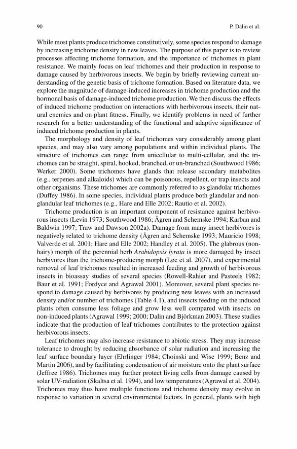
90 P. Dalin et al.
While most plants produce trichomes constitutively, some species respond to damageby increasing trichome density in new leaves. The purpose of this paper is to reviewprocesses affecting trichome formation, and the importance of trichomes in plantresistance. We mainly focus on leaf trichomes and their production in response todamage caused by herbivorous insects. We begin by briefly reviewing current un-derstanding of the genetic basis of trichome formation. Based on literature data, weexplore the magnitude of damage-induced increases in trichome production and thehormonal basis of damage-induced trichome production. We then discuss the effectsof induced trichome production on interactions with herbivorous insects, their nat-ural enemies and on plant fitness. Finally, we identify problems in need of furtherresearch for a better understanding of the functional and adaptive significance ofinduced trichome production in plants.
The morphology and density of leaf trichomes vary considerably among plantspecies, and may also vary among populations and within individual plants. Thestructure of trichomes can range from unicellular to multi-cellular, and the tri-chomes can be straight, spiral, hooked, branched, or un-branched (Southwood 1986;Werker 2000). Some trichomes have glands that release secondary metabolites(e.g., terpenes and alkaloids) which can be poisonous, repellent, or trap insects andother organisms. These trichomes are commonly referred to as glandular trichomes(Duffey 1986). In some species, individual plants produce both glandular and non-glandular leaf trichomes (e.g., Hare and Elle 2002; Rautio et al. 2002).
Trichome production is an important component of resistance against herbivo-rous insects (Levin 1973; Southwood 1986; Agren and Schemske 1994; Karban andBaldwin 1997; Traw and Dawson 2002a). Damage from many insect herbivores isnegatively related to trichome density (Agren and Schemske 1993; Mauricio 1998;Valverde et al. 2001; Hare and Elle 2002; Handley et al. 2005). The glabrous (non-hairy) morph of the perennial herb Arabidopsis lyrata is more damaged by insectherbivores than the trichome-producing morph (Løe et al. 2007), and experimentalremoval of leaf trichomes resulted in increased feeding and growth of herbivorousinsects in bioassay studies of several species (Rowell-Rahier and Pasteels 1982;Baur et al. 1991; Fordyce and Agrawal 2001). Moreover, several plant species re-spond to damage caused by herbivores by producing new leaves with an increaseddensity and/or number of trichomes (Table 4.1), and insects feeding on the inducedplants often consume less foliage and grow less well compared with insects onnon-induced plants (Agrawal 1999; 2000; Dalin and Bjorkman 2003). These studiesindicate that the production of leaf trichomes contributes to the protection againstherbivorous insects.
Leaf trichomes may also increase resistance to abiotic stress. They may increasetolerance to drought by reducing absorbance of solar radiation and increasing theleaf surface boundary layer (Ehrlinger 1984; Choinski and Wise 1999; Benz andMartin 2006), and by facilitating condensation of air moisture onto the plant surface(Jeffree 1986). Trichomes may further protect living cells from damage caused bysolar UV-radiation (Skaltsa et al. 1994), and low temperatures (Agrawal et al. 2004).Trichomes may thus have multiple functions and trichome density may evolve inresponse to variation in several environmental factors. In general, plants with high
4 Leaf Trichome Formation and Plant Resistance to Herbivory 91
Tab
le4.
1In
duce
din
crea
ses
intr
icho
me
dens
ity
reco
rded
afte
rar
tific
ialw
ound
ing
and
dam
age
byin
sect
herb
ivor
es
Plan
tSpe
cies
(lif
ehi
stor
y)1
Def
olia
tion
trea
tmen
tIn
crea
sein
tric
hom
ede
nsit
yD
amag
eE
nvir
onm
ent
Am
ount
Tis
sue
dam
aged
Mag
nitu
deT
imin
gR
efer
ence
Ara
bido
psis
thal
iana
(a)
Art
ifici
alL
abPi
nchi
ngL
eave
s25
%–1
17%
wee
ksT
raw
and
Ber
gels
on(2
003)
Aln
usin
cana
(p)
Col
eopt
era
Fiel
d30
%–1
00%
Lea
ves
500%
2%–4
wee
ksB
aur
etal
.(19
91)
Bra
ssic
ani
gra
(a)
Lep
idop
tera
Lab
25%
Lea
ves
76%
wee
ksT
raw
and
Daw
son
(200
2a)
Bra
ssic
ani
gra
(a)
Lep
idop
tera
Lab
25%
Lea
ves
113%
wee
ksT
raw
and
Daw
son
(200
2a)
Bra
ssic
ani
gra
(a)
Col
eopt
era
Lab
17%
Lea
ves
noin
duct
ion
wee
ksT
raw
and
Daw
son
(200
2a)
Bra
ssic
ani
gra
(a)
Lep
idop
tera
Lab
25%
Lea
ves
43%
4%–5
wee
ksT
raw
and
Daw
son
(200
2b)
Bra
ssic
ani
gra
(a)
Col
eopt
era
Lab
25%
Lea
ves
noin
duct
ion
4%–5
wee
ksT
raw
and
Daw
son
(200
2b)
Bra
ssic
ani
gra
(a)
Lep
idop
tera
Lab
22%
Lea
ves
38%
2%–3
wee
ksT
raw
2002
Bet
ula
pube
scen
s(p
)A
rtifi
cial
Fiel
d50
%L
eave
s30
0%2%
–7w
eeks
(199
8)R
auti
oet
al.(
2002
)
Bet
ula
pube
scen
s(p
)A
rtifi
cial
Fiel
d10
0%L
eave
s10
00%
2%–7
wee
ks(1
998)
Rau
tio
etal
.(20
02)
Bet
ula
pube
scen
s(p
)A
rtifi
cial
Fiel
d50
%L
eave
sno
indu
ctio
n2%
–7w
eeks
(199
9)R
auti
oet
al.(
2002
)
Bet
ula
pube
scen
s(p
)A
rtifi
cial
Fiel
d10
0%L
eave
sno
indu
ctio
n2%
–7w
eeks
(199
9)R
auti
oet
al.(
2002
)
Bet
ula
pube
scen
s(p
)A
rtifi
cial
Fiel
d≤
25%
Lea
ves
10%
–20%
next
year
Val
kam
aet
al.(
2005
)B
etul
apu
besc
ens
(p)
Art
ifici
alFi
eld
10cm
Shoo
tsno
indu
ctio
nne
xtye
arV
alka
ma
etal
.(20
05)

92 P. Dalin et al.
Tab
le4.
1(c
onti
nued
)
Def
olia
tion
trea
tmen
tIn
crea
sein
tric
hom
ede
nsit
yD
amag
eE
nvir
onm
ent
Am
ount
Tis
sue
dam
aged
Mag
nitu
deT
imin
gR
efer
ence
Cni
dosc
olus
texa
nus
(p)
Liv
esto
ckFi
eld
noin
fo.
Shoo
ts38
%no
info
.Po
llar
d(1
986)
Lep
idiu
mvi
rgin
icum
(a)
Lep
idop
tera
Lab
20%
–25%
Lea
ves
60%
2w
eeks
Agr
awal
(200
0)R
apha
nus
raph
anis
trum
(a)
Lep
idop
tera
Lab
50%
Lea
ves
25%
–100
%3
wee
ksA
graw
al(1
999)
Sali
xbo
real
is(p
)C
oleo
pter
aFi
eld
100%
Lea
ves
51%
–300
%ne
xtye
arZ
vere
vaet
al.(
1998
)Sa
lix
cine
rea
(p)
Col
eopt
era
Lab
5%L
eave
s72
%3
wee
ksD
alin
and
Bjo
rkm
an(2
003)
Sali
xci
nere
a(p
)A
rtifi
cial
Lab
5%L
eave
sno
indu
ctio
n3
wee
ksD
alin
and
Bjo
rkm
an(2
003)
Sali
xci
nere
a(p
)C
oleo
pter
aL
ab10
%L
eave
s93
%3
wee
ksB
jork
man
and
Ahr
ne(2
005)
Sali
xci
nere
a(p
)C
oleo
pter
aL
ab3%
Lea
ves
83%
–186
%3
wee
ksB
jork
man
etal
.(2
008)
Sali
xvi
min
alis
(p)
Col
eopt
era
Lab
5%–2
5%L
eave
s13
%3
wee
ksD
alin
etal
.(20
04)
Urt
ica
dioi
ca(p
)A
rtifi
cial
Fiel
d>
50%
Shoo
ts14
0%–7
90%
10w
eeks
Pull
inan
dG
ilbe
rt(1
989)
Urt
ica
dioi
ca(p
)A
rtifi
cial
Lab
>50
%Sh
oots
23%
–106
%10
wee
ksPu
llin
and
Gil
bert
1989
Urt
ica
dioi
cadi
oica
(p)
Art
ifici
alL
ab50
%L
eave
s26
%5–
7w
eeks
Mut
ikai
nen
and
Wal
ls(1
995)
Urt
ica
dioi
cadi
oica
(p)
Art
ifici
alL
abN
oin
fo.
Api
calb
uds
noin
duct
ion
5–7
wee
ksM
utik
aine
nan
dW
alls
(199
5)U
rtic
adi
oica
sond
enii
(p)
Art
ifici
alL
ab50
%L
eave
sno
indu
ctio
n5–
7w
eeks
Mut
ikai
nen
and
Wal
ls(1
995)
Urt
ica
dioi
caso
nden
ii(p
)A
rtifi
cial
Lab
No
info
.A
pica
lbud
s50
%5–
7w
eeks
Mut
ikai
nen
and
Wal
ls(1
995)
1T
heli
fehi
stor
yof
the
diff
eren
tpla
ntsp
ecie
sis
indi
cate
das
(p)
for
pere
nnia
l,or
(a)
for
annu
al

4 Leaf Trichome Formation and Plant Resistance to Herbivory 93
density of leaf trichomes can be expected in environments that are dry or cold, whereUV-radiation is intense, and in areas where the risk of being damaged by herbivorousinsects is high (Ehrlinger 1984; Løe et al. 2007).
4.2 Leaf Trichome Formation
4.2.1 Molecular Basis of Leaf Trichome Formation
Trichomes are initiated in the epidermis of developing leaves: some epidermal cellswill develop into trichome cells whereas surrounding cells will develop into regularepidermal cells. Present understanding of trichome formation is based on studiesof a few model species. In particular, trichome formation in A. thaliana has beenused as a model system to study various developmental and cellular mechanisms inplants. In Arabidopsis, trichomes are non-glandular, large single cells, surroundedby accessory epidermal cells on rosette leaves, stems, cauline leaves, and sepals.With mutagenesis screens, dozens of genes involved in trichome initiation, spac-ing, and shape have been identified in Arabidopsis (Marks 1997; Hulskamp andSchnittger 1998). Recent studies of A. thaliana and its close relatives have shownthat variation in some of the same genes can explain differences in trichome forma-tion in natural populations (Hauser et al. 2001; Kivimaki et al. 2007).
Studies of the genetic basis of trichome initiation have identified genes that (a)control the entry into the trichome pathway and (b) control the spacing of ini-tiation events. Loss-of-function mutations in A. thaliana have suggested that thegenes GLABROUS1 (GL1) and TRANSPARENT TESTA GLABRA1 (TTG1) are im-portant for both the initiation and spacing of leaf trichomes. GL1 encodes a myb-transcription factor that is expressed diffusively in developing leaves (Oppenheimeret al. 1991). TTG1 is expressed in most major plant organs, codes for a WD-40repeat-containing protein, and is needed for trichome cell initiation in leaves, non-hair cell type (atrichoblast) formation in roots, anthocyanin biosynthesis in leaves,and formation of mucilage layer in germinating seeds (Koornneef 1981; Walkeret al. 1999). For normal trichome initiation, the genes GLABROUS3 (GL3) and EN-HANCER OF GLABRA3 (EGL3), which code for helix-loop-helix (bHLH) proteins,are also needed (Payne et al. 2000; Zhang et al. 2003). GL1 and TTG1 have the ca-pacity to limit trichome initiation together with other genes, e.g., CAPRICE (CPC)and TRIPTYCHON (TRY) that are known to promote root hair cell fate (Szymanskiet al. 2000; Schiefelbein 2003). The gene GLABROUS2 (GL2) quantitatively regu-lates the frequency of trichome initiation and is involved in determining trichomespacing and root hair development (Ohashi and Oka 2002), but is also involved inthe control of seed oil accumulation (Shen et al. 2006). Thus, many genes involvedin trichome initiation and spacing also regulate other important phenotypic traits,which may constrain the evolution of genes and traits.
Although current understanding of the genetic basis of trichome initiation andspacing depends to a large degree on studies of mutants in Arabidopsis, some
94 P. Dalin et al.
information is also available for other plant species. Trichome initiation in cottonseems to be regulated by a similar multimeric complex between MYB, bHLHand WD repeat proteins as in Arabidopsis (Wang et al. 2004). Sequence com-parisons and functional analyses suggest that genes underlying trichome forma-tion in Arabidopsis (and perhaps cotton) may have evolved from duplication andneo-functionalization of genes involved in anthocyanin and flavonoid production,whereas the genes involved in the formation of analogous multicellular trichomes inNicotiana and Antirrhinum have a different origin (Serna and Martin 2006). Thus,trichome production appears to have evolved several times suggesting convergentadaptive evolution.
4.2.2 Genetic Basis of Natural Variation in Trichome Production
Both trichome production as such (trichome production vs. glabrousness) andthe type of trichomes produced (glandular vs. non-glandular) may vary withinspecies. Many plant species are polymorphic for trichome production with trichome-producing and glabrous morphs. In several species, the frequency of the trichome-producing morph varies geographically, among and within habitats (Bjorkman andAnderson 1990; Westerberg and Saura 1992; Barnes and Han 1993; St Hilaire andGraves 1999; Arroyo-Garcıa et al. 2001; Morrison 2002; Karkkainen et al. 2004;Løe et al. 2007). Genetic analyses have shown that the inheritance of the glabrousmorph is simple in many species (one gene with a recessive allele for glabrous-ness; e.g., Westerbergh and Saura 1992; Sharma and Waines 1994; Karkkainenand Agren 2002). In A. lyrata, association analysis of phenotypic variation andgenotypic variation in candidate genes revealed that the polymorphism in trichomeproduction was caused by variation in GLABROUS1 (Kivimaki et al. 2007), andsequence analysis suggested that several independent mutations in the same genecause glabrousness in different natural populations of A. lyrata and A. thaliana(Kivimaki et al. 2007; Hauser et al. 2001). Polymorphism in trichome morphology(glandular vs. non-glandular trichomes) can also be inherited in a simple Mendelianfashion, as documented for Datura wrightii (van Dam et al. 1999).
The number of trichomes produced and trichome density vary genetically withinseveral species. Quantitative genetic studies have indicated ample genetic vari-ation for trichome number and trichome density (Agren and Schemske 1994;Mauricio and Rausher 1997; Roy et al. 1999; Clauss et al. 2006). QTL-studies havebeen conducted to reveal the genetic basis of among-population differences in tri-chome formation. In A. thaliana, a QTL-analysis of a cross between two accessionsidentified a major QTL for trichome number (REDUCED TRICHOME NUMBER,RTN) that explained 70% of the variation in number of trichomes produced (Larkinet al. 1996). More recent studies of crosses among other accessions of A. thalianadetected additional QTLs (altogether nine QTLs in four different crosses, most ofwhich were shared among populations; Symonds et al. 2005). Preliminary resultsfrom QTL-crosses and within-population studies in A. lyrata suggest that a limited

4 Leaf Trichome Formation and Plant Resistance to Herbivory 95
number of loci contribute to variation in the number of trichomes produced, and thatsome genes segregate in several populations also in this species (Karkkainen et al.unpublished data).
4.3 Induced Trichome Production
4.3.1 Damage-Induced Trichome Production
Both artificial wounding and damage by herbivores can induce an increase intrichome density, and induction of trichome production has been observed in bothannual and perennial plants (Table 4.1). The magnitude of the reported increase intrichome density is typically between 25% and 100%, but in some cases as large as500%–1000% (Table 4.1). The responses often involve changes in trichome densityexpressed within days or weeks (Baur et al. 1991; Agrawal 1999, 2000; Rautioet al. 2002; Dalin and Bjorkman 2003). In some woody perennials, the response isdelayed and not observed until the year following initial attack (Zvereva et al. 1998;Valkama et al. 2005). Damage can also induce a change in the relative proportionsof glandular and non-glandular trichomes (Rautio et al. 2002).
Abiotic stress, such as drought and UV-radiation, may influence trichome for-mation (Nagata et al. 1999; Hoglund and Larsson 2005), and abiotic conditionsmay modify damage-induced responses in trichome density. For example, Bjorkmanet al. (2008) showed that the increase in trichome production induced by leaf beetleherbivory in the willow Salix cinerea is stronger in the shade than under direct sun-light, suggesting that the plants invested more resources into trichome defense whengrowing in the shade. The results from Table 4.1 also show that damage-inducedincreases in trichome density tend to be stronger under field conditions than in thelab. All five studies, in which plants increased the density of leaf trichomes by morethan 200% following damage, were conducted in the field. However, empirical dataare still few, and additional studies of how abiotic conditions affect damage-inducedresponses in trichome formation are clearly needed.
Damage has not been observed to induce trichome production in all studies(Table 4.1). Only about half of the 15 independent studies listed in Table 4.1, inwhich the experimental treatment consisted of artificial wounding (e.g., clippingof leaves) reported an increased trichome density. Also, the effect of damage ontrichome production may vary depending on the identity of the herbivore; not all her-bivorous insects induce a change in trichome formation (Traw and Dawson 2002a,b). Artificial wounding differs from the damage caused by the feeding of insects inseveral ways, which may reduce the chances of detecting induced responses (Hartleyand Lawton 1987; Cipollini et al. 2003). Moreover, the feeding patterns of insectsvary, especially among feeding guilds (e.g., chewers, miners and borers), whichmay affect how plants respond to damage. Further studies are therefore needed toexamine the extent to which responses vary with damage type, and whether plantsmay benefit from responding differently to different kinds of damage.
96 P. Dalin et al.
4.3.2 Hormonal Regulation of Induced Trichome Production
Recent work on Arabidopsis indicates that several regulatory networks may influencedamage-induced increases in trichome production. Jasmonic acid regulates the sys-temic expression of chemical defenses (Karban and Baldwin 1997; Schaller andStintzi this volume), and herbivore damage as well as artificial wounding cause rapidincreases in jasmonic acid (Bostock 1999; Reymond et al. 2000). In A. thaliana,artificial damage, but also application of jasmonic acid and application of giberellinincreases trichome production in new leaves (Traw and Bergelson 2003). Appli-cation of salicylic acid, on the other hand, reduces trichome production and in-hibits the response to jasmonic acid, which is consistent with negative cross-talkbetween the jasmonate- and salicylate-dependent pathways in Arabidopsis (Trawand Bergelson 2003). Concentrations of salicylic acid typically increase in responseto infection by biotrophic pathogens (Gaffney et al. 1993; Ryals et al. 1994), but mayalso increase in response to damage caused by some herbivores (Stotz et al. 2002;van Poecke and Dicke 2002). This suggests that induction of increased trichomeproduction will be affected by interactions with both herbivores and pathogens, andshould vary depending on the identity of the herbivore.
4.4 Leaf Trichomes and Plant Resistance
4.4.1 Effects on Herbivore Behavior and Performance
Trichomes influence insect oviposition and/or feeding in a wide range of insectsand other herbivores (Levin 1973). Non-glandular trichomes mainly function as astructural defense against small herbivores. They interfere with the movement ofinsects and other small arthropods over the plant surface and make it more difficultfor insects to access the leaf epidermis underneath for feeding (Southwood 1986).Trichomes are often composed of cellulose and other substances that constitute lownutritional value for the insects (Levin 1973). Insects that need to feed throughthe trichomes before accessing the leaf epidermis might therefore gain less weightand, ultimately, show increased mortality. Glandular trichomes can be viewed asa combination of a structural and chemical defense. This is because the glandulartrichomes release secondary metabolites that can be poisonous or repellent to her-bivorous organisms. Stinging trichomes, such as those produced by stinging nettles(Urtica dioica), may deter even large herbivores (Pollard and Briggs 1984).
The presence and density of leaf trichomes can influence both host-plant selec-tion behavior and performance (i.e., growth, survival, and fecundity) of herbivorousinsects, but can be considered a relatively soft ‘weapon’ in plant defense comparedwith many other traits that are lethal to insects. It has been argued that weak de-fense traits might influence a wide range of herbivorous insects because they areless likely to result in counter-adaptations in insects (Feeny 1976). For example,there are a number of studies suggesting that leaf trichomes reduce the feedingof both generalist and specialist insects (Traw and Dawson 2002a; Agrawal 1999,2004). Agrawal (1999) showed that two lepidopteran species, one specialist and

4 Leaf Trichome Formation and Plant Resistance to Herbivory 97
one generalist, were both negatively influenced by previous damage on wild radishplants, which was correlated with an increased density and number of trichomes onthe leaves. Agrawal (2004) also showed that several insect feeding guilds, both leafchewers and leaf miners, were negatively affected by an increased density of leaftrichomes on milkweed (Asclepias syriaca). Thus, non-glandular trichomes mightinfluence the feeding by a wide range of insects and therefore reduce total damagecaused by herbivores.
Insects may evolve physiological or behavioral traits that allow them to cope withstructural plant defenses. For example, several mirid bugs (Heteroptera: Miridae)have special structures on their legs, which facilitate movement across trichome-covered plant surfaces (Southwood 1986; van Dam and Hare 1998). The leaf beetlePhratora vulgatissima has been observed to remove non-glandular trichomes whenfeeding on leaves of the willow Salix viminalis (Dalin et al. 2004). This behaviorwas not observed when the same beetle species was feeding on S. cinerea, whichproduces shorter and broader trichomes that appeared more difficult for the larvae tohandle (Dalin and Bjorkman 2003). The evolution of mouthparts strong enough tohandle structural plant traits is thus one possible mechanism for insects to circum-vent structural plant defenses such as leaf trichomes (Levin 1973; Raupp 1985).
4.4.2 Effects on Tritrophic Interactions
Leaf trichomes do not only affect herbivores, but also their natural enemies. Thismay indirectly affect the intensity of damage caused by herbivores. In theory, theeffect on the abundance and effectiveness of natural enemies may be neutral (noeffect), negative or positive (Fig. 4.1). If we assume that herbivores are affectednegatively by leaf trichomes (Fig 4.1; top graph), a neutral effect on the third trophiclevel will result in natural enemies having an additive effect on plant damage causedby herbivores and on plant fitness. A negative effect on the third trophic level willact antagonistically. Depending on the relative strength of the negative effects ofleaf trichomes on herbivores and natural enemies the correlation between trichomedensity and damage may vary from negative to positive. Plant damage will be (i)negatively correlated with trichome density if the effect on natural enemies is weakerthan that on herbivores (dotted line) (ii) uncorrelated with trichome density if theeffect on the natural enemies and herbivores balance (solid line), or (iii) positivelycorrelated with trichome density if the effect on natural enemies is stronger thanthat on the herbivores (dashed line). A positive effect of leaf trichomes on the thirdtrophic level should result in a synergistic positive effect on plant fitness. From aplant protection perspective, this last scenario would be most desirable, but thereseem to be few such examples.
In most empirical studies, the abundance and effectiveness of natural enemieswere found to be negatively correlated with the density of plant trichomes (Kauffmanand Kennedy 1989; Farrar and Kennedy 1991; Nihoul 1993; Gange 1995; Heinz andZalom 1996; Romeis et al. 1998, 1999; Krips et al. 1999; Rosenheim et al. 1999;Lovinger et al. 2000; Fordyce and Agrawal 2001; Stavrinides and Skirvin 2003;Mulatu et al. 2006; Olson and Andow 2006). The effectiveness of both predators

98 P. Dalin et al.
Plant trichome density
Herbivore performanceor density without
natural enemies
Effect onnatural enemies
Plant damage
Neutral Negative Positive
Additive Antagonistic Synergistic
Fig. 4.1 Simplified graphical presentation of how the effect of leaf trichome density on theabundance or effectiveness of predators and parasitoids of herbivores may influence the relation-ship between trichome density and plant damage caused by herbivores (bottom row of graphs). Thenatural enemies may not be affected, or may be negatively or positively affected by plant trichomedensity (middle row of graphs). A basic assumption is that plant trichome density negatively affectsherbivore performance or density (top graph), i.e., trichomes have the potential to function as aplant resistance trait (cf. Hare 1992)
(in 9 out of 13 reviewed studies) and parasitoids (in 12 out of 14 reviewed studies) isaffected negatively because movement is inhibited and searching time is prolonged.Both non-glandular and glandular plant trichomes can have these effects. Glandulartrichomes, in addition, affect natural enemies negatively by (a) the release of repel-lent or toxic compounds and (b) being sticky and hindering movements. However,the effect of the sticky substances produced by glandular trichomes may sometimesbe of little importance in the field as the adhesive effect of the trichome exudates canbe negatively affected by dust, wind, and rain (Obrycki and Tauber 1984; Romeiset al. 1999).
In a few cases, the abundance or effectiveness of natural enemies were found tobe positively correlated with the density of plant trichomes. For example, Styrskyet al. (2006) recently showed that fire ant predation of herbivores on soybean washigher on isolines of soybean with high trichome density. The suggested mecha-nism was a functional response by the ants to a higher abundance of caterpillarson pubescent plants. Apple trees producing high density of trichomes had a higherabundance of predatory mites than apple trees with a low density of trichomes, and ithas been suggested that this was because pubescent plants capture more pollen andfungal spores that serve as alternative food for the predators (Roda et al. 2003).Lucas and Brodeur (1999) showed that within potato plants, an aphidophagousmidge was more abundant on leaves with a high trichome density. The authors
4 Leaf Trichome Formation and Plant Resistance to Herbivory 99
suggested that both a decreased risk of intraguild predation by e.g., coccinelids and ahigher density of food (i.e., aphids) on leaves with a high trichome density may havecontributed to this pattern. Finally, leaf trichome exudates may serve as extrafloralnectar for a scelonid egg parasitoid of squash bugs (Olson and Nechols 1995). Adultfemales of the parasitoid lived longer when they had access to squash leaves thanwhen only provided with water, and this increase in longevity was enough to allowmaximum realized fecundity.
In even fewer cases, the abundance or effectiveness of parasitoids and preda-tors were found not to be correlated with leaf trichome density (e.g., Sutterlin andvan Lenteren 1997; Bjorkman and Ahrne 2005; see also reviews by Kennedy 2003;Simmons and Gurr 2005), possibly because of a reluctance to report ‘negative’ data.
The density of prey or hosts can affect the impact of leaf trichomes on predatorsand parasitoids, but relationships appear system-specific. In some studies, an effectof trichomes on natural enemies was detectable only at high prey/host densities (e.g.,Sutterlin and van Lenteren 2000) while in other studies such an effect was observedonly at low densities (Krips et al. 1999).
4.4.3 Adaptive Significance of Induced Trichome Productionin Plants
The adaptive significance of damage-induced increases in trichome productionshould depend on the benefits and costs associated with this change in pheno-type. Plants induced to increase leaf trichome density often receive less damageby insects than non-induced plants (Baur et al. 1991; Agrawal 1999; Dalin andBjorkman 2003). This suggests that plants should benefit from induced trichomeproduction as long as the fitness costs associated with increased trichome produc-tion do not exceed the benefits of reduced damage. Agrawal (1999) showed thatinduced wild radish plants (Raphanus raphanistrum and Raphanus sativus) receivedless damage by a variety of insects, and produced more seeds and fruits than non-induced plants. Wild radish responded to herbivory by producing new leaves withincreased trichome density, but also with a systemic increase in the concentrationsof secondary metabolites (Agrawal 1999). Thus, although this study clearly showsthat the plants benefited from the induced responses, additional studies are neededto distinguish the effects of increased trichome production on fitness from those ofother induced differences in plant quality in this and other species.
Apart from reducing overall damage, induced trichome production may divertdamage away from the most valuable parts of the plant and cause insects to feed onother, less valuable leaves, or other non-induced plant individuals nearby. Becauseplants cannot change the density of trichomes on already existing leaves, an inducedincrease in trichome production can only be expressed in leaves developing duringor subsequent to attack (Traw and Bergelson 2003). Induced increases in trichomeproduction can however be relatively rapid and expressed in new leaves withindays or weeks after the initial attack (Baur et al. 1991; Agrawal 1999; Dalin andBjorkman 2003). A similarly rapid reduction in trichome production in new leaves
100 P. Dalin et al.
when damage ceases indicates that trichomes are costly to produce (Bjorkman et al.2008). Additional evidence for costs of trichome production is provided by exam-ples of trade-offs between trichome density and other putative defense traits, suchas thorns (Bjorkman and Anderson 1990). A damage-induced increase in trichomeproduction can lead to variation in trichome density within plants with older, basalleaves having lower trichome density than younger, induced apical leaves. Larvaeof the leaf beetle P. vulgatissima showed a more dispersed feeding behavior amonginduced willow leaves with high trichome density at the top of shoots, indicatingthat the insects were searching for more optimal food within the branches (Dalin andBjorkman 2003). Such damage-induced within-branch variation in trichome densitymay reflect developmental constraints, but may also be advantageous since damageto old basal leaves is likely to be less detrimental to plant fitness than damage to theyoung apical leaves (cf. Feeny 1976).
4.5 Perspectives
Several plant species respond to herbivory by producing leaves with increased den-sity and number of trichomes. The apparent benefit of this response is to reduce fur-ther damage by making it more difficult for insects to feed on the leaves. However,the magnitude of induced trichome production may vary with habitat conditions, andincreased trichome density may affect predators and parasitoids negatively, whichcould reduce the effect on leaf damage. Moreover, trichome production may alsoaffect tolerance to drought and other forms of abiotic stress. Taken together, thissuggests that an analysis of the adaptive significance of induced trichome produc-tion requires a multidimensional view of the realized fitness (see also chapter bySteppuhn and Baldwin this volume). The effects of induced trichome productionon interactions with herbivores should not be examined in isolation, but has to beconsidered in the context of effects on tritrophic interactions, and the extent to whichfitness costs and benefits vary with abiotic conditions in natural populations. Moreinformation is also needed on the genetic and hormonal basis of induced trichomeproduction. Such knowledge could be important for the development of sustainablepest control methods, using induced plant responses as a tool to prevent insect pestdamage in agricultural systems.
References
Agrawal AA (1999) Induced responses to herbivory in wild radish: effects on several herbivoresand plant fitness. Ecology 80:1713–1723
Agrawal AA (2000) Specificity of induced resistance in wild radish: causes and consequences fortwo specialist and two generalist caterpillars. Oikos 89:493–500
Agrawal AA (2004) Resistance and susceptibility of milkweed: competition, root herbivory, andplant genetic variation. Ecology 85:2118–2133
Agrawal AA, Conner JK, Stinchcombe JR (2004) Evolution of plant resistance and tolerance tofrost damage. Ecol Lett 7:1199–1208

4 Leaf Trichome Formation and Plant Resistance to Herbivory 101
Agren J, Schemske DW (1993) The cost of defense against herbivores: an experimental study oftrichome production in Brassica rapa. Am Nat 141:338–350
Agren J, Schemske DW (1994) Evolution of trichome number in a naturalized population of Bras-sica rapa. Am Nat 143:1–13
Arroyo-Garcıa R, Martınez-Zapater JM, Fernandez Prieto JA, Alvarez-Arbesu R (2001) AFLPevaluation of genetic similarity among laurel populations (Laurus L.). Euphytica 122:155–164
Barnes BV, Han FQ (1993) Phenotypic variation of Chinese aspens and their relationships tosimilar taxa in Europe and North America. Can J Botany 71:799–815
Baur R, Binder S, Benz G (1991) Non glandular leaf trichomes as short-term inducible defence ofthe grey alder, Alnus incana (L.), against the chrysomelid beetle, Agelastica alni L. Oecologia87:219–226
Benz BW, Martin CE (2006) Foliar trichomes, boundary layers, and gas exchange in the species ofepiphytic Tillandsia (Bromeliaceae). J Plant Physiol 163:648–656
Bjorkman C, Ahrne K (2005) Influence of leaf trichome density on the efficiency of twopolyphagous insect predators. Entomol Exp Appl 115:179–186
Bjorkman C, Anderson DB (1990) Trade-off among antiherbivore defences in a South Americanblackberry (Rubus bogotensis). Oecologia 85:247–249
Bostock RM (1999) Signal conflicts and synergies in induced resistance to multiple attackers.Physiol Mol Plant Pathol 55:99–109
Choinski JS, Wise RR (1999) Leaf growth and development in relation to gas exchange in Quercusmarilandica Muenchh. J Plant Physiol 154:302–309
Cipollini D, Purrington CB, Bergelson J (2003) Costs of induced responses in plants. Basic ApplEcol 4:79–85
Clauss MJ, Dietel S, Schubert G, Michell-Olds T (2006) Glucosinolate and trichome defenses in anatural Arabidopsis lyrata population. J Chem Ecol 32:2351–2373
Dalin P, Bjorkman C (2003) Adult beetle grazing induces willow trichome defense againstsubsequent larval feeding. Oecologia 134:112–118
Dalin P, Bjorkman C, Eklund K (2004) Leaf beetle grazing does not induce willow trichomedefence in the coppicing willow Salix viminalis. Agric For Entomol 6:105–109
Duffey SS (1986) Plant glandular trichomes: their partial role in defence against insects. In: JuniperB, Southwood SR (eds) Insects and the plant surface, Arnold, London, pp 151–172
Ehrlinger J (1984) Ecology and physiology of leaf pubescence in North American desert plants.In: Rodriguez E, Healey PL, Mehta I (eds) Biology and chemistry of plant trichomes. PlenumPress, New York, pp 113–132
Farrar RR, Kennedy G (1991) Inhibition of Telenomus sphingis an egg parasitoid of Manducaspp by trichome 2 tridecanone-based host plant resistance in tomato. Entomol Exp Appl60:157–166
Feeny PP (1976) Plant apparency and chemical defense. In: Wallace JM, Mansell RL (eds)Biochemical interaction between plants and insects. Plenum Press, New York, pp 1–40
Fordyce JA, Agrawal AA (2001) The role of plant trichomes and caterpillar group size on growthand defence of the pipevine swallowtail Battus philenor. J Anim Ecol 70:997–1005
Gaffney T, Frierich L, Vernooij B, Negrotto D, Nye G, Uknes S, Ward E, Kessman H, Ryals J(1993) Requirement of salicylic acid for the induction of systemic acquired resistance. Science261:754–756
Gange AC (1995) Aphid performance in an alder (Alnus) hybrid zone. Ecology 76:2074–2083Handley R, Ekbom B, Agren J (2005) Variation in trichome density and resistance against
a specialist insect herbivore in natural populations of Arabidopsis thaliana. Ecol Entomol30:284–292
Hare JD (1992) Effects of plant variation on herbivore-natural enemy interactions. In: Fritz RS,Simms EL (eds) Plant resistance to herbivores and pathogens: ecology, evolution and genetics.University of Chicago Press, Chicago, pp 278–298
Hare JD, Elle E (2002) Variable impact of diverse insect herbivores on dimorphic Datura wrightii.Ecology 83:2711–2720

102 P. Dalin et al.
Hartley SE, Lawton JH (1987) Effects of different types of damage on the chemistry of birchfoliage. Oecologia 74:432–437
Hauser M-T, Harr B, Schlotterer C (2001) Trichome distribution in Arabidopsis thaliana and itsclose relative Arabidopsis lyrata: molecular analysis of the candidate gene GLABROUS1. MolBiol Evol 18:1754–1763
Heinz KM, Zalom FG (1996) Performance of the predator Delphastus pusillus on Bemisia resistantand susceptible tomato lines. Entomol Exp Appl 81:345–352
Hoglund S, Larsson S (2005) Abiotic induction of susceptibility in insect-resistant willow. EntomolExp Appl 115:89–96
Hulskamp M, Schnittger A (1998) Spatial regulation of trichome formation Arabidopsis thaliana.Semin Cell Dev Biol 9:213–220
Jeffree CE (1986) The cuticle, epicuticular waxes and trichomes of plants, with reference to theirstructure, function and evolution. In: Juniper B, Southwood SR (eds) Insects and the plantsurface. Arnold, London, pp 23–64
Karban R, Baldwin IT (1997) Induced responses to herbivory. The University of Chicago Press,Chicago, p 319
Karkkainen K, Agren J (2002) Genetic basis of trichome production in Arabidopsis lyrata.Hereditas 136:219–226
Karkkainen K, Løe G, Agren J (2004) Population structure in Arabidopsis lyrata: evidence fordivergent selection on trichome production. Evolution 58:2831–2836
Kauffman WC, Kennedy GG (1989) Relationship between trichome density in tomato and para-sitism of Heliothis spp. (Lepidoptera, Noctuidae) eggs by Trichogramma spp (Hymenoptera,Trichogrammatidae). Environ Entomol 18:698–704
Kennedy GG (2003) Tomato, pests, parasitoids, and predators: tritrophic interactions involving thegenus Lycopersicon. Ann Rev Entomol 48:51–72
Kivimaki M, Karkkainen K, Gaudeul M, Løe G, Agren J (2007) Gene, phenotype and function:GLABROUS1 and resistance to herbivory in natural populations of Arabidopsis lyrata. MolEcol 16:453–462
Koornneef M (1981) The complex syndrome of ttg mutants. Arabid Inf Serv 18:45–51Krips OE, Kleijn PW, Willems PEL, Gols GJZ, Dicke M (1999) Leaf hairs influence search-
ing efficiency and predation rate of the predatory mite Phytoseiulus persimilis (Acari:Phytoseiidae). Exp Appl Acarol 23:119–131
Larkin JC, Young N, Prigge M, Marks MD (1996) The control of trichome spacing and number inArabidopsis. Development 122:997–1005
Levin DA (1973) The role of trichomes in plant defence. Q Rev Biol 48:3–15Løe G, Torang P, Gaudeul M, Agren J (2007) Trichome production and spatiotemporal variation
in herbivory in the perennial herb Arabidopsis lyrata. Oikos 116:134–142Lovinger A, Liewehr D, Lamp WO (2000) Glandular trichomes on alfalfa impede searching
behavior of the potato leafhopper parasitoid. Biol Control 18:187–192Lucas E, Brodeur J (1999) Oviposition site selection by the predatory midge Aphidoletes
aphidimyza (Diptera: Cecidomyiidae). Environ Entomol 28:622–627Marks MD (1997) Molecular genetic analysis of trichome development in Arabidopsis. Annu Rev
Plant Physiol Plant Mol Biol 48:137–163Mauricio R (1998) Costs of resistance to natural enemies in field populations of the annual plant
Arabidopsis thaliana. Am Nat 151:20–28Mauricio R, Rausher MD (1997) Experimental manipulations of putative selective agents pro-
vides evidence for the role of natural enemies in the evolution of plant defense. Evolution51:1435–1444
Morrison LW (2002) The geographic distribution of pubescence in the sea daisy, Borrichiaaborescens, on Bahamian Islands. Glob Ecol Biogeogr 11:247–252
Mulatu B, Applebaum SW, Coll M (2006) Effect of tomato leaf traits on the potato tubermoth and its predominant larval parasitoid: a mechanism for enemy-free space. Biol Control37:231–236

4 Leaf Trichome Formation and Plant Resistance to Herbivory 103
Mutikainen P, Walls M (1995) Growth, reproduction and defence in nettles: responses to herbivorymodified by competition and fertilization. Oecologia 104:487–495
Nagata T, Todoriki S, Hayashi T, Shibata Y, Mori M, Kanegae H, Kikuchi S (1999)Gamma-radiation induces leaf trichome formation in Arabidopsis. Plant Physiol 120:113–119
Nihoul P (1993) Do light-intensity, temperature and photoperiod affect the entrapment of mites onglandular hairs of cultivated tomatoes. Exp Appl Acarol 17:709–718
Obrycki JJ, Tauber MJ (1984) Natural enemy activity on glandular pubescent potato plants in thegreenhouse: an unreliable predictor of effects in the field. Environ Entomol 13:679–683
Ohashi Y, Oka A (2002) Entopically additive expression of GLABRA2 alters the frequency andspacing of trichome initiation. Plant J 29:359–369
Olson DM, Andow DA (2006) Walking patterns of Trichogramma nubilale Ertle & Davis(Hymenoptera; Trichogrammatidae) on various surfaces. Biol Control 39:329–335
Olson DL, Nechols JR (1995) Effects of squash leaf trichome exudates and honey on adult feeding,survival, and fecundity of the squash bug (Heteroptera, Coreidae) egg parasitoid Gryon penn-sylvanicum (Hymenoptera, Scelionidae). Environ Entomol 24:454–458
Oppenheimer DG, Herman PL, Sivakumaran S, Eschm J, Marks MD (1991) A myb gene requiredfor leaf trichome differentiation in Arabidopsis is expressed in stipules. Cell 67:483–493
Payne CT, Zhang F, Lloyd AM (2000) GL3 encodes a bHLH protein that regulates trichomedevelopment in Arabidopsis through interaction with GL1 and TTG1. Genetics 156:1349–1362
Pollard AJ (1986) Variation in Cnidoscolus texanus in relation to herbivory. Oecologia 70:411–413Pollard AJ, Briggs D (1984) Genecological studies of Urtica dioica L. III. Stinging hairs and
plant-herbivore interactions. New Phytol 97:507–522Pullin AS, Gilbert JE (1989) The stinging nettle, Urtica dioica, increases trichome density after
herbivore and mechanical damage. Oikos 54:275–280Raupp MJ (1985) Effects of leaf toughness on mandibular wear of the leaf beetle, Plagiodera
versicolora. Ecol Entomol 10:73–79Rautio P, Markkola A, Martel J, Tuomi J, Harma E, Kuikka K, Siitonen A, Riesco IL, Roitto
M (2002) Developmental plasticity in birch leaves: defoliation causes shift from glandular tononglandular trichomes. Oikos 98:437–446
Reymond P, Weber H, Damond M, Farmer EE (2000) Differential gene expression in response tomechanical wounding and insect feeding in Arabidopsis. Plant Cell 12:707–719
Roda A, Nyrop J, English-Loeb G (2003) Pubescence mediates the abundance of non-prey foodand the density of the predatory mite Typhlodromus pyri. Exp Appl Acarol 29:193–211
Romeis J, Shanower TG, Zebitz CPW (1998) Physical and chemical plant characters inhibiting thesearching behaviour of Trichogramma chilonis. Entomol Exp Appl 87:275–284
Romeis J, Shanower TG, Zebitz CPW (1999) Trichogramma egg parasitism of Helicovarpaarmigera on pigonpea and sorghum in southern India. Entomol Exp Appl 90:69–81
Rosenheim JA, Limburg DD, Colfer RG (1999) Impact of generalist predators on a biologicalcontrol agent, Chrysoperla carnea: direct observations. Ecol Appl 9:409–417
Rowell-Rahier M, Pasteels JM (1982) The significance of salicin for a Salix-feeder, Phratora(Phyllodecta) vitellinae. In: Visser JH, Mink AK (eds) Proceedings from the 5th internationalsymposium on insect-plant relationships. Pudoc, Wageningen, pp 73–79
Roy BA, Stanton ML, Eppley SM (1999) Effects of environmental stress on leaf hair density andconsequences for selection. J Evol Biol 12:1089–1103
Ryals J, Uknes S, Ward E (1994) Systemic acquired resistance. Plant Physiol 104:1109–1112Schiefelbein J (2003) Cell-fate specification in the epidermis: a common patterning mechanism in
the root and shoot. Curr Opin Plant Biol 6:74–78Serna L, Martin C (2006) Trichomes: different regulatory networks lead to convergent structures.
Trends Plant Sci 11:1360–1385Sharma HC, Waines JG (1994) Inheritance of leaf pubescence in diploid wheat. J Hered
85:286–288Shen B, Sinkevicius KW, Selinger DA, Tarczynski MC (2006) The homeobox gene GLABRA2
affects seed oil content in Arabidopsis. Plant Mol Biol 60:377–387
104 P. Dalin et al.
Simmons AT, Gurr GM (2005) Trichomes of Lycopersicon species and their hybrids: effects onpests and natural enemies. Agric For Entomol 48:51–72
Skaltsa H, Verykokidou E, Harvala C, Krabourniotis G, Manetas Y (1994) UV-protective potentialand flavonoid content of leaf hairs of Quercus ilex. Phytochemistry 37:987–990
Southwood SR (1986) Plant surfaces and insects – an overview. In: Juniper B, Southwood SR (eds)Insects and the plant surface. Arnold, London, pp 1–22
Stavrinides MC, Skirvin DJ (2003) The effect of chrysanthemum leaf trichome density and preyspatial distribution on predation of Tetranychus urticae (Acari: Tetranychidae) by Phytoseiuluspersimilis (Acari: Phytoseiidae). Bull Entomol Res 93:343–350
St Hilaire R, Graves WR (1999) Foliar traits of sugar and black maples near 43 degrees N latitudein the eastern and central United States. J Am Soc Hort Sci 124:605–611
Stotz HU, Koch T, Biedermann A, Weniger K, Boland W, Mitchell-Olds T (2002) Evidence forregulation of resistance in Arabidopsis to Egyptian cotton worm by salicylic and jasmonic acidsignaling pathways. Planta 214:648–652
Styrsky JD, Kaplan I, Eubanks MD (2006) Plant trichomes indirectly enhance tritrophic interac-tions involving a generalist predator, the red imported fire ant. Biol Control 36:375–384
Sutterlin S, van Lenteren JC (1997) Influence of hairiness of Gerbera jamesonii leave on thesearching efficiency of the parasitoid Encarsia formosa. Biol Control 9:157–165
Sutterlin S, van Lenteren JC (2000) Pre- and post-landing response of the parasitoid Encarsiaformosa to whitefly hosts on Gerbera jamesonii. Entomol Exp Appl 96:299–307
Symonds VV, Godoy AV, Alconada T, Botto JF, Juenger TE, Casal JJ, Lloyd AM (2005) Mappingquantitative trait loci in multiple populations of Arabidopsis thaliana identifies natural allelicvariation for trichome density. Genetics 169:1649–1658
Szymanski DB, Lloyd AM, Marks MD (2000) Progress in the molecular genetic analysis oftrichome initiation and morphogenesis in Arabidopsis. Trends Plant Sci 5:214–219
Traw BM (2002) Is induction response negatively correlated with constitutive resistance in blackmustard? Evolution 56:2116–2205
Traw BM, Bergelson J (2003) Interactive effects of jasmonic acid, salicylic acid, and gibberellinon induction of trichomes in Arabidopsis. Plant Physiol 133:1367–1375
Traw BM, Dawson TE (2002a) Reduced performance of two specialist herbivores (Lepidoptera:Pieridae, Coleoptera: Chrysomelidae) on new leaves of damaged black mustard plants. EnvironEntomol 31:714–722
Traw BM, Dawson TE (2002b) Differential induction of trichomes by three herbivores of blackmustard. Oecologia 131:526–532
Valkama E, Koricheva J, Ossipov V, Ossipova S, Haukioja E, Pihlaja K (2005) Delayed inducedresponses of birch glandular trichomes and leaf surface lipophilic compounds to mechanicaldefoliation and simulated winter browsing. Oecologia 146:385–393
Valverde PL. Fornoni J, Nunez-Farfan J (2001) Defensive role of leaf trichomes in resistance toherbivorous insects in Datura stramonium. J Evol Biol 14:424–432
Walker AR, Davidson PA, Bolognesi-Winfield AJ, James CM, Srinivasan N, Blundell TL, Esch JJ,Marks MD, Gray JC (1999) The TRANSPARENT TEST GLABRA1 locus, which regulatestrichome differentiation and anthocyanin biosynthesis in Arabidopsis, encodes a WD40 repeatprotein. Plant Cell 11:1337–1349
van Dam NM, Hare JD (1998) Differences in distribution and performance of two sap-suckingherbivores on glandular and non-glandular Datura wrightii. Ecol Entomol 23:22–32
van Dam NM, Hare JD, Elle E (1999) Inheritance and distribution of trichome phenotypes inDatura wrightii. J Hered 90:220–227
van Poecke RMP, Dicke M (2002) Induced parasitoid attraction by Arabidopsis thaliana:involvement of the octadecanoid and the salicylic acid pathway. J Exp Biol 53:1793–1799
Wang S, Wang JW, Yu N, Li CH, Luo B, Gou JY, Wang LJ, Chen XY (2004) Control of planttrichome development by a cotton fiber MYB gene. Plant Cell 16:2323–2334
Werker E (2000) Trichome diversity and development. Adv Bot Res 31:1–35Westerberg A, Saura A (1992) The effect of serpentine and the population structure of Silene
dioica. Evolution 46:1537–1548

4 Leaf Trichome Formation and Plant Resistance to Herbivory 105
Zhang F, Gonzales A, Zhao MZ, Payne CT, Lloyd A (2003) A network of redundant bHLH proteinsfunctions in all TTG1-dependent pathways of Arabidopsis. Development 130:4859–4869
Zvereva EL, Kozlov MV, Niemela P (1998) Effects of leaf pubescence in Salix borealis onhost-plant choice and feeding behaviour of the leaf beetle, Melasoma lapponica. Entomol ExpAppl 89:297–303

Chapter 5Resistance at the Plant Cuticle
Caroline Muller
Resistance against herbivores could potentially start at the plant cuticle, the firstcontact zone between an approaching organism and the plant. Due to its physico-mechanical and chemical properties the cuticle comprises several important phys-iological and ecological functions. Chemical compounds of or on the cuticle candirectly act deterrent or even toxic to herbivores. The physico-mechanical cuticlecharacteristics influence locomotion and attachment of herbivorous insects but alsoof antagonists, thereby affecting direct and indirect constitutive defense properties.Little is known on induced resistance with regard to the plant cuticle. Induced di-rect defense could act through a production of waxes or biosynthesis of secondarymetabolites that are deposited at the plant cuticle after herbivore attack or which areinduced by compounds of oviduct secretion or footsteps. Furthermore, the specificcuticle properties influence the sorption of compounds originating from herbivorousspecies that could directly inhibit conspecifics, or affect the searching behavior ofantagonists. An indirect induced defense can also be mediated by similarities be-tween cuticular compound profiles between plants and herbivores that influence thepalatability by predators. Finally, optical properties can change due to herbivoryand affect the host finding behavior of predators or parasitoids. The deposition andbehavior of agrochemicals used to increase crop resistance is highly determinedby various cuticle characteristics. Different aspects of the role of the plant cuticlein resistance are outlined in this chapter, as well as potential future studies in thisfield.
5.1 Introduction
The plant cuticle secludes all above-ground plant tissue from the environment andforms an important protection barrier against water loss by transpiration but alsoagainst biotic harms from outside. For any approaching organism this outer plant
C. MullerDepartment of Chemical Ecology, University of Bielefeld, D-33615 Bielefeld, Germanye-mail: [email protected]
A. Schaller (ed.), Induced Plant Resistance to Herbivory, 107C© Springer Science+Business Media B.V. 2008

108 C. Muller
surface thus builds the first contact zone. Before herbivorous insects can damagethe plant tissue, the cuticle can highly efficiently inhibit attachment, locomotion,feeding, or oviposition. Thereby it can be generally prevented that (not-adapted)insects will come into contact with diverse plant primary and secondary metabolites,and that damage could activate enzyme activities, thereby evoking induced defenseresponses of the plant.
The plant cuticle is well characterized by its physico-chemical and morpho-logical features. It forms a continuous translucent film of polymeric lipids andsoluble waxes covering all aerial primary tissues of higher plants which is onlyinterrupted by stomata. The cuticle consists of three main parts that are depositedat the outermost extracellular matrix of the epidermis: the cuticle layer forms thebasis that embeds the cuticle proper which is covered by epicuticular waxes. Thebiopolyester cutin is composed of �-hydroxy and hydroxyepoxy fatty acids andoffers a densely networked structural support. The cuticular waxes form a com-plex mixture of very long-chain fatty acids and their derivatives, and additionallyoften various cyclic compounds such as pentacyclic triterpenoids (Riederer andMarkstadter 1996; Muller and Riederer 2005). The specific chemical compositionleads to a specific ultrastructure of the cuticle, which can vary highly betweenspecies. Barthlott et al. (1998) classified a total of 23 wax types, which differ inthickness and in presence and shape of local wax projections, among other features.
The physico-chemical properties of the cuticle influence features of the plant sur-face such as color, texture and presence of phytochemicals (volatile or non-volatile;Muller and Riederer 2005) that are important determinants for host plant accep-tance by herbivorous insects (Powell et al. 1999). Generally, the cuticle will forma constitutive resistance barrier against herbivores that lack adaptations to a spe-cific host plant surface. However, its fundamental role in plant-insect interactionsis underrepresented in the scientific literature. Many more studies focused on therole of secondary plant compounds for insect acceptance or rejection (Schoonhovenet al. 2006) that are, however, often not present on the outer surface but only ininner plant tissue (Muller and Riederer 2005). Studies on resistance effects of theplant cuticle were mainly driven by investigations of crop pests (e.g., Eigenbrodeand Shelton 1990; Stoner 1990; Cervantes et al. 2002). Comparisons between ac-ceptance of plant genotypes or varieties with different levels of wax covers helpedto elucidate the role of constitutive resistance parameters of plant individuals (Yanget al. 1991; Eigenbrode and Pillai 1998; Cervantes et al. 2002). Furthermore, in-direct constitutive defense can be implemented at the plant cuticle, as the cuticledoes not only determine features for herbivorous insects but also for predators orparasitoids, specifically by influencing their locomotion abilities (Eigenbrode 2004).The knowledge of the role of the plant cuticle in induced resistance on the other handis extremely poor, and offers a comprehensive interesting field for future studies.
This chapter will start with a description of the physico-chemical nature of theplant cuticle which is essential to explain features that determine the role of thecuticle in interactions with herbivores. In the following, constitutive resistance char-acteristics of the plant against herbivores will be highlighted which can be dividedin direct resistance and indirect, the latter acting by influencing the efficiency ofpredators or parasitoids of the herbivores. The underlying mechanisms can be based
5 Resistance at the Plant Cuticle 109
on physical and/or chemical features of the cuticle. Few examples will be givenwhere the plant cuticle has been found to be involved in induced resistance, ei-ther direct or indirect. As agrochemicals need to intrude the plant tissue throughthe cuticle and thereby might change the plant surface considerably, the impact ofcrop protection treatments on plant-insect interactions will be shortly discussed.The chapter will end with an outlook, suggesting some future studies that might berelevant to gain a deeper understanding on the role of the plant cuticle as a plantresistance factor.
5.2 Characteristics of the Cuticle
5.2.1 Physico-Chemical Features
The plant cuticle forms a hydrophobic outer barrier of nearly all above-ground partsof terrestrial plants. Its thickness can vary from 0.01 to 200 �m (Nawrath 2006). Thecuticle is a bilayered membrane. At first, the cuticle proper consisting of soluble andpolymeric lipids is synthesized at the outside of the primary cell wall. During organdevelopment, a zone of the primary cell wall and later of the secondary cell wall be-comes impregnated with cutin, forming the cuticle layer beneath the cuticle properwhich can vary in thickness (Jeffree 1996, 2006; Bargel et al. 2006). The cutin ma-trix (intracuticular) is composed predominantly of C16 �-hydroxy and unsaturatedC18 hydroxyepoxy fatty acid monomers (Holloway 1982) and contains to a lesserextent also glycerol and phenolic compounds (Nawrath 2006). Furthermore, variouscell wall polysaccharides are present in the cuticle layer (Holloway 1982). Cuticularwaxes are typically comprised of a complex mixture of aliphatic long-chain hy-drocarbons together with cyclic compounds. The major classes of cuticular waxesare n-alkanes (C17–C35), smaller portions of iso- and anteiso homologues, primaryalcohols (C22–C32), fatty acids (C12–C32), aldehydes (C22–C36), secondary alco-hols (C21–C33) �-diketones (C27–C35), and n-alkyl esters (C36–C72) (Walton 1990;Jeffree 2006). Additionally, various triterpenoids can be found, most of them beingpentacyclic triterpenoids (Walton 1990; Jetter et al. 2006). For a thorough review ofthe composition of plant cuticular waxes see Jetter et al. (2006). The intracuticularwaxes of the cuticle proper mainly form well-packed subdomains, while the epi-cuticular waxes are deposited as film or form crystals (Muller and Riederer 2005).Intra- and epicuticular waxes can be sampled separately (Ensikat et al. 2000) andhave been shown to have distinct qualitative and quantitative compositions of waxcompounds (Jetter et al. 2000, 2006). After removal or abrasion of waxes, a constantregeneration usually follows (Neinhuis et al. 2001). While wax films appear to beubiquitous, thick wax layers are rare. The crystals can vary from one to tens ofmicrometers in size and their shape is determined by the qualitative and quanti-tative composition of wax compounds (Bargel et al. 2006; Jeffree 2006). Amongthe crystalloids, platelets, tubules, stomatal wax chimneys, rodlets, needles, ribbonsand others can be found (Jeffree 1986; Barthlott et al. 1998), which determine themicrorelief of the plant surface.
110 C. Muller
The microrelief influences several features of the plant surface as e.g., thewettability. While smooth surfaces, lacking wax crystals, are more or less wettable,rough surfaces offer permanent water repellency (Neinhuis and Barthlott 1998).The roughness can be caused by different microstructures such as wax crystalsand cuticular folds, but also by trichomes (Neinhuis and Barthlott 1997). A roughsurface helps to keep the cuticle dry and clean, as mechanical adhesion of smallparticles is impaired and water droplets running off the surface take along parti-cles (Barthlott and Neinhuis 1997). In the plant-insect literature, the different mi-croreliefs are usually simplified and only the terms ‘glossy’ and ‘waxy’ are used.Glossy plants have usually a reduced complexity of the microstructure and reducedamounts of epicuticular lipids in comparison to normal wax phenotypes (Eigenbrodeand Espelie 1995). Generally, glossy plants allow an easier attachment for insectsthan normal or extremely waxy surfaces (Eigenbrode and Pillai 1998; Gorb andGorb 2002; Eigenbrode 2004) but these features can also differ depending on thespecific plant-insect system.
Furthermore, the microrelief of the plant cuticle strongly influences its opticalproperties (Pfundel et al. 2006). On the one hand radiation is absorbed at the plantsurface, but on the other hand photons are also redirected at the transition of phaseswith different refractive indices causing specular reflectance (Vogelmann 1993;Barnes and Cardoso-Vilhena 1996). Electronic absorption of radiation dependson the presence of linear or cyclic compounds including several double bonds(Cockell and Knowland 1999) such as phenolics, hydroxycinnamic acids, colorlessflavonoids or anthocyanins (Krauss et al. 1997; Olsson et al. 1998; Harborne andWilliams 2000; Markstadter et al. 2001). Their primary role is the protection againstdamage caused by ultraviolet radiation, but their qualitative and quantitative occur-rence in the cuticle also determines its optical behavior. The specular reflectance ofthe radiation depends on the angle of incident radiation and on the viewing angle ofthe observer, e.g., an approaching insect (Woolley 1971). Upper surfaces of glabrousleaves exhibit reflectance values in the UV-A spectral range (315–400 nm) betweenfour and 10% (Caldwell 1968; Bullas-Appleton et al. 2004), while glaucous leaf sur-faces with a blue–green appearance reflect radiation up to 25% (Pfundel et al. 2006).In several succulent plants with powdery epicuticular waxes a reflectance of UVup to 80%, and of visible radiation (400–700 nm) up to 70% can be reached onthe upper leaf surfaces (Mulroy 1979). Furthermore, trichomes can influence thereflectance of radiation to highly variable degrees (Ehleringer and Bjorkman 1978;Robberecht et al. 1980; Baldini et al. 1997). Combination of absorption and specificwavelength reflectance properties can finally influence the colored appearance ofthe cuticle which can be used as cue for host plant finding of herbivorous insects(Prokopy and Owens 1983; Prokopy et al. 1983; Bullas-Appleton et al. 2004).
5.2.2 Further Chemical Characteristics
Next to the typical wax compounds or in combination with these, other plantmetabolites whose presence might be important in plant-insect interactions were
5 Resistance at the Plant Cuticle 111
identified in the cuticle over the last decades. Wax fatty acids can esterify withphenylpropanoids (cinnamyl alcohol), diterpenes (phytol) (Griffiths et al. 1999),benzyl and phenylethyl alcohol (Gulz and Marner 1986) or alkyl benzoates (Gulzet al. 1987). Very long-chain phenylpropyl and phenylbutyl esters have been foundin needle cuticles of Taxus baccata L. (Taxaceae) (Jetter et al. 2002). Tocopherolshave been reported in the waxes of a number of species (Griffiths et al. 2000; Gulzet al. 1992).
Solublecarbohydratesand amino acidscan leak to the leaf surface (Fialaet al.1990;Soldaat et al. 1996; Derridj et al. 1996a; Leveau 2004) and offer information aboutthe quality of the plant as a food source available to insects (Derridj et al. 1996b).However, the leaking of carbohydrates onto the surface is size-limited, since largecarbohydrates will not be able to pass the cuticle (Popp et al. 2005). Compoundslocated in trichomes can be exuded or released onto the plant surface when the cutic-ular sac of the trichomes matures. Diterpenoids, sesquiterpenes and flavonoids havebeen reported to be deposited on the leaf surface via this way (Severson et al. 1984;Talley et al. 2002; Roda et al. 2003; Bargel et al. 2004). But flavonoids and otherphenolic compounds are also found on the surface of diverse higher plant familiesindependent of trichomes (Wollenweber et al. 2004, 2005a, b). In ferns, flavonoidscan form thick farinose layers on the fronds (Wollenweber 1989; Roitman et al. 1993).
Various secondary metabolites, which can be important infochemicals in plant-insect interactions, were described to be present in leaf surface extracts, such aspyrrolicidine alkaloids in Asteraceae (Vrieling and Derridj 2003), glucosinolates inBrassicaceae (Stadler 1992; Hopkins et al. 1997; Griffiths et al. 2001; van Loonet al. 1992), terpenoids (Hubbell et al. 1984) but also phytosterols (Holloway 1971;Hubbell et al. 1984; Avato et al. 1990; Shepherd et al. 1999a). However, these find-ings are discussed controversially, specifically when the metabolites are rather nonvolatile, polar and/or charged (Muller and Riederer 2005). Surface extracts preparedwith organic solvents, used in several of the mentioned studies for isolation of ‘cu-ticle compounds’, always hold the risk that compounds are eluted from underlyingplant tissue and do not originate from the cuticle (Riederer and Markstadter 1996;Jetter et al. 2000, 2006). Soldaat et al. (1996) did not find pyrrolicidine alkaloids insurfaces of Senecio (Asteraceae) and the only alkaloids that have been confirmed ascuticle constituents with more sophisticated methods are these of fruits of Papaver(Papaveraceae) (Jetter and Riederer 1996). Also, the often reported presence of glu-cosinolates on the plant cuticle is not certain (Reifenrath et al. 2005). Keeping plantsin darkness before solvent extraction lowers the risk of dissolving compounds fromthe inner leaf through stomata (Reifenrath et al. 2005). Preferably, extractions withorganic solvents should be avoided altogether and replaced by different adhesionmethods using frozen water or glycerol (Ensikat et al. 2000; Riedel et al. 2003),cellulose acetate (Muller and Hilker 2001), or gum arabic (Jetter et al. 2000; Riedelet al. 2003; Reifenrath et al. 2005), to mechanically detach the epicuticular waxesfrom the plant surface for chemical analyses of compounds present therein (Jetteret al. 2006; Muller 2006).
Lipophilic uncharged organic chemicals, specifically volatiles, can easily accu-mulate in the cuticle and be sorbed onto plant surfaces whereby the cuticle can

112 C. Muller
function as fixative (Muller and Riederer 2005). Volatile compounds could reachthe surface via transport from the inner leaf through the cuticle (depending on themolecule’s characteristics) or by sorption from the environment. These volatiles canfunction as infochemicals for insects (Lambdon et al. 1998).
5.2.3 Variation of the Plant Cuticle
The plant cuticle may vary in microstructure, thickness and chemical compositionwithin an individual due to different endogenous and exogenous factors. Adaxialand abaxial surfaces can differ dramatically in chemical composition and ultrastruc-ture (Netting and von Wettstein 1973; Holloway et al. 1977; Baum et al. 1980) whichin turn influences feeding and oviposition by herbivores (Muller 1999; Muller andHilker 2001). Age, developmental stage and organ are important endogenous deter-minants of leaf and fruit cuticle characteristics (Freeman et al. 1979a, b; Atkin andHamilton 1982; Edwards 1982; Riederer and Schonherr 1988; Bianchi et al. 1989;Neinhuis and Barthlott 1998; Jetter and Schaffer 2001; Ficke et al. 2004). Duringsenescence, reflectance and scattering patterns change as well, influencing the op-tical properties of the leaf surface (Major et al. 1993), and thereby offering visualinformation about the plant quality. Climate conditions in a certain habitat suchas season (Gulz and Boor 1992; Neinhuis and Barthlott 1998; Jenks et al. 2002),or temperature (Gulz and Boor 1992; Riederer and Schreiber 2001) are importantexogenous factors affecting cuticle characteristics, as well as erosion by wind andrain (Baker 1974; Baker and Hunt 1986; Rogge et al. 1993), or certain gases (Percyet al. 2002). Endogenous and exogenous factors might interact, as for example thepresence of essential oils together with high temperature may lead to melting of waxplatelets (Gulz and Boor 1992). Changes of the plant cuticle can be also caused byvisiting insects (see Section 5.4) or by pathogens.
5.3 Constitutive Resistance
5.3.1 Physico-Mechanical Direct Defense
Due to the specific morphology of the surface epicuticular waxes, the plant cuti-cle forms a constitutive resistance barrier against many visiting insects. The pro-tection of flowers from undesired visitors has already early been described byKerner (1879), and is realized by ‘antirobbing’ devices that cover the pedicles.Leaves of Ilex aquifolium (Aquifoliaceae) are so smooth that many insects can-not grasp them (Jolivet 1988). Attachment and locomotion of insects’ eggs andtarsae, respectively, can be strongly inhibited depending on the amount and struc-ture of epicuticular waxes (Peter and Shanower 2001). The movement and feedingof neonate larvae in particular is affected by the specific constitution of the cuticle

5 Resistance at the Plant Cuticle 113
(Zalucki et al. 2002). The less complex epicuticular microstructure of glossysurfaces can lead to ten times lower survival of Plutella xylostella L. larvae(Lepidoptera: Plutellidae) compared to survival on waxy genotypes (Eigenbrodeand Shelton 1990). Initiation of feeding and walking speed of these larvae are re-duced on glossy as compared to waxy genotypes. Sorghum (Poaceae) genotypeswith a smooth amorphous wax layer and sparse wax crystals are more resistantto the sorghum shoot fly Atherigona soccata (Rondani) (Diptera: Muscidae) thanother genotypes (Nwanze et al. 1992). Thus, glossy surfaces can provide an impor-tant resistance factor against herbivores (Stoner 1990). Other resistance mechanismsmight involve pleiotropic effects of the glossy trait on further plant characteristics,such as e.g., water stress (Cole and Riggal 1992; Rutledge and Eigenbrode 2003).Overall, agricultural crop species with glossy surfaces and reduced wax blooms aremore often resistant to insect pests within the reported occurrences than species orvarieties with waxy surfaces, whereby the resistance was mainly investigated againstneonate caterpillars or smaller insects such as aphids or thrips (Eigenbrode 2004).The increased ability of these insects to walk on waxy surfaces might have evolvedas part of the host plant specialization process.
On the other hand, several insects have trouble adhering properly to waxy sur-faces, as e.g., flea beetles (Coleoptera: Chrysomelidae) on waxy cabbage geno-types (Bodnaryk 1992). This results in reduced feeding on such plants. Waxbloomsalso prevent attachment of the mustard leaf beetle, Phaedon cochleariae (F.)(Coleoptera: Chrysomelidae), whose traction is much better on glossy leaf surfaces(Stork 1980). Leaves can change their microstructure when maturing (Edwards1982), turning from a more diverse array of epicuticular waxes to a glossy type,as e.g., in various eucalypt species. The young eucalypt leaves are more resistantto attack by the tortoise beetle Paropsis charybdis Stal (Coleoptera: Chrysomel-idae) (Edwards 1982) and to oviposition and settling by the psyllid Ctenary-taina spatulata Taylor (Homoptera: Psyllidae; Brennan et al. 2001), whereas theyare more susceptible to the autumn gum moth, Mnesampela privata (Guenee)(Lepidoptera: Geometridae) (Steinbauer et al. 2004). Normal or waxy surfaces ofleaves and fruits can receive more (Steinbauer et al. 2004) or fewer eggs than glossyphenotypes (Neuenschwander et al. 1985; Uematsu and Sakanoshita 1989; Bluaet al. 1995), the latter leading to an increased pest resistance. Different visual ap-pearances of glossy and waxy phenotypes may contribute to acceptance by insectsthat use leaf color to distinguish between host plants (Prokopy et al. 1983; seeSection 5.2.1).
Mechanical removal of plant epicuticular waxes often led to a higher susceptibil-ity to herbivores, shown e.g., for several psyllids (Brennan and Weinbaum 2001),chrysomelid beetles (Edwards 1982; Reifenrath et al. 2005; Muller 2006), andmoths (Hagley et al. 1980; Uematsu and Sakanoshita 1989). Cellulose acetate-stripped oat leaves were penetrated earlier by aphids than intact oat leaves(Powell et al. 1999). In contrast, monophagous herbivores might need intact epicu-ticular layers for host detection (Muller and Hilker 2001). The abundance of normaland reduced wax blooms can finally have effects on arthropod diversity (Changet al. 2004b).

114 C. Muller
5.3.2 Chemical Direct Defense
The chemical composition of the cuticular lipids can cause resistance againstseveral insect herbivores (Eigenbrode and Espelie 1995) through perception ofstimuli by highly adapted sensory structures (Stadler 1984, 1986) followed byavoidance behavior. The antixenotic activity of hexane and chloroform surfaceextracts of some grasses is higher than that of others against the Hessian fly(Diptera: Cecidomyiidae; Foster and Harris 1992; Cervantes et al. 2002). Ap-plying epicuticular waxes of resistant on susceptible Rhododendron genotypes(Ericaceae) caused a reduction of feeding and oviposition of Stephanitis pyrioidesScott (Heteroptera: Tingidae; Chappell and Robacker 2006). Meridic diet contain-ing cuticular lipid extracts from foliage of four maize genotypes (Poaceae) led toreduced growth of Spodoptera frugiperda (J. E. Smith) (Lepidoptera: Noctuidae;Yang et al. 1991). Cultivars of Medicago sativa that contain 50% more wax es-ters are more resistant to the aphid Therioaphis maculata (Buckton) (Homoptera:Aphididae; Bergman et al. 1991). Deterrent effects of surfaces waxes of Sorghumseedlings against Locusta migratoria L. (Orthoptera: Acrididae) are attributableto p-hydroxybenzaldehyde, n-alkane, and ester fractions (Woodhead 1983). Ad-dition of n-alkane-1-ols, specifically C24 and C25 alcohols, to cabbage wax com-ponents deposited on a glass slide reduced the time biting, and increased the timewalking of neonate P. xylostella (Eigenbrode and Pillai 1998). The 3-O-fatty acidester derivatives (C12–C18) of two pentacyclic triterpenic acids, ursolic acid, andoleanolic acid, together with the parent acids, were found to exhibit potent an-tifeedant activities against the agricultural pest Spodoptera litura F. (Mallavadhaniet al. 2003). The triterpenols �- and �-amyrin inhibit feeding of L. migratoriaand reduce acceptance behavior of P. xylostella (Eigenbrode and Espelie 1995).Cultivars of Rubus idaeus L. (Rosaceae) with higher levels of cycloartenyl es-ters, �-amyryl esters and other compounds present in the cuticular waxes aremore resistant to the aphid Amphorophora idaei Borner (Homoptera: Aphididae;Shepherd et al. 1999b). Sesquiterpenes located in trichomes of Lycopersicon hirsu-tum f. hirsutum (Solanaceae) are toxic to neonate larvae of Leptinotarsa decemlin-eata Say (Coleoptera: Chrysomelidae; Carter et al. 1989). Adults of the Coloradopotato beetle abandon certain wild Solanum species by falling to the ground aftera few minutes of contact with the plant surface. While this behavior is induced bytrichomes, non-volatile fractions of leaf surface extracts were identified as phagode-terrents against the beetle (Yencho et al. 1994; Pelletier and Dutheil 2006). However,these extracts might contain also other compounds than cuticular waxes (see alsoSection 5.2.2).
Toxic or deterrent secondary compounds can be concentrated to higher levels inglossy as compared to waxy surfaces and thus increase plant resistance (Cole andRiggal 1992). On the other hand, epicuticular waxes might also cover secondarycompounds that act as feeding or oviposition stimulants and thereby increase plantresistance features (Reifenrath et al. 2005).

5 Resistance at the Plant Cuticle 115
5.3.3 Constitutive Indirect Defense
An indirect constitutive defense of the plant against herbivorous insects can berealized by impacting the locomotion and attachment abilities of predators or para-sitoids on the cuticle. Differences of leaf micro morphology and chemistry lead todifferent predation efficiencies of carnivores. The efficiency of predators increasedseveral-fold on glossy genotypes compared to waxy (Eigenbrode and Espelie 1995;Eigenbrode et al. 1999). Specifically, coccinnelid larvae and beetles often fall offplants, or forage aphids less efficiently on plants with a pronounced epicuticularwax bloom (Grevstad and Klepetka 1992; Shah 1982; Rutledge et al. 2003). Thus,glossy phenotypes improve effectiveness of some predators leading indirectly to ahigher resistance against herbivores. Selection and breeding of such phenotypes hasbeen used especially for management of cabbage pests (Eigenbrode 2004).
Similar effects could be observed with parasitoids. Whereas Diaeratiella rapae(M’Intosh) wasps (Hymenoptera: Braconidae) foraged more slowly, groomed witha higher frequency, and fell off from leaves of cauliflower varieties with heavierwax blooms more frequently, their foraging efficacy on cabbage aphids improvedhighly when epicuticular waxes were removed (Gentry and Barbosa 2006). Waspsof Aphidius ervi Haliday (Hymenoptera: Aphidiidae) spent more time actively for-aging on pea plants with reduced wax loads, which may contribute to a higherparasitism rate of the pea aphid Acyrthosiphon pisum Harris (Homoptera: Aphidae)observed on those varieties (Chang et al. 2004a). However, predators and parasitoidsdo not seem to prefer plants with reduced wax blooms over waxy leaves, they justshow higher success rates of predation and parasitation on these plants due to amuch better attachment on glossy leaves (Eigenbrode and Kabalo 1999; Eigenbrodeet al. 1999; Eigenbrode 2004).
In contrast, several predator species can actually forage more efficiently on waxysurfaces (Eigenbrode 2004). A few syrphid species were shown to oviposit prefer-entially on plants with heavy epicuticular wax blooms, thereby increasing the plantresistance against herbivores (Rutledge et al. 2003). In some predator species, differ-ent developmental stages have contrasting abilities to cope with the surface structureof the plant. Parasitoids were also shown to differ in their ability to walk and forageon glossy versus waxy plant cuticles (Eigenbrode 2004). Further structures on thesurface, such as trichomes, pronounced leaf veins, or smaller diameter stems ortendrils might help carnivorous insects to find a better grip to the plant (Kareiva andSahakian 1990; Federle et al. 1997; Olson and Andow 2006).
Finally, in ant-plant mutualistic relationships the physico-chemical features ofepicuticular waxes play an important role for attachment abilities of specialized andnon-specialized ant species. A stable association with an efficient ant species canincrease the resistance of the plant against herbivores which are foraged by the ants.Different Macaranga plant-ant associations, and the involvement of the particularsurface characteristics of the cuticle in these interactions have been studied in de-tail by Federle and coworkers (Federle et al. 1997, 2000; Markstadter et al. 2000;Federle and Bruening 2005).
116 C. Muller
Whereas constitutive direct defense has been shown to be influenced byphysico-mechanical characteristics of the cuticle (Section 5.3.1) and by the presenceof certain chemical compounds on the surface (Section 5.3.2), indirect constitutivedefense mechanisms are mainly driven by physico-mechanical properties.
5.4 Induced Resistance
5.4.1 Induced Direct Defense
Plants can respond with a number of induced defense responses to herbivore feed-ing. In many plant species, changes in primary composition, as e.g., the C/N ratio(Ohgushi 2005), as well as changes of secondary metabolite concentrations in re-sponse to herbivory have been found that led to increased plant performance (e.g.,Bennett and Wallsgrove 1994; Agrawal 1998; van Dam et al. 2001). Also, proteinaseinhibitorscanbeinducedbydamage(Bergeyetal.1996;Koiwaetal.1997;Ryan2000;Tamayo et al. 2000), lowering the digestibility of the plant material for herbivores andthereby increasing plant resistance. The herbivores themselves show different levelsof adaptation to overcome these plant defense responses, which can also be dividedinto constitutive and induced (Gatehouse 2002). While considerable knowledge hasaccumulated on mechanisms that lead to an induction of defense traits in the planttissue after mechanical or herbivore damage (Kessler and Baldwin 2002; Arimuraet al. 2005), relatively little is known on inducible effects on the plant cuticle.
Direct effects on wax production were observed by Bystrom et al. (1968) af-ter aphid punctures in Beta vulgaris L. (Chenopodiaceae). Infestation of leaves ofSorghum halepense (L.) (Poaceae) by Sipha flava (Forbes) (Homoptera: Aphididae)led to a reduction of crystalline epicuticular waxes compared to non-infested ones.However, infested leaves became even more susceptible to the aphid than intactplants (Gonzales et al. 2002).
At the leaf surface, the density of trichomes can increase in response to herbivory(Agrawal et al. 2002; Traw and Dawson 2002; Valkama et al. 2005; Dalin et al.this volume). The induced trichomes can exude various metabolites such as e.g.,flavonols (Roda et al. 2003), or other lipophilic compounds (Valkama et al. 2005)that change the surface chemistry of the plants. These responses can appear quitedelayed in long-living species such as trees: the density of glandular trichomes andconcentrations of several lipophilic compounds on the leaf surface increased in sil-ver birch (Betula pendula Roth) defoliated during the previous summer. Effects werealso visible in systemic branches and affected different herbivore guilds to variousdegrees (Valkama et al. 2005).
Treatment of celery leaves (Apium graveolens, cv. secalinum, Apiaceae) withjasmonic acid or amino-acid conjugates of jasmonic acid stimulates the biosynthesisof a number of furanocoumarins which are at least in part deposited on the leafsurface (Stanjek et al. 1997). Whereas these act as oviposition stimulants for somespecialists, they likely also have deterrent effects on generalist herbivores.

5 Resistance at the Plant Cuticle 117
Wounding by insects and the detection of insect elicitors originating from oralsecretions by plants can lead to the release of systemin and other polypeptides thatsystemically activate plenty of defensive genes and signal transduction pathways,including the jasmonic acid signaling pathway (Bergey et al. 1996; Ryan 2000;Kessler and Baldwin 2002; Arimura et al. 2005). But even without any damageof the plant tissue, plants can build up resistance by responding to the mere touchof a crawling insect (Bown et al. 2002). Footsteps of crawling non-wounding insectlarvae stimulated the synthesis of 4-aminobutyrate and superoxide within short timewhich might mediate rapid local resistance mechanisms (Bown et al. 2002). Thesuperoxide production, leading to local cell death, might also play an importantfunction in protection against infection by opportunistic pathogens at the site of thelarval footprint damage (Hall et al. 2004). The response of the plant to insect crawl-ing could be mimicked by suction applied with a micropipette (Bown et al. 2002),suggesting a purely physico-mechanical effect of larval pads on the plant surface.
However, attachment pads on the tarsae of insects produce also fluid secretionsthat are needed for wet adhesion to the plant surface (Eisner and Aneshansley 2000;Gorb 1998; Federle et al. 2002; Votsch et al. 2002; Federle and Bruening 2005).While the chemical composition of these fluids has only been studied in a smallnumber of species (Attygalle et al. 2000; Betz 2003; Votsch et al. 2002), theylikely etch cuticular waxes. Etched wax crystals have been found that form an amor-phous mass on adhesive pads influencing locomotion of insects (Gaume et al. 2004;Federle and Bruening 2005). These fluid secretions and their impact on epicuticu-lar waxes could potentially be important stimuli for the plant to induce resistanceresponses.
Another elicitor for early defense responses of plants may be present in oviductsecretions which are emitted during oviposition (Hilker and Meiners 2006). Wheneggs are only attached to the cuticle and leaves are not visibly damaged by ovipo-sition it is unclear how potential elicitors could enter the plant tissue. Muller andRosenberger (2006) showed that oviposition secretion can be sorbed in the plant cu-ticle in such species (i.e., in Lilioceris lilii L., Coleoptera: Chrysomelidae) and couldeven penetrate through stomata (in L. merdigera (L.)). This way signals may reachinner plant tissues allowing the plant to respond to insect attack with the inductionof direct or indirect defense responses.
Finally, sorption of compounds released from individual herbivores on the cu-ticle might affect conspecifics. The iridoid (epi)-chrysomelidial, which is the maincomponent of the larval secretion of the leaf beetle P. cochleariae, was detectablein surface extracts of leaves on which larvae had fed (Rostas and Hilker 2002).The secretion has a deterrent effect on conspecific adults. Oviposition deterringpheromones have been reported in a number of species of different orders(Anderson 2002). Female tephritid flies mark the fruit with such pheromones afteroviposition. As the fruits can only support a limited number of larvae, other femaleswill avoid marked fruits as potential hosts for their offspring. The persistence ofthe pheromone varies from a few hours up to several days and depends on the na-ture of the pheromone and the physico-chemical properties of the cuticular surface(Muller and Riederer 2005).

118 C. Muller
5.4.2 Induced Indirect Defense
Cuticular lipids can hold important functions in multitrophic interactions, as theirmajor constituents can be chemically very similar between cuticles of plants, her-bivores, and their predators. The cuticular lipid components of larvae of Manducasexta L. (Lepidoptera: Sphingidae) were shown to vary slightly depending on thesurface composition of their specific host plants. This diet-related variation influ-ences palatability by predatory ants (Espelie and Bernays 1989) and thus indirectlyplant resistance features. The migratory grasshopper Melanopus sanguinipes (F.)(Orthoptera: Acrididae) incorporates n-alkanes of the plant cuticle in its integument(Blomquist and Jackson 1973) which is exploited for host recognition by somepredators and parasitoids (Espelie et al. 1991). However, such kind of mimicry canalso lead to perfect protection: larvae of Mechanitis polymnia (L.) (Lepidoptera:Nymphalidae) are defended against the predatory ant Camponotus crassus Mayr(Hymenoptera: Formicidae) by a chemical similarity between their cuticular lipidsand those of the solanaceous host plant (Henrique et al. 2005). On other plantspecies, the larvae do not receive this chemical camouflage and experience higherpredation rates. Plant cuticular hydrocarbons of fruits and seed coats of apples weredetected in larvae and adults of codling moths, Cydia pomonella L. (Lepidoptera:Torticidae), and were furthermore identical between adult moths and their para-sitoid Ascogaster quadridentata Wesmael (Hymenoptera: Braconidae). Even in thehyperparasitoid Perilampus fulvicornis Ashmead (Hymenoptera: Perilampidae) onecuticular lipid, pentacosene, was identical with its host (Espelie and Brown 1990).These relationships may have important implications for the biology of the membersof the different trophic levels, for host acceptance and suitability. Indirectly, theyagain might affect plant resistance features.
Feeding of a leaf mining herbivore was shown to lead to increased amounts ofthe triterpene squalene on the leaf surface of apples, which mediated host locationof a parasitoid (Dutton et al. 2002). Induced wax production after aphid puncturesthat has been observed in B. vulgaris (Bystrom et al. 1968; see also Section 5.4.1)may also be used for host location by parasitoids.
The volatility of compounds which are induced by feeding (Dicke et al. 1998;Kessler and Baldwin 2001; Mattiacci et al. 2001; Rose and Tumlinson 2004;Arimura et al. 2005) or oviposition (Meiners and Hilker 1997; Hilker et al. 2002;Colazza et al. 2004; Fatouros et al. 2005), and which are attractive to membersof the third trophic level is certainly influenced by epicuticular lipids at the plantsurface which thereby influence indirect plant resistance traits. The sorption ofvolatile organic compounds in the waxes (Welke et al. 1998) and the transport path-ways through the cuticle (Riederer and Friedmann 2006) are highly determined bythe lipophilicity of the compounds, by the saturation vapor pressure in the atmo-sphere above the cuticle (Riederer et al. 1995, 2002), and by the specific physico-chemical characteristics of the plant cuticle (Muller and Riederer 2005). For studieson attraction of parasitoids by herbivore-induced volatiles it would be importantto understand the kinetics of transport, and to know the concentrations of the keycompounds, at the plant surface and in a distance.

5 Resistance at the Plant Cuticle 119
Herbivore infestation can also influence the optical appearance of the plantsurface. Predators with good color perception will react to slight changes and pre-date more efficiently. For example, willow warblers were shown to respond tochanges in UV spectral maxima which resulted from induction of flavonoids inthe surface due to sawfly damage (Mantyla et al. 2004). Endophytic feeding canlead to desiccation and discoloration, influencing chromatic and achromatic cues,mainly by changes of contrasts, for parasitoids searching for pupal hosts (Fischeret al. 2004). Also, polarized reflectance, determined by specular reflectance ofthe epicuticular waxes and surface particle scattering (Grant et al. 1993), may bechanged by mining insects and influence host finding by carnivores.
5.5 Crop Protection
The nature of the plant cuticle highly influences the deposition and subsequentbehavior of pesticides, growth regulators, foliar nutrients, or other chemicals usedfor plant cultivation. For systemic crop protection, many agrochemicals are sprayedon the leaves where they first have to penetrate the plant cuticle whose waxysurface film and embedded cuticular waxes act as an important barrier. Organicnon-electrolytes physically diffuse within and across the cuticle with varying mo-bility, depending on plant species and ontogenetic state, the solute size and shape,the temperature and the presence of accelerators (Buchholz 2006; Riederer andFriedmann 2006). Accelerators enhance the fluidity of cuticular waxes by actingas plasticizers (Schreiber 2006), thereby facilitating the subsequent penetration oflipophilic non-electrolytes (Riederer and Friedmann 2006). The plethora of com-pounds used as agrochemicals and as transport-enhancing chemicals via the cuti-cle has been reviewed from the plant perspective (Riederer and Schonherr 1990;Baur 1998; Zabkiewicz 2000). The specific nature of the cuticle also determines thepersistence of the various agrochemicals on the plant surface (Riccio et al. 2006).
However, little is known how changes of the morphological and physico-chemicalcharacteristics of the cuticle after treatment with insecticides or herbicides (Tayloret al. 1981; Whitehouse et al. 1982; Gouret et al. 1993) act on herbivorous insects.Given that the cuticle plays an important role for host recognition, host accep-tance, and also attachment abilities of insect legs and eggs, a tremendous effect onplant-insect interactions can be expected by changing plant surface features throughapplication of agrochemicals. Herbicide treatment of Brassica spp. with S-ethyldipropylthiocarbamate resulted in a reduction of epicuticular waxes (Eigenbrode andShelton1992;Justusetal.2000).Treatedplants turnedmoreacceptableforovipositionby adult P. xylostella (Justus et al. 2000). The survival of neonate larvae was signifi-cantly reduced on these plants, however (Eigenbrode and Shelton 1992). Insecticidetreatment not only kills the herbivores but may also negatively affect beneficial in-sects such as predators or parasitoids. Deltamethrin, used as insecticide, was shown torapidly diffuse in oilseed rape cuticles. The parasitoid Diaeretiella rapae (M’Intosh)(Hymenoptera: Braconidae) was only little affected by residues of this pyrethroid,and thus could limit populations of the aphid Myzus persicae (Sulzer) (Homoptera:Aphididae) efficiently (Desneux et al. 2005).
120 C. Muller
To summarize, a plant cuticle that offers resistance to a plant should comprise dif-ferent properties. For efficient direct defense, it should have a surface microstructureon which herbivores are unable to walk, and eggs cannot attach; feeding or ovipo-sition stimulants, and specifically nutrients, should be covered by cuticular waxesand not be present on the outer surface. For indirect defense, the microstructureshould enable carnivores to hunt effectively; the sorption for attractants or stimulantsfor predators or parasitoids should be optimal; the visual appearance of the plantshould betray the presence of feeding herbivores to the enemies. However, as thedifferent species of herbivores and carnivores have various adaptations to specificcuticle characteristics, this optimal cuticle will probably never be feasible.
5.6 Perspectives
In almostallplant-insect relationships, thecuticleas thefirst contact zonecan certainlybe expected to play a significant role for host acceptance or rejection by herbivores.However, for only few genera of plants have the plant-insect interactions that aremediated at the cuticular surfacebeen studied well (e.g.,Pisum,Brassica, Macaranga,Nepenthes, Eucalyptus). In these investigations, either glossy or waxy genotypes werecompared, or manipulative experiments were carried out to assess the role of spe-cific plant cuticle structures and chemistry. The different methods for investigatingthe role of the plant cuticle for insects have recently been reviewed (Muller 2006).
Aspects of plant resistance mechanisms mediated by the plant cuticle are specif-ically important for plant breeding programs (Pelletier and Dutheil 2006). Also,for genetically modifying plants, cuticle characteristics and their barrier functionneed to be carefully evaluated (Kerstiens et al. 2006). However, this subject is quitecomplex, as the epicuticular waxes can have positive or negative effects on herbiv-orous and predatory insect species, or could decrease susceptibility to one pest butsimultaneously increase susceptibility to another pest species.
Many questions are still open, specifically with regard to induced plant responsesat the plant surface: What happens morphologically at the plant cuticle after purecontact with insect legs, mouthparts, antennae or the ovipositor? What changeschemically? Does signaling occur at/through the cuticle? Do induced changes of thecuticle finally affect herbivorous insects, either the individual itself, conspecifics, orindividuals of another species? It is surprising that there is so little knowledge onthe very first contact zone of the plant for an approaching insect – a goldmine forfuture studies!
References
Agrawal AA (1998) Induced responses to herbivory and increased plant performance. Science279:1201–1202
Agrawal AA, Conner JK, Johnson MT, Wallsgrove R (2002) Ecological genetics of inducedplant defense against herbivores: additive genetic variation and costs of phenotypic plasticity.Evolution 56:2206–2213

5 Resistance at the Plant Cuticle 121
Anderson P (2002) Oviposition pheromones in herbivorous and carnivorous insects. In: HilkerM, Meiners T (eds) Chemoecology of insect eggs and egg deposition. Blackwell, Berlinpp 235–263
Arimura G, Kost C, Boland W (2005) Herbivore-induced, indirect plant defences. BiochimBiophys Acta-Molec Cell Biol Lipids 1734:91–111
Atkin DSJ, Hamilton RJ (1982) The changes with age in the epicuticular wax of Sorghum bicolor.J Nat Prod (Lloydia) 45:697–703
Attygalle AB, Aneshansley DJ, Meinwald J, Eisner T (2000) Defense by foot adhesion in achrysomelid beetle (Hemisphaerota cyanea): characterization of the adhesive oil. Zool-AnalComplex Sys 103:1–6
Avato P, Bianchi G, Pogna N (1990) Chemosystematics of surface lipids from maize and somerelated species. Phytochemistry 29:1571–1576
Baker EA (1974) The influence of environment on leaf wax development in Brassica oleraceaevar. gemmifera. New Phytol 73:955–966
Baker EA, Hunt GM (1986) Erosion of waxes from leaf surfaces by simulated rain. New Phytol102:161–173
Baldini E, Facini O, Nerozzi F, Rossi F, Rotondi A (1997) Leaf characteristics and optical proper-ties of different woody species. Trees-Struct Funct 12:73–81
Bargel H, Barthlott W, Koch K, Schreiber L, Neinhuis C (2004) Plant cuticles: multifunctionalinterfaces between plant and environment. In: Hemsley AR, Poole I (eds) The evolution ofplant physiology. Elsevier Academic Press, London, pp 171–194
Bargel H, Koch K, Cerman Z, Neinhuis C (2006) Structure-function relationships of the plantcuticle and cuticular waxes – a smart material? Funct Plant Biol 33:893–910
Barnes JD, Cardoso-Vilhena J (1996) Interactions between electromagnetic radiation and the plantcuticle. In: Kerstiens G (ed) Plant cuticles: an integrated functional approach. BIOS Scientific,Oxford, pp 157–174
Barthlott W, Neinhuis C (1997) Purity of the sacred lotus, or escape from contamination in biolog-ical surfaces. Planta 202:1–8
Barthlott W, Neinhuis C, Cutler D, Ditsch F, Meusel I, Theisen I, Wilhelmi H (1998) Classificationand terminology of plant epicuticular waxes. Bot J Linn Soc 126:237–260
Baum BR, Tulloch AP, Bailey LG (1980) A survey of epicuticular waxes among genera ofTriticeae. 1. Ultrastructure of glumes and some leaves as observed with the scanning electron-microscope. Can J Bot 58:2467–2480
Baur P (1998) Mechanistic aspects of foliar penetration of agrochemicals and the effect of adju-vants. Rec Res Devel Agric Food Chem 2:809–837
Bennett RN, Wallsgrove RM (1994) Secondary metabolites in plant defense-mechanisms. NewPhytol 127:617–633
Bergey DR, Hoi GA, Ryan CA (1996) Polypeptide signaling for plant defensivegenes exhibits analogies to defense signaling in animals. Proc Natl Acad Sci USA93:12053–12058
Bergman DK, Dillwith JW, Zarrabi AA, Caddel JL, Berberet RC (1991) Epicuticular lipids ofalfalfa relative to its susceptibility to spotted alfalfa aphids (Homoptera: Aphididae). EnvironEntomol 20:781–785
Betz O (2003) Structure of the tarsi in some Stenus species (Coleoptera, Staphylinidae): externalmorphology, ultrastructure, and tarsal secretion. J Morphol 255:24–43
Bianchi G, Avato P, Scarpa O, Murelli C, Audisio G, Rossini A (1989) Composition and structureof maize epicuticular wax esters. Phytochemistry 28:165–171
Blomquist GJ, Jackson LL (1973) Incorporation of labelled dietary n-alkanes into cuticular lipidsof the grasshopper Melanopus sanguinipes. J Insect Physiol 19:1639–1647
Blua MJ, Yoshida HA, Toscano NC (1995) Oviposition preference of 2 Bemisia Species(Homoptera, Aleyrodidae). Environ Entomol 24:88–93
Bodnaryk RP (1992) Leaf epicuticular wax, an antixenotic factor in Brassicaceae that affects therate and pattern of feeding of flea beetles, Phyllotreta cruciferae (Goeze). Can J Plant Sci72:1295–1303

122 C. Muller
Bown AW, Hall DE, MacGregor KB (2002) Insect footsteps on leaves stimulate the accumulationof 4-aminobutyrate and can be visualized through increased chlorophyll fluorescence and su-peroxide production. Plant Physiol 129:1430–1434
Brennan EB, Weinbaum SA (2001) Stylet penetration and survival of three psyllid species on adultleaves and ‘waxy’ and ’de-waxed’ juvenile leaves of Eucalyptus globulus. Entomol Exp Appl100:355–363
Brennan EB, Weinbaum SA, Rosenheim JA, Karban R (2001) Heteroblasty in Eucalyptus globulus(Myricales: Myricaceae) affects ovipositonal and settling preferences of Ctenarytaina eucalyptiand C. spatulata (Homoptera: Psyllidae). Environ Entomol 30:1144–1149
Buchholz A (2006) Characterization of the diffusion of non-electrolytes across plant cuticles: prop-erties of the lipophilic pathway. J Exp Bo 57:2501–2513
Bullas-Appleton ES, Otis G, Gillard C, Schaafsma AW (2004) Potato leafhopper (Homoptera:Cicadellidae) varietal preferences in edible beans in relation to visual and olfactory cues. Env-iron Entomol 33:1381–1388
Bystrom BG, Glater RB, Scott FM, Bowler FSC (1968) Leaf surface of Beta vulgaris-electronmicroscope study. Bot Gaz 129:133–138
Caldwell MM (1968) Solar ultraviolet radiation as an ecological factor for alpine plants. EcolMonogr 38:243–268
Carter CD, Gianfagna TJ, Sacalis JN (1989) Sesquiterpenes in glandular trichomes of a wild tomatospecies and toxicity to the Colorado potato beetle. J Agric Food Chem 37:1425–1428
Cervantes DE, Eigenbrode SD, Ding HJ, Bosque-Perez NA (2002) Oviposition responses by Hes-sian fly, Mayetiola destructor, to wheats varying in surfaces waxes. J Chem Ecol 28:193–210
Chang GC, Neufeld J, Durr D, Duetting PS, Eigenbrode SD (2004a) Waxy bloom in peas influencesthe performance and behavior of Aphidius ervi, a parasitoid of the pea aphid. Entomol Exp Appl110:257–265
Chang GC, Rutledge CE, Biggam RC, Eigenbrode SD (2004b) Arthropod diversity in peas withnormal or reduced waxy bloom. J Insect Sci 4. Art. No. 18
Chappell M, Robacker C (2006) Leaf wax extracts of four deciduous azalea genotypes affectazalea lace bug (Stephanitis pyrioides Scott) survival rates and behavior. J Am Soc Hort Sci131:225–230
Cockell CS, Knowland J (1999) Ultraviolet radiation screening compounds. Biol Rev 74:311–345Colazza S, Fucarino A, Peri E, Salerno G, Conti E, Bin F (2004) Insect oviposition induces volatile
emission in herbaceous plants that attracts egg parasitoids. J Exp Biol 207:47–53Cole RA, Riggal W (1992) Pleiotropic effects of genes in glossy Brassica oleracea resistant
to Brevicoryne brassicae. In: Menken SBJ, Visser JH, Harrewijn P (eds) Proceedings ofthe 8th international symposium on insect-plant relationships. Kluwer Academic, Dordrecht,pp 313–315
Derridj S, Boutin JP, Fiala V, Soldaat LL (1996a) Primary metabolites composition of the leek leafsurface: comparative study, impact on the host-plant selection by an ovipositing insect. ActaBot Gall 143:125–130
Derridj S, Wu BR, Stammitti L, Garrec JP, Derrien A (1996b) Chemicals on the leaf surface,information about the plant available to insects. Entomol Exp Appl 80:197–201
Desneux N, Fauvergue X, Dechaume-Moncharmont FX, Kerhoas L, Ballanger Y, Kaiser L (2005)Diaeretiella rapae limits Myzus persicae populations after applications of deltamethrin inoilseed rape. J Econ Entomol 98:9–17
Dicke M, Takabayashi J, Posthumus MA, Schutte C, Krips OE (1998) Plant-phytoseiid interactionsmediated by herbivore-induced plant volatiles: variation in production of cues and in responsesof predatory mites. Rev Exp Appl Aca 22:311–333
Dutton A, Mattiacci L, Amado R, Dorn S (2002) A novel function of the triterpene squalene in atritrophic system. J Chem Ecol 28:103–116
Edwards PB (1982) Do waxes of juvenile Eucalyptus leaves provide protection from grazing in-sects? Aust J Ecol 7:347–352
Ehleringer JR, Bjorkman O (1978) Pubescence and leaf spectral characteristics in a desert shrub,Encelia farinosa. Oecologia 36:151–162

5 Resistance at the Plant Cuticle 123
Eigenbrode SD (2004) The effects of plant epicuticular waxy blooms on attachment andeffectiveness of predatory insects. Arthr Struct Devel 33:91–102
Eigenbrode SD, Espelie KE (1995) Effects of plant epicuticular lipids on insect herbivores. AnnuRev Entomol 40:171–194
Eigenbrode SD, Kabalo NN (1999) Effects of Brassica oleracea waxblooms on predation andattachment by Hippodamia convergens. Entomol Exp Appl 92:125–130
Eigenbrode SD, Kabalo NN, Stoner KA (1999) Predation, behavior, and attachment by Chrysop-erla plorabunda larvae on Brassica oleracea with different surface waxblooms. Entomol ExpAppl 90:225–235
Eigenbrode SD, Pillai SK (1998) Neonate Plutella xylostella responses to surface wax componentsof a resitant cabbage (Brassica oleracea). J Chem Ecol 24:1611–1627
Eigenbrode SD, Shelton AM (1990) Behavior of neonate diamondback moth larvae (Lepidoptera,Plutellidae) on glossy-leafed resistant Brassica oleracea L. Environ Entomol 19:1566–1571
Eigenbrode SD, Shelton AM (1992) Survival and behavior of Plutella xylostella larvae on cabbageswith leaf waxes altered by treatment with S-ethyl dipropylthiocarbamate. Entomol Exp Appl62:139–145
Eisner T, Aneshansley DJ (2000) Defense by foot adhesion in a beetle (Hemisphaerota cyanea).Proc Natl Acad Sci USA 97:6568–6573
Ensikat HJ, Neinhuis C, Barthlott W (2000) Direct access to plant epicuticular wax crystals by anew mechanical isolation method. Int J Plant Sci 161:143–148
Espelie KE, Bernays EA (1989) Diet-related differences in the cuticular lipids of Manduca sextalarvae. J Chem Ecol 15:2003–2017
Espelie KE, Bernays EA, Brown JJ (1991) Plant and insect cuticular lipids serve as behaviouralcues for insects. Arch Insect Biochem Physiol 17:389–399
Espelie KE, Brown JJ (1990) Cuticular hydrocarbons of species which interact on four trophic lev-els: apple, Malus pumila Mill.; codling moth, Cydia pomonella L.; a hymenopteran parasitoid,Ascogaster quadridentata Wesmael; and a hyperparasite Perilampus fulvicornis Ashmead.Comp Biochem Physiol 95B:131–136
Fatouros NE, Bukovinszkine’Kiss G, Kalkers LA, Gamborena RS, Dicke M, Hilker M (2005)Oviposition-induced plant cues: do they arrest Trichogramma wasps during host location?Entomol Exp Appl 115:207–215
Federle W, Bruening T (2005) Ecology and biomechanics of slippery wax barriers and wax runningin Macaranga ant mutualisms. In: Harrel A, Speck T, Rowe N (eds) Ecology and biomechan-ics: a mechanical approach to the ecology of animals and plants. CRC Press, Boca Raton,pp 163–185
Federle W, Maschwitz U, Fiala B, Riederer M, Holldobler B (1997) Slippery ant-plants and skil-ful climbers: selection and protection of specific ant partners by epicuticular wax blooms inMacaranga (Euphorbiaceae). Oecologia 112:217–224
Federle W, Riehle M, Curtis ASG, Full RJ (2002) An integrative study of insect adhesion: mechan-ics and wet adhesion of pretarsal pads in ants. Integ and Comp Biol 42:1100–1106
Federle W, Rohrseitz K, Holldobler B (2000) Attachment forces of ants measured with a cen-trifuge: better ‘wax-runners’ have a poorer attachment to a smooth surface. J Exp Biol203:505–512
Fiala V, Glad C, Martin M, Jolivet E, Derridj S (1990) Occurrence of soluble carbohydrates on thephylloplane of maize (Zea mays L.): variations in relation to leaf heterogeneity and position onthe plant. New Phytol 115:609–615
Ficke A, Gadoury DM, Godfrey D, Dry IB (2004) Host barriers and responses to Uncinula necatorin developing grape berries. Phytopathology 94:438–445
Fischer S, Samietz J, Wackers FL, Dorn S (2004) Perception of chromatic cues during host loca-tion by the pupal parasitoid Pimpla turionellae (L.) (Hymenoptera: Ichneumonidae). EnvironEntomol 33:81–87
Foster SP, Harris MO (1992) Foliar chemicals of wheat and related grasses influencing ovipo-sition by Hessian fly, Mayetiola destructor (Say) (Diptera: Cecidomyiidae). J Chem Ecol18:1965–1980

124 C. Muller
Freeman B, Albrigo LG, Biggs RH (1979a) Cuticular waxes of developing leaves and fruit ofBlueberry, Vaccinium ashei Reade Cv bluegem. J Am Soc Hort Sci 104:398–403
Freeman B, Albrigo LG, Biggs RH (1979b) Ultrastructure and chemistry of cuticular waxes ofdeveloping Citrus leaves and fruits. J Am Soc Hort Sci 104:801–808
Gatehouse JA (2002) Plant resistance towards insect herbivores: a dynamic interaction. New Phytol156:145–169
Gaume L, Perret P, Gorb E, Gorb S, Labat JJ, Rowe N (2004) How do plant waxes cause flies toslide? Experimental tests of wax-based trapping mechanisms in three pitfall carnivorous plants.Arthr Struct Devel 33:103–111
Gentry GL, Barbosa P (2006) Effects of leaf epicuticular wax on the movement, foraging behavior,and attack efficacy of Diaeretiella rapae. Entomol Exp Appl 121:115–122
Gonzales WL, Ramirez CC, Olea N, Niemeyer HM (2002) Host plant changes produced by theaphid Sipha flava: consequences for aphid feeding behaviour and growth. Entomol Exp Appl103:107–113
Gorb SN (1998) The design of the fly adhesive pad: distal tenent setae are adapted to the deliveryof an adhesive secretion. Proc Roy Soc Lond Ser B-Biol Sci 265:747–752
Gorb EV, Gorb SN (2002) Attachment ability of the beetle Chrysolina fastuosa on various plantsurfaces. Entomol Exp Appl 105:13–28
Gouret E, Rohr R, Chamel A (1993) Ultrastructure and chemical composition of some iso-lated plant cuticles in relation to their permeability to the herbicide, diuron. New Phytol124:423–431
Grant L, Daughtry CST, Vanderbilt VC (1993) Polarized and specular reflectance variation withleaf surface-features. Physiol Plant 88:1–9
Grevstad FS, Klepetka BW (1992) The influence of plant architecture on the foraging efficienciesof a suite of ladybird beetles feeding on aphids. Oecologia 92:399–404
Griffiths DW, Deighton N, Birch ANE, Patrian B, Baur R, Stadler E (2001) Identification of glu-cosinolates on the leaf surface of plants from Cruciferae and other closely related species.Phytochemistry 57:693–700
Griffiths DW, Robertson GW, Shepherd T, Birch AN, Gordon SC, Woodford JAT (2000) A compar-ison of the composition of epicuticular wax from red raspberry (Rubus idaeus L.) and hawthorn(Crataegus monogyna Jacq.) flowers. Phytochemistry 55:111–116
Griffiths DW, Robertson GW, Shepherd T, Ramsay G (1999) Epicuticular waxes and volatiles fromfaba bean (Vicia faba) flowers. Phytochemistry 52:607–612
Gulz PG, Boor G (1992) Seasonal variations in epicuticular wax ultrastructures of Quercus roburleaves. Z Naturforsch C Biosci 47:807–814
Gulz PG, Marner FJ (1986) Esters of benzyl alcolhol and 2-phenyl-ethanol-1 in epicuticular waxesfrom Jojoba leaves. Z Naturforsch C Biosci 41:673–676
Gulz PG, Muller E, Schmitz K, Guth S (1992) Chemical composition and surface structures ofepicuticular leaf waxes of Ginkgo biloba, Magnolia grandiflora and Liriodendron tulipifera.Z Naturforsch C Biosci 47:516–526
Gulz PG, Scora RW, Muller E, Marner FJ (1987) Epicuticular leaf waxes of Citrus halimii stone.J Agric Food Chem 35:716–720
Hagley EAC, Bronskill JF, Ford EJ (1980) Effect of the physical nature of leaf and fruit surfaceson oviposition by the codling moth, Cydia pomonella (Lepidoptera, Tortricidae). Can Entomol112:503–510
Hall DE, MacGregor KB, Nijsse J, Bown AW (2004) Footsteps from insect larvae damage leafsurfaces and initiate rapid responses. Eur J Plant Pathol 110:441–447
Harborne JB, Williams CA (2000) Advances in flavonoid research since 1992. Phytochemistry55:481–504
Henrique A, Portugal A, Trigo JR (2005) Similarity of cuticular lipids between a caterpil-lar and its host plant: a way to make prey undetectable for predatory ants? J Chem Ecol31:2551–2561
Hilker M, Kobs C, Varama M, Schrank K (2002) Insect egg deposition induces Pinus sylvestris toattract egg parasitoids. J Exp Biol 205:455–461
5 Resistance at the Plant Cuticle 125
Hilker M, Meiners T (2006) Eggs of herbivorous insects inducing early alert in plants. J ChemEcol 32:1379–1397
Holloway PJ (1971) The chemical and physical characteristic of leaf surfaces. In: Preece TF,Dickinson CH (eds) Ecology of leaf surface microorganisms. Academic Press, London,pp 39–53
Holloway PJ (1982) The chemical constitution of plant cutins. In: Cutler DF, Alvin KL, Price CE(eds) The plant cuticle. Academic Press, London, pp 45–85
Holloway PJ, Hunt GM, Baker EA, Macey MJK (1977) Chemical composition and ultrastruc-ture of epicuticular wax in four mutants of Pisum sativum (L). Chem Phys Lipids 20:141–155
Hopkins RJ, Birch ANE, Griffiths DW, Baur R, Stadler E, McKinlay RG (1997) Leaf surfacecompounds and oviposition preference of turnip root fly Delia floralis: the role of glucosinolateand nonglucosinolate compounds. J Chem Ecol 23:629–643
Hubbell SP, Howard JJ, Wiemer D. (1984) Chemical leaf repellency to an Attine ant – seasonaldistribution among potential host plant-species. Ecology 65:1067–1076
Jeffree CE (1986) The cuticle, epicuticular waxes and trichomes of plants, with reference to theirstructure, functions and evolution. In: Juniper BE, Southwood TRE (eds) Insects and the plantsurface. Edward Arnold, London, pp 23–64
Jeffree CE (1996) Structure and ontogeny of plant cuticles. In: Kerstiens G (ed) Plant cuticles: anintegrated functional approach. BIOS Scientific Publishers, Oxford, pp 33–82
Jeffree CE (2006) The fine structure of the plant cuticle. In: Riederer M, Muller C (eds) Biologyof the plant cuticle. Blackwell Publishing, Oxford, pp 11–125
Jenks MA, Gaston CH, Goodwin MS, Keith JA, Teusink RS, Wood KV (2002) Seasonal varia-tion in cuticular waxes on Hosta genotypes differing in leaf surface glaucousness. Hort Sci37:673–677
Jetter R, Klinger A, Schaffer S (2002) Very long-chain phenylpropyl and phenylbutyl esters fromTaxus baccata needle cuticular waxes. Phytochemistry 61:579–587
Jetter R, Kunst L, Samuels L (2006) Composition of plant cuticular waxes. In: Riederer M, MullerC (eds) Biology of the plant cuticle. Blackwell, London, pp 145–181
Jetter R, Riederer M (1996) Cuticular waxes from the leaves and fruit capsules of eight Papaver-aceae species. Can J Bot 74:419–430
Jetter R, Schaffer S (2001) Chemical composition of the Prunus laurocerasus leaf surface.Dynamic changes of the epicuticular wax film during leaf development. Plant Physiol126:1725–1737
Jetter R, Schaffer S, Riederer M (2000) Leaf cuticular waxes are arranged in chemically andmechanically distinct layers: evidence from Prunus laurocerasus L. Plant Cell Environ23:619–628
Jolivet P (1988) Interrelationship between insects and plants. CRC Press, Boca RatonJustus KA, Dosdall LM, Mitchell BK (2000) Oviposition by Plutella xylostella (Lepidoptera:
Plutellidae) and effects of phylloplane waxiness. J Econ Entomol 93:1152–1159Kareiva P, Sahakian R (1990) Tritrophic effects of a simple architectural mutation in Pea plants.
Nature 345:433–434Kerner A (ed) (1879) Schutzmittel der Bluthen gegen unberufene Gaste. Verlag der Wagner’schen
Universitats-Buchhandlung, InnsbruckKerstiens G, Schreiber L, Lendzian KJ (2006) Quantification of cuticular permeability in geneti-
cally modified plants. J Exp Bot 57:2547–2552Kessler A, Baldwin IT (2001) Defensive function of herbivore-induced plant volatile emissions in
nature. Science 291:2141–2144Kessler A, Baldwin IT (2002) Plant responses to insect herbivory: the emerging molecular analysis.
Annu Rev Plant Biol 53:299–328Koiwa H, Bressan RA, Hasegawa PM (1997) Regulation of protease inhibitors and plant defense.
Trends Plant Sci 2:379–384Krauss P, Markstadter C, Riederer M (1997) Attenuation of UV radiation by plant cuticles from
woody species. Plant Cell Environ 20:1079–1085

126 C. Muller
Lambdon PW, Hassall M, Mithen R (1998) Feeding preferences of woodpigeons and flea-beetlesfor oilseed rape and turnip rape. Ann Appl Biol 133:313–328
Leveau JHJ (2004) Leaf surface sugars. In: Decker M (ed) Encyclopedia of plant and crop science.Dekker M, New York, pp 642–645
Major DJ, McGinn SM, Gillespie TJ, Baret F (1993) A technique for determination of single leafreflectance and transmittance in field studies. Remote Sens Environ 43:209–215
Mallavadhani UV, Mahapatra A, Raja SS, Manjula C (2003) Antifeedant activity of some penta-cyclic triterpene acids and their fatty acid ester analogues. J Agric Food Chem 51:1952–1955
Mantyla E, Klemola T, Haukioja E (2004) Attraction of willow warblers to sawfly-damaged moun-tain birches: novel function of inducible plant defences? Ecol Lett 7:915–918
Markstadter C, Federle W, Jetter R, Riederer M, Holldobler B (2000) Chemical composition of theslippery epicuticular wax blooms on Macaranga (Euphorbiaceae) ant-plants. Chemoecology10:33–40
Markstadter C, Queck I, Baumeister J, Riederer M, Schreiber U, Bilger W (2001) Epidermal trans-mittance of leaves of Vicia faba for UV radiation as determined by two different methods.Photosynth Res 67:17–25
Mattiacci L, Rocca BA, Scascighini N, D’Alessandro M, Hern A, Dorn S (2001) Systemicallyinduced plant volatiles emitted at the time of ‘danger’. J Chem Ecol 27:2233–2252
Meiners T, Hilker M (1997) Host location in Oomyzus gallerucae (Hymenoptera: Eulophidae),an egg parasitoid of the elm leaf beetle Xanthogaleruca luteola (Coleoptera: Chrysomelidae).Oecologia 112:87–93
Muller C (1999) Chemische Okologie des Phytophagenkomplexes an Tanacetum vulgare L. (Aster-aceae). Logos, Berlin
Muller C (2006) Plant-insect interactions on cuticular surfaces. In: Riederer M, Muller C (eds)Biology of the plant cuticle. Blackwell, Oxford, pp 398–422
Muller C, Hilker M (2001) Host finding and oviposition behavior in a chrysomelid specialist – theimportance of host plant surface waxes. J Chem Ecol 27:985–994
Muller C, Riederer M (2005) Review: plant surface properties in chemical ecology. J Chem Ecol31:2621–2651
Muller C, Rosenberger C (2006) Different oviposition behaviour in Chrysomelid beetles: char-acterisation of the interface between oviposition secretion and the plant surface. Arthr StructDevel 35:197–205
Mulroy TW (1979) Spectral properties of heavily glaucous and non-glaucous leaves of a succulentrosette-plant. Oecologia 38:349–357
Nawrath C (2006) Unravelling the complex network of cuticular structure and function. Curr OpinPlant Biol 9:281–287
Neinhuis C, Barthlott W (1997) Characterization and distribution of water-repellent, self-cleaningplant surfaces. Ann Bot 79:667–677
Neinhuis C, Barthlott W (1998) Seasonal changes of leaf surface contamination in beech, oak, andginkgo in relation to leaf micromorphology and wettability. New Phytol 138:91–98
Neinhuis C, Koch K, Barthlott W (2001) Movement and regeneration of epicuticular waxes throughplant cuticles. Planta 213:427–434
Netting AG, von Wettstein P (1973) Physicochemical basis of leaf wettability in wheat. Planta114:289–309
Neuenschwander P, Michelakis S, Holloway P, Berchtold W (1985) Factors affecting the suscepti-bility of fruits of different olive varieties to attack by Dacus oleae (Gmel.) (Dipt., Tephritidae).Z Angew Entomol 100:174–188
Nwanze KF, Pring RJ, Sree PS, Butler DR, Reddy YVA, Soman P (1992) Resistance in sorghumto the shoot fly, Atherigona soccata: epicuticular wax and wetness of the central whorl leaf ofyoung seedlings. Ann Appl Biol 120:373–382
Ohgushi T (2005) Indirect interaction webs: herbivore-induced effects through trait change inplants. Annu Rev Ecol Syst 36:81–105
Olson DM, Andow DA (2006) Walking pattern of Trichogramma nubilale Ertle & Davis(Hymenoptera; Trichogrammatidae) on various surfaces. Biol Control 39:329–335
5 Resistance at the Plant Cuticle 127
Olsson LC, Veit M, Weissenbock G, Bornman JF (1998) Differential flavonoid response toenhanced UV-B radiation in Brassica napus. Phytochemistry 49:1021–1028
Pelletier Y, Dutheil J (2006) Behavioural responses of the colorado potato beetle to trichomes andleaf surface chemicals of Solanum tarijense. Entomol Exp Appl 120:125–130
Percy KE, Awmack CS, Lindroth RL, Kubiske ME, Kopper BJ, Isebrands JG, Pregitzer KS,Hendrey GR, Dickson RE, Zak DR, Oksanen E, Sober J, Harrington R, Karnosky DF (2002)Altered performance of forest pests under atmospheres enriched by CO2 and O3. Nature420:403–407
Peter AJ, Shanower TG (2001) Role of plant surface in resistance to insect herbivores. In:Ananthakrishnan TN (ed) Insects and plant defence dynamics. Science Publishers, Enfield,pp 107–132
Pfundel EE, Agati G, Cerovic ZG (2006) Optical properties of plant surfaces. In: Riederer M,Muller C (eds) Biology of the plant cuticle. Blackwell, London, pp 216–249
Popp C, Burghardt M, Friedmann A, Riederer M (2005) Characterization of hydrophilic andlipophilic pathways of Hedera helix L. cuticular membranes: permeation of water and un-charged organic compounds. J Exp Bot 56:2797–2806
Powell G, Maniar SP, Picket JA, Hardie J (1999) Aphid responses to non-host epicuticular lipids.Entomol Exp Appl 91:115–123
Prokopy RJ, Collier RH, Finch S (1983) Leaf color used by cabbage root flies to distinguish amonghost plants. Science 221:190–192
Prokopy RJ, Owens ED (1983) Visual detection of plants by herbivorous insects. Ann Rev Entomol28:337–364
Reifenrath K, Riederer M, Muller C (2005) Leaf surface wax layers of Brassicaceae lack feedingstimulants for Phaedon cochleariae. Entomol Exp Appl 115:41–50
Riccio R, Trevisan M, Capri E (2006) Effect of surface waxes on the persistence of chlorpyrifos-methyl in apples, strawberries and grapefruits. Food Addit Contam 23:683–692
Riedel M, Eichner A, Jetter R (2003) Slippery surfaces of carnivorous plants: composition of epi-cuticular wax crystals in Nepenthes alata Blanco pitchers. Planta 218:87–97
Riederer M, Burghardt M, Mayer S, Obermeier H, Schonherr J (1995) Sorption of monodisperesealcohol ethoxylates and their effects on the mobility of 2,4-D in isolated plant cuticles. J AgricFood Chem 43:1067–1075
Riederer M, Daiss A, Gilbert N, Kohle H (2002) Semi-volatile organic compounds at theleaf/atmosphere interface: numerical simulation of dispersal and foliar uptake. J Exp Bot53:1815–1823
Riederer M, Friedmann A (2006) Transport of lipophilic non-electrolytes across the cuticle. In:Riederer M, Muller C (eds) Biology of the plant cuticle. Blackwell, Oxford, pp 250–279
Riederer M, Markstadter C (1996) Cuticular waxes: a critical assessment of current knowledge. In:Kerstiens G (ed) Plant cuticles – an integrated functional approach. BIOS Scientific, Oxford,pp 189–200
Riederer M, Schonherr J (1988) Development of plant cuticles: fine structure and cutin compositionof Clivia miniata Reg. leaves. Planta 174:127–138
Riederer M, Schonherr J (1990) Effects of surfactants on water permeability of isolated cuticlesand on the composition of their cuticular waxes. Pest Sci 29:85–94
Riederer M, Schreiber L (2001) Protecting against water loss: analysis of the barrier properties ofplant cuticles. J Exp Bot 52:2023–2032
Robberecht R, Caldwell MM, Billings WD (1980) Leaf ultraviolet optical-properties along a lati-tudinal gradient in the arctic-alpine life zone. Ecology 61:612–619
Roda AL, Oldham NJ, Svatos A, Baldwin IT (2003) Allometric analysis of the induced flavonolson the leaf surface of wild tobacco (Nicotiana attenuata). Phytochemistry 62:527–536
Rogge WF, Hildemann LM, Mazurek MA, Cass GR, Simoneit BRT (1993) Sources of fine organicaerosol. 4. Particulate abrasion products from leaf surfaces of urban plants. Environ Sci Technol27:2700–2711
Roitman JN, Wong RY, Wollenweber E (1993) Methylene bisflavonoids from frond exudate ofPentagramma triangularis ssp. triangularis. Phytochemistry 34:297–301

128 C. Muller
Rose USR, Tumlinson JH (2004) Volatiles released from cotton plants in response to Helicoverpazea feeding damage on cotton flower buds. Planta 218:824–832
Rostas M, Hilker M (2002) Feeding damage by larvae of the mustard leaf beetle deters conspecificfemales from oviposition and feeding. Entomol Exp Appl 103:267–277
Rutledge CE, Eigenbrode SD (2003) Epicuticular wax on pea plants decreases instantaneous searchrate of Hippodamia convergens larvae and reduces attachment to leaf surfaces. Can Entomol135:93–101
Rutledge CE, Robinson AP, Eigenbrode SD (2003) Effects of a simple plant mutation on thearthropod community and the impacts of predators on a principle insect herbivore. Oecologia135:39–50
Ryan CA (2000) The systemin signaling pathway: differential activation of plant defensive genes.Biochim Biophys Acta-Prot Struct Molec Enzymol 1477:112–121
Schoonhoven LM, van Loon JJA, Dicke M (2006) Insect-plant biology. Oxford University Press,Oxford, UK
Schreiber L (2006) Review of sorption and diffusion of lipophilic molecules in cuticular waxes andthe effects of accelerators on solute mobilities. J Exp Bot 57:2515–2523
Severson RF, Rrendale RF, Chortyk OT, Johnson AW, Jackson DM, Gwynn GR, Chaplin JF,Stephenson MG (1984) Quantitation of the major cuticular components from green leaf ofdifferent tobacco types. J Agric Food Chem 32:566–570
Shah MA (1982) The influence of plant-surfaces on the searching behavior of coccinellid larvae.Entomol Exp Appl 31:377–380
Shepherd T, Robertson GW, Griffiths DW, Birch ANE (1999a) Epicuticular wax composition inrelation to aphid infestation and resistance in red raspberry (Rubus idaeus L.). Phytochemistry52:1239–1254
Shepherd T, Robertson GW, Griffiths DW, Birch ANE (1999b) Epicuticular wax ester and tria-cylglycerol composition in relation to aphid infestation and resistance in red raspberry (Rubusidaeus L.). Phytochemistry 52:1255–1267
Soldaat LL, Boutin JP, Derridj S (1996) Species-specific composition of free amino acids on theleaf surface of four Senecio species. J Chem Ecol 22:1–12
Stadler E (1984) Contact chemoreception. In: Bell WJ, Carde RT (eds) Chemical ecology of in-sects. Chapman & Hall, London, pp 3–35
Stadler E (1986) Oviposition and feeding stimuli in leaf surface waxes-an overview. In: JuniperBE, Southwood TRE (eds) Insects and the plant surface. Edward Arnold, London, pp 1–22
Stadler E (1992) Behavioral responses of insects to plant secondary compounds. In: RosenthalGA, Berenbaum MR (eds) Herbivores: their interactions with secondary plant metabolites.Academic Press, San Diego, pp 45–88
Stanjek V, Herhaus C, Ritgen U, Boland W, Stadler E (1997) Changes in the leaf surface chemistryof Apium graveolens (Apiaceae) stimulated by jasmonic acid and perceived by a specialistinsect. Helv Chim Acta 80:1408–1420
Steinbauer MJ, Schiestl FP, Davies NW (2004) Monoterpenes and epicuticular waxes help femaleautumn gum moth differentiate between waxy and glossy Eucalyptus and leaves of differentage. J Chem Ecol 30:1117–1142
Stoner KA (1990) Glossy leaf wax and plant resistance to insects in Brassica oleracea undernatural infestation. Environ Entomol 19:730–739
Stork NE (1980) Role of waxblooms in preventing attachment to brassicas by the mustard beetle,Phaedon cochleariae. Entomol Exp Appl 28:100–107
Talley SM, Coley PD, Kursar TA (2002) Antifungal leaf-surface metabolites correlate with fungalabundance in sagebrush populations. J Chem Ecol 28:2141–2168
Tamayo MC, Rufat M, Bravo JM, San Segundo B (2000) Accumulation of a maize proteinaseinhibitor in response to wounding and insect feeding, and characterization of its activity towarddigestive proteinases of Spodoptera littoralis larvae. Planta 211:62–71
Taylor FE, Davies LG, Cobb AH (1981) An analysis of the epicuticular wax of Chenopodiumalbum leaves in relation to environmental change, leaf wettability and the penetration of theherbicide bentazone. Ann Appl Biol 98:471–478

5 Resistance at the Plant Cuticle 129
Traw MB, Dawson TE (2002) Differential induction of trichomes by three herbivores of blackmustard. Oecologia 131:526–532
Uematsu H, Sakanoshita A (1989) Possible role of cabbage leaf wax bloom in suppressing Dia-mondback moth Plutella xylostella (Lepidoptera: Yponomeutidae) oviposition. Appl EntomolZool 24:253–257
Valkama E, Koricheva J, Ossipov V, Ossipova S, Haukioja E, Pihlaja K (2005) Delayed inducedresponses of birch glandular trichomes and leaf surface lipophilic compounds to mechanicaldefoliation and simulated winter browsing. Oecologia 146:385–393
van Dam NM, Hadwich K, Baldwin IT (2001) Induced responses in Nicotiana attenuata affectbehavior and growth of the specialist herbivore Manduca sexta. Oecologia 122:371–379
van Loon JJA, Blaakmeer A, Griepink FC, van Beek TA, Schoonhoven LM (1992) Leaf sur-face compound from Brassica oleracea (Cruciferae) induces oviposition by Pieris brassicae(Lepidoptera, Pieridae). Chemoecology 3:39–44
Vogelmann TC (1993) Plant-tissue optics. Annu Rev Plant Physiol Plant Molec Biol 44:231–251Votsch W, Nicholson G, Muller R, Stierhof YD, Gorb S, Schwarz U (2002) Chemical composition
of the attachment pad secretion of the locust Locusta migratoria. Insect Biochem Molec Biol32:1605–1613
Vrieling K, Derridj S (2003) Pyrrolizidine alkaloids in and on the leaf surface of Senecio jacobaeaL. Phytochemistry 64:1223–1228
Walton TJ (1990) Waxes, cutin and suberin. In: Harwood JL, Boyer J (eds) Lipids, membranes andaspects of photobiology. Academic Press, London, pp 105–158
Welke B, Ettlinger K, Riederer M (1998) Sorption of volatile organic chemicals in plant surfaces.Environ Sci Technol 32:1099–1104
Whitehouse P, Holloway PJ, Caseley JC (1982) The epicuticular wax of wild oats in relation tofoliar entry of the herbicides diclofop-methyl and difenzoquat. In: Cutler DF, Alvin KL, PriceCE (eds) The plant cuticle. Academic Press, London, pp 315–330
Wollenweber E (1989) Exudate flavonoids in flowering plants and ferns. Naturwissenschaften76:458–463
Wollenweber E, Christ M, Dunstan RH, Roitman JN, Stevens JF (2005a) Exudate flavonoids insome Gnaphalieae and Inuleae (Asteraceae). Z Naturforsch C Biosci 60:671–678
Wollenweber E, Dorr M, Bohm BA, Roitman JN (2004) Exudate flavonoids of eight species ofCeanothus (Rhamnaceae). Z Naturforsch C Biosci 59:459–462
Wollenweber E, Dorsam M, Dorr M, Roitman JN, Valant-Vetschera KM (2005b) Chemodiversityof surface flavonoids in Solanaceae. Z Naturforsch C Biosci 60:661–670
Woodhead S (1983) Surface chemistry of Sorghum bicolor and its importance in feeding byLocusta migratoria. Physiol Entomol 8:345–352
Woolley JT (1971) Reflectance and transmittance of light by leaves. Plant Physiol 47:656–662Yang G, Isenhour DJ, Espelie KE (1991) Activity of maize leaf cuticular lipids in resistance to
leaf-feeding by the fall armyworm. Florida Entomol 74:229–236Yencho GC, Renwick JAA, Steffens JC, Tingey WM (1994) Leaf surface extracts of Solanum
berthaultii Hawkes deter Colorado beetle feeding. J Chem Ecol 20:991–1007Zabkiewicz JA (2000) Adjuvants and herbicidal efficacy – present status and future prospects.
Weed Res 40:139–149Zalucki MP, Clarke AR, Malcolm SB (2002) Ecology and behavior of first instar larval
Lepidoptera. Annu Rev Entomol 47:361–393
Chapter 6Wound-Periderm Formation
Idit Ginzberg
Herbivores, and particularly chewing insects, cause substantial damage to the plant.In addition to lost tissue, there are great concerns of pathogen invasion and waterloss at the site of the attack. One of the plant’s defense strategies is the forma-tion of wound periderm at the boundaries of the invaded or damaged region toisolate it from non-wounded healthy tissue. The development of wound peridermfollowing insect feeding has never been specifically examined; although studies ofherbivory and wound signaling have indicated extensive overlap in the respectivesets of induced genes.
The periderm protective characteristics are mainly due to the suberized walls ofits outer cell layers. Suberin is composed of aromatic and aliphatic polyester do-mains, and associated waxy material, providing biochemical and structural barriersagainst pathogen infection, and contributes to water-proofing of the periderm. Mostof the current knowledge on wound periderm derives from healing processes ofmechanically wounded potato tubers. The review summaries these studies, in lightof plant response to herbivory.
6.1 Introduction
Aerial plant organs are generally protected by the cuticle and waxes that separate theouter environment and the interior tissues, and help maintain internal homeostasis.Upon injury of plant surfaces, the inner tissues are exposed to the surroundings,and may become desiccated and/or infected by pathogens, unless impermeability israpidly reestablished. Plants do not heal wounds in the sense that damaged tissue iscompletely restored; instead, wound healing involves minimizing pathogen invasionand fluid loss.
Although wound responses may differ among plant species and organs, theyfollow a similar pattern. The first layer of intact cells adjacent to the wound ismodified by the accumulation of various antimicrobial and water-impermeable
I. GinzbergAgricultural Research Organization, The Volcani Center, Bet Dagan 50250, Israele-mail: [email protected]
A. Schaller (ed.), Induced Plant Resistance to Herbivory, 131C© Springer Science+Business Media B.V. 2008
132 I. Ginzberg
substances, including lignin and suberin (Rittinger et al. 1987), to form a sealinglayer. This may be followed by the development of periderm tissue under the sealinglayer (Lulai and Freeman 2001; Rittinger et al. 1987; Thomson et al. 1995).
Periderm is a protective tissue of secondary origin that replaces the epidermis whenthe latter is damaged. The protective characteristics of the periderm are mainly dueto the deposition of suberin macromolecules on the walls of its outer cell layers.Periderm formation is a common phenomenon in stems and roots of dicotyledonsand gymnosperms which increase in thickness by secondary growth, as well as inlenticels and following wounding. Actually, periderm formation and suberizationprocesses are considered one of the generalized responses to wounding, irrespec-tive of the nature of the natural protective polymer of the plant tissue (Bloch 1941;Dean and Kolattukudy 1976; Hawkins and Boudet 1996; Kolattukudy 2001; Rittingeret al. 1987).
Almost all herbivores, and particularly chewing insects, cause substantial injury atthe site of attack. In addition to the immediate and local damage to the plant due to losttissue, there is the great concern of water loss from the perimeter of the injury (Aldeaet al. 2005). One of the plant’s defense strategies is the formation of wound peridermat the boundaries of the invaded or damaged region to isolate it from non-woundedhealthy tissue (Franceschi et al. 2005). The wound periderm may prevent successivepest invasions (Ichihara et al. 2000) and wall off pathogen infection. However, thedevelopment of wound periderm following insect feeding has never been specificallystudied. Studies of herbivory and wound signaling have indicated extensive overlapin the respective sets of induced genes (Major and Constabel 2006; Ralph et al. 2006;Reymond et al. 2000), and insect feeding has been simulated using mechanicallywounded plants, but such experiments are typically too short for periderm to form.
Most of our current knowledge on wounding periderm and suberization is de-rived from wound-induced healing processes in potato tuber. Wound-periderm for-mation is induced by removing the tuber skin or excising discs of tuber flesh usinga cork borer, and allowing the exposed tissue to heal. This procedure enables theisolation of significant amounts of suberized periderm which are then available foranatomical and chemical characterization, as well as for direct measurements ofwater permeability. In addition to the technical advantages involved in using thepotato-tuber-disc system, there is a resemblance between the wound periderm ofpotato and that of other plants. Tissue development and anatomy are highly similar,and chemical components of suberin from other species are also found in the suber-ized periderm of potato (Gandini et al. 2006; Schreiber et al. 2005). Collectively,these characteristics of induced wound periderm in potato tubers make them a goodmodel to study suberized wound-periderm tissue in plants.
6.2 Potato Periderm: Origin and Anatomy
Periderm tissue replaces the epidermis early in potato-tuber development (Reeveet al. 1969). This type of periderm, designated natural or native periderm, is similarto wound periderm in terms of tissue origin, structure and morphology (Sabba and
6 Wound-Periderm Formation 133
Lulai 2002). Both periderm types consist of similar chemical components, althoughthey differ in their relative quantities (Lapierre et al. 1996; Negrel et al. 1996).
The native periderm is initiated by divisions of both the epidermal and subepider-mal cells, first at the stem end and later in the bud-end region of the tuber (Artschwa-ger 1924; Reeve et al. 1969). New periderm is continuously added by cell divisionsduring tuber maturation. The capacity to produce new periderm upon wounding isretained by all parenchyma tissues in the tuber, but it is most pronounced close tothe original, native periderm (Artschwager 1927).
Both native and wound periderms consist of three cell types: the external suber-ized phellem cells derive from a single-cell meristematic layer, the phellogen (corkcambium), localized underneath them; inward cell divisions of the phellogen giverise to the parenchyma-like phelloderm (Fig. 6.1). Following suberization of theirwalls, the phellem cells die, thus creating the outer protective layer. The termperiderm and those referring to its components are derived from Greek words,among which phellem means cork, gen, to produce, derma, skin, and peri, about(Esau 1965). In non-wounded tubers, the suberized phellem constitutes the potatoskin (Lulai and Freeman 2001).
In both wounded and native periderm, the phellem cells are identified by theirrectangular shape and arrangement in columnar rows and by the fact that their wallsautofluoresce under UV illumination (Fig. 6.1). This columnar pattern is a result oftheir origin from periclinal divisions in the phellogen cell layer (Artschwager 1924);the autofluorescence is due to the presence of aromatic suberin polymers in their cellwalls (Bernards and Lewis 1998). In immature periderm (both native and wound-ing), the actively dividing phellogen is labile and prone to fracture, allowing separa-tion of the suberized phellem from the underlying phelloderm and parenchyma cells(Lulai and Freeman 2001; Sabba and Lulai 2002). When the phellogen’s meristem-atic activity ceases, the cell walls thicken and become more resistant to fracture.Unesterified pectins may be involved in cell–cell adhesion via calcium-pectate for-mation (Kobayashi et al. 1999; Parker et al. 2001), and they have been detected inmaturing native periderm (Lulai and Freeman 2001; Sabba and Lulai 2002, 2005).However, maturation of wound periderm is not associated with an increase in unes-terified pectin in the phellogen walls (Sabba and Lulai 2002, 2004). Nevertheless,to gain maximal protection in the wounded area, a mature wound periderm shoulddevelop in which phellogen activity ceases and the suberized phellem layers adherestrongly to the underlying phelloderm.
6.3 Chemical Composition of the Periderm
6.3.1 Suberin
6.3.1.1 Occurrence
The periderm owes its protective characteristics to the suberized walls of thephellem cells. Identifying suberized walls in a variety of plant organs has led to

134 I. Ginzberg
B Wound periderm
3d
1–2d
6d
4–5d
Wounding byskin removal
A Native periderm
Phellem(skin)
Phelloderm
Fig. 6.1 Potato native and wound periderm. Cross sections of potato (Solanum tuberosum L. cv.Desiree) tuber surface were stained with Safranin O/Fast Green and viewed with light (left panel)and UV (right panel) microscopes to examine tissue morphology and autofluorescence of suberizedcells, respectively. (A) Native periderm of mature tuber with suberized phellem cells (the ‘skin’)and the parenchyma-like phelloderm, (B) Development of wound periderm following removal ofthe skin. Wounding induced suberization of the exposed tuber cells (1–3d). On the third day ameristematic phellogen that appears as multinuclear region developed below these cells (circled).Outward divisions of the phellogen produced the characteristic suberized phellem layers (4–6d).The dark grains are amyloplasts. Bar represents 100 �m

6 Wound-Periderm Formation 135
the conclusion that suberization of cell walls is a universal mechanism that plantsuse whenever there is a need for the cell wall to become an effective diffusion barrier(Dean and Kolattukudy 1976; Kolattukudy 1984, 2001). Examples include epider-mis, hypodermis and endodermis in roots (Schreiber et al. 1999; Zeier et al. 1999;Zimmermann et al. 2000) and in other underground plant organs, such as tubersand stolons (Espelie et al. 1980), as well as suberization of chalazal cells (Cochraneet al. 2000), the bundle sheath (Evert et al. 1996), and the base of secretory glands(Bakker and Baas 1993), where compartmentalization of nutrients or toxic materi-als is required. One specific example is the suberized walls of phenol-storing cellswhich are part of the plant’s constitutive defense mechanism against grazing animalsand feeding insects. The toxic compounds are kept compartmentalized in a reduced,poised state within the cell vacuoles, while at the same time providing the meansfor their rapid oxidation, should decompartmentalization occur, e.g. via wounding(Beckman 2000). These phenolic compounds can be further used as building blocksfor the suberin macromolecules as one means of sealing off the immediate site ofcellular penetration. If the stress persists, these same processes promote the pro-longed build-up of indole acetic acid (IAA) and ethylene, resulting in a cascade ofevents that produce a peridermal defense at depth, several centimeters below thepoint of immediate danger (Beckman 2000).
In addition to its developmentally regulated biosynthesis, suberin synthesis isinduced and its polymers are deposited in the walls of cells adjacent to wound sitesduring wound healing. Most importantly, suberization develops not only on tubersand other organs that are normally protected by suberin but also on fruits and leaves,which are normally protected by cutin (Bakker 1988; Dean and Kolattukudy 1976;Keren-Keiserman et al. 2004; Simons and Chu 1978).
6.3.1.2 Cellular Localization
Suberin is composed of aromatic and aliphatic polyester domains, as well as associ-ated waxy material, and is localized between the primary wall and the plasmalemma(Kolattukudy 1984). The model for suberin macromolecules suggests that the aro-matic domains are attached to the cell-wall carbohydrates (Yan and Stark 2000), andthe aliphatic domains are attached covalently to the phenolic domain via ester bonds(Bernards and Lewis 1992; Kolattukudy 1980, 1984). Transmission electron mi-croscopy of suberized walls shows a lamellar structure in which the light segmentsprobably constitute the aliphatic domain (Soliday et al. 1979). The suberin in potatowound periderm is deposited from the oldest, outer cell layer inward. Depositionoccurs in a segmented fashion within each cell layer, first on the outer tangentialcell walls, then on the associated radial walls and finally, on the inner tangentialwalls (Lulai and Corsini 1998).
Suberin structure and chemical composition, as well as the enzymes involvedin its biosynthesis, have been reviewed in detail (Bernards 2002; Bernards andLewis 1998; Gandini et al. 2006; Graca and Santos 2007; Kolattukudy 2001). Inthe present review, this information is given in brief, to demonstrate the complex-ity of the suberization process and the difficulties in deciphering its structure andcomposition, which to date have only been partially characterized.
136 I. Ginzberg
6.3.1.3 Purification and Detection
As suberin is deposited within the cell wall and attaches to it, it is impossible to isolatepure suberin from the cell wall. In the case of healing potato discs, the periderm tissueis peeled away from the tuber flesh and the adhering carbohydrates are removed bytreatment with a mixture of hydrolases, such as cellulase and pectinase (Kolattukudyand Agrawal 1974; Vogt et al. 1983), leaving behind an insoluble material which isenriched in suberized walls. Further hydrolysis of noncellulosic polysaccharides ispossible using trifluoroacetic acid (Arrieta-Baez and Stark 2006). The soluble waxesassociated with the polymer are then extracted with organic solvents and analyzed bygaschromatography/massspectrometry(GC/MS).Aliphaticcomponentsare releasedfrom the polymer by depolymerization with alkaline hydrolysis, transesterificationwith methanol in the presence of BF3 or NaOCH3, or exhaustive hydrogenolysiswith LiAlH4 in tetrahydrofuran (Kolattukudy 1984). The ether and C–C bonds inthe aromatic polymer are oxidatively cleaved with alkaline nitrobenzene, CuO orthioglycolate treatment (Kolattukudy 1984). The derivatized suberin monomers areidentified by GC/MS, high-pressure liquid chromatography (HPLC) or nuclear mag-netic resonance (NMR; Kolattukudy 1984; Yan and Stark 2000).
6.3.1.4 Chemical Composition and Biosynthesis
The aromatic domain of potato-periderm suberin is composed of hydroxycinna-mates and their amide derivatives as well as monolignols (Bernards and Lewis 1998;Yan and Stark 2000). The major components of the aliphatic domain are �,
�-diacids and �-hydroxyacids, with minor amounts of alkan-1-ols and alkanoicacids (Graca and Pereira 2000). Glycerol has been suggested to be involved in thecross-linking between the corresponding polyesters, allowing the formation of athree dimensional network (Graca and Pereira 2000), and recently it was shown thatglycerol-3-phosphate acetyltransferase5 gene play a critical role in suberized cellwall biogenesis (Beisson et al. 2007). In addition, the potato periderm contains up to20% extractable lipids (waxes). Besides linear long-chain aliphatic wax compounds,alkyl ferulates have been detected as significant constituents; in wound peridermthey amount to more than 60% of the total extracts (Schreiber et al. 2005).
It is worth noting that suberin aromatic and aliphatic domains differ from ligninand cutin, respectively. The major distinction between cutin and suberin is that theformer contains polar in-chain hydroxylated and/or epoxy acids as major compo-nents (Matzke and Riederer 1991). As for lignin, it is comprised of polymerizedhydroxycinnamyl alcohols, i.e. monolignols (Bernards and Razem 2001; Bernardset al. 2000).
The phenylpropanoid pathway is induced in both aromatic suberin and ligninbiosynthesis.Theactivitiesofphenylalanineammonia-lyase (PAL),4-coumaryl-CoAligase (4-CL) and cinnamyl alcohol dehydrogenase (CAD) increase in both suberiza-tionandlignification,whilecinnamoyl-CoAoxidoreductase(CCR)activity is inducedonly upon initiation of lignification (Bernards et al. 2000). The lack of CCR induc-tion in suberizing potato periderm indicates channeling of hydroxycinnamoyl-CoA

6 Wound-Periderm Formation 137
derivatives away from monolignol formation and toward that of suberin derivatives(Bernards et al. 2000). Hydroxylation and methylation of hydroxycinnamic acidsoccur at the hydroxycinnamoyl-CoA derivatives level, and putatively suberin-specificO-methyltransferase has been cloned from maize (Held et al. 1993). Channeling ofthe phenylpropanoid phenolics into suberin has been suggested to involve the ac-tivity of tyramine hydroxycinnamoyl transferase (THT; Negrel et al. 1993, 1996),and its cDNA has been cloned from potato (Schmidt et al. 1999). It has further beensuggested that the phenolic domain is polymerized via peroxidase activity (Cottle andKolattukudy1982)andrequiresH2O2 (RazemandBernards2003),andasuberization-associated anionic peroxidase has been cloned (Roberts et al. 1988; Roberts and Ko-lattukudy 1989). Cationic isoforms may also be involved, although these have not yetbeen cloned (Quiroga et al. 2000).
Reactions which are specific to aliphatic suberin include fatty-acid elongation(C-18 and C-24 to C-32) and oxidation. Ketoacyl-CoA synthase, catalyzing the firststep in the elongation of 18:0 via malonyl-CoA, has been cloned from suberizingprimary roots of maize (Schreiber et al. 2000). The activities of two NADP-dependentdehydrogenases that catalyze the oxidation of �-hydroxyacidsand �-oxoacid to theircorresponding dicarboxylic acids are considered to be key steps in aliphatic suberinbiosynthesis (Agrawal and Kolattukudy 1977); however, their corresponding cDNAshavenotyetbeencloned.Theincorporationofferulates intosuberinpolymershasbeensuggested to be through conjugation to thehydroxyfatty acids by the hydroxyfatty acidferuloyl-CoA transferase (HHT; Lofty et al. 1994).
Extensive chemical analysis of suberin has enabled the identification of itsmonomeric and oligomeric fragments and the construction of a conceptual modelof its structure in the cell wall. Nevertheless, only sketchy details, described herein,are known regarding the biosynthesis of suberin-specific components, the polymer-ization reactions, the bonding between the aromatic and aliphatic domains and theanchoring of the macromolecules to carbohydrates in the primary cell wall. This andthe regulation of suberin induction and biosynthesis await further clarification.
6.4 Protective Characteristics of Wound Periderm
Following suberization of their cell walls, the phellem cells die, creating a protectivelayer that covers the previously exposed tissue at the wounding site. The corpses ofthe cells are filled with air and therefore provide thermal insulation, the suberizedwalls prevent invasion by microorganisms (mechanically and chemically), and waxdeposits that are embedded within the suberin material prevent desiccation of thepreviously exposed plant tissue (Kolattukudy 1977). Mechanisms by which suberinmay impart disease resistance have been suggested by Kolattukudy (1984): it mayact as a barrier to the diffusion of pathogen enzymes or toxins into living tissues, as astructural barrier to pathogen ingression, or as a biochemical barrier to microbes dueto the high proportion of phenolic materials incorporated into the suberin polymer.
In potato tubers, synthesis and integration into the cell walls of hydroxycin-namic acid amides of tyramine and octopamine constitute a very early response

138 I. Ginzberg
to wounding (Negrel et al. 1993). These amides exhibit antifungal activity andare postulated to contribute to the formation of a primary phenolic barrier againstpathogen invasion, and to be the building blocks, with other phenolics, of thesuberin polyaromatic domain which then serves as a permanent barrier (Bernardset al. 1995), replacing the cuticle. The suberin aromatic domain further strengthensthe cell walls against fungal enzymatic hydrolysis (Negrel et al. 1995), and the thick-ness of the suberized cell layers is highly correlated with resistance to pathogens(Morris et al. 1989). These postulated roles of the suberin phenolic domain weresupported by experiments with inhibitors of suberin phenolic synthesis whichprevented the development of pathogen resistance (Bostock and Stermer 1989;Hammerschmidt 1984). Lulai and Corsini (1998) further suggested that the suberinphenolic domain provides resistance to bacterial (Erwinia carotovora subsp. caro-tovora) but not fungal (Fusarium sambucinum) infection, whereas deposition of thesuberin aliphatic domain is responsible for the latter.
In potato-tuber wound-healing tissue, the deposition of suberin aliphatic compo-nents has also been correlated with the development of resistance at the tissue’ssurface to diffusion of water vapor (Kolattukudy and Dean 1974). However thewaxes that associated with the suberin polymer were suggested to play the crucialrole in water-proofing of the periderm. Specific inhibition of wax synthesis inhibitedthe development of resistance to water vapor diffusion at the periderm surface, indi-cating that the waxes associated with the suberin polymer, rather than the polymeritself, constitute the major diffusion barrier formed during wound healing (Solidayet al. 1979). This role of suberin-associated waxes is analogous to the contributionof waxy material to the aerial cuticle: in cuticles, water permeability is completelydetermined by the waxes (Schonherr 1976). Extraction of the wax from the peri-derm results in an increase in water permeability (Espelie et al. 1980; Schreiberet al. 2005; Vogt et al. 1983).
Interestingly, water permeability of the wound periderm is, on average, 100times higher than that of natural periderm (Schreiber et al. 2005; Vogt et al. 1983).Schreiber et al. (2005) correlated the formation and amount of the aliphatic do-main in potato-tuber periderm with its efficiency as a barrier to water transport.The differences in suberin chemical composition and amounts between native andwound periderm were minor compared to the large differences in water perme-ability, suggesting that the molecular arrangement and precise localization of thedeposition of suberin and its associated waxes within the cell wall contribute to itsefficiency as a barrier to water transport.
6.5 Erecting the Periderm Barrier
6.5.1 Time Course of Periderm Development
Following wounding, the erection of a protective barrier before pathogen invasionand establishment is fundamental to host resistance. Hence, the rate of periderm

6 Wound-Periderm Formation 139
development and suberization of its outer phellem cells are important determinantsin plant recovery after wounding.
Cytological events that may lead to phellogen initials and periderm developmenthave been suggested to follow a transient increase in IAA and lipid hydroxyperox-ide, both reaching maximal levels 20–30 min post-wounding of potato tubers (Fab-bri et al. 2000). This is followed by mitotic activity of cells competent to produceperiderm, starting from 120 min post-wounding (Fabbri et al. 2000). Cell divisionsare accompanied by a biphasic increase in protein synthesis, 2 h and then between 12and 24 h post-wounding (Morelli et al. 1994), and with an increase in polymerizedactin and microfilament bundles in cells at the wounding site, 12 h post-wounding(Morelli et al. 1998).
The depositions of suberin aliphatic and aromatic compounds in the develop-ing periderm have been found to be separate processes (Lulai and Morgan 1992).Suberin polyphenolics accumulate first during wound-induced suberization, whilethe suberin aliphatics accumulate last and complete the suberin barrier (Lulai andCorsini 1998; Lulai and Morgan 1992).
PAL activity, which is a necessary prerequisite for the biosynthesis of suberinaromatic monomers, was found to reach a transient peak 12 h after potato-tuberwounding; however, it was not restricted to the suberizing cell layers, and was alsodetected in deeper layers (Borchert 1978). Suberin deposition initiated 24 h afterslicing the potato tuber (Thomas 1982). When radioactive cinnamic acid was fedto wound-healing potato-tuber discs, maximal uptake was measured during the firsttwo days post-wounding and declined thereafter (Cottle and Kolattukudy 1982b).Consecutively, the highest deposition of the phenolic suberin components onto thecell wall was measured between three and seven days of wound healing (Cottle andKolattukudy 1982b; Hammerschmidt 1985; Lulai and Corsini 1998). Accordingly,total peroxidase activity, presumed to be involved in polymerization of the aromaticdomain, and its anchorage to the cell wall, increased gradually in the suberizingcells four days post-wounding and remained high thereafter (Borchert 1978).
The suberin phenolic domain provides esterification sites for the aliphatic com-ponents; hence, initiation of aliphatics deposition in the periderm tissue was detectedupon completion of suberin phenolic depositions at the first, outer layer of thephellem (Lulai and Corsini 1998). With the use of cycloheximide and actino-mycin D, it was demonstrated that RNA and protein synthesis of �-hydroxyaciddehydrogenase, the enzyme involved in the formation of suberin aliphatic compo-nents, occurs between 72 and 96 h post-wounding (Agrawal and Kolattukudy 1977).Measurement of the accumulation of the aliphatic components indicated a slowerincrease up to five days post-wounding followed by a dramatic increase up to dayten of suberization (Agrawal and Kolattukudy 1977; Lulai and Corsini 1998). Thetemporal incorporation of [1−14C]oleic acid and [1−14C]acetate into aliphatic com-ponents of suberin supported this time course (Dean and Kolattukudy 1977).
Thus, in potato, deposition of the suberin components initiates one to two dayspost-wounding, and formation of fully protective tissue is complete about ten dayslater.
140 I. Ginzberg
The above time course of suberin accumulation up to the formation of a fullyprotective barrier may vary with potato genotype (Lulai and Orr 1995). Such varia-tion is also found in woody plants, e.g. suberin accumulation in peach bark woundswas found to be highly cultivar-dependent (Biggs and Miles 1988).
6.5.2 Physiological Factors that Determine the Rateof Wound Healing
The above-described time course for the development of a suberized periderm bar-rier in the potato-tuber-disc system was determined under controlled conditions;however, physiological as well as environmental conditions influence the capacityto heal wounds. The effect of temperature and relative humidity on periderm forma-tion has been extensively studied in the context of the industrial/agricultural practiceof curing skinning injuries of newly harvested potato tubers. Histological observa-tions indicated that wound-induced periderm formation occurs most rapidly at 25◦C;wound healing was delayed at lower temperatures (10◦C–15◦C), while temperaturesas high as 35◦C prevented periderm formation (Morris et al. 1989; Thomas 1982).Similarly, in wound-healing peach bark, phellogen regeneration was strongly influ-enced by temperature and occurred more rapidly under moderate climatic conditions(Biggs and Miles 1988). Nevertheless, high humidity (98%) in combination with theoptimal temperature (25◦C) hastened wound-induced periderm formation in potatotubers (Morris et al. 1989).
The effect of temperature on wound-induced suberin formation was determinedin the potato-tuber-disc system by monitoring the development of water-loss resis-tance. The optimum temperature for the formation of aliphatic suberin monomersand the development of resistance to water-vapor conduction was 26.4◦C, whereasalkane synthesis was optimal at 18.6◦C (Dean 1989). Lower temperatures reducedsuberin monomer production. The difference in optimal temperatures for the synthe-sis of suberin aliphatic and wax components may derive from the temperature sen-sitivity of fatty-acid-chain-elongation reactions: C21 alkane dominates the alkanesfound at 19.5◦C whereas at 29.4◦C, the three homologs C21, C23 and C25, are foundin equal proportions (Dean 1989). Temperature dependence of periderm develop-ment and suberin biosynthesis implies an effect of seasonal variation on the plant’sability to recover from injury.
Wound depth affects wound-healing rate: shallow tuber wounds begin to healfaster than deeper ones (Lulai and Orr 1995). The age of the wounded tissue alsoaffects the healing process. Kumar and Knowles (2003) showed that aging potatotubers lose the capacity to heal wounds during storage, together with their abilityto sustain superoxide production on the wound surface. They suggested that highactivity of superoxide dismutase (SOD) in the aging tissues limits the accumulationof superoxide radicals, thereby reducing the wounded tissue’s ability to oxidize1-aminocyclopropane-1-carboxylic acid (ACC) to wound-induced ethylene. Thelower availability of superoxide radicals may further affect the rate of the protective
6 Wound-Periderm Formation 141
layer’s evolution as they are required for polymerization of the suberin aromaticdomain (Bernards and Razem 2001; Razem and Bernards 2003). In addition, oldertubers exhibit a reduced ability to increase PAL activity in response to wounding,which potentially limits the availability of suberin aromatic precursors and furthercontributes to age-induced loss in wound-healing ability and pathogen resistance(Kumar and Knowles 2003).
6.5.3 Hormonal Involvement
Suberization, as measured by resistance of the tissue surface to water-vapor dif-fusion, was inhibited by millimolar concentrations of IAA, severely inhibited bymicromolar concentrations of cytokinin, but stimulated by abscisic acid (ABA;Lulai and Orr 1995; Soliday et al. 1978). The involvement of ABA in wound healingwas first observed when the hormone was released into the washing medium withinone day of cutting the potato discs; addition of ABA to the incubation mediumof the washed tuber discs reversed the washing-induced inhibition of suberization(Soliday et al. 1978). It was therefore proposed that during the early phase of woundhealing, ABA plays a role in triggering a chain of biochemical processes whicheventually result in the formation of a suberization-inducing factor, responsible forthe induction of enzymes involved in suberin biosynthesis. The effect of ABA onsuberization was further studied in potato-tuber tissue culture by measuring thedeposition of suberin components. ABA treatment resulted in an increase in botharomatic and aliphatic polymeric components of suberin, as well as hydrocarbonsand fatty alcohols characteristic of waxes associated with potato suberin, and certainenzymes postulated to be involved in suberization (Cottle and Kolattukudy 1982).
As ethylene is a well-known wound-response hormone (Abeles et al. 1992;O’Donnell et al. 1996), its role in wound-induced suberization of potato tuber wasexamined over a 9-day wound-healing period, using a variety of inhibitors of ethy-lene biosynthesis and action (Lulai and Suttle 2004). Ethylene evolution was stimu-lated by wounding and reached a maximum two to three days post-wounding, afterwhich it gradually declined. However, the hormone was not required for wound-induced accumulation of suberin polyphenolics in the sealing layer (i.e., suberiza-tion of existing cells at the wound surface) during the first two to four days of woundhealing, or for the subsequent suberization of phellem cells (between four and ninedays; Lulai and Suttle 2004). Although the effects of ABA, IAA and ethylene wereexamined with respect to the suberization processes, their involvement in the evolu-tion of wounding periderm, i.e. the initiation of phellogen initials, regulation of celldivision, and determination of phellem and phelloderm fate, remained unclear.
The time course of periderm formation and its suberization may differ in planta,and may depend on various physiological and environmental stimuli; nevertheless,the rate and extent of suberin accumulation are more important than the actualnumber of new phellem cells or the thickness of the new suberized layers. Thismeans that heavily suberized periderm composed of relatively few cell layers may

142 I. Ginzberg
be a more effective barrier to pathogen ingression than a thicker periderm (i.e.,one with more cell layers) that is relatively less suberized (Biggs 1989; Biggs andMiles 1988).
6.6 Perspectives
Wound-periderm formation and suberization of cell walls at the wounding site arefundamental features of the plant’s response to disruption of its natural outer pro-tective membranes. The rate of establishment of suberized periderm following in-jury is an important factor in the plant’s resistance to indirect damage followingthe wounding, such as water loss and pathogen invasion. However, the regulation,developmental stages and biosynthetic pathway of periderm development and suber-ization are still not completely understood. Moreover, herbivore feeding differs fromplain injury as the former secrete saliva which may interact with chemical factorsinvolved in the healing process. The effect of the plant-herbivore interaction andthe resultant signaling with respect to suberized periderm development has not beenconsidered experimentally, and requires further clarification.
References
Abeles F, Morgan P, Saltveit M (1992) Ethylene in plant biology. Academic Press, LondonAgrawal VP, Kolattukudy PE (1977) Biochemistry of suberization. Omega-hydroxyacid oxidation
in enzyme preparations from suberizing potato tuber disks. Plant Physiol 59:667–672Aldea M, Hamilton JG, Resti JP, Zangerl AR, Berenbaum MR, DeLucia EH (2005) Indirect effects
of insect herbivory on leaf gas exchange in soybean. Plant Cell Environ 28:402–411Arrieta-Baez D, Stark RE (2006) Using trifluoroacetic acid to augment studies of potato suberin
molecular structure. J Agric Food Chem 54:9636–9641Artschwager E (1924) Studies on the potato tuber. J Agric Res 27:809–835Artschwager E (1927) Wound periderm formation in the potato as affected by temperature and
humidity. J Agr Res 35:995–1000Bakker JC (1988) Russeting (cuticle cracking) in glasshouse tomatoes in relation to fruit growth. J
Hort Sci 63:459–463Bakker ME, Baas P (1993) Cell walls in oil and mucilage cells. Acta Bot Neer 42:133–139Beckman CH (2000) Phenolic-storing cells: keys to programmed cell death and periderm formation
in wilt disease resistance and in general defence responses in plants? Physiol Mol Plant Pathol57:101–110
Beisson F, Li Y, Bonaventure G, Pollard M, Ohlrogge JB (2007) The acyltransferase GPAT5 isrequired for the synthesis of suberin in seed coat and root of Arabidopsis. Plant Cell 19:351–368
Bernards MA (2002) Demystifying suberin. Can J Bot 80:227–240Bernards MA, Lewis NG (1992) Alkyl ferulates in wound healing potato tubers. Phytochemistry
31:3409–3412Bernards MA, Lewis NG (1998) The macromolecular aromatic domain in suberized tissue: a
changing paradigm. Phytochemistry 47:915–933Bernards MA, Lopez ML, Zajicek J (1995) Hydroxycinnamic acid derived polymers constitute the
polyaromatic domain of suberin. J Biol Chem 270:7382–7386Bernards MA, Razem FA (2001) The poly(phenolic) domain of potato suberin: a non-lignin cell
wall bio-polymer. Phytochemistry 57:1115–1122

6 Wound-Periderm Formation 143
Bernards MA, Susag LM, Bedgar DL, Anterola AM, Lewis NG (2000) Induced phenylpropanoidmetabolism during suberization and lignification: a comparative analysis. J Plant Physiol157:601–607
Biggs AR (1989) Temporal changes in the infection court after wounding of peach bark andtheir association with cultivar variation in infection by Leucostoma persoonii. Phytopathology79:627–630
Biggs AR, Miles NW (1988) Association of suberin formation in uninoculated wounds with sus-ceptibility to Leucostoma cincta and L. persoonii in various peach cultivars. Phytopathology78:1070–1074
Bloch R (1941) Wound healing in higher plants. Bot Rev 7:110–146Borchert R (1978) Time course and spatial distribution of phenylalanine ammonia-lyase and per-
oxidase activity in wounded potato tuber tissue. Plant Physiol 62:789–793Bostock RM, Stermer BA (1989) Perspective on wound healing in resistance to pathogens. Annu
Rev Phytopathol 27:342–371Cochrane MP, Paterson L, Gould E (2000) Changes in chalazal cell walls and in the per-
oxidase enzymes of the crease region during grain development in barley. J Exp Bot51:507–520
Cottle W, Kolattukudy PE (1982) Abscisic acid stimulation of suberization: induction of enzymesand deposition of polymeric components and associated waxes in tissue cultures of potato tuber.Plant Physiol 70:775–780
Cottle W, Kolattukudy PE (1982b) Biosynthesis, deposition, and partial characterization of potatosuberin phenolics. Plant Physiol 69:393–399
Dean BB (1989) Deposition of aliphatic suberin monomers and associated alkanes during aging ofSolanum tuberosum L. tuber tissue at different temperatures. Plant Physiol 89:1021–1023
Dean BB, Kolattukudy PE (1976) Synthesis of suberin during wound-healing in jade leaves, tomatofruit, and bean pods. Plant Physiol 58:411–416
Dean BB, Kolattukudy PE (1977) Biochemistry of suberization. Incorporation of [1 −14 C]oleicacid and [1 −14 C]acetate into aliphatic components of suberin in potato tuber disks (Solanumtuberosum). Plant Physiol 59:48–54
Esau K (1965) The periderm. In: Plant anatomy, 2nd edn. Wiley, New YorkEspelie KE, Sadek NZ, Kolattukudy PE (1980) Composition of suberin-associated waxes from
the subterranean storage organs of 7 plants—parsnip, carrot, rutabaga, turnip, red beet, sweetpotato and potato. Planta 148:468–476
Evert RF, Russin WA, Bosabalidis AM (1996) Anatomical and ultrastructural changes as-sociated with sink-to-source transition in developing maize leaves. Int J Plant Sci157:247–261
Fabbri AA, Fanelli C, Reverberi M, Ricelli A, Camera E, Urbanelli S, Rossini A, Picardo M,Altamura MM (2000) Early physiological and cytological events induced by wounding inpotato tuber. J Exp Bot 51:1267–1275
Franceschi VR, Krokene P, Christiansen E, Krekling T (2005) Anatomical and chemical defensesof conifer bark against bark beetles and other pests. New Phytol 167:353–375
Gandini A, Pascoal C, Silvestre AJD. (2006) Suberin: a promising renewable resource for novelmacromolecular materials. Prog Pol Sci 31:878–892
Graca J, Pereira H (2000) Suberin structure in potato periderm: glycerol, long-chain monomers,and glyceryl and feruloyl dimers. J Agric Food Chem 48:5476–5483
Graca J, Santos S (2007) Suberin: a biopolyester of plants’ skin. Mol Biosci 7:128–135Hammerschmidt R (1984) Rapid deposition of lignin in potato tuber tissue as a response to fungi
non-pathogenic on potato. Physiol Plant Pathol 24:33–42Hammerschmidt R (1985) Determination of natural and wound-induced potato tuber suberin
phenolics by thioglycolic acid derivatization and cupric oxide oxidation. Potato Res 28:123–127
Hawkins S, Boudet A (1996) Wound-induced lignin and suberin deposition in a woody angiosperm(Eucalyptus gunnii Hook): histochemistry of early changes in young plants. Protoplasma191:96–104

144 I. Ginzberg
Held BM, Wang H, John I, Wurtele ES, Colbert JT (1993) An mRNA putatively coding for anO-methyltransferase accumulates preferentially in maize roots and is located predominantly inthe region of the endodermis. Plant Physiol 102:1001–1008
Ichihara Y, Fukuda K, Suzuki K (2000) The effect of periderm formation in the cortex of Pinusthunbergii on early invasion by the pinewood nematode. Forest Pathol 30:141–148
Keren-Keiserman A, Tanami Z, Shoseyov O, Ginzberg I (2004) Peroxidase activity associatedwith suberization processes of the muskmelon (Cucumis melo) rind. Physiol Plant Pathol121:141–148
Kobayashi M, Nakagawa H, Asaka T, Matoh T (1999) Borate-rhamnogalacturonan II bondingreinforced by Ca2+ retains pectic polysaccharides in higher-plant cell walls. Plant Physiol119:199–203
Kolattukudy PE (1977) Lipid polymers and associated phenols, their chemistry, biosynthesis androle in pathogenesis. Recent Adv Phytochem 77:185–246
Kolattukudy PE (1980) Biopolyester membranes of plants: cutin and suberin. Science 208:990–1000
Kolattukudy PE (1984) Biochemistry and function of cutin and suberin. Can J Bot 62:2918–2933Kolattukudy PE (2001) Polyesters in higher plants. Adv Biochem Eng Biotechnol 71:2–49Kolattukudy P, Agrawal VP (1974) Structure and composition of aliphatic constituents of potato
tuber skin (suberin). Lipids 9:682–691Kolattukudy PE, Dean BB (1974) Structure, gas-chromatographic measurement, and function of
suberin synthesized by potato tuber tissue slices. Plant Physiol 54:116–121Kumar GXM, Knowles NR (2003) Wound-induced superoxide production and PAL activity de-
cline with potato tuber age and wound healing ability. Physiol Plant 117:108–117Lapierre C, Pollet B, Negrel J (1996) The phenolic domain of potato suberin: structural comparison
with lignins. Phytochemistry 42:949–953Lofty S, Negrel J, Javelle F (1994) Formation of �-feruloyloxypalmitic acid by an enzyme from
wound-healing potato tuber discs. Phytochemistry 35:1419–1424Lulai EC, Corsini DL (1998) Differential deposition of suberin phenolic and aliphatic domains and
their roles in resistance to infection during potato tuber (Solanum tuberosum L.) wound-healing.Physiol Mol Plant Pathol 53:209–222
Lulai EC, Freeman TP (2001) The importance of phellogen cells and their structural characteristicsin susceptibility and resistance to excoriation in immature and mature potato tuber (Solanumtuberosum L.) periderm. Ann Bot 88:555–561
Lulai EC, Morgan WC (1992) Histochemical probing of potato periderm with neutral red: a sensi-tive cytofluorochrome for the hydrophobic domain of suberin. Biotech Histochem 67:185–195
Lulai EC, Orr PH (1995) Porometric measurements indicate wound severity and tuber maturityaffect the early stages of wound-healing. Am Potato J 72:225–241
Lulai EC, Suttle JC (2004) The involvement of ethylene in wound-induced suberization of potatotuber (Solanum tuberosum L.): a critical assessment. Postharvest Biol Technol 34:105–112
Major IT, Constabel CP (2006) Molecular analysis of poplar defense against herbivory: comparisonof wound- and insect elicitor-induced gene expression. New Phytol 172:617–635
Matzke K, Riederer M (1991) A comparative study into the chemical constitution of cutinsand suberins from Picea abies (L.) Karst, Quercus robur L., and Fagus sylvatica L. Planta185:233–245
Morelli JK, Shewmaker CK, Vayda ME (1994) Biphasic stimulation of translational activity cor-relates with induction of translation elongation factor 1 subunit � upon wounding in potatotubers. Plant Physiol 106:897–903
Morelli JK, Zhou W, Yu J, Lu C, Vayda ME (1998) Actin depolymerization affects stress-inducedtranslational activity of potato tuber tissue. Plant Physiol 116:1227–1237
Morris SC, Forbessmith MR, Scriven FM (1989) Determination of optimum conditions for suber-ization, wound periderm formation, cellular desiccation and pathogen resistance in woundedSolanum tuberosum tubers. Physiol Mol Plant Pathol 35:177–190
Negrel J, Javelle F, Paynot M (1993) Wound-induced tyramine hydroxycinnamoyl transferase inpotato (Solanum tuberosum) tuber discs. J Plant Physiol 142:518–524
6 Wound-Periderm Formation 145
Negrel J, Lotfy S, Javelle F (1995) Modulation of the activity of two hydroxycinnamoyl trans-ferases in wound-healing potato tuber discs in response to pectinase or absisic acid. J PlantPhysiol 146:318–322
Negrel J, Pollet B, Lapierre C (1996) Ether-linked ferulic acid amides in natural and wound perid-erms of potato tuber. Phytochemistry 43:1195–1199
O’Donnell PJ, Calvert C, Atzorn R, Wasternack C, Leyser HMO, Bowles DJ (1996) Ethylene as asignal mediating the wound response of tomato plants. Science 274:1914–1917
Parker CC, Parker ML, Smith AC, Waldron KW (2001) Pectin distribution at the surface of potatoparenchyma cells in relation to cell–cell adhesion. J Agric Food Chem 49:4364–4371
Quiroga M, Guerrero C, Botella MA, Barcelo A, Amaga I, Medina MI, Alfonso FJ, de ForchettoSM, Tigier M, Valpnesta V (2000) A tomato peroxidase involved in the synthesis of lignin andsuberin. Plant Physiol 122:1119–1127
Ralph SG, Yueh H, Friedmann M, Aeschliman D, Zeznik JA, Nelson CC, Butterfield YSN, Kirk-patrick R, Liu J, Jones SJM, Marra MA, Douglas CJ, Ritland K, Bohlmann J (2006) Coniferdefence against insects: microarray gene expression profiling of Sitka spruce (Picea sitchensis)induced by mechanical wounding or feeding by spruce budworms (Choristoneura occidentalis)or white pine weevils (Pissodes strobi) reveals large-scale changes of the host transcriptome.Plant Cell Environ 29:1545–1570
Razem FA, Bernards MA (2003) Reactive oxygen species production in association with suberiza-tion: evidence for an NADPH-dependent oxidase. J Exp Bot 54:935–941
Reeve RM, Hautala E, Weaver ML (1969) Anatomy and compositional variation within potatoes.1. Developmental histology of the tuber. Am Potato J 46:361–373
Reymond P, Weber H, Damond M, Farmer EE (2000) Differential gene expression in response tomechanical wounding and insect feeding in Arabidopsis. Plant Cell 12:707–720
Rittinger PA, Biggs AR, Peirson DR (1987) Histochemistry of lignin and suberin deposition inboundary layers formed after wounding in various plant species and organs. Can J Bot 65:1886–1892
Roberts E, Kolattukudy PE (1989) Molecular cloning, nucleotide sequence, and abscisic acid in-duction of a suberization-associated highly anionic peroxidase. Mol Gen Genet 217:223–232
Roberts E, Kutchan T, Kolattukudy PE (1988) Cloning and sequencing of cDNA for a highlyanionic peroxidase from potato and the induction of its mRNA in suberizing potato tubers andtomato fruits. Plant Mol Biol 11:15–26
Sabba RP, Lulai EC (2002) Histological analysis of the maturation of native and wound peridermin potato (Solanum tuberosum L.) tuber. Ann Bot 90:1–10
Sabba RP, Lulai EC (2004) Immunocytological comparison of native and wound periderm matu-ration in potato tuber. Am J Potato Res 81:119–124
Sabba RP, Lulai EC (2005) Immunocytological analysis of potato tuber periderm and changesin pectin and extensin epitopes associated with periderm maturation. J Am Hort Sci 130:936–942
Schmidt A, Grimm R, Schmodt J, Scheel D, Strack D, Rosahl S (1999) Cloning and expression of apotato cDNA encoding hydroxycinnamoyl-CoA: tyramine N-(hydroxycinnamoyl)-transferase.J Biol Chem 274:4273–4280
Schonherr J (1976) Water permeability of isolated cuticular membranes: the effect of cuticularwaxes on diffusion of water. Planta 131:159–164
Schreiber L, Franke R, Hartmann K (2005) Wax and suberin development of native and woundperiderm of potato (Solanum tuberosum L.) and its relation to peridermal transpiration. Planta220:520–530
Schreiber L, Hartmann K, Skrabs M, Zeier J (1999) Apoplastic barriers in roots: chemical compo-sition of endodermal and hypodermal cell walls. J Exp Bot 50:1267–1280
Schreiber L, Skrabs M, Hartmann K, Becker D, Cassagne C, Lessire R (2000) Biochemicaland molecular characterization of corn (Zea mays L.) root elongases. Biochem Soc Trans28:647–649
Simons RK, Chu MC (1978) Periderm morphology of mature Golden Delicious apple with specialreference to russeting. Sci Hort 8:333–340

146 I. Ginzberg
Soliday CL, Dean BB, Kolattukudy PE (1978) Suberization: inhibition by washing and stimulationby abscisic acid in potato disks and tissue culture. Plant Physiol 61:170–174
Soliday CL, Kolattukudy PE, Davis RW (1979) Chemical and ultrastructural evidence that waxesassociated with the suberin polymer constitute the major diffusion barrier to water-vapor inpotato tuber (Solanum tuberosum L). Planta 146:607–614
Thomas P (1982) Wound-induced suberization and periderm development in potato tubers as af-fected by temperature and gamma irradiation. Potato Res 25:155–164
Thomson N, Evert RF, Kelman A (1995) Wound healing in whole potato tubers: a cytochemical,fluorescence, and ultrastructural analysis of cut and bruise wounds. Can J Bot 73:1436–1450
Vogt E, Schonherr J, Schmidt HW (1983) Water permeability of periderm membranes isolatedenzymatically from potato tubers (Solanum tuberosum L). Planta 158:294–301
Yan B, Stark RE (2000) Biosynthesis, molecular structure, and domain architecture of potatosuberin: a 13C NMR study using isotopically labeled precursors. J Agric Food Chem48:3298–3304
Zeier J, Ruel K, Ryser U, Schreiber L (1999) Chemical analysis and immunolocalisation of ligninand suberin in endodermal and hypodermal/rhizodermal cell walls of developing maize (Zeamays L.) primary roots. Planta 209:1–12
Zimmermann HM, Hartmann K, Schreiber L, Steudle E (2000) Chemical composition of apoplas-tic transport barriers in relation to radial hydraulic conductivity of corn roots (Zea mays L.).Planta 210:302–311
Chapter 7Traumatic Resin Ducts and PolyphenolicParenchyma Cells in Conifers
Paal Krokene, Nina Elisabeth Nagy and Trygve Krekling
Conifers integrate multiple constitutive and inducible defenses into a coordinated,multitiered defense strategy. Constitutive defenses, established before an attack,represent a fixed cost and function as an insurance against inevitable attacks. In-ducible defenses, mobilized in response to an attack, represent a variable resistancethat is turned on when it is needed. Polyphenolic parenchyma cells (PP cells) thatare specialized for synthesis and storage of phenolic compounds are abundant inthe phloem of all conifers. In addition to being a prominent constitutive defensecomponent, PP cells are also involved in a range of inducible defense responses,including activation of existing PP cells, production of new PP cells, and woundperiderm formation. Their abundance and varied defensive roles make the PP cellsthe single most important cell type in conifer defense. Another important defense aretraumatic resin ducts which are induced in many conifers after various biotic or abi-otic challenges. Traumatic resin ducts are primarily formed in the xylem where theyappear in tangential rows, but inducible resin ducts are also formed in the phloemof some conifers. Activation of PP cells and formation of traumatic resin ductstake place through the octadecanoid pathway, involving jasmonate and ethylenesignaling.
7.1 Introduction
The conifers include about 630 species of woody gymnosperms that are widelydistributed, from Arctic and alpine timber lines to tropical forests (Farjon 2001).Even though the conifers are extremely species-poor compared to the enormousdiversity of angiosperms, extant conifers are in many ways remarkably success-ful plants. Many species are the dominant plants over huge areas, and are majorforest trees with immense ecological and economical values. Conifers grow taller,stouter, and older than any other trees, with record holders such as coast redwoodSequoia sempervirens (115.6 m tall), Montezuma cypress Taxodium mucronatum
P. KrokeneNorwegian Forest and Landscape Institute, N-1432 As, Norwaye-mail: [email protected]
A. Schaller (ed.), Induced Plant Resistance to Herbivory, 147C© Springer Science+Business Media B.V. 2008

148 P. Krokene et al.
(11.4 m diameter) and Great Basin bristlecone pine Pinus longaeva (4844 years).Taxonomically, the conifers constitute the division Pinophyta, which includes a sin-gle class (Pinopsida) with a single extant order (Pinales or Coniferales). The mostspecies-rich and geographically widespread of the seven to eight recognized familiesof the Pinales is the Pinaceae with about 225 species, including important NorthernHemisphere forest trees in the genera Pinus, Picea, Larix and Abies. Other importantconifer families are the Cupressaceae (∼135 species), the Podocarpaceae (∼185species) and the Araucariaceae (41 species), all of which include many tropical andSouthern Hemisphere species (Farjon 2001).
A key to the conifer’s success are their effective defensive strategies that haveenabled them to fend of attackers over a phylogenetic history of more than 200million years. Long-lived and apparent plants such as conifers are bound to be foundby natural enemies during ecological time, and thus need a wide range of defensesthat are effective against both specialist and generalist enemies (Feeny 1976). Thisis achieved by integrating multiple constitutive and inducible defenses into a coor-dinated, multitiered defense strategy (Franceschi et al. 2005). Constitutive defenses,such as cork bark and preformed resin and phenols, act as a first line of defensethat inhibits or slows down an initial attack, and if this is not sufficient, inducibledefenses are mobilized to kill or compartmentalize the attackers. The temporal andspatial integration of the constitutive and inducible defenses probably contribute toreducing the total costs of defense. Two types of inducible defenses can be distin-guished in conifers based on their level of organizational complexity. At the simplestlevel are defense responses that are a result of changed metabolism of existing cells,such as lignification of cell walls, induction of resin production in preformed resinducts, and activation of the hypersensitive response. At a more complex organi-zational level are induced defense responses that involve changes in cell divisionand differentiation, leading to formation of e.g. traumatic resin ducts and a woundperiderm.
The constitutive and inducible defenses in conifers may be both mechanicaland chemical in nature. Mechanical defenses consist of structural elements thatprovide toughness or thickness to tissues, and thereby interfere with penetrationand degradation by attackers. Prominent mechanical defenses in conifer stems in-clude the outer defense barrier (the periderm and cork bark), tough elements ofthe phloem (e.g., sclerenchyma and calcium oxalate crystals), and tissue impreg-nation with polymers (e.g., lignins and suberin) (Wainhouse et al. 1990; Kartuchet al. 1991; Franceschi et al. 2000, 2005; Hudgins et al. 2003b). Chemical defensesinclude substances with toxic or inhibitory effects, such as allelochemicals, pro-teins, and enzymes. Chemical defenses in conifers often consist of pools of storedchemicals (e.g., phenolics or terpenoids) that are released after attack (Phillips andCroteau 1999; Schmidt et al. 2005). There is an obvious overlap between differentways of classifying conifer defenses, as the same defense traits can have both chem-ical and mechanical properties, and be both constitutively expressed and inducible.Resin for example, a major defense component in many conifers, is produced consti-tutively and stored in preformed resin ducts, but its production can also be inducedin preformed resin ducts (Ruel et al. 1998; Lombardero et al. 2000; Hudgins and
7 Induced Anatomical Defenses in Conifers 149
Franceschi 2004), or in inducible traumatic resin ducts (Bannan 1936, Hudginset al. 2003a, 2004). Furthermore, resin is chemically toxic to insects and fungi,as well as having mechanical defense properties by being sticky and exuded underpressure when attacking organisms are penetrating the bark (e.g., Raffa et al. 1985;Cook and Hain 1988).
In this chapter we describe two important anatomically and chemically baseddefense components of conifer bark and sapwood. Parenchyma cells with character-istic polyphenolic content occur in the bark of all conifers and are the most abundantliving cell type of the secondary phloem. These polyphenolic parenchyma cells (PPcells) are specialized for synthesis and storage of phenolic compounds, and due totheir ubiquitous occurrence and varied defensive roles, they may be regarded as thesingle most important cell type in conifer defense (Franceschi et al. 2005). PP cellsare produced during normal phloem development, but undergo extensive changesin response to attack, and are thus a major site of both constitutive and induced de-fenses (Franceschi et al. 1998, 2000). Another major defense component in conifersare various resin containing structures that may be constitutively present or inducedin the bark and sapwood of many species. We will focus on traumatic resin ducts(TRDs) which, as their name implies, are induced after various biotic or abioticchallenges. TRDs in the strict sense are found in the xylem, where they appear astangential rows, but inducible resin ducts are also formed in the phloem of someconifers (Hudgins et al. 2004). Induction of PP cells and TRDs has been associatedwith the phenomenon of systemic induced resistance (Bonello and Blodgett 2003)that has been described in only a few members of the Pinaceae so far (Reglinskyet al. 1989; Christiansen et al. 1999b; Krokene et al. 1999, 2000, 2003; Bonelloet al. 2001, Bonello and Blodgett 2003), but that is likely to be widespread inconifers.
7.2 Polyphenolic Parenchyma Cells
7.2.1 Anatomy and Development of PP Cells
Polyphenolic parenchyma cells seem to play a prominent role in the defense ofall conifer families that have been studied (Franceschi et al. 1998, 2000; Hudginset al. 2003a, 2004) (Fig. 7.1). Considering their abundance and importance inconifer bark, surprisingly little was published on their structure and function un-til 1998, when Franceschi and co-workers published the first in a series of paperson PP cell anatomy, development, and roles in constitutive and inducible defenses(Franceschi et al. 1998, 2000, 2005; Krekling et al. 2000, 2004; Krokene et al. 2003;Hudgins et al. 2003a, 2004; Nagy et al. 2004). Norway spruce has served as a modelspecies for most of this research, but studies on other conifers in the Cupressaceae,Podocarpaceae, Araucariaceae and Taxaceae have demonstrated the general impor-tance of PP cells in conifer defense (Hudgins et al. 2003a, 2004).

150 P. Krokene et al.
Fig. 7.1 Three-dimensional line drawing showing the basic bark and sapwood anatomy of coniferstems, using Norway spruce as an example. Characterizing features are the multiple concentric lay-ers of defensive structures in the bark, including the periderm and multiple layers of polyphenolicparenchyma cells, and the sapwood with axial and radial resin ducts
Microscopy examination of conifer bark reveals that conifer defenses are laidout in a concentric, multilayered design around the vascular cambium (Franceschiet al. 2005). This is exemplified in the Pinaceae where the PP cells are organizedin characteristic concentric rows (one cell wide) in the secondary phloem, separatedby 9–12 rows of sieve cells, and associated albuminous cells interspersed with a fewscattered brachysclereids or stone cells (Franceschi et al. 1998; Krekling et al. 2000;Figs. 7.1 and 7.2A). In temperate conifers, the PP cell cylinders form annual ringsin the phloem, since a new row is laid down at the beginning of every growthseason (Alfieri and Evert 1973; Krekling et al. 2000; Fig. 7.3). Running perpen-dicular to the tangentially oriented PP cell layers, the radial rays span from thesecondary phloem into the xylem. Close to the cambium they are usually uniseriate,consisting of ray parenchyma only, but they frequently become multiseriate in theolder parts of the phloem with a central radial resin duct (Krekling et al. 2000;Fig. 7.3A).
In the non-Pinaceae, PP cells and rays are organized similarly, but the interven-ing blocks of sieve cells are only a few cell rows thick (Fig. 7.4B), and normallyintersected by an almost complete tangential row of sclerenchymatic fiber cells(Fig. 7.2B). The fiber cells have lignified secondary wall thickenings and serve asstructural elements as well as mechanical defense (Hudgins et al. 2003a, 2004).The highly ordered arrangement of densely spaced fiber rows and PP cells probably
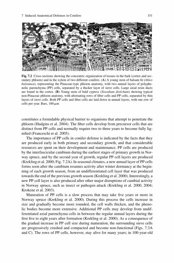
7 Induced Anatomical Defenses in Conifers 151
Fig. 7.2 Cross-sections showing the concentric organization of tissues in the bark (cortex and sec-ondary phloem) and in the xylem of two different conifers. (A) A young stem of balsam fir (Abiesbalsamea), representing the Pinaceae-type phloem anatomy, with two annual layers of polyphe-nolic parenchyma (PP) cells, separated by a thicker layer of sieve cells. Large axial resin ductsare found in the cortex. (B) Young stem of bald cypress (Taxodium distichum) showing typicalnon-Pinaceae phloem anatomy, with alternating rows of fiber cells and PP cells, separated by thinlayers of sieve cells. Both PP cells and fiber cells are laid down in annual layers, with one row ofcells per year. Bars, 100 �m
constitutes a formidable physical barrier to organisms that attempt to penetrate thephloem (Hudgins et al. 2004). The fiber cells develop from precursor cells that aredistinct from PP cells and normally require two to three years to become fully lig-nified (Franceschi et al. 2005).
The importance of PP cells in conifer defense is indicated by the facts that theyare produced early in both primary and secondary growth, and that considerableresources are spent on their development and maintenance. PP cells are producedby the interfascicular cambium during the earliest stages of primary growth in Nor-way spruce, and by the second year of growth, regular PP cell layers are produced(Krekling et al. 2000; Fig. 7.2A). In seasonal climates, a new annual layer of PP cellsforms soon after the cambium resumes activity after winter dormancy at the begin-ning of each growth season, from an undifferentiated cell layer that was producedtowards the end of the previous growth season (Krekling et al. 2000). Interestingly, anew PP cell layer is also produced after other major disruptions of cambial activityin Norway spruce, such as insect or pathogen attack (Krekling et al. 2000, 2004;Krokene et al. 2003).
Maturation of PP cells is a slow process that may take five years or more inNorway spruce (Krekling et al. 2000). During this process the cells increase insize and gradually become more rounded, the cell walls thicken, and the pheno-lic bodies become more extensive. Additional PP cells may develop from undif-ferentiated axial parenchyma cells in between the regular annual layers during thefirst five to eight years after formation (Krekling et al. 2000). As a consequence ofthe gradual increase in PP cell size during maturation, the surrounding sieve cellsare progressively crushed and compacted and become non-functional (Figs. 7.3Aand C). The rows of PP cells, however, stay alive for many years; in 100-year-old
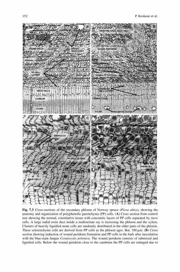
152 P. Krokene et al.
Fig. 7.3 Cross-sections of the secondary phloem of Norway spruce (Picea abies), showing theanatomy and organization of polyphenolic parenchyma (PP) cells. (A) Cross section from controltree showing the normal, constitutive tissue with concentric layers of PP cells separated by sievecells. A large radial resin duct inside a multiseriate ray is traversing the phloem and the xylem.Clusters of heavily lignified stone cells are randomly distributed in the older parts of the phloem.These sclerenchyma cells are derived from PP cells as the phloem ages. Bar, 100 �m. (B) Crosssection showing induction of wound periderm formation and PP cells in the bark after inoculationwith the blue-stain fungus Ceratocysis polonica. The wound periderm consists of suberized andlignified cells. Below the wound periderm close to the cambium the PP cells are enlarged due to

7 Induced Anatomical Defenses in Conifers 153
Norway spruce trees PP cell rows formed more than 70 year ago contained liv-ing cells (Krekling et al. 2000). PP cells in older phloem layers may undergo celldivision, and this enables the tree to maintain intact layers of PP cells as the stem di-ameter increases during growth (Krekling et al. 2000). In addition, new PP cells arerecruited in the older layers of the secondary phloem by differentiation of existingresidual parenchyma cells or ray cell derivatives (Fahn 1990; Krekling et al. 2000).
The phenolic material characterizing the PP cells is stored inside a large vacuolethat may fill most of the cell volume (the phenolic body). The phenolic nature ofthe PP cells is inferred from their bright yellow fluorescence when exposed to bluelight (450–490 nm; Fig. 7.4A), strong staining (and quenching of fluorescence) byosmium tetroxide and the periodic acid-Schiff procedure (Fig. 7.3), and the pres-ence of phenylalanine ammonia lyase (PAL), a key enzyme in phenolic synthesis(Parham and Kaustinen 1976; Franceschi et al. 1998). The vacuolar content of thePP cells is highly dynamic and changes in appearance over the year, as well asafter pathogen attack or mechanical wounding (Fig. 7.3D). In Norway spruce thedensity of the phenolic body increases during autumn and appears dense-stainingand granular by mid-winter. During spring and summer the density decreases againand reverts to a homogenous, light-staining appearance by mid-summer (Kreklinget al. 2000). Attempts have been made to classify PP cells into four types based onthe appearance of the vacuolar content (Franceschi et al. 1998), but any relationshipbetween vacuolar morphology and resistance remains unclear.
In addition to being a major storage site for phenolic compounds, PP cellsalso accumulate and store carbohydrates, in the form of starch grains, and lipids(Murmanis and Evert 1967; Alfari and Evert 1973; Krekling et al. 2000; Fig. 7.4C).Starch reserves build up through the summer months in Norway spruce, are comp-letely absent in the winter, and begin to accumulate again in the spring (Kreklinget al. 2000). In addition to serving as energy reserves, the starch and lipid of the PPcells can also be used for rapid synthesis of defensive chemicals in response to attack.
Since PP cells and ray cells make up the bulk of the living cells in the phloemthey are probably involved in radial and circumferential transmission of signals thatactivate defense responses (Franceschi et al. 2000; Krekling et al. 2004). Consistentwith this notion, there are numerous symplasmic cell connections (plasmodesmata)facilitating transport of signal molecules and nutrients between PP cells, betweenray cells and adjacent PP cells, and between ray cells (Krekling et al. 2000).
PP cell are also involved in production of important mechanical defenses that arefound in the secondary phloem of all conifers (Franceschi et al. 2005).
Fig. 7.3 (continued) mobilization of their polyphenolic vacuolar content, whereas PP cells in theimmediate vicinity of the wound periderm have emptied their content. Traumatic resin duct for-mation has been induced in the xylem. Bar, 100 �m. (C) Larger magnification of normal, non-activated annual layers of PP cells in the bark. Four annual rows of PP cells are shown, separatedby 10–12 layers of sieve cells. Bar, 50 �m. (D) Larger magnification of bark three weeks after in-oculation with Ceratocystis polonica, showing PP cells that have mobilized their phenolic contentand increased in size. The older layers of sieve cells are compressed into dense areas of cell wallsby the swelling PP cells. Bar, 50 �m
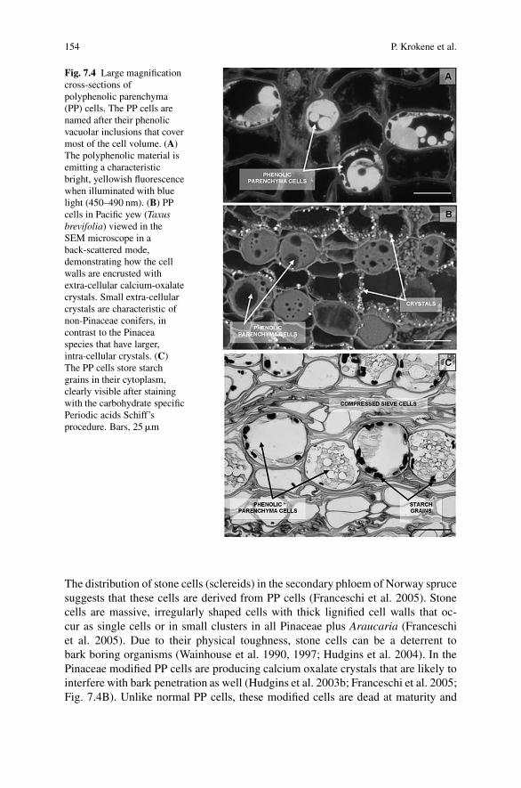
154 P. Krokene et al.
Fig. 7.4 Large magnificationcross-sections ofpolyphenolic parenchyma(PP) cells. The PP cells arenamed after their phenolicvacuolar inclusions that covermost of the cell volume. (A)The polyphenolic material isemitting a characteristicbright, yellowish fluorescencewhen illuminated with bluelight (450–490 nm). (B) PPcells in Pacific yew (Taxusbrevifolia) viewed in theSEM microscope in aback-scattered mode,demonstrating how the cellwalls are encrusted withextra-cellular calcium-oxalatecrystals. Small extra-cellularcrystals are characteristic ofnon-Pinaceae conifers, incontrast to the Pinaceaspecies that have larger,intra-cellular crystals. (C)The PP cells store starchgrains in their cytoplasm,clearly visible after stainingwith the carbohydrate specificPeriodic acids Schiff’sprocedure. Bars, 25 �m
The distribution of stone cells (sclereids) in the secondary phloem of Norway sprucesuggests that these cells are derived from PP cells (Franceschi et al. 2005). Stonecells are massive, irregularly shaped cells with thick lignified cell walls that oc-cur as single cells or in small clusters in all Pinaceae plus Araucaria (Franceschiet al. 2005). Due to their physical toughness, stone cells can be a deterrent tobark boring organisms (Wainhouse et al. 1990, 1997; Hudgins et al. 2004). In thePinaceae modified PP cells are producing calcium oxalate crystals that are likely tointerfere with bark penetration as well (Hudgins et al. 2003b; Franceschi et al. 2005;Fig. 7.4B). Unlike normal PP cells, these modified cells are dead at maturity and

7 Induced Anatomical Defenses in Conifers 155
have suberized cell walls. Crystals of calcium oxalate may also occur in the vacuolesof some of the PP cells along with the phenolic material (Franceschi et al. 1998).
7.2.2 Inducible Responses in PP Cells
In addition to being a prominent constitutive component in the secondary phloem,PP cells are also involved in a range of inducible defense responses, including acti-vation of existing cells, production of extra PP cells, and wound periderm formation.Activation or swelling of existing PP cells begins a few days after an attack andresults in a four-fold increase in the PP cell volume and a change in the appearanceof the phenolic content (Franceschi et al. 1998, 2000, 2005; Krokene et al. 2003;Fig. 7.3). The dramatic increase in PP cell size leads to extensive compression ofthe surrounding sieve cells, and at least in the Pinaceae, which produce many layersof sieve cells per year, the induced phloem is transformed into dense blocks of cellwalls separated by layers of swollen PP cells (Figs. 7.3B and D). In addition toserving as a physical barrier to penetration by invading organisms this combined cellwall/PP cell barrier appears to be reinforced by phenolics released from the inducedPP cells and deposited in the surrounding sieve cell walls (Franceschi et al. 2000).
New or extra PP cells may be induced to develop from undifferentiatedparenchyma cells in between the regular annual PP cell layers. This is not partof the normal developmental program of the secondary phloem and is only seenafter severe challenges such as massive fungal infection (Krekling et al. 2000, 2004;Krokene et al. 2003). Production of extra PP cells may also be induced by treat-ment with methyl jasmonate (Krokene et al. 2007), an elicitor of plant defenseresponses that induces similar responses as fungal pathogens and insects in conifers(Franceschi et al. 2002; Martin et al. 2002; Hudgins et al. 2004; Krokene et al.2008). With time, extra PP cells appear several centimeters away from the point ofinduction and can be quite abundant, covering 50% of the phloem circumferencein Norway spruce 15 weeks after a massive fungal infection (Krokene et al. 2003).Because differentiation of extra PP cells is a relatively slow process compared tothe activation of existing ones, it probably enhances long-term resistance rather thanresistance to an on-going attack. Production of extra PP cells seems to be involvedin the phenomenon of systemic induced resistance in Norway spruce (Krokeneet al. 2003).
PP cells are also instrumental in the process of wound periderm formation(Franceschi et al. 2000). This is an essential part of conifer defenses, as it servesto wall off damaged regions and re-establish a continuous surface barrier (see alsoGinzberg this volume). Wound periderms are produced by activation of existing PPcells, which begin to divide to form a wound phellogen or cork cambium (Fig. 7.3B).Similar to the normal periderm this meristem produces cork tissue (phellem) out-wards and phelloderm inwards, and thus regenerates a continuous surface barrier(Franceschi et al. 2005). Damaged tissues outside this new periderm are isolated

156 P. Krokene et al.
from nutrient supplies through the secondary phloem and will eventually be shed asnew phloem is produced by the vascular cambium.
7.2.3 Evidence for Defensive Roles of PP Cells
The anatomical organization, chemical nature and inducible responses of PP cellssuggest that they are important in resistance against attacking organisms. A defen-sive role for PP cells is supported by inoculation experiments with bark beetle-associated bluestain fungi, showing that tree resistance developed over a time framethat paralleled the time required to activate PP cells (Krokene et al. 2003). Treespretreated with a sublethal fungal infection three to nine weeks before a massiveartificial infection showed strong activation of PP cells, and were much more resis-tant to the subsequent challenge than untreated trees (Krokene et al. 1999, 2003).Trees pretreated one week before mass-infection showed no activation of PP cellsand were as susceptible as untreated control trees (Krokene et al. 2003). In thisexperiment, the constitutive PP cell barrier was circumvented, as fungus was arti-ficially inoculated into wounds going all the way to the vascular cambium. Whenfungal inoculations are made into shallow bark wounds, ending in the middle ofthe PP cell layers, fungal growth is completely restricted, confirming a role in de-fense also for the constitutive, concentric layers of PP cells (Franceschi et al. 2000).Many bluestain fungi are successful conifer pathogens because they circumventbark defenses by hitching a ride with scolytid bark beetles tunneling through thebark to the vascular cambium (e.g., Paine et al. 1997; Franceschi et al. 2000,2005).
The exact chemical nature of the phenolic material inside the PP cells is notknown. The conclusion that the material is polyphenolic in nature is based on mi-croscopical evidence, such as structural similarity to polyphenolics in other plantcell types, autofluorescence, staining properties, and localization of PAL to theplasma membrane and cytoplasm of PP cells by immunolocalization (Franceschiet al. 1998). Other important enzymes involved in phenol synthesis, such as chal-con and stilbene synthases, have also been found to increase in concentration inconifer bark after fungal infection, emphasizing the role of phenolics in bark de-fenses (Brignolas et al. 1995a; Richard et al. 2000; Nagy et al. 2004). There are alsonumerous direct chemical analyses of conifer phloem showing high concentrationsof soluble phenolics, such as flavonoids and stilbenes (Lindberg et al. 1992; Lieutieret al. 1991, 1996, 2003; Brignolas et al. 1995a, b, 1998; Dixon and Paiva 1995;Bois and Lieutier 1997; Evensen et al. 2000; Viiri et al. 2001; Zeneli et al. 2006).However, the soluble phenolics that lend themselves to routine chemical analysisdo not appear to be crucial for defense, as they were only weakly inducible by fun-gal inoculation, mechanical wounding, and bark beetle attack (Erbilgin et al. 2006;Zeneli et al. 2006). PP cell activation may thus be related to changes in more com-plex phenolics, such as high molecular weight condensed tannins or cell wall-boundphenolics, but this remains to be tested.

7 Induced Anatomical Defenses in Conifers 157
7.3 Traumatic Resin Ducts
7.3.1 Resin-Based Defenses in Conifers
Conifer resin is composed largely of terpenes, made up chiefly of monoterpenes(C10) and diterpenes (C20), with small amounts of sesquiterpenes (C15) and othercompounds (Gershenzon and Croteau 1991; Bohlmann this volume). Resin is gen-erally considered to be important in conifer defense because of its physical prop-erties and its toxic and repellent effects on many herbivores and pathogens (Reidet al. 1967; Shrimpton and Whitney 1968; Bordasch and Berryman 1977; Raffaet al. 1985; Gijzen et al. 1993; Bohlmann this volume). Conifer resin is synthesizedand accumulated in specialized secretory structures of different anatomical com-plexity, ranging from scattered individual resin cells and sac-like resin cavities linedwith epithelial cells, to complex networks of interconnected radial and axial resinducts. These resin structures may be present constitutively or they may be inducible,and they are found in many different tissues, including xylem, phloem, cortex andneedles.
Resin producing structures are found in many conifer lineages, but they arenot uniformly important. In fact, most conifers in the families Araucariaceae,Podocarpaceae, Cupressaceae, Cephalotaxaceae, and Taxaceae (the non-Pinaceaefamilies) lack preformed resin structures in phloem and xylem (Bannan 1936;Fahn 1979; Wu and Hu 1997), and inducible resin structures only appear in somegenera (Hudgins et al. 2004; Franceschi et al. 2005; Table 7.1). Instead, the non-Pinaceae species seem to rely mainly on massive constitutive bark defenses in theform of PP cells and lignified fiber cells (see Section 7.2.1). It is a striking andalmost paradoxical fact that the Pinaceae conifers that are relying on resin-baseddefenses are more susceptible to tree-killing bark beetles (family Scolytidae) ascompared to species in the non-Pinaceae group which rarely suffer from bark beetleattacks (Hudgins et al. 2004; Franceschi et al. 2005). The specialization of tree-killing bark beetles on resin-producing conifers includes the use of resin as a pre-cursor for production of population-aggregating pheromones (e.g., Vanderwel andOehlschlager 1987). Effective aggregation pheromones are probably essential fortree-killing, since small herbivores like bark beetles must attack in large numbersin order to overwhelm and kill large host trees (Franceschi et al. 2005). Bark bee-tles are thus using one of the tree’s primary chemical defenses to their advantage,illustrating how co-evolution between insect herbivores and host trees can turn adefense into a weakness. Still, since most conifers, both Pinaceae and non-Pinaceaespecies, usually are well-defended against bark beetles, these contrasting strategiesmay also serve as an example of how completely different defense strategies can beeffective against the same pest (Franceschi et al. 2005).
Constitutive resin structures are present in the cortex and phloem of most Pinaceaeand a few non-Pinaceae conifers, usually in the form of resin ducts or resin cavities(Fig. 7.5; Table 7.1). In addition, many Pinaceae species have scattered constitutiveresin ducts in normal xylem (Table 7.1). The axial resin ducts in the cortex (the

158 P. Krokene et al.
Tab
le7.
1D
iffe
rent
type
sof
resi
nst
ruct
ures
inco
nife
rst
ems.
The
gene
raar
egr
oupe
dby
fam
ily,
star
ting
wit
hth
ePi
nace
ae(s
how
ing
all1
1ge
nera
),co
ntin
uing
wit
hA
rauc
aria
ceae
(1of
3ge
nera
),Po
doca
rpac
eae
(1of
18–1
9ge
nera
),C
upre
ssac
eae
(7of
33ge
nera
),C
epha
lota
xace
ae(1
of3
gene
ra),
and
Taxa
ceae
(2of
3ge
nera
)
Gen
usSp
ecie
sC
orti
cal
Phlo
emX
ylem
Ref
eren
cedu
cts
Con
stitu
tive
Indu
cibl
eC
onst
itutiv
eIn
duci
ble
Abi
es>
40ye
sca
vitie
sno
?no
cavi
ties
Hud
gins
etal
.(20
04)
and
Wu
and
Hu
(199
7)C
edru
s2–
4ye
sca
viti
esno
?no
axia
lH
udgi
nset
al.(
2004
)an
dW
uan
dH
u(1
997)
Pse
udol
arix
1no
cavi
ties
no?
noca
viti
esPe
nhal
low
(190
7),B
anna
n19
36an
dW
uan
dH
u(1
997)
Tsu
ga9
yes
cavi
ties
no?
noca
viti
esH
udgi
nset
al.(
2004
)an
dW
uan
dH
u(1
997)
Not
hots
uga
1ye
sra
dial
no?
noax
ial
Wu
and
Hu
(199
7)K
etel
eeri
a3
yes
radi
alno
?ax
ial
axia
l?W
uan
dH
u(1
997)
Lar
ix14
nora
dial
no?
netw
ork1
axia
lH
udgi
nset
al.(
2003
a)an
dW
uan
dH
u(1
997)
Pse
udot
suga
5ye
sra
dial
no?
netw
ork
axia
lH
udgi
nset
al.(
2004
)an
dW
uan
dH
u(1
997)
Cat
haya
1ye
sra
dial
no?
netw
ork
axia
l?W
uan
dH
u(1
997)
Pic
ea35
yes
radi
alno
?ne
twor
kax
ial
Hud
gins
etal
.(20
04)
and
Wu
and
Hu
(199
7)P
inus
115
yes
radi
alno
?ne
twor
k(a
xial
)2H
udgi
nset
al.(
2004
)an
dW
uan
dH
u(1
997)
Ara
ucar
ia19
(no)
3ax
ial
nono
Hud
gins
etal
.(20
04)
Aga
this
21no
no?
nono
?Pe
nhal
low
(190
7)W
olle
mia
1no
?no
?no
no?
Hea
dyet
al.(
2002
)
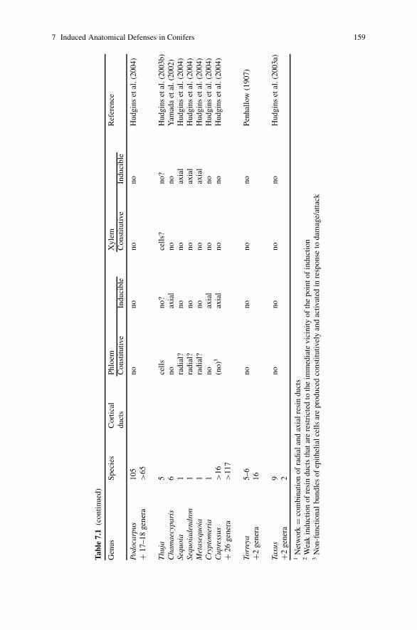
7 Induced Anatomical Defenses in Conifers 159
Tab
le7.
1(c
onti
nued
)
Gen
usSp
ecie
sC
orti
cal
Phlo
emX
ylem
Ref
eren
cedu
cts
Con
stitu
tive
Indu
cibl
eC
onst
itutiv
eIn
duci
ble
Podo
carp
us10
5no
nono
noH
udgi
nset
al.(
2004
)+
17–1
8ge
nera
>65
Thu
ja5
cell
sno
?ce
lls?
no?
Hud
gins
etal
.(20
03b)
Cha
mae
cypa
ris
6no
axia
lno
noY
amad
aet
al.(
2002
)Se
quoi
a1
radi
al?
nono
axia
lH
udgi
nset
al.(
2004
)Se
quoi
aden
dron
1ra
dial
?no
noax
ial
Hud
gins
etal
.(20
04)
Met
aseq
uoia
1ra
dial
?no
noax
ial
Hud
gins
etal
.(20
04)
Cry
ptom
eria
1no
axia
lno
noH
udgi
nset
al.(
2004
)C
upre
ssus
>16
(no)
3ax
ial
nono
Hud
gins
etal
.(20
04)
+26
gene
ra>
117
Torr
eya
5–6
nono
nono
Penh
allo
w(1
907)
+2ge
nera
16
Taxu
s9
nono
nono
Hud
gins
etal
.(20
03a)
+2ge
nera
21
Net
wor
k=
com
bina
tion
ofra
dial
and
axia
lres
indu
cts
2W
eak
indu
ctio
nof
resi
ndu
cts
that
are
rest
rict
edto
the
imm
edia
tevi
cini
tyof
the
poin
tof
indu
ctio
n3
Non
-fun
ctio
nal
bund
les
ofep
ithel
ialc
ells
are
prod
uced
cons
titut
ivel
yan
dac
tivat
edin
resp
onse
toda
mag
e/at
tack

160 P. Krokene et al.
outer part of the young stem bark) may be very large compared to other resin ducts(Figs. 7.2A and 7.5A). They are formed in a circumferential ring as the cortex isproduced during primary stem development, and may be present for at least 25 yearsin Norway spruce (Christiansen et al. 1999a). The cortex is an important defensebarrier in the early stages of stem growth, but its defensive roles are gradually takenover by the secondary phloem as this develops more extensive layers (Franceschiet al. 2005). The constitutive resin ducts in the phloem are always radial, and re-side within multiseriate radial rays surrounded by ray parenchyma cells. The cross-section of these ducts increases in size in the older parts of the bark and can be quiteextensive (Christiansen et al. 1999a; Fig. 7.3A). Radial resin ducts occur primarily inthe phloem, but some run continuously from the phloem to the sapwood, where theyare interconnected with the axial resin ducts (Fig. 7.5E; Christiansen et al. 1999a;Nagy et al. 2000). A complex constitutive resin duct network with interconnectedradial and axial ducts in phloem and xylem are found only in some Pinaceae genera(Pinus, Picea, Larix, Pseudotsuga, Keteleeria, Cathaya; Table 7.1), and is mosthighly developed in the genus Pinus which seems to rely heavily on constitutiveresin (Bannan 1936; Vite 1961; Johnson and Croteau 1987).
Resin ducts are lined with plastid-enriched epithelial cells that produce and se-crete resin into an extracellular lumen, where it is stored under pressure (Charonet al. 1987; Gershenzon and Croteau 1990; Nagy et al. 2000; Fig. 7.6A). The sim-pler resin cells accumulate resin internally under pressure and may expand into quitelarge structures (Fig. 7.5B). Resin ducts form schizogenously as the epithelial cellspull apart during resin duct formation (Nagy et al. 2000). The epithelial cells liningradial and axial resin ducts are thin-walled and long-lived, in contrast to the epithe-lial cells of the resin cavities, which are short-lived and gradually become lignifiedduring development (Bannan 1936; Fahn 1979).
7.3.2 Anatomy and Development of Traumatic Resin Ducts
In addition to constitutive resin structures many conifers develop inducible resinstructures in response to biotic and abiotic challenges, such as insect attack, pathogeninfection, frost damage, or physical wounding (e.g., Bannan 1936; Alfaro 1995;Kyto et al. 1996; Hudgins et al. 2003a, 2004). In the Pinaceae, inducible resin struc-tures usually occur as so-called pathological or traumatic axial resin ducts in thexylem, but axial resin ducts are also induced in the phloem of a few non-Pinaceaegenera (Table 7.1). The nature of the vascular cambium implies that inducibleresin ducts must be axial and not radial. The cambium can relatively quickly bere-programmed to produce functional axial resin ducts that may be several meterslong (e.g., Christiansen et al. 1999b; Nagy et al. 2000), whereas it would requireyears to produce radial ducts of any extent.
TRDs form in tangential bands in the xylem of many Pinaceae species (Figs. 7.3Band 7.5D). These bands are often surrounded by small tracheids with thickened cellwalls and are visible to the naked eye as a punctuated line within the annual ring

7 Induced Anatomical Defenses in Conifers 161
Fig. 7.5 Cross-sections of resin-containing structures in conifers. (A) An axially oriented resinduct in the outer cortical region of the phloem of balsam fir (Abies balsamea). Bar, 50 �m. Thecircular cortical resin ducts are lined with small epithelial cells which are surrounded by sheetcells containing phenolic material. (B) In grand fir (Abies grandis) resin is produced and storedin individual resin cells in the secondary phloem. Bar, 50 �m. (C) Low magnification of Norwayspruce (Picea abies) bark and sapwood, showing traumatic resin ducts within the sapwood 3 weeksafter inoculation with the blue-stain fungus Ceratocystis polonica. The traumatic resin ducts arevisible as small, tangentially oriented channels close to the cambium. (D) Closer look at traumaticresin ducts in the xylem of Norway spruce after fungal inoculation. The traumatic resin ducts arecharacteristically associated with radial rays. (E) Cross-section demonstrating luminal continuitybetween several coalescing traumatic resin ducts and a radial resin duct traversing the phloem andxylem. Bars, 50 �m
(false annual rings; Fig. 7.5C). The formation of functional TRDs with secretoryactivity is quite rapid, and requires two to four weeks in Norway spruce (Nagyet al. 2000). TRDs develop in the same way as normal axial resin ducts, by schizo-genesis between incipient epithelial cells when these are still close to the cambialzone (Fig. 7.6B). Sometimes large transverse resin ducts can be produced by tan-gential anastomosis of neighboring ducts (Gerry 1942; Fig. 7.5E). If the inducing

162 P. Krokene et al.
Fig. 7.6 Cross-sections of the epithelial cells of developing traumatic resin ducts. (A) In Norwayspruce, the epithelial cells lining the resin duct are characterized by having dense cytoplasm, withmany plastids and vacuoles, and conspicuously enlarged nuclei. The numerous plastids are theprimary site for resin synthesis. Bar, 10 �m. (B) Early in their development the traumatic resinducts are often surrounded by living parenchyma cells full of starch grains and phenolic material.Bar, 25 �m
stimulus is strong, an almost complete tangential ring of TRDs can be produced,and with time, the ducts may extend several meters from the point of induction inspruce (Christiansen et al. 1999b). In other genera, such as Abies, Tsuga, Cedrusand Pseudolarix, TRDs mainly form in the vicinity of wounds (Bannan 1936; Wuand Hu 1997).

7 Induced Anatomical Defenses in Conifers 163
The TRD system may represent a considerable volume, and contribute signif-icantly to a tree’s overall resin production. After massive induction, TRDs aremuch more abundant than normal xylem ducts, and may extend over 50%–100%of the xylem circumference in Norway spruce (Franceschi et al. 2002; Krokeneet al. 2003). TRDs are also much larger than constitutive xylem ducts; in Norwayspruce for example, they are often twice as wide and represent a volume that is fourtimes greater per unit duct length. TRDs form an interconnected network of resin-filled cavities tangentially in the new sapwood, as well as radially with luminal con-tinuity between adjacent TRDs and radial constitutive resin ducts (Fig. 7.5E). Thisorganization facilitates resin flow both axially along the trunk, as well as radiallytowards the bark surface (Nagy et al. 2000; Krokene et al. 2003).
TRDs appear to be derived from xylem mother cells (Werker and Fahn 1969;Nagy et al. 2000). The strict distribution pattern of TRDs in tangential bands impliesa combination of predisposition and a tangential spread of a stimulus resulting incoordinated induction and differentiation of the xylem mother cells. With increas-ing distance from the point of induction, TRDs are gradually reduced in size andnumber, and are found closer and closer to the cambium (Krekling et al. 2004).There thus appears to be a time-lag in the production and differentiation of theTRDs away from the stimulus, which would be consistent with a signal propagat-ing a developmental wave traveling in the axial direction at about 2.5 cm per day(Krekling et al. 2004). TRD formation appears to be activated through the octade-canoid pathway, involving jasmonate and ethylene signaling (Franceschi et al. 2002;Martin et al. 2002; Hudgins et al. 2003a, 2004, 2006; Hudgins and Franceschi 2004;Miller et al. 2005; Ralph et al. 2007).
7.3.3 Defensive Roles of TRDs
TRD formation in Norway spruce is part of a complex defense response that is rapidand coordinated, resulting in an extensive resin-secreting network. The result is mas-sive accumulation of resin and enhanced resin flow at the site of attack (Phillips andCroteau 1999; Nagy et al. 2000). Artificial inoculation or wounding, leading to TRDformation and increased synthesis of resin, can thus be used as a technique to improveand prime a tree’s resistance against insects and pathogens. For example, Norwayspruce trees that were pretreated with a sublethal fungal infection developed abundantTRDs which rendered the trees much more resistant to a subsequent massive infectionafter three to nine weeks (Krokene et al. 1999, 2003). The ability of a tree to undergotraumatic resinosis can be used to screen for resistance (Krokene et al. 2008). Forexample, resistant clones of Norway spruce and white spruce (Picea glauca) producemore TRDs more quickly as compared to susceptible clones (Tomlin et al. 1998; Nagyet al. 2000; Krekling et al. 2004). In white spruce, large inner and dense outer resincanals were correlated with resistance against the white pine weevil, Pissodes strobi(Peck) (Alfaro et al. 2004).
There is ample evidence that conifer resin is effective in defense against in-sects. In several conifers, the concentration of monoterpenes is enhanced in tissues

164 P. Krokene et al.
attacked by bark beetles and their associated blue-stain fungi (Russell and Berryman1976; Cates and Alexander 1982; Raffa and Berryman 1982, 1983; Paine et al. 1987;Lewinsohn et al. 1991). Tomlin et al. (2000) found that simulated white pine wee-vil damage enhanced the monoterpene to resin acid ratio in stem regions distantfrom the wounded site in resistant trees, but only slightly in susceptible trees.Monoterpene-enriched resin, which is less viscous than constitutive resin, proba-bly flows more readily into oviposition cavities and larval mines, thus being moreeffective in inhibiting/killing immature weevils. Resin may also inhibit invadinginsects and mycelial growth of fungal pathogens due to its toxic properties. Forexample, Klepzig et al. (1995) found that monoterpenes in red pine resin inhibitfungal germination and mycelial growth, as well as beetle tunneling. Alpha-pinenein particular, the most abundant monoterpene in red pine phloem, exhibited strongbiological activity towards insects and fungi (Klepzig et al. 1995).
Resin diterpenes are considered to be insecticidal and fungicidal, and are impor-tant in wound sealing. An open wound is a potential infection pathway for attackinginsects and pathogenic fungi, and effective wound sealing is decisive in preventinginvasions. The diterpene abietic acid is important for oxidative polymerization ofresin and dehydroabietic acid for resin viscosity and crystallization (Phillips andCroteau 1999). Resistant white spruce trees appear to produce more volatile terpenesand diterpene resin acids at the area of wounding than susceptible trees (Tomlinet al. 2000).
The induction of functional TRDs is usually too slow to be effective againstrapid mass-attacks by bark beetles, which may be completed in a few days (Paineet al. 1997). However, TRD formation may contribute to defense when bark bee-tle attacks are slowed down by e.g. unfavorable weather conditions (Franceschiet al. 2000), and may also be important in protecting the trees against opportunisticwound colonizers after e.g. mechanical damage to the bark.
7.4 Perspectives
Invaders attempting to colonize healthy conifers have to struggle their way throughmultiple layers of periderm, PP cells and/or fiber cells. However, the ray cells run-ning axially through the secondary phloem and xylem represent a potential weakpoint in this defense barrier. Since the rays are the only tissue type in the stem thatis oriented radially they form a major direct link between phloem and sapwood.This potential loophole must therefore be expected to be well-defended, and im-munolabeling studies in Norway spruce have indeed demonstrated that rays are amajor site for PAL activity (Franceschi et al. 1998), and probably other defensiveelements as well. Anatomical studies in Scots pine suggest that pathogen infectioninduces changes in ray cells, including increase in cell size and a more active cyto-plasm that may be related to increased cellular activity and resin production (Nagyet al. 2006). However, as far as we know these preliminary studies are the only oneslooking at inducible defenses in ray cells, and there are no detailed studies of ray

7 Induced Anatomical Defenses in Conifers 165
cell structure and function. The rays are not only involved in defending the bark andouter sapwood, but are probably also instrumental in heartwood formation and de-fense against e.g. wood rotting fungi, since they are the only route for translocationof substances between the living phloem and the inner sapwood (Shain 1967; Kwonet al. 2001; Franceschi et al. 2005). With their radial orientation, the rays are alsoexpected to play important roles in signaling and transport of chemicals betweenphloem and xylem.
Chemical and molecular characterization of the central cell types in conifer de-fense, such as radial rays, PP cells, and resin duct epithelial cells, is strongly needed.The data available so far represent whole tissues (e.g., the whole secondary phloem),and the results are thus an average of information of different tissue and cell types.Recent technological and analytical developments have made it possible to dissectspecific cell types and characterize their content using both chemical and moleculartechniques. Laser microdissection enables effective isolation of single cells and iswidely used in the biomedical field (Emmert-Buck et al. 1996; Day et al. 2005).More recently, laser microdissection has been adapted for use with many plantspecies, including Arabidopsis, rice, maize and others (Ohtsu et al. 2007). Theonly example from conifers so far is the dissection and chemical analysis of stonecells in Norway spruce (Li et al. 2007). Using laser microdissection in combina-tion with sensitive instrumental analysis such as mass spectroscopy and cryogenicNMR-spectroscopy, it will be possible to chemically characterize pure cell popu-lations. This represents a new approach for the cell-specific localization of low-molecular weight compounds in plants, and has been termed micro-phytochemistry(Li et al. 2007). In addition to metabolites, proteins, DNA, and RNA can be an-alyzed from microdissected samples. This powerful tool can thus give us detailedinformation about the chemical and molecular nature of all the important cell typesin conifer defense.
References
Alfaro RI (1995) An induced defense reaction in white spruce to attack by the white-pine weevil,Pissodes strobe. Can J For Res 25:1725–1730
Alfaro RI, VanAkker L, Jaquish B, King J (2004) Weevil resistance of progeny derived from puta-tively resistant and susceptible interior spruce parents. For Ecol Man 202:369–377
Alfieri FJ, Evert RF (1973) Structure and seasonal development of secondary phloem in Pinaceae.Bot Gaz 134:17–25
Bannan MW (1936) Vertical resin ducts in the secondary wood of the abietineae. New Phytol.35:11–46
Bois E, Lieutier F (1997) Phenolic response of Scots pine clones to inoculation with Lep-tographium wingfieldii, a fungus associated with Tomicus piniperda. Plant Physiol Biochem35:819–825
Bonello P, Gordon TR, Storer AJ (2001) Systemic induced resistance in Monterey pine. For Pathol31:99–106
Bonello P, Blodgett JT (2003) Pinus nigra–Sphaeropsis sapinea as a model pathosystem to in-vestigate local and systemic effects of fungal infection of pines. Physiol Mol Plant Pathol63:249–261

166 P. Krokene et al.
Bordasch RP, Berryman AA (1977) Host resistance to the fir engraver beetle, Scolytus ventralis(Coleoptera: Scolytidae). 2. Repellency of Abies grandis resins and some monoterpenes. CanEntomol 109:95–100
Brignolas F, Lacroix B, Lieutier F, Sauvard D, Drouet A, Claudot AC, Yart A, Berryman AA,Christiansen E (1995a) Induced responses in phenolic metabolism in two Norway spruce clonesafter wounding and inoculation with Ophiostoma polonicum, a bark beetle-associated fungus.Plant Physiol 109:821–827
Brignolas F, Lieutier F, Sauvard D, Yart A, Drouet A, Claudot AC (1995b) Changes in soluble-phenol content of Norway-spruce (Picea abies) phloem in response to wounding and inocula-tion with Ophiostoma polonicum. Eur J For Pathol 25:253–265
Brignolas F, Lieutier F, Sauvard D, Christiansen E, Berryman AA (1998) Phenolic predictors forNorway spruce resistance to the bark beetle lps typographus (Coleoptera: Scolytidae) and anassociated fungus, Ceratocystis polonica. Can J For Res 28:720–728
Cates RG, Alexander H (1982) Host resistance and susceptibility. In: Mitton JB, Sturgeon KB(eds) Bark beetles in North American conifers. A system for the study of evolutionary biology.University of Texas Press, Austin, pp 212–263
Charon J, Launay J, Carde J-P (1987) Spatial organization and volume density of leucoplasts inpine secretory cells. Protoplasma 138:45–53
Christiansen E, Franceschi VR, Nagy NE, Krekling T, Berryman AA, Krokene P, Solheim H(1999a) Traumatic resin duct formation in Norway spruce after wounding or infection witha bark beetle-associated blue-stain fungus, Ceratocystis polonica. In: Lieutier F, Mattson WJ,Wagner MR (eds) Physiology and genetics of tree-phytophage interactions. Les Colloques deI’INRA, INRA Editions, Versailles, pp 79–89
Christiansen E, Krokene P, Berryman AA, Franceschi VR, Krekling T, Lieutier F, Lonneborg A,Solheim H (1999b) Mechanical injury and fungal infection induce acquired resistance in Nor-way spruce. Tree Physiol 19:399–403
Cook SP, Hain FP (1988) Toxicity of host monoterpenes to Dendroctonus frontalis and Ips cal-ligraphus (Coleoptera: Scolytidae). J Entomol Sci 23:287–292
Day RC, Grossniklaus U, Macknight RC (2005) Be more specific! Laser-assisted microdissectionof plant cells. Trends Plant Sci 10:397–406
Dixon RA, Paiva NL (1995) Stress-induced phenylpropanoid meatabolism. Plant Cell7:1085–1097
Emmert-Buck MR, Bonner RF, Smith PD, Chuaqui RF, Zhuang ZP, Goldstein SR, Weiss RA,Liotta LA (1996) Laser capture microdissection. Science 274:998–1001
Erbilgin N, Kroken, P, Christiansen E, Zeneli G, Gershenzon J (2006) Exogenous application ofmethyl jasmonate elicits defenses in Norway spruce (Picea abies) and reduces host colonizationby the bark beetle Ips typographus. Oecologia 148:426–436
Evensen PC, Solheim H, Høiland K, Stenersen J (2000) Induced resistance of Norway spruce,variation of phenolic compounds and their effects on fungal pathogens. For Pathol 30:97–108
Fahn A (1990) Plant anatomy. Pergamon Press, OxfordFahn A (1979) Resin ducts of the coniferae. In: Secretory tissues in plants. Academic Press,
LondonFarjon A (2001) World checklist and bibliography of conifers, 2nd edn. Royal Botanical Gardens,
Kew, RichmondFeeny P (1976) Plant apparency and chemical defense. In: Wallace JW, Mansell RT (eds)
Biochemichal interaction between plants and insects. Plenum Press, New York, pp 1–40Franceschi VR, Krekling T, Berryman AA, Christiansen E (1998) Specialized phloem parenchyma
cells in Norway spruce (Pinaceae) bark are an important site of defense reactions. Am J Bot85:601–615
Franceschi VR, Krokene P, Krekling T, Christiansen E (2000) Phloem parenchyma cells are in-volved in local and distant defense responses to fungal inoculation or bark beetle attack inNorway spruce (Pinaceae). Am J Bot 87:314–326
Franceschi VR, Krekling T, Christiansen E (2002) Application of methyl jasmonate on Piceaabies (Pinaceae) stems induces defense related responses in phloem and xylem. Am J Bot89:578–586

7 Induced Anatomical Defenses in Conifers 167
Franceschi VR, Krokene P, Christiansen E, Krekling T (2005) Anatomical and chemical defencesof conifer bark against bark beetles and other pests (Tansley Review). New Phytol 167:353–376
Gershenzon J, Croteau RB (1991). Terpenoids. In: Rosenthal GA, Berenbaum MR (eds) Herbi-vores: their interactions with secondary plant metabolites. Vol I, the chemical participants.Academic Press, San Diego, pp 165–219
Gijzen M, Lewinsohn E, Savage TJ, Croteau RB (1993) Conifer monoterpenes – biochemistry andbark beetle chemical ecology. ACS Symp. Ser. 525:8–22
Heady RD, Banks JG, Evans PD (2002) Wood anatomy of wollemi pine (Wollemia nobilis, Arau-cariaceae). IAWA Journal 23:339–357
Hudgins JW, Christiansen E, Franceschi VR (2003a) Methyl jasmonate induced changes mimick-ing anatomical defenses in diverse members of the Pinaceae. Tree Physiol 23:361–371
Hudgins JW, Krekling T, Franceschi VR (2003b) Distribution of calcium oxalate crystals in thesecondary phloem of conifers: a constitutive defense mechanism? New Phytol 159:677–690
Hudgins JW, Christiansen E, Franceschi VR (2004) Induction of anatomically based defense re-sponses in stems of diverse conifers by methyl jasmonate: a phylogenetic perspective. TreePhysiol 24:251–264
Hudgins JW, Franceschi VR (2004) Methyl jasmonate-induced ethylene production is responsiblefor conifer phloem defense responses and reprogramming of stem cambial zone for traumaticresin duct formation. Plant Physiol 13:2134–2149
Johnson MA, Croteau R (1987) Biochemistry of conifer resistance to bark beetles and their fungalsymbionts. In: Fuller G, Nes WD (eds) Ecology and metabolism of plant lipids. AmericanChemical Society, Washington DC, pp 76–92
Kartuch B, Karuch R, Weilgong P (1991) Site-specific differences in calcium oxalate content ofthe secondary phloem of spruce (Picea abies Karst). Flora 185:377–384
Klepzig KD, Kruger EL, Smalley EB, Raffa KF (1995) Effects of biotic and abiotic stress on in-duced accumulation of terpenes and phenolics in red pines inoculated with bark beetle-vectoredfungus. J Chem Ecol 21:601–626
Krekling T, Franceschi VR, Berryman AA, Christiansen E (2000) The structure and developmentof polyphenolic parenchyma cells in Norway spruce (Picea abies) bark. Flora 195:354–369
Krekling T, Franceschi VR, Krokene P, Solheim H (2004) Differential anatomical responses ofNorway spruce tissues to sterile and fungus-infected wounding. Trees 18:1–9
Krokene P, Christiansen E, Solheim H, Berryman AA, Franceschi VR (1999) Induced resistanceto pathogenic fungi in Norway spruce. Plant Physiol 121:565–570
Krokene P, Nagy NE, Solheim H (2008) Methyl jasmonate and oxalic acid treatment of Norwayspruce: anatomically based defence responses and increased resistance against fungal infection.Tree Physiol 28:29–35
Krokene P, Solheim H, Langstrom B (2000) Fungal infection and mechanical wounding inducedisease resistance in Scots pine. Eur J Plant Pathol 106:537–541
Krokene P, Solheim H, Christiansen E (2003) Inducible anatomical defense responses in Norwayspruce stems and their possible role in induced resistance. Tree Physiol 23:191–197
Kwon M, Davin LB, Lewis NG (2001) In situ hybridization and immunolocalization of lignanreductases in woody tissue: implications for heartwood formation and other forms for vasculartissue preservation. Phytochemistry 57:899–914
Kyto M, Niemela P, Annila E (1996) Vitality and bark beetle resistance of fertilized Norway spruce.For Ecol Manag 84:149–157
Lewinsohn E, Gijzen M, Croteau R (1991) Defense-mechanisms of conifers – differencesin constitutive and wound-induced monoterpene biosynthesis among species. Plant Physiol96:44–49
Li SH, Schneider B, Gershenzon J (2007). Microchemical analysis of laser-microdissectedstone cells of Norway spruce by cryogenic nuclear magnetic resonance spectroscopy. Planta225:771–779
Lieutier F, Yart A, Jay-Allemand C, Delorme L (1991) Preliminary investigations on phenolics as aresponse of Scots pine phloem to attacks by bark beetles and associated fungi. Eur J For Pathol21:354–364

168 P. Krokene et al.
Lieutier F, Sauvard D, Brignolas F, Picron V, Yart A, Bastien C, Jay-Allemand C (1996) Changes inphenolic metabolites of Scots-pine phloem induced by Ophiostoma brunneo-ciliatum, a bark-beetle-associated fungus. Eur J For Pathol 26:145–158
Lieutier F, Brignolas F, Sauvard D, Yart A, Galet C, Brunet M, van de Sype H (2003) Intra- andinter-provenance variability in phloem phenols of Picea abies and relationship to a bark beetle-associated fungus. Tree Physiol 23:247–256
Lindberg M, Lundgren L, Gref R, Johansson M (1992) Stilbenes and resin acids in relation tothe penetration of Heterobasidion annosum through the bark of Picea abies. Eur J For Pathol22:95–106
Lombardero MJ, Ayres MP, Lorio PL, Ruel JJ (2000) Environmental effects on constitutive andinducible resin defences of Pinus taeda. Ecol Lett 3:329–339
Martin D, Tholl D, Gershenzon J, Bohlmann J (2002) Methyl jasmonate induces traumatic resinducts, terpenoid resin biosynthesis, and terpenoid accumulation in developing xylem of Norwayspruce stems. Plant Physiol 129:1003–1018
Miller B, Madilao LL, Ralph S, Bohlmann J (2005) Insect-induced conifer defense. White pineweevil and methyl jasmonate induce traumatic resinosis, de novo formed volatile emissions,and accumulation of terpenoid synthase and putative octadecanoid pathway transcripts in Sitkaspruce. Plant Physiol 137:369–382
Murmanis L, Evert RF (1967) Parenchyma cells of secondary phloem in Pinus strobus. Planta73:301–318
Nagy NE, Fossdal CG, Krokene P, Krekling T, Lonneborg A, Solheim H (2004) Inducedresponses to pathogen infection in Norway spruce phloem: changes in polyphenolicparenchyma cells, chalcone synthase transcript levels and peroxidase activity. Tree Physiol24:505–515
Nagy NE, Franceschi VR, Solheim H, Krekling T, Christiansen E (2000) Wound-induced traumaticresin duct development in stems of Norway spruce (Pinaceae): anatomy and cytochemical traits.Am J Bot 87:302–313
Nagy NE, Krokene P, Solheim H (2006) Anatomical-based defense responses of Scots pine (Pinussylvestris) stems to two fungal pathogens. Tree Physiol 26:159–167
Ohtsu K, Takahashi H, Schnable PS, Nakazono M (2007) Cell type-specific gene expression profil-ing in plants by using a combination of laser microdissection and high-throughput technologies.Plant Cell Physiol 48:3–7
Paine TD, Blanche CA, Nebeker TE, Stephen FM (1987) Composition of loblolly pine resin de-fenses: comparison of monoterpenes from induced lesion and sapwood resin. Can J For Res17:1202–1206
Paine TD, Raffa KF, Harrington TC (1997) Interactions among scolytid bark beetles, their associ-ated fungi, and live host conifers. Ann Rev Entomol 42:179–206
Parham RA, Kaustinen HM (1976) Differential staining of tannin in sections of epoxy-embeddedplant cells. Stain Technol 51:237–240
Penhallow DP (1907) A manual of the North American gymnosperms: exclusive of the Cycadalesbut together with certain exotic species. Athenaeum Press, Boston, MA. pp 404
Phillips MA, Croteau RB (1999) Resin-based defense in conifers. Trends Plant Sci 4:184–190Raffa KF, Berryman AA (1982) Accumulation of monoterpenes and associated volatiles follow-
ing inoculation of grand fir with a fungus transmitted by the fir engraver Scolytus ventralis(Coleoptera: Scolytidae). Can Entomol 114:797–810
Raffa KF, Berryman AA (1983). Physiological aspects of lodgepole pine wound responses to a fun-gal symbiont of the mountain pine beetle, Dendroctonus ponderosae (Coleoptera: Scolytidae).Can Entomol 115:723–734
Raffa KF, Berryman AA, Simasko J, Teal W, Wong BL (1985) Effects of grand fir monoterpenes onthe fir engraver, Scolytus ventralis (Coleoptera: Scolytidae), and its symbiotic fungus. EnvironEntomol 14:552–556
Ralph SG, Hudgins JW, Jancsik S, Franceschi VR, Bohlmann J (2007) Aminocyclopropanecarboxylic acid synthase is a regulated step in ethylene-dependent induced conifer defense.Full-length cDNA cloning of a multigene family, differential constitutive, and wound- and

7 Induced Anatomical Defenses in Conifers 169
insect-induced expression, and cellular and subcellular localization in spruce and Douglas fir.Plant Physiol 143:410–424
Reglinski T, Stavely FJL, Taylor JT (1998) Induction of phenylalanine ammonia lyase activity andcontrol of Sphaeropsis sapinea infection in Pinus radiata by 5-chlorosalicylic acid. Eur J ForPathol 28:153–158
Reid RW, Whitney HS, Watson JA (1967) Reactions of lodgepole pine to attack by Dendroctonusponderosae Hopkins and blue stain fungus. Can J Bot 45:1115–1126
Richard S, Lapointe G, Rutledge G, Seguin A (2000) Induction of chalcone synthase expression inwhite spruce by wounding and jasmonate. Plant Cell Physiol 41:982–987
Ruel JJ, Ayres MP, Lorio PL (1998) Loblolly pine responds to mechanical wounding with increasedresin flow. Can J For Res 28:596–602
Russell CE, Berryman AA (1976) Host resistance to the fir engraver beetle. 1. Monoterpene com-position of Abies grandis pitch blisters and fungus-infected wounds. Can J Bot 54:14–18
Schmidt A, Zeneli G, Hietala AM, Fossdal CG, Krokene P, Christiansen E, Gershenzon J (2005)Induced chemical defenses in conifers: biochemical and molecular approaches to studyingtheir function. In: Romeo J (ed) Chemical ecology and phytochemistry of forests and forestecosytems. Elsevier, Amsterdam, pp 1–28
Shain L (1967) Resistance of sapwood in stems of loblolly pine to infection by Fomes annosus.Phytopathology 57:1034–1045
Shrimpton DM, Whitney HS (1968) Inhibition of growth of blue stain fungi by wood extractives.Can J Bot 46:757–761
Tomlin ES, Alfaro RI, Borden JH, He FL (1998) Histological response of resistant and susceptiblewhite spruce to simulated white pine weevil damage. Tree Physiol 18: 21–28
Tomlin ES, Antonejevic E, Alfaro RI, Borden JH (2000) Changes in volatile terpene and diterpeneresin acid composition of resistant and susceptible white spruce leaders exposed to simulatedwhite pine weevil damage. Tree Physiol 20:1087–1095
Vanderwel D, Oehlschlager AC (1987) Biosynthesis of pheromones and endocrine regulation ofpheromone production in Coleoptera. In: Prestwich GD, Blomquist GJ (eds) Pheromone bio-chemistry. Academic Press, New York, pp 175–215
Vite JP (1961) The influence of water supply on oleoresin exudation pressure and resistance to barkbeetle attack in Pinus ponderosa. Contributions from the Boyce Thompson Institute 21:37–66
Viiri H, Annila E, Kitunen V, Niemela P (2001) Induced responses in stilbenes and terpenes infertilized Norway spruce after inoculation with blue-stain fungus, Ceratocystis polonica. Trees15:112–122
Wainhouse D, Cross DJ, Howell RS (1990) The role of lignin as a defense against the spruce barkbeetle Dendroctonus micans – effect on larvae and adults. Oecologia 85:257–265
Wainhouse D, Rose DR, Peace AJ (1997) The influence of preformed defences on the dynamicwound response in spruce bark. Funct Ecol 11:564–572
Werker E, Fahn A (1969) Resin ducts of Pinus halepensis Mill.—their structure, development andpattern of arrangement. Bot J Linn Soc 62:379–411
Wu H, Hu ZH (1997) Comparative anatomy of resin ducts of the Pinaceae. Trees Struct Funct11:135–143
Zeneli G, Krokene P, Christiansen E, Krekling T, Gershenzon J (2006) Methyl jasmonate treat-ment of large Norway spruce (Picea abies) trees increases the accumulation of terpenoid resincomponents and protects against infection by Ceratocystis polonica, a bark beetle-associatedfungus. Tree Physiol 26:977–988

Part BProduction of Secondary Metabolites

Chapter 8Insect-Induced Terpenoid Defenses in Spruce
Jorg Bohlmann
Conifers produce an array of hundreds of different terpenoids as part of their com-plex chemical, physical, and ecological defenses against insect pests and pathogens.Terpenoid chemicals exist both as constitutive and as massively induced defensesin conifers. It is thought that the diversity of terpenoid chemicals serves as a multi-layered chemical shield in long-lived conifer trees that provides a lasting protectionagainst the much faster evolving insect pests and potential pathogens. The forma-tion of terpenoid defenses in conifers involves the activity of two pathways, themethylerythritol phosphate pathway and the mevalonate pathway, which lead tothe five-carbon precursors of terpenoid biosynthesis. The many monoterpenoids,sesquiterpenoids, and diterpene resin acids, which are present in oleoresin mixtures,are then formed by families of enzymes belonging to the classes of prenyl trans-ferases, terpenoid synthases, and cytochrome P450 dependent monooxygenases.The genes for almost all the enzymatic steps in terpenoid oleoresin biosynthesishave been identified in species of spruce, which have thus been established as aconifer reference system to study constitutive and induced terpenoid defenses us-ing biochemical, molecular genetic, and genomic and proteomic approaches. At thehistological and cellular levels, oleoresin terpenoids are produced and accumulateconstitutively in large quantities in specialized anatomical structures that are foundin most organs and tissues. In many conifer species, biosynthesis and accumulationof terpenoids is further enhanced as part of the induced defense in response to insectattack or fungal infection. In this chapter, I will discuss selected aspects of terpenoiddefenses in conifers against insects and pathogens.
8.1 Introduction
Conifers (i.e., members of the pine family; Pinaceae) include some of the longest-living and tallest organisms on earth. Their tall physical structure and long lifespan
J. BohlmannMichael Smith Laboratories, University of British Columbia, Vancouver, B.C., Canadae-mail: [email protected]
A. Schaller (ed.), Induced Plant Resistance to Herbivory, 173C© Springer Science+Business Media B.V. 2008
174 J. Bohlmann
make them prominent targets in space and time for many potential herbivores andpathogens. The successful defense and resistance of conifers against most herbivoresand pathogens can be explained, at least in part, by the formation of a diverse array ofmonoterpenoid, sesquiterpenoid, and diterpene resin acid defense chemicals. Thesecompounds accumulate in large amounts in form of preformed or induced oleoresinmixtures. Some terpenoids, in particular monoterpenes and sesquiterpenes, can alsobe actively emitted as volatile organic compounds with functions as semiochemicalsfrom conifer needles. Conifer oleoresin is stored in specialized anatomical structuressuch as resin canals, resin blisters, or resin cells in stems, roots, or needles. Thedevelopmental programs for the constitutive formation of these specialized, resin-accumulating structures are not understood; except, it is now well established thatthe formation of so-called traumatic resin ducts is induced in the cambium zone ofconifer stems in response to insect or fungal attack, by mechanical wounding, or bychemical elicitation with methyl jasmomate (MeJA) or ethylene treatment (Krokeneet al. this volume). In recent years, several reviews have been published on the topic ofterpenoid oleoresin defenses in conifers (e.g., Bohlmann and Croteau 1999; Phillipsand Croteau 1999; Trapp and Croteau 2001a; Keeling and Bohlmann 2006a, b).
The families of terpenoid synthases (TPS) and cytochrome P450 dependentmonooxygenases (P450) play a central role in the formation of terpenoid chemicaldiversity and for phenotypic plasticity in conifer defense (Bohlmann et al. 1998a;Martin et al. 2004; Ro et al. 2005). Both, the TPS and P450 are encoded in multi-gene families in conifers (Martin et al. 2004; Hamberger and Bohlmann 2006).These gene families are thought to contribute, on the genomic, molecular, and bio-chemical levels, much to the diversity and plasticity of constitutive and inducedterpenoid defenses in the long-lived conifers. In contrast to the TPS and P450, weknow relatively little about the early steps of terpenoid biosynthesis in conifers withonly a few studies published on prenyltransferases (PT; Tholl et al. 2001; Burke andCroteau 2002; Martin et al. 2002; Schmidt and Gershenzon 2007) or earlier steps inconifer isoprenoid biosynthesis (Phillips et al. 2007). However, a recent large-scalespruce genomics project has identified genes (i.e., expressed sequences tags, ESTs;and full-length cDNAs, FL-cDNAs) for almost every single step in conifer monoter-penoid, sesquiterpenoid, and diterpenoid biosynthesis (Ralph et al. 2006; Ralph andBohlmann, in preparation).
In building on pioneering work by Clarence (Bud) Ryan and coworkers, the useof MeJA as an elicitor to induce plant defenses enabled us and others to develop adetailed characterization of inducible terpenoid defenses in several conifer speciessuch as Norway spruce (Picea abies), Sitka spruce (P. sitchensis), White spruce(P. glauca) and Douglas fir (Pseudotsuga menziesii; e.g., Franceschi et al. 2002;Martin et al. 2002, 2003a; Faldt et al. 2003; Hudgins et al. 2003; Huber et al.2005a, b; Miller et al. 2005; Ralph et al. 2006; Erbilgin et al. 2006; Zeneli et al. 2006;Phillips et al. 2007). By treating conifer seedlings, mature trees, or even cell cultureswith MeJA, it became possible to accurately measure the quantitative and qualitativechanges of traumatic resin terpenoids, detect the dynamics of enzyme activities andtranscript levels induced in the traumatic terpenoid defense response, and analyze
8 Insect-Induced Terpenoid Defenses in Spruce 175
the induced and active emission of terpenoid volatiles. In addition, the non-invasivetreatment with MeJA, as opposed to mechanical wounding or insect attack, allowedfor the analysis of temporal and spatial patterns of cell differentiation during trau-matic resin duct formation in the cambium zone of spruce stems. Application ofMeJA provided a powerful tool to mimic at least some of the effects of insect feed-ing in spruce, as has been shown in the defense response of Sitka spruce to whitepine weevil (Pissodes strobi; Miller et al. 2005). Beyond the use of MeJA as anexogenous elicitor, both ethylene and octadecanoid signaling appear to be involvedin the endogenous signaling of the induced traumatic resin response in spruce andDouglas fir (Hudgins and Franceschi 2004; Miller et al. 2005; Hudgins et al. 2006;Ralph et al. 2007).
The MeJA- or insect-induced accumulation of terpenoids in traumatic resin ductsin stems and roots, as well as the induced emission of terpenoid volatiles fromneedles, is controlled at least in part by up-regulation of terpenoid biosynthesis(Martin et al. 2002, 2003a; Huber et al. 2005b; Miller et al. 2005; Keeling andBohlmann, 2006a). In this process, transcript levels and/or enzyme activities forseveral steps in terpenoid formation are up-regulated in the methylerythritol phos-phate (MEP) pathway, in the subsequent prenyltransferase steps, and in the latesteps catalyzed by TPS and P450s. The analysis of more than 200,000 spruce ESTsand 6,464 high-quality finished FLcDNAs along with transcriptome and proteomeanalyses have identified many other insect- and MeJA-regulated processes in theinduced chemical defense and resistance of Sitka spruce against white pine wee-vil and spruce budworms (Choristoneura occidentalis; Ralph et al. 2006, 2007b;Lippert et al. 2007; Ralph and Bohlmann, in preparation).
8.2 Biochemistry of Terpenoid Biosynthesis in Conifer Defense
The progress in research on the biochemistry and molecular biology of terpenoidsin conifer defense has recently been reviewed (Martin and Bohlmann 2005; Keelingand Bohlmann 2006a, b; Phillips et al. 2006) and a number of earlier reviews on thistopic are available (e.g., Bohlmann and Croteau 1999; Phillips and Croteau 1999;Trapp and Croteau 2001a). Terpenoids represent the largest group of known plantsecondary metabolites. The three large classes of monoterpenoids, sesquiterpenoids,and diterpene resin acids are the most abundant terpenoid defense chemicals inconifers. In particular, the monoterpenoids and diterpene resin acids constitute muchof the conifer oleoresin volume. In addition, sesquiterpenoids contribute substan-tially to the structural diversity of chemicals in the oleoresin mixture. The biosyn-thesis of monoterpenoids, sesquiterpenoids, and diterpene resin acids begins withthe formation of the five carbon building blocks, isopentenyl diphosphate (IDP) andits isomer, dimethylallyl diphosphate (DMAPP). In plants, two pathways exist forthe formation of these precursors, the mevalonic acid (MEV) pathway and the MEPpathway. Based on large-scale EST and FLcDNA sequencing, all but one gene forthe MEV and MEP pathways have been cloned from Sitka spruce and/or white
176 J. Bohlmann
spruce (Ralph et al. 2006; Ralph and Bohlmann, in preparation). These genomicsefforts in Sitka spruce and white spruce, by extension of gene discovery to otherconifer species, led to the identification and functional characterization of a smallfamily of differentially MeJA- and fungal-induced genes for the first step of the MEPpathway, deoxyxylulose phosphate synthase (DXPS), in Norway spruce (Phillipset al. 2007). Based on our studies with Norway spruce DXPS in cell suspensioncultures, it appears that the different isoforms of DXPS may have specific functionsin regulation of substrate flux in primary isoprenoid metabolism and secondaryterpenoid defense metabolism. Following the formation of IDP and DMAPP, agroup of PTs catalyze 1’-4 condensation reactions coupling IPP with an allylicprenyl diphosphate. Specifically, geranyl diphosphate (GDP) synthase forms the C10
precursor of monoterpenoids, farnesyl diphosphate (FDP) synthase forms the C15
precursor of sesquiterpenoids, and geranylgeranyl diphosphate (GGDP) synthaseproduces the C20 precursor for diterpenoids. A few conifer PTs have been charac-terized in grand fir (Abies grandis; Tholl et al. 2001; Burke and Croteau 2002) andmore recently in Norway spruce (Schmidt and Gershenzon 2007) and a large num-ber of distinct PTs have been identified with ESTs and FLcDNAs in Sitka spruceand white spruce (Ralph and Bohlmann, in preparation). Although it is thought thatPTs may control flux of pathway intermediates at branch points of monoterpenoid,sesquiterpenoid, and diterpenoid biosynthesis in the induced defense of conifers,except for a few studies showing up-regulation of some PT activities and transcriptlevels, this aspect of the regulation of terpenoid biosynthesis is not well understoodand will require much further characterization (Martin et al. 2002; Schmidt andGershenzon 2007; Ralph and Bohlmann, in preparation).
TPS utilize the three prenyl diphosphates GDP, FDP, and GGDP as substratesin the formation of the hundreds of structurally diverse monoterpenoids, sesquiter-penoids, and diterpenoids. The TPS exist in large gene families and are arguably thebest characterized genes and enzymes of chemical defenses in conifers (Bohlmannet al. 1998a; Martin et al. 2004; Keeling and Bohlmann 2006a). Much of the workon TPS in induced conifer defense is based on the early and pioneering studies byRodney Croteau and coworkers at Washington State University. The TPS employan electrophilic reaction mechanism, assisted by divalent metal ion cofactors (Davisand Croteau 2000). The prenyl diphosphate substrates are ionized or protonated byTPS to produce reactivecarbocation intermediates. The carbocation intermediates canthen rearrange within the spatial constraints of the TPS active site and are eventuallyquenched to yield the many different cyclic and acyclic terpenoid products (Starkset al. 1997; Cane 1999; Wise and Croteau 1999; Christianson 2006). While someTPS form only a single product, the majority of TPS characterized to date, includingmany TPS identified in conifers, produce arrays of multiple products from a singlesubstrate (Steele et al. 1998; Faldt et al. 2003; Martin et al. 2004). Based on theirsubstrate specificities, the single- and multi-product TPS are grouped into classesof mono-TPS, sesqui-TPS and di-TPS, which are responsible for the formation ofthe many simple (acyclic or single ring structure) and more intricate (two or morering structures) terpene skeletons of conifer mono-, sesqui-, and diterpenoids. Manyof the known conifer TPS appear to exert tight control over the stereochemistry of

8 Insect-Induced Terpenoid Defenses in Spruce 177
products formed and usually one enantiomer dominates any given TPS product pro-file. To date, approximately 50 different conifer TPS have been cloned and manyof them have been functionally characterized in Norway spruce, Sitka spruce andwhite spruce (Byun McKay et al. 2003, 2006; Faldt et al. 2003; Martin et al. 2004;Keeling et al. 2006a; Ralph and Bohlmann, in preparation). In addition, TPS havebeen cloned and characterized from grand fir (e.g., Stofer Vogel et al. 1996; Bohlmannet al. 1997, 1998a, 1999; Steele et al. 1998), loblolly pine (Phillips et al. 2003; Ro andBohlmann 2006) and Douglas fir (Huber et al. 2005a).
While the majority of terpenoid products formed by the conifer mono-TPS andsesqui-TPS accumulate in the oleoresin without any further apparent biochemi-cal modifications, the diterpene olefins produced by the di-TPS may be subjectto oxidations catalyzed by P450s (Keeling and Bohlmann 2006b). To date only asingle P450 in the formation of diterpene resin acids has been cloned and func-tionally characterized (Ro et al. 2005). The loblolly pine PtAO P450 enzyme, abi-etadienol/abietadienal oxidase, is a multi-substrate and multi-functional diterpeneoxidase in the CYP720 family of plant P450 enzymes. PtAO catalyzes at leasttwo of the three consecutive oxidation steps in the formation of conifer diterpeneresin acids and the enzyme efficiently uses several different diterpene alcohols andditerpene aldehydes as substrates. Through the combination of accepting multiplediterpenoid substrates and by catalyzing at least two consecutive oxidations, thisP450 enzyme leads to the formation of several different diterpene resin acids foundin conifer oleoresin. The ability of PtAO to catalyze consecutive oxidation steps issimilar to the activity of angiosperm P450s in the biosynthesis of the diterpene gib-berellin phytohormones (Ro et al. 2005; Keeling and Bohlmann 2006b). A furtheranalysis of the nearly half a million conifer ESTs from spruce and loblolly pinerevealed a large number of new P450s in the CYP85 clan that may be involvedin terpenoid oxidation in conifers (Hamberger and Bohlmann 2006). While coniferP450s putatively involved in terpenoid phytohormone formation, such as gibberellicacid biosynthesis, appear to be expressed as single-copy genes in Sitka spruce andloblolly pine, we found substantial expansion of conifer-specific P450 subfamiliesthat are likely associated with terpenoid secondary metabolism and defense. Sinceconifer P450 enzymes are extremely difficult to study as native enzymes in crudeor cell-free extracts from bark, needle, or wood tissues, it was necessary to developan efficient recombinant expression and assay system for functional biochemicalcharacterization of candidate full-length P450 cDNAs. The system developed forthe characterization of the PtAO enzyme involved a combination of yeast expressionof P450 candidate cDNAs together with expression of cytochrome P450 reductase,in vivo feeding experiments with diterpene substrates, in vitro enzyme assays, andultimately the development of an engineered yeast strain that produces conifer diter-pene resin acids de novo based on the simultaneous expression of GGDP synthase,diterpene synthase, P450, and P450 reductase (Ro et al. 2005).
Based on all current information, thebiosynthesisof terpenoids in conifers involvesseveral subcellular compartments (Keeling and Bohlmann 2006a). Enzymes of theMEV pathway are thought to be localized to the cytosol and endoplasmic reticulum,and the MEP pathway is localized in plastids. While GDP and GGDP synthases are
178 J. Bohlmann
most likely localized to plastids, FDP synthase is thought to reside in the cytosoland possibly in mitochondria. Based on the presence or absence of transit peptides,mono-TPS and di-TPS are also present in plastids, while sesqui-TPS appear to becytosolic. Finally, the PtAO P450 is associated with the endoplasmic reticulum (Roet al. 2005; Ro and Bohlmann 2006). It has been speculated that enzymes of terpenoidoleoresin formation are predominantly present in specialized epithelial cells liningthe surface of resin ducts or resin blisters (Keeling and Bohlmann 2006b). However,testing this idea requires further validation by immuno-localization or cell-specifictranscriptome and proteome analysis. Nevertheless, the involvement of several sub-cellular compartments in oleoresin terpenoid biosynthesis, the possible associationof terpenoid biosynthesis with specialized cells, as well as the massive sequestrationand accumulation of oleoresin terpenoids in the extracellular space of resin ducts orresin blistermandatesefficientand specialized transport systemsfor intermediatesandend-products of terpenoid biosynthesis. Such transport systems would be essential todeliver the large amounts of lipophilic terpenoids against steep concentration gradi-ents and across cell membranes and the cell wall into the extracellular storage sites ofspecialized anatomical structures. The combined genomic and proteomic analysis ofspecialized cells types based on laser-assisted microdissection along with yeast ex-pression systemsdeveloped for thedenovo formation ofconifer terpenoids in vivo (Roet al. 2005) are now being explored for the discovery of candidate transport systemsin conifer oleoresin biosynthesis and terpenoid accumulation.
8.3 Evolution of the Conifer TPS and P450 Gene Familiesfor Terpenoid Defense
The cloning and characterization of several dozen different TPS from severalconifer species, including Norway spruce, Sitka spruce, loblolly pine, grand fir, andDouglas fir, enabled phylogenetic reconstruction of the gymnosperm TPS family,thereby shedding some light on the evolution of the larger TPS family in plantsin general. Based on overall protein sequence relatedness, gene structure similar-ities, and catalytic mechanisms, all plant TPS are believed to have arisen from acommon ancestor, which may have been closely related to the known conifer di-TPS and to the recently characterized bifunctional ent-kaurene synthase from themoss Physcomitrella patens (Bohlmann et al. 1998a; Trapp et al. 2001b; Martinet al. 2004; Hayashi et al. 2006). It is possible that such an ancestral TPS was in-volved in the biosynthesis of the precursors of gibberellic acid, copalyl diphosphateand ent-kaurene. The evolution of the large family of plant TPS enzymes as weknow them today occurred apparently to some large extend independently in theseparate angiosperm and gymnosperm lineages involving numerous events of geneduplication and subsequent functional specialization, i.e. neo-functionalization andsub-functionalization (Martin et al. 2004; Keeling and Bohlmann 2006a; Keelinget al. 2008).

8 Insect-Induced Terpenoid Defenses in Spruce 179
Based on amino acid sequence similarity, the TPS family can be divided intoseven subfamilies designated TPS-a through TPS-g (Bohlmann et al. 1998a; Martinetal.2004).Theconifermono-TPS,sesqui-TPSanddi-TPSclusterseparatelyfromtheangiposperm TPS into the TPS-d subfamily, which is further divided into groups TPS-d1 (mostly mono-TPS), TPS-d2 (mostly sesqui-TPS) and TPS-d3 (mostly di-TPS;all containing an ancestral 200-amino acid motif). Both phylogenetic analyses andanalysis of gene structure position the conifer di-TPS closest to the putative ancestorof plant TPS (Bohlmann et al. 1998a; Trapp and Croteau 2001b; Martin et al. 2004).
TPS of conifer secondary metabolism are believed to have evolved from TPSinvolved in primary metabolism. In the evolution of conifer TPS in terpenoid sec-ondary metabolism a massive expansion of gene families and radiation of a diversityof biochemical functions has occurred (Martin et al. 2004). The same may be thecase for the P450s (Hamberger and Bohlmann 2006). In contrast to the TPS andP450 of terpenoid secondary metabolism with their adaptive functions in coniferdefense and resistance against insects and pathogens, to the best of current knowl-edge, the TPS and P450s in primary gibberellic acid phytohormone formation seemto be conserved and have undergone very little if any radiation in conifers.
8.4 Biosynthesis and Accumulation of Terpenoid DefensesRequires Specialized Cells
Accumulation of large amounts of hydrophobic terpenoids in constitutive andinduced (i.e., traumatic) oleoresin requires specialized anatomical structures inconifers stems, roots, foliage, and cones. Unless sequestered into extracellularspaces of such specialized structures, the hydrophobic terpenoids would interferewith biochemical processes, integrity of membranes and cell structure of the ter-penoid producing cells. Sequestration and accumulation of terpenoid oleoresin canbe achieved with simple, short-lived resin blisters or with complex, long-lived resinduct systems commonly found in conifer species that produce large quantities ofoleoresin. As part of their ability to massively increase the biosynthesis and accumu-lation of terpenoid defenses, conifers also develop additional traumatic resin ducts(TRD) when challenged by insect attack or fungal inoculation or in response to sim-ulated insect attack induced by treatment with MeJA or ethylene (e.g., Alfaro 1995;Nagy et al. 2000; Franceschi et al. 2002, 2005; Martin et al. 2002; Byun McKayet al. 2003; Hudgins et al. 2003; Krekling et al. 2004; Hudgins and Franceschi 2004;Huber et al. 2005b; Krokene et al. this volume). The de novo formation of TRDoccurs within the cambium zone and outermost layers of developing xylem due to atransient change in cambial activity that initiates resin duct epithelial cells in lieu ofwood-forming tracheids. Lumenal continuity between TRD, radial ducts, and resinducts in the bark establishes a three-dimensional resin duct reticulum enabling en-hanced biosynthesis, accumulation and flow of resin. The induced formation of TRDis associated with increased biosynthesis and accumulation of terpenoid oleoresinand involves induced gene expression and enzyme activities of several mono-TPS,sesqui-TPS, and di-TPS (Martin et al. 2002; Byun McKay et al. 2003, 2006; Faldt
180 J. Bohlmann
et al. 2003; Huber et al. 2005b; Miller et al. 2005) and also involves increasedlevels of transcripts for octadecanoid and ethylene formation (Miller et al. 2005;Hudgins et al. 2006; Ralph et al. 2007a). Contact of TRD with ray parenchymacells may enable signaling of induced terpenoid defenses between xylem, cambium,and bark tissues (Nagy et al. 2000; Franceschi et al. 2005; Hudgins et al. 2006;Ralph et al. 2007a). Given the abundance of relevant gene probes available fromspruce EST and FLcDNAs projects (Ralph et al. 2006; Ralph and Bohlmann, inpreparation) combined with immuno-localization, in-situ hybridization, and cell- ortissue specific micro-dissection techniques, it is now possible to test the cell-specificlocalization of transcripts and proteins for constitutive and induced terpenoid for-mation and its defense signaling in conifers (Hudgins et al. 2006; Keeling andBohlmann 2006b; Ralph et al. 2007a).
8.5 Molecular Biology of Insect-and MeJA-Induced TerpenoidDefenses in Conifers
The use of MeJA to induce chemical and anatomical defenses in conifers has beenof critical importance in the characterization of insect-induced terpenoid defenseresponses in species of spruce (Franceschi et al. 2002; Martin et al. 2002, 2003a;Faldt et al. 2003; Miller et al. 2005; Erbilgin et al. 2006; Zeneli et al. 2006). Sim-ilar to the effect of real insect attack or fungal inoculations, exogenous treatmentof conifers with MeJA induces the development of TRD, terpenoid accumulation,as well as terpenoid volatile emissions. Treatment of trees with MeJA provided ameans by which to induce conifer defense responses without mechanically injuringthe tree. This is especially vital to the quantification of induced terpenoid defenseswhere any injury may reduce the biological capacity of the bark, cambium, or xylemtissue to respond, but would also enhance the loss of terpenoids through the woundsite. Instead, treatment with MeJA allowed for the characterization of the inducedenzyme activities and gene expression of terpenoid biosynthesis in stems and fo-liage of spruces, and enabled the detailed quantitative and qualitative analysis ofterpenoids in control and induced trees. This approach also enabled the characteri-zation of active emission of terpenoid volatiles from the needles of Sitka spruce andNorway spruce (Martin et al. 2002, 2003a; Miller et al. 2005).
The cloning and functional characterization of families of TPS and P450 inspecies of spruce and other conifers provided an initial set of valuable probes forgene expression analysis of MeJA- or insect-induced terpenoid defenses (Faldtet al. 2003; Byun McKay et al. 2003, 2006; Miller et al. 2005; Ro et al. 2005).TPS of all biochemical classes were induced by weevil-feeding or MeJA-treatmentin stems of Sitka spruce, with some of the strongest responses observed for themono-TPS and di-TPS. In loblolly pine, we also found a strong MeJA-induced in-crease of transcript levels of the PtAO P450 gene along with increased transcriptlevels of a diterpene synthase that produces the substrate precursor for PtAO (Roet al. 2005; Ro and Bohlmann 2006). TPS gene expression and TPS enzyme activi-ties are also elevated in foliage of Norway spruce and Sitka spruce following MeJA

8 Insect-Induced Terpenoid Defenses in Spruce 181
elicitation or weevil attack (Martin et al. 2003a; Miller et al. 2005). Induced geneexpression of TPS in Norway spruce and Sitka spruce is associated with increasedTPS enzyme activities and with the elevated accumulation of oleoresin terpenoidsin stems or the release of terpenoid volatiles from needles (Martin et al. 2002; Milleret al. 2005). In recent work with Sitka spruce, we have established comprehensivemaps of weevil-induced gene expression profiles for additional steps of terpenoidbiosynthesis, indicating that TPS, P450, PTs, and a few genes in the MEP and MEVpathways are the strongest up-regulated transcripts in the weevil-induced terpenoiddefense response in Sitka spruce (Ralph and Bohlmann, in preparation). In addition,microarray analyses on platforms with ∼21,800 spotted spruce cDNAs identifiedthousands of transcript species as differentially expressed in response to insect at-tack in Sitka spruce (Ralph et al. 2006; Ralph and Bohlmann, in preparation).
8.6 Effects of Conifer Terpenoids on Insectsand Insect-Associated Pathogens
Terpenoids in form of oleoresins and volatile emissions are a critical componentof conifer interactions with other organisms such as defense and resistance againstinsects and pathogens (Langenheim 2003; Raffa et al. 2005). In general, terpenoidchemicals can protect conifers by providing mechanical barriers in form of ‘pitch’or crystallized resin at wound sites, through toxicity against insects and insect-associated pathogens, or by interrupting essential processes in insect biology. Inaddition, in indirect defense systems, conifer terpenoids may function as signals forpredators and parasites of the attacking herbivore.
Specific functions of individual terpenoid compounds in conifer defense havebeen difficult to establish experimentally (Keeling and Bohlmann 2006a). While thecomplexity of hundreds of terpenoid chemicals that form the constitutive and in-ducible oleoresin blends is likely to provide a major advantage for the sustainabilityof a conifer defense system that is both stable and flexible, the same chemical com-plexity also poses a substantial challenge for researchers to pinpoint specific defenseactivities conclusively to individual terpenoid compounds. It is also possible that themixture of terpenoids may be more effective than any individual compound alone.The lack of appropriate mutants for most conifers and the slow process of genetictransformation of conifers in general make it extremely difficult to manipulate com-plete terpenoid profiles or individual terpenoid compounds for in vivo elucidation oftheir defense functions with intact trees. Therefore, most tests of conifer terpenoidsfor their effects on insects or insect-associated pathogens have relied on exposureof insects or pathogens to isolated compounds or blends of these compounds. Insuch experiments, for example, the sesquiterpenoid compounds juvabione, farnesol,and farnesal have been shown to interrupt development and maturation of insects byinterfering with insect endocrine systems (Schmialek 1963; Slama et al. 1965).
The tree-killing activity of certain bark beetles involves an association of theseinsects with a community of fungi and bacteria some of which are known to be

182 J. Bohlmann
pathogenic and contribute directly to tree mortality. It is therefore important to es-tablish the effect of terpenoid defenses with these pathogens as well. For example, thediterpene resin acids abietic acid and isopimaric acid strongly inhibit spore germina-tion of the blue-stain fungus Ophiostoma ips, a conifer pathogen that is symbioticallyassociated with the pine engraver Ips pini (Kopper et al. 2005). The same study alsoshowed that abietic acid inhibits mycelial growth of Ophiostoma ips.
Several possible approaches can be considered for future research to test the roleof individual terpenoids or groups of terpenoids in conifer defense. Targeted manip-ulation of terpenoid biosynthesis or genetic association studies are likely to providethe most informative approaches. Both approaches require funding commitments ofmany years, and genetic association studies also require access to substantial bio-logical resources in form of established breeding programs and provenance trials.Fortunately, relevant biological materials are available to study the role of consti-tutive and traumatic terpenoids in the resistance of white spruce and Sitka spruceagainst the white pine weevil (Alfaro et al. 2002, 2004; King et al. 1997, 2004).In Sitka spruce, lines with extremely divergent profiles for individual terpenoidshave been identified in metabolite profiling studies and the chemical profiles havebeen associated with resistance against the white pine weevil (Roberts, Keeling, andBohlmann, unpublished results). In studies with white spruce, it has been establishedthat insect-induced formation of traumatic resin ducts is positively correlated withresistance to white pine weevil (Alfaro et al. 1996, 2002; Tomlin et al. 1998). Asfar as targeted manipulation of terpenoids in conifers is concerned, as a proof ofprinciple, we have recently transformed a sesquiterpenoid synthase, E-�-bisabolenesynthase, under the control of a wound- and insect-inducible promoter in whitespruce (Godard et al. 2007). The use of an insect-inducible promoter will allowus to manipulate and test terpenoid defenses in a more realistic fashion than whatcan be accomplished with constitutive over-expression (Godard et al. 2007).
Although terpenoids have important roles in the protection of conifers againstinsects, they are also vital for the successful attack against conifers by certain barkbeetles and their associated tree-killing fungi. The large topic of conifer terpenoidsand terpenoid pheromones as signals in bark beetle biology, as well as the topicof de novo formation of terpenoid pheromones in bark beetles have recently beenaddressed in several excellent reviews (e.g., Seybold and Tittiger 2003; Seyboldet al. 2006; Raffa et al. 2005). It is well established that terpenoid volatiles emit-ted from conifer host trees function as semiochemicals in the identification of hostand non-host conifer species as well as in the identification of individual suscep-tible trees within a given species. Once a tree has been attacked by a bark bee-tle, enzymes of the insect and the insect-associated microorganisms are requiredfor detoxification of host terpenoid chemicals. A recent analysis of ESTs of thefungal pathogen Ophiostoma clavigerum, a blue-stain fungus associated with themountain pine beetle (Dendroctonus ponderosa), revealed an overrepresentation oftranscripts encoding for P450 and transport proteins in fungi exposed to terpenoidspresent in the conifer host tree (Diguistini et al. 2007). Beyond simple detoxifi-cation of terpenoid defenses, oxidation of monoterpenoids by bark beetles or bytheir associated fungi results in the formation of pheromones for the coordination

8 Insect-Induced Terpenoid Defenses in Spruce 183
of mass attack and mating of hundreds or thousands of insects on individual trees.Recent biochemical and molecular studies showed that bark beetle P450 enzymesplay a central role in the conversion of conifer host defense chemicals into insectterpenoid pheromones (Huber et al. 2007; Sandstrom et al. 2006). A mass-attackby bark beetles that is coordinated by terpenoid pheromones allows the insect tooverwhelm and exhaust the defense system of the much larger host tree and thuspermits the insects and fungal pathogens to kill not only individual conifer treesbut in epidemic situations leads to the destructions of conifer forest over landscapeareas of millions of hectares. While much research has focused on the role of hostmonoterpenes as precursors for bark beetle sex and aggregation pheromones, it isnow well established, based on precursor feeding experiments, enzyme character-ization and gene discovery, that some species of coniferophagus bark beetles areable to produce monoterpenoid pheromones de novo (e.g., Gilg et al. 2005; Huberet al. 2007; Keeling et al. 2004, 2006; Martin et al. 2003b; Sandstrom et al. 2006;Seybold and Tittiger 2003; Seybold et al. 1995, 2000, 2006).
In addition to the prominent role of terpenoids in the direct defense of conifers,indirect defense systems mediated by conifer volatiles have also been established inseveral conifers. For example, the egg deposition by the sawfly Diprion pini elic-its emissions of terpenoid and other volatiles from Scots pine (P. sylvestris) whichcan result in the attraction of a parasitic wasp (Mumm and Hilker 2006). Over themillions of years of co-evolution of conifers with insects and insect-associated mi-croorganisms, terpenoids may have played a substantial role in shaping the manyinteractions of conifers with insects and pathogens.
Acknowledgments Research on conifer defenses in my laboratory has been generously supportedwith grants from the Natural Sciences and Engineering Research Council of Canada (NSERC), theHuman Frontiers Science Program (HFSP), Genome Canada, Genome British Columbia, and theProvince of British Columbia. I wish to thank the members of my group, both past and present,for their many excellent contributions to our program and our collaborators in the BC Ministryof Forests and Range and the Canadian Forest Service for continuous support of the research inmy group. Salary support has been provided, in part, by a UBC Distinguished University ScholarAward and an NSERC E.W.R. Steacie Memorial Fellowship. My research on conifer defense hasbeen inspired by a very fine group of Washington State University eminent scholars: Dr. RodneyCroteau, who pioneered biochemical research on conifer terpenoids; Dr. Vince Franceschi, whointroduced me to the fascinating system of traumatic resin cells; and last but not least Dr. Clarence(Bud) Ryan whose pioneering work on induced plant defense against insects provided a foundationfor my research on induced defenses in conifers.
References
Alfaro RI (1995) An induced defense reaction in white spruce to attack by the white-pine weevil,Pissodes strobi. Can J For Res 25:1725–1730
Alfaro RI, Kiss GK, Yanchuk A (1996) Variation in the induced resin response of white spruce,Picea glauca, to attack by Pissodes strobi. Can J For Res 26:967–972
Alfaro RI, Borden JH, King JN, Tomlin ES, McIntosh RL, Bohlmann J (2002) Mechanisms ofresistance in conifers against shoot infesting insects. In: Wagner MR, Clancy KM, Lieutier F,

184 J. Bohlmann
Paine TD (eds) Mechanisms and deployment of resistance in trees to insects. Kluwer AcademicPress, Dordrecht, pp 101–126
Alfaro RI, van Akker L, Jaquish B, King JN (2004) Weevil resistance of progeny derived fromputatively resistant and susceptible interior spruce parents. For Ecol Manage 202:369–377
Bohlmann J, Steele CL, Croteau R (1997) Monoterpene synthases from grand fir (Abies grandis).cDNA isolation, characterization, and functional expression of myrcene synthase, (–)-(4S)-limonene synthase, and (–)-(1S,5S)-pinene synthase. J Biol Chem 272:21784–21792
Bohlmann J, Meyer-Gauen G, Croteau R (1998a) Plant terpenoid synthases: Molecular biologyand phylogenetic analysis. Proc Natl Acad Sci USA 95:4126–4133
Bohlmann J, Crock J, Jetter R, Croteau R (1998b) Terpenoid-based defenses in conifers: cDNAcloning, characterization, and functional expression of wound-inducible (E)-�-bisabolene syn-thase from grand fir (Abies grandis). Proc Natl Acad Sci USA 95:6756–6761
Bohlmann J, Croteau R (1999) Diversity and variability of terpenoid defenses in conifers: molecu-lar genetics, biochemistry and evolution of the terpene synthase gene family in grand fir (Abiesgrandis). In: Chadwick DJ, Goode JA (eds) Insect plant interactions and induced plant fefense.John Wiley, West Sussex, pp 132–146
Bohlmann J, Phillips M, Ramachandiran V, Katoh S, Croteau R (1999) cDNA cloning, characteri-zation, and functional expression of four new monoterpene synthase members of the Tpsd genefamily from grand fir (Abies grandis). Arch Biochem Biophys 368:232–243
Burke C, Croteau R (2002) Geranyl diphosphate synthase from Abies grandis: cDNA isolation,functional expression, and characterization. Arch Biochem Biophys 405:130–136
Byun McKay SA, Hunter W, Goddard KA, Wang S, Martin DM, Bohlmann J, Plant AL (2003)Insect attack and wounding induce traumatic resin duct development and gene expression of(–)-pinene synthase in Sitka spruce. Plant Physiol 133:368–378
Byun McKay A, Godard KA, Toudefallah M, Martin DM, Alfaro R, King J, Bohlmann J, Plant AL(2006) Wound-induced terpene synthase gene expression in Sitka spruce that exhibit resistanceor susceptibility to attack by the white pine weevil. Plant Physiol 140:1009–1021
Cane DE (1999) Sesquiterpene biosynthesis: cyclization mechanisms. In: Cane DE (ed) Com-prehensive natural products chemistry: isoprenoids, including carotenoids and steroids, vol 2.Pergamon Press, Oxford, pp 155–200
Christianson DW (2006) Structural biology and chemistry of the terpenoid cyclases. Chem Rev106:3412–3442
Davis EM, Croteau R (2000) Cyclization enzymes in the biosynthesis of monoterpenes, sesquiter-penes, and diterpenes. Top Curr Chem 209:53–95
DiGuistini S, Ralph S, Lim Y, Holt R, Jones S, Bohlmann J, Breuil C (2007) Generation andannotation of lodgepole pine and oleoresin-induced expressed sequences from the blue-stainfungus Ophiostoma clavigerum, a Mountain Pine Beetle-associated pathogen. FEMS MicrobiolLett 267:151–158
Erbilgin N, Krokene P, Christiansen E, Zeneli G, Gershenzon J (2006) Exogenous application ofmethyl jasmonate elicits defenses in Norway spruce (Picea abies) and reduces host colonizationby the bark beetle Ips typographus. Oecologia 148:426–436
Faldt J, Martin D, Miller B, Rawat S, Bohlmann J (2003) Traumatic resin defense in Norwayspruce (Picea abies): Methyl jasmonate-induced terpene synthase gene expression, and cDNAcloning and functional characterization of (+)-3-carene synthase. Plant Mol Biol 51:119–133
Franceschi VR, Krekling T, Christiansen E (2002) Application of methyl jasmonate on Piceaabies (Pinaceae) stems induces defense-related responses in phloem and xylem. Am J Bot89:578–586
Franceschi VR, Krokene P, Christiansen E, Krekling T (2005) Anatomical and chemical defensesof conifer bark against bark beetles and other pests. New Phytol 167:353–376
Gilg AB, Bearfield JC, Tittiger C, Welch WH, Blomquist GJ (2005) Isolation and functional ex-pression of the first animal geranyl diphosphate synthase and its role in bark beetle pheromonebiosynthesis. Proc Natl Acad Sci, USA 102:9760–9765
Godard KA, Byun-McKay A, Levasseur C, Plant A, Seguin A, Bohlmann J (2007) Testing of aheterologous, wound- and insect-inducible promoter for functional genomics studies in coniferdefense. Plant Cell Rep 26:2083–2090 (doi:10.1007/s00299-007-0417-5)

8 Insect-Induced Terpenoid Defenses in Spruce 185
Hamberger B, Bohlmann J (2006) Cytochrome P450 monooxygenases in conifer genomes: Dis-covery of members of the terpenoid oxygenase superfamily in spruce and pine. Biochem SocTransactions 34:1209–1214
Hayashi KI, Kawaide H, Notomi M, Sagiki Y, Matsuo A, Nozaki H (2006) Identification andfunctional analysis of bifunctional ent-kaurene synthase from the moss Physcomitrella patens.FEBS Lett 580:6175–6181
Huber DPW, Philippe RN, Godard KA, Bohlmann J (2005a) Characterization of four terpene syn-thase cDNAs from methyl jasmonate-induced Douglas-fir, Pseudotsuga menziesii. Phytochem-istry 66:1427–1439
Huber DPW, Phillippe RN, Madilao L, Sturrock RN, Bohlmann J (2005b) Changes in anatomy andterpene chemistry in roots of Douglas-fir seedlings following treatment with methyl jasmonate.Tree Physiol 25:1075–1083
Huber DPW, Erickson ML, Leutenegger CM, Bohlmann J, Seybold SJ (2007) Isolation and ex-treme sex-specific expression of cytochrome P450 genes in the bark beetle, Ips paraconfusus,following feeding on the phloem of host ponderosa pine, Pinus ponderosa. Insect Mol Biol16:335–349
Hudgins JW, Christiansen E, Franceschi VR (2003) Methyl jasmonate induces changes mimickinganatomical defenses in diverse members of the Pinaceae. Tree Physiol 23:361–371
Hudgins JW, Franceschi VR (2004) Methyl jasmonate-induced ethylene production is responsiblefor conifer phloem defense responses and reprogramming of stem cambial zone for traumaticresin duct formation. Plant Physiol 135:2134–2149
Hudgins JW, Ralph SG, Franceschi VR, Bohlmann J (2006) Ethylene in induced coniferdefense: cDNA cloning, protein expression, and cellular and subcellular localization of1-aminocyclopropane-1-carboxylate oxidase in resin duct and phenolic parenchyma cells.Planta 224:865–877
Keeling CI, Blomquist GJ, Tittiger C (2004) Coordinated gene expression for pheromone biosyn-thesis in the pine engraver beetle, Ips pini (Coleoptera: Scolytidae). Naturwissenschaften91:324–328
Keeling CI, Bohlmann J (2006a) Genes, enzymes and chemicals of terpenoid diversity in theconstitutive and induced defence of conifers against insects and pathogens. New Phytol170:657–675
Keeling CI, Bohlmann J (2006b) Diterpene resin acids in conifers. Phytochemistry 67:2415–2423Keeling CI, Bearfield JC, Young S, Blomquist GJ, Tittiger C (2006) Effects of juvenile hormone
on gene expression in the pheromone-producing midgut of the pine engraver beetle, Ips pini.Insect Mol Biol 15:207–216
Keeling CI, Weißhaar S, Lin R, Bohlmann J (2008) Functional plasticity of paralogous diterpenesynthases involved in conifer defense. Proc Natl Acad Sci USA, in press
King JN, Yanchuk AD, Kiss GK, Alfaro RI (1997) Genetic and phenotypic relationships be-tween weevil (Pissodes strobi) resistance and height growth in spruce populations of BritishColumbia. Can J For Res 27:732–739
King JN, Alfaro RI, Cartwright C (2004) Genetic resistance of Sitka spruce (Picea sitchensis)populations to the white pine weevil (Pissodes strobi): distribution of resistance. Forestry4:269–278
Kopper BJ, Illman BL, Kersten PJ, Klepzig KD, Raffa KF (2005) Effects of diterpene acids oncomponents of a conifer bark beetle-fungal interaction: tolerance by Ips pini and sensitivity byits associate Ophiostoma ips. Environ Entomol 34:486–493
Krekling T, Franceschi VR, Krokene P, Solheim H (2004) Differential anatomical response ofNorway spruce stem tissues to sterile and fungus infected inoculations. Trees-Struct Funct18:1–9
Langenheim JH (2003) Plant resins: chemistry, evolution, ecology, and ethnobotany. Timber PressInc, Portland, OR, p 586
Lippert D, Chowrira S, Ralph SG, Zhuang J, Aeschliman D, Ritland C, Ritland K, Bohlmann J(2007) Conifer defense against insects: proteome analysis of Sitka spruce (Picea sitchensis)bark induced by mechanical wounding or feeding by white pine weevils (Pissodes strobi).Proteomics 7:248–270

186 J. Bohlmann
Martin D, Tholl D, Gershenzon J, Bohlmann J (2002) Methyl jasmonate induces traumatic resinducts, terpenoid resin biosynthesis, and terpenoid accumulation in developing xylem of Norwayspruce stems. Plant Physiol 129:1003–1018
Martin DM, Gershenzon J, Bohlmann J (2003a) Induction of volatile terpene biosynthesis anddiurnal emission by methyl jasmonate in foliage of Norway spruce (Picea abies). Plant Physiol132:1586–1599
Martin D, Bohlmann J, Gershenzon J, Francke W, Seybold SJ (2003b) A novel sex-specific andinducible monoterpene synthase activity associated with a pine bark beetle, the pine engraver,Ips pini. Naturwissenschaften 90:173–179
Martin DM, Faldt J, Bohlmann J (2004) Functional characterization of nine Norway spruce TPSgenes and evolution of gymnosperm terpene synthases of the TPS-d subfamily. Plant Physiol135:1908–1927
Martin D, Bohlmann J (2005) Molecular biochemistry and genomics of terpenoid defenses inconifers. Rec Adv Phytochem 39:29–56
Miller B, Madilao LL, Ralph S, Bohlmann J (2005) Insect-induced conifer defense. White pineweevil and methyl jasmonate induce traumatic resinosis, de novo formed volatile emissions,and accumulation of terpenoid synthase and octadecanoid pathway transcripts in Sitka spruce.Plant Physiol 137:369–382
Mumm R, Hilker M (2006) Direct and indirect chemical defense of pine against folivorous insects.Trends Plant Sci 11:351–358
Nagy NE, Franceschi VR, Solheim H, Krekling T, Christiansen E (2000) Wound-induced traumaticresin duct development in stems of Norway spruce (Pinaceae): anatomy and cytochemical traits.Am J Bot 87:302–313
Phillips MA, Croteau RB (1999) Resin-based defenses in conifers. Trends Plant Sci 4:184–190Phillips MA, Wildung MR, Williams DC, Hyatt DC, Croteau R (2003) cDNA isolation, func-
tional expression, and characterization of (+)-�-pinene synthase and (–)-�-pinene synthasefrom loblolly pine (Pinus taeda): Stereocontrol in pinene biosynthesis. Arch Biochem Biophys411:267–276
Phillips MA, Bohlmann J, Gershenzon J (2006) Molecular regulation of induced terpenoid biosyn-thesis in conifers. Phytochem Rev 5:179–189
Phillips MA, Walter MH, Ralph SG, Dabrowska P, Luck K, Uros EM, Boland W, Strack D,Rodrıguez-Conception M, Bohlmann J, Gershenzon J (2007) Functional identification and dif-ferentential expression of 1-deoxy-D-xylulose-5-phosphate synthase in induced terpenoid resinformation of Norway spruce (Picea abies). Plant Mol Biol 65:243–257 (doi:10.1007/s11103-007-9212-5)
Raffa KF, Aukema BH, Erbilgin N, Klepzig KD, Wallin KF (2005) Interactions among coniferterpenoids and bark beetles across multiple levels of scale: An attempt to understandlinks between population patterns and physiological processes. Rec Adv Phytochem 32:79–118
Ralph SG, Yueh H, Friedmann M, Aeschliman D, Zeznik JA, Nelson CC, Butterfield YSN,Kirkpatrick R, Liu J, Jones SJM, Marra MA, Douglas CJ, Ritland K, Bohlmann J (2006)Conifer defense against insects: Microarray gene expression profiling of Sitka spruce (Piceasitchensis) induced by mechanical wounding or feeding by spruce budworms (Choristoneuraoccidentalis) or white pine weevils (Pissodes strobi) reveals large-scale changes of the hosttranscriptome. Plant Cell Environ 29:1545–1570
Ralph SG, Hudgins JW, Jancsik S, Franceschi VR, Bohlmann J (2007a) Aminocyclopropanecarboxylic acid synthase is a regulated step in ethylene-dependent induced conifer defense.Full-length cDNA cloning of a multigene family, differential constitutive, and wound- andinsect-induced expression, and cellular and subcellular localization in spruce and Douglas fir.Plant Physiol 143:410–424
Ralph SG, Jancsik S, Bohlmann J (2007b) Dirigent proteins in conifer defense II: Extended genediscovery, phylogeny, and constitutive and stress-induced gene expression in spruce (Piceaspp.). Phytochemistry 68:1974–1990
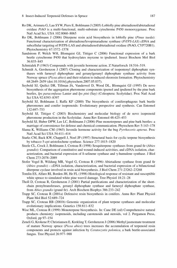
8 Insect-Induced Terpenoid Defenses in Spruce 187
Ro DK, Arimura G, Lau SYW, Piers E, Bohlmann J (2005) Loblolly pine abietadienol/abietadienaloxidase PtAO is a multi-functional, multi-substrate cytochrome P450 monooxygenase. ProcNatl Acad Sci, USA 102:8060–8065
Ro DK, Bohlmann J (2006) Diterpene resin acid biosynthesis in loblolly pine (Pinus taeda):Functional characterization of abietadiene/levopimaradiene synthase (PtTPS-LAS) cDNA andsubcellular targeting of PtTPS-LAS and abietadienol/abietadienal oxidase (PtAO, CYP720B1).Phytochemistry 67:1572–1578
Sandstrom P, Welch WH, Blomquist GJ, Tittiger C (2006) Functional expression of a barkbeetle cytochrome P450 that hydroxylates myrcene to ipsdienol. Insect Biochem Mol Biol36:835–845
Schmialek P (1963) Compounds with juvenile hormone action. Z Naturforsch 18:516–519.Schmidt A, Gershenzon J (2007) Cloning and characterization of isopentenyl diphosphate syn-
thases with farnesyl diphosphate and geranylgeranyl diphosphate synthase activity fromNorway spruce (Picea abies) and their relation to induced oleoresin formation. Phytochemistry68:2649–2659 (doi:10.1016/j.phytochem.2007.05.037).
Seybold SJ, Quilici DR, Tillman JA, Vanderwel D, Wood DL, Blomquist GJ (1995) De novobiosynthesis of the aggregation pheromone components ipsenol and ipsdienol by the pine barkbeetles, Ips paraconfusus Lanier and Ips pini (Say) (Coleoptera: Scolytidae). Proc Natl AcadSci USA 92:8393–8397
Seybold SJ, Bohlmann J, Raffa KF (2000) The biosynthesis of coniferophagous bark beetlepheromones and conifer isoprenoids: Evolutionary perspective and synthesis. Can Entomol132:697–753
Seybold SJ, Tittiger C (2003) Biochemistry and molecular biology of de novo isoprenoidpheromone production in the Scolytidae. Annu Rev Entomol 48:425–453
Seybold SJ, Huber DPW, Lee LC, Bohlmann J (2006) Pine monoterpenes and pine bark beetles: amarriage of convenience for defense and chemical communication. Phytochem Rev 5:143–178
Slama K, Williams CM (1965) Juvenile hormone activity for the bug Pyrrhocoris apterus. ProcNatl Acad Sci USA 54:411–414
Starks CM, Back KW, Chappell J, Noel JP (1997) Structural basis for cyclic terpene biosynthesisby tobacco 5-epi-aristolochene synthase. Science 277:1815–1820
Steele CL, Crock J, Bohlmann J, Croteau R (1998) Sesquiterpene synthases from grand fir (Abiesgrandis): Comparison of constitutive and wound-induced activities, and cDNA isolation, char-acterization, and bacterial expression of �-selinene synthase and �-humulene synthase. J BiolChem 273:2078–2089
Stofer Vogel B, Wildung MR, Vogel G, Croteau R (1996) Abietadiene synthase from grand fir(Abies grandis) – cDNA isolation, characterization, and bacterial expression of a bifunctionalditerpene cyclase involved in resin acid biosynthesis. J Biol Chem 271:23262–23268
Tomlin ES, Alfaro RI, Borden JH, He FL (1998) Histological response of resistant and susceptiblewhite spruce to simulated white pine weevil damage. Tree Physiol 18:21–28
Tholl D, Croteau R, Gershenzon J (2001) Partial purifications and characterization of the short-chain prenyltransferases, geranyl diphosphate synthase and farnesyl diphosphate synthase,from Abies grandis (grand fir). Arch Biochem Biophys 386:233–242
Trapp SC, Croteau R (2001a) Defensive resin biosynthesis in conifers. Annu Rev Plant PhysiolPlant Mol Biol 52:689–724
Trapp SC, Croteau RB (2001b) Genomic organization of plant terpene synthases and molecularevolutionary implications. Genetics 158:811–832
Wise ML, Croteau R (1999) Monoterpene biosynthesis. In: Cane DE (ed) Comprehensive naturalproducts chemistry: isoprenoids, including carotenoids and steroids, vol 2. Pergamon Press,Oxford, pp 97–154
Zeneli G, Krokene P, Christiansen E, Krekling T, Gershenzon J (2006) Methyl jasmomate treatmentof mature Norway spruce (Picea abies) trees increases the accumulation of terpenoid resincomponents and protects against infection by Ceratocystis polonica, a bark beetle-associatedfungus. Tree Physiol 26:977–988
Chapter 9Phenylpropanoid Metabolism Inducedby Wounding and Insect Herbivory
Mark A. Bernards and Lars Bastrup-Spohr
Wounding of plant tissue, whether by biotic or abiotic means, results in a massivere-arrangement of metabolism, with the primary objectives of isolating the affectedtissue and limiting the extent of further damage. Within this context, newly syn-thesized phenylpropanoid-derived compounds are known to provide anti-feedant,anti-microbial, and cytotoxic effects, as well as building blocks for structural macro-molecules (e.g., lignin, suberin) and condensed tannins. The literature describinghydroxycinnamates (and their conjugates), flavonoids and condensed tannins visa vis their roles in plant/herbivore interactions is incomplete. Nevertheless, it isclear that the biosynthesis of phenylalanine, a necessary precursor, is induced byherbivore damage, and there is a coordinate induction of transcripts/enzymes as-sociated with hydroxycinnamic acid, flavonoid, and condensed tannin formation.Recent genome scale studies have provided evidence for the involvement of manygenes in the response to herbivory, including those of phenylpropanoid metabolism.The next steps will certainly involve the functional characterization of these genes.Forward progress is dependent on expanding analyses to more plant-insect interac-tions, as well as monitoring plant reactions to herbivory (or wounding) over relevanttime frames. Defining the biochemistry of the phenylpropanoid metabolism of spe-cific plant-insect interactions, both at the transcript and enzyme levels, will leadto the discovery of new pathways, or variations on known pathways. In definingplant/herbivore-specific variations in phenylpropanoid metabolism we will discoverthe underlying strategies used by plants to defend themselves and gain insights intothe role(s) of phenylpropanoids in these processes.
9.1 Introduction
The overall success of plants defending themselves against herbivores likely de-pends on a combination of both pre-formed defenses (phytochemicals, physicalbarriers, etc.) and those induced by the chewing, boring, or sucking of insects.
M.A. BernardsDepartment of Biology, The University of Western Ontario, London, ON, Canada, N6A 5B7e-mail: [email protected]
A. Schaller (ed.), Induced Plant Resistance to Herbivory, 189C© Springer Science+Business Media B.V. 2008

190 M.A. Bernards, L. Bastrup-Spohr
This chapter summarizes the field of wound- and insect herbivore-induced phenyl-propanoid metabolism, with an emphasis on the types of molecules involved, and theenzymes and pathways associated with their biosynthesis. The chapter begins withan overview of the types of phenolic compounds known to accumulate in responseto wounding or herbivory, and some of their putative roles in defense against herbiv-orous insects, followed by a summary of the literature describing the enzymology ofphenylpropanoid metabolism induced by wounding and/or herbivory. It is importantto note up front that the literature on herbivore-induced phenylpropanoid enzymesat the protein/enzyme activity level is sparse. Few contributions describing the en-zymology of wound- and/or herbivory-induced phenylpropanoid metabolism havebeen made since the summary chapter written by Constable (1999). Instead, muchof the recent literature has focused on the induction of specific genes at the transcriptlevel and the use of genome wide screening of herbivory and wound-induced genesusing available microarrays. With the latter, there is much reliance on database an-notation for the identification of genes, with only a small subset of these having beenverified by functional analysis, so far. As we look ahead to what needs to be accom-plished in the future to further our understanding of the role of phenolics in theoverall interaction between plants and their insect pests, functional characterizationof the dozens of genes putatively involved in induced phenylpropanoid metabolismwill be high priority.
Since there are chapters elsewhere in this book that specifically address volatilearomatics (Chapters 19–21), wound periderm (Chapter 6), and polyphenol oxidase(Chapter 12), these topics will not be repeated herein. Similarly, there is an entiresection in this book devoted to signals in defense; as such the topic of wound-induced signaling (e.g., via the jasmonate pathway) will not be covered.
9.2 Phenolic Compounds and Plant-HerbivoreInteractions
9.2.1 Roles for Phenolics in Plant-Herbivore Interactions
Phenolic compounds are aromatic compounds bearing one or more hydroxyl groupon an aromatic ring. Phenylpropanoids, strictly speaking, are phenolic compoundsderived from phenylalanine. And while there are >8000 known phenolic com-pounds, roughly categorized into 14 compound classes based on the number ofcarbons and their arrangement (e.g., Strack 1997), only a small fraction of themhave been implicated in plant-herbivore interactions. Amongst these are the benzoicacids, hydroxycinnamic acids (and their conjugates), furanocoumarins, coumarins,stilbenes, flavonoids (especially flavonols), hydrolysable tannins, condensed tannins(catechin polymers; Fig. 9.1), and lignin (reviewed in Constable 1999). Woundingand herbivory also induce the formation of other poly(phenolic)-based cell wallmodifications, especially suberin (Ginzberg this volume).
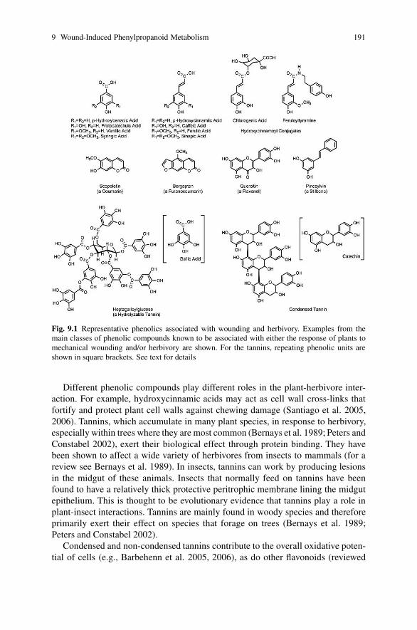
9 Wound-Induced Phenylpropanoid Metabolism 191
Fig. 9.1 Representative phenolics associated with wounding and herbivory. Examples from themain classes of phenolic compounds known to be associated with either the response of plants tomechanical wounding and/or herbivory are shown. For the tannins, repeating phenolic units areshown in square brackets. See text for details
Different phenolic compounds play different roles in the plant-herbivore inter-action. For example, hydroxycinnamic acids may act as cell wall cross-links thatfortify and protect plant cell walls against chewing damage (Santiago et al. 2005,2006). Tannins, which accumulate in many plant species, in response to herbivory,especially within trees where they are most common (Bernays et al. 1989; Peters andConstabel 2002), exert their biological effect through protein binding. They havebeen shown to affect a wide variety of herbivores from insects to mammals (for areview see Bernays et al. 1989). In insects, tannins can work by producing lesionsin the midgut of these animals. Insects that normally feed on tannins have beenfound to have a relatively thick protective peritrophic membrane lining the midgutepithelium. This is thought to be evolutionary evidence that tannins play a role inplant-insect interactions. Tannins are mainly found in woody species and thereforeprimarily exert their effect on species that forage on trees (Bernays et al. 1989;Peters and Constabel 2002).
Condensed and non-condensed tannins contribute to the overall oxidative poten-tial of cells (e.g., Barbehenn et al. 2005, 2006), as do other flavonoids (reviewed

192 M.A. Bernards, L. Bastrup-Spohr
in Treutter 2005). Paradoxically, one of the hallmark features of phenylpropanoidsin general is their anti-oxidative properties (i.e., they are readily oxidized to rela-tively stable quinone intermediates), but in the context of resistance to herbivoresthis translates into a class of compounds that, once oxidized can interfere withproteins in the digestive tract of chewing insects and mammals. This can resultin an overall decline in nutritional quality of plant material and/or cause oxidativestress in herbivores (Bi and Felton 1997; Barbehenn et al. 2005, 2006). The effectof defense compounds on herbivores also varies according to the compound. Forexample, defense compounds can influence herbivore settling, feeding, oviposition,growth fecundity and/or fertility (Walling 2000).
Since several types of phenolic compounds (e.g., hydroxycinnamates, hydrox-ycinnamoyl conjugates, flavonoids) possess anti-microbial properties (e.g.,Malmberg 1984; Ding et al. 2000), their accumulation in tissue damaged by wound-ing (including chewing damage) could play a dual role: direct defense againstherbivores and protection against opportunistic pathogens. For example, in thewound response of potato tubers, where the predominant role of induced phenyl-propanoid metabolism appears to be associated with the formation of the poly(phenolic) domain of suberin, other phenolics (e.g., hydroxycinnamoyl amides,chlorogenic acid) that are not only involved in suberin formation also accumulate(Cottle and Kolattukudy 1982; Malmberg 1984; Razem and Bernards 2002). Thesuberin phenolics play a role in forming a physical barrier, while the soluble phe-nolics possess anti-microbial properties and presumably protect against opportunis-tic pathogens.
9.2.2 Constitutive vs. Induced Accumulation of Phenolics
Phenolics are widely distributed in plants, and accumulate during normal growthand development. Consequently, phenolics are constitutively present in plants priorto insect or mammalian herbivory-induced damage (i.e., wounding). Indeed, rolesfor phenolics as pre-formed (constitutive) defenses against herbivory are well doc-umented (e.g., Ding et al. 2000; Mutikainen et al. 2000; Treutter 2005; Santiagoet al. 2005, 2006) and include physical barriers such as cell wall bound phenolics,lignins, suberin, and cuticle-associated phenolics as well as stored compounds thathave a deterring (anti-feedant) or directly toxic (insecticidal) effect on herbivores(Walling 2000). Induced defenses, on the other hand, are characterized by the syn-thesis of new compounds or structural barriers after herbivore-induced damage hasoccurred, and includes both proteins and secondary metabolites. Induced defensescan work both locally, near the point of attack and/or systemically, i.e., throughoutthe plant.
Since producing defense related compounds is metabolically expensive, consti-tutive and induced defense mechanisms can be seen as two different strategies foravoiding herbivores. Constitutive defenses are always present, but potentially ener-getically costly. For example, tannins accumulate as constitutive defense in poplarleaves (Arnold et al. 2004), but the extent of accumulation is dependent on the sink

9 Wound-Induced Phenylpropanoid Metabolism 193
strength of the developing leaves and associated with the import of carbohydratesfrom older source leaves. If other sinks, such as the roots, are removed by girdlingand thus decreasing competition for the carbohydrates, an increase in leaf tanninconcentration occurs. Increased phenolic accumulation in association with high sinkstrength, does not appear to be universal, however, since the opposite phenomenonhas been found in the gymnosperm species Pinus sylvestris, where the productionof phenolics was negatively correlated with sink strength (Honkanen et al. 1999).
Similarly, phenolics can also be induced in poplar leaves as a response to grazingby gypsy moth larvae, mechanical wounding and treatment with jasmonic acids(Arnold et al. 2004). In this study phenolic responses in leaves, induced by her-bivores, were found in the first six to eight days post emergence, when the wholecanopy was treated with jasmonic acid. During this period leaves are characterizedby sink strength indicated by the activity of the enzyme cell wall invertase thatfacilitates phloem unloading to the cells. Arnold et al. (2004) found that when car-bohydrate source leaves were removed, the induced response to jasmonic acid wassignificantly reduced. These results indicate that even a local defense response indeveloping foliage depends to some degree on long distance carbohydrate trans-port from source tissue. Thus, the role of tannins in plant defense, and maybe otherphenylpropanoids as well, is not only dependent on the plant’s genetic basis to pro-duce them, but also on what stage a particular leaf is in and if there is a source ofcarbohydrates to supply the production.
Induced defenses are only invoked after tissue damage has occurred. While thispotentially provides defense at a lower energetic cost it also comes with some loss ofbiomass. Thus plants need to balance the allocation of carbon and nitrogen resourcesbetween these two types of defenses and vegetative and reproductive growth toinsure survival in the long term (Walling 2000). However, it is not always easy to dis-tinguish between constitutive and induced defense mechanisms since some defense-related compounds are constitutively synthesized and stored, and also synthesizedde novo as a response to herbivore damage (Ding et al. 2000; Gatehouse 2002).
To add to this complexity, induced metabolic pathways differ when plants areexposed to different types of herbivory, and even species of herbivores. Generallyinsects that use a piercing/sucking mode of feeding elicit a response similar to thatof fungal and bacterial plant pathogens, while chewing insects produce a responsesimilar to that of mechanical wounding (e.g., Walling 2000). In the case of chewinginsects, it has been reported that the damage made by insects is not equivalent tomechanical wounding alone (Hartley and Firn 1989) since, for example, insect re-gurgitant can increase the response to chewing herbivory over that induced by me-chanical wounding alone (Korth and Dixon 1997). Further to this, some plants reactspecifically to one species of herbivore or even a specific life stage within a species(van der Ven et al. 2000), which may correspond to the content of the saliva in theinsect species (Walling 2000). However, the general response of plants to wound-ing and herbivore damage, vis a vis induced phenylpropanoid metabolism and theaccumulation of phenolic compounds, is essentially the same: i.e., the same typesof compounds are induced to accumulate, and thereby the same enzymes involved.Notwithstanding the subtle details defining specific plant-herbivore interactions, the

194 M.A. Bernards, L. Bastrup-Spohr
wound response literature, which is more complete than that of herbivore responseliterature, can provide a model for the phenylpropanoid metabolic processes in-volved in the response of plants to herbivore damage.
9.3 Induced Biosynthesis of Phenolic Compounds
Historically, phenylpropanoid metabolism has been divided into two main stages,‘general phenylpropanoid metabolism’ (i.e., the biosynthesis of hydroxycinnamicacids) and pathway specific metabolism (i.e., hydroxycinnamic acids as precur-sors to specific classes of phenylpropanoids such as flavonoids or coumarins; e.g.,Strack 1997). More recently it has been shown that the interconversion betweenhydroxycinnamic acids does not necessarily lie within a defined general pathway(reviewed in Dixon et al. 2001; Humphreys and Chapple 2002), and in fact branchesinto compound class-specific pathways as early as cinnamate (or cinnamoyl-CoA),or after its hydroxylation to form p-coumarate. Consequently, ‘general phenyl-propanoid metabolism’ only consists of two common steps: the deamination ofphenylalanine by phenylalanine ammonia-lyase (PAL) and the 4-hydroxylation ofcinnamate by cinnamate-4-hydroxylase (C4H).
The main classes of soluble phenolic compounds involved in the response ofplants to wounding and herbivory are hydroxycinnamic acids (and their conjugatesand derivatives) and tannins (via flavonoids), and these will be considered separatelybelow. However, phenylpropanoids are derived from phenylalanine, which also hasto be synthesized de novo as part of the overall wound/herbivory response, and it isimportant to consider this as an early stage of induced phenylpropanoid metabolism.
9.3.1 The Shikimate Pathway
All three aromatic amino acids, phenylalanine, tyrosine, and tryptophan are syn-thesized via the shikimate pathway. With respect to induced phenolic metabolism,the synthesis of phenylalanine (Fig. 9.2; Table 9.1) is of particular importancesince a significant flux of carbon must go through this intermediate. The first en-zyme in the shikimate pathway is 3-deoxy-D-arabino-heptulosonate-7-phosphate(DAHP) synthase, which catalyses the condensation of one molecule each of phos-phoenolpyruvate and erythrose-4-phosphate; both of these starting materials are de-rived from primary metabolism. DAHP synthase has been shown to be induced, atthe transcript, protein, and enzyme activity levels, in wounded potato tubers andtomato fruits (Dyer et al. 1989). More recently, DAHP synthase gene up-regulationin Sitka spruce bark following mechanical wounding and weevil feeding, as well asin buds following spruce bud worm larvae feeding has been demonstrated at the tran-script level, using a microarray gene profiling approach (Ralph et al. 2006). In addi-tion, shikimate kinase was also shown to be coordinately up-regulated with DAHPsynthase in the same tissues (Ralph et al. 2006). Similarly, wounding has been

9 Wound-Induced Phenylpropanoid Metabolism 195
Fig. 9.2 The shikimate pathway and N-recycling in plants. The shikimic acid pathway, fromphospho-enol-pyruvate and erythrose-4-phosphate to phenylalanine is shown. Cofactors and co-substrates are not shown for clarity. Refer to Table 9.1. Bold arrows indicate steps for whichwounding and/or herbivory have been shown to result in induced gene expression and/or en-zyme activity. Enzymes are: (1) 3-deoxy-D-arabino-heptulosonate-7-phosphate (DAHP) syn-thase; (2) 3-dehydroquinate synthase; (3) 3-dehydroquinate dehydratrase; (4) shikimate de-hydrogenase; (5) shikimate kinase; (6) 5-enolpyruvylshikimate-3-phosphate (EPSP) synthase;

196 M.A. Bernards, L. Bastrup-Spohr
shown to induce enzyme activity levels of dehydroquinate dehydratase/shikimatedehydrogenase in sweet potato (Minamikawa et al. 1966) and chorismate mutase inpotato tubers (Kuroki and Conn 1988). These studies provide a critical link betweenprimary carbon metabolism and the de novo synthesis of phenolics in response towounding and herbivory. No other enzymes of the shikimate pathway have beenshown to be directly involved in the response of plants to wounding or herbivory, ei-ther at the transcript or protein levels. However, a recent genomic-scale explorationof gene expression during active lignin formation in poplar (Ehlting et al. 2005) hasdemonstrated that several shikimate pathway genes are co-coordinately regulated,including 3-dehydroquinate synthase, 3-dehydroquinate dehydratrase/shikimate de-hydrogenase, shikimate kinase, chorismate synthase, chorismate mutase, putativeaspartate: prephenate aminotransferases and arogenate dehydratase. Significantly,neither arogenate dehydrogenase (which forms tyrosine from arogenate), nor any ofthe genes associated with tryptophan biosynthesis (which branches off of the mainshikimate pathway at chorismate) were up-regulated during lignification, suggest-ing a process-specific subset of shikimate pathway genes to be involved. The syn-chronized upregulation of genes for shikimate pathway enzymes and PAL was alsoobserved in tomato cell cultures in response to fungal elicitors (Gorlach et al. 1995).Thus, while not strictly wound- or herbivory-based studies, these data illustratethe coordinate regulation of the biosynthetic pathway providing the precursor forphenylpropanoid metabolism. Presumably, this level of coordinate regulation is inoperation when phenylpropanoid metabolism is induced by wounding or herbi-vore damage.
Herbivory and wounding have been clearly linked to the induction of PAL atthe transcript, protein and enzyme levels (e.g., Bi and Felton 1995; Hartley andFirn 1989; Bernards et al. 2000; Devoto et al. 2005; Qiuju et al. 2005; Saltveitet al. 2005; Ralph et al. 2006). Phenylalanine ammonia-lyase serves as the gatewayenzyme into phenylpropanoid metabolism, and while it is considered in more detailin Section 9.3.2, it is mentioned here since metabolism of phenylalanine representsan exit point from the shikimate pathway and provides a strong metabolic draw onit. The deamination of phenylalanine by PAL yields two products: cinnamic acid,the metabolic fate of which will be discussed in greater detail in Section 9.3.2, andammonia. As noted above, the production of defense compounds is metabolicallyexpensive, and the ‘cost’ of inducing phenylpropanoid metabolism for chemical de-fense could potentially be very high if one mole of ammonia were lost for each moleof phenylpropanoid metabolite formed. However, plants have evolved an efficientN-recycling mechanism in which the ammonia liberated from phenylalanine byPAL is reused in the production of more phenylalanine (Fig. 9.2). This recycling
�Fig. 9.2 (continued) (7) chorismate synthase; (8) chorismate mutase; (9) aspartate:prephenateaminotransferase; (10) arogenate dehydratase; (11) phenylalanine ammonia-lyase; (12) glu-tamine synthetase; (13) ferredoxin-dependent glutamine:2-oxoglutarate aminotransferase. �-KG,�-ketoglutarate (2-oxoglutarate); Fdxred, reduced ferredoxin; Fdxox, oxidized ferredoxin. (Adaptedin part from Razal et al. 1996; Strack 1997; Croteau et al. 2000)

9 Wound-Induced Phenylpropanoid Metabolism 197T
able
9.1
Enz
ymes
ofth
esh
ikim
ate
path
way
sle
adin
gto
phen
ylal
anin
ean
dth
eir
corr
espo
ndin
gge
nes
inA
rabi
dops
is
Enz
yme
nam
eE
.Cnu
mbe
r1R
eact
ion
cata
lyze
dR
epre
sent
ativ
eA
rabi
dops
isge
nes2
133-
Deo
xy-D
-ara
bino
-he
ptul
oson
ate-
7-ph
osph
ate
Synt
hase
(DA
HPS
)
2.5.
1.54
phos
phoe
nolp
yruv
ate
+er
ythr
ose
4-ph
osph
ate
+H
2O
→3-
Deo
xy-D
-ara
bino
-he
ptul
oson
ate-
7-ph
osph
ate
+P i
At1
g224
10,A
t4g3
3510
,At4
g399
80
23-
Deh
ydro
quin
ate
Synt
hase
4.2.
3.4
3-D
eoxy
-D-a
rabi
no-h
eptu
loso
nate
-7-p
hosp
hate
→3-
dehy
droq
uina
te+
P iA
t5g6
6120
3/4
3-D
ehyd
roqu
inat
eD
ehyd
rata
se/S
hiki
mat
eD
ehyd
roge
nase
4.2.
1.10
/1.
1.1.
253-
dehy
droq
uina
te→
3-de
hydr
oshi
kim
ate+
H2O
3-de
hydr
oshi
kim
ate
+N
AD
P+→
shik
imat
e+
NA
DPH
,H+
At3
g063
50
5Sh
ikim
ate
Kin
ase
(SK
)2.
7.1.
71sh
ikim
ate+
AT
P→
shik
imat
e-3-
phos
phat
e+
AD
PA
t1g0
6890
,At1
g605
30,A
t2g1
4050
,At2
g167
90,
At2
g219
40,A
t2g3
5500
,At3
g509
50,A
t3g2
6900
,A
t4g3
9540
,At5
g223
70,A
t5g4
5210
,At5
g470
506
5-en
olpy
ruvy
lshi
kim
ate
3-ph
osph
ate
Synt
hase
(EPS
PS)
2.5.
1.19
shik
imat
e-3-
phos
phat
e+
phos
phoe
nolp
yruv
ate
→5-
enol
pyru
vyls
hiki
mat
e-3-
phos
phat
e+
P iA
t1g4
8860
,At2
g453
00
7C
hori
smat
eSy
ntha
se(C
S)4.
2.3.
55-
enol
pyru
vyls
hiki
mat
e-3-
phos
phat
e→
chor
ism
ate
+P i
At1
g488
50
8C
hori
smat
eM
utas
e(C
M)
5.4.
99.5
chor
ism
ate
→pr
ephe
nate
At1
g693
70,A
t3g2
9200
,At5
g108
709
Asp
arta
te-P
reph
enat
eA
min
otra
nsfe
rase
2.6.
1.78
prep
hena
te+
aspa
rtat
e→
arog
enat
e+
oxal
oace
tate
Unk
now
nin
Ara
bido
psis
10A
roge
nate
Deh
ydra
tase
(AD
T)
4.2.
1.91
arog
enat
e→
phen
ylal
anin
e+
CO
2+
H2O
At1
g082
50,A
t1g1
1790
,At2
g278
20,A
t3g0
7630
,A
t3g4
4720
,At5
g226
3011
Phen
ylal
anin
eA
mm
onia
-lya
se(P
AL
)4.
3.1.
5ph
enyl
alan
ine
→ci
nnam
ate
+N
H+ 4
At2
g370
40,A
t3g1
0340
,At3
g532
60,A
t5g0
4230
12G
luta
min
eSy
nthe
tase
(GS)
6.3.
1.2
glut
amat
e+
NH
+ 4+
AT
P→
glut
amin
e+
AD
P+
P iA
t1g4
8470
,At1
g662
00,A
t3g1
7820
,At3
g531
80,
At5
g165
70,A
t5g3
5630
,At5
g376
0013
Ferr
edox
in-d
epen
dent
glut
amat
esy
ntha
se1.
4.7.
1gl
utam
ine+
2-ox
oglu
tara
te+
Fdx r
ed→
2gl
utam
ate+
Fdx o
x
At2
g412
20
1E
nzym
eN
omen
clat
ure
(web
vers
ion)
ww
w.c
hem
.qm
ul.a
c.uk
/iub
mb/
enzy
me/
2G
ene
iden
tifier
sar
efr
omE
hltin
get
al.2
005,
Ral
phet
al.
2006
,an
dT
heA
rabi
dops
isIn
form
atio
nR
esou
rce
(TA
IR)
(ww
w.a
rabi
dops
is.o
rg).
Not
all
gene
sli
sted
have
been
func
tion
ally
char
acte
rize
d;no
rar
eth
eli
sts
cons
ider
edco
mpl
ete.
3E
nzym
enu
mbe
rre
fer
toth
eco
rres
pond
ing
reac
tion
inFi
g.9.
2.

198 M.A. Bernards, L. Bastrup-Spohr
mechanism, which involves the glutamine synthetase/ferredoxin-dependent glu-tamine 2-oxoglutarate amino transferase (GS/GOGAT) ammonia assimilation sys-tem, has been demonstrated in wound healing potato tubers (Razal et al. 1996),sweet potato tubers (Singh et al. 1998), and in lignifying cell cultures of Pinustaeda (van Heerden et al. 1996). Thus, the heavy demand for phenylalanine broughtabout by wound-induced phenylpropanoid metabolism is met by the induction of theshikimate pathway and an efficient N-recycling system. However, with the exceptionof demonstrated GS up-regulation post wounding and/or herbivore damage in Sitkaspruce (Ralph et al. 2006), these aspects of induced phenylpropanoid metabolismhave not been demonstrated in plant-herbivore interactions. While it is logical toassume that the shikimate and N-recycling pathways are more broadly associated inplant-herbivore interactions resulting in de novo synthesis of phenolics, confirma-tion of their participation is lacking.
9.3.2 Induced General Phenylpropanoid Metabolismand the Origin of Hydroxycinnamates
The induction of phenylpropanoid metabolism is implied in the accumulation ofnewly formed phenolic compounds in several plants in response to herbivore dam-age, e.g., p-coumaric and ferulic acids in wheat exposed to Sitodipolis mosellana(Ding et al. 2000) and corn exposed to Sesamina noonagrioides (Santiago et al. 2005,2006), tannins in many tree species (Bernays et al. 1989; Peters and Constabel 2002;Arnold et al. 2004), and phenolics in Medicago truncatula damaged by Spodopteralittoralis (Leitner et al. 2005). There are relatively few reports in which the activityof phenylpropanoid biosynthesis enzymes is measured directly.
Phenylalanine ammonia-lyase (PAL) is the gateway enzyme providing C6–C3
carbon skeletons for the formation of essentially all phenylpropanoids and theirderivatives (Fig. 9.3, Table 9.2). The conversion of Phe to cinnamate + NH+
4 isconsidered to be physiologically irreversible, and thus is a good indicator of car-bon commitment to phenylpropanoid metabolism. Phenylalanine ammonia-lyaseis represented by a small gene family (reviewed in Dixon and Paiva 1995; Dixonet al. 2001). For example, at least four genes have been annotated as PAL in theArabidopsis database, while ten uniquely expressed sequence tags (ESTs) are de-scribed for Sitka spruce (Ralph et al. 2006). Not surprisingly, this step in the phenyl-propanoid metabolic pathway has received a great deal of attention in the literature,albeit largely as a function of biotic and abiotic stress induction; e.g., responseto pathogen attack, exposure to UV light, wounding etc. (Strack 1997; Croteauet al. 2000). From a herbivory induction standpoint, increased PAL activity has beendirectly measured in a few insect/plant systems, e.g., Helicoverpa zea damaged soy-bean leaves (Bi and Felton 1995); Apocheima pilosaria damaged birch (Hartley andFirn 1989); gypsy moth larvae damaged poplar leaves (Arnold et al. 2004). At thetranscript level, PAL has been shown to be induced by insect damage of Arabidopsis(Reymond et al. 2004) and Sitka spruce (Ralph et al. 2006). There are many more

9 Wound-Induced Phenylpropanoid Metabolism 199
Fig
.9.3
(con
tinu
ed)
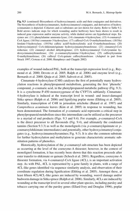
200 M.A. Bernards, L. Bastrup-Spohr
Fig. 9.3 (continued) Biosynthesis of hydroxycinnamic acids and their conjugates and derivatives.The biosynthesis of hydroxycinnamates, hydroxycinnamoyl conjugates, and derivatives of hydrox-ycinnamates is depicted. Cofactors and co-substrates are not shown for clarity. Refer to Table 9.2.Bold arrows indicate steps for which wounding and/or herbivory have been shown to result ininduced gene expression and/or enzyme activity, while dashed arrows are hypothetical steps. En-zymes are: (11) phenylalanine ammonia-lyase; (14) cinnamate-4-hydroxylase; (15) 4-coumaroyl-CoA ligase; (16/16a) coumarate-3-hydroxylase; (17) caffeoyl-CoA O-methyltransferase; (18/18a)ferulate-5-hydroxylase; (19/19a) caffeic acid/5-hydroxyferulic acid O-methyltransferase; (20)hydroxycinnamoyl CoA:shikimate/quinate hydroxycinnamoyltransferase; (21) cinnamoyl-CoAreductase; (22) cinnamyl alcohol dehydrogenase; (23) hydroxycinnamoyl CoA:tyramine hy-droxycinnamoyltransferase; (24) p-coumaroyltyramine-3-hydroxylase; (25) caffeoyltyramineO-methyltransferase; (26) caffeoyl CoA:quinate caffeoyltransferase. (Adapted in part fromStrack 1997; Croteau et al. 2000; Humphreys and Chapple 2002)
examples of wound-induced PAL, both at the transcript expression level (e.g., Rey-mond et al. 2000; Devoto et al. 2005; Ralph et al. 2006) and enzyme level (e.g.,Bernards et al. 2000; Qiuju et al. 2005; Saltveit et al. 2005).
Cinnamate-4-hydroxylase (C4H) catalyses the first of potentially many hydrox-ylation reactions in phenylpropanoid metabolism, generating the first ‘phenolic’compound, p-coumaric acid, in the phenylpropanoid metabolic pathway (Fig. 9.3).It is a cytochrome P-450 monooxygenase of the CYP73A subfamily. Cinnamate-4-hydroxylase is induced at the transcript level in wound- and insect-damagedSitka spruce (Ralph et al. 2006) and lignifying poplar stems (Ehlting et al. 2005).Similarly, transcription of C4H in jerusalem artichoke (Batard et al. 1997) andCamptotheca acuminata leaves (Kim et al. 2005) in response to wounding, hasbeen demonstrated. The formation of p-coumaric acid represents a critical step inphenylpropanoid metabolism since this intermediate can be utilized as the precursorto a myriad of end products (Figs. 9.3 and 9.4). For example, p-coumaroyl-CoAis the direct precursor to all flavonoids (Fig. 9.4), and ultimately the condensedtannins (Section 9.3.3) as well as the monolignols (via p-coumaroylquinnate or p-coumaroylshikimate intermediates) and potentially, other hydroxycinnamoyl conju-gates (e.g., hydroxycinnamoyltyramines; Fig. 9.3). It is also the common substratefor further hydroxylation and methylation to generate characteristic aromatic sub-stitution patterns of phenolics.
Historically, hydroxylation of the p-coumaroyl sub-structure has been depictedas occurring at the level of the coenzyme-A thioester; however, in the context ofmonolignol formation, it has recently been shown that this reaction proceeds afterester transfer to shikimate or quinate (Schoch et al. 2001). Regardless, coenzyme-Athioester formation, via 4-coumaroyl-CoA ligase (4CL), is an important activationstep. As with PAL, 4CL is represented by a gene family in plants, with a range of4CL-like genes identified via sequence similarity analysis, and observation of theircoordinate regulation during lignification (Ehlting et al. 2005). Amongst these, atleast fifteen 4CL/4CL-like genes are induced by wounding, weevil damage and/orbudworm damage in Sitka spruce (Ralph et al. 2006). Similarly, 4CLs are induced bywounding at the transcript level in several other plant species, including parsley andtobacco carrying one of the parsley genes (Ellard-Ivey and Douglas 1996), poplar

9 Wound-Induced Phenylpropanoid Metabolism 201
Tab
le9.
2E
nzym
esof
phen
ylpr
opan
oid
met
abol
ism
lead
ing
tohy
drox
ycin
nam
icac
ids
and
thei
rco
njug
ates
,and
thei
rco
rres
pond
ing
gene
sin
Ara
bido
psis
Enz
yme
nam
eE
.Cnu
mbe
r1R
eact
ion
cata
lyze
dR
epre
sent
ativ
eA
rabi
dops
isge
nes2
143
Cin
nam
ate-
4-H
ydro
xyla
se(C
4H)
(CY
P73A
)1.
14.1
3.11
cinn
amat
e+
NA
DPH
,H+
+O
2→
p-co
umar
ate
+N
AD
P++
H2O
At2
g304
90,A
t2g3
0160
,At2
g306
00,
At2
g305
80,
154-
Cou
mar
oyl-
CoA
Lig
ase
(4C
L)
6.2.
1.12
p-co
umar
ate
+co
enzy
me
A+
AT
P→
p-co
umar
oyl-
CoA
+A
MP
+PP
i
At1
g516
80,A
t1g6
5060
,At3
g212
30,
At3
g212
4016
/16a
Cou
mar
ate-
3-H
ydro
xyla
se(C
3H)
(CY
P98A
)1.
14.1
3.36
p-co
umar
oyl-
CoA
[qui
nate
/shi
kim
ate]
+N
AD
PH,H
++
O2→
caff
eoyl
-CoA
[qui
nate
/shi
kim
ate]
+N
AD
P++
H2O
At1
g745
40,A
t1g7
4550
,At2
g408
90
17C
affe
oyl-
CoA
O-M
ethy
ltra
nsfe
rase
(CoA
OM
T)
2.1.
1.10
4ca
ffeo
yl-C
oA+
S-ad
enos
yl-L
-met
hion
ine
→fe
rulo
yl-C
oA+
S-ad
enos
yl-L
-ho
moc
yste
ine
At1
g247
35,A
t1g6
7980
,At1
g679
90,
At3
g619
90,A
t4g2
6220
,At4
g340
50
18/1
8aFe
rula
te-5
-Hyd
roxy
lase
(F5H
)(C
YP8
4A)
1.14
.13.
XX
feru
late
+N
AD
PH,H
++
O2→
5-hy
drox
yfer
ulat
e+
NA
DP+
+H
2O
At4
g362
20,A
t5g0
4330
19/1
9aC
affe
icA
cid/
5-H
ydro
xyfe
ruli
cac
idO
-Met
hylt
rans
fera
se(C
OM
T)
2.1.
1.68
caff
eate
/5-h
ydro
xyfe
rula
te+S
-ade
nosy
l-L
-met
hion
ine
→fe
rula
te/s
inap
ate+
S-ad
enos
yl-L
-ho
moc
yste
ine
At5
g541
60
20H
ydro
xyci
nnam
oyl
CoA
:Shi
kim
ate/
Qui
nate
hy-
drox
ycin
nam
oylt
rans
fera
se(C
ST/Q
ST)
2.3.
1.13
3p-
coum
aroy
l-C
oA+
quin
ate/
shik
imat
e→
p-co
umar
oyl-
quin
ate/
shik
imat
e+
coen
zym
eA
At5
g489
30
21C
inna
moy
l-C
oAR
educ
tase
(CC
R)
1.2.
1.44
hydr
oxyc
inna
moy
l-C
oA+
NA
DPH
,H+
→hy
drox
ycin
nam
alde
hyde
+N
AD
P++
coen
zym
eA
At1
g159
50,A
t1g8
0820
22C
inna
myl
Alc
ohol
Deh
ydro
gena
se(C
AD
)1.
1.1.
195
hydr
oxyc
inna
mal
dehy
de+
NA
DPH
,H+
→hy
drox
ycin
nam
ylal
coho
l+N
AD
P+A
t1g7
2680
,At3
g194
50,A
t4g3
4230
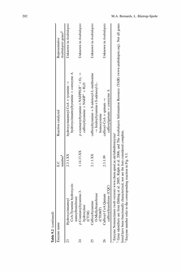
202 M.A. Bernards, L. Bastrup-Spohr
Tab
le9.
2(c
onti
nued
)
Enz
yme
nam
eE
.Cnu
mbe
r1R
eact
ion
cata
lyze
dR
epre
sent
ativ
eA
rabi
dops
isge
nes2
23H
ydro
xyci
nnam
oyl
CoA
:Tyr
amin
ehy
drox
ycin
-na
moy
ltra
nsfe
rase
2.3.
1.X
Xhy
drox
ycin
nam
oyl-
CoA
+ty
ram
ine
→hy
drox
ycin
nam
oylt
yram
ine
+co
enzy
me
AU
nkno
wn
inA
rabi
dops
is
24p-
Cou
mar
oylt
yram
ine-
3-hy
drox
ylas
e(C
T3H
)
1.14
.13.
XX
p-co
umar
oylt
yram
ine+
NA
DPH
,H+
+O
2→
caff
eoyl
tyra
min
e+
NA
DP+
+H
2O
Unk
now
nin
Ara
bido
psis
25C
affe
oylt
yram
ine
O-M
ethy
ltra
nsfe
rase
(CT
OM
T)
2.1.
1.X
Xca
ffeo
ylty
ram
ine
+S-
aden
osyl
-L-m
ethi
onin
e→
feru
loyl
tyra
min
e+S-
aden
osyl
-L-
hom
ocys
tein
e
Unk
now
nin
Ara
bido
psis
26C
affe
oyl
CoA
:Qui
nate
caff
eoyl
tran
sfer
ase
(CQ
C)
2.3.
1.99
caff
eoyl
-CoA
+qu
inat
e→
caff
eoyl
quin
ate
+co
enzy
me
AU
nkno
wn
inA
rabi
dops
is
1E
nzym
eN
omen
clat
ure
(web
vers
ion)
ww
w.c
hem
.qm
ul.a
c.uk
/iub
mb/
enzy
me/
2G
ene
iden
tifier
sar
efr
omE
hltin
get
al.2
005,
Ral
phet
al.
2006
,an
dT
heA
rabi
dops
isIn
form
atio
nR
esou
rce
(TA
IR)
(ww
w.a
rabi
dops
is.o
rg).
Not
all
gene
sli
sted
have
been
func
tion
ally
char
acte
rize
d;no
rar
eth
eli
sts
cons
ider
edco
mpl
ete.
3E
nzym
enu
mbe
rre
fer
toth
eco
rres
pond
ing
reac
tion
inFi
g.9.
3.

9 Wound-Induced Phenylpropanoid Metabolism 203
Fig
.9.4
(con
tinu
ed)
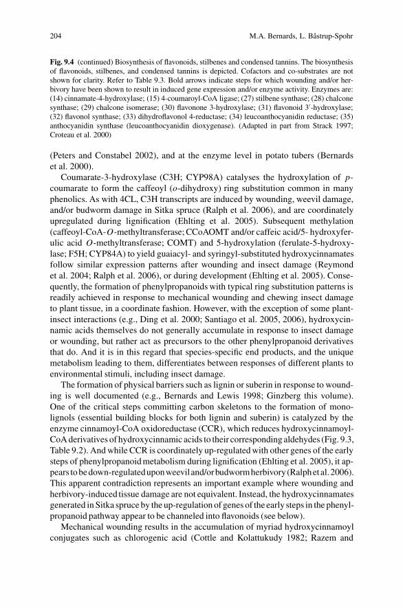
204 M.A. Bernards, L. Bastrup-Spohr
Fig. 9.4 (continued) Biosynthesis of flavonoids, stilbenes and condensed tannins. The biosynthesisof flavonoids, stilbenes, and condensed tannins is depicted. Cofactors and co-substrates are notshown for clarity. Refer to Table 9.3. Bold arrows indicate steps for which wounding and/or her-bivory have been shown to result in induced gene expression and/or enzyme activity. Enzymes are:(14) cinnamate-4-hydroxylase; (15) 4-coumaroyl-CoA ligase; (27) stilbene synthase; (28) chalconesynthase; (29) chalcone isomerase; (30) flavonone 3-hydroxylase; (31) flavonoid 3′-hydroxylase;(32) flavonol synthase; (33) dihydroflavonol 4-reductase; (34) leucoanthocyanidin reductase; (35)anthocyanidin synthase (leucoanthocyanidin dioxygenase). (Adapted in part from Strack 1997;Croteau et al. 2000)
(Peters and Constabel 2002), and at the enzyme level in potato tubers (Bernardset al. 2000).
Coumarate-3-hydroxylase (C3H; CYP98A) catalyses the hydroxylation of p-coumarate to form the caffeoyl (o-dihydroxy) ring substitution common in manyphenolics. As with 4CL, C3H transcripts are induced by wounding, weevil damage,and/or budworm damage in Sitka spruce (Ralph et al. 2006), and are coordinatelyupregulated during lignification (Ehlting et al. 2005). Subsequent methylation(caffeoyl-CoA-O-methyltransferase; CCoAOMT and/or caffeic acid/5- hydroxyfer-ulic acid O-methyltransferase; COMT) and 5-hydroxylation (ferulate-5-hydroxy-lase; F5H; CYP84A) to yield guaiacyl- and syringyl-substituted hydroxycinnamatesfollow similar expression patterns after wounding and insect damage (Reymondet al. 2004; Ralph et al. 2006), or during development (Ehlting et al. 2005). Conse-quently, the formation of phenylpropanoids with typical ring substitution patterns isreadily achieved in response to mechanical wounding and chewing insect damageto plant tissue, in a coordinate fashion. However, with the exception of some plant-insect interactions (e.g., Ding et al. 2000; Santiago et al. 2005, 2006), hydroxycin-namic acids themselves do not generally accumulate in response to insect damageor wounding, but rather act as precursors to the other phenylpropanoid derivativesthat do. And it is in this regard that species-specific end products, and the uniquemetabolism leading to them, differentiates between responses of different plants toenvironmental stimuli, including insect damage.
The formation of physical barriers such as lignin or suberin in response to wound-ing is well documented (e.g., Bernards and Lewis 1998; Ginzberg this volume).One of the critical steps committing carbon skeletons to the formation of mono-lignols (essential building blocks for both lignin and suberin) is catalyzed by theenzyme cinnamoyl-CoA oxidoreductase (CCR), which reduces hydroxycinnamoyl-CoA derivatives of hydroxycinnamic acids to their corresponding aldehydes (Fig. 9.3,Table 9.2). And while CCR is coordinately up-regulated with other genes of the earlysteps of phenylpropanoid metabolism during lignification (Ehlting et al. 2005), it ap-pears to bedown-regulateduponweeviland/orbudwormherbivory(Ralphetal.2006).This apparent contradiction represents an important example where wounding andherbivory-induced tissue damage are not equivalent. Instead, the hydroxycinnamatesgenerated in Sitka spruce by the up-regulation of genes of the early steps in the phenyl-propanoid pathway appear to be channeled into flavonoids (see below).
Mechanical wounding results in the accumulation of myriad hydroxycinnamoylconjugates such as chlorogenic acid (Cottle and Kolattukudy 1982; Razem and

9 Wound-Induced Phenylpropanoid Metabolism 205
Bernards 2002) and hydroxycinnamoyl amides (Malmberg 1984; Razem andBernards 2002) in potatoes and tomatoes (Pearce et al. 1998). With respect tohydroxycinnamoyltyramines, a hydroxycinnamoyl CoA:tyramine hydroxycinna-moyltransferase (HTH) enzyme has been shown to be induced by wounding(Negrel et al. 1995), which utilizes either p-coumaroyl-CoA or feruloyl-CoA ashydroxycinnamoyl donor (Fig. 9.3). Interestingly, even though feruloyltyramine hasbeen shown to be incorporated into the phenolic domain of wound suberin (Negrelet al. 1996), it is the p-coumaroyl derivative that accumulates when poly(phenolic)domain formation is inhibited (Razem and Bernards 2002). Subsequently, a uniquecoumarate-3-hydroxylase (C3H) that utilizes p-coumaroyltyramine as substrate inwound-healing potato tubers has been measured in vitro (Pabani and Bernards, un-published results). This result serves to emphasize the central role of p-coumaroyl-CoA in hydroxycinnamic acid metabolism as well as the power of using wound-induced (i.e., either mechanical of herbivory associated) tissues as a source ofdiscovering novel biochemistry.
9.3.3 Flavonoids and Tannins
Flavonoids are compounds with a C15 aglycone skeleton and form the building blocksof the condensed tannins (Fig. 9.4, Table 9.3). The first compound in the pathway,naringenin chalcone (4,2′,4′,6′-tetrahydroxychalcone), is formed by the condensationof p-coumaroyl-CoA with three molecules of malonyl-CoA, catalyzed by the enzymechalcone synthase (CHS). Chalcone synthase is considered to be the rate limiting en-zyme in the flavonoid pathway (Strack 1997), and has been shown to be induced at boththe protein and transcript level by wounding of Norway spruce (Brignolas et al. 1995)and white spruce (Richard et al. 2000), respectively, as well as at the transcript level bywounding and weevil and/or budworm herbivory of Sitka spruce (Ralph et al. 2006).Interestingly, stilbene synthase, which catalyzes the same condensation reaction asCHS,but results in adifferent folding and cyclyzationsequence to generate stilbenoids(e.g., pinosylvin; Fig. 9.4), is also induced at the protein level by wounding in Norwayspruce (Brignolas et al. 1995) and at transcript level by wounding and weevil and/orbudworm herbivory of Sitka spruce (Ralph et al. 2006). Presumably the combinedsynthesis of stilbenes and condensed tannins (from CHS-derived flavonoids) repre-sents a concerted response to both deter insects and prevent opportunistic pathogensfrom establishing themselves in wounded tissues.
Naringenin chalcone is converted into naringenin via a stereospecifc ring clo-sure and isomerization step catalyzed by the enzyme chalcone isomerase (CHI).Chalcone isomerase transcription is induced by wounding and weevil and/or bud-worm herbivory of Sitka spruce (Ralph et al. 2006). The metabolic fate of narin-genin is an important branchpoint in flavonoid biosynthesis, giving rise to theflavonols (e.g., kaempferol, quercitin), dihydroflavonols (e.g., dihydrokaempferol,dihydroquercitin), isoflavonoids (e.g., genistein) and flavones (e.g., apigenin). Col-lectively, these compounds are referred to as flavonoids. However, while flavonoidsthemselves have been reported to have deterring effects on insect herbivores (e.g.,

206 M.A. Bernards, L. Bastrup-Spohr
Tab
le9.
3Se
lect
eden
zym
esof
flavo
noid
san
dst
ilbe
nesy
nthe
sis,
and
thei
rco
rres
pond
ing
gene
sin
Ara
bido
psis
Enz
yme
nam
eE
.Cnu
mbe
r1R
eact
ion
cata
lyze
dR
epre
sent
ativ
eA
rabi
dops
isge
nes2
273
Stil
bene
Synt
hase
(ST
S)2.
3.1.
146
cinn
amoy
l-C
oA+
3m
alon
yl-C
oA→
pino
sylv
in+
4co
enzy
me
A+
4CO
2
Unk
now
nin
Ara
bido
psis
28C
halc
one
Synt
hase
(CH
S)2.
3.1.
74p-
coum
aroy
l-C
oA+
3m
alon
yl-C
oA→
nari
ngen
inch
alco
ne+
4co
enzy
me
A+
3CO
2
At1
g020
50,A
t4g0
0040
,At4
g348
50,A
t5g1
3930
29C
halc
one
Isom
eras
eC
HI)
5.5.
1.6
nari
ngen
inch
alco
ne→
nari
ngen
inA
t1g5
3520
,At5
g052
70,A
t3g5
5120
,At5
g662
2030
Flav
anon
e3-
Hyd
roxy
lase
(FH
T)
1.14
.11.
9na
ring
enin
+2-
oxog
luta
rate
+O
2→
dihy
drok
aem
pfer
ol+
succ
inat
e+
CO
2+
H2O
At1
g785
50,A
t3g1
9000
,At3
g512
40,A
t4g1
0490
,A
t4g1
6330
,At5
g245
30
31Fl
avon
oid
3′ -Hyd
roxy
lase
(F3H
)1.
14.1
3.21
dihy
drok
aem
pfer
ol+N
AD
PH,H
++O
2→
dihy
droq
uerc
itin
+N
AD
P++
H2O
At5
g079
90
32Fl
avon
olSy
ntha
se(F
LS)
1.14
.11.
23di
hydr
okae
mpf
erol
/di
hydr
oque
rcit
in+
2-ox
oglu
tara
te+
O2
→ka
empf
erol
/que
rcit
in+
succ
inat
e+
CO
2+
H2O
At1
g493
90,A
t2g4
4800
,At3
g190
10,A
t3g5
0210
,A
t5g0
8640
,At5
g439
35,A
t5g6
3580
,A
t5g6
3590
,At5
g635
95,A
t5g6
3600
33D
ihyd
rofla
vono
l4-R
educ
tase
(DFR
)1.
1.1.
219
quer
citi
n+
NA
DPH
,H+
→le
ucoc
yani
din
+N
AD
P+A
t5g4
2800
34L
euco
anth
ocya
nidi
nR
educ
tase
(LA
R)
1.17
.1.3
leuc
ocya
nidi
n+
NA
DPH
,H+
→ca
tech
in+
NA
DP+
+H
2O
At1
g617
20
35A
ntho
cyan
idin
Synt
hase
(AN
S)(L
euco
anth
ocya
nidi
nD
ioxy
gena
se)
1.14
.11.
19le
uco(
anth
o)cy
anid
in+
2-ox
oglu
tara
te+
O2
→an
thoc
yani
din+
succ
inat
e+C
O2+2
H2O
At2
g382
40,A
t4g2
2880
1E
nzym
eN
omen
clat
ure
(web
vers
ion)
ww
w.c
hem
.qm
ul.a
c.uk
/iub
mb/
enzy
me/
2G
ene
iden
tifier
sar
efr
omE
hltin
get
al.2
005,
Ral
phet
al.
2006
,an
dT
heA
rabi
dops
isIn
form
atio
nR
esou
rce
(TA
IR)
(ww
w.a
rabi
dops
is.o
rg).
Not
all
gene
sli
sted
have
been
func
tion
ally
char
acte
rize
d;no
rar
eth
eli
sts
cons
ider
edco
mpl
ete.
3E
nzym
enu
mbe
rre
fer
toth
eco
rres
pond
ing
reac
tion
inFi
g.9.
4.
9 Wound-Induced Phenylpropanoid Metabolism 207
Treutter 2005), they are not as prominent as the condensed tannins with respect toplant-insect interactions.
Condensed tannins, also called proanthocyanindins, are formed from the cova-lent linking of flavan-4-ols (e.g., catechin) and/or flavan-3,4-diols (Fig. 9.4). InSitka spruce, transcripts for several key steps in the formation of (+)-catechin(the major condensed tannin monomer) are induced several fold by wounding andweevil and/or budworm herbivory (Ralph et al. 2006). These include flavonoid 3′-hydroxylase (a CYP75B1 enzyme that hydroxylates dihydrokaempferol to yielddihydroquercitin) and dihydroflavonol 4-reductase. This latter gene, which codesfor the enzyme that catalyzes the reduction of the 4′-ketone of the dihydroflavonolring structure to yield the flavan-3,4-diol leucocyanidin, represents a key step in theformation of condensed tannins. In an earlier report, Peters and Constabel (2002)showed that (in addition to the accumulation of condensed tannins) herbivory in-duced the expression of this enzyme. Dihydroflavonol 4-reductase catalyzes thepenultimate step in the pathway to condensed tannins; the last known step, whichconverts leuco(antho)cyanidin to (+)-catechin is catalyzed by leucoanthocyanidinreductase. There are no reports of this enzyme being induced by either woundingor herbivory. Similarly, the key step converting naringenin to dihydrokaempferol,catalyzed by flavonone 3-hydroxylase (a 2-oxoglutarate-dependent dioxygenase)does not appear to be induced. It may be that there is sufficient constitutive ac-tivity of these key enzymes, the latter of which is also important in the forma-tion of other flavonoids, to support wound/herbivory-induced accumulation. Inter-estingly, in the Sitka spruce response to wounding, weevil, and budworm dam-age, the anthocyanidin synthase (leucoanthocyanidin dioxygenase) gene is alsoinduced (Ralph et al. 2006). The enzyme product of this gene catalyses the firststep in the formation of anthocyanidins from leucocyanidin, and therefore rep-resents an alternative fate for the carbon skeletons otherwise destined for tanninbiosynthesis.
The formation of condensed tannins from (+)-catechin and other flavan-4-olsand flavan-3,4-diols remains uncharacterized. It is interesting to note, however, thata number of laccase/diphenol oxidase genes are also induced by wounding andherbivory in Sitka spruce (Ralph et al. 2006); however, whether they are involvedin tannin formation remains unknown. It seems clear from the work of Peters andConstabel (2002) and more recently by Ralph et al. (2006) that there is a concerted,coordinate induction of many genes associated with condensed tannin formation,in response to wounding and herbivory, at least in tree species that normally formcondensed tannins as defense compounds.
9.4 Concluding Remarks and Perspectives
In the preceding sections, the induction of specific subclasses of phenylpropanoidmetabolites (hydroxycinnamates and their conjugates; flavonoids; condensed tan-nins) were described. This treatment of induced phenylpropanoid metabolism, at

208 M.A. Bernards, L. Bastrup-Spohr
the transcript and/or enzyme level, is by no means exhaustive. Indeed, a numberof other plant/herbivore interactions involving other types of phenylpropanoidshave been described, including furanocoumarins in wild parsnip and parsely (e.g.,Zangerl 1990, 1999; Lois 1991; Olson and Roseland 1991; Berenbaum and Zangerl1996), benzoic acids in Nicotiana species (Spencer and Towers 1991; Wilder-muth 2006), and hydrolysable tannins in red oak (Schultz and Baldwin 1982).These represent other plant/herbivore specific phenomena that could also provideinsights into how plants respond to this type of environmental stress. Nevertheless,the literature describing hydroxycinnamates (and their conjugates), flavonoids, andcondensed tannins vis a vis their roles in plant/herbivore interactions may serveas model, providing some general points. First, it is clear that the biosynthesisof phenylalanine, a necessary precursor to most phenylpropanoids, is induced byherbivore damage, presumably along with elements of a nitrogen recycling sys-tem that minimizes the energetic impact of using this amino acid as a precursor.Second, there is a coordinate induction of transcripts/enzymes associated with hy-droxycinnamic acid formation. These too are essential as precursors to the morespecific phenylpropanoids that define different plant-herbivore interactions. Third,there is a coordinate induction of several genes in the flavonoid pathway, leading tothe accumulation of flavonoids as well as providing precursors for the condensedtannins.
Clearly the literature describing phenylpropanoid metabolism induced by her-bivory is incomplete. Recent microarray (genome scale) studies (Reymond et al.2004; Ehlting et al. 2005; Ralph et al. 2006) have provided evidence for the involve-ment of many genes in the response of plants to herbivory, including confirmationof the involvement of many expected ones. The next step will certainly involvethe functional characterization of these genes, including their selective knock-downand cloning. Forward progress is dependent on expanding analyses to more plantand insect species, as well as monitoring plant reactions to herbivory (or wounding)over relevant time frames. In addition, much needs to be done to advance fromour current state of knowledge, i.e., of which genes are transcribed in response toherbivory, to knowledge of how the gene products are assembled into functionalmetabolic pathways (e.g., post-transcriptional and post-translational modifications,activators, inhibitors, etc.).
The study of wound and/or herbivory induced phenylpropanoid metabolismalso provides the opportunity to discover novel biochemistry. Defining thephenylpropanoid metabolism of specific plant-insect interactions, both at the tran-script and enzyme levels, will lead to the discovery of new pathways, or at leastvariations on known pathways. It is in defining plant/herbivore-specific variationsin phenylpropanoid metabolism that we will discover the underlying strategies usedby plants to defend themselves, as well as gain insights into the role(s) of phenyl-propanoids in these processes.
Acknowledgments The authors are indebted to Dr. S.E. Kohalmi for thoughtful reading of themanuscript. Work conducted in MAB’s lab was supported by an NSERC Discovery Grant.

9 Wound-Induced Phenylpropanoid Metabolism 209
References
Arnold T, Appel H, Patel V, Stochum E, Kavalie A, Schultz J (2004) Carbohydrate translocationdetermines the phenolic content of Populus foliage: a test of the sink-source model of plantdefense. New Phytol 164:157–164
Barbehenn RV, Cheek S, Gasperut A, Lister E, Maben R (2005) Phenolic compounds in red oakand sugar maple leaves have prooxidant activities in the midguts of Malacosoma disstria andOrgyia leucostigma caterpillars. J Chem Ecol 31:969–988
Barbehenn RV, Jones CP, Karonen M, Salminen J-P (2006) Tannin composition affects the oxida-tive activities of tree leaves. J Chem Ecol 32:2235–2251
Batard Y, Schalk M, Pierrel M-A, Zimmerlin A, Durst F, Werck-Reichart D (1997) Regulation ofthe cinnamate 4-hydroxylase (CYP73A1) in jerusalem artichoke tubers in response to wound-ing and chemical treatments. Plant Physiol 113:951–959
Berenbaum MR, Zangerl AR (1996) Phytochemical diversity: adaptation or random variation? In:Romeo JT, Saunders JA, Barbosa P (eds) Phytochemical diversity and redundancy in ecologicalinteractions. Plenum Press, New York, pp 1–24
Bernards MA, Lewis NG (1998) The macromolecular aromatic domain in suberized tissue: achanging paradigm. Phytochemistry 47:915–933
Bernards MA, Susag LM, Bedgar DB, Anterola AM, Lewis NG (2000) Induced phenylpropanoidmetabolism during suberization and lignification: a comparative analysis. J Plant Physiol157:601–607
Bernays EA, Driver GC, Bilgener M (1989) Herbivores and plant tannins. Adv Ecol Res 19:263–302
Bi JL, Felton GW (1995) Foliar oxidative stress and insect herbivory: primary compounds, sec-ondary metabolites, and reactive oxygen species as components of induced resistance. J ChemEcol 21:1511–1530
Bi JL, Felton GW (1997) Antinutritive and oxidative components as mechanisms of induced resis-tance in cotton. J Chem Ecol 23:97–117
Brignolas F, Lacroix B, Lieutier F, Sauvard D, Drouet A, Claudot, A-C, Yart A, Berryman AA,Christiansen E (1995) Induced responses in phenolic metabolism in two Norway spruce clonesafter wounding and inoculations with Ophiostoma polonicum, a bark beetle-associated fungus.Plant Physiol 109:821–827
Constable CP (1999) A survey of herbivore-induced defense proteins and phytochemicals. In:Agrawal AA, Tuzun S, Bent E (eds) Induced plant defenses against pathogens and herbivores:biochemistry, ecology and agriculture. The American Phytopathological Society, St. Paul,pp 137–166
Cottle W, Kolattukudy PE (1982) Biosynthesis, deposition, and partial characterization of potatosuberin phenolics. Plant Physiol 69:393–399
Croteau R, Kutchan TM, Lewis NG (2000) Natural products (Secondary Metabolites). In:Buchanan B, Gruissem R, Jones R (eds) Biochemistry & molecular biology of plants. AmericanSociety of Plant Biologists, Rockville, pp 1250–1318
Devoto A, Magusin A, Chang H-S, Chilcott C, Zhu T, Turner JG (2005) Expression profilingreveals COI1 to be a key regulator of genes involved in wound- and methyl jasmonate-inducedsecondary metabolism, defense, and hormone interactions. Plant Mol Biol 58:497–513
Ding H, Lamp RL, Ames N (2000) Inducible production of phenolic acids in wheat and antibioticresistance to Sitodiplosis mosellana. J Chem Ecol 26:969–984
Dixon RA, Chen F, Guo D, Parnathi K (2001) The biosynthesis of monolignols: a ‘metabolic grid’,or independent pathways to guaiacyl and syringyl units? Phytochemistry 57: 1069–1084
Dixon RA, Paiva NL (1995) Stress-induced phenylpropanoid metabolism. Plant Cell 7:1085–1097Dyer WE, Henstrand JM, Handa AK, Herrmann KM (1989) Wounding induces the first enzyme of
the shikimate pathway in Solanaceae. Proc Natl Acad Sci USA 86:7370–7373Ehlting J, Mattheus N, Aeschliman DS, Li E, Hamberger B, Cullis IF, Zhuang J, Kaneda M,
Mansfield SD, Samuels L, Ritland K, Ellis BE, Bohlmann J, Douglas CJ (2005) Global tran-script profiling of primary stems from Arabidopsis thaliana identifies candidate genes for
210 M.A. Bernards, L. Bastrup-Spohr
missing links in lignin biosynthesis and transcriptional regulators of fiber differentiation. PlantJ 42:618–640
Ellard-Ivey M, Douglas CJ (1996) Role of jasmonates in the elicitor- and wound-inducible expres-sion of defense genes in parsley and transgenic tobacco. Plant Physiol 112: 183–192
Gatehouse JA (2002) Plant resistance towards insect herbivores: a dynamic interaction. New Phytol156:145–169
Gorlach J, Raesecke HR, Rentsch D, Regenass M, Roy P, Zla M, Keel C, Boller T, AmrheinN, Schmid J (1995) Temporally distinct accumulation of transcripts encoding enzymes ofthe prechorismate pathway in elicitor-treated, cultured tomato cells. Proc Natl Acad Sci USA92:3166–3170
Hartley SE, Firn RD (1989) Phenolic biosynthesis, leaf damage, and insect herbivory in birch(Betula pendula). J Chem Ecol 15(1):275–283
Honkanen T, Haukioja E, Kitunen V (1999) Responses of Pinus sylvestris branches to simulatedherbivory are modified by tree sink/source dynamics and by external resources. Functional Ecol13:126–140
Humphreys JM, Chapple C (2002) Rewriting the lignin road map. Current Opin Plant Biol 5:224–229
Kim D-G, Kim T-J, Lee S-H, Lee I (2005) Effect of wounding and chemical treatments on expres-sion of the gene encoding cinnamate-4-hydroxylase in Campotheca acuminata leaves. J PlantBiol 48:298–303
Korth KL, Dixon RA (1997) Evidence for chewing insect-specific molecular events distinct fromgeneral wound response in leaves. Plant Physiol 115:1299–1305
Kuroki G, Conn EE (1988) Increased chorismate mutase levels as a response to wounding inSolanum tuberosum L. tubers. Plant Physiol 86: 895–898
Leitner M, Boland W, Mithofer A (2005) Direct and indirect defenses induced by piercing-suckingand chewing herbivores in Medicago truncatula. New Phytol 167:597–606
Lois R, Hahlbrock K (1991) Differential wound activation of members of the phenylalanineammonia-lyase and 4-coumarate: CoA ligase gene families in various organs of parsley plants.Z Naturforschung 47c:90–94
Malmberg A (1984) N-Feruloylputrescine in infected potato tubers. Acta Chem Scand B 38:153–155
Minamikawa T, Kojima M, Uritani I (1966) Dehydroquinate hydro-lyase and shikimate: NADPoxidoreductase in sliced roots of sweet potato. Arch Biochem Biophys 117:194–195
Mutikainen P, Walls M, Ovaska J, Keinanen M, Julkunen-Tiitto R, Vapaavouri E (2000) Herbivoreresistance in Betula pendula: Effect of fertilization, defoliation and plant genotype. Ecology81:49–65
Negrel J, Lotfy S, Javelle F (1995) Modulation of the activity of two hydroxycinnamoyl trans-ferases in wound-healing potato tuber discs in response to pectinase or abscisic acid. J PlantPhysiol 146:318–322
Negrel J, Pollet B, Lapierre C (1996) Ether-linked ferulic acid amides in natural and wound perid-erms of potato tuber. Phytochemistry 43:1195–1199
Olson MM, Roseland CR (1991) Induction of the coumarins scopoletin and ayapin in sunflower byinsect-feeding stress and effects of coumarins on the feeding of sunflower beetle (Coleoptera:Chrysomelidae). Environ Entomol 20:1166–1172
Pearce G, Marchand PA, Griswold J, Lewis NG, Ryan CA (1998) Accumulation of feruloyltyra-mine and p-coumaroyltyramine in tomato leaves in response to wounding. Phytochemistry47(4):659–664
Peters D, Constabel CP (2002) Molecular analysis of herbivore-induced condensed tannin syn-thesis: cloning and expression of dihydroflavonol reductase from trembling aspen (Populustremulus). Plant J 32:701–712
Qiuju Q, Xueyan S, Liang P, Xiwu G (2005) Induction of phenylalanine ammonia-lyase andlipoxygenase in cotton seedlings by mechanical wounding and aphid infestation. Prog Nat Sci15(5):419–423

9 Wound-Induced Phenylpropanoid Metabolism 211
Ralph SG, Yueh H, Friedmann M, Aeschliman D, Zeznik JA, Nelson CC, Butterfield YSN, Kirk-patrick R, Liu J, Jones SJM, Marra MA, Douglas CJ, Ritland K, Bohlmann J (2006) Coniferdefence against insects: microarray gene expression profiling of Sitka spruce (Picea sitchensis)induced by mechanical wounding or feeding by spruce budworms (Choristoneura occidentalis)or white pine weevils (Pissodes strobi) reveals large-scale changes of the host transcriptome.Plant Cell Environ 29:1545–1570
Razal RA, Ellis S, Singh S, Lewis NG, Towers GHN (1996) Nitrogen recycling in phenylpropanoidmetabolism. Phytochemistry 41:31–36
Razem FA, Bernards MA (2002) Hydrogen peroxide is required for poly(phenolic) domain forma-tion during wound-induced suberization. J Agric Food Chem 50:1009–1015
Reymond P, Bodenhausen N, van Poecke RMP, Krishnamurthy V, Dicke M, Farmer EE (2004) Aconserved transcript pattern in response to a specialist and a generalist herbivore. Plant Cell16:3132–3147
Reymond P, Weber H, Damond M, Farmer EE (2000) Differential gene expression in response tomechanical wounding and insect feeding in Arabidopsis. Plant Cell 12: 707–719
Richard S, Lapointe G, Rutledge RG, Seguin A (2000) Induction of chalcone synthase expressionin white spruce by wounding and jasmonate. Plant Cell Physiol 41(8): 982–987
Saltveit ME, Choi Y-J, Tomas-Berberan FA (2005) Involvement of components of thephospholipid-signaling pathway in wound-induced phenylpropanoid metabolism in lettuce(Lactuca Sativa) leaf tissue. Physiol Plant 125:345–355
Santiago R, Butron A, Arnason JT, Reid LM, Souto XC, Malvar RA (2006) Putative role of cellwall phenylpropanoids in Sesamia noonagrioides (Lepidoptera: Noctuidae) resistance. J AgricFood Chem 54:2274–2279
Santiago R, Malvar RA, Baamonde MD, Revilla P, Souto XC (2005) Free phenols in maize pithand their relationship with resistance to Sesamia nonagrioides (Lepidoptera: Noctuidae) attack.J Econ Entomol 98 (4):1349–1356
Schoch G, Goepfert S, Morant M, Hehn A, Meyer D, Ullmann P, Werck-Reichhart D (2001)CYP98A3 from Arabidopsis thaliana is a 3′-hydroxylase of phenolic esters, a missing linkin the phenylpropanoid pathway. J Biol Chem 276:36566–36574
Schultz JC, Baldwin IT (1982) Oak leaf quality declines in response to defoliation by gypsy mothlarvae. Science 217:149–151
Singh S, Lewis NG, Towers GHN (1998) Nitrogen recycling during phenylpropanoid metabolismin sweet potato tubers. J Plant Physiol 153:316–323
Spencer PA, Towers GHN (1991) Restricted occurrence of acetophenone signal compounds.Phytochemistry 30:2933–2937
Strack D (1997) Phenolic metabolism. In: Dey PM, Harborne JB (eds) Plant biochemistry. Aca-demic Press, New York, pp 387–416
Treutter D (2005) Significance of flavonoids in plant resistance and enhancement of their biosythe-sis. Plant Biol 7:581–591
van der Ven WTG, LeVesque CS, Perring TM, Walling LL (2000) Local and systemic changes insquash gene expression in response to silverleaf whitefly feeding. Plant Cell 12:1409–1423
van Heerden PS, Towers GHN, Lewis NG (1996) Nitrogen metabolism in lignifying Pinus taedacell cultures. J Biol Chem 271:12350–12355
Walling LL (2000) The myriad plant responses to herbivores. J Plant Growth Regul 19:195–216Wildermuth MC (2006) Variations on a theme: synthesis and modification of plant benzoic acids.
Current Opin Plant Biol 9:288–296Zangerl AR (1990) Furanocoumarin induction in wild parsnip: evidence for an induced defense
against herbivores. Ecology 71:1933–1940Zangerl AR (1999) Locally-induced responses in plants: the ecology and evolution of restrained
defense. In: Agrawal AA, Tuzun S, Bent E (eds) Induced plant defenses against pathogens andherbivores: biochemistry, ecology and agriculture. The American Phytopathological Society,St. Paul, pp 231–249
Chapter 10Defense by Pyrrolizidine Alkaloids: Developedby Plants and Recruited by Insects
Thomas Hartmann and Dietrich Ober
Pyrrolizidine alkaloids are constitutively expressed toxic plant secondary com-pounds with sporadic occurrence in distantly related angiosperm families. Phys-iological and ecological aspects of their role in defense against herbivores arediscussed. In Senecio species senecionine N-oxide is synthesized as backbone struc-ture in roots, distributed through the phloem all over the plant and diversified byperipheral reactions yielding the species-specific alkaloid profiles. Except structuraldiversification the alkaloids do not exhibit any turnover or degradation. They are,however, spatially mobile, slowly allocated and accumulated at strategic importantsites of defense, e.g. inflorescences and epidermal tissues. Based on the molecularevolution of the first pathway-specific enzyme, evidence is presented that the path-ways of pyrrolizidine alkaloids evolved independently in the various angiospermtaxa. Pyrrolizidine alkaloids are the best studied example for a plant defense systemrecruited by herbivorous insects. Various adapted insects sequester the alkaloidsand utilize them for their own protection against predators and parasitoids. Adaptedarctiids (Lepidoptera) are chosen to illustrate the specific integration of a plant de-fense system into an insect’s biology. This integration concerns sensory recogni-tion and efficient exploitation of plant alkaloid sources as well as maintenance andallocation of the alkaloids in a non-toxic and metabolically safe state. These bio-chemical adaptations assure the unique role of the alkaloids in the insect’s behavior,for instance, male-to-female transfer of the alkaloids that guarantees an efficientprotection of the eggs by coating with alkaloids from both parents.
10.1 Introduction
Pyrrolizidine alkaloids (PAs) are a class of constitutively expressed plant defensecompounds against herbivores (Hartmann and Witte 1995; Hartmann and Ober2000). Why should a constitutive defense be considered in a book dedicated to
T. HartmannInstitute of Pharmaceutical Biology, Technical University of Braunschweig, D-38106Braunschweig, Germanye-mail: [email protected]
A. Schaller (ed.), Induced Plant Resistance to Herbivory, 213C© Springer Science+Business Media B.V. 2008
214 T. Hartmann, D. Ober
induced plant defenses? There are good reasons to do so. Firstly, for many chemicaldefenses the borders between constitutive and induced defenses are floating. Ter-penoids (see Bohlmann this volume), phenolics (see Bernards and Bastrup-Spohrthis volume), and even alkaloids such as nicotine (see Steppuhn and Baldwin thisvolume) are generally expressed constitutively at a certain level which under her-bivore pressure can be boosted by induced synthesis. Secondly, in comparison topreformed defenses, induced defenses are less expensive due to lower metaboliccosts and allow the focused accumulation of high levels of defense compounds.Constitutive defenses are likely to be more ancient, and provided the basis for theevolution of efficiently regulated inducible defenses. The survival of an herbivorousinsect depends greatly on its ability to cope with the plant anti-herbivore endow-ments. For an herbivore switching to a new potential food-plant it should be easierto become adapted to a preformed defense than to an inducible one. Some her-bivores that successfully adapted to a constitutive plant defense even adopted theplant defense system for their own benefit. The mechanistic background of suchinsect–plant adaptations provides fascinating insights into the abilities of insects tointegrate plant-derived defense devices into their own biology.
In the first part we review the present knowledge of plant PAs as defense com-pounds that evolved probably independently in different angiosperm taxa. The sec-ond part deals with specialized herbivorous insects that independently acquired theability to sequester plant PAs. It will be shown that the acquisition of plant PAs maygreatly affect the biology of the sequestering insect herbivore.
10.2 Plant Pyrrolizidine Alkaloids
10.2.1 Occurrence and Structural Types
The occurrence of PAs is restricted to the angiosperms. They are found scatteredand often sporadically in distantly related families. Well known sources are thetribes Senecioneae and Eupatorieae of the Asteraceae, and many genera of theBoraginaceae (Hartmann and Witte 1995). In the Fabaceae, PAs are only foundin the tribe Crotalariae where they substitute the quinolizidine alkaloids (Wink andMohamed 2003). Within the Apocynaceae and Orchidaceae, few genera are knownto contain PAs (Hartmann and Witte 1995) while in the Convolvulaceae PAs areonly found in a few related species (Jenett-Siems et al. 2005).
PAs encompass a rich diversity of about 400 compounds. Despite their diversitythey all have one feature in common, they are ester alkaloids composed of a necinebase esterified to one or more necic acids (Fig. 10.1). All necine bases are deriva-tives of the unique bicyclic aminoalcohol, 1-hydroxymethylpyrrolizidine. The necicacids represent a variety of often rather complex branched-chain aliphatic and lessabundant aromatic acids. The majority of known PAs can be attributed either to thesenecionine type (Fig. 10.1B) or the lycopsamine type (Fig. 10.1C). The formerare found mainly in the Asteraceae, tribe Senecioneae, the latter type in three plant
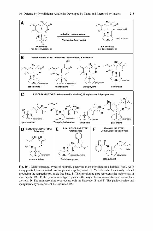
10 Defense by Pyrrolizidine Alkaloids: Developed by Plants and Recruited by Insects 215
reduction (spontaneous)
N-oxidation (enzymatic)
PA N-oxidenon-toxic (hydrophilic)
N
OO
O
OHO
H
O
R1
2
7
9
necine base
necic acid
N
OO
O
OHO
H
PA free basepro-toxic (lipophilic)
N
OO
O
O
H
OHOH
monocrotaline
N
OH
O
HO
OAc
O
OH O
ipanguline B
platynecineN
OH
O
HO
O
O
T-phalaenopsine
trachelanthamidine
SENECIONINE TYPE: Asteraceae (Senecioneae) & Fabaceae
LYCOPSAMINE TYPE: Asteraceae (Eupatorieae), Boraginaceae & Apocynaceae
A
B
C
MONOCROTALINE TYPE:Fabaceae
PHALAENOPSINE TYPE:Orchidaceae
IPANGULINE TYPE:Convolvulaceae (Ipomoea)D E
parsonsine
N
OH
O
OHOH
H
amabiline
supinidine
lycopsamine
N
OH
O
OH
HOOH
H
heliotridineN
OH
O
OH
OOH
HO
7-angeloylechinatine
triangularine
N
OO
O
O
H
OH
N
OO
O
OHO
O
senkirkine
otonecineN
OO
O
OHO
H
platyphylline
platynecine
senecionine
N
OO
O
OHO
H
retronecine
N
O
O
OHO O O
HOHO
F
retronecine
retronecine
retronecine
retronecine
Fig. 10.1 Major structural types of naturally occurring plant pyrrolizidine alkaloids (PAs). A: Inmany plants 1,2-unsaturated PAs are present as polar, non-toxic N-oxides which are easily reducedproducing the respective pro-toxic free base. B: The senecionine type represents the major class ofmacrocyclic PAs. C: the lycopsamine type represents the major class of monoesters and open-chaindiesters. D: The monocrotaline type occurs only in Fabaceae. E and F: The phalaenopsine andipangularine types represent 1,2-saturated PAs

216 T. Hartmann, D. Ober
taxa, the Asteraceae, tribe Eupatorieae, the Boraginaceae, and the Apocynaceae.PAs of the senecionine type represent typically macrocyclic diesters occasionallyaccompanied by related open-chain diesters such as triangularine (Fig. 10.1B). PAsof the lycopsamine type are open-chain monoesters or diesters containing at leastone unit of 2-isopropyl-2,3-dihydroxybutyrate, the unique C7-necic acid, which innature occurs almost exclusively in PAs. In the Apocynaceae, these PAs are occa-sionally found as macrocyclic triesters such as parsonsine (Fig. 10.1C). In both typesof PAs, retronecine is the prevailing necine base but occasionally replaced by othernecines. Another unique feature of PAs is that at least in the Asteraceae, Boragi-naceae, and Fabaceae they are found as polar N-oxides which, however, are easilyspontaneously reduced to the respective free bases (Hartmann and Toppel 1987).Only otonecine esters like senkirkine (Fig. 10.1B) which cannot be N-oxidized aregenuinely present as free bases. Species of the Apocynaceae appear to contain PAsonly as free bases. Orchidaceae and Convolvulaceae contain only PAs with 1,2-saturated necine bases (Fig. 10.1E and F). These PAs are either found as free baseslike in the Convolvulaceae (Jenett-Siems et al. 1998), or as mixtures of free basesand their N-oxides like in the orchids (Frolich et al. 2006).
10.2.2 Toxicity
PA-containing plants are probably the most common poisonous plants affectinglivestock, wildlife, and humans (Cheeke 1998; Prakash et al. 1999; Stegelmeieret al. 1999). Toxicity induced by PAs has been studied most intensively in verte-brates. Only PAs with a 1,2-double bond in the necine base moiety, e.g. retronecine,heliotridine, supinidine, otonecine (Fig. 10.1) are potentially toxic, but they requiremetabolic activation to exert toxicity. There are three principle metabolic path-ways for the metabolism of 1,2-unsaturated PAs in vertebrates (Fu et al. 2004;Fig. 10.2): (i) hydrolysis of the ester bonds yielding the free necine base; (ii)N-oxidation of the free base, and (iii) oxidation of the necine base moiety tothe corresponding dehydropyrrolizidine (pyrrolic ester) derivative. Hydrolysis andN-oxidation are considered as detoxification pathways whereas the formation of adehydropyrrolizidine creates reactive intermediates that easily form adducts withbiological nucleophiles resulting in severe cell toxicity (Fig. 10.2). In vertebrates,this bioactivation is mediated by hepatic cytochrome P450 monooxygenases, par-ticularly CYP3A and CYP2B isoforms (Huan et al. 1998a). Multisubstrate flavinemonooxygenases (Huan et al. 1998b) and to a lesser extent P450 enzymes (Williamset al. 1989a, b) are involved in the detoxification of PAs to the correspondingN-oxides.
In herbivorous insects, bioactivation of PAs comparable to that of vertebratesexists, since insects possess a rich range of P450 enzymes involved in xenobi-otic metabolism (Brattsten 1992; Glendinning and Slansky 1995; Scott et al. 1998;Glendinning 2002). Mutagenic effects of PAs have been demonstrated in Drosophila(Frei et al. 1992), and acute toxicity in larval development of Philosamia ricini(Saturniidae; Narberhaus et al. 2005).

10 Defense by Pyrrolizidine Alkaloids: Developed by Plants and Recruited by Insects 217
Fig. 10.2 Detoxification of pro-toxic 1,2-unsaturated pyrrolizidine alkaloids in vertebrates andtheir conversion into toxic pyrrolic intermediates by cytochrom P450-mediated bioactivation
Many plants store their PAs as non-toxic N-oxides (see Section 5.2.1). How-ever, any herbivorous vertebrate or insect feeding on these plants absorb the PAs aspro-toxic free base since in the reducing gut milieu any ingested N-oxide is easilyconverted into the respective free base (Mattocks 1986; Lindigkeit et al. 1997).Thus, for a non-adapted herbivore, both forms are pro-toxic. PAs that lack the1,2-double-bond are not bioactivated (Fu et al. 2004) and therefore in the abovediscussed sense are non-toxic.
10.2.3 Features of a Constitutive Chemical Defense:Physiological Aspects
The basic requirements for a constitutive defense are simple: The defense com-pound(s) should accumulate stably in the target tissues at concentrations needed toprevent damage by an antagonist, e.g. an insect herbivore. PAs fulfill these require-ments in general: they are accumulated in sufficient quantities and are metabolicallystable, i.e. they do not show turnover or degradation (Hartmann and Dierich 1998;Frolich et al. 2007). However, PAs are spatially mobile and carried from their sitesof synthesis to their sites of accumulation (Hartmann et al. 1989) where they areconcentrated within cellular vacuoles (Ehmke et al. 1988). These metabolic char-acteristics have been established for Senecio species which synthesize senecionineN-oxide as PA backbone structure in the roots. Senecionine N-oxide is distributedthrough the phloem all over the plant, and is chemically diversified (see below) to
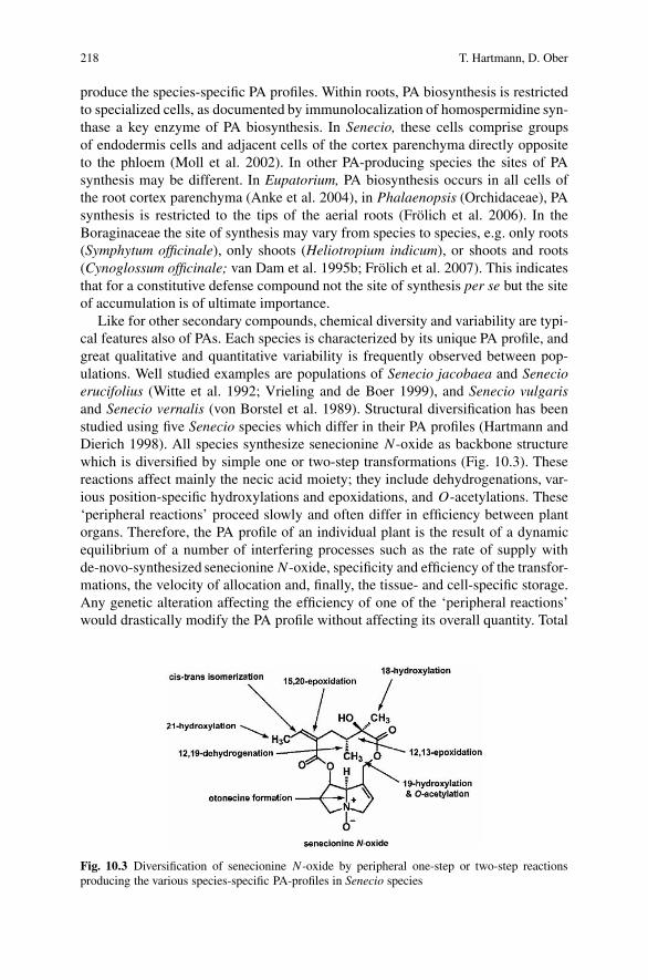
218 T. Hartmann, D. Ober
produce the species-specific PA profiles. Within roots, PA biosynthesis is restrictedto specialized cells, as documented by immunolocalization of homospermidine syn-thase a key enzyme of PA biosynthesis. In Senecio, these cells comprise groupsof endodermis cells and adjacent cells of the cortex parenchyma directly oppositeto the phloem (Moll et al. 2002). In other PA-producing species the sites of PAsynthesis may be different. In Eupatorium, PA biosynthesis occurs in all cells ofthe root cortex parenchyma (Anke et al. 2004), in Phalaenopsis (Orchidaceae), PAsynthesis is restricted to the tips of the aerial roots (Frolich et al. 2006). In theBoraginaceae the site of synthesis may vary from species to species, e.g. only roots(Symphytum officinale), only shoots (Heliotropium indicum), or shoots and roots(Cynoglossum officinale; van Dam et al. 1995b; Frolich et al. 2007). This indicatesthat for a constitutive defense compound not the site of synthesis per se but the siteof accumulation is of ultimate importance.
Like for other secondary compounds, chemical diversity and variability are typi-cal features also of PAs. Each species is characterized by its unique PA profile, andgreat qualitative and quantitative variability is frequently observed between pop-ulations. Well studied examples are populations of Senecio jacobaea and Senecioerucifolius (Witte et al. 1992; Vrieling and de Boer 1999), and Senecio vulgarisand Senecio vernalis (von Borstel et al. 1989). Structural diversification has beenstudied using five Senecio species which differ in their PA profiles (Hartmann andDierich 1998). All species synthesize senecionine N-oxide as backbone structurewhich is diversified by simple one or two-step transformations (Fig. 10.3). Thesereactions affect mainly the necic acid moiety; they include dehydrogenations, var-ious position-specific hydroxylations and epoxidations, and O-acetylations. These‘peripheral reactions’ proceed slowly and often differ in efficiency between plantorgans. Therefore, the PA profile of an individual plant is the result of a dynamicequilibrium of a number of interfering processes such as the rate of supply withde-novo-synthesized senecionine N-oxide, specificity and efficiency of the transfor-mations, the velocity of allocation and, finally, the tissue- and cell-specific storage.Any genetic alteration affecting the efficiency of one of the ‘peripheral reactions’would drastically modify the PA profile without affecting its overall quantity. Total
Fig. 10.3 Diversification of senecionine N-oxide by peripheral one-step or two-step reactionsproducing the various species-specific PA-profiles in Senecio species
10 Defense by Pyrrolizidine Alkaloids: Developed by Plants and Recruited by Insects 219
plant PA quantity is determined exclusively by the rate of root-specific formationof senecionine N-oxide. This mechanistic scenario explaining the great variation ofPA profiles between populations is supported by other studies. A genetic analysisof S. jacobaea populations (Vrieling et al. 1993) revealed that the large phenotypicvariation in both total PA content and the qualitative PA profiles is largely due togenetic variation. A comparative analysis of the PA profiles of 24 species of theSenecio section Jacobaea based on a maximum parsimony cladogram inferred fromDNA sequence and morphological data (Pelser et al. 2005) resulted in a largelyincidental distribution of 26 biogenetically related PAs within the whole clade. Dif-ferences in the PA profiles cannot be explained by gain and loss of PA-specific genesduring evolution, but rather by a transient switch-off and switch-on of gene expres-sion. Apparently the species are able to randomly shuffle patterns of senecionine-derived derivatives from the ‘genetic pool’ of the clade, and thus generating PAprofiles of changing diversity and variability.
10.2.4 Features of a Constitutive Defense System:Ecological Aspects
As most plant toxins, PAs are bitter tasting for humans and strong feeding deter-rents for vertebrates and invertebrates (Boppre 1986). However, direct evidence fortheir defense function in plants is still sparse. In contrast to an inducible defensewhere cause (attack) and effect (formation of a defense compound) are obvious, aconstitutive defense can only be judged by the absence ore presence of potentialherbivores. Therefore, evidence supporting a function of PAs in plant defense ismostly indirect. One line of indirect evidence is tissue-specific accumulation ofPAs. Flowers are often the organs showing the highest PA concentrations. Thishas been demonstrated for Senecio (Hartmann and Zimmer 1986), Heliotropium(Boraginaceae; Frolich et al. 2007), and Phalaenopsis (Orchidaceae; Frolich et al.2006). In flowering specimens, the amounts of PAs associated with the inflores-cences account for 50%–90% of total PAs, indicating an efficient protection ofthe most valuable reproductive plant organs. In rosette leaves of C. officinale, theyoungest leaves contain up to 190 times higher PA levels than older leaves (vanDam et al. 1994). Generalist herbivorous insects only feed on the PA-poor old leaveswhereas a specialist herbivore, adapted to PAs, prefers the protein-rich young leaves(van Dam et al. 1995a). In vegetative stems of herbal species like Senecio, PAsaccumulate preferentially in the peripheral cell layers (Hartmann et al. 1989), andthey are even found in trace amounts on the leaf-surface (Vrieling and Derridj 2003).Again, the functionally valuable young tissues as well as peripheral tissues whichprovide the first contact for an herbivore are efficiently protected. Another line ofindirect evidence indicating a defense role of PAs is the chemical diversity and greatvariability of the PA profiles. The effect of pure PAs on five generalist insect her-bivores revealed that structurally related PAs differ in their deterrent effects andcan even act synergistically if applied in mixtures (Macel et al. 2005). This resultwas taken as argument to emphasize the importance of generalist herbivores on theevolution and maintenance of PA diversity. Further evidence is derived from the

220 T. Hartmann, D. Ober
observation that the geographical distribution of certain S. jacobaea chemotypes isapparently not related to common ancestry but due to similar selection pressure, e.g.herbivores, in a certain region (Macel et al. 2004).
10.2.5 Molecular Evolution of the PA Pathways in Angiosperms
All plant PAs share the carbon skeleton of 2-hydroxymethylpyrrolizidine in theirnecine base moiety (Fig. 10.1). Detailed tracer studies carried out in the late 1970sand 1980s identified the symmetrical polyamine homospermidine as biosyntheticprecursor of the necine base moiety (Spenser 1985; Robins 1989). The formationof homospermidine is catalyzed by homospermidine synthase (EC 2.5.1.45, HSS)which was first isolated and characterized from Senecio root cultures (Bottcheret al. 1993, 1994). The enzyme was purified, cloned and functionally expressedin Escherichia coli (Ober and Hartmann 1999). It is encoded by a gene that orig-inated by gene duplication and subsequent diversification from an ancestral genefor deoxyhypusine synthase (EC 2.5.1.46, DHS; Ober and Hartmann 2000). DHScatalyzes the first of two steps in the posttranslational activation of the eukaryoticinitiation factor 5A (eIF5A; Caraglia et al. 2001). DHS is a highly conserved en-zyme, ubiquitously present in all Eukarya and Archaea. In the activation process ofeIF5A, it catalyzes the transfer of the aminobutyl moiety of spermidine to a specificlysine residue of the eIFA precursor protein (Fig. 10.4A). As a side-reaction, DHS isable to catalyze the formation of homospermidine from putrescine (Fig. 10.4A) andthus precisely the main-reaction of HSS. A comparison of the enzymatic and molec-ular properties of the two enzymes revealed that HSS retained all properties of DHSexcept the ability to bind the eIF5A precursor protein (Fig. 10.4B; Ober et al. 2003).The evolution of HSS is a conclusive example for evolution by change of function:the duplicate of a ubiquitous gene (DHS) functionally involved in the activation ofa regulatory protein was recruited and integrated as HSS into a completely differentfunctional environment, i.e. the biosynthesis of low molecular defense compounds(Fig. 10.4C; Ober and Hartmann 2000; Ober 2005).
The confirmed ancestry of HSS from ubiquitously occurring DHS offered theopportunity to address the intriguing question whether the PA pathways within theangiosperms are of monophyletic or polyphyletic origin. Analyzing the pairs ofcDNA sequences encoding HSS and DHS from various angiosperm species revealedat least four independent duplication events of the gene encoding DHS resulting ineach case in a new gene encoding HSS (Reimann et al. 2005; Fig. 10.5). These du-plications occurred early in the evolution of the monocots and of the Boraginaceae,respectively, and even twice within the family of the Asteraceae (i.e., once withinthe Senecioneae and the Eupatorieae, respectively). Further duplication events arelikely in those families with PA-producing species that were not included in thisstudy. These data show that the scattered occurrence of PAs within the angiospermsis a result of convergent evolution. This observation is remarkable against the back-ground that the PA structures produced are often identical. PAs of the lycopsaminetype, for example, occur within the tribe Eupatorieae (Asteraceae) and within the

10 Defense by Pyrrolizidine Alkaloids: Developed by Plants and Recruited by Insects 221
Fig. 10.4 Origin of homospermidine synthase (HSS) by duplication of the gene encoding de-oxyhypusine synthase (DHS). A: DHS catalyzes the aminobutylation of a specific lysine residueof the eIF5A precursor protein (a) and, as side reaction, converts putrescine to homospermidine(b). B: HSS is a DHS that lost the ability to bind the eIF5A precursor protein. Its reactivity andkinetics with putrescine remain unaltered. C: HSS is only known from PA containing plants, it wasrecruited and integrated into PA biosynthesis and catalyzes the formation of homospermidine, theunique carbon skeleton of the necine bases
families of the Boraginaceae and the Apocynaceae. Despite the independent originof HSS within these families, the PAs share a number of identical structural fea-tures (Fig. 10.1C), such as retronecine as necine base, and the unique C7-necid acid,2-isopropyl-2,3-dihydroxybutyrate. Further enzymes of PA biosynthesis will haveto be analyzed to get an impression of how the whole pathway was recruited inseparate angiosperm lineages.

222 T. Hartmann, D. Ober
Fig. 10.5 Phylogenetic tree of HSS and DHS encoding cDNA sequences of various angiospermspecies. Branches of HSS encoding sequences are shown in bold, originating in each case atthe duplication event of the DHS-encoding gene (labeled by circles). Modified from Reimannet al. (2004). Species abbreviations: A.t ., Arabidopsis thaliana; C.o., Cynoglossum officinale;C.r., Crotalaria retusa; D.c., Dianthus caryophyllus; E .c., Eupatorium cannabinum; H.i .,Heliotropium indicum; I.h., Ipomoea hederifolia; L .e., Lycopersicon esculentum; M.a., Musaacuminata; N .t ., Nicotiana tabacum; P.h., Petasites hybridus; Phal, Phalaenopsis spec.; S. j.,Senecio jacobaea; S.v., Senecio vernalis; S.o., Symphytum officinale
10.3 Utilization of Plant Derived Pyrrolizidine Alkaloidsby Insects
10.3.1 Occurence and Distribution of PA SequestrationAmong Insects
Almost 40 years ago, Miriam Rothschild and her colleagues showed that the larvaeof the European cinnabar moth, Tyria jacobaeae (Arctiidae) sequester PAs fromtheir host plant S. jacobaea and maintain the alkaloids through all life stages (Aplinet al. 1968). In the following decades, many further examples of PA-sequestering

10 Defense by Pyrrolizidine Alkaloids: Developed by Plants and Recruited by Insects 223
herbivorous insects became known (Hartmann and Witte 1995; Hartmann 1999;Hartmann and Ober 2000). PA sequestration is common within the tiger moth fam-ily (Arctiidae; Conner and Weller 2004). About 70 species are known that eithersequester PAs with their larval food, like T. jacobaeae, or obtain the alkaloids asadults from dead or withered parts of PA-containing plants. In the latter case, adultmoths regurgitate on the plant surface and then reimbibe the fluid containing the dis-solved alkaloids (Boppre 1986; Schneider 1987). This behavior in which the plant isonly used as ‘drug source’ is called pharmacophagy (Boppre 1984). Pharmacophagyis also common among PA-sequestering butterflies including danaine and ithomi-ine nymphalid butterflies (Boppre 1990). Pioneering work on PA-sequestration bydanaid and ithomiine butterflies has been performed in Australia (Edgar 1982) andBrasil (Brown 1984, 1987; Trigo et al. 1996).
Other examples for PA sequestration are found among beetle species.Chrysomelid leaf beetles (Coleoptera) are known for their diverse endogenouschemical defenses. Some beetles possess numerous exocrine glands, from whichscores of diminutive droplets of defensive secretions are released when the bee-tle is attacked. Leaf beetles generally synthesize their own defense compounds,but in some cases, they recruit plant defenses. Species of the palaearctic genusOreina feeding on PA-containing plants like Adenostyles alliariae (Asteraceae)sequester PAs from their host plants, and concentrate them up to 0.3 mol l−1 intheir defense secretions (Pasteels et al. 1988; Hartmann et al. 1997). The sameadaptation evolved independently in species of the neotropical genus Platyphorafeeding on species containing PAs of the lycopsamine type (Pasteels et al. 2001).Further examples of Coleoptera that sequester plant PAs are flea beetles of the genusLongitarsus feeding on PA-containing species of the Asteraceae and Boraginaceae(Haberer and Dobler 1999; Dobler et al. 2000), and the Brasilian soldier beetleChauliognathus falax (Cantharidae) feeding on Senecio inflorescences (Klitzke andTrigo 2000). Within the Orthoptera, a single PA-sequestering species was described,the polyphagous grasshopper Zonocerus variegatus (Bernays et al. 1977). Withinthe Hemiptera, PA-sequestering species are found in three unrelated taxa, includingthe largid bug Largus rufipennis sucking on Senecio stems (Klitzke and Trigo 2000),and two phloem feeders, i.e. the aphid Aphis jacobaeae feeding on various Seneciospecies (Witte et al. 1990), and the Mexican scale insect Ceroplastes albolineatusfeeding on Pittocaulon (ex Senecio) praecox (Marın Loaiza et al. 2007).
All quoted examples for PA sequestration must have evolved independently in-dicating a significant benefit of PA acquisition for the insects. In most cases, PAsequestration was found to be accompanied by highly specific biochemical adapta-tions. Leaf beetles, for instance, possess different but specific adaptations to preventthe accumulation of detrimental concentrations of PAs as pro-toxic free bases inmetabolically active tissues. They have most efficient membrane carriers whichcatalyze the transfer of PAs into their defensive glands, and concentrate them intheir defense secretions (Hartmann et al. 1999, 2001). In the next chapter, PA-sequestering arctiid moths are selected to exemplify the amazingly sophisticatedintegration of plant-derived PAs into the moth’s biology.
224 T. Hartmann, D. Ober
10.3.2 PA Sequestration by Arctiid Moths
10.3.2.1 Behavioral and Chemo-Ecological Aspects: The Utetheisaornatrix Story
The role of plant-derived PAs in the performance of an insect has most completelybeen elucidated in U. ornatrix (Eisner et al. 2002). Larvae of this moth obtain theirPAs from Crotalaria spp. and retain the alkaloids through metamorphosis. At mat-ing, the male advertises his PA load to the female through the PA-derived malecourtship pheromone, hydroxydanaidal. The pheromone is emitted from a pair ofandroconial scent-brushes (coremata) which the male everts during close-range pre-copulatory interactions with the female. Females are able to measure the pheromoneconcentration and differentiate between males that contain unequal pheromonequantities and accept eventually males having higher levels (Conner et al. 1990).During insemination the male transmits a portion of his PAs to the female. Thesealkaloids are transmitted together with the female’s own load to the eggs duringoviposition (Dussourd et al. 1988; Iyengar et al. 2001).
PAs protect adults against predation by spiders (Eisner and Eisner 1991;Gonzalez et al. 1999). The PA endowment was also shown to protect the eggs, asthe most endangered life stage of the moth, against predation by coccinelid beetles(Dussourd et al. 1988), ants (Hare and Eisner 1993), and chrysopid larvae (Eisneret al. 2000), as well as parasitization by a parasitoid wasp, Trichogramma ostriniae(Bezzerides et al. 2004).
10.3.2.2 Physiological and Biochemical Aspects
Arctiids that sequester PAs from their host plants must have acquired at least twonovel capabilities: they need to recognize PAs and they must be able to preventself-poisoning. It is known that PAs are feeding stimulants for PA-adapted arcti-ids (Boppre 1986; Schneider 1987), but only recently have arctiid caterpillars beenshown to possess single sensory neurons in their mouthparts that respond specifi-cally and sensitively (threshold of response <10−9 M) to all major structural typesof PAs (Fig 10.1), but not to other alkaloids (Bernays et al. 2002a, b). Arctiidsabsorb PAs as pro-toxic free bases since any N-oxide ingested with the food is re-duced in the gut. The absorbed free bases are immediately converted into the respec-tive non-toxic PA N-oxides. This N-oxidation is catalyzed by a flavine-dependentmonooxygenase (senecionine N-oxygenase; EC 1.14.13.101) localized as solubleenzyme in the hemolymph. The enzyme specifically converts any pro-toxic freebase into its non-toxic N-oxide (Lindigkeit et al. 1997). Its acquisition in ancestralarctiids appears to be the prerequisite for PA sequestration (Naumann et al. 2002).Sensory recognition and specific detoxification allow adapted monophagous arctiidslike U. ornatrix and T. jacobaeae to perceive and maintain PAs from their host plant.However, polyphagous species like Estigmene acrea or Grammia geneura for whichmore than 65 larval host plant species have been recorded need additional adapta-tions. Both species exploit PA-containing plants such as Senecio or Crotalaria in

10 Defense by Pyrrolizidine Alkaloids: Developed by Plants and Recruited by Insects 225
their natural environment, e.g. Arizona grass-lands, as ‘drug sources’ (Hartmannet al. 2005a, b). Since they cannot survive on PA containing plants as sole foodsource, extensive feeding on a PA plant causes a transient loss in the response ofthe PA-sensitive neurons allowing the caterpillar to leave its alkaloid source andsearch for better quality food (Bernays et al. 2003). This mechanism apparentlyallows the caterpillar to find a suitable balance between the intake of nutrients and‘defense drugs’ (Singer et al. 2004a, b). Moreover, the phagostimulatory responseto all kinds of PAs requires the ability of polyphagous caterpillar to cope with anynaturally occurring plant PA source. There are at least three ways how they handlePAs (Fig. 10.6): (1) Macrocyclic PAs and PA of the lycopsamine type (Fig. 10.1)are sequestered and maintained unaltered through all life-stages (Fig. 10.6 IIA); (2)PAs that cannot be detoxified by N-oxidation like the otonecine derivatives (e.g.,senkirkine; Fig. 10.1) are excluded from sequestration (Fig. 10.6 IIC), and (3) PAsthat are sequestered by larvae but need to be trans-esterified before transmission tothe subsequent life stages (Fig. 10.6 IIB). The latter case includes mainly PA mo-noesters and open-chain diesters that do not belong to the lycopsamine type. ThesePAs are hydrolyzed in the larvae and the resulting necine bases are re-esterifiedwith necic acids of insect origin. Arctiid-specific retronecine esters were frequentlyobserved, i.e. callimorphine (Edgar et al. 1980; Ehmke et al. 1990) and creatono-tine (Hartmann et al. 1990; Schulz et al. 1993). These compounds represent twoclasses of arctiid-specific PAs, the callimorphines and the creatonotines (Fig 10.6I; Hartmann et al. 2005a, b; Beuerle et al. 2007). Arctiids are able to re-esterifyall kinds of necine bases derived from plant PAs and are thus able to salvage necinebases and maintain them as insect-made esters through all life stages. These amazingmetabolic adaptations allow polyphagous arctiids to utilize almost the entire diver-sity of naturally occurring plant PAs efficiently. This also accounts for the forma-tion of the male courtship pheromone (hydroxydanaidal) that signals the female themale’s PA load. Its biosynthesis proceeds via a retronecine O7-monoester (Schulzet al. 1993). Probably a certain proportion of all naturally occurring retronecineesters are partially hydrolyzed and the resulting retronecine is converted into thepheromone via the creatonotines as common intermediates (Fig. 10.6 IID; Hartmannet al. 2003, 2005b).
10.4 Perspectives
The evidence discussed here favors the hypothesis that PAs represent efficientdefense compounds that may protect organisms on two trophic levels: the plant levelwhere they evolved probably under the pressure of herbivory and the insects levelwhere certain species evolved adaptations that allowed them to adopt these plantdefense compounds and utilize them for their own protection against predators andparasitoids.
In both cases selected mechanisms are required, either, as in plants, to synthesize,maintain and apply these compounds as a chemical defense barrier, or, as in adapted

226 T. Hartmann, D. Ober
Fig. 10.6 (I) The creatonotines and callimorphines are two classes of arctiid-specific pyrrolizidinealkaloids made of a necine base of plant origin and necic acids provided by the insect. (II)Polyphagous arctiid larvae are able to cope with all structural types of plant PAs. Depending onthe structure, PAs are either utilized unaltered (route A), or they are hydrolyzed and the necinebases are salvaged as creatonotines or callimorphines (route B), or they are excluded from seques-tration like the otonecine esters (route C). Species which produce the male courtship pheromone,hydroxydanaidal, synthesize this ‘PA signal’ via creatonotines as common intermediates (route D)

10 Defense by Pyrrolizidine Alkaloids: Developed by Plants and Recruited by Insects 227
insects, to recognize, ingest, safely maintain, and employ the adopted compoundsin their species-specific behavior and defense strategies. To unravel the evolutionaryorigin of the acquired mechanisms involved in these processes on both trophic levelsis one of the most challenging future goals. The independent evolutionary originsof the biosynthetic pathways in plants, and the multiple independent adaptations ofvarious insects to PA sequestration and utilization pose further exciting questions.
Acknowledgments The authors thank the Deutsche Forschungsgemeinschaft (DFG) for continuesfinancial support.
References
Anke S, Niemuller D, Moll S, Hansch R, Ober D (2004) Polyphyletic origin of pyrrolizidinealkaloids within the Asteraceae. Evidence from differential tissue expression of homospermi-dine synthase. Plant Physiol 136:4037–4047
Aplin RT, Benn MH, Rothschild M (1968) Poisonos alkaloids in the body tissues of the cinnabarmoth (Callimorpha jacobaeae L.). Nature 219:747–748
Bernays EA, Chapman RF, Hartmann T (2002a) A highly sensitive taste receptor cell forpyrrolizidine alkaloids in the lateral galeal sensillum of a polyphagous caterpillar, Estigmeneacrea. J Comp Physiol A 188:715–723
Bernays EA, Chapman RF, Hartmann T (2002b) A taste receptor neurone dedicated to the per-ception of pyrrolizidine alkaloids in the medial galeal sensillum of two polyphagous arctiidcaterpillars. Physiol Entomol 27:1–10
Bernays EA, Edgar JA, Rothschild M (1977) Pyrrolizidine alkaloids sequestered and stored by theaposematic grasshopper, Zonocerus variegatus. J Zool, 182:85–87
Bernays EA, Rodrigues D, Chapman RF, Singer MS, Hartmann T (2003) Loss of gustatory re-sponses to pyrrolizidine alkaloids after their extensive ingestion in the polyphagous caterpillarEstigmene acrea. J Exp Biol 206:487–4496
Beuerle T, Theuring C, Klewer N, Schulz S, Hartmann T (2007) Absolute configuration of thecreatonotines and callimorphines, two classes of arctiid-specific pyrrolizidine alkaloids. InsectBiochem Mol Biol 37:80–89
Bezzerides A, Yong TH, Bezzerides J, Husseini J, Ladau J, Eisner M, Eisner T (2004) Plant-derivedpyrrolizidine alkaloid protects eggs of a moth (Utetheisa ornatrix) against a parasitoid wasp(Trichogramma ostriniae). Proc Natl Acad Sci USA 101:9029–9032
Boppre M (1984) Redefining ‘pharamcophagy’. J Chem Ecol 10:1151–1154Boppre M (1986) Insects pharmacophagously utilizing defensive plant chemicals (pyrrolizidine
alkaloids). Naturwissenschaften 73:17–26Boppre M (1990) Lepidoptera and pyrrolizidine alkaloids: exemplification of complexity in chem-
ical ecology. J Chem Ecol 16:165–185Bottcher F, Adolph RD, Hartmann T (1993) Homospermidine synthase, the first pathway-specific
enzyme in pyrrolizidine alkaloid biosynthesis. Phytochemistry 32:679–689Bottcher F, Ober D, Hartmann T (1994) Biosynthesis of pyrrolizidine alkaloids: putrescine and
spermidine are essential substrates of enzymatic homospermidine formation. Can J Chem72:80–85
Brattsten LB (1992) Metabolic defenses against plant allelochemicals. In: Rosenthal GA, Beren-baum MR (eds) Herbivores: their interactions with secondary metabolites, vol 2. AcademicPress, San Diego, pp 175–242
Brown KSJ (1984) Adult-obtained pyrrolizidine alkaloids defend ithomiine butterflies against aspider predator. Nature 309:707–709

228 T. Hartmann, D. Ober
Brown KSJ (1987) Chemistry at the solanaceae/ithomiinae interface. Ann Missouri Bot Gard 74:359–397
Caraglia M, Marra M, Giuberti G, D’Alessandro AM, Budillon A, del Prete S, Lentini A, Beni-nati S, Abbruzzese A (2001) The role of eukaryotic initiation factor 5A in the control of cellproliferation and apoptosis. Amino Acids 20:91–104
Cheeke PR (ed) (1998) Natural toxicants in feeds, forages, and poisonous plants. Interstate,Danville
Conner WE, Roach B, Benedict E, Meinwald J, Eisner T (1990) Courtship pheromone productionand body size as correlates of larval diet in males of the arctiid moth Utetheisa ornatrix. J ChemEcol 16:543–552
Conner WE, Weller, SJ (2004) A quest for alkaloids: the curious relationship between tiger mothsand plants containing pyrrolizidine alkaloids. In: Carde RT, Millar JG (eds) Advances in insectchemical ecology. University Press, Cambridge, pp 248–282
Dobler S, Haberer W, Witte L, Hartmann T (2000) Selective sequestration of pyrrolizidine alka-loids from diverse host plants by Longitarsus flea beetles. J Chem Ecol 26:1281–1298
Dussourd DE, Ubik K, Harvis C, Resch J, Meinwald J, Eisner T (1988) Biparental defensive en-dowment of eggs with acquired plant alkaloid in the moth Utetheisa ornatrix. Proc Natl AcadSci USA 85:5992–5996
Edgar JA (1982) Pyrrolizidine alkaloids sequestered by Salomon Island Danainae butterflies. Thefeeding preferences of the Danainae and Ithomiinae. J Zool 196:385–399
Edgar JA, Culvenor CCJ, Cockrum PA, Smith LW (1980) Callimorphine: identification andsymthesis of the cinnabar moth ‘metabolite’. Tetrahedron Lett 21:1383–1384
Ehmke A, Von Borstel K, Hartmann T (1988) Alkaloid N-oxides as transport and vacuolar storagecompounds of pyrrolizidine alkaloids in Senecio vulgaris L. Planta 176:83–90
Ehmke A, Witte L, Biller A, Hartmann T (1990) Sequestration, N-oxidation and transformationof plant pyrrolizidine alkaloids by the arctiid moth Tyria jacobaeae L. Z . Naturforsch C 45:1185–1192
Eisner T, Eisner M (1991) Unpalatability of the pyrrolizidine alkaloid-containing moth Utetheisaornatrix and its larva to wolf spiders. Psyche Cambridge 98:111–118
Eisner T, Eisner M, Rossini C, Iyengar VK, Roach BL, Benedikt E, Meinwald J (2000) Chemicaldefense against predation in an insect egg. Proc Natl Acad Sci USA 97:1634–1639
Eisner T, Rossini C, Gonzalez A, Iyengar VK, Siegler MVS, Smedley SR (2002) Paternal invest-ment in egg defence. In: Hilker M, Meiners T (eds) Chemoecology of insect eggs and eggdeposition. Blackwell, Oxford, pp 91–116
Frei H, Luthy J, Brauchli J, Zweifel U, Wurgler FE, Schlatter C (1992) Structure/activity relation-ships of the genotoxic potencies of sixteen pyrrolizidine alkaloids assayed for the inductionof somatic mutation and recombination in wing cells of Drosophila melanogaster. Chem BiolInteract 83:1–22
Frolich C, Hartmann T, Ober D (2006) Tissue distribution and biosynthesis of 1,2-saturatedpyrrolizidine alkaloids in Phalaenopsis hybrids (Orchidaceae). Phytochemistry 67:1493–1502
Frolich C, Ober D, Hartmann T (2007) Tissue distribution, core biosynthesis and diversification ofpyrrolizidine alkaloids of the lycopsamine type in three Boraginaceae species. Phytochemistry68:1026–1037
Fu PP, Xia Q, Lin G, Chou MW (2004) Pyrrolizidine alkaloids – genotoxicity, metabolism en-zymes, metabolic activation, and mechanisms. Drug Metab Rev 36:1–55
Glendinning JI (2002) How do herbivorous insects cope with noxious secondary plant compoundsin their diet? Entomol Exp Appl 104:15–25
Glendinning JI, Slansky F (1995) Consumption of a toxic food by caterpillars increases withdietary exposure: support for a role of induced detoxification enzymes. J Comp Physiol A176:337–345
Gonzalez A, Rossini C, Eisner M, Eisner T (1999) Sexually transmitted chemical defense in amoth (Utetheisa ornatrix). Proc Natl Acad Sci USA 96:5570–5574

10 Defense by Pyrrolizidine Alkaloids: Developed by Plants and Recruited by Insects 229
Haberer W, Dobler S (1999) Quantitative analysis of pyrrolizidine alkaloids sequestered fromdiverse host plants in Longitarsus flea beetles (Coleoptera, Chrysomelidae). Chemoecology9:169–179
Hare JF, Eisner T (1993) Pyrrolizidine alkaloid deters ant predators of Utetheisa ornatrix eggs:effects of alkaloid concentration, oxidation state, and prior exposure of ants to alkaloid-ladenprey. Oecologia 96:9–18
Hartmann T (1999) Chemical ecology of pyrrolizidine alkaloids. Planta 207:483–495Hartmann T, Biller A, Witte L, Ernst L, Boppre M (1990) Transformation of plant pyrrolizidine
alkaloids into novel insect alkaloids by arctiid moths (Lepidoptera). Biochem Syst Ecol18:549–554
Hartmann T, Dierich B (1998) Chemical diversity and variation of pyrrolizidine alkaloids of thesenecionine type: biological need or coincidence? Planta 206:443–451
Hartmann T, Ehmke A, Eilert U, von Borstel, K, Theuring C (1989) Sites of synthesis, translocationand accumulation of pyrrolizidine alkaloid N-oxides in Senecio vulgaris L. Planta 177:98–107
Hartmann T, Ober D (2000) Biosynthesis and metabolism of pyrrolizidine alkaloids on plants andspecialized insect herbivores. Top Curr Chem 209:207–243
Hartmann T, Theuring C, Bernays EA (2003) Are insect-synthesized retronecine esters (cre-atonotines) the precursors of the male courtship pheromone in the arctiid moth Estigmeneacrea? J Chem Ecol 29:2603–2608
Hartmann T, Theuring C, Beuerle T, Bernays EA, Singer MS (2005a) Acquisition, transformationand maintenance of plant pyrrolizidine alkaloids by the polyphagous arctiid Grammia geneura.Insect Biochem Mol Biol 35:1083–99
Hartmann T, Theuring C, Beuerle T, Klewer N, Schulz S, Singer, MS, Bernays EA (2005b) Spe-cific recognition, detoxification and metabolism of pyrrolizidine alkaloids by the polyphagousarctiid Estigmene acrea. Insect Biochem Mol Biol 35:391–411
Hartmann T, Theuring C, Schmidt J, Rahier M, Pasteels JM (1999) Biochemical strategy of se-questration of pyrrolizidine alkaloids by adults and larvae of chrysomelid leaf beetles. J InsectPhysiol 45:1085–1095
Hartmann T, Theuring C, Witte L, Pasteels JM (2001) Sequestration, metabolism and partial syn-thesis of tertiary pyrrolizidine alkaloids by the neotropical leaf-beetle Platyphora boucardi.Insect Biochem Mol Biol 31:1041–1056
Hartmann T, Toppel G (1987) Senecionine N-oxide, the primary product of pyrrolizidine alkaloidbiosynthesis in root cultures of Senecio vulgaris. Phytochemistry 26:1639–1644
Hartmann T, Witte L (1995) Pyrrolizidine alkaloids: chemical, biological and chemoecologicalaspects. In: Pelletier SW (ed) Alkaloids: chemical and biological perspectives, vol 9. PergamonPress, Oxford, pp 155–233
Hartmann T, Witte L, Ehmke A, Theuring C, Rowell-Rahier M, Pasteels JM (1997) Selective se-questration and metabolism of plant derived pyrrolizidine alkaloids by chrysomelid leaf beetles.Phytochemistry 45:489–497
Hartmann T, Zimmer M (1986) Organ-specific distribution and accumulation of pyrrolizidine al-kaloids during the life history of two annual Senecio species. J Plant Physiol 122:67–80
Huan JY, Miranda CL, Buhler DR, Cheeke PR (1998a) The roles of CYP3A and CYP2B isoformsin hepatic bioactivation and detoxification of the pyrrolizidine alkaloid senecionine in sheepand hamsters. Toxicol Appl Pharmacol 151:229–235
Huan JY, Miranda CL, Buhler DR, Cheeke PR (1998b) Species differences in the hepatic micro-somal enzyme metabolism of the pyrrolizidine alkaloids. Toxicol Lett 99:127–137
Iyengar VK, Rossini C, Eisner T (2001) Precopulatory assessment of male quality in an arctiidmoth (Utetheisa ornatrix): hydroxydanaidal is the only criterion of choice. Behav Ecol Socio-biol 49:283–288
Jenett-Siems K, Ott SC, Schimming T, Siems K, Muller F, Hilker M, Witte L, Hartmann T, AustinDF, Eich E (2005) Ipangulines and minalobines, chemotaxonomic markers of the infragenericIpomoea taxon subgenus Quamoclit, section Mina. Phytochemistry 66:223–231

230 T. Hartmann, D. Ober
Jenett-Siems K, Schimming T, Kaloga M, Eich E, Siems K, Gupta MP, Witte L, Hartmann T(1998) Pyrrolizidine alkaloids of Ipomoea hederifolia and related species. Phytochemistry47:1551–1560
Klitzke CF, Trigo JR (2000) New records of pyrrolizidine alkaloid-feeding insects. Hemiptera andColeoptera on Senecio brasiliensis. Biochem Syst Ecol 28:313–318
Lindigkeit R, Biller A, Buch M, Schiebel HM, Boppre M, Hartmann T (1997) The two faces ofpyrrolizidine alkaloids: the role of the tertiary amine and its N-oxide in chemical defense ofinsects with acquired plant alkaloids. Eur J Biochem 245:626–636
Macel M, Bruinsma M, Dijkstra SM, Ooijendijk T, Niemeyer HM, Klinkhamer PGL (2005) Dif-ferences in effects of pyrrolizidine alkaloids on five generalist insect herbivore species. J ChemEcol 31:1493–1508
Macel M, Vrieling K, Klinkhamer PG (2004) Variation in pyrrolizidine alkaloid patterns of Seneciojacobaea. Phytochemistry 65:865–873
Marın Loaiza JC, Cespedes CL, Beuerle T, Theuring C, Hartmann T (2007) Ceroplastes albo-lineatus, the first scale insect shown to sequester pyrrolizidine alkaloids from its host-plantPittocaulon praecox. Chemoecology 17:109–115
Mattocks AR (ed) (1986) Chemistry and toxicology of pyrrolizidine alkaloids. Academic Press,London
Moll S, Anke S, Kahmann U, Hansch R, Hartmann T, Ober D (2002) Cell specific expression ofhomospermidine synthase, the entry enzyme of the pyrrolizidine alkaloids in Senecio vernalisin comparison to its ancestor deoxyhypusine synthase. Plant Physiol 130:47–57
Narberhaus I, Zintgraf V, Dobler S (2005) Pyrrolizidine alkaloids on three trophic levels – evidencefor toxic and deterrent effects on phytophages and predators. Chemoecology 15:121–125
Naumann C, Hartmann T, Ober D (2002) Evolutionary recruitment of a flavin-dependent monooxy-genase for the detoxification of host plant-acquired pyrrolizidine alkaloids in the alkaloid-defended arctiid moth Tyria jacobaeae. Proc Natl Acad Sci USA 99:6085–6090
Ober D (2005) Seeing double – gene duplication and diversification in plant secondary metabolism.Trends Plant Sci 10:444–449
Ober D, Harms R, Witte L, Hartmann T (2003) Molecular evolution by change of function:alkaloid-specific homospermidine synthase retained all properties of deoxyhypusine synthaseexcept binding the eIF5A precursor protein. J Biol Chem 278:12805–12815
Ober D, Hartmann T (1999) Homospermidine synthase, the first pathway-specific enzyme ofpyrrolizidine alkaloid biosynthesis, evolved from deoxyhypusine synthase. Proc Natl Acad SciUSA 96:14777–14782
Ober D, Hartmann T (2000) Phylogenetic origin of a secondary pathway: the case of pyrrolizidinealkaloids. Plant Mol Biol 44:445–450
Pasteels JM, Rowell-Rahier M, Randoux T, Braekman JC, Daloze D (1988) Pyrrolizidine alkaloidsof probable host-plant origin in the pronotal and elytral secretion of the leaf beetle Oreinacacaliae. Entomol Exp Appl 49:55–88
Pasteels JM, Termonia A, Windsor D, Witte L, Theuring C, Hartmann T (2001) Pyrrolizidinealkaloids and pentacyclic triterpene saponins in the defensive secretions of Platyphora leafbeetles. Chemoecology 11:113–120
Pelser PB, de Vos H, Theuring C, Beuerle T, Vrieling K, Hartmann T (2005) Frequent gain andloss of pyrrolizidine alkaloids in the evolution of Senecio section Jacobaea (Asteraceae). Phy-tochemistry 66:1285–1295
Prakash AS, Pereira TN, Reilly PEB, Seawright AA (1999) Pyrrolizidine alkaloids in human diet.Mutat Res 443:53–67
Reimann A, Nurhayati N, Backenkohler A, Ober D (2004) Repeated evolution of the pyrrolizidinealkaloid-mediated defense system in separate angiosperm lineages. Plant Cell 16:2772–2784
Robins DJ (1989) Biosynthesis of pyrrolizidine alkaloids. Chem Soc Rev 18:375–408Schneider D (1987) The strange fate of pyrrolizidine alkaloids. In: Chapman RF, Bernays EA, Stof-
folano JG (eds) Perspectives in chemoreception and behavior. Springer, New York, pp 123–142

10 Defense by Pyrrolizidine Alkaloids: Developed by Plants and Recruited by Insects 231
Schulz S, Francke W, Boppre M, Eisner T, Meinwald J (1993) Insect pheromone biosynthesis:stereochemical pathway of hydroxydanaidal production from alkaloidal precursors in Cre-atonotos transiens (Lepidoptera, Arctiidae). Proc Natl Acad Sci USA 90:6834–6838
Scott JG, Liu N, Wen Z (1998) Insect cytochromes P450: diversity, insecticide resistance andtolerance to plant toxins. Comp Biochem Physiol C Pharmacol Toxicol Endocrinol 121:147–155
Singer MS, Carriere Y, Theuring C, Hartmann T (2004a) Disentangeling food quality from resis-tance against parasitoids: diet choice by a generalist caterpillar. Am Nat 164:424–429
Singer MS, Rodrigues D, Stireman JOI, Carriere Y (2004b) Roles of food quality and enemy-freespace in host use by a generalist insect herbivore. Ecology 85:2747–2753
Spenser ID (1985) Stereochemical aspects of the biosynthetic routes leading to the pyrrolizidineand quinolizidine alkaloids. Pure Appl Chem 57:453–470
Stegelmeier BL, Edgar JA, Colegate SM, Gardner DR, Schoch TK, Coulombe RA, Molyneux RJ(1999) Pyrrolizidine alkaloid plants, metabolism and toxicity. J Nat Tox 8:95–116
Trigo JR, Brown KS, Henriques SA, Barata LES (1996) Qualitative patterns of pyrrolizidine alka-loids in ithomiinae butterflies. Biochem Syst Ecol 24:181–188
van Dam NM, Verporte R, van der Mejden E (1994) Extreme differences in pyrrolizidine alkaloidlevels between leaves of Cynoglossum officinale. Phytochemistry 37:1013–1016
van Dam NM, Vuister LWM, Bergshoff C, de Vos H, van der Meijden E (1995a) The ‘raisond’etre’ of pyrrolizidine alkaloids in Cynoglossum officinale: deterrent effects against generalistherbivores. J Chem Ecol 21:507–523
van Dam NM, Witte L, Theuring C, Hartmann T (1995b) Distribution, biosynthesis and turnoverof pyrrolizidine alkaloids in Cynoglossum officinale. Phytochemistry 39:287–292
von Borstel K, Witte L, Hartmann T (1989) Pyrrolizidine alkaloid patterns in populations ofSenecio vulgaris, Senecio vernalis and their hybrids. Phytochemistry 28:1635–1638
Vrieling K, de Boer NJ (1999) Host-plant choice and larval growth in the cinnabar moth: dopyrrolizidine alkaloids play a role. Entomol Exp Appl 91:251–257
Vrieling K, de Vos H, van Wijk CAM (1993) Genetic analysis of the concentrations of pyrrolizidinealkaloids in Senecio jacobaea. Phytochemistry 32:1141–1144
Vrieling K, Derridj S (2003) Pyrrolizidine alkaloids in and on the leaf surface of Senecio jacobaeaL. Phytochemistry 64:1223–1228
Williams DE, Reed RL, Kedzierski B, Dannan GA, Guengerich FP, Buhler DR (1989a) Bioactiva-tion and detoxication of the pyrrolizidine alkaloid senecionine by cytochrome P-450 enzymesin rat liver. Drug Metab Dispos 17:387–392
Williams DE, Reed RL, Kedzierski B, Ziegler DM, Buhler DR (1989b) The role of flavin-containing monooxygenase in the N-oxidation of the pyrrolizidine alkaloid senecionine. DrugMetab Dispos 17:380–386
Wink M, Mohamed GIA (2003) Evolution of chemical defense traits in the Leguminosae: map-ping of distribution patterns of secondary metabolites on a molecular phylogeny inferred fromnucleotide sequences of the rbcL gene. Biochem Syst Ecol 31:897–917
Witte L, Ehmke A, Hartmann T (1990) Interspecific flow of pyrrolizidine alkaloids; from plantsvia aphids to ladybirds. Naturwissenschaften 77:540–543
Witte L, Ernst L, Adam H, Hartmann T (1992) Chemotypes of two pyrrolizidine alkaloid-containing Senecio spp. Phytochemistry 31:559–566

Part CAnti-nutritional Enzymes and Proteins
Chapter 11Plant Protease Inhibitors: Functional Evolutionfor Defense
Maarten A. Jongsma and Jules Beekwilder
In this chapter we review the ways in which plants and plant predators have evolvedcreative solutions in the battle for scarce essential amino acids. We show that onboth the plant and insect side many different gene families of inhibitors and pro-teases are involved with intricate specialized expression patterns among the differentmembers. They provide an evolutionary and ecological genetic fingerprint of theirfunctional role. Some of the hypotheses we put forward are currently only supportedby circumstantial evidence, but provide attractive alleys which functional evolutionmay have gone. At first glance, this topic of plant-insect/pathogen interactions ap-pears to be only a clash of two protein molecules. However, due to the opportunis-tic nature of evolution, this battle may have taken on many new appearances. Theavailability of fully sequenced genomes on both the plant and insect side, and theexcellent tools to specifically knock down and overexpress specific genes provideexactly the instruments needed to allow a whole new set of discoveries in this area.
11.1 Introduction
The research in the field of plant protease inhibitors (PIs) has a 60 year long his-tory dating back to the years just after the second world war when Kunitz (1946),Bowman (1946) and Birk et al. (1963) first purified and characterized the proteaseinhibitors in soybeans. Then, in the following decades, new gene families wereadded to the initial spectrum, to the extent that in plants we are now aware of 13 dif-ferent protease inhibitor gene families known to target all main families of proteases(Rawlings et al. 2006), and often aimed at inhibiting the proteases of herbivores orplant pathogens.
The antinutritional effects of proteinase inhibitors in animal and human nutri-tion were already known early on, but their role in plant defense became much
M.A. JongsmaPlant Research International B.V., Wageningen University and Research Center, 6700 AAWageningen, The Netherlandse-mail: [email protected]
A. Schaller (ed.), Induced Plant Resistance to Herbivory, 235C© Springer Science+Business Media B.V. 2008

236 M.A. Jongsma, J. Beekwilder
more evident when Green and Ryan discovered that also leaves of plants inducedhigh levels of protease inhibitors in a specific response to mechanical and insectdamage (Green and Ryan 1972). The advent of genetic modification allowed theoverexpression of protease inhibitors which subsequently provided the first in plantademonstrations of the role of protease inhibitors in protecting plants against insects(Hilder et al. 1987; Abdeen et al. 2005), but also against microbes (Qu et al. 2003),and viruses (Gutierrez-Campos et al. 1999). Natural mutants of trypsin inhibitorsin wild tobacco plants recently provided the first true evidence that the inhibitorsexert a certain level of control even against their natural ‘adapted’ pests (Zavalaet al. 2004).
Despite the dramatically raised plant PI levels in defense against herbivorousinsects, the effects on herbivore mortality or development are often relatively minoror even absent. Yet, even small effects on development may still be ecologicallyrelevant, and affect the next generation with severely reduced fecundity (De Leo andGallerani 2002). However, the observation that some insects circumvent the effectsof the inhibitors by responding with the induction of PI-insensitive proteases createdthe awareness that here an intriguing battle of inhibitor and protease molecules wasbeing staged (Jongsma et al. 1995; Jongsma and Bolter 1997). It was subsequentlyshown that proteases had acquired mutations rendering them sterically insensitiveto the inhibitors (Bown et al. 1997; Volpicella et al. 2003; Bayes et al. 2005).Other insects were very effective in degrading the inhibitors with specific proteases(Michaud 1997; Giri et al. 1998). But despite these insights, we think that we haveactually advanced only a little bit on the track of unraveling the mutual plays of trickand deceit that have evolved around this form of plant defense.
This chapter essentially deals with a description of how these interactions havebeen understood so far and what may be in for the future. Despite being an ‘old’ fieldof research, tantalizing results lie ahead if the subject matter is placed in its ecolog-ical, physiological, cytological, and finally molecular context with plant predatorsand pathogens.
11.2 An Array of Gene Families with a Diversity of Roles
11.2.1 The Arabidopsis and Rice Genomes
So far, the rich resource of fully sequenced plant genomes has not been exploited toits full extend with respect to the specific area of protease inhibitors. For the purposeof this review we therefore made an inventory of the complete genetic repertoire ofprotease inhibitors of two plant species: the fully annotated genomes of Arabidopsisthaliana (thale cress, a crucifer), and Oryza sativa (rice). With Arabidopsis being adicotyledonous species, and rice being a monocot, these plant species are only dis-tantly related in the plant kingdom. Conveniently, the MEROPS database (Rawlingset al. 2006) has interrogated these genomes for the presence of sequences homolo-gous to known protease inhibitor sequences.

11 Plant Protease Inhibitors 237
Across all organisms, the inhibitors in MEROPS have been classified into 68families, referred to as I01, I02, etc. Thirteen of these families are present in plants.In a specific plant species like Arabidopsis the genome contains 81 putative in-hibitor genes, representing seven of those families (Fig. 11.1A), while the genomeof Oryza contains 121 putative inhibitor genes, representing eight inhibitor fami-lies (Fig. 11.1B). There is considerable overlap in the inhibitor families representedin both plants. Six families I03 (Kunitz-type inhibitors), I04 (serpins), I13 (eglinC-homologues), I25 (cystatins), I29 (cathepsin prodomains) and I51 (CPY-inhibitorhomologues) occur in both plants. One could consider those families as generalplant inhibitors, likely occurring in most plant species.
In addition to general protease inhibitor families, many plant families have in-hibitors that are uniquely found only in certain groups of plants. In Oryza this in-cludes members of the families I06 (trypsin/alpha-amylase inhibitors), which aretypical for monocots, and I13 (Bowman–Birk inhibitors), which apart from mono-cots also occur in Leguminosae, potato and sunflower, but not in Arabidopsis.Arabidopsis on the other hand expresses members of the I18 family (mustardtrypsin inhibitor homologues), which is unique for Cruciferae. There are more ofthose species-specific protease inhibitors. For example, only solanaceous speciesare known to have inhibitors of family I20 (potato inhibitor 2) and I37 (potato met-allocarboxypeptidase inhibitor), and only Cucurbitaceae are known to have familyI07 (squash inhibitors), while family I67 (bromein) is uniquely found in pineapple.
The overview in Fig. 11.1 is based on sequence data only and is likely to over-estimate the actual number of encoded inhibitors. Not every sequence that wasannotated as such has actually been demonstrated to encode an active proteaseinhibitor. For instance, the I51 CPY inhibitor homologues are related to the yeast
Fig. 11.1 Phylogenetic tree based on the mature protein sequences of all the protease inhibitors ofArabidopsis and rice in the MEROPS database (Rawlings et al. 2006)

238 M.A. Jongsma, J. Beekwilder
carboxypeptidase Y inhibitor, but also to a collection of known kinase inhibitors.So far, protease inhibitor activity has not been demonstrated for any of these plantproteins. Also noteworthy in this respect is the largest family of inhibitors in bothplants, the I29 cathepsin-prodomains. I29s can occur either as free inhibitors, or asprodomain of a cathepsin. In all Arabidopsis and most of the Oryza I29 members,the inhibitor module in the protein is followed by a cathepsin domain, indicating thatthese proteins merely function as pro-domain to prevent premature activation of thecathepsins. In Oryza, a small group of four sequences (indicated in Fig. 11.1B) isformed by I29 members that are not part of a cathepsin sequence, and may thusfunction as inhibitors of other proteases. However, this potential function has notyet been assessed.
Most inhibitors in Oryza and Arabidopsis target serine proteases, such as trypsinsand chymotrypsins, which may relate to the fact that many herbivores, such aslepidopteran insects but also mammals, use these proteases for protein digestion.Only family I25 (cystatins) targets cysteine proteases, while family I03, the Kunitzinhibitors, also comprises members specific for cysteine and aspartic proteases.
11.2.2 Roles in Defense, but Also in Other Fields
A direct relationship to defense against arthropods, nematodes, fungi, and/or bac-teria has been established for most inhibitor families, such as I12 (Bowman–Birk),I03 (Kunitz), I06 (trypsin/�-amylase), I25 (cystatins), I13 (eglin C) and I18 (mustardtrypsin inhibitor). An exception may be found in the serpin family (I04). They havebeen demonstrated to possess inhibitor activity in plants, but their functional role isstill unclear. Serpins do accompany storage proteins in seeds of monocots, wherethey could protect these from proteolysis or still act in defense (Hejgaard 2005). InArabidopsis, however, none of its members are induced by jasmonate or salicylatetreatments and expression levels are generally very low (Genevestigator; data notshown). We observed that when potato cystatins, known for their defense role, areoverexpressed in tomato cytoplasm this tends to lead to dwarfing and chlorosis oftomato plants whereas these effects are not observed when the inhibitors are tar-geted to the secretory pathway (unpublished results). This does emphasize that theseproteins are not inert to the host plant physiology and may play subtle secondaryroles in a range of processes unrelated to defense and dependent on the subcellularlocalization of the inhibitor.
The availability for several years now of full genome DNA microarrays ofArabidopsis has resulted in a rich source of expression data for each gene in differenttissues under different physiological conditions and stresses. Without any wetlab ex-periments, these data can be mined and used to hypothesize on the functions of indi-vidual members of gene families (Zimmermann et al. 2004). As an example, we didthis exercise for the unique cruciferous MTI-2 gene family with six gene memberswhich was also studied by Clauss and Mitchell-Olds (2004). Table 11.1 summarizesthe presently available data for tissue-specific gene expression in Arabidopsis. It is

11 Plant Protease Inhibitors 239
Tab
le11
.1T
issu
e-sp
ecifi
cco
nstit
utiv
eex
pres
sion
with
out
indu
cer
ofal
lsi
xm
embe
rsof
the
MT
I-2
gene
fam
ilyin
Ara
bido
psis
.T
heav
erag
eflu
ores
cenc
esi
gnal
sfo
rth
ese
tiss
ues
are
show
n
Ana
tom
yA
cc.n
r.A
t2g4
3510
At2
g435
20A
t2g4
3530
At2
g435
35A
t1g4
7540
At2
g435
50
Pote
ntia
lP.
syri
ngae
jasm
onat
eda
rkne
ssja
smon
ate
indu
cer
sene
scen
cew
ound
ing
AT
TI1
AT
TI2
AT
TI3
AT
TI4
AT
TI7
AT
TI6
#of
Chi
psM
ean
Mea
nM
ean
Mea
nM
ean
Mea
n
0ca
llus
656
0419
044
020
3011
339
1ce
llsu
spen
sion
8757
6625
530
757
7170
298
2se
edlin
g49
318
3269
721
2359
4491
1879
21co
tyle
dons
976
361
838
7911
2742
4049
22hy
poco
tyl
645
341
361
0435
4082
3552
23ra
dicl
e9
121
173
357
7690
8467
23
inflo
resc
ence
232
1939
3428
4203
827
6685
2710
31flo
wer
8520
5892
551
1651
362
3842
311
carp
el15
1314
563
7154
827
4738
4431
11ov
ary
416
1337
650
2147
949
3334
3112
stig
ma
323
6314
823
4017
069
1868
312
peta
l6
991
7515
6615
650
4727
313
sepa
l6
1377
633
722
2923
647
2098
314
stam
en15
575
154
1056
191
101
1253
3141
poll
en2
452
8659
396
157
1231
5pe
dice
l3
489
652
2716
752
6935
4632
sili
que
1915
4220
0447
4673
893
7010
8933
seed
5326
6198
6480
116
2425
735
176
34st
em28
1212
533
3827
489
5619
61

240 M.A. Jongsma, J. Beekwilder
Tab
le11
.1(c
onti
nued
)
Ana
tom
yA
cc.n
r.A
t2g4
3510
At2
g435
20A
t2g4
3530
At2
g435
35A
t1g4
7540
At2
g435
50
Pote
ntia
lP.
syri
ngae
jasm
onat
eda
rkne
ssja
smon
ate
indu
cer
sene
scen
cew
ound
ing
AT
TI1
AT
TI2
AT
TI3
AT
TI4
AT
TI7
AT
TI6
#of
Chi
psM
ean
Mea
nM
ean
Mea
nM
ean
Mea
n
35no
de3
260
536
4362
454
3949
5736
shoo
tape
x25
1858
3680
7164
807
4138
7337
caul
ine
leaf
316
3617
821
2919
338
392
4ro
sett
e71
034
7314
9960
3592
555
4228
41ju
veni
lele
af87
2036
782
3846
722
6923
7742
adul
tlea
f24
363
4624
4985
6010
0769
5533
43pe
tiol
e12
559
467
4303
548
2739
8644
sene
scen
tle
af3
3454
102
995
7949
131
45hy
poco
tyl
1227
0338
5834
7020
9783
415
451
xyle
m3
3441
4618
1676
587
7111
645
2co
rk3
812
225
1249
383
9057
15
root
s23
619
6710
922
947
9372
408
52la
tera
lroo
t4
611
303
1300
7892
101
548
53ro
otti
p4
148
162
281
2351
147
8954
elon
gati
onzo
ne7
190
116
131
4685
129
157
55ro
otha
irzo
ne4
296
114
126
1008
9211
256
endo
derm
is3
483
127
187
1037
170
4957
endo
derm
is+
cort
ex3
155
108
300
974
104
306
58ep
idat
rich
obla
sts
349
815
632
690
1213
688
59la
tera
lroo
tcap
383
816
259
415
7913
117
460
stel
e3
244
137
163
1576
190
94
Indu
cer
refe
rsto
circ
umst
ance
sw
hich
wil
lac
tivat
eth
ege
neon
top
ofth
ede
velo
pmen
tall
yre
gula
ted
expr
essi
onin
diff
eren
tti
ssue
spr
ovid
edin
the
tabl
e.R
esul
tsw
ere
obta
ined
from
the
Gen
eA
tlas
ofG
enev
estig
ator
(Zim
mer
man
net
al.2
004)
atht
tps:
//w
ww
.gen
eves
tiga
tor.e
thz.
ch/.
Mos
tdat
apo
sses
sed
stan
dard
erro
rsof
5%–2
0%of
the
mea
n.A
TT
Inu
mbe
rsre
fer
toC
laus
san
dM
itch
ell-
Old
s(2
004)
.

11 Plant Protease Inhibitors 241
remarkable to see that the apparent functional redundancy of these genes (they allencode trypsin inhibitors) at least partly finds its explanation in the fact that theirtissue specificity differs widely between for example roots (mainly At2g43535),leaves (mainly At2g43530; At2g43550), or seeds (mainly At1g47540; At2g43520).Furthermore, they are also regulated very differently under different stresses andphysiological conditions. Remarkably, only At2g43530 and At2g43550 are clearlyresponsive to jasmonates whereas At2g43510 is specifically responsive to microbialinfection by Pseudomonas syringae (not shown).
11.3 Structure–Function Relationships
11.3.1 Specificity for Proteases: The Different Models
As mentioned in Section 11.1, plants have evolved different classes of moleculesto inhibit different types of proteases often occurring outside their own system aspart of the herbivore gut or the microbial extracellular fluid. The different classesof inhibitors are distinguished by the structure of their polypeptide backbone. Themechanism of inhibition, however, is mostly the same for all classes, and involvesan interaction of inhibitor residues with the active site of the target protease in acanonical (substrate-like) manner through an exposed reactive site loop (Bode andHuber 2000). For serine proteinase inhibitors, most residues interacting with theproteinase are located on a single loop, to which the P1 residue is central. The P1residue of protease substrates is the one on the amino-side of the hydrolyzed bond,and is often crucial for substrate recognition. The surrounding residues (P2, P3 etc.at its N-terminus, and P1’, P2’ etc. at its C-terminus) play secondary roles in theinteraction of proteases and their substrates (Berger and Schechter 1970). Both ininhibitors and in substrates, the P1 residue fits into the S1 substrate binding siteof the proteinase. Unlike normal peptide substrates, the inhibitor residues aroundP1 interact with the enzyme through complementary polar and hydrophobic inter-actions, and are held in position by strong bonds with the inhibitor scaffold. Thisprevents immediate dissociation of the complex, thus keeping the enzyme inactive(Bode and Huber 2000).
The P1 residue is usually the primary determinant of inhibitor specificity. In thecase of trypsin inhibitors, the P1 residue is usually a positively charged residue (Argor Lys), while in the case of chymotrypsin inhibitors, the P1 is hydrophobic (Leu,Ile, Trp or Phe). The relative importance of the P1 residue varies between inhibitors.For instance, the mustard trypsin inhibitor MTI-2, with an Arg at position P1, is avery potent trypsin inhibitor, but is unable to inhibit chymotrypsin. When the MTI-2P1 is changed into Leu, the inhibitor becomes active against chymotrypsin, andlooses most of its activity towards trypsin (Volpicella et al. 2001). Thus, the speci-ficity of MTI-2 is clearly governed by the P1 residue. For PI-2, on the other hand,inhibitor variants with Arg at the P1 position inhibit both trypsin and chymotrypsin(Beekwilder et al. 2000). This indicates that, in the case of PI-2, contacts between

242 M.A. Jongsma, J. Beekwilder
enzyme and inhibitor outside the P1 position play an essential role in recognition.In general, however, the nature of the P1 residue is quite predictive of the enzymeclass which is inhibited.
The contact residues outside the P1 play a role in the recognition between enzymeand substrate. This is especially so for regulatory proteinases which specificallyactivate pro-proteins. Thrombin for instance has a canyon-like active site groove,build up from two relatively large loops around residues 60 and 150. As a result, onlyappropriately shaped substrates (such as fibrinogen) and inhibitors have access toits active site (Bode and Huber 2000). Most proteinases contain a substrate groove,but it is usually much wider, and thereby much less selective than in the case ofthrombin. This is particularly true for trypsin-like proteinases with a wide substraterange, such as those involved in digestion of dietary protein.
Still, the inhibitor residues around P1 within a gene family are hypervariablerelative to the rest of the protein. By using arrays of fluorogenic substrates, theimportance of residues adjacent to the P1 residue for cleavage efficiency can beexplored (Gosalia et al. 2005). Such exercises reveal that related proteinases candevelop different affinities for substrates (and inhibitors) by interacting with theamino-acids around the primary cleavage site. Laskowski and coworkers analyzedthe enzyme-binding loop of inhibitors from the ovomucoid trypsin inhibitor fam-ily in over 100 bird species, and found that the residues known to be in contactwith the target enzymes are particularly variable between these species (Laskowskiet al. 1987). These differences likely indicate adaptations of the inhibitor familytowards different target proteinases, which, in turn, exhibit amino-acid changes atthe binding interface.
More profound differences exist between different families of proteinase in-hibitors. These differences include the number of contacts they make to differentloops of the proteinase. In Table 11.2 we have summarized the contacts found incrystal structures of three classes of plant inhibitors with trypsin-like proteinases.While all inhibitors make contacts through their P1 residue, the other residues make
Table 11.2 Interaction between proteinase residues and different inhibitors. The first columnshows the regions of the proteinase, exposed to the inhibitor (Bode and Huber 2000). The secondcolumn indicates to which substrate-binding pockets the region contributes. The third, fourth, andfifth columns show the residues within the proteinase loops which are in contact to the inhibitor,according to Greenblatt et al. (1989), Song and Suh (1998), and Koepke et al. (2000). Residues arenumbered according to the chymotrypsin nomenclature
Region Pocket Contacts to SKTI Contacts to PI-2 Contacts to BBI
40 loop S1’ S2’ S3’ 40–42 38–42 41S1 57 57 57
60 loop S1’ S2 60 – –90 loop S2 94, 96, 97, 99, 102 – 97, 99150 loop S2’ 151 – –170 loop S2, S3, S4 – 169,171 –
S1 189–195 190–195 189–195217 loop S3, S4, S5 214–217, 219, 220 214–217, 219 213, 215–217, 219
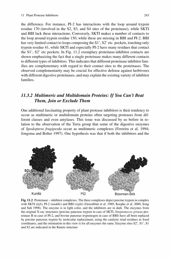
11 Plant Protease Inhibitors 243
the difference. For instance, PI-2 has interactions with the loop around trypsinresidue 170 (involved in the S2, S3, and S4 sites of the proteinase), while SKTIand BBI lack these interactions. Conversely, SKTI makes a number of contacts tothe loop around trypsin residue 150, while these are missing in BBI and PI-2. BBIhas very limited contact to loops composing the S1’, S2’ etc. pockets, touching onlytrypsin residue 41, while SKTI and especially PI-2 have many residues that contactthe S1’, S2’ etc pockets. In Fig. 11.2 exemplary proteinase-inhibitor contacts areshown emphasizing the fact that a single proteinase makes many different contactsto different types of inhibitors. This indicates that different proteinase inhibitor fam-ilies are complementary with regard to their contact sites to the proteinases. Theobserved complementarity may be crucial for effective defense against herbivoreswith different digestive proteinases, and may explain the existing variety of inhibitorfamilies.
11.3.2 Multimeric and Multidomain Proteins: If You Can’t BeatThem, Join or Exclude Them
One additional fascinating property of plant protease inhibitors is their tendency tooccur as multimeric or multidomain proteins often targeting proteases from dif-ferent classes and even amylases. This issue was discussed by us before in re-lation to the observation of the Terra group that some of the digestive enzymesof Spodoptera frugiperda occur as multimeric complexes (Ferreira et al. 1994;Jongsma and Bolter 1997). One hypothesis was that if both the inhibitors and the
Fig. 11.2 Proteinase – inhibitor complexes. The three complexes depict porcine trypsin in complexwith SKTI (left), PI-2 (middle) and BBI (right) (Greenblatt et al. 1989; Koepke et al. 2000; Songand Suh 1998). The enzyme is in light color, and the inhibitors are in dark. The enzymes fromthe original X-ray structures (porcine pancreas trypsin in case of SKTI, Streptomyces griseus pro-teinase B in case of PI-2, and bovine pancreas trypsinogen in case of BBI) have all been replacedby porcine pancreas trypsin by molecular replacement, using the catalytic triad residues as fixedcoordinates, and the orientation in this view is for all enzymes the same. Enzyme sites S2’, S1’, S1and S2 are indicated in the Kunitz structure
244 M.A. Jongsma, J. Beekwilder
(multimeric) proteases/amylases had two or more contact sides to each other thiswould lead to polymerization in one or more dimensions. Such a polymer of con-catenated protein would be less effective in accessing protein substrates, and, withfewer degrees of freedom, better prevent the dissociation of the inhibitor-enzymecomplex. In this way protease-inhibitor complexes with poor dissociation constantscould still remain effective in controlling the enzyme activity. Multidomain or multi-meric associations of inhibitors could, thus, be an evolutionary advantageous trait tothe plant. It was not clear, however, how multimeric associations of proteases couldbe advantageous to the insect. This was discussed in a second study on Heliothisvirescens (Brito et al. 2001) where the authors found the multimeric forms of trypsinto be specifically induced in response to PIs and, furthermore, to be less sensitive toPIs. This led them to hypothesize that the multimeric association might render themsterically less sensitive to the inhibitors. If the enzymes would be complexed not injuxtaposition but with the active sites close together this would certainly be a realpossibility as previously observed for human tryptase (Pereira et al. 1998). Thus,there are ways for both plants and insects of associating proteases or inhibitorsto potentially increase or decrease inhibition. More detailed research is needed todemonstrate the reality and success of these strategies both on the plant and insectside. Surely, however, these structural features provide an extra dimension to thestudy and understanding of this form of plant defense.
11.4 The Insects Fight Back
11.4.1 Evolution of Steric Occlusion
Plants have evolved different inhibitor classes as a presumably successful strategyof defending themselves against insect and pathogen attack (Ryan 1990; Jongsmaand Bolter 1997; Ferry et al. 2006). Most plants produce protease inhibitors uponwounding, and fill protein storage organs such as seeds, fruits or tubers with proteaseinhibitors, to make them nutritionally unsuitable for herbivores. However, insectsdepend on plant proteins as the source of amino acids and strong selection pressureshave operated to avoid the action of the inhibitors. In the previous paragraph wealready discussed multimeric associations as one way which possibly evolved tosterically avoid the entry of inhibitors in the active sites of the protease. This pos-sibility is still largely hypothetical, however, and requires experimental verification.A second well-documented strategy has evolved mainly in generalist insects suchas Helicoverpa and Spodoptera species, which must adapt to many different config-urations of inhibitors. The successful colonization of many different plant speciesby these insects is based on their ability to switch to a different set of proteasesupon encountering protease inhibitors in their diet. Characteristic of this inducedset of proteases is their insensitivity to a broad range of inhibitors. This specialproperty was first noted a decade ago (Jongsma et al. 1995), and has since thenbeen found in many insect pests (Bown et al. 1997, 2004; Mazumdar-Leighton and

11 Plant Protease Inhibitors 245
Broadway 2001a; Volpicella et al. 2003; Gruden et al. 2004; Liu et al. 2004; Moonet al. 2004; Bayes et al. 2005; Chougule et al. 2005).
What causes the resistance of these proteases to inhibition? Several different linesof research have been deployed to understand the induced proteinases. By analyzinggene expression in the gut, sequences of proteases have been identified that are in-duced upon ingestion of protease inhibitors (Bown et al. 1997; Mazumdar-Leightonand Broadway 2001b; Gruden et al. 2004). However, in the absence of a suitablerecombinant expression system, it has so far not been possible to analyze the reac-tivity of different inhibitors to individual proteases. Proteinases have been purifiedfrom herbivore guts to identify the dominant proteinases involved in this switch,and to analyze their interaction with plant inhibitors in inhibition assays and byprotein modeling (Volpicella et al. 2003, 2006; Gruden et al. 2004). Proteinases inthe Helicoverpa midgut, induced by ingestion of SKTI, were compared to enzymesisolated from uninduced midguts. Both classes of enzymes were equally active onproteinaceous substrates, but differed extremely in their sensitivity to inhibitors fromfour different classes. The dominant trypsin isolated from SKTI-induced guts wascompletely insensitive to >1000 fold molar excess of any of the four tested in-hibitors, each of them with different structural folds (Volpicella et al. 2003). On theother hand, also enzymes with differential sensitivity to different inhibitor classeswere observed. For instance, a chymotrypsin induced by SKTI was more susceptibleto PI-2 than to other inhibitors (Volpicella et al. 2006). Other groups have observeddifferences in effectiveness of chickpea-derived and potato-derived inhibitors tosemi-purified Helicoverpa armigera proteinases (Telang et al. 2005). These datashow that a panel of different proteinases can be deployed by adapted pest insects,each with different inhibitor sensitivity.
What makes a proteinase insensitive to an inhibitor? The use of syntheticsubstrates revealed potentially relevant differences between SKTI-induced and con-stitutive enzymes. Benzoyl-Arg-pNA, which carries a benzoyl group at the P2 po-sition, was digested ten times less efficiently by SKTI-induced trypsin as comparedto uninduced trypsin, while substrates with Phe or Arg in the P2 position did notdiscriminate the two proteinases (Volpicella et al. 2003). This suggests that an im-portant structural feature exists at the S2 site of induced trypsin, which correlateswith its resistance to inhibitors. However, the nature of that structural feature cannotbe further explored in the absence of a successful recombinant expression systemand crystal structures of the proteins.
Unequivocal insight in the structural basis of the resistance of insect proteasesto plant inhibitors was obtained for the first time from the crystal structure ofHelicoverpa zea carboxypeptidase B (Bayes et al. 2005, 2006). This enzyme, re-ferred to as CPBHz, resists inhibition by potato carboxypeptidase inhibitor (PCI),which inhibits all other known enzymes from this class, including Helicoverpa car-boxypeptidase A (CPAHa). The CPBHz crystal structure clearly showed that twosurface loops of the enzyme assumed an unusual position in comparison to relatedinhibited enzymes such as CPAHa. In high-affinity complexes, these regions stabi-lize the interaction between the carboxypeptidase and the inhibitor. This interactionis initially formed by insertion of the flexible C-terminal tail of PCI, which presents

246 M.A. Jongsma, J. Beekwilder
the P1 and P1’ residue to the active site pocket (Rees and Lipscomb 1982; Molinaet al. 1994). However, due to the displacement of these loops on the surface of theenzyme, the inhibitor binds to a more distant position relative to the active site. Asa consequence, the P1 and P1’ residues are unable to penetrate the active site deepenough to achieve a strong interaction, and the interaction is easily lost.
The evolutionary mechanism by which the proteases evolve towards insensitivityfor the inhibitors can be deduced in part from a comparison of the mutated sequencewith its closest PI-sensitive homologue. Displacement of protease loops is clearlyinvolved, as is apparent from the CPBHz structure. In one of the two loops, dis-placement is accompanied by insertion of a residue relative to inhibitor-sensitiveenzymes, while in the other loop, a single residue is deleted. In both loops, a theobserved number of mutations was higher as compared to other parts of the molecule(Bayes et al. 2005). Interestingly, one could predict that the plant could re-establishinhibition of CPBHz by inserting an additional residue in the substrate-like tail ofPCI, thus allowing its P1 residue to reach the active site pocket, but such an inhibitorwith an extended tail has not yet been found or evolved.
11.4.2 Inactivation with Specific Proteases
Although generally stable against many proteases, some insects have evolved spe-cific proteases capable of effectively degrading plant protease inhibitors. We ob-served this phenomenon recently with Plutella xylostella degrading MTI-2 whenexpressed in Arabidopsis (Yang et al. submitted). Others found degradation of di-etary protease inhibitors by insects like black vine weevil, Phaedon cochleria andH. armigera (Michaud 1997; Girard et al. 1998; Giri et al. 1998). In our opinion,many cases of specific inactivation may have escaped detection due to the exper-imental set up chosen by most authors, in which comparing short and prolongedpreincubation of the inhibitors with the gut proteases is not standard procedure.
11.5 Perspectives
11.5.1 Regulation is the Key: Did Insects and Plants BothThink of It. . .?
The existence of PI-insensitive proteases in the insect gut and their specific up-regulation in response to proteinase inhibitors in the diet provides a clear benefitto the insect and is likely monitored by some kind of feedback mechanism whichhas not yet been elucidated. However, the parallel observation of sensitive enzymesbeing downregulated could be beneficial to both insects and plants alike. The insectsdon’t waste their energy on useless (inhibited) proteases, but the plants similarly,if they would govern this process, could spend less energy on synthesizing an ef-fective concentration of inhibitors. Indeed if plants could successfully antagonize
11 Plant Protease Inhibitors 247
the regulatory feedback system operational in insect guts, for PI-sensitive and-insensitive proteases alike, they would have found a second way independent ofdirect inhibition to achieve the same net result: lower protease levels in the gut,leading to slower development and higher mortality. Hijacking the regulation ofproteases in the insect gut is, therefore, a holy grail for plants, and preventing themfrom achieving this goal is the lifeline of the insects: an attractive second stage ofevolutionary battle between proteases and inhibitors.
However, has the script been written? How likely is it, that plants have evolvedsuch signals for the specific purpose of downregulating proteases in insects? Cur-rently, there are a few peptide hormones known for receptor-mediated up- or down-regulation of serine proteases. In mammals the monitor peptide, member of the PSTIgene family, and the luminal cholecystokinin releasing factor LCRF are known toupregulate pancreatic protease secretion by stimulating cholecystokinin release inthe blood (Iwai et al. 1988; Spannagel et al. 1996). Excess proteases subsequentlydegrade the monitor/LCRF peptides and stop the secretion. In mosquitoes after ablood meal the oostatic hormone or trypsin modulating oostatic factor (TMOF) wasthe only peptide found so far to actively signal the downregulation of trypsin secre-tion into the gut (Borovsky et al. 1990).
Remarkably the TMOF peptide, which consists of only ten amino acids, isalso active in the lepidopteran herbivore H. virescens. TMOF actively downregu-lates proteinase secretion when injected into the hemolymph of the larvae (Nauenet al. 2001), but surprisingly also when it is expressed in plants fused to otherpeptides, and when it reaches the larvae through the gut after eating the plantleaves. Expressed as a potentially cleavable six-domain repeat, or replacing sys-temin in prosystemin, or in C-terminal fusion to the coat protein of TMV, TMOFreduced larval development and decreased both gut trypsin and chymotrypsin activ-ity (Tortiglione et al. 2002, 2003; Borovsky et al. 2006).
The TMOF peptide presumably signals through the interaction with receptorspresent on cells lining the gut. Such receptors are also potential targets for plantpeptides or proteins to upset gut physiology. This brings us to the last unexploredarea of plant protease inhibitors: the function(s) of the N- and C-terminal peptideswhich extend from most of the basic inhibitor domains that have been characterizedin plants. The extensions appear in significant variation, both in length and sequence,within most inhibitor gene families. As an example, Table 11.3 shows the peptidesextending from the mature forms of the Bowman–Birk inhibitors found in soybeanand peanut. These sequences represent specific insertions and/or deletions. The ex-tensions are not likely to be involved in the subcellular targeting of inhibitors, sinceonly the mature proteins are shown and the targeting peptides are already removed.Therefore, these peptide domains may serve a direct role in plant defense. Potentialfunctions include the modification of the pI of the protein molecule to favor theinteraction with the protease, or else, the binding to cellular receptors lining theherbivore gut to modulate or antagonize the regulatory processes of the insect. Un-derstanding the regulation of protease expression in insects and the potential role ofplant protease inhibitors to interfere with this regulation promises to be a rewardingresearch area.

248 M.A. Jongsma, J. Beekwilder
Table 11.3 Comparison of the N- and C-terminal peptides of the Bowman–Birk inhibitors of soy-bean and peanut extending outside the basic inhibitory domain for which no function has beenassigned
Source Code1 N-terminal sequence3 Inhibitor2 C-terminal sequence3
Soybean MER21210 SDQSSSYDDDEYSKPCC- -inhibitor- -CKSRDDSoybean MER21212 SDHSSSDDESSKPCC- -inhibitor- -CKSSDEDDDSoybean MER19956 SDHHQHSNDDDSSKPCC- -inhibitor- -CKPSEDDKENYSoybean MER19957 SDHHQHSNDDESSKPCC- -inhibitor- -CKPSQDDKENYPeanut MER18102 EASSSSDDNVCC- -inhibitor- -CRSPeanut MER18107 AASDCC- -inhibitor- -CA1 Codes refer to the MEROPS protease inhibitor database: http://merops.sanger.ac.uk/2 This represents the conserved two domains involved in protease inhibition3 The sequences represent the mature stored forms after removal of the signal peptide and othertargeting peptides
11.5.2 Creative Evolution
In this chapter we reviewed the ways in which plants and plant predators haveevolved creative solutions in the battle for scarce essential amino acids. We showedthat on both the plant and insect side many different gene families are involvedwith intricate specialized expression patterns among the different members. Theyprovide an evolutionary and ecological genetic fingerprint of their functional role,and, if anything, this chapter highlighted the little that is actually known about theintricate fabric of functional relationships that are still out there to be discoveredand admired.
Some of the hypotheses we put forward are currently only supported by circum-stantial evidence, but provide attractive alleys which functional evolution may havegone. At first glance, this topic of plant–insect/pathogen interactions appears to beonly a clash of two protein molecules. However, due to the opportunistic nature ofevolution, this battle may have taken on many new appearances. We hope our accountof these possibilities provides inspiration to researchers to investigate them. The avail-ability of fully sequenced genomes on both the plant and insect side, and the excellenttools to specifically knock down and overexpress specific genes provide exactly theinstruments needed to allow a whole new set of discoveries in this area.
References
Abdeen A, Virgos A, Olivella E, Villanueva J, Aviles X, Gabarra R, Prat S (2005) Multiple in-sect resistance in transgenic tomato plants over-expressing two families of plant proteinaseinhibitors. Plant Mol Biol 57:189–202
Bayes A, Comellas-Bigler M, de la Vega MR, Maskos K, Bode W, Aviles FX, Jongsma MA,Beekwilder J, Vendrell J (2005) Structural basis of the resistance of an insect carboxypeptidaseto plant protease inhibitors. Proc Natl Acad Sci USA 102:16602–16607
Bayes A, de la Vega MR, Vendrell J, Aviles FX, Jongsma MA, Beekwilder J (2006) Response ofthe digestive system of Helicoverpa zea to ingestion of potato carboxypeptidase inhibitor andcharacterization of an uninhibited carboxypeptidase B. Insect Biochem Mol Biol 36:654–664
11 Plant Protease Inhibitors 249
Beekwilder J, Schipper B, Bakker P, Bosch D, Jongsma M (2000) Characterization of potato pro-teinase inhibitor II reactive site mutants. Eur J Biochem 267:1975–1984
Berger A, Schechter I (1970) Mapping the active site of papain with the aid of peptide substratesand inhibitors. Philos Trans R Soc Biol Sci 257:249–264
Birk Y, Gertler A, Khalef S (1963) A pure trypsin inhibitor from soya beans. Biochem J87:281–284
Bode W, Huber R (2000) Structural basis of the endoproteinase–protein inhibitor interaction.Biochim Biophys Acta 1477:241–252
Borovsky D, Carlson DA, Griffin PR, Shabanowitz J, Hunt DF (1990) Mosquito oostatic fac-tor: a novel decapeptide modulating trypsin-like enzyme biosynthesis in the midgut. FASEB J4:3015–3020
Borovsky D, Rabindran S, Dawson WO, Powell CA, Iannotti DA, Morris TJ, Shabanowitz J, HuntDF, DeBondt HL, DeLoof A (2006) Expression of Aedes trypsin-modulating oostatic factor onthe virion of TMV: a potential larvicide. Proc Natl Acad Sci USA 103:18963–18968
Bowman DE (1946) Differentiation of soy bean antitryptic factors. Proc Soc Exp Biol Med63:547–550
Bown DP, Wilkinson HS, Gatehouse JA (1997) Differentially regulated inhibitor-sensitive andinsensitive protease genes from the phytophagous insect pest, Helicoverpa armigera, are mem-bers of complex multigene families. Insect Biochem Mol Biol 27:625–638
Bown DP, Wilkinson HS, Gatehouse JA (2004) Regulation of expression of genes encoding di-gestive proteases in the gut of a polyphagous lepidopteran larva in response to dietary proteaseinhibitors. Physiol Entomol 29:278–290
Brito LO, Lopes AR, Parra JRP, Terra WR, Silva MC (2001) Adaptation of tobacco budwormHeliothis virescens to proteinase inhibitors may be mediated by the synthesis of new pro-teinases. Comp Biochem Physiol Biochem Mol Biol 128:365–375
Chougule NP, Giri AP, Sainani MN, Gupta VS (2005) Gene expression patterns of Helicoverpaarmigera gut proteases. Insect Biochem Mol Biol 35:355–367
Clauss MJ, Mitchell-Olds T (2004) Functional divergence in tandemly duplicated Arabidopsisthaliana trypsin inhibitor genes. Genetics 166:1419–1436
De Leo F, Gallerani R (2002) The mustard trypsin inhibitor 2 affects the fertility of Spodopteralittoralis larvae fed on transgenic plants. Insect Biochem Mol Biol 32:489–496
Ferreira C, Capella AN, Sitnik R, Terra WR (1994) Properties of the digestive enzymes and the per-meability of the peritrophic membrane of Spodoptera frugiperda (Lepidoptera) larvae. CompBiochem Physiol Physiol 107:631–640
Ferry N, Edwards MG, Gatehouse J, Capell T, Christou P, Gatehouse AMR (2006) Transgenicplants for insect pest control: a forward looking scientific perspective. Transgenic Res 15:13–19
Girard C, Le Metayer M, Bonade-Bottino M, Pham-Delegue MH, Jouanin L (1998) High level ofresistance to proteinase inhibitors may be conferred by proteolytic cleavage in beetle larvae.Insect Biochem Mol Biol 28:229–237
Giri AP, Harsulkar AM, Deshpande VV, Sainani MN, Gupta VS, Ranjekar PK (1998) Chickpeadefensive proteinase inhibitors can be inactivated by podborer gut proteinases. Plant Physiol116:393–401
Gosalia DN, Salisbury CM, Ellman JA, Diamond SL (2005) High throughput substrate specificityprofiling of serine and cysteine proteases using solution-phase fluorogenic peptide microarrays.Mol Cell Prot 4:626–636
Green TR, Ryan CA (1972) Wound-induced proteinase inhibitor in plant leaves – possible defensemechanism against insects. Science 175:776–777
Greenblatt HM, Ryan CA, James MN (1989) Structure of the complex of Streptomyces griseusproteinase B and polypeptide chymotrypsin inhibitor-1 from russet burbank potato tubers at2.1 A resolution. J Mol Biol 205:201–228
Gruden K, Kuipers AGJ, Guncar G, Slapar N, Strukelj B, Jongsma MA (2004) Molecular basisof colorado potato beetle adaptation to potato plant defence at the level of digestive cysteineproteinases. Insect Biochem Mol Biol 34:365–375

250 M.A. Jongsma, J. Beekwilder
Gutierrez-Campos R, Torres-Acosta JA, Saucedo-Arias LJ, Gomez-Lim MA (1999) The use ofcysteine proteinase inhibitors to engineer resistance against potyviruses in transgenic tobaccoplants. Nature Biotech 17:1223–1226
Hejgaard J (2005) Inhibitory plant serpins with a sequence of three glutamine residues in the reac-tive center. Biol Chem 386:1319–1323
Hilder VA, Gatehouse AMR, Sheerman SE, Barker RF, Boulter D (1987) A novel mechanism ofinsect resistance engineered into tobacco. Nature 330:160–163
Iwai K, Fushiki T, Fukuoka S (1988) Pancreatic-enzyme secretion mediated by novel peptide:monitor peptide hypothesis. Pancreas 3:720–728
Jongsma MA, Bakker PL, Peters J, Bosch D, Stiekema WJ (1995) Adaptation of Spodoptera ex-igua larvae to plant proteinase-inhibitors by induction of gut proteinase activity insensitive toinhibition. Proc Natl Acad Sci USA 92:8041–8045
Jongsma MA, Bolter C (1997) The adaptation of insects to plant protease inhibitors. J Insect Phys-iol 43:885–895
Koepke J, Ermler U, Warkentin E, Wenzl G, Flecker P (2000) Crystal structure of cancer chemo-preventive Bowman–Birk inhibitor in ternary complex with bovine trypsin at 2.3 A resolution.Structural basis of janus-sfaced serine protease inhibitor specificity. J Mol Biol 298:477–491
Kunitz M (1946) Crystalline soybean trypsin inhibitor. J Gen Physiol 29:149–154Laskowski M, Kato I, Ardelt W, Cook J, Denton A, Empie MW, Kohr WJ, Park SJ, Parks K,
Schatzley BL, Schoenberger OL, Tashiro M, Vichot G, Whatley HE, Wieczorek A, WieczorekM (1987) Ovomucoid 3rd domains from 100 avian species – isolation, sequences, and hyper-variability of enzyme-inhibitor contact residues. Biochemistry 26:202–221
Liu YL, Salzman RA, Pankiw T, Zhu-Salzman K (2004) Transcriptional regulation in southerncorn rootworm larvae challenged by soyacystatin N. Insect Biochem Mol Biol 34:1069–1077
Mazumdar-Leighton S, Broadway RM (2001a) Identification of six chymotrypsin cDNAs fromlarval midguts of Helicoverpa zea and Agrotis ipsilon feeding on the soybean (Kunitz) trypsininhibitor. Insect Biochem Mol Biol 31:633–644
Mazumdar-Leighton S, Broadway RM (2001b) Transcriptional induction of diverse midguttrypsins in larval Agrotis ipsilon and Helicoverpa zea feeding on the soybean trypsin inhibitor.Insect Biochem Mol Biol 31:645–657
Michaud D (1997) Avoiding protease-mediated resistance in herbivorous pests. Trends Biotechnol15:4–6
Molina MA, Marino C, Oliva B, Aviles FX, Querol E (1994) C-tail valine is a key residuefor stabilization of complex between potato inhibitor and carboxypeptidase-A. J Biol Chem269:21467–21472
Moon J, Salzman RA, Ahn JE, Koiwa H, Zhu-Salzman K (2004) Transcriptional regulation incowpea bruchid guts during adaptation to a plant defence protease inhibitor. Insect Mol Biol13:283–291
Nauen R, Sorge D, Sterner A, Borovsky D (2001) Tmof-like factor controls the biosynthesisof serine proteases in the larval gut of Heliothis virescens. Arch Insect Biochem Physiol47:169–180
Pereira PJB, Bergner A, Macedo-Ribeiro S, Huber R, Matschiner G, Fritz H, Sommerhoff CP,Bode W (1998) Human �-tryptase is a ring-like tetramer with active sites facing a central pore.Nature 392:306–311
Qu LJ, Chen J, Liu MH, Pan NS, Okamoto H, Lin ZZ, Li CY, Li DH, Wang JL, Zhu GF, Zhao X,Chen X, Gu HG, Chen ZL (2003) Molecular cloning and functional analysis of a novel type ofBowman–Birk inhibitor gene family in rice. Plant Physiol 133:560–570
Rawlings ND, Morton FR, Barrett AJ (2006) MEROPS: the peptidase database. Nucl Acids Res34:D270–D272
Rees DC, Lipscomb WN (1982) Refined crystal-structure of the potato inhibitor complex ofcarboxypeptidase-A at 2.5-A resolution. J Mol Biol 160:475–498
Ryan CA (1990) Protease inhibitors in plants – genes for improving defenses against insects andpathogens. Ann Rev Phytopathol 28:425–449

11 Plant Protease Inhibitors 251
Song HK, Suh SW (1998) Kunitz-type soybean trypsin inhibitor revisited: refined structure of itscomplex with porcine trypsin reveals an insight into the interaction between a homologousinhibitor from Erythrina caffra and tissue-type plasminogen activator. J Mol Biol 275:347–363
Spannagel AW, Green GM, Guan DF, Liddle RA, Faull K, Reeve JR (1996) Purification and char-acterization of a luminal cholecystokinin-releasing factor from rat intestinal secretion. ProcNatl Acad Sci USA 93:4415–4420
Telang MA, Giri AP, Sainani MN, Gupta VS (2005) Characterization of two midgut proteinasesof Helicoverpa armigera and their interaction with proteinase inhibitors. J Insect Physiol51:513–522
Tortiglione C, Fanti P, Pennacchio F, Malva C, Breuer M, De Loof A, Monti LM, Tremblay E, RaoR (2002) The expression in tobacco plants of Aedes aegypti trypsin modulating oostatic factor(aea-tmof) alters growth and development of the tobacco budworm, Heliothis virescens. MolBreed 9:159–169
Tortiglione C, Fogliano V, Ferracane R, Fanti P, Pennacchio F, Monti LM, Rao R (2003) An insectpeptide engineered into the tomato prosystemin gene is released in transgenic tobacco plantsand exerts biological activity. Plant Mol Biol 53:891–902
Volpicella M, Ceci LR, Cordewener J, America T, Gallerani R, Bode W, Jongsma MA, Beek-wilder J (2003) Properties of purified gut trypsin from Helicoverpa zea, adapted to proteinaseinhibitors. Eur J Biochem 270:10–19
Volpicella M, Ceci LR, Gallerani R, Jongsma MA, Beekwilder J (2001) Functional expressionon bacteriophage of the mustard trypsin inhibitor MTI-2. Biochem Biophys Res Commun280:813–817
Volpicella M, Cordewener J, Jongsma MA, Gallerani R, Ceci LR, Beekwilder J (2006) Identifica-tion and characterization of digestive serine proteases from inhibitor-resistant Helicoverpa zealarval midgut. J Chrom B Analyt Technol Biomed Life Sci 833:26–32
Yang L, Fang ZY, Dicke M, van Loon JJA, Jongsma MA (2008) The diamondback moth, Plutellaxylostella specifically inactivates Mustard Trypsin Inhibitor 2(MTI2) to overcome host plantdefence (submitted)
Zavala JA, Patankar AG, Gase K, Hui DQ, Baldwin IT (2004) Manipulation of endogenous trypsinproteinase inhibitor production in Nicotiana attenuata demonstrates their function as antiher-bivore defenses. Plant Physiol 134:1181–1190
Zimmermann P, Hirsch-Hoffmann M, Hennig L, Gruissem W (2004) Genevestigator. Arabidopsismicroarray database and analysis toolbox. Plant Physiol 136:2621–2632

Chapter 12Defensive Roles of Polyphenol Oxidase in Plants
C. Peter Constabel and Raymond Barbehenn
Plant polyphenol oxidases (PPOs) are widely distributed and well-studied oxidativeenzymes, and their effects on discoloration in damaged and diseased plant tissueshave been known for many years. The discovery in C.A. Ryan’s laboratory in themid-1990s that tomato PPO is induced by the herbivore defense signals systeminand jasmonate, together with seminal work on PPO’s possible effects on herbiv-orous insects by G. Felton and S. Duffey has motivated many studies of PPO inthe context of plant-herbivore defense. The cloning and characterization of PPOcDNAs from multiple plant species now allows for direct testing of defensive func-tions of PPO using transgenic plants. These have shown that PPO can contributeto insect herbivore and pathogen resistance, although how this occurs is only nowbeing investigated more closely. Here we review progress in the functional analysisof PPO in plant defense against pests, and describe recent results that address themechanisms of PPO as an anti-herbivore protein. We suggest that assumptions ofhow PPO functions as an anti-nutritive defense against lepidopterans needs to be re-examined in light of the near anoxic conditions in lepidopteran midguts. Ultimately,the efficacy of PPO should be directly tested in a greater variety of plant-insectinteractions. In addition, the identification of the endogenous PPO substrates willhelp to define defensive and potentially other roles for PPO in plants.
12.1 Introduction
Polyphenol oxidases (PPOs) are ubiquitous copper-containing enzymes which usemolecular oxygen to oxidize common ortho-diphenolic compounds such as caf-feic acid and catechol to their respective quinones. PPO-generated quinones arehighly reactive and may cross-link or alkylate proteins, leading to the commonly ob-served brown pigments in damaged plant tissues and plant extracts. The conspicuouspigments are generally undesirable in food products, and the role of PPO in browning
C.P. ConstabelCentre for Forest Biology and Department of Biology, University of Victoria, Victoria,BC, Canada V8W 3N5,e-mail: [email protected]
A. Schaller (ed.), Induced Plant Resistance to Herbivory, 253C© Springer Science+Business Media B.V. 2008

254 C.P. Constabel, R. Barbehenn
has prompted numerous studies on PPO in food and beverages. In parallel, the po-tential roles for PPO in plant defense against pests have motivated many studies onPPO in an ecological context, though few of these have used a transgenic approach.Functional and mechanistic studies on PPO in plant-insect interactions using PPO-modified transgenic plants have recently been reported, providing new insight intothe biology of this versatile enzyme.
12.2 The Biochemistry of Plant Polyphenol Oxidase
Polyphenol oxidase (catechol oxidase; E.C. 1.10.3.2) has been purified and char-acterized from a wide range of plant species and a variety of tissues (Constabelet al. 1996; Mayer 2006), and activity levels using common substrates vary widely(Constabel and Ryan 1998). Key features of PPOs are two conserved copper-bindingdomains, and N-terminal chloroplast and thylakoid transit peptides (van Gelderet al. 1997; Marusek et al. 2006). The size of the predicted mature PPO proteins istypically 54–62 kD (Constabel et al. 1996; van Gelder et al. 1997). However, somePPOs are processed further, and in some cases only the processed form is fully active(Rathjen and Robinson 1992). Many PPOs are predicted to contain a proteolyticprocessing site near the C-terminus of the polypeptide (Marusek et al. 2006).
Based on its association with browning reactions in crop plants, PPO has beencharacterized in a wide variety of food plants including banana, wheat, quince, andavocado, and a number of chemical inhibitors have been identified (reviewed inMayer 2006; Yoruk and Marshall 2003). Although PPO is found at significant levelsin a variety of fruits, vegetables and grains, its biological function in these tissues hasrarely been studied. PPO is expressed in many different tissues and organs, includingroots, leaves, flowers, and vascular tissue (Constabel et al. 1996). Its presence inchloroplasts led to the proposal that PPO may function in pseudocyclic photophos-phorylation or as a modulator of oxygen levels, yet to date little evidence supportsthese roles (Steffens et al. 1994). A complicating issue regarding PPO function hasbeen the separate localization of its phenolic substrates in plant vacuoles, so thatthe cell would have to be broken in order for PPO to oxidize phenols; this is mostlikely to occur following pest or pathogen challenge. Roles of PPO in plant defenseare thus commonly discussed, although direct evidence for such roles has becomeavailable only recently.
A puzzling feature of PPO has been a variable degree of latency, so that PPOsfrom some species need to be activated with detergents or proteases for full activ-ity. For example, tomato leaf PPO is extracted in its fully active form (Constabelet al. 1995), but poplar leaf PPO requires activation with protease or detergent(Constabel et al. 2000). Recent experiments suggest that such activating treatmentsfunction by removal of a peptide from the active site, via proteolysis or partial un-folding of the polypeptide, respectively (Gandia-Herrero et al. 2005). Treatmentwith low pH can also activate latent PPOs, presumably via a conformational changeof the active site, increasing its accessibility to substrates (Kanade et al. 2006).

12 Polyphenol Oxidases in Plant Defense 255
PPO latency may be significant for its defensive function; we recently showed thatlatent poplar PPO is activated by its passage through caterpillar guts (Wang andConstabel 2004).
PPOs are known to have broad substrate specificities. Thus, an enzyme from anygiven source may be capable of oxidizing a variety of simple ortho-diphenolics,such as caffeic acid and its conjugates, catechol derivatives, or dihydroxyphe-nylalanine (DOPA). However, enzymes from different plant species exhibit distinctpreference profiles (reviewed in Constabel et al. 1996). Flavonoids withortho-dihydroxy phenolic rings have been found to be PPO substrates, for example,catechin, (−)-epicatechin, and myricetin (Guyot et al. 1996; Jimenez and Garcia-Carmona 1999). Some reports also describe the oxidation of trihydroxy phenolics,such as gallic acid, by PPO (Shin et al. 1997), but it is not clear how widespreadthis is. Furthermore, for some PPOs a monophenolase activity has been described(Wuyts et al. 2006). Such enzymes may hydroxylate a monophenol such as tyro-sine, which can then undergo further oxidation by the polyphenol oxidase activityto the quinone. Tyrosine hydroxylation by PPO has been described in pokeweed,where this reaction constitutes part of the biosynthetic pathway leading to betalains(Gandia-Herrero et al. 2005).
While the range of substrates accepted by isolated PPOs can be readily definedin vitro, knowledge of the substrates that are utilized in planta or during defensereactions is much scarcer. Caffeic acid esters such as chlorogenic acid (caffeoyl-quinate) are excellent PPO substrates and are very common plant metabolites. Intomato and coffee, chlorogenic acid has been identified as the most likely in vivoPPO substrate (Melo et al. 2006; Li and Steffens 2002). Caffeic acid esters areubiquitous as lignin precursors (Humphreys and Chapple 2002), but in most speciesthese may not accumulate to sufficient levels to be considered likely PPO substrates.In Populus tremuloides, catechol is postulated to be released by the breakdown ofthe abundant phenolic glycosides (Clausen et al. 1989), and would therefore beavailable as a substrate in damaged tissues (Haruta et al. 2001). For most PPOs,however, the endogenous substrates are unknown. The importance of identifyingthe in planta PPO substrates and understanding the overall phytochemical contextof PPO-containing plants is emphasized by recent reports of PPO-like enzymeswith biosynthetic roles as hydroxylases of secondary metabolites (Cho et al. 2003;Nakayama et al. 2000).
PPO has been extensively studied by biologists, plant pathologists, and ecolo-gists interested in mechanisms of defense against pests and pathogens. Based on thebrowning reactions resulting from the reactive PPO-generated quinones, PPO hasoften been suggested to function as a defense against pests and pathogens. A mostdramatic illustration of the efficacy of PPO in this context comes from work onSolanum berthaultii in which extremely high PPO levels (45% of soluble protein)are found in glandular trichomes (Kowalski et al. 1992). Breakage of the trichomesby small-bodied insects such as aphids leads to rapid PPO-mediated oxidationand polymerization of phenolics, ultimately entrapping insects, or occluding theirmouthparts with a sticky polymer (Kowalski et al. 1992). In most species, however,leaf PPO is found not in trichomes but in mesophyll cells. Here, the PPO-generated

256 C.P. Constabel, R. Barbehenn
quinones were proposed to alkylate dietary protein during insect feeding, and to de-grade essential amino acids in insect guts (Felton et al. 1989, 1992). PPO-mediatedprotein alkylation has been demonstrated to operate against lepidopteran pests inthe presence of oxygen and in artificial diets, and is now being investigated in moredetail in midgut fluids (see Section 12.5 below).
12.3 PPO and Induced Herbivore Defense in Tomatoand Other Plants
The idea that PPO may act as an anti-nutritive defense against leaf-eating insectswas first suggested by G. Felton and S. Duffey, who showed an inverse correlationof Heliothis zea growth and PPO levels in tomato plants (Felton et al. 1989). Strongsupport for an anti-herbivore role of PPO came from the discovery that the her-bivore defense-inducing signal molecules systemin and methyl jasmonate (MeJA)induce PPO activity and PPO mRNA levels in tomato leaves (Constabel et al. 1995).Systemin is a short peptide required for systemic wound signaling in tomato whichstrongly upregulates tomato herbivore defenses (Ryan 2000; Narvaez-Vasquez andOrozco-Cardenas, this volume). Though no longer considered to be the primarysystemic signal, it is required for the generation of such a signal (Schilmiller andHowe 2005). PPO and other defenses were also found to be strongly inducedby MeJA and oligogalacturonic acid, major plant defense signaling compounds(Constabel et al. 1995). Since PPO induction in tomato by multiple signals oc-curs in parallel with a suite of other anti-herbivore proteins including several typesof protease inhibitors (PIs) and the anti-nutritive enzymes arginase and threoninedeaminase (Bergey et al. 1996; Chen et al. 2005), PPO is thought to play a similarrole in defense against insects.
In tobacco, PPO and PIs are upregulated by tobacco systemin as well as byMeJA (Constabel and Ryan 1998; Ren and Lu 2006). Likewise, strong herbivore-,wound-, and MeJA-induction of PPO was shown in leaves of several poplar species(Constabel et al. 2000; Haruta et al. 2001). Hybrid poplar (Populus trichocarpa xP. deltoides) has a strong systemic inducible defense response, which also includestrypsin inhibitors and chitinases, both with confirmed anti-insect activities (Parsonset al. 1989; Lawrence and Novak 2001, 2006). Recent large scale genomics exper-iments have underscored the complexity of the herbivore defense response in hy-brid poplar, which involves upregulation of many additional putative defense genes(Christopher et al. 2004; Ralph et al. 2006; Major and Constabel 2006). Overall, theco-induction of PPO with other herbivore defense proteins in several plant specieshas provided support for its anti-herbivore function.
The induction of PPO in tomato by both insects and MeJA has been replicatedin both laboratory and field studies (Stout et al. 1998; Thaler et al. 1996, 2002).Furthermore, the early work in tomato stimulated numerous studies on induciblePPO in diverse plant species. The inducibility of PPO by wounding or MeJA treat-ment has been confirmed in other plants, including both herbaceous crops and trees(Constabel and Ryan 1998). The induction of PPO by herbivory has now been shown

12 Polyphenol Oxidases in Plant Defense 257
Table 12.1 Studies of PPO induction in plants by herbivores with unknown effects on herbivores
Plant taxa Insect species (Order)1 Induction Reference
SolanaceaeTomato Leptinotarsa decemlineata (C) ∼2X Felton (1992)Potato L. decemlineata regurgitant ∼3–7X Kruzmane et al. (2002)
SalicaceaePoplar (hybrid) Malacosoma disstria (L) ∼12X Constabel et al. (2000)
BetulaceaeBlack alder Agelastica alni (C) ∼3X Tscharntke et al. (2001)
PoaceaeBuffalograss Blissus occiduus (H) None Heng-Moss et al. (2004)Barley, wheat, oats Diuraphis noxia (Ho) None Ni et al. (2001)
FabaceaeCommon bean Melanoplus differentialis (O) ∼2X Alba-Meraz and Choe (2002)Soybean Helicoverpa zea (L) None Bi and Felton (1995)Soybean Ceratoma trifurcata (C) None Felton et al. (1994)Soybean Spissistilus festinus (H) 1.6X Felton et al. (1994)
MalvaceaeCotton Helicoverpa zea (L) Not detectable Bi et al. (1997)
TheaceaeTea Helopeltis theivora (H) ∼2–3X Chakraborty and
Chakraborthy (2005)1 C = Coleoptera, L = Lepidoptera, H = Heteroptera, Ho = Homoptera, O = Orthoptera.
for a taxonomically diverse group of plants (Table 12.1), and this induction hascommonly been interpreted as a direct response against the herbivore. By contrast,studies of the potential impact of induced PPO against the herbivores have largelybeen done with a more limited range of plants and herbivores, primarily noctuidcaterpillars on tomato and other species in the Solanaceae (Table 12.2). The resultsof 11 of 16 experiments demonstrate PPO induction and are consistent with thehypothesis that induced PPO contributes to defense against herbivores. As with allcorrelative studies, however, it is not possible to determine the specific impact ofinduced PPO on herbivores due to the other biochemical changes that occur in dam-aged plants (e.g., Hermsmeier et al. 2001; Thaler et al. 2001; Chen et al. 2005; Majorand Constabel 2006). The direct effects of PPO on insect herbivores can thus bestbe tested using transgenic plants, where PPO levels are manipulated independentlyof other traits (see Section 12.4).
Other studies of the association between PPO activity and insect herbivore per-formance using (1) plant genotypes that vary in resistance to herbivory, (2) on-togenetic variation in PPO activity within the plant, and (3) leaves treated withPPO, yielded mixed results. A potato genotype with high PPO activity had in-creased resistance to the Colorado potato beetle (Leptinotarsa decemlineata;Castanera et al. 1996), while resistance to the coffee leaf miner (Leucopteracoffeella; Diptera) was apparently unaffected by higher levels of PPO in coffee

258 C.P. Constabel, R. Barbehenn
Tab
le12
.2In
duct
ion
ofPP
Os
inpl
ants
wit
hef
fect
ste
sted
onhe
rbiv
ores
Plan
ttax
aIn
duci
ngag
ent(
Ord
er)1
Indu
ctio
nE
ffec
tH
erbi
vore
1an
dre
spon
se2
Ref
eren
ce
Sola
nace
aeTo
mat
oH
elic
over
paze
a(L
)∼
2X(−
)Sp
odop
tera
exig
ua(L
)G
RSt
oute
tal.
(199
8)To
mat
oH
elic
over
paze
a(L
)∼
2X(−
)Sp
ider
mit
e(A
)nu
mbe
rsSt
oute
tal.
(199
8)To
mat
oJa
smon
icac
id∼
3X(0
)M
andu
case
xta
(L)
RG
RT
hale
ret
al.(
2002
)To
mat
oJa
smon
icac
id∼
3X(−
)Tr
icho
plus
iani
(L)
RG
RT
hale
ret
al.(
2002
)To
mat
oJa
smon
icac
id∼
3X(−
)T
hrip
s(T
)da
mag
eT
hale
ret
al.(
1999
,200
2)To
mat
oJa
smon
icac
id∼
3X(−
)Sp
ider
mit
e(A
)nu
mbe
rsT
hale
ret
al.(
2002
)To
mat
o(w
ild)
Jasm
onic
acid
∼3X
(0)
Spod
opte
raex
igua
(L)
RG
RT
hale
ret
al.(
2002
)To
mat
oJa
smon
icac
id∼
5X(−
)Sp
odop
tera
exig
ua(L
)R
GR
Tha
ler
etal
.(19
99)
Tom
ato
Mac
rosi
phum
euph
orbi
ae(H
)D
ecre
ased
(+)
Spod
opte
raex
igua
(L)
RG
RSt
oute
tal.
(199
8)To
mat
oW
ind
stre
ssD
ecre
ased
(0)
Man
duca
sext
a(L
)R
GR
Cip
ollin
iand
Red
man
(199
9)To
mat
oJa
smon
icac
id∼
2X(−
)M
andu
case
xta
(L)
RG
RR
edm
anet
al.(
2001
)To
bacc
o(w
ild)
clip
ped
sage
brus
h∼
4X(−
)Fi
eld
dam
age
by(L
)an
d(O
)K
arba
net
al.(
2000
)Po
tato
(wil
d)H
igh
cons
titu
tive
PPO
∼4–7
X(−
)L
epti
nota
rsa
dece
mli
neat
a(C
)R
GR
Cas
tane
raet
al.(
1996
)H
orse
nett
leH
igh
Nfe
rtil
izer
Dec
reas
ed(0
)M
andu
case
xta
(L)
herb
ivor
yC
ipol
linie
tal.
(200
2)H
orse
nett
leH
igh
Nfe
rtil
izer
Dec
reas
ed(0
)E
pitr
ixsp
.(C
)he
rbiv
ory
Cip
olli
niet
al.(
2002
)
Bet
ulac
eae
Mon
tain
birc
hE
pirr
ita
autu
mna
ta(L
)∼
3X(−
)E
pirr
ita
autu
mna
ta(L
)R
GR
Ruu
hola
and
Yan
g(2
006)
1C
=C
oleo
pter
a,L
=L
epid
opte
ra,O
=O
rtho
pter
a,T
=T
hysa
nopt
era,
A=
Ara
chni
da.
2G
R=
grow
thra
te(m
g/da
y),R
GR
=re
lativ
egr
owth
rate
(mg/
mg/
day)
.

12 Polyphenol Oxidases in Plant Defense 259
leaves (Melo et al. 2006). Relative growth rates of Helicoverpa zea caterpillarswere negatively correlated with tomato leaf and tomato fruit PPO levels (Feltonet al. 1989), while Manduca quinquemaculata caterpillars showed greater perfor-mance on younger tobacco leaves, which contain higher PPO levels (Kessler andBaldwin 2002). Lymantria dispar caterpillars were unaffected by an eight-fold in-crease in PPO levels, using mushroom PPO applied to the leaf surface (Barbehennet al. 2007).
Correlations of PPO activity with defense may be confounded by the complexityof PPO gene families. For example, the poplar genome contains as many as 10–12PPO genes (I.T. Major, L. Tran and C.P. Constabel, unpublished data), but to date,only three PPO genes have been studied. PtdPPO1 is exclusively expressed in dam-aged leaves, while PtdPPO2 and PtdPPO3 are predominantly expressed in stems,petioles, or roots. Both PtdPPO1 and PtdPPO2 are inducible, but in different tissues(Wang and Constabel 2004). Tissue-specific PPO expression has been most care-fully studied in tomato, where seven PPO genes were characterized by Steffens andco-workers (Hunt et al. 1993; Steffens et al. 1994). In elegant work using promoter-GUS fusions, in situ hybridization, and immunolocalization, a highly complexcell- and tissue-specific pattern of expression was established (Thipyapong 1997;Thipyapong and Steffens 1997). Each PPO showed a distinctive expression profile,but at least one PPO gene showed constitutive expression for any given tissue. In-terestingly, only the PPO-F gene was found to be herbivore-inducible. In additionto systemin and MeJA, pathogen and abiotic stress signals such as salicylic acidand ethylene were also shown to regulate PPO-F, consistent with additional roles ofPPO in pathogen or other stress resistance. In general, diverse expression patternsof PPO in tomato and poplar in response to both developmental and stress signalsindicate that PPO may have additional stress-related functions in different systemsor situations.
12.4 Some Defensive Functions of PPO have been Demonstratedin Transgenic Tomato and Poplar Plants
In tomato and poplar, the induction and regulation of PPO in the herbivore defenseresponse, in parallel with many confirmed defense genes, has provided indirect ev-idence for its role in defense (‘guilt by association’). Nevertheless, it is possiblethat the herbivore-induced PPO contributes to wound healing and defense againstopportunistic pathogens, rather than direct defense against insects. This questioncan best be addressed using transgenic plants. The availability of PPO cDNAs froma diversity of species has facilitated this approach in species susceptible to genetictransformation. Based on the commercial interest in the role of PPO in browning,several studies reported the successful anti-sense suppression of PPO in potato tuberand apple fruit (Bachem et al. 1994; Murata et al. 2001; Coetzer et al. 2001). Con-versely, the overexpression of PPO in transgenic sugarcane resulted in darker juice(Vickers et al. 2005). However such PPO-modified plants were not used to addressquestions of biological functions or plant defense.
260 C.P. Constabel, R. Barbehenn
The defensive roles of PPO were first directly tested with PPO-overexpressingtomato plants, which showed fewer lesions and increased resistance to the bacterialpathogen Pseudomonas syringae pv tomato (Li and Steffens 2002), while antisensePPO-suppressed tomato showed greater susceptibility (more bacterial replicationand more lesions; Thipyapong et al. 2004a). These plants also provide the strongestsupport to date for a defensive role of PPO against insect herbivores. Noctuidcaterpillars Heliothis armigera and Spodoptera litura showed negative effects ongrowth when fed on PPO-overexpressing lines, and conversely, positive effectson growth when fed PPO-suppressed lines (P. Thipyapong, personal communica-tion). In transgenic Populus, overexpression of the induced leaf PPO gene in lowPPO poplar lines facilitated assays for roles of PPO against tree-feeding caterpil-lars. These studies have provided mixed results. First-instar caterpillars of Mala-cosoma disstria had decreased growth rates on elevated-PPO poplar (Wang andConstabel 2004), but only when experiments were performed in the fall, presum-ably a result of decreased caterpillar vigor. Similarly, fourth-instar caterpillars ofLymantria dispar had decreased growth rates on elevated-PPO poplar in the winter(Barbehenn et al. 2007). A second species of lymantriid caterpillar, Orgyia leu-costigma, had decreased growth rates on elevated-PPO poplars in one experiment,but no negative effects were observed in another experiment (both in the winter).Here it appears that the varying and small effects of large increases in PPO activityin poplar (five to 40-fold increases) would make induced PPO alone ineffective as adefense against these tree-feeding caterpillars.
The differences in the apparent effectiveness of overexpressing PPO in tomatovs. poplar may be due to several factors, including the differential susceptibilitiesof test insects. The cell-specific localization of PPO may also be important; intomato a significant proportion of overexpressed PPO is found in glandular tri-chomes (P. Thipyapong, personal communication). This may favor pre-ingestivePPO oxidation of phenolics and avoid the anoxic environment of the gut (see be-low). Similarly, a rapid oxidation of phenolics in tomato may be favored by thelack of latency for the tomato, but not the poplar enzyme (Constabel et al. 1995,2000).
PPO-overexpressing transgenic poplars have been useful tools for probing otheraspects of PPO, such as its stability after ingestion. Defense proteins are predictedto be relatively stable in the harsh conditions found in insect digestive systems, andthe recovery of significant amounts of PPO in frass of forest tent caterpillar feedingon transgenic foliage is consistent with this expectation (Wang and Constabel 2004).Furthermore, PPO was activated by its passage through the insect gut, since unlikePPO extracted from leaves, PPO in frass extracts was fully active. Western blotanalysis using PPO-specific antibodies showed that PPO in frass migrated at a lowermolecular weight, indicating that proteolytic processing at a discrete site occurredin the gut (Wang and Constabel 2004). As mentioned above, PPOs are frequentlyobserved to have a C-terminal proteolytic processing site (Marusek et al. 2006).The biological significance of PPO activation by gut enzymes is not clear, but itmay impact its effectiveness.
12 Polyphenol Oxidases in Plant Defense 261
12.5 PPO Activity Against Insects: Mechanisms of Actionand Limitations
The many studies of PPO induction by herbivores attest to the general belief thatPPOs play a key role in defense against herbivores. However, since the work ofFelton et al. (1989, 1992), studies have rarely examined the mode of action of in-gested PPOs. At least three mechanisms have been proposed by which PPO mightaffect insect herbivores: (1) PPO-generated quinones could alkylate essential aminoacids, decreasing plant nutritional quality, (2) redox cycling of quinones may pro-duce oxidative stress in the gut lumen, and (3) phenolic oxidation products, suchas quinones and reactive oxygen species (hydrogen peroxide) generated by quinoneredox cycling, could be absorbed and have toxic effects on herbivores. The workthat has addressed these mechanisms in insect herbivores is summarized below.
As originally shown by Felton et al. (1989, 1992), PPO can directly reduce pro-tein quality in vitro, when incubated with dietary protein and chlorogenic acid atambient oxygen at pH 7.0. The alkylation of essential amino acids with quinonesunder these conditions significantly decreased noctuid caterpillar performance, andup to 50% of radiolabeled chlorogenic acid was found bound to protein in frassof noctuid caterpillars fed on tomato foliage (Felton et al. 1989). High pH envi-ronments, such as the lepidopteran midgut, favor protein alkylation, and severalessential amino acids (lysine, histidine, cysteine, methionine) are particularly sus-ceptible to quinone alkylation (Felton et al. 1992). Under optimal conditions forPPO activity, PPO clearly has an effect on protein nutritional quality. However,limiting factors in insect digestive systems may be low oxygen levels and the pres-ence of antioxidants such as ascorbate or glutathione (see below). Another potentialeffect of PPO is elevated oxidative stress in the gut lumen, which we examinedrecently using PPO-overexpressing poplar foliage (Barbehenn et al. 2007). HighPPO levels had little effect on observed levels of oxidized proteins or semiquinoneradical production in two tree-feeding caterpillar species. Coating leaf disks with acommercial PPO (fungal tyrosinase) likewise produced no increase in semiquinoneradical levels. These data suggest that there is little increase in quinone formationfollowing ingestion of high levels of PPO in poplar, contrary to expectations basedon the model of Felton et al. (1989, 1992) and previous results with forest tent cater-pillars (Wang and Constabel 2004). By contrast, Thipyapong and coworkers haverecently shown decreased growth rates and decreased nutritional indices of somenoctuid caterpillars on PPO-overexpressing tomato lines (P. Thipyapong, personalcommunication), consistent with post-ingestive mechanism(s) of PPO activity. Weare unaware of any work that has examined the potential effect of PPO on oxidativestress or toxicity at the tissue level in insects.
The activity of ingested PPO is dependant on the chemical environment of theinsect gut, such as oxygen and phenolic substrate levels, reductants, inhibitors,and pH. Surprisingly little work has been done to determine how the physiologi-cal conditions present in insect gut fluids influence PPO and other defensive reac-tions. Phenolic substrates must be present for PPO to be effective, but unfortunatelythese are typically not analyzed and are assumed to be present at sufficient levels.

262 C.P. Constabel, R. Barbehenn
Likewise, molecular oxygen is an absolute requirement for PPO, and its activity ishalted by purging oxygen from the reaction mixture (Duckworth and Coleman 1970;Fig. 12.1). Significantly, the gut contents of caterpillars and grasshoppers containlow steady-state concentrations of oxygen, and are sometimes anaerobic (Johnsonand Barbehenn 2000). Oxygen drops from ambient levels of 150 mm Hg (21.0%)to less than 10 mm Hg (1.4%) over a distance of several mm into the foreguts ofsome caterpillars. The midgut oxygen levels of 0.1–0.5 mm Hg seen in many specieswould be expected to decrease PPO activities to less than 1% of maximal PPO ratesat ambient oxygen levels (Fig. 12.1). Some caterpillar species have enlarged foregutswith much higher oxygen levels than the midgut. Nevertheless, in our work with onesuch species, Lymantria dispar, we found little evidence for a strong effect of PPO(Barbehenn et al. 2007). The limited oxygen availability in midguts of many insectsshould be kept in mind when using purified proteins or macerated leaf tissues tomodel chemical processes within herbivores. Where an impact of PPO on herbivoresis found, limiting factors in the gut argue for a preingestive mode of action of PPO.
Although pH optima of PPOs are commonly broad, the reactivity of quinoneswith amino acids in an acidic medium is greatly reduced. Thus, Felton et al. (1992)concluded that PPOs would likely be ineffective against the Colorado potato beetledue to the low pH (5.5–6.5) of the beetle’s midgut. Consistent with this conclusion,no significant decreases in the levels of four essential amino acids were found in thefeces of L. decemlineata that fed on potato leaves from varieties with higher PPOlevels (Castanera et al. 1996). Grasshopper gut pH is also acidic, but we are unawareof work on the effects of PPO in the Orthoptera. The high pH found in lepidopteranmidguts (ca. pH 9–10) would be expected to decrease the activities of ingested PPO,but the basic conditions favor protein alkylation (Felton et al. 1989).
Extensive work by food scientists has identified many PPO inhibitors that reducebrowning of processed foods. Among these inhibitors, ascorbate is ubiquitous inleaves and present at high levels, and would be co-ingested with PPO. When presentin midgut fluid or in vitro reaction mixtures at 0.2–0.5 mM, ascorbate can chemi-cally reduce quinones and semiquinone radicals, thereby limiting the effectivenessof PPO as an oxidative defense (Martinez-Cayuela et al. 1988; Janovitz-Klappet al. 1990; Felton and Duffey 1992; Barbehenn et al. 2007). Levels of ascorbate
Fig. 12.1 Effect of molecularoxygen concentration on PPOreaction rate [modified fromDuckworth and Coleman(1970)]. Reaction mixturescontained PPO (125 �g/ml)and pyrocatechol (0.25 mM)in 0.1 M sodium phosphatebuffer (pH 7.0)
0.00 25 50 75 100 125 150
0.2
0.4
0.6
0.8
1.0
[O2] (mm Hg)
Rea
ctio
n ra
te (a
rbitr
ary
units
)
12 Polyphenol Oxidases in Plant Defense 263
in the midgut fluids of tree-feeding caterpillars feeding on young trembling aspen,red oak, and hybrid poplar foliage are sufficiently high to prevent the net productionof quinones by PPO (Barbehenn et al. 2003, 2007). Furthermore, an ascorbate recy-cling system is considered to be central to the antioxidant defenses of tree-feedingcaterpillars (Barbehenn et al. 2001). However, it is not known whether the ascorbateingested by tomato-feeding caterpillars, such as noctuids, is maintained in their gutfluids at sufficiently high levels to inhibit PPO.
Thiols such as glutathione and cysteine also decrease the net production ofquinones by PPO and may inhibit the enzyme directly (Negishi and Ozawa 2000).In leaves, glutathione concentrations are roughly 5% as high as ascorbate lev-els, but can be equal to ascorbate levels in caterpillar midgut fluids (Barbehennet al. 2001, 2003). Midgut glutathione at 50–100 �M in tree-feeding caterpillarsis thus theoretically high enough to inhibit PPO activity (Barbehenn et al. 2003;Nagai and Suzuki 2003; Aydemir and Akkanh 2006), although apparently higherlevels of cysteine are necessary to inhibit PPO from some plants (e.g., Janovitz-Klapp et al. 1990). Other potential PPO inhibitors reported include simple pheno-lics and quercetin (Walker and McCallion 1980; Le Bourvellec et al. 2004; Neryaet al. 2004).
12.6 PPO and Pathogen Defense
A role for PPO in defense against pathogens has been postulated from the earli-est days of PPO research. This hypothesis has been supported by many correlativestudies, such as the upregulation of PPO in pathogen-challenged plants (reviewedin Constabel et al. 1996; Mayer 2006). Pathogen-induced PPO activity continues tobe reported for a variety of plant taxa, including monocots and dicots (e.g., Chenet al. 2000; Deborah et al. 2001). Similarly, studies describing correlations of highPPO levels in cultivars or lines with high pathogen resistance continue to providesupport for a pathogen defense role of PPO (Raj et al. 2006). Several groups havealso attempted to correlate the protective effects of rhizosphere bacteria with aninduction of defense enzymes including PPO, with mixed success (Chen et al. 2000;Ramamoorthy et al. 2002).
Direct evidence for a role of PPO in inhibiting pathogen ingress or growthcomes from transgenic tomato plants with enhanced or suppressed PPO levels.When challenged by the bacterial pathogen Pseudomonas syringae pv tomato,PPO-overexpressing plants showed reduced bacterial growth, whereas PPO anti-sense-suppressed lines supported greater bacterial numbers (Li and Steffens 2002;Thipyapong et al. 2004a). These studies are the only direct demonstrations to dateof PPO’s importance in pathogen defense. Whether such a function extends to othertypes of pathogens such as fungi remains to be tested. In poplar, infection withMelampsora medusae (a foliar rust pathogen) does not induce PPO, but represses itsexpression together with many other herbivore defense genes (Miranda et al. 2007).

264 C.P. Constabel, R. Barbehenn
The demonstration that PPO can inhibit bacterial plant pathogens suggests that,in addition to effects on some herbivores, PPO is important for inhibiting microbesintroduced into damaged leaves via the mouthparts of feeding insects. These mightbe opportunistic bacteria or pathogens that are vectored by specific insects, althoughlittle is known about the microflora of caterpillar mouthparts and digestive sys-tems. Thus, the distinction between pest and pathogen defenses may be artificial,and these may be seen as synergistic and complementary responses. This contrastswith the view that pest and pathogen defense responses are mutually exclusive.This hypothesis was based on early work on tomato showing that salicylic acid,a potent inducer of pathogen defense and systemic acquired resistance, inhibitsthe jasmonate-regulated herbivore defense response (Doares et al. 1995; Thaleret al. 2002). Current models of defense signaling outline a complex and overlappingset of responses, regulated by jasmonate, ethylene, and salicylic acid-based signals,leading to several possible outcomes (Devoto and Turner 2005). Functionally, cross-talk is supported by the observation of herbivore-induced resistance of Arabidopsisagainst microbial pathogens, including P. syringae (De Vos et al. 2006). In tomato,the PPO-F gene is induced during both the wound response and infection with thepathogen P. syringae (Thipyapong 1997, 2004a).
To date, no clear mechanism for the potential anti-pathogen effects of PPO hasbeen demonstrated. Li and Steffens (2002) suggest several possibilities, including(1) general toxicity of PPO-generated quinones to pathogens and plant cells, acceler-ating cell death, (2) alkylation and reduced bioavailability of cellular proteins to thepathogen, (3) cross-linking of quinones with protein or other phenolics, forming aphysical barrier to pathogens in the cell wall, and (4) quinone redox cycling leadingto H2O2 and other reactive oxygen species (Jiang and Miles 1993). While reactiveoxygen species are known to be important factors in plant pathogen interactions anddefense signaling, and PPO is implicated in the formation of melanin-like polymersin potato blackspot lesions (Stevens et al. 1998), none of these hypotheses of howPPO might affect pathogens has been tested rigorously so far.
12.7 Conclusions and Future Directions
Although the transgenic approach has led to the direct demonstration of the efficacyof inducible PPO as a defense against some lepidopteran herbivores in some cases,the mechanisms of action against these attackers are still unclear. More detailedanalyses of midgut chemistry in a greater variety of insects feeding on high-PPOfoliage will begin to address this question. In particular, the extent of quinonebinding to protein and amino acids in the gut contents of herbivores needs to beestablished. The interpretation of feeding studies would be enhanced by testing notonly PPO levels and insect performance, but consumption rate and plant nutritionaland chemical quality, so that deterrence or compensatory feeding can be detected.In addition, our knowledge of PPO effects would benefit from greater attention toendogenous PPO substrates, rather than simply extrapolating from substrate speci-
12 Polyphenol Oxidases in Plant Defense 265
ficities observed in vitro. In few cases have the probable substrates been identified,yet the biochemical reactivities of PPO-produced quinones are dependent on thespecific structure (Jiang and Miles 1993).
Much recent work on PPO has emphasized potential roles in defense. Neverthe-less, alternate roles for this enzyme are likely and have been demonstrated. For ex-ample, antisense PPO tomato plants have enhanced pathogen resistance and droughttolerance (Thipyapong et al. 2004b), and PPO-like enzymes can act as hydroxy-lases in secondary metabolism (Steiner et al. 1999; Nakayama et al. 2000; Choet al. 2003). Given the tremendous variation in PPO expression patterns, activitylevels, and potential substrates in different species, similar variation in the adaptiveroles played by PPO in defense and other processes may be anticipated.
Acknowledgments The authors would like to thank Piyada Thipyapong for providing unpub-lished data. Grant support for work in the authors’ laboratories is from the Natural Sciences andEngineering Research Council (NSERC) of Canada (CPC), the University of Victoria’s Centrefor Forest Biology (CPC), and the National Research Initiative of the USDA Cooperative StateResearch, Education and Extension Service, grant number 2004-35302-14840 to RVB and CPC.
References
Alba-Meraz A, Choe HT (2002) Systemic effects on oxidative enzymes in Phaseolus vulgarisleaves that have been wounded by the grasshopper Melanoplus differentialis (Thomas) or havehad a foliar application of jasmonic acid. Int J Plant Sci 163:317–328
Aydemir T, Akkanh G (2006) Partial purification and characterization of polyphenol oxidase fromcelery root (Apium graveolens L.) and the investigation of the effects on the enzyme activity ofsome inhibitors. Int J Food Sci Tech 41:1090–1098
Bachem CWB, Speckmann GJ, Vanderlinde PCG, Verheggen FTM, Hunt MD, Steffens JC, ZabeauM (1994) Antisense expression of polyphenol oxidase genes inhibits enzymatic browning inpotato tubers. Biotechnology 12:1101–1105
Barbehenn RV, Bumgarner SL, Roosen E, Martin MM (2001) Antioxidant defenses in caterpillars:role of the ascorbate recycling system in the midgut lumen. J Insect Physiol 47:349–357
Barbehenn RV, Walker AC, Uddin F (2003) Antioxidants in the midgut fluids of a tannin-tolerantand a tannin-sensitive caterpillar: effects of seasonal changes in tree leaves. J Chem Ecol29:1099–1116
Barbehenn RV, Jones CP, Yip L, Tran L, Constabel CP (2007) Does the induction of polyphenoloxidase defend trees against caterpillars? Assessing defenses one at a time with transgenicpoplar. Oecologia 154:129–400
Bergey DR, Howe GA, Ryan CA (1996) Polypeptide signaling for plant defensive genes exhibitsanalogies to defense signaling in animals. Proc Natl Acad Sci USA 93:12053–12058
Bi JL, Felton GW (1995) Foliar oxidative stress and insect herbivory: primary compounds, sec-ondary metabolites, and reactive oxygen species as components of induced resistance. J ChemEcol 21:1511–1530
Bi JL, Murphy JB, Felton GW (1997) Antinutritive and oxidative components as mechanisms ofinduced resistance in cotton to Helicoverpa zea. J Chem Ecol 23:97–117
Castanera P, Steffens JC, Tingey WM (1996) Biological performance of Colorado potato beetlelarvae on potato genotypes with differing levels of polyphenol oxidase. J Chem Ecol 22:91–101
Chakraborty U, Chakraborty N (2005) Impact of environmental factors on infestation of tea leavesby Helopeltis theivora, and associated changes in flavonoid flavor components and enzymeactivities. Phytoparasitica 33:88–96

266 C.P. Constabel, R. Barbehenn
Chen C, Belanger RR, Benhamou N, Paulitz TC (2000) Defense enzymes induced in cucumberroots by treatment with plant growth-promoting rhizobacteria (PGPR) and Pythium aphanider-matum. Physiol Mol Plant Path 56:13–23
Chen H, Wilkerson CG, Kuchar JA, Phinney BS, Howe GA (2005) Jasmonate-inducible plantenzymes degrade essential amino acids in the herbivore midgut. Proc Natl Acad Sci USA102:19237–19242
Cho MH, Moinuddin SGA, Helms GL, Hishiyama S, Eichinger D, Davin LB, Lewis NG (2003)(+)-Larreatricin hydroxylase, an enantio-specific polyphenol oxidase from the creosote bush(Larrea tridentata). Proc Natl Acad Sci USA 100:10641–10646
Christopher ME, Miranda M, Major IT, Constabel CP (2004) Gene expression profiling of system-ically wound-induced defenses in hybrid poplar. Planta 219:936–947
Cipollini DF, Redman AM (1999) Age-dependent effects of jasmonic acid treatment and windexposure on foliar oxidase activity and insect resistance in tomato. J Chem Ecol 25:271–281
Cipollini ML, Paulk E, Cipollini DF (2002) Effect of nitrogen and water treatment on leaf chem-istry in horsenettle (Solanum carolinense), and relationship to herbivory by flea beetles (Epitrixspp.) and tobacco hornworm (Manduca sexta). J Chem Ecol 28:2377–2398
Clausen TP, Reichardt PB, Bryant JP, Werner RA, Post K, Frisby K (1989) Chemical model forshort-term induction in quaking aspen (Populus tremuloides) foliage against herbivores. J ChemEcol 15:2335–2346
Coetzer C, Corsini D, Love S, Pavek J, Tumer N (2001) Control of enzymatic browning in potato(Solanum tuberosum L.) by sense and antisense RNA from tomato polyphenol oxidase. J AgricFood Chem 49:652–657
Constabel CP, Ryan CA (1998) A survey of wound- and methyl jasmonate-induced leaf polyphenoloxidase in crop plants. Phytochemistry 47:507–511
Constabel CP, Bergey DR, Ryan CA (1996) Polyphenol oxidase as a component of the inducibledefense response in tomato against herbivores. In: Romeo JT, Saunders JA, Barbosa P (eds)Phytochemical diversity and redundancy in ecological interactions. Plenum Press, New York,pp 231–252
Constabel CP, Bergey DR, Ryan CA (1995) Systemin activates synthesis of wound-inducibletomato leaf polyphenol oxidase via the octadecanoid defense signaling pathway. Proc NatlAcad Sci USA 92:407–411
Constabel CP, Yip L, Patton JJ, Christopher ME (2000) Polyphenol oxidase from hybrid poplar.Cloning and expression in response to wounding and herbivory. Plant Physiol 124:285–295
De Vos M, Van Zaanen W, Koornneef A, Korzelius JP, Dicke M, Van Loon LC, Pieterse CMJ(2006) Herbivore-induced resistance against microbial pathogens in Arabidopsis. Plant Physiol142:352–363
Deborah SD, Palaniswami A, Vidhyasekaran P, Velazhahan R (2001) Time-course study of theinduction of defense enzymes, phenolics and lignin in rice in response to infection by pathogenand non-pathogen. J Plant Dis Prot 108:204–216
Devoto A, Turner JG (2005) Jasmonate-regulated Arabidopsis stress signalling network. PhysiolPlant 123:161–172
Doares SH, Narvaez-Vasquez J, Conconi A, Ryan CA (1995) Salicylic acid inhibits synthesis ofproteinase inhibitors in tomato leaves induced by systemin and jasmonic acid. Plant Physiol108:1741–1746
Duckworth HW, Coleman JE (1970) Physicochemical and kinetic properties of mushroom tyrosi-nase. J Biol Chem 245:1613–1625
Felton GW, Donato K, Delvecchio RJ, Duffey SS (1989) Activation of plant foliar oxidases byinsect feeding reduces nutritive quality of foliage for noctuid herbivores. J Chem Ecol 15:2667–2694
Felton GW, Duffey SS (1992) Avoidance of antinutritive plant defense: role of midgut pH inColorado potato beetle. J Chem Ecol 18:571–583
Felton GW, Donato KK, Broadway RM, Duffey SS (1992) Impact of oxidized plant phenolics onthe nutritional quality of dietary protein to a noctuid herbivore, Spodoptera exigua. J InsectPhysiol 38:277–285

12 Polyphenol Oxidases in Plant Defense 267
Felton GW, Summers CB, Mueller AJ (1994) Oxidative responses in soybean foliage to herbivoryby bean leaf beetle and three-cornered alfalfa hopper. J Chem Ecol 20:639–650
Gandia-Herrero F, Jimenez-Atienzar M, Cabanes J, Garcia-Carmona F, Escribano J (2005) Evi-dence for a common regulation in the activation of a polyphenol oxidase by trypsin and sodiumdodecyl sulfate. Biol Chem 386:601–607
Guyot S, Vercauteren J, Cheynier V (1996) Structural determination of colourless and yellowdimers resulting from (+)-catechin coupling catalysed by grape polyphenoloxidase. Phyto-chemistry 42:1279–1288
Haruta M, Pedersen JA, Constabel CP (2001) Polyphenol oxidase and herbivore defense in trem-bling aspen (Populus tremuloides): cDNA cloning, expression, and potential substrates. PhysiolPlant 112:552–558
Heng-Moss T, Sarath G, Baxendale F, Novak D, Bose S, Ni XH, Quisenberry S (2004) Character-ization of oxidative enzyme changes in buffalograsses challenged by Blissus occiduus. J EconEntomol 97:1086–1095
Hermsmeier D, Schittko U, Baldwin IT (2001) Molecular interactions between the specialistherbivore Manduca sexta (Lepidoptera, Sphingidae) and its natural host Nicotiana attenuata.I. Large-scale changes in the accumulation of growth- and defense-related plant mRNAs. PlantPhysiol 125:683–700
Humphreys JM, Chapple C (2002) Rewriting the lignin roadmap. Curr Op Plant Biol 5:224–229Hunt MD, Eannetta NT, Yu HF, Newman SM, Steffens JC (1993) cDNA cloning and expression
of potato polyphenol oxidase. Plant Mol Biol 21:59–68Janovitz-Klapp AH, Richard FC, Goupy PM, Nicolas JJ (1990) Inhibition studies on apple
polyphenol oxidase. J Agric Food Chem 38:926–931Jiang Y, Miles PW (1993) Generation of H2O2 during enzymatic oxidation of catechin. Phyto-
chemistry 33:29–34Jimenez M, Garcia-Carmona F (1999) Myricetin, an antioxidant flavonol, is a substrate of polyphe-
nol oxidase. J Sci Food Agric 79:1993–2000Johnson KS, Barbehenn RV (2000) Oxygen levels in the gut lumens of herbivorous insects. J Insect
Physiol 46:897–903Kanade SR, Paul B, Rao AGA, Gowda LR (2006) The conformational state of polyphenol oxidase
from field bean (Dolichlos lablah) upon SDS and acid-pH activation. Biochem J 395:551–562Karban R, Baldwin IT, Baxter KJ, Laue G, Felton GW (2000) Communication between plants: in-
duced resistance in wild tobacco plants following clipping of neighboring sagebrush. Oecologia125:66–71
Kessler A, Baldwin IT (2002) Manduca quinquemaculata’s optimization of intra-plant ovipositionto predation, food quality, and thermal constraints. Ecology 83:2346–2354
Kowalski SP, Eannetta NT, Hirzel AT, Steffens JC (1992) Purification and characterization ofpolyphenol oxidase from glandular trichomes of Solanum berthaultii. Plant Physiol 100:677–684
Kruzmane D, Jankevica L, Ievinsh G (2002) Effect of regurgitant from Leptinotarsa decemlineataon wound responses in Solanum tuberosum and Phaseolus vulgaris. Physiol Plant 115:577–584
Lawrence SD, Novak N (2001) A rapid method for the production and characterization of recom-binant insecticidal proteins in plants. Molec Breeding 8:139–146
Lawrence SD, Novak NG (2006) Expression of poplar chitinase in tomato leads to inhibition ofdevelopment in colorado potato beetle. Biotech Lett 28:593–599
Le Bourvellec C, Le Quere J-M, Sanoner P, Drilleau J-F, Guyot S (2004) Inhibition of applepolyphenol oxidase by procyanidins and polyphenol oxidation products. J Agric Food Chem52:122–130
Li L, Steffens JC (2002) Overexpression of polyphenol oxidase in transgenic tomato plants resultsin enhanced bacterial disease resistance. Planta 215:239–247
Major IT, Constabel CP (2006) Molecular analysis of poplar defense against herbivory. Compari-son of wound- and insect elicitor-induced gene expression. New Phytol 172:617–635
268 C.P. Constabel, R. Barbehenn
Martinez-Cayuela M, Faus MJ, Gil A (1988) Effects of some reductants on the activity of cheri-moya polyphenol oxidase. Phytochemistry 27:1589–1592
Marusek CM, Trobaugh NM, Flurkey WH, Inlow JK (2006) Comparative analysis of polyphenoloxidase from plant and fungal species. J Inorg Biochem 100:108–123
Mayer AM (2006) Polyphenol oxidases in plants and fungi: going places? A review. Phytochem-istry 67:2318–2331
Melo GA, Shimizu MM, Mazzafera P (2006) Polyphenoloxidase activity in coffee leaves and itsrole in resistance against coffee leaf miner and coffee leaf rust. Phytochemistry 67:277–285
Miranda M, Ralph SG, Mellway R, White R, Heath MC, Bohlmann J, Constabel CP (2007) Thetranscriptional response of hybrid poplar (Populus trichocarpa x P. deltoides) to infection byMelampsora medusae leaf rust involves induction of flavonoid pathway genes leading to theaccumulation of proanthocyanidins. Molec Plant Microbe Interact 20:816–831
Murata M, Nishimura M, Murai N, Haruta M, Homma S, Itoh Y (2001) A transgenic apple cal-lus showing reduced polyphenol oxidase activity and lower browning potential. Biosc BiotechBiochem 65:383–388
Nagai T, Suzuki N (2003) Polyphenol oxidase from bean sprouts (Glycine max L.). J Food Sci68:16–20
Nakayama T, Yonekura-Sakakibara K, Sato T, Kikuchi S, Fukui Y, Fukuchi-Mizutani M, Ueda T,Nakao M, Tanaka Y, Kusumi T, Nishino T (2000) Aureusidin synthase: a polyphenol oxidasehomolog responsible for flower coloration. Science 290:1163–1166
Negishi O, Ozawa T (2000). Inhibition of enzymatic browning and protection of sulfhydryl en-zymes by thiol compounds. Phytochemistry 54:481–487
Nerya O, Musa R, Khatib S, Tamir S, Vaya J (2004) Chalcones as potent tyrosinase inhibitors: theeffect of hydroxyl positions and numbers. Phytochemistry 65:1389–1395
Ni X, Quisenberry SS, Heng-Moss T, Markwell J, Sarath G, Klucas R, Baxendale F (2001) Oxida-tive responses of resistant and susceptible cereal leaves to symptomatic and nonsymptomaticcereal aphid (Hemiptera: Aphididae) feeding. J Econ Entomol 94:743–751
Parsons TJ, Bradshaw HD, Gordon MP (1989). Systemic accumulation of specific mRNAs inresponse to wounding in poplar trees. Proc Natl Acad Sci USA 86:7895–7899
Raj SN, Sarosh BR, Shetty HS (2006) Induction and accumulation of polyphenol oxidase activitiesas implicated in development of resistance against pearl millet downy mildew disease. FunctPlant Biol 33:563–571
Ralph S, Oddy C, Cooper D, et al. (2006) Genomics of hybrid poplar (Populus trichocarpa xdeltoides) interacting with forest tent caterpillars (Malacosoma disstria): normalized and full-length cDNA libraries, expressed sequence tags, and cDNA microarray for the study of insect-induced defences in poplar. Mol Ecol 15:1275–1297
Ramamoorthy V, Raguchander T, Samiyappan R (2002) Induction of defense-related proteins intomato roots treated with Pseudomonas fluorescens Pf1 and Fusarium oxysporum f. sp lycop-ersici. Plant Soil 239:55–68
Rathjen AH, Robinson SP (1992) Aberrant processing of polyphenol oxidase in a variegatedgrapevine mutant. Plant Physiol 99:1619–1625
Redman AM, Cipollini DF, Schultz JC (2001) Fitness costs of jasmonic acid-induced defense intomato, Lycopersicon esculentum. Oecologia 126:380–385
Ren F, Lu YT (2006) Overexpression of tobacco hydroxyproline-rich glycopeptide systemin pre-cursor A gene in transgenic tobacco enhances resistance against Helicoverpa armigera larvae.Plant Sci 171:286–292
Ruuhola T, Yang S (2006) Wound-induced oxidative responses in mountain birch leaves. Ann Bot97:29–37
Ryan CA (2000) The systemin signaling pathway: differential activation of plant defensive genes.Biochim Biophys Acta 1477:112–121
Schilmiller AL, Howe GA (2005) Systemic signaling in the wound response. Curr Opin Plant Biol8:369–377
12 Polyphenol Oxidases in Plant Defense 269
Shin R, Froderman T, Flurkey WH (1997) Isolation and characterization of a mung bean leafpolyphenol oxidase. Phytochemistry 45:15–21
Steffens JC, Harel E, Hunt MD (1994) Polyphenol oxidase. In: Ellis BE, Kuroki GW, StaffordHA (eds) Genetic engineering of plant secondary metabolism. Plenum Press, New York,pp 276–304
Steiner U, Schliemann W, Bohm H, Strack D (1999) Tyrosinase involved in betalain biosynthesisof higher plants. Planta 208:114–124
Stevens LH, Davelaar E, Kolb RM, Pennings EJM, Smit NPM (1998) Tyrosine and cysteine aresubstrates for blackspot synthesis in potato. Phytochemistry 49:703–707
Stout MJ, Workman KV, Bostock RM, Duffey SS (1998) Specificity of induced resistance in thetomato, Lycopersicon esculentum. Oecologia 113:74–81
Thaler JS, Stout MJ, Karban R, Duffey SS (1996) Exogenous jasmonates simulate insect wound-ing in tomato plants (Lycopersicon esculentum) in the laboratory and field. J Chem Ecol 22:1767–1781
Thaler JS, Fidantsef AL, Duffey SS, Bostock RM (1999) Trade-offs in plant defense againstpathogens and herbivores: a field demonstration of chemical elicitors of induced resistance.J Chem Ecol 25:1597–1609
Thaler JS, Stout MJ, Karban R, Duffey SS (2001) Jasmonate-mediated induced plant resistanceaffects a community of herbivores. Ecol Entomol 26:312–324
Thaler JS, Karban R, Ullman DE, Boege K, Bostock RM (2002) Cross-talk between jasmonate andsalicylate plant defense pathways: effects on several plant parasites. Oecologia 131:227–235
Thipyapong P, Steffens JC (1997) Tomato polyphenol oxidase – Differential response of thepolyphenol oxidase F promoter to injuries and wound signals. Plant Physiol 115:409–418
Thipyapong P, Joel DM, Steffens JC (1997) Differential expression and turnover of the tomatopolyphenol oxidase gene family during vegetative and reproductive development. Plant Physiol113:707–718
Thipyapong P, Hunt MD, Steffens JC (2004a) Antisense downregulation of polyphenol oxidaseresults in enhanced disease susceptibility. Planta 220:105–117
Thipyapong P, Melkonian J, Wolfe DW, Steffens JC (2004b) Suppression of polyphenol oxidasesincreases stress tolerance in tomato. Plant Sci 167:693–703
Tscharntke T, Thiessen S, Dolch R, Boland W (2001) Herbivory, induced resistance, and interplantsignal transfer in Alnus glutinosa. Biochem Syst Ecol 29:1025–1047
van Gelder CWG, Flurkey WH, Wichers HJ (1997) Sequence and structural features of plant andfungal tyrosinases. Phytochemistry 45:1309–1323
Vickers JE, Grof CPL, Bonnett GD, Jackson PA, Knight DP, Roberts SE, Robinson SP (2005)Overexpression of polyphenol oxidase in transgenic sugarcane results in darker juice and rawsugar. Crop Sci 45:354–362
Walker JRL, McCallion RF (1980) Selective inhibition of ortho-diphenol and para-diphenol oxi-dases. Phytochemistry 19:373–377
Wang JH, Constabel CP (2004) Polyphenol oxidase overexpression in transgenic Populus enhancesresistance to herbivory by forest tent caterpillar (Malacosoma disstria). Planta 220:87–96
Wuyts N, De Waele D, Swennen R (2006) Extraction and partial characterization of polyphe-nol oxidase from banana (Musa acuminata Grande naine) roots. Plant Physiol Biochem 44:308–314
Yoruk R, Marshall MR (2003) Physicochemical properties and function of plant polyphenol oxi-dase: a review. J Food Biochem 27:361–422
Chapter 13Action of Plant Defensive Enzymesin the Insect Midgut
Hui Chen, Eliana Gonzales-Vigil and Gregg A. Howe
Insect feeding activates the expression of host plant defensive proteins that exertdirect effects on the attacker. In addition to the well-studied proteinase inhibitors, theplant’s defensive protein arsenal includes enzymes that disrupt various aspects of in-sect digestive physiology. In this chapter, we summarize recent studies on isoformsof arginase and threonine deaminase (TD) that degrade the essential amino acidsarginine and threonine, respectively, in the alkaline environment of the lepidopterangut. We also discuss a vegetative storage protein (VSP2) whose phosphatase activityis responsible for potent insecticidal effects on dipteran and coleopteran insects. Acommon feature of VSP2, TD, and arginase is their wound-inducible expression viathe jasmonate signaling pathway. Jasmonate-regulated defensive enzymes may haveevolved from pre-existing housekeeping enzymes that catabolize essential nutrientsduring normal plant development. The application of proteomics-based approachesto identify plant proteins in the digestive tract of phytophagous insects is facilitatingthe discovery of novel plant proteins with insecticidal activity.
13.1 Introduction
The optimal growth of insect herbivores depends on their ability to acquire aminoacids from dietary protein. Like other animals, leaf-eating insects require the essen-tial amino acids arginine, threonine, isoleucine, leucine, lysine, histidine, methion-ine, phenylalanine, tryptophan, and valine (Chang 2004). The low protein content ofplant tissue poses a major challenge to insects that require these nutrients, as proteinis both the major macronutrient and the most commonly limiting nutrient for insectgrowth (Mattson 1980; Bernays and Chapman 1994). A large body of evidenceindicates that many plant defensive compounds impair herbivore performance byrestricting the availability of amino acids or other plant-derived nutrients. Much ofthis literature concerns plant secondary metabolites such as tannins and phenolic
G.A. HoweDOE Plant Research Laboratory, Michigan State University, East Lansing, MI 48824, USAe-mail: [email protected]
A. Schaller (ed.), Induced Plant Resistance to Herbivory, 271C© Springer Science+Business Media B.V. 2008
272 H. Chen et al.
resins that interfere with the digestibility of dietary protein (Feeny 1976; Rhoadesand Cates 1976; Berenbaum 1995).
Higher plants also produce a variety of proteins that disrupt amino acid acqui-sition and other aspects of insect digestive physiology (Felton 1996). Proteinaseinhibitors (PIs) initially described by Ryan and colleagues are perhaps the best ex-ample of this type of post-ingestive defense (Ryan 1990; Jongsma and Beekwilderthis volume). Upon consumption of plant tissue by the herbivore, PIs bind to andinhibit digestive proteases in the gut lumen. The negative effect of PIs on herbivoregrowth results from a compensatory response by the insect to overproduce digestiveproteinases, which, in turn, depletes pools of essential amino acids (Broadway andDuffey 1986). Because PIs are not catalytic, their ability to thwart insect growth isdependent on accumulation to relatively high concentrations in the digestive system.Plant enzymes have the potential to exert defensive effects at much lower concen-trations. This idea has received relatively little attention until recently (Duffey andStout 1996; Felton 1996; Chen et al. 2005; Felton 2005). Research on plant enzymesthat serve a post-ingestive role in defense has focused mainly on polyphenol oxi-dase and other oxidative enzymes that covalently modify dietary protein and thusimpair the digestibility of plant food (Constabel et al. 1995; Duffey and Stout 1996;Felton 1996;Wang and Constabel2004;Constabeland Barbehenn thisvolume).Otherdefensive enzymes, including cysteine proteases, target structural components of theinsect digestive apparatus (Pechan et al. 2002; Konno et al. 2004; Mohan et al. 2006).The collective evidence thus indicates that plant enzymes play a pivotal role inanti-insect defense, and broadens the view that secondary metabolites are the majordeterminant of host plant selection by insects (Fraenkel 1959; Berenbaum 1995).
Plant proteins that serve a post-ingestive role in defense are typically synthesizedin response to wounding and herbivore attack. This central tenet of induced resistancewas established 35 years ago by Green and Ryan’s pioneering work on serine PIsin potato and tomato (Green and Ryan 1972). Induced expression of PIs and otherdefense-related proteins is controlled in large part by the jasmonate signaling pathway(Walling 2000; Kessler and Baldwin 2002; Gfeller and Farmer 2004; Howe 2004;Schilmiller and Howe 2005). Recent research on jasmonate-signaled processes indi-cates that enzymes previously thought to be involved in primary metabolism serve animportant role in post-ingestive defense. In this chapter, we summarize recent findingson two jasmonate-regulated enzymes, arginase and threonine deaminase (TD), whichhave the capacity to degrade essential amino acids in the lepidopteran gut. Recent re-search concerning the role of jasmonate-regulated vegetative storage proteins (VSPs)in plant defense against insect herbivores is also discussed.
13.2 Arginase
L-arginine (Arg) is one of the most functionally diverse amino acids in living cells.In addition to being a building block for proteins, Arg is a precursor for the biosyn-thesis of polyamines, agmatine, and proline, as well as the cell-signaling moleculesglutamate, �-aminobutyric acid, and nitric oxide (NO; Fig. 13.1). Two of the most

13 Defensive Enzymes in the Insect Midgut 273
ARG
L-Arginine
Ornithine+ Urea
ODC
Polyamines
Glutamate
Proline
NOHA Citrulline + NO NOS
NOS
Fig. 13.1 Arginine metabolism in higher plants. Arginine is a substrate for both arginase (ARG)and nitric oxide synthase (NOS). NOS catalyzes the synthesis of nitric oxide (NO) by a two-step reaction involving an N-hydroxy-arginine (NOHA) intermediate. Metabolism of arginine viathe arginase-ornithine decarboxylase (ODC) pathway gives rise to proline, glutamate, and simplepolyamines. Arginine is also metabolized by arginine decarboxylase to agmatine (not shown),which also is used as a precursor for polyamine synthesis
intensively studied pathways of Arg metabolism are those involving arginase and NOsynthase (NOS). Arginase (L-arginine amidino hydrolase, EC 3.5.3.1) is a binuclearmanganese metalloenzyme that hydrolyzes Arg to urea and ornithine, the latter ofwhich isaprecursor forpolyaminebiosynthesis. In animal systems, increasedarginaseexpression stimulates the production of polyamines that promote cell proliferationand wound healing (Satriano 2004). In contrast to the growth-promoting effects ofpolyamines are the cytostatic effects of NO produced by activated macrophages. Theswitch between the arginase and NOS branches of Arg metabolism is controlled byvarious signaling and metabolic pathways that regulate Arg availability (Mori andGotoh 2004; Satriano 2004). Because arginase and NOS compete for a commonsubstrate (Fig. 13.1), increased arginase expression can effectively inhibit the NOSpathway. Many animal pathogens, for example, induce arginase expression as a meansof evading NO-mediated host defenses (Vincendeau et al. 2003).
Very little is known about the physiological function of plant arginase. A pre-requisite for addressing this question is the unambiguous identification of plantgenes that encode arginase. Because the predicted sequences of plant arginase aremore similar to agmatinase than to authentic arginases from vertebrates, fungi, andbacteria, some workers have suggested that plant genes annotated as arginase mayencode agmatinase or a related amidinohydrolase (Perozich et al. 1998; Sekowskaet al. 2000). Recent work on arginases from cultivated tomato (Solanum lycoper-sicum) helped to resolve this issue. The tomato genome contains two arginase genesdesignated ARG1 and ARG2 (Chen et al. 2004). Both genes are predicted to en-code 338-amino-acid proteins, which are 89% identical to each other. Heterologousexpression studies showed that both enzymes have very similar biochemical proper-ties, including high specificity for Arg. The apparent Km of Arg is ∼30 mM for bothisozymes, which is consistent with Km values reported for arginases purified fromvarious plant tissues. These studies demonstrate that tomato ARG1 and 2, despitetheir sequence similarity to agmatinase and phylogenetic distinction from non-plantarginases, are genuine arginases (Chen et al. 2004).
Additional insight into the function of plant arginase was obtained by analy-sis of the enzyme’s tissue-specific expression pattern. Tomato ARG1 and ARG2

274 H. Chen et al.
genes, for example, are expressed to their highest levels in reproductive tissues(Chen et al. 2004). This observation agrees with previous studies showing thattomato ovaries and immature fruit contain relatively high levels of arginase activity(Alabadi et al. 1996). Research on soybean indicates that arginase may be involvedin mobilizing Arg during early seedling germination (Goldraij and Polacco 1999,2000). The first clue that arginase may have a role in plant defense came from theobservation that expression of tomato ARG2 (but not ARG1) is massively inducedin foliar tissue in response to mechanical wounding and methyl jasmonate treatment(Chen et al. 2004). Induced expression of ARG2 was accompanied by increases inarginase activity, and was abolished in the jasmonic acid insensitive1 (jai1) mutantthat is defective in the jasmonate signaling pathway (Li et al. 2004). The absence ofdetectable ARG2 expression in jai1 plants suggests that this isoform is not essentialfor normal plant growth and development.
An unusual feature of plant arginase is its ability to metabolize Arg at high pH.The pH optimum of tomato ARG2 is ∼9.5, with little or no activity detected at pH7.0 (Fig. 13.2). The dependency of ARG2 activity on high pH led to the idea thatthe enzyme, following its wound-induced accumulation in tomato leaves, acts in thealkaline midgut of lepidopteran insects that feed on tomato foliage; by depletingthe pool of Arg available for uptake into the intestine, arginase may reduce thenutritional quality of the plant food. This hypothesis is consistent with the proposalby Broadway and Duffey (1988) that reduced availability of basic amino acids (i.e.,Arg and lysine) increases the toxicity of PIs. Several lines of evidence support arole for arginase in post-ingestive defense. First, jai1 mutant plants that fail to ex-press ARG2 are compromised in defense against herbivore attack (Li et al. 2004;Chen et al. 2005). Second, the midgut content of tobacco hornworm (Manducasexta) larvae reared on foliage from induced tomato plants contained high levelsof ARG2 activity (Chen et al. 2005). Finally, increased arginase activity in midgutextracts was associated with reduced levels of free Arg in the same extract (Chen
Fig. 13.2 pH optimum oftomato arginase2. Ahexahistidine-taggedderivative of tomatoarginase2 was expressed inE. coli and purified bynickel-affinitychromatography (Chenet al. 2004). The recombinantenzyme was assayed forarginase activity in buffersystems (K-PO4, triangles;Tris, diamonds; glycine,squares; K-PO4, circles)adjusted to the indicated pH
0
0.1
0.2
0.3
0.4
0.5
0.6
0.7
pH
Arg
inas
e ac
tivity
(ar
bitr
ary
units
)
6 7 8 9 10 11 12
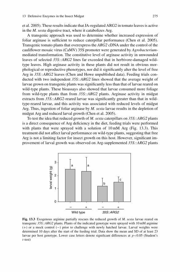
13 Defensive Enzymes in the Insect Midgut 275
et al. 2005). These results indicate that JA-regulated ARG2 in tomato leaves is activein the M. sexta digestive tract, where it catabolizes Arg.
A transgenic approach was used to determine whether increased expression offoliar arginase is sufficient to reduce caterpillar performance (Chen et al. 2005).Transgenic tomato plants that overexpress the ARG2 cDNA under the control of thecauliflower mosaic virus (CaMV) 35S promoter were generated by Agrobacterium-mediated transformation. The constitutive level of arginase activity in unwoundedleaves of selected 35S::ARG2 lines far exceeded that in herbivore-damaged wild-type leaves. High arginase activity in these plants did not result in obvious mor-phological or reproductive phenotypes, nor did it significantly alter the level of freeArg in 35S::ARG2 leaves (Chen and Howe unpublished data). Feeding trials con-ducted with two independent 35S::ARG2 lines showed that the average weight oflarvae grown on transgenic plants was significantly less than that of larvae reared onwild-type plants. These bioassays also showed that larvae consumed more foliagefrom wild-type plants than from 35S::ARG2 plants. Arginase activity in midgutextracts from 35S::ARG2-reared larvae was significantly greater than that in wild-type-reared larvae, and this activity was associated with reduced levels of midgutArg. Thus, ingestion of foliar arginase by M. sexta larvae results in the depletion ofmidgut Arg and reduced larval growth (Chen et al. 2005).
To test the idea that reduced growth of M. sexta caterpillars on 35S::ARG2 plantsis a direct consequence of Arg deficiency in the diet, feeding trials were performedwith plants that were sprayed with a solution of 10 mM Arg (Fig. 13.3). Thistreatment did not affect larval performance on wild-type plants, suggesting that freeArg is not a limiting factor for insect growth on this host. However, significant im-provement of larval growth was observed on Arg-supplemented 35S::ARG2 plants
larv
al w
eigh
t (g)
0
1
2
3
– + – +
Wild type 35S::ARG2
aa
cb
Fig. 13.3 Exogenous arginine partially rescues the reduced growth of M. sexta larvae reared ontransgenic 35S::ARG2 plants. Plants of the indicated genotype were sprayed with 10 mM arginine(+) or a mock control (−) prior to challenge with newly hatched larvae. Larval weights weredetermined 10 days after the start of the feeding trial. Data show the mean and SD of at least 23larvae per host genotype. Lower case letters denote significant differences at p<0.05 (Student’st-test)

276 H. Chen et al.
compared to the same genotype treated with a mock control. The ability of exoge-nous Arg to partially rescue the reduced growth of M. sexta larvae on 35S::ARG2plants supports the hypothesis that Arg is a limiting nutrient for insect growth on thisline. These results are also consistent with the notion that reduced Arg availabilityin the diet increases the toxicity of PIs (Broadway and Duffey 1998). Because lar-vae reared on Arg-supplemented transgenic plants weighed significantly less thaninsects grown on wild-type plants (Fig. 13.3), it is possible that the defensive role ofarginase involves mechanisms in addition to Arg depletion.
13.3 Threonine Deaminase
Threonine deaminase (EC 4.2.1.16), also known as threonine dehydratase or thre-onine ammonia-lyase, is a pyridoxal phosphate-dependent enzyme that catalyzesthe dehydratation of L-threonine (Thr) to �-ketobutyrate and ammonia. AlthoughTD can also use L-serine as a substrate to produce pyruvate and ammonia, Thris generally regarded as the enzyme’s preferred substrate. Many microorganismspossess distinct biosynthetic and biodegradative forms of TD. The biosyntheticenzyme catalyzes the committed step in the synthesis of Ile and is feedback in-hibited by Ile. Biosynthetic TDs from bacteria have been studied extensively as amodel for allosteric inhibition (Umbarger 1956). Biodegradative TDs are involvedin anaerobic breakdown of Thr to propionate and are insensitive to Ile. Higherplants possess a biosynthetic-type TD whose structure, function, and regulationby Ile is very similar to that of bacterial TDs (Mourad and King 1995; Halgandet al. 2002). This conclusion has been confirmed by molecular cloning and analysisof TD-encoding genes from several plants, including potato (Hildmann et al. 1992),tomato (Samach et al. 1991), chickpea (John et al. 1995), Arabidopsis (Mouradand King 1995; Wessel et al. 2000), and the native tobacco Nicotiana attenuata(Hermsmeier et al. 2001). The physiological importance of biosynthetic TD in plantgrowth and development was demonstrated by studies of TD-deficient mutants ofN. plumbaginifolia and N. attenuata (Sidorov et al. 1981; Colau et al. 1987; Kanget al. 2006). Although Ile-insensitive TD activity has been described in senescingtomato leaves and other plant tissues (Szamosi et al. 1993, and citations therein),genes encoding a biodegradative-type enzyme have not been identified in plants.
The first indication that biosynthetic TD might serve a role in plant defense camefrom work by Hildmann et al. (1992) showing that a TD gene in potato is highlyexpressed in response to mechanical wounding or treatment with the phytohormonesabscisic acid (ABA) and MeJA. Subsequent studies showed that TD expressionin leaves of tomato and N. attenuata is also induced by the jasmonate signalingpathway in response to wounding and herbivory (Samach et al. 1995; Hermsmeieret al. 2001; Strassner et al. 2002; Li et al. 2004). In these solanaceous species, TD isconstitutively expressed to high levels in reproductive organs (Hildmann et al. 1992;Kang and Baldwin 2006). In fact, TD is the most abundant protein in tomato flow-ers (Samach et al. 1991). The overall pattern of TD expression in vegetative and

13 Defensive Enzymes in the Insect Midgut 277
reproductive tissues is very similar to that of PIs and other JA-inducible proteins thathave a role in anti-insect defense. The phenomenon of stress-induced TD expressionhas not been reported for plants outside the Solanaceae.
Recent work from our laboratory indicates that tomato possesses two TD genes(designated SlTD1 and SlTD2) that have distinct roles in plant growth and develop-ment (Chen et al. 2007). TD1 is constitutively expressed in all tissues, whereas TD2expression is dependent on an intact jasmonate signaling pathway (Li et al. 2004;Chen et al. 2007). Plant TD sequences cluster into two major phylogenetic groups(Fig. 13.4). The sequence of SlTD1 is more similar to that of TDs from rice, poplar,and Arabidopsis than it is to SlTD2. The fact that Arabidopsis, poplar, and rice eachpossess a single TD gene supports the hypothesis that SlTD1 performs a housekeep-ing role in Ile biosynthesis. Jasmonate-inducible isoforms from tomato (SlTD2)and potato (StTD2) comprise a distinct subgroup of proteins that are similar to aTD sequence from chickpea. The lack of detectable TD2 expression in jai1 mu-tant plants, together with the fact that this mutant does not exhibit symptoms (e.g.,stunted growth) of Ile deficiency, indicates that TD1 can produce Ile pools that areutilized for normal growth and development in the absence of TD2 (Li et al. 2004;Chen et al. 2007).
Direct evidence for the idea that TD has a role in anti-insect defense came fromthe observation that TD2 accumulates in the food bolus and frass of M. sexta larvae
Maize
Sugarcane
Rice
Poplar
Arabidopsis
Aquilegia
Tomato TD1
Potato TD1
N.attenuata
Tomato TD2
Potato TD2
Chickpea
2
1
Fig. 13.4 Phylogeny of plant threonine deaminase. Unrooted Neighbor-Joining Tree wasconstructed with the deduced amino acid sequence of TDs from the indicated plants. Acces-sion numbers: Maize (CO446428), Sugarcane (CA208490), Rice (NP 001051069), Poplar (es-tExt fgenesh4 pg.C 280257), Arabidopsis (NP 187616), Aquilegia (DT735861), Tomato TD1(ABK20067), Potato TD1 (BI436101), N. attenuata (AAX22214), Chickpea (Q39469), TomatoTD2 (P25306), and Potato TD2 (X67846). The sequences cluster into two subgroups designated“1” and “2”. Because of its ambiguous position in the tree, N. attenuata TD was not assigned toeither group
278 H. Chen et al.
raised on tomato foliage (Chen et al. 2005). High TD2 activity in the midgut wascorrelated with reduced levels of free Thr. The level of midgut ammonia, which isproduced from TD-catalyzed deamination of Thr, was also correlated with TD2 ac-tivity. A recent study by Kang et al. (2006) provided direct evidence for a role of TDin anti-insect defense. These workers showed that TD-deficient N. attenuata plantsare compromised in resistance to M. sexta larvae. Supplementation of N. attenuataleaves with Thr resulted in increased caterpillar performance, suggesting that Thravailability in the leaf diet is limiting for larval growth. Unlike tomato and potato,N. attenuata has a single TD gene (Kang et al. 2006). Silencing of this gene thusled to a deficiency in Ile and stunted plant growth. Reduced Ile levels in the silencedlines caused decreased production of jasmonoyl-Ile (JA-Ile), which is an importantsignal for induced plant defense responses to pathogens (Staswick et al. 1998) andinsects (Kang et al. 2006). Thus, TD’s defensive role in N. attenuata is attributedboth to its involvement in JA-Ile synthesis and its function in post-ingestive defense(Kang et al. 2006). The dual role of N. attenuata TD may explain the protein’ssomewhat ambiguous position in the TD phylogenetic tree (Fig. 13.4), and suggeststhat the evolution of this isoform as an antinutritional enzyme is constrained by itsessential function in Ile biosynthesis. Tomato TD2 is presumably not subjected tosuch constraint because of the presence of TD1. Thus, TD2 may be highly special-ized as a midgut-active defensive enzyme.
Biosynthetic TDs contain an N-terminal catalytic domain and a C-terminal reg-ulatory domain (Gallagher et al. 1998). Binding of Ile to the regulatory domainresults in an allosteric transition that strongly inhibits catalytic activity. The highlevel of TD2 activity in the midgut of tomato-reared M. sexta suggested that theregulatory properties of the enzyme were altered in a way that confers insensitivityto Ile (Chen et al. 2005). In vitro enzyme assays showed that TD2 isolated fromtomato foliage is strongly inhibited by Ile, whereas TD2 activity in midgut andfrass extracts is insensitive to Ile. Using LC-MS/MS analysis of midgut extracts,we detected numerous peptides corresponding to the catalytic domain. However,we failed to detect peptides from the regulatory domain. It was thus proposed thatthe regulatory domain of TD2 is proteolytically cleaved from the catalytic domainfollowing ingestion of leaf material by the insect (Chen et al. 2005). The resultingtruncated enzyme can efficiently degrade Thr, which is a dietary requirement forphytophagous insects, in the Ile-rich insect gut.
To prove that proteolytic processing is involved in the production of an Ile-insensitive variant of TD2, it was necessary to establish a product-precursor re-lationship between the ∼55-kDa form of the protein that accumulates in tomatoleaves (Samach et al. 1995) and the putatively processed form of the enzyme.Immunoblot studies showed that anti-TD2 antibodies cross-react with a ∼55-kDaprotein in insect-damaged tomato foliage (Chen et al. 2007). In contrast, a lowermolecular-weight (∼40-kDa) protein, designated pTD2, was the predominant formof the protein in the food bolus and frass. These results demonstrate that pTD2 isproduced by proteolytic processing of TD2 following ingestion of tomato foliageby M. sexta. The near absence of unprocessed TD2 in midgut extracts suggests thatthe processing reaction occurs rapidly in response to maceration and ingestion of

13 Defensive Enzymes in the Insect Midgut 279
leaf tissue. It was also found that pTD2 is the predominant form of the enzyme infrass from tomato-reared Trichoplusia ni (cabbage looper), a generalist caterpillar.Consistent with this finding, T. ni frass contained TD activity that is insensitive toinhibition by Ile (Chen et al. 2007). Thus, ingestion of foliage by specialist andgeneralist insect herbivores results in proteolytic removal of the regulatory domainof TD2, resulting in an enzyme that efficiently degrades Thr without being inhibitedthrough feedback by Ile. A proposed model depicting that fate of tomato TD2 in thelepidopteran gut lumen is shown in Fig. 13.5. The structure of TD2 suggests thatthe processing reaction involves either a plant- or insect-derived endoprotease thatcleaves the neck region between the regulatory and catalytic domains. Additionalwork is needed to test this hypothesis.
Several properties of pTD2 presumably facilitate its post-ingestive role in de-fense (Chen et al. 2007). First, pTD2 is insensitive to feedback inhibition by Ile.Second, pTD2 is highly resistant to insect digestive proteases as determined by thehigh level to which the active enzyme is excreted in frass. Third, purified pTD2is active in an alkaline pH range that matches the lepidopteran midgut; little or noactivity was observed at pH values below 6.0. Fourth, pTD2 is active at temperaturesexceeding 60◦C. This thermostability indicates that the enzyme would be active atelevated body temperatures, which for M. sexta caterpillars in natural conditionscan exceed 35◦C (Casey 1976). Finally, the expression pattern of TD2 is consistentwith a function in antiherbivore defense. TD2 is coordinately induced with otherdefensive genes in response to wounding and JA treatment (Hildmann et al. 1992;Samach et al. 1995; Li et al. 2004). The high level of constitutive TD2 expres-sion in tomato reproductive tissues (Samach et al. 1991, 1995) is similar to manyother JA-regulated defensive proteins, including PIs, arginase, LAP-A, and AtVSP2(Hildmann et al. 1992; Utsugi et al. 1998; Chao et al. 1999; Chen et al. 2004).These observations support the idea that TD accumulation in floral tissues reflects amechanism that protects reproductive structures from herbivory.
Functional divergence of SlTD1 and SlTD2 indicates that the latter defensiveisozyme may have evolved from a ‘housekeeping’ TD. It is reasonable to assume,for example, that TD2 arose from a gene duplication event, and that selectivepressure imposed by herbivory led to the evolution of TD2. An important featureacquired by TD2 and other post-ingestive defense proteins during evolution wasregulation by the jasmonate signaling pathway. It is unclear whether TD2 evolvednew biochemical or structural properties that enhance its ability to degrade Thr in theinsect gut. Future research aimed at comparing the stability, activity, and structureof TD1 and TD2 may provide additional insight into this question.
Fig. 13.5 Working model forproteolytic activation oftomato TD2 in the responseto herbivore attack. CAT:catalytic domain; REG:regulatory domain Tomato Lepidopteran gut
Biosynthetic TD (Ile-sensitive)
CAT REG CAT
Degradative TD (Ile-insensitive)
Proteolysis Ingestion

280 H. Chen et al.
13.4 Vegetative Storage Proteins
Vegetative storage proteins (VSPs) comprise a diverse group of proteins that ac-cumulate to high levels in various vegetative organs of the plant. The dynamics ofVSP accumulation and degradation in plant tissues is controlled by both develop-mental and environmental cues (Berger et al. 1995). The first identified and mostextensively characterized VSPs are soybean VSP� and VSP� (Staswick 1994). Acommonly held theory is that accumulated VSPs function as a source of aminoacids and other nutrients for developing seeds. However, results from transgenicexperiments with VSP-deficient soybean plants indicate that VSPs contribute littleif any to plant productivity (Staswick et al. 2001). Thus, alternative roles for VSPsin nutrient storage or other aspects of plant physiology must be considered.
A possible clue to understanding VSP function can be found by studying theenzymatic activity of these proteins. Soybean and Arabidopsis VSPs exhibit phos-phatase activity (Leelapon et al. 2004; Liu et al. 2005), whereas patatin from potatotubers is a lipid acyl hydrolase (Andrews et al. 1988). The predominant VSPs inbark from mulberry are jacalin-like lectins (Van Damme et al. 2002). VSPs fromsweet potato and alfalfa are reported to function as trypsin inhibitors and chitinases,respectively (Yeh et al. 1997; Meuriot et al. 2004). These observations raise thepossibility that VSPs are enzymes that subsequently acquired a protein storagefunction (Staswick 1994). Alternatively, the function of VSPs may be intimatelylinked to their enzymatic activity. In this context, it is noteworthy that many ofthe above-mentioned activities are typically associated with plant defensive pro-cesses. Additional support for a role of VSPs in plant defense comes from thewell- established fact that VSP expression in vegetative tissues is strongly pro-moted by mechanical wounding, herbivory, and jasmonate treatment (Mason andMullet 1990; Staswick 1990; Franceschi and Grimes 1991; Staswick et al. 1991;Major and Constabel 2006). The absence of VSP expression in vegetative and repro-ductive tissues of the Arabidopsis coi1 mutant further demonstrates that expressionof this protein is tightly controlled by the jasmonate signaling pathway (Benedettiet al. 1995).
A significant breakthrough in our understanding of VSP function came fromthe recent analysis of Arabidopsis VSP2 (Liu et al. 2005). Purified VSP2, incor-porated into artificial diets, exhibited potent anti-insect activity against dipteran andcoleopteran insects that have an acidic gut lumen. The growth-inhibiting effect ofVSP2 on these insects was at least 10-fold greater than other anti-insect proteinsincluding cystatin and lectin. Significantly, site-directed mutagenesis showed thatthe phosphatase activity of VSP2 is essential for the protein’s insecticidal effects.These results provide compelling evidence that VSP2 is an anti-insect protein andthat the protein’s phosphatase activity is critical for this function. The mode of actionof VSP2 remains to be determined. Given the importance of dietary phosphorousfor the growth of leaf-eating caterpillars (Perkins et al. 2004), it is conceivable thatthe defensive effects of VSP2 may be attributed to the enzyme’s ability to perturbphosphate homeostasis in the gut lumen.

13 Defensive Enzymes in the Insect Midgut 281
13.5 Perspectives
Post-ingestive interactions between plant food and components of the insect di-gestive system have played a major role in shaping patterns of host plant utiliza-tion by insects. The vast majority of research on this topic has focused on plantsecondary metabolites that exert toxic or antinutritive effects on insect attackers(Fraenkel 1959). The pioneering work of Ryan and colleagues on defensive PIsintroduced the concept that wound-inducible plant proteins act directly in the in-sect gut as a defense. The recent discovery of jasmonate-regulated isoforms of TD,arginase, and VSP as anti-insect proteins extends this concept to include plant en-zymes that metabolize essential nutrients in the insect digestive system. Additionalwork is needed to clarify the mode of action of these enzymes, and to elucidate thestructural features that allow them to function in the extreme environment of themidgut. These and other examples support the idea that selective pressure imposedby insect herbivores led to the evolution of defensive enzymes from pre-existingisoforms that function in primary metabolism. The tremendous diversity of plantenzymes further suggests that many additional anti-insect proteins remain to bediscovered. The application of proteomics-based approaches to identify plant pro-teins that accumulate in the insect food bolus (Chen et al. 2005) and frass (Chenet al. 2007) is expected to facilitate this goal. Future research in this area promisesto contribute to the development of sound pest management approaches, includingthe generation of crop varieties with enhanced pest resistance.
Acknowledgments This work was supported by grants from the USDA National Research Initia-tive Competitive Grants Program award (2007-35604-17791) and the US Department of Energy(DE-FG02-91ER20021).
References
Alabadi D, Aguero MS, Perez-Amador MA, Carbonell J (1996) Arginase, arginine decarboxylase,ornithine decarboxylase, and polyamines in tomato ovaries. Changes in unpollinated ovariesand parthenocarpic fruits induced by auxin or gibberellin. Plant Physiol 112:1237–1244
Andrews DL, Beames B, Summers MD, Park WD (1988) Characterization of the lipid acyl hy-drolase activity of the major potato (Solanum tuberosum) tuber protein, patatin, by cloning andabundant expression in a baculovirus vector. Biochem J 252:199–206
Benedetti CE, Xie D, Turner JG (1995) COI1-dependent expression of an Arabidopsis vegetativestorage protein in flowers and siliques and in response to coronatine or methyl jasmonate. PlantPhysiol 109:567–572
Berenbaum MR (1995) Turnabout is fair play – Secondary roles for primary compounds. J ChemEcol 21:925–940
Berger S, Bell E, Sadka A, Mullet JE (1995) Arabidopsis thaliana AtVsp is homologous to soybeanVspA and VspB, genes encoding vegetative storage protein acid-phosphatases, and is regu-lated similarly by methyl jasmonate, wounding, sugars, light and phosphate. Plant Mol Biol27:933–942
282 H. Chen et al.
Bernays EA, Chapman RF (1994) Host-plant selection by phytophagous insects. Chapman & Hall,New York
Broadway RM, Duffey SS (1986) Plant proteinase inhibitors – Mechanism of action and effect onthe growth and digestive physiology of larval Heliothis zea and Spodoptera exiqua. J InsectPhysiol 32:827–833
Broadway RM, Duffey SS (1988) The effect of plant protein quality on insect digestive physiologyand the toxicity of plant proteinase inhibitors. J Insect Physiol 34:1111–1117
Casey TM (1976) Activity patterns, body temperature and thermal ecology in two desert caterpil-lars (Lepidoptera: Sphingidae). Ecology 57:485–497
Chang CL (2004) Effect of amino acids on larvae and adults of Ceratitis capitata (Diptera: Tephri-tidae). Ann Entomol Soc Am 97:529–535
Chao WS, Gu YQ, Pautot VV, Bray EA, Walling LL (1999) Leucine aminopeptidase RNAs,proteins, and activities increase in response to water deficit, salinity, and the wound signalssystemin, methyl jasmonate, and abscisic acid. Plant Physiol 120:979–992
Chen H, Gonzales-Vigil E, Wilkerson CG, Howe GA (2007) Stability of plant defense proteins inthe gut of insect herbivores. Plant Physiol 143:1954–1967
Chen H, McCaig BC, Melotto M, He SY, Howe GA (2004) Regulation of plant arginase by wound-ing, jasmonate, and the phytotoxin coronatine. J Biol Chem 279:45998–46007
Chen H, Wilkerson CG, Kuchar JA, Phinney BS, Howe GA (2005). Jasmonate-inducible plantenzymes degrade essential amino acids in the herbivore midgut. Proc Natl Acad Sci USA102:19237–19242
Colau D, Negrutiu I, Vanmontagu M, Hernalsteens JP (1987) Complementation of a threoninedehydratase-deficient Nicotiana plumbaginifolia mutant after Agrobacterium-tumefaciens-mediated transfer of the Saccharomyces cerevisiae ILV1 gene. Mol Cell Biol 7:2552–2557
Constabel CP, Bergey DR, Ryan CA (1995) Systemin activates synthesis of wound-inducibletomato leaf polyphenol oxidase via the octadecanoid defense signaling pathway. Proc NatlAcad Sci USA 92:407–411
Duffey SS, Stout MJ (1996) Antinutritive and toxic components of plant defense against insects.Arch Insect Biochem Physiol 32:3–37
Feeny P (1976) Plant apparency in chemical defense. Recent Adv Phytochem 10:1–40Felton GW (1996) Nutritive quality of plant protein: sources of variation and insect herbivore
responses. Arch Insect Biochem Physiol 32:107–130Felton GW (2005) Indigestion is a plant’s best defense. Proc Natl Acad Sci USA 102:18771–18772Fraenkel GS (1959) The raison d’etre of secondary plant substances; these odd chemicals arose as a
means of protecting plants from insects and now guide insects to food. Science 129:1466–1470Franceschi VR, Grimes HD (1991) Induction of soybean vegetative storage proteins and antho-
cyanins by low-level atmospheric methyl jasmonate. Proc Natl Acad Sci USA 88:6745–6749Gallagher DT, Gilliland GL, Xiao GY, Zondlo J, Fisher KE, Chinchilla D, Eisenstein E (1998)
Structure and control of pyridoxal phosphate dependent allosteric threonine deaminase. Struc-ture 6:465–475
Gfeller A, Farmer EE (2004) Keeping the leaves green above us. Science 306:1515–1516Goldraij A, Polacco JC (1999) Arginase is inoperative in developing soybean embryos. Plant Phys-
iol 119:297–304Goldraij A, Polacco JC (2000) Arginine degradation by arginase in mitochondria of soybean
seedling cotyledons. Planta 210:652–658Green TR, Ryan CA (1972) Wound-induced proteinase inhibitor in plant leaves – possible defense
mechanism against insects. Science 175:776–777Halgand F, Wessel PM, Laprevote O, Dumas R (2002) Biochemical and mass spectrometric
evidence for quaternary structure modifications of plant threonine deaminase induced byisoleucine. Biochemistry 41:13767–13773
Hermsmeier D, Schittko U, Baldwin IT (2001) Molecular interactions between the specialist her-bivore Manduca sexta (Lepidoptera, Sphingidae) and its natural host Nicotiana attenuata. I.Large-scale changes in the accumulation of growth- and defense-related plant mRNAs. PlantPhysiol 125:683–700

13 Defensive Enzymes in the Insect Midgut 283
Hildmann T, Ebneth M, Pena-Cortes H, Sanchez-Serrano JJ, Willmitzer L, Prat S (1992) Generalroles of abscisic and jasmonic acids in gene activation as a result of mechanical wounding.Plant Cell 4:1157–1170
Howe GA (2004) Jasmonates as signals in the wound response. J Plant Growth Regul 23:223–237John SJ, Srivastava V, Guhamukherjee S (1995) Cloning and sequencing of chickpea cDNA coding
for threonine deaminase. Plant Physiol 107:1023–1024Kang JH, Baldwin IT (2006). Isolation and characterization of the threonine deaminase promoter
in Nicotiana attenuata. Plant Sci 171:435–440Kang JH, Wang L, Giri A, Baldwin IT (2006) Silencing threonine deaminase and JAR4 in Nico-
tiana attenuata impairs jasmonic acid-isoleucine-mediated defenses against Manduca sexta.Plant Cell 18:3303–3320
Kessler A, Baldwin IT (2002) Plant responses to insect herbivory: the emerging molecular analysis.Annu Rev Plant Biol 53:299–328
Konno K, Hirayama C, Nakamura M, Tateishi K, Tamura Y, Hattori M, Kohno K (2004) Pa-pain protects papaya trees from herbivorous insects: role of cysteine proteases in latex. Plant J37:370–378
Leelapon O, Sarath G, Staswick PE (2004) A single amino acid substitution in soybean VSP alphaincreases its acid phosphatase activity nearly 20-fold. Planta 219:1071–1079
Li L, Zhao Y, McCaig BC, Wingerd BA, Wang J, Whalon ME, Pichersky E, Howe GA (2004) Thetomato homolog of CORONATINE-INSENSITIVE1 is required for the maternal control of seedmaturation, jasmonate-signaled defense responses, and glandular trichome development. PlantCell 16:126–143
Liu YL, Ahn JE, Datta S, Salzman RA, Moon J, Huyghues-Despointes B, Pittendrigh B, MurdockLL, Koiwa H, Zhu-Salzman K (2005) Arabidopsis vegetative storage protein is an anti-insectacid phosphatase. Plant Physiol 139:1545–1556
Major IT, Constabel CP (2006) Molecular analysis of poplar defense against herbivory: comparisonof wound- and insect elicitor-induced gene expression. New Phytol 172:617–635
Mason HS, Mullet JE (1990) Expression of two soybean vegetative storage protein genes dur-ing development and in response to water deficit, wounding, and jasmonic acid. Plant Cell2:569–579
Mattson WJ (1980) Herbivory in relation to plant nitrogen-content. Annu Rev Ecol Syst11:119–161
Meuriot F, Noquet C, Avice JC, Volenec JJ, Cunningham SM, Sors TG, Caillot S, Ourry A (2004)Methyl jasmonate alters N partitioning, N reserves accumulation and induces gene expressionof a 32-kDa vegetative storage protein that possesses chitinase activity in Medicago sativataproots. Physiol Plant 120:113–123
Mohan S, Ma PWK, Pechan T, Bassford ER, Williams WP, Luthe DS (2006) Degradation ofthe S. frugiperda peritrophic matrix by an inducible maize cysteine protease. J Insect Physiol52:21–28
Mori M, Gotoh T (2004) Arginine metabolic enzymes, nitric oxide and infection. J Nutr134:2820S–2825S
Mourad G, King J (1995) L-O-Methylthreonine resistant mutant of Arabidopsis defective inisoleucine feedback regulation. Plant Physiol 107:43–52
Pechan T, Cohen A, Williams WP, Luthe DS (2002) Insect feeding mobilizes a unique plant de-fense protease that disrupts the peritrophic matrix of caterpillars. Proc Natl Acad Sci USA99:13319–13323
Perkins MC, Woods HA, Harrison JF, Elser JJ (2004) Dietary phosphorus affects the growth oflarval Manduca sexta. Arch Insect Biochem Physiol 55:153–168
Perozich J, Hempel J, Morris SM Jr (1998) Roles of conserved residues in the arginase family.Biochim Biophys Acta 1382:23–37
Rhoades DF, Cates RG (1976) Toward a general theory of plant antiherbivore chemistry. RecentAdv Phytochem 10:168–213
Ryan CA (1990) Protease inhibitors in plants – Genes for improving defenses against insects andpathogens. Annu Rev Phytopath 28:425–449
284 H. Chen et al.
Samach A, Broday L, Hareven D, Lifschitz E (1995) Expression of an amino acid biosynthesis genein tomato flowers: developmental upregulation and MeJa response are parenchyma-specific andmutually compatible. Plant J 8:391–406
Samach A, Hareven D, Gutfinger T, Ken-Dror S, Lifschitz E (1991) Biosynthetic threonine deam-inase gene of tomato: isolation, structure, and upregulation in floral organs. Proc Natl Acad SciUSA 88:2678–2682
Satriano J (2004) Arginine pathways and the inflammatory response: interregulation of nitric oxideand polyamines: review article. Amino Acids 26:321–329
Schilmiller AL, Howe GA (2005) Systemic signaling in the wound response. Curr Opin Plant Biol8:369–377
Sekowska A, Danchin A, Risler JL (2000) Phylogeny of related functions: the case of polyaminebiosynthetic enzymes. Microbiology 146:1815–1828
Sidorov V, Menczel L, Maliga P (1981) Isoleucine-requiring Nicotiana plant deficient in threoninedeaminase. Nature 294:87–88
Staswick PE (1990) Novel regulation of vegetative storage protein genes. Plant Cell 2:1–6Staswick PE (1994) Storage proteins of vegetative plant tissue. Annu Rev Plant Physiol Plant Mol
Biol 45:303–322Staswick PE, Huang JF, Rhee Y (1991) Nitrogen and methyl jasmonate induction of soybean veg-
etative storage protein genes. Plant Physiol 96:130–136Staswick PE, Yuen GY, Lehman CC (1998) Jasmonate signaling mutants of Arabidopsis are sus-
ceptible to the soil fungus Pythium irregulare. Plant J 15:747–754Staswick PE, Zhang ZY, Clemente TE, Specht JE (2001) Efficient down-regulation of the major
vegetative storage protein genes in transgenic soybean does not compromise plant productivity.Plant Physiol 127:1819–1826
Strassner J, Schaller F, Frick UB, Howe GA, Weiler EW, Amrhein N, Macheroux P, Schaller A(2002) Characterization and cDNA-microarray expression analysis of 12-oxophytodienoate re-ductases reveals differential roles for octadecanoid biosynthesis in the local versus the systemicwound response. Plant J 32:585–601
Szamosi I, Shaner DL, Singh BK (1993) Identification and characterization of a biodegradativeform of threonine dehydratase in senescing tomato (Lycopersicon esculentum) leaf. Plant Phys-iol 101:999–1004
Umbarger HE (1956) Evidence for a negative-feedback mechanism in the biosynthesis ofisoleucine. Science 123:848
Utsugi S, Sakamoto W, Murata M, Motoyoshi F (1998) Arabidopsis thaliana vegetative stor-age protein (VSP) genes: gene organization and tissue-specific expression. Plant Mol Biol38:565–576
Van Damme EJM, Hause B, Hu JL, Barre A, Rouge P, Proost P, Peumans WJ (2002) Two distinctjacalin-related lectins with a different specificity and subcellular location are major vegetativestorage proteins in the bark of the black mulberry tree. Plant Physiol 130:757–769
Vincendeau P, Gobert AP, Daulouede S, Moynet D, Mossalayi MD (2003) Arginases in parasiticdiseases. Trends Parasitol 19:9–12
Walling LL (2000) The myriad plant responses to herbivores. J Plant Growth Regul 19:195–216Wang JH, Constabel CP (2004) Polyphenol oxidase overexpression in transgenic Populus
enhances resistance to herbivory by forest tent caterpillar (Malacosoma disstria). Planta220:87–96
Wessel PM, Graciet E, Douce R, Dumas R (2000) Evidence for two distinct effector-binding sitesin threonine deaminase by site-directed mutagenesis, kinetic, and binding experiments. Bio-chemistry 39:15136–15143
Yeh KW, Chen JC, Lin MI, Chen YM, Lin CY (1997) Functional activity of sporamin from sweetpotato (Ipomoea batatas Lam): a tuber storage protein with trypsin inhibitory activity. PlantMol Biol 33:565–570
Chapter 14Plant Lectins as Part of the Plant DefenseSystem Against Insects
Els J.M. Van Damme
Many plants contain carbohydrate-binding proteins which are commonly designatedas lectins. It is believed that some of these lectins play a role in plant defense againstinsects. This chapter gives an overview of the state of the art in the field of plantlectin research and highlights the most important results with regard to the insecti-cidal activity of plant lectins belonging to different families. A distinction is madebetween the ‘classical lectins’ that are constitutively expressed and the ‘induciblelectins’. Furthermore, the possible mode of action and target sites for the lectins inthe insect body are discussed.
14.1 Introduction
During evolution plants have developed a wide range of sophisticated defense mech-anisms to counteract attack by pathogens and herbivory. These include both mechan-ical and chemical defenses, and allow the plant to survive in the same environmentas its potential attackers. As part of their chemical defense mechanism plants synthe-size a whole battery of so-called defense proteins (Carlini and Grossi-de-Sa 2002).One particular class of defense proteins are the plant lectins, a heterogeneous groupof proteins that can specifically interact with particular carbohydrates (Peumans andVan Damme 1995; Van Damme et al. 1998a, 2007b). This chapter will focus on therole of carbohydrate-bindingproteins (lectins) in plant defense against insects. A de-tailed discussion on the insecticidal activity of different families of carbohydrate-binding proteins will be given. In addition, some recent results on inducible lectinsinvolved in plant defense against insects will be discussed.
E.J.M. Van DammeLaboratory of Biochemistry and Glycobiology, Department of Molecular Biotechnology,Ghent University, 9000 Gent, Belgiume-mail: [email protected]
A. Schaller (ed.), Induced Plant Resistance to Herbivory, 285C© Springer Science+Business Media B.V. 2008

286 E.J.M. Van Damme
14.2 Distribution of Plant Lectins
Many plants including important food plants such as wheat, rice, potato, tomato,soybean and bean contain carbohydrate-binding proteins commonly referred to as‘lectins’, ‘agglutinins’ or ‘hemagglutinins’. According to a recently proposed def-inition, this group of proteins comprises ‘all plant proteins possessing at least onenon-catalytic domain that binds reversibly to specific mono- or oligosaccharides’.Hitherto, about 500 different plant lectins have been isolated and (partially) charac-terized. On a first glance, all these lectins form a heterogeneous group of proteinsbecause of the obvious differences in structure, specificity and biological activities.However, the majority of all plant lectins can be classified on the basis of structuralanalyses and sequence data in seven families of structurally and evolutionary re-lated proteins. These lectin families are: (1) the amaranthins, (2) the Cucurbitaceaephloem lectins, (3) the chitin binding lectins composed of hevein domains, (4) theGNA-related lectins, (5) the jacalin-related lectins, (6) the legume lectins and (7) thelectins with ricin-B domains (Van Damme et al. 1998a, 2007b). Recently, evidencewas reported for the occurrence in plants of homologs of the Agaricus bisporusagglutinin (Peumans et al. 2007) and homologs of class V chitinases with sugar-binding activity (Van Damme et al. 2007a).
Although lectins have for a long time been considered typical seed proteins, theoccurrence of plant lectins in vegetative plant tissues is now very well documented.There are, however, striking differences for what concerns the location and con-centration of the different lectins. Seed lectins are primarily found in the cotyledons(e.g., legume species) or the endosperm, but can also be confined to the primary axis(e.g., wheat). Seed lectins generally account for 0.1%–5% of the total seed protein.Non-seed lectins have been detected in almost all vegetative tissues ranging fromleaves, stems, bark, bulbs, rhizomes, roots to flowers, fruits, phloem sap and evennectar. Lectin concentrations in vegetative storage tissues can amount to 50% ofthe total protein, whereas in other tissues the lectin is a very minor protein (e.g., inleaves of leek). On average, lectins in vegetative tissues also account for 0.1%–5%of the total protein.
14.3 Classical and Inducible Plant Lectins
For many years research on plant lectins has been focused on a group of lectinsthat occur in reasonable to high concentrations in different plant tissues. Becauseof the high lectin concentrations and the developmental regulation it was diffi-cult to imagine a specific role for these lectins within the plant cell. Several linesof evidence indicated that the majority of these lectins may be involved in plantdefense. In this respect, plant lectins basically differ from animal lectins becausethe latter are involved in specific recognition processes within the organism itself.Taking into consideration the increasing evidence that protein–carbohydrate inter-actions are very important for the normal development and functioning of animalorganisms, and the fact that different types of lectins are the mediators of these

14 Insecticidal Activity of Plant Lectins 287
protein–carbohydrate interactions, the question arose whether the same holds truefor plants. Recently, this search for potential physiologically active plant lectinsresulted in the discovery of different lectin families, which are candidates to me-diate essential protein–carbohydrate interactions in the plant itself (Van Dammeet al. 2004, 2007b). Evidence has accumulated that plants synthesize well-definedcarbohydrate binding proteins (lectins) upon exposure to stress situations likedrought, high salt, wounding, or treatment with some plant hormones (Zhanget al. 2000; Chen et al. 2002). Localization studies demonstrated that – in contrast tothe ‘classical’ plant lectins, which are typically found in vacuoles – the ‘inducible’lectins are exclusively located in the cytoplasm and the nucleus. It is hypothesizedthat this new class of lectins may play a specific role within the plant cell. Thissection will give a brief overview of the classical and inducible lectins that havebeen identified to date (Table 14.1).
Table 14.1 Overview of different plant lectin families, their location, carbohydrate binding prop-erties and insecticidal activity
Lectin family Vacuolar lectins Nucleocytoplasmiclectins
Carbohydratespecificity
Insecticidalactivity∗
Amaranthinfamily
No examplesknown
Documented inAmaranthus sp.and Prunus sp.
GalNAc/T-antigen
+
Cucurbitaceaephloemlectins/Lectins withNictabadomain
No examplesknown
Wide taxonomicdistribution
(GlcNAc)nN-glycans
?
GNA-relatedlectins
Wide taxonomicdistribution
Found in diverse taxa Man High ManN-glycans
+ + +
Chitin-bindinglectinscomposed ofheveindomains
Wide taxonomicdistribution
No examples known (GlcNAc)nN-glycans
++
Jacalin-relatedlectins
Onlydocumented ina fewMoraceae sp.
Ubiquitous Gal/T-antigenManN-glycans
+
Legume lectins Common inFabaceae andLamiaceae
No examples known Man/GlcGal/GalNAc(GlcNAc)nFuc, Sia�2-3Gal/GalNAcComplex
++
Lectins withRicin-Bdomains
Wide taxonomicdistribution
No examples known Gal/GalNAcSia�2-6Gal/GalNAc
+
∗Insecticidal activity has been reported for many (+++), several (++), a few (+) or no (?) lectinsfrom this plant lectin family
288 E.J.M. Van Damme
Amaranthin family: The Amaranthin family, which is called after the Ama-ranthus caudatus seed lectin is a rather small family of closely related lectinsfound in different Amaranthus species. All known amaranthins are homodimersbuilt of 33 kDa subunits. Detailed specificity studies have shown that amaranthinpreferentially recognizes the T-antigen disaccharide Gal�(1,3)GalNAc (Rinderleet al. 1989).
Cucurbitaceae phloem lectins: The family of Cucurbitaceae phloem lectins is asmall group of chitin-binding agglutinins found in the phloem exudates of a numberof Cucurbitaceae species (Sabnis and Hart 1978). These lectins, also called phloemproteins PP2, are dimeric proteins built up of subunits of approximately 22 kDa.
GNA-related lectins: In 1987 a lectin with exclusive specificity towards mannose(Man) was isolated and characterized in snowdrop (Galanthus nivalis) bulbs (VanDamme et al. 1987). The so-called G. nivalis agglutinin (abbreviated GNA) turnedout to be the prototype of an extended family of mannose-binding lectins occurring innumerous monocot plant families (e.g., Alliaceae, Liliaceae, Orchidaceae, Araceae,Bromeliaceae, Ruscaceae, Iridaceae; Yagi et al. 1996; Van Damme et al. 1998a, b;Neuteboom et al. 2002). Therefore this group of lectins was originally referred toas the ‘monocot mannose-binding lectins’ (Van Damme et al. 1998b). Recently,very similar lectins have been identified in plants other than Liliopsida (e.g., inthe liverwort, Marchantia polymorpha; Peumans et al. 2002) as well as in bacteria(Parret et al. 2003, 2005), and animals (Tsutsui et al. 2003) Therefore, this groupof lectins is now referred to as ‘GNA-related lectins’ after the first identified member.
All plant lectins with GNA-domains are composed of lectin subunits of approx-imately 12 kDa, that show specificity towards mannose. Nevertheless, GNA-relatedlectins from different plants exhibit subtle but marked differences in specificitytowards oligomannosides and N-glycans (Kaku et al. 1990, 1992; Van Dammeet al. 2007b).
Chitin-binding lectins composed of hevein domains: The name ‘hevein’ refers toa chitin-binding polypeptide of 43 amino acids found in the latex of Hevea brasilien-sis (rubber tree; Waljuno et al. 1975). Many plant proteins owe their chitin-bindingactivity to the presence of one or more hevein domain(s). However, it was shown thatvarious lectins with hevein domains also have high affinity for N-glycosylated ani-mal glycoproteins (Goldstein and Poretz 1986). This group of lectins is widespreadand occurs both in monocotyledonous (e.g., wheat germ agglutinin (WGA)) as wellas dicotyledonous (e.g,. potato lectin) plant species.
Jacalin-related lectins: The family of jacalin-related lectins comprises all pro-teins with one or more domains that are structurally equivalent to ‘jacalin’, agalactoside-binding lectin from jack fruit (Artocarpus integrifolia) seeds(Sastry et al. 1986). In recent years several lectins related to jacalin but with speci-ficity towards mannose were discovered and characterized in detail (Van Dammeet al. 1996). Therefore the family of jacalin-related lectins is now subdivided intotwo subfamilies with a distinct specificity and molecular structure. The galactose(Gal)-specific jacalin-related lectins are built up of four cleaved protomers compris-ing a small (�; 20 amino acid residues) and a large (�; 133 amino acid residues) sub-unit and exhibit a clear preference for galactose over mannose (Bourne et al. 2002).

14 Insecticidal Activity of Plant Lectins 289
In contrast, mannose-specific jacalin-related lectins are built up of uncleaved pro-tomers of approximately 150 amino acids each, which exhibit exclusive specificitytowards mannose.
Legume lectins: Historically, the term ‘legume lectin’ refers to proteins that arestructurally and evolutionary related to a well-defined type of lectin that was orig-inally discovered in seeds of legumes (Fabaceae) like jack bean, common bean,pea, peanut and soybean. Until recently, legume lectins were exclusively found inFabaceae. However, the isolation and cloning of a structurally and evolutionaryclosely related agglutinin from leaves of Glechoma hederacea demonstrated thatclosely related lectins also occur in the family Lamiaceae (Wang et al. 2003a, b).
The family of legume lectins (from both Fabaceae and Lamiaceae species) isfairly homogeneous as far as the molecular structure of the native lectins is con-cerned (Van Damme et al. 1998a, 2007b). All lectins are composed of subunits ofapproximately 30 kDa and many of them are glycoproteins.
Although the overall fold of the lectin and its carbohydrate-binding sites arehighly conserved the legume lectin family exhibits a marked heterogeneity in sugar-binding activity and specificity (Young and Oomen 1992; Sharma and Surolia 1997).Legume lectins with specificity directed against galactose (Gal), N-acetylgalac-tosamine (GalNAc), mannose/glucose (Man/Glc), fucose (Fuc), N-acetylglucosa-mine (GlcNAc) and sialic acid have been found as well as numerous lectins thatbind exclusively more complex glycan structures (Van Damme et al. 1998a). Incontrast, the lectins from Lamiaceae species exhibit specificity that is primarilydirected against the T-antigen (GalNAc�1-Ser/Thr; Wu 2005; Singh et al. 2006).
Lectins with ricin-B domains: This group of plant lectins was previously referredto as the family of ‘ribosome-inactivating proteins’ (abbreviated RIPs), due to thefact that these proteins have been first identified as plant proteins that inactivateeukaryotic ribosomes through the removal of a conserved adenine residue from thelarge ribosomal RNA (Van Damme et al. 2001; Stirpe 2004). At present, RIPs areconsidered enzymes, namely N-glycosylases (EC 3.2.2.22) that are capable of cleav-ing adenine residues from different polynucleotide substrates. Based on their molec-ular structure RIPs are subdivided in type-1, type-2 and type-3 RIPs. Type-1 RIPsconsist of a single enzymatically active polypeptide chain. Type-2 RIPs are chimericproteins built up of protomers consisting of an enzymatically active A-chain and acarbohydrate-binding B-chain, which are both derived from a single precursor andare held together by a disulfide bond. Finally, type-3 RIPs also are chimeric proteinsbuilt up of an N-terminal N-glycosylase domain linked to a C-terminal domain witha still unknown activity/function. Taking the definition of plant lectins into accountonly the B-chain of type-2 RIPs can be classified as a lectin domain. Therefore, thelectin domain is referred to as the ricin-B domain, named after ‘ricin’ the first andmost famous RIP isolated from seeds of Ricinus communis (Lord et al. 1994).
Most plant ricin-B domains preferentially bind to galactose or GalNAc. How-ever, some lectins exhibit a clear preference for sialylated glycans (e.g., Sambucusnigra lectin I). As already mentioned above, type-2 RIPs are not only lectins butalso enzymes capable of (catalytically) inactivating ribosomes. They are thereforeextremely potent cytotoxins, at least at the condition that they succeed in entering
290 E.J.M. Van Damme
the cell. The most notorious example of a type-2 RIP is the extremely toxic ricin.It should be emphasized, however, that most type-2 RIPs are only moderately oreven weakly toxic. The huge difference in toxicity is not linked to differences in thecatalytic activity of the A-chain but is primarily determined by the lectin’s ability topenetrate into the cell, which in turn depends on the sugar-binding specificity of theB-chain (Peumans et al. 2001; Van Damme et al. 2001; Stirpe 2004).
Inducible lectins: Unambiguous evidence for the existence of jasmonate-inducedlectins was obtained recently from experiments with jasmonate-treated tobaccoplants (Nicotiana tabacum cv Samsun NN; Chen et al. 2002). Untreated plants donot contain any detectable lectin activity. After jasmonate treatment however, leavesof young tobacco plants express low concentrations of a lectin (called N. tabacumagglutinin or Nictaba) with specificity towards oligomers of N-acetylglucosamine.Nictaba is a homodimer of 19 kDa subunits. Sequence analysis of Nictaba andcomparison with other sequences in the databases revealed that this plant lectin is arepresentative of a new family of inducible cytoplasmic proteins with sequence sim-ilarity to the Cucurbitaceae phloem lectins. Immunolocalisation studies confirmedthat Nictaba is expressed in the cytoplasm and the nucleus. Furthermore, evidencewas presented for the interaction of the lectin with several proteins in the plantnucleus (Lannoo et al. 2006).
Another example of an inducible lectin is the mannose-binding lectin (calledOrysata) that is induced in rice plants by salt stress, desiccation, pathogen infectionand the phytohormones jasmonic acid and abscisic acid (ABA). This protein wasalready described in 1990 as SalT (a salt-inducible protein; Claes et al. 1990) but waslater identified as a lectin belonging to the family of mannose-specific jacalin-relatedlectins (Zhang et al. 2000). Since Orysata is synthesized only under specific stressconditions, and in addition, is located in the cytoplasm and the nucleus, we presumethat this lectin plays a role in the plant response to well-defined stress factors.
Recently we have also identified other families of nucleocytoplasmic plantlectins, presumably with specificity towards mannose and galactose (unpublishedresults). Unfortunately the study of these lectins is hampered by the very low ex-pression levels of the lectins (even after induction).
14.4 Insecticidal Activity of Plant Lectins
During the last two decades, numerous reports have been published on the insec-ticidal activity of plant lectins against many pest insects belonging to the ordersLepidoptera, Coleoptera, Diptera and Homoptera (Sharma et al. 2004). Both in vitroassays with lectin spiked artificial diets, and in vivo assays with transgenic plantsexpressing a foreign lectin gene have demonstrated the potential of several plantlectins as insecticidal proteins (Carlini and Grossi-de-Sa 2002). This section willgive a brief overview of the insecticidal properties that have been reported for lectinsbelonging to the different lectin families (Table 14.1).
Amaranthins: Taking into consideration the exclusive location of the amaran-thins in the seeds and their high affinity for the T-antigen, it can be envisaged that

14 Insecticidal Activity of Plant Lectins 291
these lectins are involved in the plant’s defense against seed predators. However,at present very little information is available with regard to the physiological roleof amaranthins and their involvement in plant defense. Some recent studies withphloem-specific expression of the A. caudatus agglutinin (ACA) in tobacco as wellas in cotton suggest that the ACA gene can be considered an effective aphid-resistantgene (Guo et al. 2004; Wu et al. 2006). Insect bioassays with Myzus persicae andAphis gossypii showed up to 75% and 64% inhibition, respectively, of aphid pop-ulation growth on transgenic lines, with the highest inhibition on those transgeniclines expressing the highest lectin levels.
Chitin-binding lectins composed of hevein domains: By virtue of their interactionwith GlcNAc-oligomers (chitin), lectins with hevein domains were always believedto be associated with the plant’s defense against insects. Feeding trials with arti-ficial diets confirmed that several of these chitin-binding lectins interfere with thegrowth and development of insects. For example, WGA, rice lectin, stinging nettlelectin, and the potato and thorn apple lectins had a moderate inhibitory effect onthe development of the larvae of the cowpea weevil (Callosobruchus maculatus;Murdock et al. 1990; Huesing et al. 1991). WGA inhibits the larval growth of theSouthern corn rootworm (Diabrotica undecimpunctata) and kills the neonate Os-trinia nubilalis (European corn borer) larvae at fairly low concentrations (Czaplaand Lang 1990). In addition, WGA shows larvicidal activity on the blowfly Lu-cilia cuprina (Eisemann et al. 1994). Similarly, the Phytolacca americana lectin islethal to larvae of the Southern corn rootworm (Czapla and Lang 1990). RecentlyGupta et al. (2005) also reported an effect of WGA against neonates of Americanbollworm (Helicoverpa armigera). The results of feeding trials leave no doubt thatsome chitin-binding lectins have deleterious effects on chewing insects when testedin artificial diets. At present, it still remains to be demonstrated that the expressionof a chitin-binding lectin in a transgenic plant offers an increased protection againstchewing insects. Tests with artificial diets further indicated that WGA negativelyaffects the growth and reproduction of the phloem feeding rice brown planthopper(Nilaparvata lugens; Powell et al. 1995a) and the mustard aphid (Lipaphis erysimi).In addition, transformation of Indian mustard with the cDNA encoding WGA re-sulted in transgenic plants causing high mortality and reduced fecundity of aphids(L. erysimi; Kanrar et al. 2002).
GNA-related lectins: GNA-related lectins are undoubtedly the most intensivelystudied family of plant lectins that are actually being investigated for their role inthe plant’s defense against phytophagous insects and other invertebrates. One argu-ment in favor of the defensive role against sucking insects is the observation thatsome of these lectins accumulate in the phloem sap. The fact that a typical mono-cot mannose-binding lectin is the most abundant protein in the phloem exudate offlowering stalks of onion (Allium cepa) plants is in agreement with the presumeddefensive role against phloem feeding insects (Peumans et al. 1997).
GNA was the first lectin found to affect the growth and development of sap-sucking insects, and its insecticidal properties have been studied in most detail.Numerous experiments with artificial diets clearly demonstrated that the snowdroplectin has detrimental effects on the development and reproduction of Homopteran

292 E.J.M. Van Damme
pests belonging to the Aphididae (Rahbe et al. 1995; Sauvion et al. 1996), Cicadel-lidae and Delphacidae (Powell et al. 1993, 1995b, 1998). In the meantime, studieswith transgenic tobacco, wheat, tomato, potato and rice plants have confirmed theinsecticidal activity of GNA towards Homoptera (aphids) and demonstrated thatectopically expressed GNA has deleterious effects on aphids, leaf hoppers and planthoppers (Hilder et al. 1995; Down et al. 1996; Rao et al. 1998; Stoger et al. 1999).In addition to Homoptera, GNA exerts also noxious effects both in artificial dietsand transgenic plants on the larval growth and development of several Lepidopteraincluding the tomato moth (Lacanobia oleracea), the Mexican rice borer (Eoreumaloftini), the sugarcane borer (Diatraea saccharalis) and the bollworm (H. armigera;Gatehouse et al. 1997; Setamou et al. 2003; Shukla et al. 2005).
Though GNA is certainly a very promising insecticidal protein, intensive ef-forts aim at the identification of superior alternatives within the same lectin family.Comparative analyses indicated that some GNA-related lectins found in Alliaceaespecies are substantially more active than GNA itself in terms of e.g. specific ag-glutination activity (Van Damme et al. 1993). Since one can reasonably assumethat the insecticidal activity of GNA and related lectins is somehow related totheir sugar-binding activity/specificity it seemed worthwhile to check whether someGNA-related lectins possibly have a more promising insecticidal potency. Over thelast few years numerous studies have shown that not only GNA but also manyGNA-related lectins are detrimental especially to sap-sucking insects and also tosome caterpillars. For instance bioassays with artificial diets revealed that the garlic(Allium sativum) leaf lectin ASAL was highly toxic to the hemipteran pests N. lu-gens and Nilaparvata virescens (Bandyopadhyay et al. 2001; Majumder et al. 2004).Analysis of transgenic tobacco and mustard plants expressing ASAL revealed a de-crease in survival and fecundity of these phloem feeding aphids (Dutta et al. 2005a,b). In addition, Sadeghi et al. (2008) have shown that transgenic tobacco plantsexpressing ASAL and ASAII significantly (p<0.05) reduced the weight gain of4th instar larvae of Spodoptera littoralis. The lectins retarded the development ofthe larvae and their metamorphosis, and were also detrimental to the pupal stageresulting in weight reduction and lethal abnormalities. Mortality was 100% withASAL compared to 60% with ASAII.
Similarly, the Allium porrum (leek) lectin (APA) was recently shown to be apotent insecticidal protein. Ectopically expressed APA provides transgenic tobaccoplants significant levels of protection against the cotton leafworm S. littoralis due toits effect on larval survival, growth and development (unpublished results).
Insecticidal activity was recently also shown for some Araceae lectins. The Arummaculatum lectin was tested in an artificial diet against the aphids L. erysimi andAphis craccivora (Majumder et al. 2005). When incorporated in an artificial dietArisaema jacquemontii lectin affected the development of Bactrocera cucurbitaelarvae (Kaur et al. 2006). Transgenic tobacco expressing Pinellia ternata agglutininexhibited enhanced resistance to peach potato aphid (M. persicae; Yao et al. 2003).
Jacalin-related lectins: Feeding trials with artificial diets have shown that theMoraceae seed lectins have anti-insect properties. For example, the Maclurapomifera lectin exerted a significant inhibitory effect on the larvae of the cowpea

14 Insecticidal Activity of Plant Lectins 293
weevil (C. maculatus) (Murdock et al. 1990). Similarly, jacalin and the M. pomiferalectin inhibited larval growth of the Southern corn rootworm (D. undecimpunctata;Czapla and Lang 1990). Jacalin also affected the survival of the potato leafhopper(Empoasca fabae; Habibi et al. 1993). Based on these observations it seems likelythat the galactose-binding Moraceae seed lectins have a defensive function againstinsects. At present, there is little information about the role of mannose-bindingjacalin-related lectins. Chang et al. (2003) have shown that the Helianthus tubero-sus agglutinin confers resistance to peach potato aphid (M. persicae). The averagepopulation of aphids on transgenic plants expressing the lectin gene decreased by53%–70% as compared to control populations during an 11-day assay. In addition,the development of aphids was notably retarded, suggesting that this lectin genemight be another candidate for genetic engineering of plants for resistance againsthomopteran insect pests (Chang et al. 2003).
Legume lectins: Many legume seeds contain high concentrations of lectins. It hasbeen suggested that under normal conditions these lectins act as genuine storageproteins. However, as soon as the plant is eaten, the lectins end up in the gastro-intestinal tract of the predator and act as defense proteins. Some legume lectinswere shown to be toxic to insects or interfere with insect development when testedin vitro. For example, the galactose-binding peanut (Arachis hypogeae) agglutinin aswell as the basic seed lectin from winged bean (Psophocarpus tetragonolobus) showan inhibitory effect on development of cowpea weevil larvae (Murdock et al. 1990;Gatehouse et al. 1991). Similarly, the Bauhinia purpurea lectin is lethal to neonatelarvae of O. nubilalis and inhibits growth of D. undecimpunctata larvae (Czaplaand Lang 1990). Recently, Bauhinia monandra leaf lectin was also shown to cause50% mortality of C. maculatus and Zabrotes subfasciatus when incorporated intoan artificial diet at 0.5% and 0.3% (w/w), respectively but did not significantlydecrease the survival of Anagasta kuehniella at 1% (w/w; Macedo et al. 2006).African yam bean (Sphenostylis stenocarpa) lectins affected nymphal survival, re-duced growth and delayed total developmental time of Clavigralla tomentosicollisresulting in a strong overall toxicity (Okeola and Machuka 2001). Incorporationof the N-acetylglucosamine-specific lectin GS-II from leaves and seeds of Griffo-nia simplicifolia in an artificial diet doubled the developmental time of the cowpeaweevil (C. maculatus; Zhu et al. 1996). Similarly, an N-acetylglucosamine-bindinglectin from Koelreuteria paniculata (golden rain) affected larval development of C.maculatus as well as A. kuehniella (Macedo et al. 2003).
The effects of Man/Glc-specific lectin from Canavalia ensiformis (concanavalinA, ConA; jackbean) have been tested on crop pests from two different orders, Lepi-doptera and Homoptera. When fed to larvae of tomato moth (L. oleracea) in artificialdiet, ConA retarded development and decreased survival with up to 90% mortalityat a lectin concentration of 2.0% (w/w) of the total protein in artificial diet. In liquidartificial diet, ConA also reduced the size of peach potato aphids (M. persicae) by upto 30%, retarded development to maturity, and reduced fecundity, but had little effecton survival. Bioassays with L. oleracea larvae on ConA expressing potato plantsshowed that the lectin retarded larval development, and decreased larval weightsby >45%, but had no significant effect on survival. It also decreased consumption

294 E.J.M. Van Damme
of plant tissue by the larvae. In agreement with the diet bioassay results, ConAexpressing potatoes also decreased the fecundity of M. persicae by up to 45%.Therefore ConA has potential as a protective agent against insect pests in transgeniccrops (Gatehouse et al. 1999).
Tests with transgenic plants have shown that tobacco plants expressing the pealectin gene have increased resistance to Heliothis virescens (Boulter et al. 1990). Inaddition, transgenic oilseed rape expressing pea lectin reduced the growth rate ofpollen beetle larvae (Meligethes aeneus; Melander et al. 2003). A negative correla-tion was observed between lectin concentration and larval growth.
Although some older reports claim toxicity of bean (Phaseolus vulgaris) lectinPHA (with complex specificity) to insects, Murdock et al. (1990) have clearly shownthat PHA is not toxic to cowpea weevil (C. maculatus), and that the toxic effectspreviously attributed to PHA are due to contamination of the lectin preparation withthe �-amylase inhibitor. It was demonstrated, indeed, both by in vitro assays andwith transgenic plants that two lectin-related proteins from P. vulgaris, namely the�-amylase inhibitor and arcelin, are far more potent anti-insect proteins than thebean lectin itself. For example, low levels of dietary P. vulgaris �-amylase inhibitoreffectively inhibit larval growth of typical seed predating insects like the cow-pea weevil (C. maculatus) and the Azuki bean weevil (Callosobruchus chinensis)(Ishimoto and Kitamura 1989). Moreover transgenic pea seeds expressing the bean�-amylase inhibitor acquired resistance against the cowpea and Azuki bean weevil(Shade et al. 1994). Although these lectin-related proteins are evolutionary related tothe genuine lectin from bean, they do not bind carbohydrates (Ishimoto et al. 1996;Osborn et al. 1988; Paes et al. 2000).
Lectins with ricin-B domains: Although the role of lectins with ricin-B domainsis not fully understood yet, there is growing belief that these proteins play a rolein plant defense. At present, there is good evidence for a role of these proteins indefense against some viruses and fungi (Van Damme et al. 2001; Vandenbusscheet al. 2004a, b), but very little research addressed the effect of these lectins oninsects. However, in recent years evidence is accumulating to suggest a role forribosome-inactivating proteins in protection of plants against insects. For example,toxicity of cinnamomin, a type-2 RIP from Cinnamomum camphora, was observedtowards cotton bollworm (H. armigera) and mosquito (Culex pipines pallens) aswell as domestic silkworm (Bombyx mori) larvae (Zhou et al. 2000; Wei et al. 2004).Recently insecticidal activity of a type-2 RIP from iris was also reported. The IrisRIP showed activity towards both Myzus nicotianae and Spodoptera exigua whenexpressed in tobacco (Noghabi et al. 2006).
14.5 Insect Herbivory Induces the Expression of Some Lectinsin Plants
The expression of the previously mentioned jasmonate-inducible lectin in tobaccoleaves in response to many biotic and abiotic factors has been investigated. Of allplant hormones tested only jasmonic acid was able to induce expression of the lectin

14 Insecticidal Activity of Plant Lectins 295
(15–20 mg/g leaf after 3 days floating on 50 �M jasmonate solution). Wounding,however, did not induce lectin expression. Since jasmonates are important signal-ing molecules in plant defense against herbivores (Halitschke and Baldwin 2004;Schaller and Stintzi this volume), the effect of insect herbivory was also tested on16 week-old greenhouse grown tobacco plants. Leaves were infested with larvae ofthe cotton leaf worm (S. littoralis) resulting in higher jasmonate concentrations inthe leaf, which in turn induced lectin expression (150–200 �g/g leaf). When lar-vae were allowed to feed on a single leaf, systemic induction of lectin activity wasobserved in all leaves of the plant. Preliminary experiments also showed that thetobacco lectin exerts a repellent effect on chewing insects (unpublished data).
Zhu-Salzman et al. (1998) reported previously that the expression of GlcNAc-binding G. simplicifolia lectin II (GS-II) in leaves is systemically (but not locallyat the site of treatment) up-regulated after wounding as well as by jasmonic acidtreatment. Similarly, insect attack up-regulates GS-II expression only in systemicleaves but not in local leaves. In contrast to the tobacco lectin, which can only bedetected after induction, the Griffonia lectin is constitutively expressed at low butdetectable levels in leaf tissue.
Wheat plants were also shown to respond to insects with the expression of lectingenes (Williams et al. 2002). The feeding of first-instar larvae of specific biotypes ofHessian fly (Mayetiola destructor) on wheat caused rapid changes in the expressionof several transcripts at the feeding site including the mRNA for Hfr-1, a jacalin-related mannose-binding lectin. It was reported that Hfr-1 expression is also inducedby salicylic acid. Sequence analysis revealed that the C-terminal domain of the Hfr-1deduced amino acid sequence shows sequence similarity with jacalin-related lectins(Williams et al. 2002; Subramanyam et al. 2006).
More recently, Puthoff et al. (2005) and Giovanini et al. (2007) identified theHfr-2 and Hfr-3 genes, two other Hessian fly-responsive wheat genes containinglectin domains similar to amaranthin and hevein, respectively. Infestation of wheatby virulent Hessian fly larvae resulted in the upregulation of Hfr-2 gene expres-sion by about 80-fold compared to uninfested controls. Sequence analysis of Hfr-2 revealed an N-terminal lectin domain similar to amaranthin fused to a regionsimilar to haemolytic lectins and channel-forming toxins. Expression of Hfr-2 isalso upregulated by methyl jasmonate treatment, phloem feeding of bird cherry-oataphids (Rhopalosiphum padi) and fall armyworm (Spodoptera frugiperda) chewing,whereas wounding, salicylic acid and abscisic acid treatment only had a slight effect(Puthoff et al. 2005). mRNA levels for Hfr-3 (with four predicted chitin-bindinghevein domains) were shown to increase about 3000-fold above the uninfestedcontrol in the incompatible interaction three days after egg hatch of Hessian fly.Since the mRNA abundance for Hfr-3 was dependent on the number of larvae perplant it was suggested that resistance is localized rather than systemic (Giovaniniet al. 2007). The Hfr-3 gene was also responsive to bird cherry-oat aphid (R. padi)but not to fall armyworm (S. frugiperda) attack. Treatment with jasmonate, sali-cylic acid or abscisic acid, or wounding did not influence Hfr-3 expression. Allthese data suggest the involvement of a set of lectins in resistance of wheat toHessian fly.
296 E.J.M. Van Damme
14.6 Mode of Action
14.6.1 Classical Lectins
As defined above, plant lectins comprise all plant proteins that bind reversibly tospecific mono- or oligosaccharides. A typical lectin is multivalent, and thereforecapable of agglutinating or clumping cells. Over the last two decades many plantlectins have been crystallized and their three-dimensional structure studied, oftenin complex with a complementary carbohydrate. These structural studies yieldeda lot of valuable information regarding the interaction of the sugar-binding site ofplant lectins with carbohydrates or glycoproteins. It was shown that within a givenfamily lectins exhibit very similar three-dimensional structures even though thereare important differences in their carbohydrate-binding specificities. For example,all legume lectins have the same �-sandwich fold notwithstanding the fact that thereare marked differences in specificity within this particular lectin family. It is com-mon belief that the specific interaction of the lectin with a carbohydrate is at themolecular basis for its activity in plant defense. Furthermore, it is obvious that thespecificity of plant lectins is not directed against simple sugars but rather againstmore complex carbohydrates such as the N- and O-glycans present on several gly-coproteins (Van Damme et al. 2007b). Most of the ‘classical’ lectins specificallyrecognize typical animal glycans (e.g., sialic acid and GalNAc containing O- andN-linked oligosaccharides) that are abundantly present on the surface of the epithe-lial cells exposed along the intestinal tract of higher and lower animals. Since theseglycans are accessible for dietary proteins they represent potential binding sites fordietary plant lectins. If binding to these receptors results in an adverse effect thelectin will exert harmful or toxic effects. Feeding trials with insects and higher an-imals confirmed that some plant lectins provoke toxic effects ranging from a slightdiscomfort to a deadly intoxication (Peumans and Van Damme 1995), which leaveslittle doubt that lectins play a role, indeed, in plant defense against insects and/orpredating animals. In this respect the insecticidal activity of the GNA-related lectinsis in agreement with their preferential binding to high mannose-type glycan chains,which are typical constituents of insect glycoproteins.
At present, our knowledge about the mechanism of action of plant lectins is stilllimited. Several types of interactions can be envisaged within the insect body: plantlectins, especially chitin-binding lectins, can interact with the chitin matrix of theperitrophic membrane. In addition, any other lectin with a carbohydrate bindingsite complementary to glycoconjugates in the peritrophic membrane, or exposedalong the surface of the midgut epithelial cells, can bind to these carbohydratestructures. Finally, one can also imagine that lectin binding to certain glycosylatedenzymes/proteins could interfere with cellular processes important for normal func-tioning of the organism (Van Damme and Peuman 1995; Czapla 1997).
In the case of the chitin-binding lectins the detrimental effect on insects hasbeen ascribed to an impairment of the peritrophic membrane after extensive bind-ing of the lectin to the chitin present in this specialized structure (Lehane 1997;Harper et al. 1998; Wang et al. 2004). Additional target sites (proteins and/or

14 Insecticidal Activity of Plant Lectins 297
oligosaccharides) in the insect body (e.g., fat tissue, hemolymph, reproductionorgans) are possible, however (Carlini and Grossi-de-Sa 2002; Murdock and Shade2002). The peritrophic membrane consists of a matrix of chitin fibres and proteins,and is present as an envelope in the lumen of the insect gut, which physically pro-tects the epithelial cells. This membrane also has an important role in compartimen-talisation of the digestive enzymes. It is very likely that chitin-binding lectins canbind to the chitin in the peritrophic membrane and as such disrupt its structure. Anyinterference with the peritrophic membrane will have devastating effects leading tostarvation and finally death of the insect. This hypothesis was confirmed by Harperet al. (1998) who showed that the morphology of the peritrophic membrane of Euro-pean corn borer larvae changed within 24 h after feeding on a WGA diet. In contrastto the control larvae, those fed on WGA showed a multilayered and unorganizedperithrophic membrane with pieces of desintegrated microvilli, and embedded foodparticles and bacteria. After 72 h, the peritrophic membrane of WGA-fed larvaewas multilayered and discontinuous allowing cell wall fragments to penetrate intothe microvilli of the epithelium. Scanning electron microscopy futher revealed adesintegration of the chitinous meshwork and a reduced proteinaceous matrix.
There is presently no clear correlation between lectin specificity and toxicity, butbinding to glycoproteins in the midgut of the insect appears to be a prerequisitefor any toxic effect of lectins (Czapla 1997). Experiments with G. simplicifolialectin (GS-II) for example, revealed a clear correlation between receptor bindingand toxicity towards the cowpea bruchid C. maculatus (Zhu-Salzman et al. 1996).Furthermore, lectins that caused a significant mortality or a decrease in weight gainin O. nubilalis also bound strongly to O. nubilalis brush border membrane proteinsHowever, not all lectins that bound strongly were insecticidal, and therefore, lectinbinding is not an absolute predictor of toxicity (Harper et al. 1995; Murdock andShade 2002).
The mannose-binding lectins have been shown to interact with glycosylated re-ceptors in the insect midgut. One of the major receptors for GNA in N. lugens wasidentified as a subunit of ferritin suggesting that GNA may interfere with the insects’iron metabolism (Du et al. 2000). ConA was shown to interact with glycosylatedreceptors at the cell surface (Sauvion et al. 2004), one of which was later identifiedas an aminopeptidase in Acyrthosiphon pisum (Cristofoletti et al. 2006). In the caseof the garlic leaf lectin ASAL, Bandyopadhyay et al. (2001) reported binding tothe carbohydrate part of the 55 and 45 kDa brush border membrane vesicle receptorproteins in mustard aphid (L. erysimi) and red cotton bug (D. cingulatus), respec-tively. It was suggested that binding of ASAL to these receptors may decrease thepermeability of the membrane, as shown for the mannose-binding lectins GNA andConA that reduced the uptake and absorption of nutrients in peach potato aphids(Sauvion et al. 1996).
In addition to lectin binding to certain carbohydrate structures in the insect, asecond prerequisite for insecticidal activity is the resistance of the lectin to prote-olytic degradation. Zhu-Salzman et al. (1998) reported a good correlation betweenthe insecticidal activity of the GlcNAc-specific Griffonia lectin GS-II and its resis-tance to proteolytic degradation by C. maculatus midgut extracts. Experiments with
298 E.J.M. Van Damme
mutant proteins lacking carbohydrate-binding and insecticidal activity revealed thatthese proteins are rapidly digested by gut digestive proteases (Zhu-Salzman andSalzman 2001). Similarly, it has been shown that e.g. the mannose-binding lectinsGNA and ASAL remain stable and survive the insect gut (Powell et al. 1998; Bandy-opadhyay et al. 2001).
Interestingly, Fitches and Gatehouse (1998) demonstrated binding of GNA andConA to the soluble and brush border membrane enzymes in the midgut of L. oler-acea, which affected the activities of soluble and brush border membrane enzymes.In short term bioassays, both GNA and ConA increased gut protein levels and brushborder membrane amino peptidase activity. The lectins also increased trypsin activityin the gut (ConA) and the faeces (GNA). GNA also increased �-glucosidase activity,but neither lectin had an effect on alkaline phosphatase activity (Fitches and Gate-house 1998). In long-term bioassays a reduced �-glucosidase activity was seen withboth lectins, but almost no changes were observed for the other enzyme activities.
Experiments with rats revealed that in some cases, the eventual discomfort causedby ingestion of lectins is so severe that experimental animals refuse to continue eat-ing a lectin-containing diet, indicating a repellent effect of lectins (Larue-Achagiotiset al. 1992). In recent years a number of behavioral studies have also been per-formed with insects, to look for sensory mediation of lectin activity, and effectsof lectins on insect behavior. A decrease in the quantity of honeydew excreted hasbeen reported for some lectins, such as e.g. GNA and Psophocarpus lectin (Powellet al. 1995a; Powell 2001) suggesting that these lectins act as a feeding deterrent.However, Sauvion et al. (2004) have shown that feeding deterrency observed afterConA treatment of A. pisum was a consequence of intoxication rather than a sen-sory mediated process. Sadeghi et al. (2006) have shown that plant lectins can alsointerfere with oviposition. Analyses were done with a set of 14 plant lectins usinga binary choice bioassay for inhibitory activity on cowpea weevil (C. maculatus)oviposition. Coating of chickpea seeds (Cicer arietinum L.) with a 0.05% (w/v)solution of plant lectins caused a significant reduction in egg laying. However, noclear correlation could be established between deterrent activity and sugar-bindingspecificity/molecular structure of the lectins.
14.6.2 Inducible Lectins
Plants have different constitutive as well as inducible defense mechanisms at theirdisposal to protect themselves against e.g. insect attack. The inducible systems,which are thought to be more durable on the long term, play an important rolein resistance and result in different effects on phytophagous pest insects such asa higher toxicity, retardation of larval development or enhanced susceptibility tonatural enemies.
Over the last few years it has become very clear that the jasmonate pathwayplays an important role in plant resistance against a wide variety of herbivorouspest insects. Within three to five hours after insect attack of Arabidopsis plants the

14 Insecticidal Activity of Plant Lectins 299
expression levels of hundreds of transcripts change (Gfeller and Farmer 2004). Ac-cording to Reymond et al. (2004), the expression of 67%–84% of these proteins iscontrolled by the jasmonate pathway. At present it is not known how the jasmonateinducible tobacco lectin exerts its insecticidal activity. However, it is clear that thedefense system based on (inducible) lectins -as exemplified by the tobacco lectin- isdifferent from other known plant defense proteins such as the proteinase inhibitors.These proteinase inhibitors are known to exert their effect through inactivation ofdietary enzymes and -in contrast to the inducible tobacco lectin- can be induced bya whole range of stress factors (such as wounding, infection by pathogens, insectattack, treatment with UV light, or salicylic acid; Haq et al. 2004).
14.7 Perspectives
Although there are still a lot of unanswered questions regarding the physiologicalrole of plant carbohydrate-binding proteins, substantial progress has been made dur-ing the last decade in our general understanding of the role of at least those plantlectins, which are constitutively expressed in reasonable quantities. Biochemical andmolecular studies of numerous lectins eventually demonstrated that only a limitednumber of carbohydrate-binding motifs evolved in plants (Peumans et al. 2000).Since the specificity of these binding motifs is primarily directed against foreignglycans, it is generally accepted now that many plant lectins are involved in therecognition and binding of glycans from foreign organisms, and accordingly play arole in plant defense (Peumans and Van Damme 1995; Van Damme et al. 1998a).Animal and insect feeding studies with purified lectins and experiments with trans-genic plants confirmed that at least some lectins enhance the plant’s resistanceagainst herbivorous higher animals or phytophagous invertebrates. To reconcile thepresumed defensive role with the high concentration, the concept was developedthat many plant lectins are storage proteins which serve additional roles in defensein case the plant is challenged by a predator.
In the last decade, protective proteins of higher plants have received much atten-tion from plant breeders, because of their potential to render crop plants resistant tostresses. Genetically modified plants that express defensive proteins at elevated levelsmay thus be of great agricultural (and commercial) value. Therefore, these defensiveproteins are also being investigated as a new type of agrochemical. In principal, anyplant lectin with insecticidal activity can be used to make a whole plant or a specifictissue unpalatable to insects. However, the toxicity of some lectins restricts their useas resistance factors. While most lectins are only moderately toxic, there are a fewexamples of lectins that are highly toxic or even lethal (e.g., ricin, abrin). One shouldalso keep in mind that many food plants and crops contain lectins (Peumans and VanDamme 1996). The fact that a given lectin occurs naturally in crop plants will cer-tainly improve its acceptability as an insect resistance factor in transgenic crop plants.Based on these considerations, GNA-related lectins in leek (A. porrum agglutinin orAPA), and garlic (A. sativum agglutinin or ASAL) have been suggested to be moreappropriate candidate insecticidal proteins than GNA itself.
300 E.J.M. Van Damme
However, at present there is little concern about the use of GNA in crop pro-tection. GNA is considered non-toxic to mammals because of its very low bindingcapacity to the jejunum (Pusztai et al. 1990). A rice variety expressing the G. ni-valis lectin gene (1.25% GNA of the total soluble protein in the seed) was recentlyselected as a model crop and subjected to a 90-day feeding study in order to assessthe safety of this transgenic food crop in rats. In this animal study different clini-cal, biological, immunological, microbiological and pathological parameters wereexamined. Although a number of significant differences were seen between groupsfed the genetically modified plant and control diets, none of them were consideredto be adverse (Poulsen et al. 2007).
Besides repellent effects against herbivores, several recent publications reportthat plant defense proteins and lectins can have additional positive effects on plantresistance by attracting beneficial insects, such as predators and parasitic wasps(Agrawal 2003; Gatehouse 2002; Kessler and Baldwin 2002; Musser et al. 2002).These researchers confirm that plant lectins and insect-resistant transgenic plantscan play an important role in the tritrophic relations among plants, pest insects, andbeneficial insects.
Many phloem feeding insects are vectors of viral transmission. Saha et al. (2006)found that transgenic rice plants expressing ASAL (1.01% of total soluble protein)under a phloem-specific promoter exhibited less or no incidence of tungro diseaseafter attack of green leafhopper (N. virescens). These results show that phloem ex-pression of the lectin not only helps to control the phloem feeding insects but alsothe viral diseases they transmit.
The discovery that following ingestion, snowdrop lectin remains stable and activewithin the insect gut, and is able to cross the gut epithelium (Powell et al. 1998;Fitches et al. 2001), offered the opportunity to use GNA as a carrier molecule to de-liver other peptides to the circulatory system. Fitches et al. (2002) made fusion pro-teins with allostatin to deliver this insect neuropeptide hormone to the hemolymphof L. oleracea. A similar strategy was used to deliver a venom protein of the spiderSegestria florentina to the hemolymph of L. oleracea (Fitches et al. 2004). The latterfusion protein was also shown to affect survival of N. lugens and M. persicae whenincorporated into artificial diets (Down et al. 2006). Finally a fusion protein con-sisting of a toxin from the red scorpion (Mesobuthus tamulus) N-terminally fusedto GNA was acutely toxic when fed to L. oleracea larvae (Pham Trung et al. 2006).These results show that GNA can successfully be exploited as a carrier for toxinsthat are normally only toxic when injected into the insect hemolymph, across theinsect gut.
As already mentioned above several mannose-binding GNA-related lectinsproved to be promising candidates for the control of hemipteran pests. Because oftheir different modes of action, these lectins may be used in combination with otherinsecticidal proteins like Bacillus thuringiensis (Bt) toxins and protease inhibitors(Christou et al. 2006). Bano-Maqbool et al. (2001) reported on transgenic rice plantsexpressing two Cry Bt proteins in combination with GNA. Whereas the Bt genestarget the rice leaf folder (Cnaphalocrocis medinalis) and the yellow stemborer(Scirpophaga incertulas), the GNA gene targets the leafhopper (N. lugens). The
14 Insecticidal Activity of Plant Lectins 301
triple transgenic lines were more resistant as compared to the binary counterparts,indicating that the simultaneous introduction of multiple resistance genes can beadvantageous.
References
Agrawal AA (2003) Mechanisms, ecological consequences and agricultural implications of tri-trophic interactions. Curr Opin Plant Biol 3:329–335
Bandyopadhyay S, Roy A, Das S (2001) Binding of garlic (Allium sativum) leaf lectin to the gutreceptors of homopteran pests is correlated to its insecticidal activity. Plant Sci 161:1025–1033
Bano-Maqbool S, Riazuddin S, Loc NT, Gatehouse AMR., Gatehouse JA, Christou P (2001) Ex-pression of multiple insecticidal genes confers broad resistance against a range of different ricepests. Mol Breed 7:85–93
Boulter D, Edwards GA, Gatehouse AMR, Gatehouse JA, Hilder VA (1990) Additive protectiveeffects of different plant-derived insect resistance genes in transgenic tobacco plants. Crop Prot9:351–354
Bourne Y, Astoul CH, Zamboni V, Peumans WJ, Menu-Bouaouiche L, Van Damme EJM, BarreA, Rouge P (2002) Structural basis for the unusual carbohydrate-binding specificity of jacalintowards galactose and mannose. Biochem J 364:173–180
Carlini CR, Grossi-de-Sa MF (2002) Plant toxic proteins with insecticidal properties. A review ontheir potentialities as bioinsecticides. Toxicon 40:1515–1539
Chang T, Chen L, Chen S, Cai H, Liu X, Xiao G, Zhu Z (2003) Transformation of tobaccowith genes encoding Helianthus tuberosus agglutinin (HTA) confers resistance to peach-potatoaphid (Myzus persicae). Transgenic Res 12:607–614
Chen Y, Peumans WJ, Hause B, Bras J, Kumar M, Proost P, Barre A, Rouge P, Van DammeEJM (2002) Jasmonic acid methyl ester induces the synthesis of a cytoplasmic/nuclearchitooligosaccharide-binding lectin in tobacco leaves. FASEB J 16:905–907
Christou P, Capell T, Kohli A, Gatehouse JA, Gatehouse AMR (2006) Recent developments andfuture prospects in insect pest control in transgenic crops. Trends Plant Sci 11:302–308
Claes B, Dekeyser R, Villarroel R, Van den Bulcke M, Bauw G, Van Montagu M, Caplan A (1990)Characterization of a rice gene showing organ-specific expression in response to salt stress anddrought. Plant Cell 2:19–27
Cristofoletti PT, de Sousa FA, Rahbe Y, Terra WR (2006) Characterization of a membrane-boundaminopeptidase purified from Acyrthosiphon pisum midgut cells. A major binding site for toxicmannose lectins. FEBS J 273:5574–5588
Czapla TH (1997) Plant lectins as insect control proteins in transgenic plants. In: Carozzi N, KozielM (eds) Advances in insect control: the role of transgenic plants. Taylor and Francis, London,pp 123–138
Czapla TH, Lang BA (1990) Effect of plant lectins on the larval development of Europeancorn borer (Lepidoptera: Pyralidae) and southern corn rootworm (Coleoptera: Chrysomelidae).J Econ Entomol 83:2480–2485
Down RE, Fitches EC, Wiles DP, Corti P, Bell HA, Gatehouse JA, Edwards JP (2006) Insecticidalspider venom toxin fused to snowdrop lectin is toxic to the peach-potato aphid, Myzus persi-cae (Hemiptera: Aphididae) and the rice brown planthopper, Nilaparvata lugens (Hemiptera:Delphacidae). Pest Manag Sci 62:77–85
Down RE, Gatehouse AMR, Hamilton WD, Gatehouse JA (1996) Snowdrop lectin inhibits de-velopment and decreases fecundity of the glasshouse potato aphid (Aulacorthum solani) whenadministered in vitro and via transgenic plants both in laboratory and glasshouse trials. J InsectPhysiol 42:1035–1045
Du J, Foissac X, Carss A, Gatehouse AMR, Gatehouse JA (2000) Ferritin acts as the most abundantbinding protein for snowdrop lectin in the midgut of rice brown planthoppers (Nilaparvatalugens). Insect Biochem Mol Biol 30:297–305

302 E.J.M. Van Damme
Dutta I, Majumder P, Saha P, Sakar A, Ray K, Das S (2005a) Constitutive and phloem specificexpression of Allium sativum leaf agglutinin (ASAL) to engineer aphid (Lipaphis erysimi)resistance in transgenic Indian mustard (Brassica juncea). Plant Sci 169:996–1007
Dutta I, Saha P, Majumder, P, Sakar A, Chakraborti D, Banerjee S, Das S (2005b) The efficacyof a novel insecticidal protein, Allium sativum leaf lectin (ASAL), against homopteran insectsmonitored in transgenic tobacco. Plant Biotechnol J 3:601–611
Eisemann CH, Donaldson RA, Pearson RD, Cadogan LC, Vuocolo T, Tellam RL (1994) Larvicidalactivity of lectins on Lucilia cuprina – Mechanism of action. Entomol Exp Appl 72:1–10
Fitches E, Audsley N, Gatehouse JA, Edwards JP (2002) Fusion proteins containing neuropeptidesas novel insect contol agents: snowdrop lectin delivers fused allatostatin to insect haemolymphfollowing oral ingestion. Insect Biochem Mol Biol 32:1653–1661
Fitches E, Edwards MG, Mee C, Grishin E, Gatehouse AM, Edwards JP, Gatehouse JA (2004) Fu-sion proteins containing insect-specific toxins as pest control agents: snowdrop lectin deliversfused insecticidal spider venom toxin to insect haemolymph following oral ingestion. J InsectPhysiol 50:61–71
Fitches E, Gatehouse JA (1998) A comparison of the short and long term effects of insecticidallectins on the activities of soluble and brush border enzymes of tomato moth larvae (Lacanobiaoleracea). J Insect Physiol 44:1213–1224
Fitches E, Woodhouse SD, Edwards JP, Gatehouse, J.A. (2001) In vitro and in vivo binding ofsnowdrop (Galanthus nivalis agglutinin; GNA) and jack bean (Canavalia ensiformis; ConA)lectins within tomato moth (Lacanobia oleracea) larvae: mechanisms of insecticidal action.J Insect Physiol 47:777–787
Gatehouse AMR, Davison GM, Stewart JN, Gatehouse LN, Kumar A, Geoghegan IE, Birch ANE,Gatehouse JA (1999) Concanavalin A inhibits development of tomato moth (Lacanobia oler-acea) and peach-potato aphid (Myzus persicae) when expressed in transgenic potato plants.Mol Breed 5:153–165
Gatehouse AMR, Davison GM, Newell CA, Merryweather A, Hamilton WDO, Burgess EPJ,Gilbert RJC, Gatehouse JA (1997) Transgenic potato plants with enhanced resistance to thetomato moth, Lacanobia oleracea: growth room trials. Mol Breed 3:49–63
Gatehouse AMR, Howe DS, Flemming JE, Hilder VA, Gatehouse JA (1991) Biochemical basisof insect resistance in winged bean (Psophocarpus tetragonolobus) seeds. J Sci Food Agric55:63–74
Gatehouse JA (2002) Plant resistance towards insect herbivores: a dynamic interaction. New Phytol156:145–169
Gfeller A, Farmer EE (2004). Keeping the leaves green above us. Science 306:1515–1516Giovanini MP, Saltmann KD, Puthoff DP, Gonzalo M, Ohm HW, Williams CE (2007) A novel
wheat gene encoding a putative chitin-binding lectin is associated with resistance against Hes-sian fly. Mol Plant Pathol 8:69–82
Goldstein IJ, Poretz RD (1986) Isolation, physicochemical characterization, and carbohydrate-binding specificity of lectins. In: Liener IE, Sharon N, Goldstein IJ (eds) The lectins, prop-erties, functions, and applications in biology and medicine. Academic Press, Orlando, USA,pp 33–247
Guo HN, Jia YT, Zhou YG, Zhang ZS, Ouyang Q, Jiang Y, Tian YC (2004) Effects of transgenictobacco plants expressing ACA gene from Amaranthus caudatus on the population develop-ment of Myzus persicae. Acta Bot Sin 46:1100–1105
Gupta GP, Birah A, Rani S (2005) Effect of plant lectins on growth and development of Americanbollworm (Helicoverpa armigera). Indian J Agric Sci 75:207–212
Habibi J, Backus EA, Czapla TH (1993) Plant lectins affect survival of the potato leafhopper(Homoptera: Cicadellidae). J Econ Entomol 86:945–951
Halitschke R, Baldwin IT (2004) Jasmonates and related compounds in plant-insect interactions.J Plant Growth Regul 23:238–245
Haq SK, Atif SM, Khan RH (2004) Protein proteinase inhibitor genes in combat against in-sects, pests, and pathogens: natural and engineered phytoprotection. Arch Biochem Biophys431:145–159

14 Insecticidal Activity of Plant Lectins 303
Harper SM, Crenshaw RW, Mullins MA, Privalle LS (1995) Lectin binding to insect brush bordermembranes. J Econ Entomol 88:1197–1202
Harper MS, Hopkins TL, Czapla TH (1998) Effect of wheat germ agglutinin on formation andstructure of the peritrophic membrane in European corn borer (Ostrinia nubilalis) larvae. TissueCell 30:166–176
Hilder VA, Powell KS, Gatehouse AMR, Gatehouse JA, Gatehouse LN, Shi Y, Hamilton WDO,Merryweather A, Newell C, Timans JC, Peumans WJ, Van Damme EJM, Boulter D (1995)Expression of snowdrop lectin in transgenic tobacco plants results in added protection againstaphids. Transgenic Res 4:18–25
Huesing JE, Murdock LL, Shade RE (1991) Effect of wheat germ isolectins on development ofcowpea weevil. Phytochemistry 30:785–788
Ishimoto M, Kitamura K (1989) Growth inhibitory effects of an �-amylase inhibitor from kidneybean, Phaseolus vulgaris (L.) on three species of bruchids (Coleoptera: Bruchidae). Appl EntZool 24:281–286
Ishimoto M, Sato T, Chrispeels MJ, Kitamura K (1996) Bruchid resistance of transgenic azuki beanexpressing seed alpha-amylase inhibitor of common bean. Entomol Exp Appl 79:309–315
Kaku H, Van Damme EJM, Peumans WJ, Goldstein IJ (1990) Carbohydrate-binding specificity ofthe daffodil (Narcissus pseudonarcissus) and amaryllis (Hippeastrum hybr.) bulb lectins. ArchBiochem Biophys 279:298–304
Kaku H, Van Damme EJM, Peumans WJ, Goldstein IJ (1992) New mannose-specific lectins fromgarlic (Allium sativum) and ramsons (Allium ursinum) bulbs. Carbohydr Res 229:347–353
Kanrar S, Venkateswari J, Kirti PB, Chopra VL (2002) Transgenic Indian mustard (Bras-sica juncea) with resistance to the mustard aphid (Lipaphis erysimi Kalt.). Plant Cell Rep20:976–981
Kaur M, Singh K, Rup PJ, Kamboj SS, Saxena AK, Sharma M, Bhagat M, Sood SK, Singh J(2006) A tuber lectin from Arisaema jacquemontii Blume with anti-insect and anti-proliferativeproperties. J Biochem Mol Biol 39:432–440
Kessler A, Baldwin IT (2002). Plant responses to insect herbivory: the emerging molecular analy-sis. Ann Rev Plant Biol 53:299–328
Lannoo N, Peumans WJ, Van Pamel E, Alvarez R, Xiong TC, Hause G, Mazars C, Van DammeEJM (2006) Localization and in vitro binding studies suggest that the cytoplasmic/nucleartobacco lectin can interact in situ with high-mannose and complex N-glycans. FEBS Lett580:6329–6337
Larue-Achagiotis C, Picard M, Louis-Sylvestre J (1992) Feeding behavior in rats on a completediet containing Concanavalin A. Reprod Nutr Dev 32:343–350
Lehane MJ (1997) Peritrophic membrane, structure and function. Ann Rev Entomol 42:525–550Lord JM, Roberts LM, Robertus JD (1994) Ricin: structure, mode of action, and some current
applications. FASEB J 8:201–208Macedo MLR, Damico DCS, Freire MD, Toyama MH, Marangoni S, Novello JC (2003) Pu-
rification and characterization of an N-acetylglucosamine-binding lectin from Koelreuteriapaniculata seeds and its effect on the larval development of Callosobruchus maculatus(Coleoptera: Bruchidae) and Anagasta kuehniella (Lepidoptera: Pyralidae). J Agric Food Chem51:2980–2986
Macedo ML, Freire MD, da Silva MB, Coelho LC (2006) Insecticidal action of Bauhinia mo-nandra leaf lectin (BmoLL) against Anagasta kuehniella (Lepidoptera: Pyralidae), Zabrotessubfasciatus and Callosobruchus maculatus (Coleoptera: Bruchidae). Comp Biochem PhysiolA Mol Integr Physiol 146:486–498
Majumder P, Banerjee S, Das S (2004) Identification of receptors responsible for binding ofthe mannose specific lectin to the gut epithelial membrane of the target insects. Glycoconj J20:525–530
Majumder P, Mondal HA, Das S (2005) Insecticidal activity of Arum maculatum tuber lectin andits binding to the glycosylated insect gut receptors. J Agric Food Chem 53:6725–6729
Melander M, Ahman I, Kamnert I, Stromdahl AC (2003) Pea lectin expressed transgenically inoilseed rape reduces growth rate of pollen beetle larvae. Transgenic Res 12:555–567

304 E.J.M. Van Damme
Murdock LL, Huesing JE, Nielsen SS, Pratt RC, Shade RE (1990) Biological effects of plant lectinson the cowpea weevil. Phytochemistry 29:85–89
Murdock LL, Shade RE (2002) Lectins and protease inhibitors as plant defenses against insects. JAgric Food Chem 50:6605–6611
Musser RO, Hum-Musser SM, Eichenseer H, Peiffer M, Ervin G, Murphy JB, Felton GW (2002).Caterpillar salvia beats plant defenses. Nature 416:599–600
Neuteboom LW, Kunimitsu WY, Webb D, Christopher DA (2002) Characterization and tissue-regulated expression of genes involved in pineapple (Ananas comosus L.) root development.Plant Sci 163:1021–1035
Noghabi SS, Van Damme EJM, Smagghe G (2006) Bioassays for insecticidal activity of irisribosome-inactivating proteins expressed in tobacco plants. Commun Agric Appl Biol Sci71:285–289
Okeola OG, Machuka J (2001) Biological effects of African yam bean lectins on Clavigralla to-mentosicollis (Hemiptera: Coreidae). J Econ Entomol 94:724–729
Osborn TC, Alexander DC, Sun SSM, Cardona C, Bliss FA (1988) Insecticidal activity and lectinhomology of arcelin seed protein. Science 240:207–210
Paes NS, Gerhardt IR, Coutinho MV, Yokoyama M, Santana E, Harris N, Chrispeels MJ, de SaMFG (2000) The effect of arcelin-1 on the structure of the midgut of bruchid larvae and im-munolocalization of the arcelin protein. J Insect Physiol 46:393–402
Parret AH, Schoofs G, Proost P, De Mot R (2003) Plant lectin-like bacteriocin from a rhizosphere-colonizing Pseudomonas isolate. J Bacteriol 185:897–908
Parret AH, Temmerman K, De Mot R (2005) Novel lectin-like bacteriocins of biocontrol strainPseudomonas fluorescens Pf-5. Appl Environ Microbiol 71:5197–5207
Peumans WJ, Barre A., Bras J, Rouge P, Proost P, Van Damme EJM (2002) The liverwort containsa lectin that is structurally and evolutionary related to the monocot mannose-binding lectins.Plant Physiol 129:1054–1065
Peumans WJ, Barre A, Hao Q, Rouge P, Van Damme EJM (2000) Higher plants developedstructurally different motifs to recognize foreign glycans. Trends Glycosci Glycotechnol 12:83–101
Peumans WJ, Fouquaert E, Jauneau A, Rouge P, Lannoo N, Hamada H, Alvarez R, Devreese B,Van Damme EJM (2007) The liverwort Marchantia polymorpha expresses orthologs of thefungal Agaricus bisporus agglutinin family. Plant Physiol 144:637–647
Peumans WJ, Hao Q, Van Damme EJM (2001) Ribosome-inactivating proteins from plants: morethan RNA N-glycosidases? FASEB J 15:1493–1506
Peumans WJ, Smeets K, Van Nerum K, Van Leuven F, Van Damme EJM. (1997) Lectin and al-liinase are the predominant proteins in the nectar from leek (Allium porrum) flowers. Planta201:298–302
Peumans WJ, Van Damme EJM (1995) Lectins as plant defense proteins. Plant Physiol 109:347–352
Peumans WJ, Van Damme EJM (1996) Prevalence, biological activity and genetic manipulation oflectins in foods. Trends Food Sci Technol 7:132–138
Pham Trung N, Fitches E, Gatehouse JA (2006) A fusion protein containing a lepidopteran-specifictoxin from the South Indian red scorpion (Mesobuthus tamulus) and snowdrop lectin shows oraltoxicity to target insects. BMC Biotechnol 6:34–42
Poulsen M, Kroghsbo S, Schroder M, Wilcks A, Jacobsen H, Miller A, Frenzel T, Danier J, RychlikM, Shu Q, Emami K, Sudhakar D, Gatehouse A, Engel KH, Knudsen I (2007) A 90-day safetystudy in Wistar rats fed genetically modified rice expressing snowdrop lectin Galanthus nivalis(GNA). Food Chem Toxicol 45:350–363
Powell KS (2001) Antimetabolic effects of plant lectins towards nymphal stages of theplanthoppers Tarophagous proserpina and Nilaparvata lugens. Entomol Exp Appl 99:71–77
Powell KS, Gatehouse AMR, Hilder VA, Gatehouse JA (1993) Antimetabolic effects of plantlectins and plant and fungal enzymes on the nymphal stages of two important rice pests, Nila-parvata lugens and Nephotettex cinciteps. Entomol Exp Appl 66:119–126
14 Insecticidal Activity of Plant Lectins 305
Powell KS, Gatehouse AMR, Hilder VA, Gatehouse JA (1995a) Antifeedant effects of plant lectinsand an enzyme on the adult stage of the rice brown planthopper, Nilaparvata lugens. EntomolExp Appl 75:51–59
Powell KS, Gatehouse AMR, Peumans WJ, Van Damme EJM, Boonjawat J, Horsham K, Gate-house JA (1995b) Different antimetabolic effects of related plant lectins towards nymphalstages of Nilaparvata lugens. Entomol Exp Appl 75:61–65
Powell KS, Spance J, Bharathi M, Gatehouse JA, Gatehouse AMR (1998) Immnuohistochemicaland development studies to elucidate the mechanism of action of the snowdrop lectin on therice brown planthopper, Nilaparvata lugens (Stal). J Insect Physiol 67:529–539
Pusztai A, Ewen SWB, Grant G, Peumans WJ, Van Damme EJM, Rubio L, Bardocz S (1990) Therelationship between survival and binding of plant lectins during small intestinal passage andtheir effectiveness as growth factors. Digestion 46:308–316
Puthoff DP, Sardesai N, Subramanyam S, Nemacheck JA, Williams CE (2005) Hfr-2, a wheatcytolytic toxin-like gene, is upregulated by virulent Hessian fly larval feeding. Mol Plant Pathol6:41–423
Rahbe Y, Sauvion N, Febvay G, Peumans WJ, Gatehouse AMR (1995) Toxicity of lectins andprocessing of ingested proteins in the pea aphid Acyrthosiphon pisum. Entomol Exp Appl76:143–155
Rao KV, Rathore KS, Hodges TK, Fu X, Stoger E, Sudhakar D, Williams S, Christou P, BharathiM, Bown DP, Powell KS, Spence J, Gatehouse AMR, Gatehouse JA (1998) Expression ofsnowdrop lectin (GNA) in transgenic rice plants confers resistance to rice brown planthopper.Plant J 15:469–477
Reymond P, Bodenhausen N, Van Poecke RM, Krishnamurthy V, Dicke M, Farmer EE (2004) Aconserved transcript pattern in response to a specialist and a generalist herbivore. Plant Cell16:3132–3147
Rinderle SJ, Goldstein IJ, Matta KL, Ratcliffe RM (1989) Isolation and characterization of ama-ranthin, a lectin present in the seeds of Amaranthus caudatus, that recognizes the T- (or crypticT)-antigen. J Biol Chem 264:16123–16131
Sabnis DD, Hart JW (1978) The isolation and some properties of a lectin (haemagglutinin) fromCucurbita phloem exudate. Planta 142:97–101
Sadeghi A, Smagghe G, Broeders S, Hernalsteens JP, De Greve H, Peumans WJ, Van DammeEJM (2008) Ectopically expressed leaf and bulb lectins from garlic (Allium sativum L.) pro-tect transgenic tobacco plants against cotton leafworm (Spodoptera littoralis). Transgenic Res17:9–18
Sadeghi A, Van Damme EJM, Peumans WJ, Smagghe G (2006) Deterrent activity of plant lectinson cowpea weevil Callosobruchus maculatus (F.) oviposition. Phytochemistry 67:2078–2084
Saha P, Majumder P, Dutta I, Ray T, Roy SC, Das S (2006) Transgenic rice expressing Alliumsativum leaf lectin with enhanced resistance against sap-sucking insect pests. Planta 223:1329–1343
Sastry MVK, Banerjee P, Patanjali SR, Swamy MJ, Swarnalatha GV, Surolia A (1986) Analysis ofthe saccharide binding to Artocarpus integrifolia lectin reveals specific recognition of T-antigen(�-D-Gal(1,3)D-GalNAc). J Biol Chem 261:11726–11733
Sauvion N, Charles H, Febvay G, Rahbe Y (2004) Effects of jackbean lectin (ConA) on the feedingbehaviour and kinetics of intoxication of the pea aphid, Acyrthosiphon pisum. Entomol ExpAppl 110:31–44
Sauvion N, Rahbe Y, Peumans WJ, Van Damme EJM, Gatehouse JA, Gatehouse AMR (1996)Effects of GNA and other mannose binding lectins on development and fecundity of the peachpotato aphid Myzus persicae. Entomol Exp Appl 79:285–293
Setamou M, Bernal JS, Mirkov TE, Legaspi JC (2003) Effects of snowdrop lectin on Mexican riceborer (Lepidoptera: Pyralidae) life history parameters. J Econ Entomol 96:950–956
Shade RE, Schroeder HE, Pueyo JJ, Tabe LM, Murdock LL, Higgins TJV, Chrispeels MJ (1994)Transgenic pea seeds expressing the �-amylase inhibitor of the common bean are resistant tobruchid beetles. Biotechnology 12:793–796
Sharma HC, Sharma KK, Crouch JH (2004) Genetic transformation of crops for insect resistance:potential and limitations. Crit Rev Plant Sci 23:47–72
306 E.J.M. Van Damme
Sharma V, Surolia A (1997) Analyses of carbohydrate recognition by legume lectins: size of thecombining site loops and their primary specificity. J Mol Biol 267:433–445
Shukla S, Arora R, Sharma HC (2005) Biological activity of soybean trypsin inhibitor and plantlectins against cotton bollworm/legume pod borer, Helicoverpa armigera. Plant Biotechnol22:1–6
Singh T, Wu JH, Peumans WJ, Rouge P, Van Damme EJM, Alvarez RA, Blixt O, Wu AM (2006)Carbohydrate specificity of an insecticidal lectin isolated from the leaves of Glechoma heder-acea (ground ivy) towards mammalian glycoconjugates. Biochem J 393:331–341
Stirpe F (2004) Ribosome-inactivating proteins. Toxicon 44:371–383Stoger E, William S, Christou P, Down RE, Gatehouse JA (1999) Expression of the insecticidal
lectin from snowdrop (Galanthus nivalis agglutinin; GNA) in transgenic wheat plants: effectson predation by the grain aphid Sitobion avenae. Mol Breed 5:65–73
Subramanyam S, Sardesai N, Puthoff DP, Meyer JM, Nemacheck JA, Gonzalo M, Williams CE(2006) Expression of two wheat defense-response genes, Hfr-1 and Wci-1, under biotic andabiotic stress. Plant Sci 170:90–103
Tsutsui S, Tasumi S, Suetake H, Suzuki Y (2003) Lectins homologous to those of monocotyle-donous plants in the skin mucus and intestine of pufferfish, Fugu rubripes. J Biol Chem278:20882–20889
Van Damme EJM, Allen AK, Peumans WJ (1987) Isolation and characterization of a lectin withexclusive specificity towards mannose from snowdrop (Galanthus nivalis) bulbs. FEBS Lett215:140–144
Van Damme EJM, Barre A, Rouge P, Peumans WJ (2004). Cytoplasmic/nuclear plant lectins: anew story. Trends Plant Sci 9:484–489
Van Damme EJM, Barre A, Verhaert P, Rouge P, Peumans WJ (1996) Molecular cloning of themitogenic mannose/maltose-specific rhizome lectin from Calystegia sepium. FEBS Lett 397:352–356
Van Damme EJM, Culerrier R, Barre A, Alvarez R, Rouge, P, Peumans WJ (2007a) A novel familyof lectins evolutionarily related to class V chitinases: an example of neofunctionalization inlegumes. Plant Physiol 144:662–672
Van Damme EJM, Hao Q, Chen Y, Barre A, Vandenbussche F, Desmyter S, Rouge P, Peumans WJ(2001) Ribosome-inactivating proteins: a family of plant proteins that do more than inactivateribosomes. Crit Rev Plant Sci 20:395–465
Van Damme EJM, Peumans WJ, Barre A, Rouge P (1998a) Plant lectins: a composite of severaldistinct families of structurally and evolutionary related proteins with diverse biological roles.Crit Rev Plant Sci 17:575–692
Van Damme EJM, Peumans WJ, Pusztai A, Bardocz S (1998b) Handbook of plant lectins: proper-ties and biomedical applications. John Wiley & Sons, Chichester, UK
Van Damme EJM, Rouge P, Peumans WJ (2007b) Carbohydrate–protein interactions: plant lectins.In: Kamerling JP, Boons GJ, Lee YC, Suzuki A, Taniguchi N, Voragen AGJ (eds) Com-prehensive glycoscience – from chemistry to systems biology. Elsevier, Oxford, UK, vol 3,pp 563–599
Van Damme EJM, Smeets K, Engelborghs I, Aelbers H, Balzarini J, Pusztai A, Van Leuven F,Goldstein IJ, Peumans WJ (1993) Cloning and characterization of the lectin cDNA clones fromonion, shallot and leek. Plant Mol Biol 23:365–376
Vandenbussche F, Desmyter S, Ciani M, Proost P, Peumans WJ, Van Damme EJM (2004a)Analysis of the in planta antiviral activity of elderberry ribosome-inactivating proteins. EurJ Biochem 271:1508–1515
Vandenbussche F, Peumans WJ, Desmyter S, Proost, P, Ciani M, Van Damme EJM (2004b) Thetype-1 and type-2 ribosome-inactivating proteins from Iris confer transgenic tobacco plantslocal but not systemic protection against viruses. Planta 220:211–221
Waljuno K, Scholma RA, Beintema J, Mariono A, Hahn AM (1975) Amino acid sequence ofhevein. In: Proceedings of the International Rubber Conference, Kuala Lumpur, vol 2. RubberResearch Institute of Malaysia, Kuala Lumpur, pp 518–531

14 Insecticidal Activity of Plant Lectins 307
Wang P, Li G, Granados RR (2004). Identification of two new peritrophic membrane proteins fromlarval Trichoplusia ni: structural characteristics and their functions in the protease rich insectgut. Insect Biochem Mol Biol 34:215–227
Wang W, Hause B, Peumans WJ, Smagghe G, Mackie A, Fraser R, Van Damme EJM (2003b) TheTn antigen-specific lectin from ground ivy is an insecticidal protein with an unusual physiology.Plant Physiol 132:1322–1334
Wang W, Peumans WJ, Rouge P, Rossi C, Proost P, Chen J, Van Damme EJM (2003a) Leaves ofthe Lamiaceae species Glechoma hederacea (ground ivy) contain a lectin that is structurallyand evolutionary related to the legume lectins. Plant J 33:293–304
Wei GQ, Liu RS, Wang Q, Liu WY (2004) Toxicity of two type II ribosome-inactivating proteins(cinnamomin and ricin) to domestic silkworm larvae. Arch Insect Biochem Physiol 57:160–165
Williams CE, Collier CC, Nemacheck JA, Liang C, Cambron SE (2002) A lectin-like wheat generesponds systemically to attempted feeding by avirulent first-instar Hessian fly larvae. J ChemEcol 28:1411–1428
Wu AM (2005) Lectinochemical studies on the glyco-recognition factors of a Tn (α–>Ser/Thr)specific lectin isolated from the seeds of Salvia sclarea. J Biomed Sci 12:167–184
Wu J, Luo X, Guo H, Xiao J, Tian Y (2006) Transgenic cotton, expressing Amaranthus caudatusagglutinin, confers enhanced resistance to aphids. Plant Breed 125:390–394
Yagi F, Hidaka M, Minami Y, Tadera K (1996) A lectin from leaves of Neoregelia flandria rec-ognizes D-glucose, D-mannose and N-acetylglucosamine, differing from the mannose-specificlectins from other monocotyledonous species. Plant Cell Physiol 37:1007–1012
Yao J, Pang Y, Qi H, Wan B, Zhao X, Kong W, Sun X, Tang K (2003) Transgenic tobacco ex-pressing Pinellia ternata agglutinin confers enhanced resistance to aphids. Transgenic Res12:715–722
Young NM, Oomen RP (1992) Analysis of sequence variation among legume lectins. A ringof hypervariable residues forms the perimeter of the carbohydrate-binding site. J Mol Biol228:924–934
Zhang W, Peumans WJ, Barre A, Houles-Astoul C, Rovira P, Rouge P, Proost P, Truffa-BachiP, Jalali AAH, Van Damme EJM (2000) Isolation and characterization of a jacalin-relatedmannose-binding lectin from salt-stressed rice (Oryza sativa) plants. Planta 210:970–978
Zhou X, Li XD, Yuan JZ, Tang ZH, Liu WY (2000) Toxicity of cinnamomin – a new type IIribosome-inactivating protein to bollworm and mosquito. Insect Biochem Mol Biol 30:259–264
Zhu K, Huesing JE, Shade RE, Bressan RA, Hasegawa PM, Murdock LL (1996) An insecticidalN-acetylglucosamine-specific lectin gene from Griffonia simplicifolia (Leguminosae). PlantPhysiol 110:195–202
Zhu-Salzman K, Salzman RA (2001) Functional mechanics of the plant defensive Griffonia simpli-cifolia lectin II: resistance to proteolysis is independent of glycoconjugate binding in the insectgut. J Econ Entomol 94:1280–1284
Zhu-Salzman K, Salzman RA, Koiwa H, Murdock LL, Bressan RA, Hasegawa PM (1998)Ethylene negatively regulates local expression of plant defense lectin genes. Physiol Plant104:365–372
Zhu-Salzman K, Shade RE, Koiwa H, Salzman RA, Narasimhan M, Bressam RA, Hasegawa PM,Murdock LL (1996) Carbohydrate binding and resistance to proteolysis control insecticidalactivity of Griffonia simplicifolia lectin II. Proc Natl Acad Sci USA 95:15123–15128

Section IIIDefense Signaling

Part AActivation of Plant Defenses
Chapter 15Systemins and AtPeps: Defense-RelatedPeptide Signals
Javier Narvaez-Vasquez and Martha L. Orozco-Cardenas
Plants have evolved different families of functionally related peptide signals (15–23amino acids) that serve to amplify the defense response against insect/herbivoreand pathogen attacks through the activation of the octadecanoid signaling pathway.The peptides are derived from larger precursor proteins and released into the ex-tracellular space to interact with membrane receptors by still unknown processingmechanisms. Besides scarce sequence similarities among defense-related peptidesignals, their functional properties still suggest some common ancestral origin fordefense peptide signaling systems in plants and animals.
15.1 Introduction
In the Solanaceae, a family of defense-related peptide hormones called systeminsare involved in the activation of defense genes in response to wounding by her-bivore attacks (Ryan and Pearce 2003). Tomato systemin (Sys) is an 18-aminoacid oligopeptide located at the C-terminus of a cytosolic precursor protein of 200amino acids called prosystemin (ProSys; Fig. 15.1A; Ryan and Pearce 1998). Uponwounding, Sys seems to be proteolytically processed from ProSys and released intothe cell wall apoplast, where Sys interacts with a membrane receptor to activatedefense signaling. In addition to Sys, tomato plants possess three functionally re-lated hydroxyproline-rich glycopeptides (called TomHypSys I, II, and III) of 15–20residues that are synthesized from a single polyprotein precursor unrelated to ProSys(Pearce and Ryan 2003; Fig. 15.1C). The ProHypSys precursor proteins are synthe-sized with a signal peptide through the secretory pathway and localized in the cellwall (Narvaez-Vasquez et al. 2005).
Recently, a novel wound-inducible, defense-related peptide signal has been iden-tified in Arabidopsis (Huffaker et al. 2006a). The Arabidopsis peptide1 (AtPep1;Fig. 15.1D) is part of a small family of peptide signals involved in the innate immune
J. Narvaez-VasquezDepartment of Botany and Plant Sciences, University of California Riverside, Riverside, CA92521, USAe-mail: [email protected]
A. Schaller (ed.), Induced Plant Resistance to Herbivory, 313C© Springer Science+Business Media B.V. 2008

314 J. Narvaez-Vasquez, M.L. Orozco-Cardenas
A
1 MGTPSYDIKNKGDDMQEEPKVKLHHEKGGDEKEKIIEKETPSQDINNKDTISSYVLRDDTQEIPK 66 MEHEEGGYVKEKIVEKETISQYIIKIEGDDDAQEKLKVEYEEEEYEKEKIVEKETPSQDINNKGD 131 DAQEKPKVEHEEGDDKETPSQDIIKMEGEGALEITKVVCEKIIVREDLAVQSKPPSKRDPPKMQTD197 NNKL
B
1 MRVLFLIYLILSPFGAEARTLLENHEGLNVGSGYGRGANLPPPSPASSPPSKEVSNSVSPTRTDE 66 KTSENTELVMTTIAQGENINQLFSFPTSADNYYQLASFKKLFISYLLPVSYVWNLIGSSSFDHDL 131 VDIFDSKSDERYWNRKPLSPPSPKPADGQRPLHSY
C
1 MISFFRAFFLIIIISFLIFVGAQARTLLGNYHDDEMLIELKLESGNYGRTPYKTPPPPTSSSPTHQ67 EIVNGRHDSVLPPPSPKTDPIIGQLTTITTTPHHDDTVAAPPVGGRHDYVASPPPPKPQDEQRQII133 ITSSSSTLPLQASY
D
1 MEKSDRRSEESHLWIPLQCLDQTLRAILKCLGLFHQDSPTTSSPGTSKQPKEEKEDVTME 61 KEEVVVTSRATKVKAKQRGKEKVSSGRPGQHN
Fig. 15.1 Deduced amino acid sequences of the precursors of defense-related peptides in plants.(A) Tomato systemin precursor protein, prosystemin (200 residues). The systemin sequence (18residues) at the C-terminus is bold and underlined. Five imperfect repeats (underlined) suggestthat the ProSys gene has resulted from gene duplication and elongation events (see text for discus-sion). (B–C) Tobacco (B, 165 residues) and tomato (C, 146 residues) hydroxyproline-rich systeminpolyprotein precursors. Sequences corresponding to the bioactive peptides (15–20 residues) arebold and underlined. The putative signal peptide sequence in both precursors is shown in italics. Ashort conserved sequence at the N-terminus is also underlined. (D) Arabidopsis peptide1 (AtPep1)precursor protein (92 residues). The sequence of AtPep1 (23 residues) is bold and underlined
response against pathogens, with orthologs in species from several plant families,including the Solanaceae. Like Tomato Sys, AtPep1-related peptides are synthe-sized from larger precursor proteins that lack a classical signal sequence (Huffakeret al. 2006).
The activation of defense genes by systemins and AtPep1, including the genesencoding their own precursor proteins, is mediated by the octadecanoid signalingpathway (Ryan and Pearce 2003; Huffaker et al. 2006). In this pathway (Schalleret al. 2005), linolenic acid is released from cell membranes by the action of phos-pholipases, and transformed by a series of enzymatic reactions in the oxylipins phy-todienoic acid (OPDA) and jasmonic acid (JA). These oxylipins are potent inducersof defense gene activation (see other contributions in this volume). Recent geneticevidence and localization studies suggest that JA, or JA-derived oxylipins functionas the systemic signals involved in the systemic wound response against herbivoresin tomato (Howe 2005; Wasternack et al. 2006), and in the pathogen-induced sys-temic acquired resistance (SAR) in Arabidopsis plants (Truman et al. 2007).
The cumulative evidence thus indicates that plants have evolved a peptide-basedsignaling system that activates and amplifies the synthesis of widely spread ox-ilipin signals in plants for defense against pathogen and herbivore attacks. This
15 Defense-Related Peptide Signals 315
is analogous to the activation of the innate immune and inflammatory defenseresponses mediated by polypeptide cytokines in animals through the productionof eicosanoid lipid signals (Bergey et al. 1996), and suggest that defense-relatedpeptide signals may have evolved from a similar ancestor gene in eukaryotes. Thegenetic, biochemical, and functional properties, and the evolution of the aforemen-tioned plant defense peptide signals are further discussed in this chapter.
15.2 Tomato Systemin (Sys)
Sys was the first extracellular signal peptide discovered in plants (Pearce et al. 1991),and it is one of the best studied (Ryan and Pearce 1998). Sys is involved in the ac-tivation of more than 20 wound-inducible genes in some species of the Solanaceaefamily, and shares several characteristics with peptide hormones in animals andyeast (Harris 1989). First, Sys is synthesized as a larger precursor protein, sug-gesting that it should be released by proteolytic cleavage (Fig. 15.1A; McGurlet al. 1992). Second, Sys is active at very low concentrations. In a plant bioas-say, femtomol amounts of Sys supplied to young excised tomato plants throughtheir cut stems cause the leaves to accumulate defense proteinase inhibitor pro-teins (PIs) within 24 hours (Pearce et al. 1991). In addition, subnanomolar con-centrations of Sys cause a rapid increase in the pH of the extracellular mediumof cell suspension cultures of Lycopersicon peruvianum, known as the alkaliniza-tion response (Felix and Boller 1995). Third, Sys action is mediated by its inter-action with a membrane receptor at the cell surface, indicating that Sys shouldbe released into the extracellular space to exert its biological activity (Scheer andRyan 1999, 2002).
15.2.1 The ProSys Gene and Protein
The tomato genome possesses a single copy of the ProSys gene. In addition to Syslocated at the C-terminus, there are five imperfect repeats of unknown function inProSys, indicating that the ProSys gene resulted from several cycles of gene du-plication and elongation events (McGurl and Ryan 1992). ProSys homologs haveonly been found in species of the Solaneae subtribe of the Solanaceae family, in-cluding tomato, potato, bell pepper, and nightshade, but it is not found in tobacco orArabidopsis (Constabel et al. 1998). Pairwise analysis of the deduced amino acidsequences of ProSys orthologs indicate that there is a high degree of conserva-tion among them (73%–88% identity). Synthetic Sys from all investigated Solaneaespecies are capable of inducing defense PIs in the tomato plant bioassay (Constabelet al. 1998).
The ProSys gene is under tissue/cell-type specific, developmental, and envi-ronmental regulation (Ryan and Pearce 1998). Low constitutive levels of ProSysmRNA are found within the vascular phloem parenchyma (PP) cells of leaves,

316 J. Narvaez-Vasquez, M.L. Orozco-Cardenas
petioles, stems, but not in roots (Narvaez-Vasquez and Ryan 2004). ProSys isalso developmentally regulated in all floral organs, where high levels of ProSysmRNA and protein accumulate. In addition, ProSys gene expression is also upregu-lated in aerial vegetative organs by mechanical wounding, chewing insects, JA andits methyl ester (MeJA), plant and pathogen-derived oligosaccharides, and otherpathogen elicitors (Ryan and Pearce 1998).
ProSys does not comprise a predicted signal sequence, transmembrane domains,or ER/Golgi-dependent post-translational modifications, suggesting that the Sysprecursor is synthesized on free ribosomes in the cytoplasm (McGurl et al. 1992).The amino acid composition of ProSys (Fig. 15.1A) is characterized by an abun-dance of acidic amino acids (27.5%), but also a large number of basic Lys residues(15%). ProSys contain stretches of alternating Lys (K) and Glu (E) residues, resem-bling the so-called ‘KEKE’ motifs (Realini et al. 1994). Similar motifs have beenshown to be involved in protein–protein interactions through the formation of ‘polarzippers’ (Zhang et al. 1997).
15.2.2 ProSys Processing
Sys is an internal peptide that should be released from its protein precursor byproteolytic cleavage by a processing peptidase, similar to other peptide hormonesin eukaryotes (Seidah and Chretien 1997). The processing sites and the responsi-ble enzyme(s) that cleave(s) Sys from ProSys are currently unknown. Unlike mostprohormone precursors in animals and yeast, ProSys is not synthesized through thesecretory pathway.
ProSys does not have classical dibasic sites for processing by subtilisin/kexin-like, prohormone convertases (PCs; Seidah and Chretien 1997), except for a dibasicKR sequence present in the middle of Sys (Fig. 15.1A and Table 15.1). A Sys-binding protease in tomato cell membranes related to furin in the PC family maycleave Sys at this site (Schaller and Ryan 1994). The protease may be involved inSys turnover, since the proteolytic products have significantly reduced biologicalactivities (Pearce et al. 1993). Accordingly, substitution of these basic residues withchemically-modified amino acids increases the half-life and activity of the modifiedSys (Schaller 1998).
ProSys is also enriched in acidic amino acid doublets (E/D-E/D, Fig. 15.1). Infact, the Sys sequence is flanked by Asp residues at both the N- and the C-termini.Interestingly, acidic pairs of amino acids are also present in the protein precursors ofArabidopsis AtPep peptides (Fig. 15.1D; Huffaker et al. 2006). Plant endoproteaseswith specificities for P1 Asp residues have been reported (D’Hont et al. 1993; Fran-cois et al. 2002; Rojo et al. 2004; Coffeen and Wolpert 2004), and several peptidasesare known to be wound-inducible (Pautot et al. 1993; Bergey et al. 1996; Schallerand Ryan 1996; Horn et al. 2005). Notably, the processing enzyme of the signal-lessprecursor protein of the potent inflammatory cytokine interleukin-1� (IL-1�) in an-imal cells is a caspase-like Cys protease that requires substrates with Asp adjacent
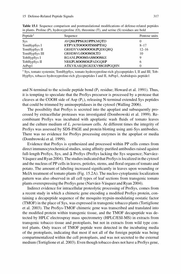
15 Defense-Related Peptide Signals 317
Table 15.1 Sequence comparison and posttranslational modifications of defense-related peptidesin plants. Proline (P), hydroxyproline (O), threonine (T), and serine (S) residues are bold
Peptide1 Sequence Pentose units
Sys AVQSKPPSKRDPPKMQTD 0TomHypSys I RTPYKTOOOOTSSSPTHQ 8–17TomHypSys II GRHDYVASOOOOKPQDEQRQ 12–16TomHypSys III GRHDSVLOOOSOKTD 10TobHypSys I RGANLPOOSOASSOOSKE 9TobHypSys II NRKPLSOOSOKPADGQRP 6AtPep1 ATKVKAKQRGKEKVSSGRPGQHN 01 Sys, tomato systemin; TomHypSys, tomato hydroxyproline-rich glycopeptides I, II and III; Tob-HypSys, tobacco hydroxyproline-rich glycopeptides I and II, AtPep1, Arabidopsis peptide1
and N-terminal to the scissile peptide bond (P1 residue; Howard et al. 1991). Thus,it is tempting to speculate that the ProSys precursor is processed by a protease thatcleaves at the COOH side of Asp (P1), releasing N-terminal extended Sys peptidesthat could be trimmed by aminopeptidases in the cytosol (Walling 2006).
The possibility that ProSys is secreted into the apoplast and subsequently pro-cessed by extracellular proteases was investigated (Dombrowski et al. 1999). Re-combinant ProSys was incubated with apoplastic wash fluids of tomato leavesand the culture medium of L. peruvianum cells. At different times the integrity ofProSys was assessed by SDS-PAGE and protein blotting using anti-Sys antibodies.There was no evidence for ProSys processing enzymes in the apoplast or media(Dombrowski et al. 1999).
Evidence that ProSys is synthesized and processed within PP cells comes fromdirect immunocytochemical studies, using affinity-purified antibodies raised againstfull-length ProSys, Sys, and � ProSys (ProSys lacking the Sys sequence; Narvaez-Vasquez and Ryan 2004). The studies indicated that ProSys is localized in the cytosoland the nucleus of PP cells in leaves, petioles, stems, and floral organs of tomato andpotato. The amount of labeling increased significantly in leaves upon wounding orMeJA treatment of tomato plants (Fig. 15.2A). The nucleo-cytoplasmic localizationpattern was also observed in all cell types of leaf sections from transgenic tomatoplants overexpressing the ProSys gene (Narvaez-Vasquez and Ryan 2004).
Indirect evidence for intracellular proteolytic processing of ProSys, comes froma recent study in which a chimeric gene encoding a modified ProSys protein, con-taining a decapeptide sequence of the mosquito trypsin-modulating oostatic factor(TMOF) in the place of Sys, was expressed in transgenic tobacco plants (Tortiglioneet al. 2003). The ProSys-TMOF chimeric gene was transcribed and translated intothe modified protein within transgenic tissue, and the TMOF decapeptide was de-tected by HPLC electrospray mass spectrometry (HPLC/ESI-MS) in extracts fromtransgenic tobacco tissue and protoplasts, but not in extracts from wild type con-trol plants. Only traces of TMOF peptide were detected in the incubating mediaof the protoplasts, indicating that most if not all of the foreign peptide was beingcompartmentalized within the cell protoplasts, and was not secreted to the externalmedium (Tortiglione et al. 2003). Even though tobacco does not have a ProSys gene,

318 J. Narvaez-Vasquez, M.L. Orozco-Cardenas
(A) (B)
Fig. 15.2 Immuno-cytochemical localization of ProSys and ProHypSys in vascular bundles oftomato leaves. Leaf samples from two-week-old tomato plants exposed for 18 hours to methyljasmonate vapors were fixed and immuno-gold labeled as previously described (Narvaez-Vasquezand Ryan 2004). (A) Silver-enhanced, immuno-gold labeled ProSys localized in vascular phloemparenchyma (PP) cells visualized under the confocal microscope. (B) Transmission electron mi-crograph showing immuno-gold labeled ProHypSys in the cell walls of PP cells (Narvaez-Vasquezet al. 2005). Arrows point to gold particles. pp, phloem parenchyma; cc, companion cell; se, sieveelements; x, xylem vessel. Bars = A, 5.0 �m; B, 0.5 �m
this result suggests that ProSys is synthesized, processed and/or stored intracellu-larly in tobacco leaves, most probably in a similar fashion as in tomato.
Genetic screens have been conducted in tomato to find mutants in the Sys signal-ing pathway (Howe and Ryan 1999). These efforts have led to the identificationof several mutants incapable to respond to Sys, wounding, or JA (Howe 2005).However, mutations that affect the processing of ProSys have not been identifiedyet. The failure of these screens to identify genes required for processing, compart-mentalization and/or transport of ProSys or Sys suggests that the ProSys processingpathway involves genes that are either redundant or required for cell/plant viability.
15.2.3 The Sys Signaling Pathway
A Sys receptor protein, SR160, has been isolated and its gene cloned (Scheerand Ryan 1999, 2002). SR-160 belongs to a family of Ser/Thr receptor kinaseswith extracellular Leu-rich repeats, which was found to be highly similar to theArabidopsis brassinolide receptor BRI1 (Scheer and Ryan 2002). Brassinolides are

15 Defense-Related Peptide Signals 319
a class of plant steroid hormones with different roles in plant development, but notin plant defense. Isolation of the tomato brassinolide receptor confirmed that it isalso the Sys receptor (Montoya et al. 2002). The function of SR160 as the Sysreceptor was reconfirmed by expressing SR160 cDNA in tobacco plants (Scheeret al. 2003). Tobacco plants lack a ProSys gene and Sys perception. In transgenictobacco expressing SR-160 mRNA constitutively, the Sys receptor is membrane lo-calized, binds Sys and activates early defense responses (Scheer et al. 2003). Thisis the first example of a protein receptor in plants having a dual function in defenseand development.
Ala substitution and deletion analyses of the Sys polypeptide indicated thatthe entire 18 amino acids are necessary for full biological activity, and that theN-terminal region might be important for recognition/binding to the Sys receptor,while theC-terminalresiduesaremoreessential for activity (Pearceetal. 1993;Meindlet al. 1998). Upon binding to its receptor, Sys activates a complex cascade of signalingevents leading to the activation of early (signaling-related) and late (defense-related)wound response genes, and theproduction of H2O2 (Ryan 2000). In tomato leaves, Syscauses a rapid (within minutes) depolarization of the plasma membrane in mesophyllcells, the alkalinization of the extracellular medium or cell wall apoplast, transient in-creases in cytoplasmic free Ca2+ concentrations, the activation of a mitogen-activatedprotein kinase (MAPK), and the release of linolenic acid (18:3 fatty acid) from cellmembranes by the activation of a phospolipase A2, leading to the biosynthesis ofOPDA and JA through the octadecanoid pathway (Ryan 2000).
15.2.4 Systemin and the Systemic Wound Signal
The plant wound defense response against insects or herbivore attacks, discoveredby Green and Ryan (1972), is characterized by the accumulation of defense chem-icals not only at the site of damage (local response), but also in other undamagedparts of the plant (systemic response). This implies that ‘wound’ signal(s) are gen-erated at the sites of damage to activate the transcription of defense genes and thesynthesis of antinutritional compounds throughout the plant (Green and Ryan 1972).
Sys has the characteristics of a systemic wound signal. When radiolabeledSys is placed in wounds made on leaves, Sys is loaded into the phloem andtransported systemically throughout the plants within a few hours, in a simi-lar fashion as the transport of 14C-sucrose (Pearce et al. 1991; Narvaez-Vasquezet al. 1994, 1995). In addition, the overexpression of the ProSys precursor geneunder the regulation of the CaMV 35S promoter in transgenic tomato plants led tothe constitutive accumulation of high levels of several defensive proteins in leaves(McGurl et al. 1994; Bergey et al. 1996). The ProSys overexpressing plants pro-duced a graft-transmissible signal that activates defense genes in a wild type scion(McGurl et al. 1994). Conversely, when the 35S:ProSys transgene was expressedin the antisense orientation in transgenic tomato plants, the systemic activation ofwound-inducible defense genes was strongly reduced and the plants were more sus-ceptible to insect damage (McGurl et al. 1992; Orozco-Cardenas et al. 1993). Thus,

320 J. Narvaez-Vasquez, M.L. Orozco-Cardenas
the cumulative results supported the hypothesis that Sys is an important componentof a mobile systemic signal.
Recent genetic evidence, however, indicated that JA or a JA-derived oxylipin,functions as the systemic wound signal in tomato, and that Sys main role is toup-regulate JA biosynthesis (Howe 2005). This was demonstrated using recipro-cal grafting experiments with tomato mutants deficient in JA biosynthesis and JAperception, which were also defective in systemic wound signaling (Howe 2005;see also Howe and Schaller this volume). Accordingly, several enzymes of the oc-tadecanoid pathway have been localized within the vascular bundle cells of tomatoleaves, including LOX, AOS, and AOC (Wasternack et al. 2006). Tomato AOC isa rate-limiting enzyme required for wound-induced JA biosynthesis during Sys sig-naling (Stenzel et al. 2003). Therefore, Sys synthesis and JA synthesis have nowbeen associated with the phloem. Altogether, the results of the grafting and local-ization studies indicated a new role for both Sys and JA in the amplification of thesystemic signaling process as they are translocated through the plant vasculature(Ryan and Moura 2002; Howe 2005; Wasternack et al. 2006).
15.3 Hydroxyproline-Rich Systemin Glycopeptides (HypSys)
The search for a functional homologue of Sys in tobacco, using a cell alkaliniza-tion assay (Felix and Boller 1995), resulted in the identification of two bioactive18-aa glycopeptides in purified HPLC fractions obtained from tobacco leaf extracts(Pearce et al. 2001). The isolated peptides (called TobHypSys I and II) were capa-ble of inducing the medium alkalinization of tobacco and tomato cell suspensioncultures, and also of activating a 48-kDa MAP kinase and defense gene expressionwhen supplied to excised tobacco leaves at nM concentrations (Pearce et al. 2001).
TobHypSys I and II are hydroxyproline-rich glycopeptides, decorated with nineand six pentose units respectively (Table 15.1). The glycosyl moieties are requiredfor full biological activity. TobHypSys I and II are derived from a single polyproteinprecursor of 165 amino acids that has a signal peptide (Pearce et al. 2001), repre-senting the first example in plants of multiple signaling peptides derived from thesame protein precursor, a common feature found in animals (Tanaka 2003).
Three HypSys glycopeptides weresubsequently purified from tomato leaves, usingthe same medium alkalinization assay with L. peruvianum suspension cell cultures(Pearce and Ryan 2003). Tomato HypSys I, II, and III of 20, 18, and 15 residuesrespectively, are also derived from a single precursor protein of 146 amino acids,which is likely to be secreted by virtue of a predicted signal peptide (Fig. 15.1C).The tomato glycopeptides have between 8 and 16 pentose units, which appear tobe arabinose residues bonded to hydroxyproline residues (Table 15.1; Pearce andRyan 2003). The amino acid sequences of TomHypSys II and III peptides are verysimilar, with nine identical amino acids (Table 15.1), suggesting they have resultedfrom gene duplication and elongation events similar to ProSys.
15 Defense-Related Peptide Signals 321
The presence of a signal peptide in both the tobacco and tomato HypSys precur-sors and the post-translational hydroxylation of proline and glycosylation indicatesthat the bioactive peptide precursors are synthesized through the secretory pathway.Immunolocalization studies using affinity-purified antibodies that only recognizeepitopes in the tomato ProHypSys precursor indicates that the proprotein is localizedin the cell walls of leaf vascular parenchyma cells (Fig. 15.2B; Narvaez-Vasquezet al. 2005).
Multiple HypSys glycopeptide signals derived from single protein precursorshave also been found in other species of the Solanaceae, including petunia,nightshade, and potato (Pearce et al. 2006). In both tobacco and tomato, the Pro-HypSys genes are upregulated in the leaves by wounding, Sys, MeJA, and theHypSys peptides themselves (Pearce et al. 2001; Pearce and Ryan 2003). The To-bacco ProHypSys gene is also induced by ABA, chewing and phloem-feeding in-sects (Rocha-Granados et al. 2005). In tomato, low constitutive levels of HypSysmRNAs are found in leaves, but the basal levels are higher in other organs, includ-ing roots (Narvaez-Vasquez and Ryan, unpublished results). Overexpression of thenative HypSys genes in tobacco (Ren and Lu 2006) and tomato (Narvaez-Vasquezet al. 2007) caused the constitutive upregulation of both signaling and defense-related genes in the transgenic plants, similar to wounding and Sys in tomato. The35S:HypSys transgenic tobacco plants were more resistant to insect attacks (Ren andLu 2006). In addition, downregulation of TomHypSys gene expression by antisensetechnology also diminished the systemic wound inducibility of defense genes, sug-gesting that Sys and HypSys work cooperatively to upregulate the systemic wound-defense response in tomato (Narvaez-Vasquez et al. 2007).
The processing events responsible for the release of the HypSys peptides in thecell wall in response to wounding are still unknown. The presence of the –N/GR se-quence at the N-termini of tobacco and tomato HypSys peptides suggests that theseamino acids may be substrates for specific proteases in both species (Table 15.1).Wounding may cause intracellular proteases to mix with the precursor of Hyp-Sys peptides in the cell wall matrix as a consequence of cell damage. Wound- orSys-induced ion fluxes and pH changes in leaf cells may as well induce secretionand/or activation of processing enzymes in the extracellular matrix (Schaller andOecking 1999; Tanaka 2003). Alternatively, pathogen-derived peptidases may alsobe involved in processing of bioactive peptides leading to the activation of defensesignaling cascades. In any event, the proteolytic release of multiple peptide signalsfrom polyprotein precursors located in the cell walls may be among the plant earliestevents in response to insect and pathogen attacks.
15.4 Arabidopsis Defense-Related Peptides (AtPeps)
AtPep1 is the first endogenous defense-related peptide purified from leaves of Ara-bidopsis thaliana (Huffaker et al. 2006a). The AtPep1 peptide has 23 amino acidsand is derived from the carboxy terminus of a signal-less 92-amino acid precursorprotein (ProAtPep1; Fig. 15.1D). AtPep1 regulates the expression of defense genes

322 J. Narvaez-Vasquez, M.L. Orozco-Cardenas
induced by pathogens through the octadecanoid signaling pathway. The gene en-coding ProAtPep1 is part of a small family of at least seven members in Ara-bidopsis, with orthologs in species of many other families, including importantcrop plants (Table 15.2). The size range of ProAtPep1-related precursor proteinsreported to date is between 75 and 154 amino acids (Table 15.2). Based on theirchromosomal location the Arabidopsis AtPep1 gene family has been classified inthree subfamilies. Members of subfamilies I (ProAtPep1, 2, and 3) and II (ProAt-Pep4, 5, and 7) are grouped in two different locations in Chromosome V, andonly one gene (ProAtPep6) belongs to the subfamily III located on chromosomeII (Huffaker et al. 2006b). The Arabidopsis paralogs share the 10-amino acid motifSSG(K/R)xGxxN that appear to be important for full biological activity. Notably,Gly17 is the only conserved amino acid in all the other known AtPep1 orthologs(Table 15.2). Ala-substitution studies indicated that Gly17 is essential for AtPep1biological activity (Huffaker et al. 2006b).
In Arabidopsis, the ProAtPep1 gene is expressed at low constitutive levelsin all tissues, with higher basal levels observed in flowers and roots (Huffakeret al. 2006a). In leaves, the ProAtPep1 gene is induced by wounding, MeJA andethylene. Other gene family members show similar expression patterns in a widerange of tissues, and appear to be upregulated by MeJA (Huffaker et al. 2006a,b). Analysis of microarray gene expression data in Arabidopsis indicated thatsome of the ProAtPep1 paralogs are differentially regulated by bacterial and fungalpathogens and pathogen-derived elicitors (Huffaker et al. 2006a). AtPep1 suppliedto excised Arabidopsis leaves through the cut petioles causes the activation ofseveral genes involved in pathogen defense, such as those encoding for defensin(PDF-1.2), other pathogenesis-related (PR) proteins, ProAtPep1, and some of theProAtPep1 paralogs (Huffaker et al. 2006b). Feeding AtPep1 to leaves also causedthe production of H2O2 in the vascular bundles (Huffaker et al. 2006a).
When the ProAtPep1 gene was overexpressed in Arabidopsis under the regu-lation of the 35S promoter, the pathogen-related defensin gene was constitutivelyexpressed and the transgenic plants were more resistant to infection by the soil bornepathogen Pythium irregulare (Huffaker et al. 2006a). Infected wild-type controlplants grew smaller, especially their roots, than the transgenic plants. This result in-dicates that the overproduced ProAtPep1 has been processed to release the bioactivepeptide to constitutively activate the pathogen-defense response in the transgenictissue (Huffaker et al. 2006a). No endogenous peptides have been reported beforethat could activate genes associated with innate immunity in plants.
An AtPep1-binding protein (∼170-kD) was isolated from microsomal mem-branes of Arabidopsis suspension cultured cells by photoaffinity labeling(Yamaguchi et al. 2006). The protein binds an iodinated-AtPep1 analog with aKd of 0.25 nM, similar to the binding constant of the iodinated Sys analog to itsSR-160 receptor (Kd of 0.17 nM; Scheer and Ryan 1999). Mass spectroscopy oftryptic digests identified the binding protein as a receptor-like protein kinase of1123 amino acids, encoded by the gene At1g73080 in Arabidopsis (Yamaguchiet al. 2006). The AtPep1 receptor protein, called AtPepR1, contains 27 leucine-rich repeats, a transmembrane region, and an intracellular Ser/Thr protein kinase

15 Defense-Related Peptide Signals 323
Tab
le15
.2C
ompa
riso
nof
the
C-t
erm
inal
amin
oac
idse
quen
ces
ofA
tPep
1pa
ralo
gsan
dor
thol
ogs
cont
aini
ngpu
tativ
eA
tPep
sequ
ence
sin
plan
ts.T
hese
quen
ceof
AtP
ep1
(23
resi
dues
)is
alig
ned
wit
hth
eC
-ter
min
ided
uced
from
six
para
logs
and
seve
nor
thol
ogs.
The
amin
oac
ids
ofth
eco
nser
ved
SSG
R/K
xGxx
Nm
otif
pres
enti
nal
lAtP
ep1
para
logs
are
bold
.Ada
pted
from
Huf
fake
ret
al.(
2006
a,b)
Pept
ide
Prec
urso
rge
neco
deC
-ter
min
alam
ino
acid
sequ
ence
Prec
urso
rle
ngth
(am
ino
acid
s)
AtP
ep1
At5
g649
0069-ATKVKAKQRGKEKV
SSG
RP
GQH
N∗
92A
tPep
2A
t5g6
4890
74-DNKAKSKKRDKEKP
SSG
RP
GQT
NSVPNAAIQVYKED
∗10
9A
tPep
3A
t5g6
4905
73-EIKARGKNKTKPTP
SSG
KG
GKH
N∗
96A
tPep
4A
t5g0
9980
55-GLPGKKNVLKKSRE
SSG
KP
GGT
NKKPF
∗81
AtP
ep5
At5
g099
9059-SLNVMRKGIRKQPV
SSG
KR
GGV
NDYDN
∗86
AtP
ep6
At2
g220
0053-ITAVLRRRPRPPPY
SSG
RP
GQN
N∗
75A
tPep
7A
t5g0
9975
81-VSGNVAARKGKQQT
SSG
KG
GGT
N∗
104
Can
ola
CD
8166
4574-VARLTRRRPRPP-Y
SSGQP
GQI
N∗
95Po
tato
CV
5053
8893-PTERRGRPPSRPKVG
SGPPPQN
N∗
116
Popl
arC
V23
975
94-DAAVSALARRTPPV
SRGGG
GQT
NTTTS
∗12
1M
edic
ago
BI3
1144
187-LSSMGRGGPRRTPLTQ
GPPPQH
N∗
111
Soyb
ean
CD
4012
8193-ASLMATRGSRGSKI
SDGSGPQH
N∗
115
Ric
e1C
F333
408
131-ARLRPKPPGNPREG
SGGNG
GHHH
∗15
4R
ice2
AK
1111
1365-DDSKPTRPGAPAEG
SGGNG
GAIHTAASS
∗93
324 J. Narvaez-Vasquez, M.L. Orozco-Cardenas
domain. Radiolabeled AtPep1 analog did not bind to microsomal membranes fromtwo insertional mutants of At1g73080, and tobacco suspension cells transformedwith the AtPepR1 gene were capable of binding to AtPep1 and showed the typicalalkalinization response. These results demonstrate that PepR1 is a functional recep-tor (Yamaguchi et al. 2006). Four out of six AtPep1 homologs synthesized basedon the deduced C-terminal amino acid sequence of ProPep1 paralogs in Arabidop-sis were biologically active in the alkalinization assay with Arabidopsis cells. Thebioactive synthetic peptides were also able to compete with the radiolabeled AtPep1analog for binding to PepR1. This result suggested that PepR1 might actually be thereceptor for at least five of the homologous AtPep1 peptides possibly generatedduring pathogen attacks (Yamaguchi et al. 2006).
15.5 Evolutionary Considerations
The discovery of genetically different defense-related peptide signals in the Solan-aceae, acting through the same octadecanoid-signaling pathway prompted Prof.C.A. Ryan and collaborators to ‘broaden the definition of systemins to includeany signaling peptide from plants that activates plant defensive genes’ (Ryan andPearce 2003). Thus, this definition should be extended to the Arabidopsis AtPeppeptides, which are also upregulated by wounding and pathogens to activate genesinvolved in pathogen defense through the octadecanoid pathway. AtPep orthologsare also present in the Solanaceae (Table 15.2; Huffaker et al. 2006a).
There is no sequence homology between the protein precursors of Sys, HypSys,and AtPep1 peptides (Fig. 15.1). The Sys and HypSys peptides have only beenfound in species of the Solanaceae, although the systemic wound defense responseis ubiquitous in plants. Even the tomato and tobacco HypSys precursors only sharea limited amino acid identity (compare Fig. 15.1B and C). However, the bioactivepeptides show some degree of conservation suggestive of a common origin. For in-stance, they are enriched in proline/hydroxyproline, serine, and threonine residues,flanked by charged amino acids (Table 15.1). The HypSys glycopeptides contain –OOS- motifs, which are posttranslational modifications derived from –PPS- primarytranslation motifs also present in Sys (Table 15.1). AtPep1 lacks this motif, butsome AtPep1 paralogs and orthologs are proline-rich and highly charged peptides(Table 15.2). Proline residues are known to confer structural conformations in thebackbone chains of bioactive peptides that are important for the interactions of pep-tide ligands with their receptors (Rath et al. 2005).
Like tomato Sys, AtPep1-related peptides are synthesized from larger precursorproteins that lack a classical signal sequence. In addition, the AtPep precursors arehighly charged proteins, with stretches of lysine (K) and glutamic acid (E) residuesthat resemble the KEKE-like motifs (Realini et al. 1994), also found in ProSys (com-pare Fig. 15.1A and D). Because of this, and the similarities mentioned above, it hasbeen proposed that peptide signals that activate defense responses against pathogenand herbivore attacks may have evolved from a common ancestor gene (Ryan and
15 Defense-Related Peptide Signals 325
Pearce 2003; Huffaker et al. 2006a, b). Mutational events, such as gene insertions,followed by duplication and elongation events appear to be involved in the evo-lution of peptide signal precursors in plants (Ryan and Pearce 2003). This mightexplain why the prohormone precursors of defense related peptides have divergedmore quickly than the actual bioactive peptides.
Since AtPep1 peptides are widely spread in the plant kingdom, they may haveevolved prior to the systemins in the Solanaceae. Since HypSys peptides are foundin most species of the Solanaceae, and Sys only in the Solaneae subtribe, HypSyspeptides appear to have evolved earlier than Sys. It is possible that the brassino-lide receptor evolved initially and then was later recruited to facilitate systeminsignaling. It would be interesting to see whether the HypSys peptides interact withhomologs of the systemin receptor or have entirely different receptors. Notably,overexpression of the tomato ProSys gene in transgenic potato causes the accumu-lation of developmentally regulated proteins in the tubers (Narvaez-Vasquez andRyan 2002). The link between signaling pathways involved in plant developmentand defense is not well understood yet.
15.6 Perspectives
The evidence suggests that defense-related peptide hormones play a primary rolein the amplification of signals generated by insects and pathogen attacks in plantsthrough the activation of a lipid-based signaling pathway in the phloem. The mainpurpose being the rapid systemic accumulation of deterrents and antinutritionalfactors throughout the plant that will diminish food quality, slow down growth,and/or prevent the colonization of invading organisms. This peptide-based signal-ing mechanism for plant defense is analogous to the activation of the inflammatoryand acute-wound defense response mediated by peptide cytokines such as the inter-leukins and TNF-�, through the eicosanoid signaling pathway in animals (Bergeyet al. 1996).
Although thoroughly studied in animal systems and yeast, the regulation of syn-thesis, processing, storage and secretion of peptide signals in plants is poorly un-derstood. The processing sites that release the active peptides are not known, nor isthe nature of the peptidases involved. The mechanism by which Sys and AtPep1 arereleased into the cell wall apoplast is still unknown. About half a dozen families ofplant peptide hormones identified to date are synthesized from precursors lackinga signal sequence (Matsubayashi and Sakagami 2006), suggesting that more oftenthan in animals and yeast, plant regulatory peptides are processed and transportedthrough non-classical mechanisms to the extracellular space (Nickel 2003). Furtherstudies on the biogenesis of defense-related peptide hormones, and the discoveryof new peptide signals and their receptors in plants, will probably provide moreevidence for the evolution of defense peptide signaling systems in eukaryotes froma common ancestor.

326 J. Narvaez-Vasquez, M.L. Orozco-Cardenas
References
Bergey DR, Howe GA, Ryan CA (1996) Polypeptide signaling for plant defensive genes exhibitsanalogies to defense signaling in animals. Proc Natl Acad Sci USA 93:12053–12058
Coffeen WC, Wolpert TJ (2004) Purification and characterization of serine proteases that exhibitcaspase-like activity and are associated with programmed cell death in Avena sativa. Plant Cell16:857–873
Constabel CP, Yip L, Ryan CA (1998) Prosystemin from potato, black nightshade, and bell pepper:primary structure and biological activity of predicted systemin polypeptides. Plant Mol Biol36:55–62
D’Hont K, Bosch D, Van Damme J, Goethals M, Vanderkerkhove J, Krebbers E (1993) An as-partic endoproteinase present in seed cleaves Arabidopsis 2S albumins in vitro. J Biol Chem268:20884–20891
Dombrowski JE, Pearce G, Ryan CA (1999) Proteinase inhibitor-inducing activity of the prohor-mone prosystemin resides exclusively in the C-terminal systemin domain. Proc Natl Acad SciUSA 96:12947–12952
Felix G, Boller T (1995) Systemin induces rapid ion fluxes and ethylene biosynthesis in Lycoper-sicon peruvianum cells. Plant J 7:381–389
Francois I, De Bolle MFC, Dwyer G, Goderis I, Woutors PFJ, Verhaert PD, Proost P, SchaaperWMM, Cammue BPA, Broekaert WF (2002) Transgenic expression in Arabidopsis of apolyprotein construct leading to production of two different antimicrobial proteins. Plant Phys-iol 128:1346–1358
Green TR, Ryan CA (1972) Wound-induced proteinase inhibitor in plant leaves – possible defensemechanism against insects. Science 175:776–777
Harris R (1989) Processing of pro-hormone precursor proteins. Arch Biochem Biophys 275:315–333
Horn M, Patankar AG, Zavala JA, Wu JQ, Doleckova-Maresova L, Vujtechova M, MaresM, Baldwin IT (2005) Differential elicitation of two processing proteases controls theprocessing pattern of the trypsin proteinase inhibitor precursor in Nicotiana attenuata. PlantPhysiol 139:375–388
Howard AD, Kostura MJ, Thornberry N, Ding GJF, Limjuco G, Weidner J, Salley JP, Hogquist KA,Chaplin DD, Mumford RA, Schmidt JA, Tocci MJ (1991) IL-1�-Converting enzyme requiresaspartic-acid residues for processing of the IL-1� precursor at 2 distinct sites and does notcleave 31-kDa IL-1�. J Immunol 147:2964–2969
Howe GA (2005) Jasmonates as signals in the wound response. J Plant Growth Regul 23:223–237Howe GA, Ryan CA (1999) Suppressors of systemin signaling identify genes in the tomato wound
response pathway. Genetics 153:1411–1421Huffaker A, Pearce G, Ryan CA (2006a) An endogenous peptide signal in Arabidopsis activates
components of the innate immune response. Proc Natl Acad Sci USA 103:10098–10103Huffaker A, Yamagushi Y, Pearce G, Ryan CA (2006b) AtPep1 peptides. In: Kastin AJ (ed) Hand-
book of biologically active peptides. Academic Press, San Diego, pp 5–8Matsubayashi Y, Sakagami Y (2006) Peptide hormones in plants. Ann Rev Plant Biol 57:649–674McGurl B, Orozco-Cardenas M, Pearce G, Ryan CA (1994) Overexpression of the prosys-
temin gene in transgenic tomato plants generates a systemic signal that constitutively inducesproteinase-inhibitor synthesis. Proc Natl Acad Sci USA 91:9799–9802
McGurl B, Ryan CA (1992) The organization of the prosystemin gene. Plant Mol Biol 20:405–409McGurl B, Pearce G, Orozco-Cardenas M, Ryan CA (1992) Structure, expression, and antisense
inhibition of the systemin precursor gene. Science 255:1570–1573Meindl T, Boller T, Felix G (1998) The plant wound hormone systemin binds with the N-terminal
part to its receptor but needs the C-terminal part to activate it. Plant Cell 10:1561–1570Montoya T, Nomura T, Farrar K, Kaneta T, Yokota T, Bishop GJ (2002) Cloning the tomato curl3
gene highlights the putative dual role of the leucine-rich repeat receptor kinase tBRI1/SR160in plant steroid hormone and peptide hormone signaling. Plant Cell 14:3163–3176

15 Defense-Related Peptide Signals 327
Narvaez-Vasquez J, Orozco-Cardenas ML, Ryan CA (1994) A sulfhydryl reagent modulates sys-temic signaling for wound-induced and systemin-induced proteinase-inhibitor synthesis. PlantPhysiol 105:725–730
Narvaez-Vasquez J, Orozco-Cardenas ML, Ryan CA (2007) Systemic wound signaling intomato leaves is cooperatively regulated by multiple plant peptides. Plant Mol Biol65:711–718
Narvaez-Vasquez J, Pearce G, Orozco-Cardenas ML, Franceschi VR, Ryan CA (1995) Autoradio-graphic and biochemical-evidence for the systemic translocation of systemin in tomato plants.Planta 195:593–600
Narvaez-Vasquez J, Pearce G, Ryan CA (2005) The plant cell wall matrix harbors a precursor ofdefense signaling peptides. Proc Natl Acad Sci USA 102:12974–12977
Narvaez-Vasquez J, Ryan CA (2002) The systemin precursor gene regulates both defensive anddevelopmental genes in Solanum tuberosum. Proc Natl Acad Sci USA 99:15818–15821
Narvaez-Vasquez J, Ryan CA (2004) The cellular localization of prosystemin: a functional rolefor phloem parenchyma in systemic wound signaling. Planta 218:360–369
Nickel W (2003) The mystery of nonclassical protein secretion – A current view on cargo proteinsand potential export routes. Eur J Biochem 270:2109–2119
Orozco-Cardenas ML, McGurl B, Ryan CA (1993) Expression of an antisense prosystemin genein tomato plants reduces resistance toward Manduca sexta larvae. Proc Natl Acad Sci USA90:8273–8276
Pautot V, Holzer FM, Reish B, Walling LL (1993) Leucine aminopeptidase: an inducible compo-nent of the defense response in Lycopersicon esculentum (tomato). Proc Natl Acad Sci USA90:9906–9910
Pearce G, Johnson S, Ryan CA (1993) Structure-activity of deleted and substituted systemin, an18-amino acid polypeptide inducer of plant defensive genes. J Biol Chem 268:212–216
Pearce G, Moura DS, Stratmann J, Ryan CA (2001) Production of multiple plant hormones from asingle polyprotein precursor. Nature 411:817–820
Pearce G, Narvaez-Vasquez J, Ryan CA (2006) Systemins. In: Kastin AJ (ed) Handbook of bio-logically active peptides. Academic Press, San Diego, pp 49–53
Pearce G, Ryan CA (2003) Systemic signaling in tomato plants for defense against herbivores –isolation and characterization of three novel defense-signaling glycopeptide hormones codedin a single precursor gene. J Biol Chem 278:30044–30050
Pearce G, Strydom D, Johnson S, Ryan CA (1991) A polypeptide from tomato leaves activates theexpression of proteinase inhibitor genes. Science 253:895–898
Rath A, Davison AR, Deber CM (2005) The structure of ‘unstructured’ regions in peptides andproteins: role of the polyproline II helix in protein folding and recognition. Biopolymers 80:179–185
Realini C, Rogers SW, Rechsteiner M (1994) KEKE motifs – proposed roles in protein–proteinassociation and presentation of peptides by MHC Class-I receptors. FEBS Lett 348:109–113
Ren F, Lu Y-T (2006) Overexpression of tobacco hydroxyproline-rich glycopeptide systemin pre-cursor A in transgenic tobacco enhances resistance against Helicoverpa armigera larvae. PlantSci 171:286–292
Rocha-Granados MC, Sanchez-Hernandez C, Sanchez-Hernandez C, Martınez-Gallardo NA,Ochoa-Alejo N, Delano-Frier JP (2005) The expression of the hydroxyproline-rich glycopep-tide systemin precursor A in response to (a)biotic stress and elicitors is indicative of its role inthe regulation of the wound response in tobacco (Nicotiana tabacum L.). Planta 222:794–810
Rojo E, Martin R, Carter C, Zouhar J, Pan SQ, Plotnikova J, Jin HL, Paneque M, Sanchez-SerranoJJ, Baker B, Ausubel FM, Raikhel NV (2004) VPE gamma exhibits a caspase-like activity thatcontributes to defense against pathogens. Curr Biol 14:1897–1906
Ryan CA (2000) The systemin signaling pathway: differential activation of plant defensive genes.Biochim Biophys Acta 1477:112–121
Ryan CA, Moura DS (2002) Systemic wound signalling in plants: a new perception. Proc NatlAcad Sci USA 99:6519–6520

328 J. Narvaez-Vasquez, M.L. Orozco-Cardenas
Ryan CA, Pearce G (1998) Systemin: a polypeptide signal for plant defensive genes. Ann Rev CellDev Biol 14:1–17
Ryan CA, Pearce G (2003) Systemins: a functionally defined family of peptide signal that regulatedefensive genes in Solanaceae species. Proc Natl Acad Sci USA 100:14577–14580
Schaller A (1998) Action of proteolysis-resistant systemin analogues in wound signalling. Phyto-chemistry 47:605–612
Schaller A, Oecking C (1999) Modulation of plasma membrane H+-ATPase activity differentiallyactivates wound and pathogen defense responses in tomato plants. Plant Cell 11:263–272
Schaller A, Ryan CA (1994) Identification of a 50-KDa systemin-binding protein in tomatoplasma-membranes having Kex2p-like properties. Proc Natl Acad Sci USA 91:11802–11806
Schaller A, Ryan CA (1996) Molecular cloning of a tomato leaf cDNA encoding an aspartic pro-tease, a systemic wound response protein. Plant Mol Biol 31:1073–1077
Schaller F, Schaller A, Stintzi A (2005) Biosynthesis and metabolism of jasmonates. J Plant GrowthRegul 23:179–199
Scheer JM, Pearce G, Ryan CA (2003) Generation of systemin signaling in tobacco by transforma-tion with the tomato systemin receptor kinase gene. Proc Natl Acad Sci USA 100:10114–10117
Scheer JM, Ryan CA (1999) A 160-kD systemin receptor on the surface of Lycopersicon peru-vianum suspension-cultured cells. Plant Cell 11:1525–1535
Scheer JM, Ryan CA (2002) The systemin receptor SR160 from Lycopersicon peruvianum is amember of the LRR receptor kinase family. Proc Natl Acad Sci USA 99:9585–9590
Seidah NG, Chretien M (1997) Eukaryotic protein processing: endoproteolysis of precursor pro-teins. Curr Opin Biotechnol 8:602–607
Stenzel I, Hause B, Maucher H, Pitzschke A, Mierch O, Ziegler J, Ryan CA, Wasternack C (2003)Allene oxide cyclase dependence of the wound response and vascular bundle-specific genera-tion of jasmonates in tomato – amplification in wound signaling. Plant J 33:577–589
Tanaka S (2003) Comparative aspects of intracellular proteolytic processing of peptide hormoneprecursors: studies of proopiomelanocortin processing. Zool Sci 20:1183–1198
Tortiglione C, Fogliano V, Ferracane R, Fanti P, Pennacchio F, Monti LM, Rao R (2003) An insectpeptide engineered into the tomato prosystemin gene is released in transgenic tobacco plantsand exerts biological activity. Plant Mol Biol 53:891–902
Truman W, Bennett MH, Kubigsteltig I, Turnbull C, Grant M (2007) Arabidopsis systemic im-munity uses conserved signaling pathways and is mediated by jasmonates. Proc Natl Acad SciUSA 104:1075–1080
Walling LL (2006) Recycling or regulation? The role of amino-terminal modifying enzymes. CurrOpin Plant Biol 9:227–233
Wasternack C, Stenzel I, Hause B, Hause G, Kutter C, Maucher H, Neumerkel J, Feussner I,Miersch O (2006) The wound response in tomato – role of jasmonic acid. J Plant Physiol163:297–306
Yamaguchi Y, Pearce G, Ryan CA (2006) The cell surface leucine-rich repeat receptor for AtPep1,an endogenous peptide elicitor in Arabidopsis, is functional in transgenic tobacco cells. ProcNatl Acad Sci USA 103:10104–10109
Zhang L, Kelley J, Schmeisser G, Kobayashi YM, Jones LR (1997) Complex formation betweenjunction, triadin, calsequestrin, and the ryanodine receptor – proteins of the cardiac junctionalsarcoplasmic reticulum membrane. J Biol Chem 272:23389–2339

Chapter 16MAP Kinases in Plant Responses to Herbivory
Johannes Stratmann
Mitogen-activated protein kinases (MAPKs) are signal transducing enzymesinvolved in many plant responses to the environment including defense responsesto herbivory. Phytophagous insects generate various plant- and insect-derived pri-mary signals such as the plant signaling peptide systemin, mechanical woundsignals, and fatty acid-amino acid conjugates. These signals activate a set of spe-cific MAPKs. Reducing the function of these MAPKs impairs the synthesis ofsecondary stress signals including the key wound signal jasmonic acid, which isrequired for the upregulation of defense proteins. Consequently, loss-of-MAPK-function results in reduced resistance of plants to herbivorous insects. Insect-responsive MAPKs also function as essential signaling relays in defense responsesto pathogens and abiotic stress. It is a major challenge for future research to un-ravel the molecular mechanisms that perceive and integrate stress signals to generatestimulus-specific MAPK signaling patterns which result in specific cellular defenseresponses.
16.1 Introduction
A wealth of genomic information in all eukaryotes has revealed the preeminentrole of protein kinases for the regulation of cellular processes. PlantsP, an inter-active website dedicated to the study of plant phosphorylation, estimated that theArabidopsis genome contains about 1000 putative protein kinases that constitute 5major classes with a total of 80 distinct families (http://plantsp.genomics.purdue.edu/html). Unlike in animals and yeast, tyrosine-specific phosphorylation is rare inplants. Almost all plant protein kinases are Ser/Thr-specific kinases. They are struc-turally defined by the presence of 12 conserved subdomains that constitute the cat-alytic core of the enzyme. The kinase domain is often extended N- and C-terminallyby other protein domains (Hanks and Hunter 1995). Many protein kinases become
J. StratmannDepartment of Biological Sciences, University of South Carolina, Columbia, SC 29208, USAe-mail: [email protected]
A. Schaller (ed.), Induced Plant Resistance to Herbivory, 329C© Springer Science+Business Media B.V. 2008

330 J. Stratmann
activated by phosphorylation via other kinases and inactivated by dephosphorylationvia protein phosphatases, which comprise about 300 members in the Arabidopsisgenome (PlantP).
Reversible phosphorylation is known to be a part of many signaling pathwaysthat regulate developmental processes and plant responses to the environment. Thischapter reviews the role of mitogen-activated protein kinase (MAPK) cascades inplant responses to herbivorous insects and mechanical wounding. To understand thefunction of MAPKs, it is important to note that many plant stress responses overlapat various levels of signal transduction and cellular responses. Global transcrip-tome analyses revealed overlaps among gene expression profiles induced by differ-ent stress stimuli, but also stimulus-specific gene expression (Cheong et al. 2002;Reymond et al. 2000; Schenk et al. 2000). It appears that a limited number ofsecondary stress signals, such as ion fluxes (e.g., Ca2+ and H+), jasmonic acid(JA), ethylene, reactive oxygen species (ROS), and salicylic acid (SA), functionin multiple signaling pathways that can be induced by structurally and functionallydiverse stress signals. MAPKs are known to function as convergence points for mul-tiple stress signals. The interaction and coordination of these signaling componentsis thought to occur in a signaling network where multiple signaling elements arehighly interconnected and mutually affect each other. While this review will focuson signal transduction in plant responses to herbivory, it will take into account thatwound-responsive MAPKs also function in other stress signaling pathways.
Only very few studies address the role of MAPKs and other protein kinases inresponse to damage inflicted by herbivorous insects. Most studies have focusedon the role of protein kinases in the response to leaf wounding with mechanicaldevices, such as a hemostat or forceps for crushing, razor blades for cutting, orcarborundum for leaf surface abrasion. It should be noted that mechanical wound-ing and wounding by chewing insects result in the generation of several primarywound signals, such as systemically propagated rapid electrical and hydraulicsignals (Malone 1996; Rhodes et al. 1996; Stratmann and Ryan 1997; Wildonet al. 1992), and peptides such as systemin and hydroxyproline-rich systemins(Pearce et al. 1991, 2001; Pearce and Ryan 2003). In addition, insects deliver fattyacid-amino acid conjugates (Alborn et al. 1997; Halitschke et al. 2003) or otherinsect-specific elicitors like �-glucosidase (Mattiacci et al. 1995) to wound siteswhich function as elicitors of direct and indirect defense responses. For some in-sect species, however, the mechanical stimuli produced by insect mandibles alonemay be sufficient to trigger a full-fledged wound response and resistance to insects(Mithofer et al. 2005). Furthermore, unlike wounding by insects, wounding withmechanical devices causes desiccation stress in the wounded leaf with concomitantactivation of water stress-related responses (Birkenmeier and Ryan 1998; Reymondet al. 2000). If protein kinases are activated as a consequence of wounding withmechanical devices, it is unclear what the actual activating signal is and whetherthis is relevant to wound responses elicited by insects.
Chewing insects represent only one feeding guild among phytophagous insects.With regard to the mechanics of plant damage, they do not have much in com-mon with sucking-piercing insects such as aphids, whiteflies, and thrips. This group

16 MAP Kinases in Plant Responses to Herbivory 331
of insects employs sophisticated strategies to imbibe cell or phloem contents. Theactual wound stimulus is more confined and is produced by elaborate specializedstylets that can penetrate single cells. However, sucking-piercing insects seem toactivate defense pathways that are also induced by microbial pathogens, nematodes,and chewing insects (Li et al. 2002b, 2006; reviewed in Walling 2000).
Protein phosphatases remove phosphate groups from MAPKs and thereby in-activate them. With a few exceptions (Meskiene et al. 1998; Seo et al. 2007;Xu et al. 1998), not much is known on the role of protein phosphatases in responsesto wounding and herbivory. This is also true for the protein substrates of MAPKs andthe upstream kinases that activate MAPKs. Therefore, these groups of proteins willnot be discussed in detail in this chapter, even though they are of utmost importancefor understanding the role and function of MAPKs.
16.2 MAP Kinases in Plant Responses to Herbivory
16.2.1 Mitogen-Activated Protein Kinase (MAPK) Cascades
A MAPK cascade is a three-tiered phosphorelay cascade which is initiated by aMAPK kinase kinase (MAPKKK, also known as MEKK). MAPKKKs are oftenactivated as a consequence of receptor-mediated perception of stress signals. Theyphosphorylate and thereby activate MAPK kinases (MAPKKs, also known as MEKsor MKKs) on two Ser/Thr residues in the kinase activation loop. MAPKKs are dual-specificity kinases which activate MAPKs (also known as MPKs) through phos-phorylation of a Thr and a Tyr residue in a TE/DY phosphorylation motif locatedin the MAPK activation loop between kinase subdomains VII and VIII (Ichimuraet al. 2002). In plants, MAPKKs are the only well-known protein kinases that canphosphorylate Tyr residues. Some MAPKs are transcriptionally regulated, but post-translational activation of preformed MAPKs via phosphorylation often precedestranscriptional activation and is required for kinase activity. This might explain whya microarray analysis found only a relatively small number of kinase transcriptsupregulated in response to herbivory by a generalist and a specialist caterpillarin Arabidopsis plants (Reymond et al. 2004). MAPKs are Ser/Thr kinases whichphosphorylate a range of cytosolic and nuclear substrate proteins that function inmetabolism or transcription, at least in metazoan and yeast cells. In plants, not manyin vivo substrates have been identified, but a picture is emerging that is consistentwith animal models.
Plant genomes contain a large number of MAPKs, e.g., 20 in Arabidopsis, 21 inpoplar, and 15 in rice (Hamel et al. 2006), but only half the number of MAPKKs.The number of functional MAPKKKs is controversial (Champion et al. 2004)and may exceed 60 kinases from various gene families. The general function andgenome representation of MAPK cascade components has been described in excel-lent reviews (Champion et al. 2004; Hamel et al. 2006; Ichimura et al. 2002) andwill not be covered here.

332 J. Stratmann
Phylogenetic comparisons of plant MAPKs revealed two major clades, onecharacterized by a TEY, and the other one by a TDY phosphorylation motif(Champion et al. 2004; Hamel et al. 2006; Ichimura et al. 2002). Among dicots,only members of the TEY clade are known to be wound-responsive. They fall intothe three distinct subgroups A1, A2, and B1. None of these MAPKs is exclusivelyactivated by wounding or wound signals. It is a general feature of all character-ized MAPKs that they respond to a wide range of stimuli and signals (reviewed inNakagami et al. 2005). Wound-responsive TDY MAPKs have also been identifiedin monocots. Possible differences between monocot and dicot MAPK signaling inthe wound response were recently reviewed and will not be discussed here (Rakwaland Agrawal 2003).
16.2.2 SIPK and WIPK
The best studied plant MAPKs are the stress-induced MAPK (SIPK) and thewound-induced MAPK (WIPK) from tobacco, and their orthologs in Arabidop-sis, alfalfa, parsley, and tomato. They belong to the A2 and A1 subgroups, re-spectively. Most stress signals that activate SIPK also activate WIPK (reviewed inNakagami et al. 2005; Zhang and Klessig 2000). SIPK activity increases in responseto almost any disturbance in the cellular environment, including biotic and abioticstimuli, and even seemingly innocuous stimuli like touch (Nakagami et al. 2005).SIPK and orthologs are preformed and activated posttranslationally without con-comitant changes in transcript levels (Higgins et al. 2007; Ichimura et al. 2000;Zhang and Klessig 1998). In contrast, WIPK and orthologs are transcriptionallyand posttranslationally regulated in response to wounding and other stress signals.However, increases in transcript levels do not always result in higher WIPK proteinlevels. Whether this is due to increased protein turnover is presently unclear (Bogreet al. 1997; Higgins et al. 2007; Hirt 1999; Seo et al. 1999; Zhang and Klessig 2000).
Detailed analyses of MAPKs activated by wounding or wound signals are onlyavailable for WIPK and SIPK in tobacco and their orthologs in tomato. Leaf wound-ing, stem excision, and feeding by Manduca sexta (Lepidoptera) larvae as well asthe wound signals systemin, hydroxyproline-rich systemins, oligosaccharides, anda novel wound-inducible diterpene (11E ,13E)-labda-11,13-diene-8�,15-diol, all in-duced MAPK activity in tobacco and tomato (Higgins et al. 2007; Holley et al. 2003;Seo et al. 1995, 2003, 2007; Stratmann and Ryan 1997). In response to woundingand insect attack, the MAPK activity increased systemically, at least over shortdistances (Seo et al. 1999; Stratmann and Ryan 1997). The function of systemicincreases in MAPK activity is not known.
Seo et al. (1995) presented the first loss-of-function analysis of a plant MAPK.In wild type tobacco plants, wounding leads to increases in JA levels followedby expression of basic pathogenesis-related (PR) genes and proteinase inhibitor II(PI-II). Surprisingly, in a transgenic line in which the WIPK gene was cosuppressed,wounding induced salicylic acid (SA) synthesis and expression of acidic PR genes,

16 MAP Kinases in Plant Responses to Herbivory 333
while JA synthesis and expression of basic PR genes and PI-II was suppressed. Thisis characteristic of defense responses to microbial pathogens. Since responses topathogens and wounding are often mutually exclusive (Felton et al. 1999), the co-suppression results suggested that WIPK is participating in a switch-like mechanismthat determines whether the wound response or the response to pathogens will beexpressed.
To further test the role of WIPK for JA synthesis in response to wounding, trans-genic tobacco plants were generated that overexpressed WIPK (Seo et al. 1999).Surprisingly, unwounded plants exhibited constitutively elevated WIPK activity (1.5fold), 3–4 fold higher JA levels as compared to control plants, and constitutive ex-pression of the PI-II gene. These results raise several questions. How does over-expression lead to increased kinase activity in the absence of wounding? Were theupstream kinases (MAPKKs and MAPKKKs) also activated? The authors suggestedthat overexpression may lead to autoactivation of WIPK, which is a well-knownphenomenon for recombinant MAPKs. The results were questioned by Kim et al.who showed that expression of a constitutively active mutant of NtMEK2, the com-mon MAPKK for SIPK and WIPK (Yang et al. 2001), leads to increased activity ofboth MAPKs and ethylene synthesis (Kim et al. 2003). However, they were not ableto detect increases in JA levels. These differences may be explained by the differ-ent gain-of-function approaches; overexpression of a constitutively active MAPKKwhich activates at least two MAPKs, versus overexpression of WIPK. In any case,the conflicting data indicate that it is important to complement gain-of-function withloss-of-function studies.
In their most recent study, the Ohashi group employed the RNA interference(RNAi) technique to silence WIPK and SIPK both individually and simultaneously(Seo et al. 2007). SIPK has an almost identical paralog, Ntf4, which has recentlybeen shown to be largely functionally redundant with SIPK, at least with regard topathogenesis-related responses (Ren et al. 2006). The RNAi construct that targetedSIPK also silenced Ntf4. These transgenic plants exhibited normal growth and devel-opment, but reduced wound-induced JA levels. Wound-induced ethylene synthesisand accumulation of transcripts for the JA-biosynthetic enzymes ALLENE OXIDESYNTHASE (AOS) and ALLENE OXIDE CYCLASE (AOC) were at least partiallySIPK-dependent. Wounding of SIPK-silenced plants also resulted in higher levelsof acidic PR transcripts, a phenomenon also observed in the WIPK-cosuppressedline (Seo et al. 1995). But WIPK-RNAi lines did not produce this response. Further-more, SA levels, which were increased in the WIPK-cosuppressed lines, were notincreased in either wounded WIPK-RNAi or SIPK-RNAi lines, but they went up inSIPK/WIPK-cosilenced plants. A significant reduction in wound response markergenes was not observed in any of the three RNAi lines.
Taken together, the three papers from Ohashi’s group point to the following sce-nario. In response to wounding, JA and ethylene levels increase in a SIPK- and/orWIPK-dependent manner. Since silencing of the kinases was strong, but the reduc-tion of JA and ethylene levels only partial, additional regulatory mechanisms maybe in place. To reconcile conflicting data, the authors suggested that either SIPKor WIPK regulate the synthesis of an unknown wound-induced inhibitor which may

334 J. Stratmann
act together with JA to suppress SA synthesis. Is the inhibition of JA synthesis in thesilenced tobacco plants a consequence of cross-inhibition by SA, a known inhibitorof JA synthesis and signaling (Doares et al. 1995)? The authors point out that thisis unlikely since silencing of SIPK and WIPK alone did not result in SA synthesis,yet the JA levels were much lower than in control plants. It remains a challenge toreconcile the different results obtained by using RNAi or cosuppression of WIPK.Since WIPK and SIPK are convergence points of many stress signals that functionin a signaling network, the ratio of SIPK to WIPK levels, which probably differsamong the various MAPK-silenced lines, may be important to generate appropriateresponses (see 16.3 Perspectives).
16.2.3 LeMPK1, LeMPK2, and LeMPK3
In tomato, the wound response is mediated by the wound signaling peptide systemin(Narvaez-Vasquez and Orozco-Cardenas this volume). Systemin is both necessaryand sufficient for the successful defense of tomato plants against herbivorous in-sects as shown by antisense inhibition and prosystemin overexpression experiments(Orozco-Cardenas et al. 1993; Chen et al. 2005; McGurl et al. 1994). In responseto leaf wounding or the application of systemin through cut stems, MAPK activityincreased within minutes in a systemic manner. The stem excision itself rapidlyinduced MAPK activity in the leaves indicating that a rapidly propagated mechan-ical signal, probably of hydraulic nature, caused the systemic MAPK activation(Stratmann and Ryan 1997). Stem excision alone induced a transient increase inMAPK activity which lasted only ∼15 min. In the presence of systemin, MAPKactivity was stronger and more prolonged (>60 min). Activation of MAPKs bysystemin was receptor-dependent. A systemin analog that binds to the systemin re-ceptor SR160 without activating it (Meindl et al. 1998; Scheer and Ryan 1999) pre-vented systemin-induced MAPK activation (Stratmann and Ryan 1997). In addition,the naphthylurea compound suramin, a known inhibitor of cytokine–cytokine recep-tor interactions in animals, was shown to prevent binding of systemin to SR160 andblocked MAPK activation (Stratmann et al. 2000a). Systemin- and wound-inducedMAPK activation was not altered in the JA-deficient mutant def1, indicating thatMAPKs function either upstream of JA synthesis or in a parallel pathway. Systemin-induced MAPK activity and PI-II synthesis exhibited the same dose-response curve,indicating that MAPKs are functioning in the signaling pathway that leads to ex-pression of wound response genes (Stratmann and Ryan 1997). Later, it was shownthat systemin and wounding activate three MAPKs, called LeMPK1, LeMPK2, andLeMPK3 (for Lycopersicon esculentum MAPK), which represent the tomato or-thologs of the tobacco SIPK, Ntf4, and WIPK, respectively (Higgins et al. 2007;Holley et al. 2003).
Wounding generates various signals, and application of systemin through cutstems involves a wound stimulus. To study systemin-specific responses, we usedtransgenic plants that overexpress prosystemin under the control of the 35S promoter.
16 MAP Kinases in Plant Responses to Herbivory 335
These plants exhibit constitutive accumulation of proteinase inhibitors (McGurlet al. 1994) and had been used earlier to identify JA biosynthetic and signalingmutants as suppressors of prosystemin-mediated responses (spr mutants; Howe andRyan 1999). Silencing the LeMPK1, LeMPK2, and LeMPK3 genes in these trans-genic plants by virus-induced gene silencing (VIGS) resulted in reduced levels ofdefensive proteinase inhibitors. This demonstrated that the function of these MAPKsis required for prosystemin-induced gene expression. Cosilencing of LeMPK1/2 in35S::Prosystemin plants also reduced JA synthesis in response to wounding, andcomplementation of LeMPK1/2-silenced plants with methyl-JA (MeJA) restoredproteinase inhibitor synthesis. These data support a role of LeMPK1/2 in regu-lating JA synthesis (Kaitheri Kandoth et al. 2007). They are consistent with ex-periments showing that MAPK activity does not increase after application of JAor its precursor 12-oxo-phytodienoic acid (Stratmann and Ryan 1997). Since it iswell-established that the LeMPK3 ortholog WIPK is required for JA synthesis intobacco (Seo et al. 1995, 1999, 2007), it is likely that systemin activates at leastthree MAPKs which function upstream of JA synthesis.
LeMPK1 and 2 are 95% identical at the amino acid level, and they are activatedin a concerted manner by a range of stress signals such as oligosaccharide elicitors,wounding, ultraviolet-B radiation, and the fungal toxin fusicoccin suggesting thatthey are functionally redundant (Higgins et al. 2007; Holley et al. 2003). Surpris-ingly, we found that they were both required for defense gene activation by sys-temin. VIGS of either LeMPK1 or LeMPK2 reduced proteinase inhibitor levels in35S::Prosystemin plants (Kaitheri Kandoth et al. 2007). There is evidence that SIPKactivates WIPK, possibly by regulating WIPK expression (Liu et al. 2003), but thisis not consistent with our results in tomato. Since LeMPK1 and 2 appear to share thesame functions in leaf tissue, gene dosage effects may be important for modulatingsignaling flow and thus cellular responses. Many MAPKs in various plant speciesevolved as a consequence of recent gene duplication events (Hamel et al. 2006).Most of them are expressed, but it is not understood why the presence of paralogsis adaptive. Additional gene knock-out experiments targeting paralogous MAPKsshould help to elucidate this phenomenon.
JA synthesis is initiated in the chloroplasts resulting in the synthesis of the JAprecursor 12-oxo-phytodienoic acid which is then further metabolized in peroxi-somes to JA (Schaller and Stintzi this volume). It is not known how the systeminsignal is transduced from the plasma membrane-bound receptor kinase SR160 viathe cytosol to the chloroplast. Most plant MAPKs are cytosolic proteins. Therefore,the LeMPKs are likely to further relay the systemin signal to activate JA biosyn-thetic processes in the chloroplasts. The identification of LeMPK substrates is es-sential to understand the highly compartmentalized wound/systemin response intomato cells (Schilmiller and Howe 2005; Wasternack et al. 2006). Known substratesof the LeMPK1/2 ortholog AtMPK6 are the cytosolic ethylene-forming enzymes1-aminocyclopropane-1-carboxylate synthase 2 and 6 (ACS2 and ACS6; Liu andZhang 2004).AtACS2/6 werephosphorylated byAtMPK6 resulting in increasedACSactivity and ethylene synthesis in response to the bacterial flagellin elicitor. Ethyleneis also an essential second messenger in the wound response (O’Donnell et al. 1996)

336 J. Stratmann
and is generated in response to systemin in suspension-cultured L. peruvianum cells(Felix and Boller 1995). In tobacco, wound-induced ethylene synthesis was par-tially regulated by SIPK (LeMPK1/2 ortholog). It is conceivable that JA synthesisis indirectly regulated by SIPK via ethylene.
Based on remarkable parallels between the animal inflammatory response andthe tomato wound response, it was proposed that a MAPK activates a cytosolicphospholipase A2 (cPLA2) which would initiate JA synthesis via production oflinolenic acid from membrane lipids (Stratmann et al. 2000a). In animal cells, cer-tain growth factors activate a MAPK which in turn activates cPLA2 via phospho-rylation (Lin et al. 1993). A cPLA2 activity was identified in tomato leaf extractsin response to wounding and systemin (Narvaez-Vasquez et al. 1999). However, aspecific wound-responsive cPLA2 has not been identified, and it is now evident thatthe first steps of JA synthesis occur in the chloroplasts (Schilmiller and Howe 2005;Wasternack et al. 2006). This is difficult to reconcile with the activation of a cytoso-lic PLA2 by MAPKs. It is also not known from other model systems that MAPKscan translocate from the cytosol into organelles.
Another intermediate signal in the wound response are reactive oxygen species(ROS), which are thought to function downstream of JA and are generated by aNADPH oxidase activity (Orozco-Cardenas et al. 2001; Sagi et al. 2004). SinceLeMPK1-3 function upstream of JA, they might also regulate ROS synthesis. ROSare known as activators of MAPKs (Kovtun et al. 2000) and might cause oxidativedamage to signaling components that regulate MAPKs, e.g., protein phosphatases.But the exact mechanism of MAPK activation by ROS is not known. In responseto fungal elicitors which trigger an oxidative burst, MAPKs are activated indepen-dently of ROS (Cazale et al. 1999; Ligterink et al. 1997; Romeis et al. 1999). Viceversa, there is evidence for a MAPK-independent oxidative burst in host-specificfungal-plant interactions (Romeis et al. 1999). Together this suggests that, at leastin some signaling pathways, MAPK activity and ROS generation are induced inparallel and may function independently of each other. However, the relation ofwound-induced ROS and MAPK activity remains to be resolved.
Systemin perception by SR160 induces extracellular alkalinization (EA) and aconcomitant membrane depolarization (Felix and Boller 1995; Moyen and Johannes1996; Schaller and Oecking 1999; Stratmann et al. 2000a). The alkalinization re-sponse is not unique to systemin, but induced by almost any stress signal. However,it is not known what the consequences of the EA are for signal transduction. Revers-ing the proton gradient by activators of the plasma membrane proton ATPase (PMA)compromised systemin-induced gene expression suggesting that the PMA regulatesthe extracellular pH in response to systemin, and that EA is an essential signal-ing event for the systemin-induced wound response (Schaller and Oecking 1999). Ithas been proposed that systemin inactivates the PMA in a calcium-dependent man-ner via phosphorylation through a calcium-dependent protein kinase (Rutschmannet al. 2002; Schaller and Oecking 1999; Schaller 1999). The fungal toxin fusicoccin(FC), which permanently activates the PMA, caused extracellular acidification andinhibited the systemin-induced wound response. Moreover, FC application resultedin expression of PR-genes. This prompted Schaller and Oecking (1999) to propose

16 MAP Kinases in Plant Responses to Herbivory 337
that PMA activity functions as a switch that determines whether wound responsegenes or PR-genes will be expressed.
Since most stress stimuli and signals that induce EA also activate A1- and A2-MAPKs, we wondered whether EA and the consequences thereof are required forMAPK activation or vice versa. We performed a comparative analysis of pH effectsand MAPK activity in response to systemin and fusicoccin. Fusicoccin activated thewounding/systemin-responsive LeMPK1, 2, and 3, showing that both extracellularalkalinization and acidification lead to the activation of the same MAPKs. However,the MAPK activation kinetics were distinct, with systemin causing transient and FCcausing prolonged MAPK activity. Simultaneous application of FC and systemindid not lead to immediate pH changes but resulted in rapid increases in MAPK ac-tivity. To test whether changes in the extracellular pH can be uncoupled from MAPKactivation, conditioned growth medium of L. peruvianum suspension-cultured cellswas exchanged with fresh medium. This resulted in a strong medium acidificationwithout concomitant MAPK activation. Together, these experiments demonstratedthat changes in the extracellular pH are neither required nor sufficient for MAPKactivation. It could also be excluded that MAPKs regulate EA. Therefore, systemin-induced MAPK activation and EA most likely operate in parallel and are not part ofone linear signaling pathway that induces wound response gene expression (Higginset al. 2007).
Systemin is an essential component in the tomato defense response against her-bivorous insects and is required for the regulation of JA synthesis (see above). JAfunctions not only as an intracellular second messenger but also as the long-distancewound signal (Schilmiller and Howe 2005; Stratmann 2003). JA biosynthetic andsignaling mutants are strongly impaired in the resistance to herbivorous insects(Howe et al. 1996; Li et al. 2002a, 2004). Consistent with a role of MAPKs in theregulation of JA synthesis, we showed that cosilencing of LeMPK1 and LeMPK2severely compromised resistance to M. sexta larvae in 35S::Prosystemin plants. Wealso showed that Manduca sexta larvae systemically induce LeMPK1, LeMPK2,and LeMPK3 activity (Kaitheri Kandoth et al. 2007).
The wound/systemin-responsive LeMPKs also play a role in resistance of tomatoplants to sucking-piercing insects such as aphids. The mechanics of the damageinflicted by aphid stylets is different from chewing insects with regard to scale andnature. Resistance of tomato plants to aphids and certain whitefly and nematodespecies is dependent on the Mi-1 gene (Li et al. 2006). Mi-1-mediated resistancerequires salicylic acid (SA) and results in upregulation of SA-inducible PR genes.This is reminiscent of tomato defense responses against pathogens which are reg-ulated by MAPK cascades (Del Pozo et al. 2004; Ekengren et al. 2003; Pedleyand Martin 2004). An involvement of MAPK(K)s in aphid resistance was testedby VIGS of LeMPK1/2, LeMPK3, and LeMKK2. All three VIGS constructs resultedin increased aphid survival on Mi-1-expressing tomato leaves demonstrating thatMi-1-mediated resistance to aphids is regulated by a MAPK cascade (Li et al. 2006).These two reports represent the only loss-of-function analyses showing that specifickinases function as essential signaling components in plant defense responses thatconfer resistance to herbivorous insects.

338 J. Stratmann
As pointed out earlier, MAPKs of the A1 and A2 subgroup are highlypromiscuous kinases responding to almost any disturbance in the cellular envi-ronment. LeMPK1 and 2 function as convergence points for stress stimuli andsignals such as feeding insects, wounding, systemin, hydroxyproline-rich systemins,oligosaccharide elicitors, and ultraviolet-B and -C radiation (Holley et al. 2003;Higgins et al. 2007; Pearce et al. 2001; Stratmann et al. 2000b). The response oftomato leaves to ultraviolet-B (UV-B) radiation is interesting with regard to thespecificity and interconnectedness of MAPK-regulated stress responses. While UV-B alone strongly induced MAPK activation, it induced neither 12-oxo-phytodienoicacid and JA formation, nor proteinase inhibitor synthesis. However, a weak woundstimulus given prior to UV-B irradiation resulted in a synergistic systemic accumu-lation of proteinase inhibitors (Stratmann et al. 2000b). Since proteinase inhibitorgene expression is strictly JA-dependent, this experiment indicates that MAPK ac-tivation by UV-B is not sufficient to bring about JA synthesis and defense geneexpression. Similar experiments with Nicotiana attenuata plants in a field set-ting demonstrated that UV-B-responsive genes show a strong overlap with insect-induced genes (Izaguirre et al. 2003). Taken together, it seems evident that activationof the same MAPKs by different stress signals and stimuli is associated with bothsignal-specific responses and overlaps among stress responses. No mechanism hasbeen identified so far that would explain this phenomenon (see 16.3 Perspectives).
16.2.4 The Role of MPK4 in the Wound Response
MAPKs of the A1 and A2 subgroups are not the only wound-responsive MAPKs.In Arabidopsis, the B1-MAPK, AtMPK4, can be activated by wounding, touch(gentle moving of the plants with hands) and some abiotic stresses (Ichimuraet al. 2000). MAPK mutants are notoriously difficult to identify in standard mu-tant screens. Therefore, it was surprising that an mpk4 mutant could be identi-fied (Petersen et al. 2000), especially considering that there is a closely relatedhomolog, AtMPK11, in the same subclade, and MPK5 and MPK12 in two addi-tional subclades of the B1 subgroup (Ichimura et al. 2002). The isolation of mpk4also demonstrates that neither the other B1-MAPKs nor the multistress-responsiveA-group MAPKs are able to compensate for the loss of MPK4. The mpk4 mutant ismore resistant to bacterial and oomycete pathogens as compared to wild type plants.This is accompanied by constitutive expression of PR genes and elevated SA levels,which is indicative of the pathogen-induced systemic acquired resistance (SAR).SAR is a broad-spectrum resistance to a wide range of microbial pathogens. Mutantmpk4 plants were MeJA-insensitive, even in transgenic NahG-expressing plants thatdo not accumulate SA. This demonstrated that the JA insensitivity was not causedby SA – JA crosstalk. The mpk4 mutant showed a dwarf phenotype, but respondednormally to multiple abiotic stress treatments and plant hormones. The effect ofthe mutation was specific as determined by a microarray analysis that found only asmall number of genes whose expression was affected by the mutation.

16 MAP Kinases in Plant Responses to Herbivory 339
The insensitivity of mpk4 plants to JA suggests that MPK4 functions in JAsignaling downstream of JA synthesis, unlike the A group MAPKs that most likelyfunction upstream of JA synthesis. It will be interesting to unravel the relation-ship of MPK4 and the E3-ligase SCFCOI1, which also functions downstream of JAand has recently been shown to target repressors of JA-induced gene expressionfor degradation by the proteasome (Chini et al. 2007; Thines et al. 2007). WhilePetersen et al. (2000) did not test whether the Arabidopsis mpk4 mutant shows acompromised wound response, this was studied in tobacco by Gomi et al. (2005).The putative tobacco ortholog NtMPK4 can be transiently activated by leaf wound-ing, similarly to A group MAPKs. NtMPK4-RNAi plants showed a phenotype thatclosely resembled the mpk4 phenotype in Arabidopsis with dwarfed morphology,elevated SA levels, and expression of SA-responsive PR genes. Wound-induced ex-pression of the wound response marker gene PI-II was reduced, while AOS expres-sion was not. JA levels were not measured in the NtMPK4-RNAi plants, and it wasnot tested whether PI-II expression can be restored in JA-treated NtMPK4-RNAiplants. It will be important to continue these studies to establish whether MPK4 isrequired for defenses against herbivorous insects, and how MPK4 acts together withthe other wound-responsive MAPKs to orchestrate the wound response.
16.3 Perspectives
We know surprisingly little about reversible phosphorylation in responses to feed-ing insects. The studies reviewed here point to a role of MAPKs in herbivory. Butthis conclusion is mostly based on experiments that apply mechanical wounding orwound signals such as systemin. MAPKs involved in responses to pathogens havegained much greater attention including multiple loss- and gain-of-function studiesand detailed biochemical analyses. To my knowledge, there are only two MAPKloss-of-function studies that directly involve insects (Kaitheri Kandoth et al. 2007;Li et al. 2006). In the future, it will be important to study the role of each MAPKin greater detail by exposing genetically manipulated plants to herbivorous insects.Since different insect feeding guilds cause different types of damage to plants (see16.1 Introduction), it will also be important to further delineate whether certainMAPKs function in defenses against all or only a limited subset of insect species.The loss-of-function studies regarding the tomato MAPKs showed that LeMPK1,2, and 3 are important for systemin-mediated resistance to chewing M. sexta larvaeand for Mi-1-mediated resistance to aphids, two feeding guilds which cause verydifferent types of damage and induce distinct cellular responses (Li et al. 2006;Thompson and Goggin 2006). Some insects are known to induce direct and indirectdefenses via fatty acid-amino acid conjugates (FACs) present in oral secretions orregurgitates (Alborn et al. 1997; Halitschke et al. 2003). It will be exciting to findout whether FACs employ the same signaling components as mechanical wounding.
Stress-responsive MAPKs of the A and B1 groups seem to be major conver-gence points for many stress signals. Loss-of-function of these MAPKs has large

340 J. Stratmann
consequences for defenses and protection against stress. Silencing of SIPK, WIPKand their tomato orthologs renders plants not only susceptible to pathogens, butalso to chewing insects, aphids, and abiotic stress (Del Pozo et al. 2004; Ekengrenet al. 2003; Kaitheri Kandoth et al. 2007; Li et al. 2006; Samuel and Ellis 2002; Seoet al. 2007; Sharma et al. 2003). On the other hand, gain-of-function studies revealeda more specific function, e.g., in the induction of the hypersensitive response (Renet al. 2002; Yang et al. 2001; Zhang and Liu 2001). Since pathogen- and wound-induced signaling components often crosstalk, it is likely that overexpression ofcertain active MAPKs will not increase resistance to insects. However, this wasnever directly tested so far.
Defense responses to wounding or herbivory and responses to pathogens areclearly distinct but both mediated by the same MAPKs. This raises a fundamen-tal question. How is signaling fidelity and specificity generated via these highlypromiscuous kinases? Moreover, what are the molecular mechanisms that lead tosignal-specific cellular responses? This is a much debated problem in animal andyeast signal transduction (Pouyssegur and Lenormand 2003; Sabbagh et al. 2001).Explanations are based on the concept of a signaling network. In such a network,specific signaling components (nodes) are highly interconnected, respond to multi-ple input signals and relay the signal to various downstream nodes. MAPKs of theA-group represent focal nodes that respond to a high number of input signals. Positiveand negative feedback-mechanisms among nodes, switch-like signaling behavior andsignal amplification increase the complexity of the network. This complexity enablesflexible responses and integration of multiple signals which is important for naturalsettings in which plants may be successively or synchronously challenged by multiplestressors. Several solutions have been proposed for the problem of signaling fidelitybased on work with yeast or animal systems. There is some evidence that similarmechanisms may operate in plants; however, they are generally not well corroborated.
MAPKs can be activated in either a prolonged or a transient manner. For spe-cific mammalian MAPKs it has been established that the kinase activation kineticsdetermine the cellular response. Nerve growth factor induces prolonged activity ofthe mammalian MAPKs ERK1 and ERK2 and differentiation of PC12 cells intoneuron-like cells. In contrast, epidermal growth factor induces transient ERK1/2 ac-tivation and PC12 cell proliferation, but not differentiation. The different activationkinetics were generated through differential interaction of growth factor receptorswith interacting proteins which in turn resulted in assembly of stable or short-livedsignaling complexes (Kao et al. 2001).
Stress-responsive plant MAPKs are also known to exhibit different activationkinetics in response to different stimuli. It had been suggested that wounding-relatedsignals lead to transient, and pathogen-related signals lead to prolonged MAPK ac-tivation (Higgins et al. 2007; Ren et al. 2002). The activation kinetics are likely tobe a function of the activating signal, the nature of the ligand-receptor interaction,and transcriptional and postranslational activation of MAPKs and negative regula-tors such as protein phosphatases. In addition, negative and positive feedback loopsaffect activation kinetics. It had been shown that wound- and systemin-inducibleMAPK cascades enter a refractory state after an initial activation indicating the

16 MAP Kinases in Plant Responses to Herbivory 341
presence of transient negative regulators of the cascade (Bogre et al. 1997; Holleyet al. 2003; Yalamanchili and Stratmann 2002). Such a behavior indicates that sus-tained activity and inducibility of MAPKs is actively prevented, at least in responseto wound-related signals. However, no mechanism has been revealed in plants thatwould explain how MAPK activation kinetics are interpreted by the signaling net-work to result in specific cellular responses.
Interaction with other proteins may modulate the activity of MAPKs or determinesignaling specificity. Well known examples from animals and yeast are scaffold-ing proteins which specifically interact with MAPKs (Breitkreutz and Tyers 2002;Morrison and Davis 2003; Pouyssegur and Lenormand 2003; Sabbagh et al. 2001).A specific MAPK cascade can be assembled by tethering a specific MAPK to thescaffolding protein which also interacts with specific MAPKKs and MAPKKKs. Thiswould establish signaling fidelity by excluding competitor MAPKs. Since homologsof the yeast and animal scaffolding proteins are not present in plants, it is not knownwhether similar mechanisms are used in plants to combinecascade components. How-ever, A MAPKKK from alfalfa had been shown to interact with a MAPK suggestingthat the MAPKKK itself may function as a scaffold (Nakagami et al. 2004).
Another way to generate specific responses via a signaling network is by dif-ferential activation and inactivation of multiple nodes that respond to multiple inputsignals. For example A-group MAPKs, ethylene, reactive oxygen species, Ca2+, andproton fluxes participate in almost any stress signaling pathway. Each input signalhas the potential to activate all these intermediate signals and signal transducers inan input-specific way via specific interactions with receptors that generate a specificpattern of network activity. A receptor-mediated input-specific signaling fingerprintis reflected in the kinetics and amplitude of the activity of individual nodes. This sce-nario can explain seemingly inconsistent MAPK gain- and loss-of-function experi-ments. Both approaches severely perturb the signaling network by overstimulationor disruption of a central node. Therefore, loss- and gain-of-function approachesmight sometimes be more informative with regard to the rank of a node within thecellular signaling network than for the function of individual nodes in response toa particular input signal. It will be essential in the future to complement such stud-ies with detailed biochemical analyses which address the dynamics of interactionsamong signaling nodes. Recent progress in phosphoproteomics (Peck 2006) andtechniques used to study protein-protein interactions should reveal the dynamicsof posttranslational modifications as well as protein complex assembly and dis-assembly in a signaling network in response to various input signals includingherbivory. This would also involve the characterization of enzymatic parameterswhich are largely unknown for plant MAPKs, with only a few exceptions (Zhangand Klessig 1997). Such data were used to model the response of animal MAPKcascades which revealed that MAPK cascades function more like an on-off switchrather than in signal amplification (Ferrell 1996; Kolch et al. 2005).
Compartmentalization and tissue-specific expression may further contribute tosignaling specificity. To date, this has not been explored for most MAPKs; how-ever, some MAPKs are known to be expressed in many different cell types(Ren et al. 2006). Upon activation, some MAPKs translocate from the cytosol

342 J. Stratmann
into the nucleus to activate transcription (Ahlfors et al. 2004; Lee et al. 2004;Ligterink et al. 1997). Others stay in the cytosol to activate cytosolic substrates(Liu and Zhang 2004), still others are permanently present in the nucleus (Munniket al. 1999), or translocate from the nucleus to the cytosol (Samaj et al. 2002).MAPKs are likely to exhibit a broad substrate specificity (Feilner et al. 2005), buta more specific MAPK-substrate interaction may be determined by compartment-specific substrate availability.
Unfortunately, only a few in vivo plant MAPK substrates have been identified sofar. Protein arrays were developed to determine putative substrates for AtMPK3 andAtMPK6. Both MAPKs phosphorylated 48 and 39 functionally diverse proteins,respectively, and many of these substrates were phosphorylated by both MAPKs(Feilner et al. 2005). Similarly, Stulemeijer et al. (2007) carried out a PepChipKinomics� Slide Analysis and found that LeMPK1, 2, and 3 share many tripep-tide phosphorylation motifs. In addition, each LeMPK phosphorylated specific sub-strates. This shows that the potential substrate range for plant MAPKs is very broad.It also implies that mechanisms which establish signaling fidelity through restrictingsubstrate availability are important for fine tuning signaling networks in order to pro-duce adequate output responses. A recent exciting development, based on the chro-matin immunoprecipitation technique, is that yeast MAPKs may directly interactwith transcribed regions of their target genes or associated histones and componentsof the transcriptional machinery (Chow and Davis 2006).
An exciting future task will be to make use of the ever increasing amount ofdata on plant MAPKs and associated proteins to generate mathematical modelsof stress signaling networks. The predictive power of such models is increasinglybeing recognized for MAPK signaling in animal cells. Models revealed unexpectedbehavior of the signaling network that could not have been identified experimentally(reviewed in Kolch et al. 2005).
Note added in proof: After submission of this article, an important paper was pub-lished by Wu et al. showing that FACs from M. sexta oral secretions activate SIPKand WIPK in N. attenuata. Silencing of these MAPKs revealed their critical rolefor FAC-induced JA-, SA-, and ethylene biosynthesis, and for the expression of arange of defense and signaling genes. (Wu J, Hettenhausen C, Meldau S, Baldwin IT(2007) Herbivory rapidly activates MAPK signaling in attacked and unattacked leafregions but not between leaves of Nicotiana attenuata. Plant Cell 19:1096–1122).
References
Ahlfors R, Macioszek V, Rudd J, Brosche M, Schlichting R, Scheel D, Kangasjarvi J (2004) Stresshormone-independent activation and nuclear translocation of mitogen-activated protein kinasesin Arabidopsis thaliana during ozone exposure. Plant J 40:512–522
Alborn HT, Turlings TCJ, Jones TH, Stenhagen G, Loughrin JH, Tumlinson JH (1997) An elicitorof plant volatiles from Beet Armyworm oral secretion. Science 276:945–949
Birkenmeier GF, Ryan CA (1998) Wound signaling in tomato plants. Evidence that ABA is not aprimary signal for defense gene activation. Plant Physiol 117:687–693

16 MAP Kinases in Plant Responses to Herbivory 343
Bogre L, Ligterink W, Meskiene I, Barker PJ, Heberle-Bors E, Huskisson NS, Hirt H (1997)Wounding induces the rapid and transient activation of a specific MAP kinase pathway. PlantCell 9:75–83
Breitkreutz A, Tyers M (2002) MAPK signaling specificity: it takes two to tango. Trends Cell Biol12:254–257
Cazale AC, Droillard MJ, Wilson C, Heberle-Bors E, Barbier-Brygoo H, Lauriere C (1999) MAPkinase activation by hypoosmotic stress of tobacco cell suspensions: towards the oxidative burstresponse? Plant J 19:297–307
Champion A, Picaud A, Henry Y (2004) Reassessing the MAP3K and MAP4K relationships.Trends Plant Sci 9:123–129
Chen H, Wilkerson CG, Kuchar JA, Phinney BS, Howe GA (2005) Jasmonate-inducible plantenzymes degrade essential amino acids in the herbivore midgut. Proc Natl Acad Sci USA102:19237–19242
Cheong YH, Chang H-S, Gupta R, Wang X, Zhu T, Luan S (2002) Transcriptional profiling re-veals novel interactions between wounding, pathogen, abiotic stress, and hormonal responsesin Arabidopsis. Plant Physiol 129:661–677
Chow CW, Davis RJ (2006) Proteins kinases: chromatin-associated enzymes? Cell 127: 887–890Chini A, Fonseca S, Fernandez G, Adie B, Chico JM, Lorenzo O, Garcıa-Casado G, Lopez-
Vidriero I, Lozano FM, Ponce MR, Micol JL, Solano R (2007) The JAZ family of repressors isthe missing link in jasmonate signalling. Nature 448:666–671
Del Pozo O, Pedley KF, Martin G (2004) MAPKKKa is a positive regulator of cell death associatedwith both plant immunity and disease. EMBO J 23:3072–3082
Doares SH, Narvaez-Vasquez J, Conconi A, Ryan CA (1995) Salicylic acid inhibits synthesis ofproteinase inhbitors in tomato leaves induced by systemin and jasmonic acid. Plant Physiol108:1741–1746
Ekengren SK, Liu Y, Schiff M, Dinesh-Kumar SP, Martin GB (2003) Two MAPK cascades, NPR1,and TGA transcription factors play a role in Pto-mediated disease resistance in tomato. Plant J36:905–917
Feilner T, Hultschig C, Lee J, Meyer S, Immink RG, et al. (2005) High throughput identificationof potential Arabidopsis mitogen-activated protein kinases substrates. Mol Cell Proteomics4:1558–1568
Felix G, Boller T (1995) Systemin induces rapid ion fluxes and ethylene biosynthesis in Lycoper-sicon peruvianum cells. Plant J 7:381–389
Felton GW, Korth KL, Bi JL, Wesley SV, Huhman DV, Mathews MC, Murphy JB, Lamb C,Dixon RA (1999) Inverse relationship between systemic resistance of plants to microorganismsand to insect herbivory. Curr Biol 9:317–320
Ferrell JE Jr. (1996) Tripping the switch fantastic: how a protein kinase cascade can convert gradedinputs into switch-like outputs. Trends Biochem Sci 21:460–466
Gomi K, Ogawa D, Katou S, Kamada H, Nakajima N, Saji H, Soyano T, Sasabe M, Machida Y,Mitsuhara I, Ohashi Y, Seo S (2005) A Mitogen-activated protein kinase NtMPK4 activatedby SIPKK is required for jasmonic acid signaling and involved in ozone tolerance via stomatalmovement in tobacco. Plant Cell Physiol 46:1902–1914
Halitschke R, Gase K, Hui D, Schmidt DD, Baldwin IT (2003) Molecular interactions between thespecialist herbivore Manduca sexta (Lepidoptera, Sphingidae) and its natural host Nicotianaattenuata. VI. Microarray analysis reveals that most herbivore-specific transcriptional changesare mediated by fatty acid-amino acid conjugates. Plant Physiol 131:1894–1902
Hamel L-P, Nicole M-C, Sritubtim S, Morency M-J, Ellis M, et al. (2006) Ancient signals:comparative genomics of plant MAPK and MAPKK gene families. Trends Plant Sci11:192–198
Hanks SK, Hunter T (1995) The eukaryotic protein kinase superfamily: kinase (catalytic) domainstructure and classification. FASEB J 9:576–596
Higgins R, Lockwood T, Holley S, Yalamanchili R, Stratmann JW (2007) Changes in extracel-lular pH are neither required nor sufficient for activation of mitogen-activated protein kinases(MAPKs) in response to systemin and fusicoccin in tomato. Planta 225:1535–1546

344 J. Stratmann
Hirt H (1999) Transcriptional upregulation of signaling pathways: more complex than anticipated?Trends Plant Sci 4:7–8
Holley SR, Yalamanchili RD, Moura SD, Ryan CA, Stratmann JW (2003) Convergence of sig-naling pathways induced by systemin, oligosaccharide elicitors, and ultraviolet-B radiation atthe level of mitogen-activated protein kinases in Lycopersicon peruvianum suspension-culturedcells. Plant Physiol 132:1728–1738
Howe GA, Ryan CA (1999) Suppressors of systemin signaling identify genes in the tomato woundresponse pathway. Genetics 153:1411–1421
Howe GA, Lightner J, Browse J, Ryan CA (1996) An octadecanoid pathway mutant (JL5) oftomato is compromised in signaling for defense against insect attack. Plant Cell 8:2067–2077
Ichimura K, Mizoguchi T, Yoshida R, Yuasa T, Shinozaki K (2000) Various abiotic stresses rapidlyactivate Arabidopsis MAP kinases ATMPK4 and ATMPK6. Plant J 24:655–666
Ichimura K, Shinozaki K, Tena G, Sheen J, Henry Y, Champion A, et al. (2002) Mitogen-activatedprotein kinase cascades in plants: a new nomenclature. Trends Plant Sci 7:301–308
Izaguirre M, Scopel AL, Baldwin IT, Ballare CL (2003) Convergent responses to stress. SolarUV-B radiation and Manduca sexta herbivory elicit overlapping transcriptional responses infield-grown plants of Nicotiana longiflora. Plant Physiol 132:1755–1767
Kaitheri Kandoth P, Ranf S, Pancholi SS, Jayanty S, Walla MD, Miller W, Howe GA, Lincoln DE,Stratmann JW (2007) Tomato MAPKs LeMPK1, LeMPK2, and LeMPK3 function in thesystemin-mediated defense response against herbivorous insects. Proc Natl Acad Sci USA104:12205–12210
Kao S-C, Jaiswal RK, Kolch W, Landreth GE (2001) Identification of the mechanisms regulatingthe differential activation of the MAPK cascade by epidermal growth factor and nerve growthfactor in PC12 Cells. J Biol Chem 276:18169–18177
Kim CY, Liu Y, Thorne ET, Yang H, Fukushige H, Gassmann W, Hildebrand D, Sharp RE, Zhang S(2003) Activation of a stress-responsive mitogen-activated protein kinase cascade induces thebiosynthesis of ethylene in plants. Plant Cell 15:2707–2718
Kolch W, Calder M, Gilbert D (2005) When kinases meet mathematics: the systems biology ofMAPK signalling. FEBS Lett 579:1891–1895
Kovtun Y, Chiu W-L, Tena G, Sheen J (2000) Functional analysis of oxidative stress-activatedmitogen-activated protein kinase cascade in plants. Proc Natl Acad Sci USA 97:2940–2945
Lee J, Rudd JJ, Macioszek VK, Scheel D (2004) Dynamic changes in the localization of MAPKcascade components controlling pathogenesis-related (PR) gene expression during innate im-munity in parsley. J Biol Chem 279:22440–22448
Li C, Williams MM, Loh Y-T, Lee GI, Howe GA (2002a) Resistance of cultivated tomato to cellcontent-feeding herbivores is regulated by the octadecanoid-signaling pathway. Plant Physiol130:494–503
Li C, Williams MM, Loh YT, Lee GI, Howe GA (2002b) Resistance of cultivated tomato to cellcontent-feeding herbivores is regulated by the octadecanoid-signaling pathway. Plant Physiol130:494–503
Li L, Zhao Y, McCaig BC, Wingerd BA, Wang J, Whalon ME, Pichersky E, Howe GA (2004) Thetomato homolog of CORONATINE-INSENSITIVE1 is required for the maternal control ofseed maturation, jasmonate-signaled defense responses, and glandular trichome development.Plant Cell 16:126–143
Li Q, Xie Q-G, Smith-Becker J, Navarre DA, Kaloshian I (2006) Mi-1-mediated aphid resistanceinvolves salicylic acid and mitogen-activated protein kinase signaling cascades. Mol Plant-Microbe Interact 19:655–664
Ligterink W, Kroj T, zur Nieden U, Hirt H, Scheel D (1997) Receptor-mediated activation of aMAP kinase in pathogen defense of plants. Science 276:2054–2057
Lin L-L, Wartmann M, Lin AY, Knopf JL, Seth A, Davis RJ (1993) cPLA2 is phosphorylated andactivated by MAP kinase. Cell 72:269–278
Liu Y, Jin H, Yang KY, Kim CY, Baker B, Zhang S (2003) Interaction between two mitogen-activated protein kinases during tobacco defense signaling. Plant J 34:149–160

16 MAP Kinases in Plant Responses to Herbivory 345
Liu Y, Zhang S (2004) Phosphorylation of 1-Aminocyclopropane-1-carboxylic acid synthase byMPK6, a stress-responsive mitogen-activated protein kinase, induces ethylene biosynthesis inArabidopsis. Plant Cell 16:3386–3399
Malone M (1996) Rapid, long-distance signal transmission in higher plants. In: Callow JA (ed)Advances in Botanical Research. Academic Press, San Diego, pp 163–228
Mattiacci L, Dicke M, Posthumus MA (1995) �-Glucosidase: an elicitor of herbivore-induced plantodor that attracts host-searching parasitic wasps. Proc Natl Acad Sci USA 92:2036–2040
McGurl B, Orozco-Cardenas ML, Pearce G, Ryan CA (1994) Overexpression of the prosystemingene in transgenic tomato plants generates a systemic signal that constitutively induces pro-teinase inhibitor synthesis. Proc Natl Acad Sci USA 91:9799–9802
Meindl T, Boller T, Felix G (1998) The plant wound hormone systemin binds with the N-terminalpart to its receptor but needs the C-terminal part to activate it. Plant Cell 10:1561–1570
Meskiene I, Bogre L, Glaser W, Balog J, Brandstotter M, Zwerger K, Ammerer G, Hirt H (1998)MP2C, a plant protein phosphatase 2C, functions as a negative regulator of mitogen-activatedprotein kinase pathways in yeast and plants. Proc Natl Acad Sci USA 95:1938–1943
Mithofer A, Wanner G, Boland W (2005) Effects of feeding Spodoptera littoralis on lima beanleaves. II. Continuous mechanical wounding resembling insect feeding is sufficient to elicitherbivory-related volatile emission. Plant Physiol 137:1160–1168
Morrison DK, Davis RJ (2003) Regulation of MAP kinase signaling modules by scaffold proteinsin mammals. Ann Rev Cell Dev Biol 19:91–118
Moyen C, Johannes E (1996) Systemin transiently depolarizes the tomato mesophyll cell mem-brane and antagonizes fusicoccin-induced extracellular acidification of mesophyll tissue. PlantCell Environ 19:464–470
Munnik T, Ligterink W, Meskiene I, Calderini O, Byerly J, Musgrave A, Hirt H (1999) Distinctosmo-sensing protein kinase pathways are involved in signalling moderate and severe hyper-osmotic stress. Plant J 20:381–388
Nakagami H, Kiegerl S, Hirt H (2004) OMTK1, a novel MAPKKK, channels oxidative stresssignaling through direct MAPK interaction. J Biol Chem 279:26959–26966
Nakagami H, Pitzschke A, Hirt H (2005) Emerging MAP kinase pathways in plant stress signalling.Trends Plant Sci 10:339–346
Narvaez-Vasquez J, Florin-Christensen J, Ryan CA (1999) Positional specificity of a phospholi-pase A2 activity induced by wounding, systemin, and oligosaccaride elicitors in tomato leaves.Plant Cell 11:2249–2260
O’Donnell PJ, Calvert C, Atzorn R, Wasternack C, Leyser HMO, Bowles DJ (1996) Ethylene as asignal mediating the wound response of tomato plants. Science 274:1914–1917
Orozco-Cardenas ML, McGurl B, Ryan CA (1993) Expression of an antisense prosystemin genein tomato plants reduces resistance toward Manduca sexta larvae. Proc Natl Acad Sci USA90:8273–8276
Orozco-Cardenas ML, Narvaez-Vasquez J, Ryan CA (2001) Hydrogen peroxide acts as a secondmessenger for the induction of defense genes in tomato plants in response to wounding, sys-temin, and methyl jasmonate. Plant Cell 13:179–191
Pearce G, Strydom D, Johnson S, Ryan CA (1991) A polypeptide from tomato leaves inducedwound-inducible proteinase inhibitor proteins. Science 253:895–898
Pearce G, Moura D, Stratmann J, Ryan CA (2001) Production of multiple plant hormones from asingle polyprotein precursor. Nature 411:817–820
Pearce G, Ryan CA (2003) Systemic signaling in tomato plants for defense against herbivores.Isolation and characterization of three novel defense-signaling glycopeptide hormones codedin a single precursor gene. J Biol Chem 278:30044–30050
Peck SC (2006) Analysis of protein phosphorylation: methods and strategies for studying kinasesand substrates. Plant J 45:512–522
Pedley KF, Martin GB (2004) Identification of MAPKs and their possible MAPK kinase activatorsinvolved in the Pto-mediated defense response of tomato. J Biol Chem 279:49229–49235
Petersen M, Brodersen P, Naested H, Andreasso E, Lindhart U, Johansen B, Nielsen HB, Lacy M,Austin MJ, Parker JE, Sharma SB, Klessig DF, Martienssen R, Mattsson O, Jensen AB, Mundy

346 J. Stratmann
J (2000) Arabidopsis MAP kinase 4 negatively regulates systemic acquired resistance. Cell103:1111–1120
Pouyssegur J, Lenormand P (2003) Fidelity and spatio-temporal control in MAP kinase (ERKs)signalling. Eur J Biochem 270:3291–3299
Rakwal R, Agrawal GK (2003) Wound signaling-coordination of the octadecanoid and MAPKpathways. Plant Physiol Biochem 41:855–861
Ren D, Yang H, Zhang S (2002) Cell death mediated by MAPK is associated with hydrogen per-oxide production in Arabidopsis. J Biol Chem 277:559–565
Ren D, Yang KY, Li GJ, Liu Y, Zhang S (2006) Activation of Ntf4, a tobacco mitogen-activatedprotein kinase, during plant defense response and its involvement in hypersensitive response-like cell death. Plant Physiol 141:1482–1493
Reymond P, Weber H, Damond M, Farmer EE (2000) Differential gene expression in response tomechanical wounding and insect feeding in Arabidopsis. Plant Cell 12:707–720
Reymond P, Bodenhausen N, Van Poecke RMP, Krishnamurthy V, Dicke M, Farmer EE (2004)A conserved transcript pattern in response to a specialist and a generalist herbivore. Plant Cell16:3132–3147
Rhodes JD, Thain JF, Wildon CD (1996) The pathway for systemic electrical signal conduction inthe wounded tomato plant. Planta 200:50–57
Romeis T, Piedras P, Zhang S, Klessig DF, Hirt H, Jones JDG (1999) Rapid avr9- and cf9-dependent activation of MAP kinases in tobacco cell cultures and leaves: convergence of resis-tance gene, elicitor, wound, and salicylate responses. Plant Cell 11:273–287
Rutschmann F, Stalder U, Piotrowski M, Oecking C, Schaller A (2002) LeCPK1, a calcium-dependent protein kinase from tomato. Plasma membrane targeting and biochemical charac-terization. Plant Physiol 129:156–168
Sabbagh WJ, Flatauer L, Bardwell J, Bardwell L (2001) Specificity of MAP kinase signaling inyeast differentiation involves transient versus sustained MAPK activation. Mol Cell 8:683–691
Sagi M, Davydov O, Orazova S, Yesbergenova Y, Ophir R, Stratmann JW, Fluhr R (2004) Rbohimpinges on wound responsiveness and plant development. Plant Cell 16:616–628
Samaj J, Ovecka M, Hlavacka A, Lecourieux F, Meskiene I, Lichtscheidl I, Lenart P, Salaj J,Volkmann D, Bogre L, Baluska F, Hirt H (2002) Involvement of the mitogen-activated proteinkinase SIMK in regulation of root hair tip growth. EMBO J 21:3296–3306
Samuel MA, Ellis BE (2002) Double jeopardy: both overexpression and suppression of a redox-activated plant mitogen-activated protein kinase render tobacco plants ozone sensitive. PlantCell 14:2059–2069
Schaller A (1999) Oligopeptide signaling and the action of systemin. Plant Mol Bio 40:763–769Schaller A, Oecking C (1999) Modulation of plasma membrane H+-ATPase activity differentially
activates wound and pathogen defense responses in tomato plants. Plant Cell 11:263–272Scheer J, Ryan CA (1999) A 160-kD systemin receptor on the surface of Lycopersicon peruvianum
suspension-cultured cells. Plant Cell 11:1525–1535Schenk PM, Kazan K, Wilson I, Anderson JP, Richmond T, Somerville SC, Manners JM (2000)
Coordinated plant defense responses in Arabidopsis revealed by microarray analysis. Proc NatlAcad Sci USA 97:11655–11660
Schilmiller AL, Howe GA (2005) Systemic signaling in the wound response. Curr Opin Plant Biol8:369–377
Seo S, Okamoto M, Seto H, Ishizuka K, Sano H, Ohashi Y (1995) Tobacco MAP kinase: a possiblemediator in wound signal transduction pathways. Science 270:1988–1992
Seo S, Sano H, Ohashi Y (1999) Jasmonate-based wound signal transduction requires activationof WIPK, a tobacco mitogen-activated protein kinase. Plant Cell 11:289–298
Seo S, Seto H, Koshino H, Yoshida S, Ohashi Y (2003) A diterpene as an endogenous signal forthe activation of defense responses to infection with tobacco mosaic virus and wounding intobacco. Plant Cell 15:863–873
Seo S, Katou S, Seto H, Gomi K, Ohashi Y (2007) The mitogen-activated protein kinases WIPKand SIPK regulate the levels of jasmonic and salicylic acids in wounded tobacco plants. Plant J49:899–909

16 MAP Kinases in Plant Responses to Herbivory 347
Sharma PC, Ito A, Shimizu T, Terauchi R, Kamoun S, Saitoh H (2003) Virus-induced silencingof WIPK and SIPK genes reduces resistance to a bacterial pathogen, but has no effect on theINF1-induced hypersensitive response (HR) in Nicotiana benthamiana. Mol Genet Genomics269:583–591
Stratmann JW, Ryan CA (1997) Myelin basic protein kinase activity in tomato leaves is inducedsystemically by wounding and increases in response to systemin and oligosaccharide elicitors.Proc Natl Acad Sci USA 94:11085–11089
Stratmann J, Scheer J, Ryan CA (2000a) Suramin inhibits initiation of defense signaling by sys-temin, chitosan and pmg-elicitor in suspension cultured Lycopersicon peruvianum cells. ProcNatl Acad Sci USA 97:8862–8867
Stratmann JW, Stelmach BA, Weiler EW, Ryan CA (2000b) UVB/UVA radiation activates a 48kDa myelin basic protein kinase and potentiates wound signaling in tomato leaves. PhotochemPhotobiol 71:116–123
Stratmann JW (2003) Long distance run in the wound response – jasmonic acid is pulling ahead.Trends Plant Sci 8:247–250
Stulemeijer IJE, Stratmann JW, Joosten MHAJ (2007) The tomato MAP kinases LeMPK1, -2,and -3 are activated during the Cf-4/Avr4-induced HR, and have overlapping but also differentphosphorylation specificities. Plant Physiol 144:1481–1494
Thompson GA, Goggin FL (2006) Transcriptomics and functional genomics of plant defence in-duction by phloem-feeding insects. J Exp Bot 57:755–766
Thines B, Katsir L, Melotto M, Niu Y, Mandaokar A, Liu G, Nomura K, He SY, Howe GA,Browse J (2007) JAZ repressor proteins are targets of the SCFcoi1 complex during jasmonatesignalling. Nature 448:661–665
Walling LL (2000) The myriad plant responses to herbivores. J Plant Growth Regul 19:195–216Wasternack C, Stenzel I, Hause B, Hause G, Kutter C, Maucher H, Neumerkel J, Feussner I,
Miersch O (2006) The wound response in tomato – Role of jasmonic acid. J Plant Physiol163:297–306
Wildon DC, Thain JF, Minchin PEH, Gubb IR, Reilly AJ, Skipper YD, Doherty HM, O’DonnellPJ, Bowles DJ (1992) Electrical signalling and systemic proteinase inhibitor induction in thewounded plant. Nature 360:62–65
Xu Q, Fu HH, Gupta R, Luan S (1998) Molecular characterization of a tyrosine-specificprotein phosphatase encoded by a stress-responsive gene in Arabidopsis. Plant Cell 10:849–857
Yalamanchili RD, Stratmann J (2002) Ultraviolet-B activates components of the systemin sig-naling pathway in Lycopersicon peruvianum suspension-cultured cells. J Biol Chem 277:28424–28430
Yang K-Y, Liu Y, Zhang S (2001) Activation of a mitogen-activated protein kinase pathway isinvolved in disease resistance in tobacco. Proc Natl Acad Sci USA 98:741–746
Zhang S, Klessig DF (1997) Salicylic acid activates a 48-kD MAP kinase in tobacco. Plant Cell9:809–824
Zhang S, Klessig DF (1998) The tobacco wounding-activated mitogen-activated protein kinase isencoded by SIPK. Proc Natl Acad Sci USA 95:7225–7230
Zhang S, Klessig DF (2000) Pathogen-induced MAP kinases in tobacco. Results Probl Cell Differ27:65–84
Zhang S, Liu Y (2001) Activation of salicylic acid-induced protein kinase, a mitogen-activatedprotein kinase, induced multiple defense responses in tobacco. Plant Cell 13:1877–1889
Chapter 17Jasmonate Biosynthesis and Signalingfor Induced Plant Defense against Herbivory
Andreas Schaller and Annick Stintzi
Jasmonates are a growing class of signaling molecules and plant hormones whichare derived from polyunsaturated fatty acids via the octadecanoid pathway, andcharacterized by a pentacyclic ring structure. Until recently, jasmonic acid has beenviewed as the end product of the pathway and as the bioactive hormone. It becomesincreasingly clear, however, that biological activity is not limited to jasmonic acid,but extends to, and may even differ between its many metabolites and conjugatesas well as its biosynthetic precursors. Like other plant hormones, jasmonates ex-hibit a broad spectrum of physiological activities, ranging from seed germination,over reproductive development, all the way to senescence. Jasmonates also serveimportant roles as signaling molecules in plant defense, particularly defense againstinsect herbivores and necrotrophic patghogens. In this chapter, we will briefly dis-cuss each step of the octadecanoid pathway, emphasizing on those that are relevantfor the regulation of jasmonic acid biosynthesis, and on insights derived from therecently solved crystal structures of two of the pathway’s enzymes. With respect tojasmonate signaling, we will focus on their role as signal molecules in the systemicdefense response against insect herbivores, and on jasmonate-dependent activationof defense gene expression.
17.1 Introduction
Since the identification of methyl jasmonate (MeJA) as a secondary metabolite inessential oils of jasmine in 1962 (Demole et al. 1962), many related compoundshave been discovered in a wide range of plants, and are collectively referred to asjasmonates. Jasmonates share their biosynthetic origin from oxygenated polyunsat-urated fatty acids, and a substituted pentacyclic ring as a common structural ele-ment. In the early 1980s, their widespread occurrence throughout the plant king-dom (Meyer et al. 1984), and their growth-inhibitory (Dathe et al. 1981), and
A. StintziUniversity of Hohenheim, Institute of Plant Physiology and Biotechnology, D-70599 Stuttgart,Germanye-mail: [email protected]
A. Schaller (ed.), Induced Plant Resistance to Herbivory, 349C© Springer Science+Business Media B.V. 2008

350 A. Schaller, A. Stintzi
senescence-promoting activities (Ueda and Kato 1980) were established. Firstindications for a role of jasmonates in the regulation of gene expression were ob-tained by Parthier and co-workers who observed the accumulation of jasmonate-inducible proteins (JIPs) in senescing barley leaves (Weidhase et al. 1987; Mueller-Uri et al. 1988). Seminal work of Farmer and Ryan subsequently demonstrated thatMeJA and jasmonic acid (JA) induce the accumulation of proteinase inhibitors as adirect defense against insect herbivores (Farmer and Ryan 1990; Farmer et al. 1991).Shortly after, jasmonates were shown to mediate the elicitor-induced accumulationof antimicrobial phytoalexins in cell cultures (Gundlach et al. 1992), and the in-duction of vegetative storage proteins in soybean and Arabidopsis (Franceschi andGrimes 1991; Staswick et al. 1992). These findings greatly stimulated the interestin jasmonates as a new class of signaling molecules in plant defense against bothinsects and pathogens.
In subsequent years, numerous mutants were characterized that are impaired ineither jasmonate synthesis or response clearly establishing their function as plant de-fense regulators. Mutants impaired in jasmonate perception and signaling, includingthe coi1, jin1, and jai3 mutants in Arabidopsis (Feys et al. 1994; Berger et al. 1996;Lorenzo et al. 2004; Lorenzo and Solano 2005), coi1 in N. attenuata (Pascholdet al. 2007), and the jai1 mutant in tomato (Li et al. 2004) fail to mount appropriatedefense responses. Also compromised in the induction of defense responses are mu-tants that are deficient in the polyunsaturated fatty acid precursors of jasmonates (thefad3fad7fad8 and the spr2/LeFad7 mutants in Arabidopsis and tomato; McConnet al. 1997; Li et al. 2003), and mutants affected in the octadecanoid pathway forjasmonate biosynthesis, i.e. dad1, aos, acx1/5, jar1 in Arabidopsis (Park et al. 2002;Staswick et al. 2002; von Malek et al. 2002; Schilmiller et al. 2007), def1 in tomato(Howe et al. 1996) and jar4 in N. attenuata (Kang et al. 2006).
In addition to their role as defense regulators, the characterization of biosynthesisand perception mutants established jasmonates as phytohormones in plant reproduc-tive development. Many of the Arabidopsis mutants are male sterile due to defects inanther and pollen maturation (Feys et al. 1994; McConn and Browse 1996; Sanderset al. 2000; Stintzi and Browse 2000; Ishiguro et al. 2001; Park et al. 2002; vonMalek et al. 2002; Schilmiller et al. 2007) while a defect in the maternal controlof seed maturation appears to cause sterility in jai1 in tomato (Li et al. 2004).Other jasmonate-regulated developmental processes include root growth (Staswicket al. 1992), glandular trichome development (Li et al. 2004), tuber formation(Yoshihara et al. 1989; Pelacho and Mingo-Castel 1991), laticifer differentiation(Hao and Wu 2000), seed germination (Corbineau et al. 1988; Finch-Savage et al.1996), carbon/nitrogen allocation (Creelman and Mullet 1997), senescence (Uedaand Kato 1980; Creelman and Mullet 1997), and tendril coiling (Falkenstein et al.1991).
A number of excellent review articles have recently been published on thebiosynthesis of jasmonates and on their activity, particularly their contribution tothe regulation of plant defense responses (Blee 2002; Liechti and Farmer 2002;Halitschke and Baldwin 2004; Howe 2004; Pozo et al. 2004; Schaller et al. 2004;Browse 2005; Lorenzo and Solano 2005; Schilmiller and Howe 2005; Delker

17 Jasmonate Biosynthesis and Signaling 351
et al. 2006; Wasternack 2006; Cheong and Choi 2007; Wasternack 2007), and werefer the reader to these articles for a comprehensive discussion of the availableliterature. In this chapter, we will emphasize on the most recent findings with re-spect to jasmonate biosynthesis, and discuss those aspects of jasmonate signalingthat are relevant for the systemic induction of herbivore defenses and defense geneactivation.
17.2 Jasmonate Biosynthesis
The synthesis of jasmonates and many other oxylipins is initiated by lipoxygenases(LOXs), which catalyze the regio- and stereoselective dioxygenation of polyunsatu-rated fatty acids (reviewed by Blee 2002; Feussner and Wasternack 2002; Howe andSchilmiller 2002; Schaller et al. 2004; Wasternack 2007). Linoleic acid (18:2) andlinolenic acid (18:3) are oxygenated by specific LOXs at C9 or C13 to result in thecorresponding (9S)- or (13S)-hydroperoxy-octadecadi(tri)enoic acids, which feedinto at least seven alternative pathways resulting in the formation of a large varietyof oxylipins (Blee 2002; Feussner and Wasternack 2002). The first committed stepin the two parallel pathways for JA biosynthesis (Fig. 17.1), i.e. the octadecanoidpathway from 18:3 and the hexadecanoid pathway from 16:3 (Weber et al. 1997), isperformed by allene oxide synthase (AOS), an unusual cytochrome P450 which usesits hydroperoxide substrate as source for reducing equivalents and as oxygen donor,and is thus independent of molecular oxygen and NAD(P)H. AOS catalyzes thedehydration of 13(S)-hydroperoxy-octadecatrienoic acid (13-HPOT) to form an un-stable allene oxide, 12,13(S)-epoxy-octadecatrienoic acid (12,13-EOT). In aqueousmedia, 12,13-EOT rapidly decomposes to �- and �-ketols, or undergoes cyclizationto form 12-oxo-phytodienoic acid (OPDA). As opposed to spontaneous cyclizationwhich results in a racemic mixture of OPDA enantiomers, allene oxide cyclase(AOC) ensures the formation of the optically pure 9S,13S enantiomer. Dinor-OPDA(dnOPDA) is generated in the parallel pathway from 16:3 (Fig. 17.1). The shorthalf-life of 12,13-EOT in aqueous media (26 s at 0◦C and pH 6.7; Hamberg andFahlstadius 1990; Ziegler et al. 1999) and the optical purity of endogenous OPDA(Laudert et al. 1997) suggest tight coupling of the AOS and AOC reactions in vivo.However, physical contact of AOS and AOC in an enzyme complex does not seem tobe required for stereochemical control of the cyclization reaction (Zerbe et al. 2007).
Only 9S,13S-OPDA, i.e. one out of four possible OPDA stereoisomers, is apercursor for biologically active JA. AOC is thus crucially important to establishthe enantiomeric structure of the cyclopentenone ring. The crystal structure ofArabidopsis AOC2 has recently been solved shedding light on how the enzyme ex-erts stereochemical control on the cyclization reaction (Hofmann et al. 2006). Con-sidering the fact that cyclization occurs spontaneously in aqueous solution, AOC2does not need to be much of a catalyst in terms of lowering the activation energybarrier. Indeed, binding of the substrate or the transition state does not involve anyinduced fit mechanism. The hydrophobic protein environment and very few ionicinteractions with a glutamate residue (Glu23) and a tightly bound water molecule,

352 A. Schaller, A. Stintzi
Fig. 17.1 Biosynthesis of jasmonic acid. Polyunsaturated fatty acids (18:3 and 16:3) are precursorsfor jasmonic acid biosynthesis via the octa- and hexadecanoid pathways, respectively. The firstthree steps are localized in plastids and lead to the formation of 12-oxophytodienoic acid (OPDA)and dinor 12-oxophytodienoic acid (dnOPDA). The subsequent steps, including the reduction ofOPDA (dnOPDA) to OPC8:0 (OPC6:0) followed by three (two) cycles of �-oxidation result in theproduction of jasmonic acid in peroxisomes. Arrows and broken arrows are used to differentiatebetween well characterized reactions and those steps that are still hypothetical and for which thecorresponding enzymes remain to be identified. Further detail is given in the text
ensure binding and correct positioning of the substrate 12,13-EOT. Steric restric-tions imposed by the protein environment enforce the necessary conformationalchanges of the substrate’s hydrocarbon tail resulting in the absolute stereoselectivityof the AOC2-mediated as opposed to the chemical cyclization reaction (Hofmannet al. 2006).

17 Jasmonate Biosynthesis and Signaling 353
The passive role of AOC2 in the steroselective synthesis of 9S13S-OPDA isreminiscent of dirigent proteins and the way in which they promote stereoselectiveformation of lignans. Lignan formation involves the regio- and sterochemically con-trolled oxidative coupling of two phenols, e.g. the formation of (+)-pinoresinol fromtwo molecules of coniferylalcohol (Davin et al. 1997). In absence of dirigent pro-teins, resonance-stabilized phenoxy radical intermediates couple randomly to forma mixture of racemic lignans. Dirigent proteins, which lack a catalytic center, arebelieved to bind and orient the free radical intermediates allowing stereoselectivecoupling to occur (Davin et al. 1997).
Interestingly, both dirigent proteins and AOC2 are distant members of thelipocalin family (Charron et al. 2005; Hofmann et al. 2006; Pleiss and Schallerunpublished observation). Lipocalins are �-barrel proteins comprising a centralhydrophobic cavity for binding of small lipophilic molecules like steroids, phero-mones, odorants, or retinoids. In fact most lipocalins do not function as enzymes butrather as binding proteins in olfaction, pheromone transport, retinol transport, andinvertebrate cryptic coloration (Charron et al. 2005). Likewise, dirigent proteins andAOC2 act as binding proteins of the unstable lipophilic precursors (phenoxy radicalsand allene oxides, respectively) to exert stereochemical control in the synthesis oflignans and OPDA.
The formation of 9S,13S-OPDA as the first member of the jasmonate family withsignaling activity concludes the plastid-localized part of the octa(hexa)decanoidpathway. Little is known about how OPDA (and/or dnOPDA) is transferred fromplastids to peroxisomes, where the final steps of JA biosynthesis occur. Recently, theABC transporter COMATOSE (CTS, also known as PXA1 (Zolman et al. 2001) orPED3 (Hayashi et al. 2002)) was shown to be involved in this process (Theodoulouet al. 2005). CTS catalyzes the ATP-dependent import of fatty acids into peroxi-somes as substrates for �-oxidation. The cts mutant has reduced levels of JA, isimpaired in wound-induced JA accumulation, and expresses the JA-dependent VSP1gene at a lower level suggesting that CTS delivers substrates for JA synthesis into theperoxisomes, most likely (dn)OPDA, or the corresponding CoA esters (Theodoulouet al. 2005). However, other pathways for (dn)OPDA import must exist, as indicatedby the residual levels of JA, and the lack of JA-deficiency symptoms (e.g., malesterility) in the cts mutant. Additional pathways for fatty acid import into peroxi-somes, by diffusion or by an as yet unidentified mechanism, were also suggested byFulda et al. (2004). Because of the higher pH in peroxisomes as compared to thecytoplasm, weak acids are predicted to be trapped in peroxisomes as the anion, andthis may account for some of the (dn)OPDA import (Theodoulou et al. 2005).
Once within the peroxisomes, 9S,13S-OPDA is reduced by 12-oxophytodienoatereductase (OPR3) to yield 3-oxo-2-(2’(Z)-pentenyl)-cyclopentane-1-octanoic acid(OPC-8:0), and dnOPDA is reduced to the corresponding hexanoic acid derivative(OPC-6:0; Schaller et al. 2000; Stintzi and Browse 2000; Fig. 17.1). The signal-ing properties of jasmonates bearing a cyclopentenone ring (OPDA, dnOPDA) areclearly distinct from JA and its derivatives which are characterized by the reducedcyclopentanone ring (Blechert et al. 1999; Stintzi et al. 2001; Taki et al. 2005). OPR3may therefore be particularly important for the regulation of the relative levels ofthese two classes of signaling molecules.

354 A. Schaller, A. Stintzi
Even though OPR3 belongs to a small family of related flavin-dependent oxi-doreductases (at least three in tomato, six genes in Arabidopsis, six in pea, eight inmaize, ten in rice) it is functionally unique, as evident from JA-deficiency symp-toms (male-sterility) of the OPR3 loss-of-function mutant in Arabidopsis (Sanderset al. 2000; Stintzi and Browse 2000) and tomato (Schaller and Stintzi unpublished).The genetic evidence is consistent with biochemical and structural data which sug-gest that all OPRs catalyze the reduction of �,�-unsaturated carbonyls (conjugatedenones) while only OPR3s and the rice ortholog OsOPR7 (Tani et al. 2007) are ca-pable of reducing the 9S,13S enantiomer of OPDA (Schaller et al. 2000; Breithauptet al. 2001, 2006; Strassner et al. 2002). Therefore, these are the only OPRs con-tributing to JA biosynthesis. The OPR1 isoforms from Arabidopsis, tomato, and rice,on the other hand, were shown to reduce numerous conjugated enones including9R,13R-OPDA, but they do not accept 9S,13S-OPDA the precursor of biologicallyactive JA (Schaller and Weiler 1997; Straßner et al. 1999; Schaller et al. 2000;Strassner et al. 2002; Sobajima et al. 2003; Tani et al. 2007).
Recent crystal structure analyses provided insights into the mechanisms of sub-strate reduction and the remarkable differences in stereospecificity between OPR1and 3 (Breithaupt et al. 2001, 2006; Fox et al. 2005; Malone et al. 2005). Consistentwith the reaction mechanism proposed for the related Old Yellow Enzyme fromyeast, the carbonyl moiety of the substrate forms hydrogen bonds with two histidineresidues (His187 and His197 in tomato OPR1) which leads to a polarization of the�,�-double bond. Consequently, hydride transfer from the reduced flavin cofactorto the substrate C� is facilitated, followed by a protonation of the C� by a tyrosineresidue (Breithaupt et al. 2001). A comparison of the OPR1 and OPR3 structuresrevealed a more open active site cavity in OPR3, explaining its more relaxed speci-ficity allowing reduction of both the (9S,13S) and (9R,13R) isomers of OPDA.Two residues in OPR1, Tyr78 and Tyr246 seem to act as gatekeepers, narrowing theactive site and blocking the entry of 9S,13S-OPDA (Breithaupt et al. 2001, 2006).
The shortening of the hexanoic and octanoic acid side chains in OPC-6:0 andOPC-8:0 to yield JA involves two or three rounds of �-oxidation, respectively. Priorto entry into the �-oxidation cycle, the carboxylic moiety needs to be activatedas CoA ester. Co-expression with genes known to be involved in JA biosynthe-sis suggested At1g20510, a member of the ATP-dependent acyl-activating familyof enzymes, as a candidate for the required Acyl-CoA ligase. The recombinantenzyme was found to activate OPDA and OPC-8:0 in vitro, as well as medium-to-long straight-chain fatty acids (Koo et al. 2006). Loss-of-function mutants inArabidopsis accumulated less JA in response to mechanical wounding providingdirect evidence for a role in JA biosynthesis. Hyper accumulation of OPC-8:0 in thenull mutants supported OPC-8:0 as the physiological substrate, and the enzyme wasthus named OPC-8:0 CoA Ligase1 (OPCL1; Koo et al. 2006; Fig 17.1). A closelyrelated enzyme (At4g05160) was suggested by Schneider et al. (2005) as a candidateOPC-6:0-CoA ligase. Consistent with such a role, the enzyme is located in perox-isomes and its expression is induced by MeJA. However, the recombinant enzymewas found to prefer medium-chain fatty acids over OPC-6:0 as substrates in vitro(Schneider et al. 2005), and the corresponding loss-of-function mutant exhibited
17 Jasmonate Biosynthesis and Signaling 355
wild-type levels of JA (Koo et al. 2006). Its contribution to JA biosynthesis is thusquestionable.
Beta-oxidation itself involves three core enzymes, acyl-CoA oxidase (ACX),multifunctional protein (MFP; comprising enoyl-CoA hydratase and �-hydroxy-acyl-CoA dehydrogenase activities), and 3-ketoacyl-CoA thiolase (Fig. 17.1).Despite early findings implicating �-oxidation in JA biosynthesis (Vick and Zim-merman 1984), direct evidence for the contribution of these enzymes is very recent.ACX1A was shown to catalyze the first step in the �-oxidation of OPC-8:0-CoA,and was found to be responsible for the bulk of wound-induced JA production intomato. Consistant with its essential role in JA biosynthesis, the acx1 tomato mu-tant was impaired in wound-induced defense gene activation and insect resistance(Li et al. 2005). In Arabidopsis, ACX1 is responsible for about 80% of JA pro-duction after wounding (Cruz Castillo et al. 2004; Schilmiller et al. 2007), andonly the acx1/5 double mutant showed hallmarks of severe JA deficiency, includ-ing impaired insect resistance and reduced male reproductive function (Schilmilleret al. 2007). Also the aim1 mutant in Arabidopsis, which is disrupted in one of twoMFP genes, is impaired in wound-induced accumulation of JA and expression ofJA-dependent genes (Delker et al. 2007). Among the five 3-ketoacyl-CoA thiolasegenes in Arabidopsis, KAT2 appears to be the one most relevant for JA biosynthesis.In transgenic plants silenced for KAT2 expression, wound-induced JA accumulationwas markedly reduced (65%–80%), and the induction of a JA-dependent markergene (JR2) impaired, both locally at the site of wounding and systemically (CruzCastillo et al. 2004). As the final step in JA biosynthesis, the JA-CoA ester hasto be hydrolyzed to release the free acid. Candidate acyl-thioesterases have beenidentified in Arabidopsis, two of which appear to be peroxisomal (AtACH1 andAtACH2; Tilton et al. 2000), but a direct involvement in JA biosynthesis remains tobe shown.
Traditionally, JA has been viewed as the end product of the pathway and thebioactive hormone. The many different metabolites of JA and the enzymatic steps intheir interconversion were thought to contribute to hormone homeostasis to sustainand control the levels of active JA. This view is changing, however, and E.E. Farmertakes the extreme position to consider JA as a mere precursor of bioactive hormones,i.e. JA conjugates (Farmer 2007). Indeed, there is clear evidence showing that someJA metabolites have unique signaling properties, and that some processes are notcontrolled by JA but rather by JA derivatives (reviewed by Wasternack 2007). Forexample, the nyctinastic leaf movement in Albizzia depends on a specific enantiomerof 12-OH-JA-O-glucoside (Nakamura et al. 2006a, b), 12-OH-JA (tuberonic acid)has long been implicated in potato tuber formation (Yoshihara et al. 1989), andin defense signaling, the active signal appears to be the amide-linked isoleucineconjugate JA-Ile, rather than JA itself (Staswick and Tiryaki 2004; Kang et al. 2006).
The relevance of JA-Ile as a signal in its own right was first demonstrated byWasternack and co-workers who observed that JA and JA-Ile induce different re-sponses when applied exogenously to barley leaves, and that they are both ac-tive without being inter-converted (Kramell et al. 1997; Wasternack et al. 1998).Biosynthesis of JA-Ile involves the adenylation of JA, followed by the exchange of
356 A. Schaller, A. Stintzi
AMP with isoleucine, and is catalyzed by the amino acid conjugate synthetase JAR1(Staswick et al. 2002; Staswick and Tiryaki 2004). The Arabidopsis jar1 mutant isinsensitive to exogenously applied JA with respect to root growth inhibition, inducedresistance to Phytium irregulare, induced systemic resistance (ISR), and protectionagainst ozone damage (Staswick and Tiryaki 2004, and references therein). Further-more, silencing of the JAR1 ortholog in N. attenuata compromised defense geneinduction and resistance against M. sexta (Kang et al. 2006). Therefore, conjugationto Ile appears to be necessary for at least a subset of the jasmonate-regulated pro-cesses. Further support for the specific signaling function of JA-Ile is provided bythe recent observation that unlike JA or OPDA, JA-Ile mediates the specific degra-dation of repressors of jasmonate-dependent gene expression (Thines et al. 2007,see below).
17.3 Jasmonate Signaling in the Systemic Inductionof Herbivore Defenses
The systemic induction of defensive proteinase inhibitors in tomato plants in re-sponse to local wounding or herbivore attack was discovered by Ryan and coworkers35 years ago and has since served as a model system to study long-range signalingprocesses in plants (Green and Ryan 1972). A number of chemical signals wereidentified in the Ryan lab that are intricately involved in the systemic induction ofdefense responses. This includes systemin peptides which are derived from largerprecursor proteins by proteolytic processing (Pearce et al. 1991; McGurl et al. 1992;Ryan and Pearce 2003; Narvaez-Vasquez and Orozco-Cardenas this volume), oli-gogalacturonides which are generated by a polygalacturonase systemically inducedafter wounding (Bergey et al. 1999), and jasmonates which are derived from theoctadecanoid pathway (Farmer and Ryan 1990; Farmer et al. 1991). Farmer andRyan proposed a model according to which primary wound signals like oligogalac-turonides and systemin trigger the activation of the octadecanoid pathway result-ing in a burst of JA production which ultimately leads to the activation of defensegenes (Farmer and Ryan 1992). The wound signaling pathway, its interaction withother hormone and defense signaling pathways, and the role of individual signalingmolecules were subject of numerous recent reviews (Pieterse et al. 2006; Ryan andPearce 2003; Stratmann 2003; Howe 2004; Lorenzo and Solano 2005; Schilmillerand Howe 2005; Wasternack et al. 2006; Cheong and Choi 2007), and systemicwound signaling is discussed in detail by Schaller and Howe (this volume). Herewe will focus on the specific role of jasmonates in the activation of defense geneexpression.
Since the discovery of the systemic wound response researchers have been fas-cinated by the question of what the long-distance signal might be (Ryan 1992;Bowles 1998). The search for the systemic wound signal in tomato plants led tothe discovery of systemin in the Ryan lab, and its role as the systemically mo-bile signal was supported by numerous observations: Systemin was shown to be

17 Jasmonate Biosynthesis and Signaling 357
necessary and sufficient for the systemic wound response, it triggers the synthesisand accumulation of JA, it is mobile within the phloem when applied to wound sites,and its precursor protein is specifically expressed in the vasculature. Based on thesefindings, a model had been proposed, according to which systemin is proteolyti-cally released from its precursor upon wounding, loaded into sieve elements, anddelivered along source-to-sink gradients into systemic tissues where it stimulatesthe synthesis of jasmonic acid as a secondary signal for the induction of defensegene expression (Ryan 2000).
However, this model is not entirely consistent with recent findings from the Howelab, demonstrating that systemic signaling requires the activity of systemin and thesynthesis of JA only in the wounded, not in the systemic leaves. Furthermore, theinduction of defense genes in systemic tissues depends on JA perception and sig-naling, but not on the capacity to synthesize JA (Li et al. 2002, 2003; Schaller andHowe this volume). These findings are in complete agreement with the observationthat expression of octadecanoid pathway genes is induced by wounding at the siteof tissue damage but not systemically, and that there is a dramatic increase in JAlevels in wounded leaves but not in unwounded tissues (Strassner et al. 2002). Thedata suggest that systemin acts at the site of tissue damage, to strengthen the sys-temic wound response by boosting the octadecanoid pathway, for the generationof the long-distance signal, maybe JA itself or one of its derivatives (Ryan andMoura 2002; Stratmann 2003; Schilmiller and Howe 2005; Schaller and Howe thisvolume).
The joint role of (pro)systemin and the octadecanoid pathway in the generation ofa phloem-mobile signal for systemic induction of defense genes is consistent withtheir localization in the vasculature of tomato plants. Several of the octadecanoidpathway enzymes – LOX, AOS, and AOC – are located in the companion cell-sieve element complex (Hause et al. 2000, 2003a), whereas prosystemin accumu-lates in cells of the phloem parenchyma (Jacinto et al. 1997; Narvaez-Vasquez andRyan 2004; Narvaez-Vasquez and Orozco-Cardenas this volume). The presence ofprosystemin and octadecanoid pathway enzymes in different cell types of the vascu-lar bundle suggests a model in which systemin is released from phloem parenchymacells in response to wounding, is then perceived at the cell surface of neighboringcompanion cells, where it triggers the activation of the octadecanoid pathway forthe production of the systemic signal, and its release into the sieve elements forlong-distance transport (Schilmiller and Howe 2005; Wasternack 2006).
The onset of JA accumulation at the site of tissue damage is almost instantaneousand peaks at around 1 hour after wounding (Doares et al. 1995; McConn et al. 1997;Ziegler et al. 2001; Strassner et al. 2002), and must therefore be independent fromchanges in gene expression. How the initial burst in jasmonate production is con-trolled, and how the production of jasmonates is limited in unstressed tissues arestill open questions. It is obviously not the level of octadecanoid pathway enzymes,which are constitutively expressed and abundant in unstressed tomato leaves (Hauseet al. 2003b; Stenzel et al. 2003b; Li et al. 2004). These enzymes do either not haveaccess to their respective substrates, in this case substrate availability would be limit-ing for jasmonate production, and/or constitutively expressed octadecanoid pathway
358 A. Schaller, A. Stintzi
enzymes may be inactive and require posttranslational modification for activation.Both hypotheses are consistent with the repeated observation that transgenic plantsoverexpressing octadecanoid pathway enzymes do not have elevated resting levelsof JA, but show increased JA production after wounding (Wang et al. 1999; Laudertet al. 2000; Park et al. 2002; Stenzel et al. 2003a).
Primary substrates for the octa(hexa)decanoid pathway are polyunsaturated fattyacids (18:3, 16:3) which are abundant in chloroplast lipids, but not readily availableas precursors for JA production in unwounded plants. Additional lipid-bound sub-strates may include fatty acid hydroperoxides, OPDA, and dnOPDA generated byLOX, AOS, and AOC from esterified rather than free fatty acids (Fig. 17.1; Busemanet al. 2006). The availability of these substrates as precursors for JA biosynthesis iscontrolled by lipolytic activities, which may include different types of phospholi-pases and acyl hydrolases (reviewed by Delker et al. 2006). However, in contrastto DAD1 which is required for JA production during male reproductive develop-ment (Ishiguro et al. 2001), the lipase(s) involved in the wound-induced burst of JAproduction have not been identified at the molecular level.
An interesting scenario of how post-translational modification may contribute tothe regulation of octadecanoid pathway activity and the initial burst of JA produc-tion after wounding has recently been derived from the crystal structure of tomatoOPR3. The enzyme was found to crystallize as a homodimer, in which each pro-tomer blocks the active site of the other (Breithaupt et al. 2006). Dimerization ofOPR3 concomitant with a loss of activity was also observed in solution, suggestingthat OPR3 activity may be controlled in vivo by regulation of the monomer/dimerequilibrium. The crystallized OPR3 dimer was found to be stabilized by a sulfate ionat the dimer interface. Intriguingly, the sulfate is located close to Tyr364 and is posi-tioned perfectly to mimic a phosphorylated tyrosine residue suggesting that dimer-ization and hence OPR3 activity may be regulated by phosphorylation of Tyr364in vivo (Breithaupt et al. 2006). This scenario would be consistent with a proposedrole for reversible protein phosphorylation in JA biosynthesis and the regulation ofthe wound response (Rojo et al. 1998; Schaller and Oecking 1999; Stratmann thisvolume).
Following the production of JA at the site of tissue damage and transmissionof the long distance signal, the activation of defense responses in systemic tissuesrequires COI1 (Li et al. 2002, 2003). COI1 was identified many years ago as anessential component of the JA signaling pathway in a screen for insensitivity tocoronatine, a structural analogue of JA-Ile (Feys et al. 1994; Xie et al. 1998). Thefinding that COI1 is part of the Skp/Cullin/Fbox (SCF)COI1 complex, a type of E3
ubiquitin ligase, suggested that JA signaling is controlled by a negative regulatorthat is ubiquinated specifically by SCFCOI1, targeted for destruction by the 26S pro-teasome, resulting in the activation of JA responses (Xu et al. 2002). The negativeregulators of JA signaling have remained elusive until, very recently, two indepen-dent studies led to the identification of JAZ (jasmonate ZIM domain) proteins astargets of SCFCOI1 (Chini et al. 2007; Thines et al. 2007).
JAI3 was identified in Arabidopsis as a member of the JAZ protein family whichnegatively regulates MYC2, the key transcriptional activator of JA-dependent genes

17 Jasmonate Biosynthesis and Signaling 359
(Lorenzo et al. 2004; Chini et al. 2007). In the jai3-1 mutant, a splicing acceptorsite mutation results in the formation of a truncated JAI3 (JAZ3) protein, which isno longer subject to COI1-dependent degradation resulting in a JA-insensitive phe-notype (Chini et al. 2007). Thines et al. (2007) identified the JAZ family in a searchfor transcripts rapidly upregulated by JA. JAZ1 was shown to be a negative regulatorof JA-dependent genes, which is degraded in a COI1-dependent manner in responseto JA. They further demonstrated that the physical interaction of COI1 and JAZ1 ispromoted by JA-Ile, but not JA, MeJA, or OPDA. This interaction was independentof other protein factors suggesting that the COI1-JAZ1 complex is the site of JA-Ileperception (Thines et al. 2007). These exciting findings explain beautifully howjasmonate signals may activate defense responses in systemic tissues by triggeringthe COI1-dependent degradation of JAZ proteins, thus relieving MYC transcriptionsfactors from negative regulation, resulting in the transcriptional activation of defensegenes.
17.4 Perspectives
Despite tremendous progress in recent years, many open questions remain withrespect to jasmonate biosynthesis and signaling. As far as JA biosynthesis is con-cerned, surprisingly little is known of how it all starts. The question is not as trivialas it may seem. Obviously, plastid-localized 13-LOXs catalyze the formation offatty acid hydroperoxides as the first step in JA biosynthesis, but LOX substratesmay include free polyunsaturated fatty acids liberated from chloroplast lipids byunidentified lipases (Bachmann et al. 2002), or fatty acids that remain esterified inmembrane lipids (Brash et al. 1987). Indeed, octa(hexa)decanoid pathway interme-diates are present in complex lipids in Arabidopsis. Seventeen complex lipids havebeen identified already, including mono- and digalactosyldiacylglycerols (MGDGsand DGDGs), as well as phosphatidylglycerol, with OPDA and dnOPDA esterifiedin the sn1 and/or sn2 positions (Stelmach et al. 2001; Hisamatsu et al. 2003, 2005;Buseman et al. 2006). The very rapid (within 15 min) and very large (200–1000fold) increase of complex lipids containing two oxylipin chains after wounding, aswell as the relative abundance and positional specificity of OPDA and dnOPDAin the different galactolipid species which reflects the composition of galactolipidsprior to wounding, suggest that they were generated in situ by direct conversionof esterified 18:3 and 16:3 (Buseman et al. 2006). Consistent with this notion,oxylipin-containing galactolipids and AOS are co-localized in the thylakoid mem-brane fraction of Arabidopsis chloroplasts (Bottcher and Weiler 2007). In the lightof such a diversity of oxylipin-containing lipids in Arabidopsis, the identification ofthe lipases that are involved in their release, and of the mechanisms in control oftheir activities are urgent problems to be addressed in the future.
A long-standing question with respect to jasmonate signaling concerns the activ-ities of different jasmonate family members. The traditional view of JA being thebioactive hormone was challenged by Seo et al. who suggested that defense signaling

360 A. Schaller, A. Stintzi
relies on the formation of MeJA by a JA carboxyl methyltransferase (Seo et al. 2001).Furthermore, the JAR1-dependent conjugation to Ile was found to be required for(at least a subset of) JA responses (Staswick and Tiryaki 2004). The identificationof the COI1-JAZ1 complex as the perception site for JA-Ile but not for JA, MeJA, orOPDA provided strong support for the importanceof JA-Ileas a regulatorof jasmonateresponse genes (Thines et al. 2007). However, this finding does not preclude thepossibility that jasmonates other than JA-Ile may control subsets of COI1-dependentgenes, as it was shown for OPDA (Stintzi et al. 2001). Conceivably, different membersof the JAZ protein family may interact with different transcription factors, and thus,may each regulate part of the response to jasmonates. The degradation of these JAZproteins and, consequently, transcriptional activation depend on their interaction withCOI1, which in case of JAZ1 is stimulated by JA-Ile. Other jasmonates may promotethe interaction of COI1 with different JAZ repressors, target them for degradation, andrelease inhibition of jasmonate-responsive genes.
Despite the progress made in recent years, there are still many open questionsand discoveries waiting to be made concerning both, jasmonate biosynthesis andsignaling, but also related to the transport of jasmonates and communication be-tween organelles, plant organs, and organisms.
Acknowledgments The authors thank the German Research Foundation (DFG) and the GermanAcademic Exchange Service (DAAD) for support.
References
Bachmann A, Hause B, Maucher H, Garbe E, Voros K, Weichert H, Wasternack C, Feussner I(2002) Jasmonate-induced lipid peroxidation in barley leaves initiated by distinct 13-LOXforms of chloroplasts. Biol Chem 383:1645–1657
Berger S, Bell E, Mullet JE (1996) Two methyl jasmonate-insensitive mutants show altered ex-pression of AtVSP in response to methyl jasmonate and wounding. Plant Physiol 111:525–531
Bergey DR, Orozco-Cardenas M, de Moura DS, Ryan CA (1999) A wound- and systemin-inducible polygalacturonase in tomato leaves. Proc Natl Acad Sci USA 96:1756–1760
Blechert S, Bockelmann C, Fußlein M, Von Schrader T, Stelmach B, Niesel U, Weiler EW (1999)Structure-activity analyses reveal the existence of two separate groups of active octadecanoidsin elicitation of the tendril-coiling response of Bryonia dioica Jacq. Planta 207:470–479
Blee E (2002) Impact of phyto-oxylipins in plant defense. Trends Plant Sci 7:315–321Bottcher C, Weiler EW (2007) Cyclo-Oxylipin-galactolipids in plants: occurrence and dynamics.
Planta 226:629–637Bowles D (1998) Signal transduction in the wound response of tomato plants. Phil Transact Royal
Soc 353:1495–1510Brash AR, Ingram CD, Harris TM (1987) Analysis of a specific oxygenation reaction of soybean
lipoxygenase-1 with fatty acids esterified in phospholipids. Biochemistry 26:5465–5471Breithaupt C, Kurzbauer R, Lilie H, Schaller A, Strassner J, Huber R, Macheroux P, Clausen T
(2006) Crystal structure of 12-oxophytodienoate reductase 3 from tomato: self-inhibition bydimerization. Proc Natl Acad Sci USA 103:14337–14342
Breithaupt C, Strassner J, Breitinger U, Huber R, Macheroux P, Schaller A, Clausen T (2001)X-ray structure of 12-oxophytodienoate reductase 1 provides structural insight into substratebinding and specificity within the family of OYE. Structure 9:419–429

17 Jasmonate Biosynthesis and Signaling 361
Browse J (2005) Jasmonate: an oxylipin signal with many roles in plants. Vitam Horm 72:431–456Buseman CM, Tamura P, Sparks AA, Baughman EJ, Maatta S, Zhao J, Roth MR, Esch SW, Shah
J, Williams TD, et al. (2006) Wounding stimulates the accumulation of glycerolipids contain-ing oxophytodienoic acid and dinor-oxophytodienoic acid in Arabidopsis leaves. Plant Physiol142:28–39
Charron JBF, Ouellet F, Pelletier M, Danyluk J, Chauve C, Sarhan F (2005) Identification, expres-sion, and evolutionary analyses of plant lipocalins. Plant Physiol 139:2017–2028
Cheong J-J, Choi YD (2007) Signaling pathways for the biosynthesis and action of jasmonates.J Plant Biol 50:122–133
Chini A, Fonseca S, Fernandez G, Adie B, Chico JM, Lorenzo O, Garcıa-Casado G, Lopez-Vidriero I, Lozano FM, Ponce MR, et al. (2007) The JAZ family of repressors is the missinglink in jasmonate signalling. Nature 448:666–671
Corbineau F, Rudnicki RM, Come D (1988) The effects of methyl jasmonate on sunflower(Helianthus annuus L.) seed germination and seedling development. Plant Growth Regul 7:157–169
Creelman RA, Mullet JE (1997) Biosynthesis and action of jasmonates in plants. Annu Rev PlantPhysiol Plant Mol Biol 48:355–381
Cruz Castillo M, Martinez C, Buchala A, Metraux JP, Leon J (2004) Gene-specific involvement ofbeta-oxidation in wound-activated responses in Arabidopsis. Plant Physiol 135:85–94
Dathe W, Ronsch H, Preiss A, Schade W, Sembdner G, Schreiber K (1981) Endogenous plant hor-mones of the broad bean, Vicia faba L. (–)-Jasmonic acid, a plant growth inhibitor in pericarp.Planta 153:530–535
Davin LB, Wang HB, Crowell AL, Bedgar DL, Martin DM, Sarkanen S, Lewis NG (1997) Stere-oselective bimolecular phenoxy radical coupling by an auxiliary (dirigent) protein without anactive center. Science 275:362–366
Delker C, Stenzel I, Hause B, Miersch O, Feussner I, Wasternack C (2006) Jasmonate biosynthesisin Arabidopsis thaliana – enzymes, products, regulation. Plant Biol 8:297–306
Delker C, Zolman BK, Miersch O, Wasternack C (2007) Jasmonate biosynthesis in Arabidopsisthaliana requires peroxisomal beta-oxidation enzymes – additional proof by properties of pex6and aim1. Phytochemistry 68:1642–1650
Demole E, Lederer E, Mercier D (1962) Isolement et determination de la structure du jasmonatede methyle, constituant odorant characteristique de l’essence de jasmin. Helv Chim Acta 45:675–685
Doares SH, Syrovets T, Weiler EW, Ryan CA (1995) Oligogalacturonides and chitosan acti-vate plant defensive genes through the octadecanoid pathway. Proc Natl Acad Sci USA 92:4095–4098
Falkenstein E, Groth B, Mithofer A, Weiler EW (1991) Methyljasmonate and linolenic acid arepotent inducers of tendril coiling. Planta 185:316–322
Farmer EE (2007) Plant biology: jasmonate perception machines. Nature 448:659–660Farmer EE, Johnson RR, Ryan CA (1991) Regulation of expression of proteinase inhibitor genes
by methyl jasmonate and jasmonic acid. Plant Physiol 98:995–1002Farmer EE, Ryan CA. (1990) Interplant Communication: Airborne methyl jasmonate induces syn-
thesis of proteinase inhibitors in plant leaves. Proc Natl Acad Sci USA 87:7713–7716Farmer EE, Ryan CA (1992) Octadecanoid precursors of jasmonic acid activate the synthesis of
wound-inducible proteinase inhibitors. Plant Cell 4:129–134Feussner I, Wasternack C (2002) The lipoxygenase pathway. Annu Rev Plant Biol 53:275–297Feys BJF, Benedetti CE, Penfold CN, Turner JG (1994) Arabidopsis mutants selected for resistance
to the phytotoxin coronatine are male sterile, insensitive to methyl jasmonate, and resistant toa bacterial pathogen. Plant Cell 6:751–759
Finch-Savage WE, Blake PS, Clay HA (1996) Desiccation stress in recalcitrant Quercus roburL seeds results in lipid peroxidation and increased synthesis of jasmonates and abscisic acid.J Exp Bot 47:661–667
362 A. Schaller, A. Stintzi
Fox BG, Malone TE, Johnson KA, Madson SE, Aceti M, Bingman CA, Blommel PG, Buchan B,Burns B, Cao J, et al. (2005) X-ray structure of Arabidopsis Atlg77680, 12-oxophytodienoatereductase isoform 1. Proteins 61:206–208
Franceschi VR, Grimes HD (1991) Induction of soybean vegetative storage proteins and antho-cyanins by low-level atmospheric methyl jasmonate. Proc Natl Acad Sci USA 88:6745–6749
Fulda M, Schnurr J, Abbadi A, Heinz E, Browse J (2004) Peroxisomal Acyl-CoA synthetase ac-tivity is essential for seedling development in Arabidopsis thaliana. Plant Cell 16:394–405
Green TR, Ryan CA (1972) Wound-induced proteinase inhibitor in plant leaves: a possible defensemechanism against insects. Science 175:776–777
Gundlach H, Muller MJ, Kutchan TM, Zenk MH (1992) Jasmonic acid as a signal transducer inelicitor-induced plant cell cultures. Proc Natl Acad Sci USA 89:2389–2393
Halitschke R, Baldwin IT (2004) Jasmonates and related compounds in plant-insect interactions.J Plant Growth Regul 23:238–245
Hamberg M, Fahlstadius P (1990) Allene oxide cyclase: a new enzyme in plant lipid metabolism.Arch Biochem Biophys 276:518–526
Hao BZ, Wu JL (2000) Laticifer differentiation in Hevea brasiliensis: induction by exogenousjasmonic acid and linolenic acid. Ann Bot-London 85:37–43
Hause B, Hause G, Kutter C, Miersch O, Wasternack C (2003a) Enzymes of jasmonate biosynthe-sis occur in tomato sieve elements. Plant Cell Physiol 44:643–648
Hause B, Stenzel I, Miersch O, Maucher H, Kramell R, Ziegler J, Wasternack C (2000) Tissue-specific oxylipin signature of tomato flowers: allene oxide cyclase is highly expressed in distinctflower organs and vascular bundles. Plant J 24:113–126
Hause B, Stenzel I, Miersch O, Wasternack C (2003b) Occurrence of the allene oxide cyclase indifferent organs and tissues of Arabidopsis thaliana. Phytochemistry 64:971–980
Hayashi M, Nito K, Takei-Hoshi R, Yagi M, Kondo M, Suenaga A, Yamaya T, Nishimura M (2002)Ped3p is a peroxisomal ATP-binding cassette transporter that might supply substrates for fattyacid beta-oxidation. Plant Cell Physiol 43:1–11
Hisamatsu Y, Goto N, Hasegawa K, Shigemori H (2003) Arabidopsides A and B, two new oxylip-ins from Arabidopsis thaliana. Terahedron Lett 44:5553–5556
Hisamatsu Y, Goto N, Sekiguchi M, Hasegawa K, Shigemori H (2005) Oxylipins arabidopsidesC and D from Arabidopsis thaliana. J Nat Prod 68:600–603
Hofmann E, Zerbe P, Schaller F (2006) The crystal structure of Arabidopsis thaliana allene oxidecyclase: insights into the oxylipin cyclization reaction. Plant Cell 18:3201–3217
Howe GA (2004) Jasmonates as signals in the wound response. J Plant Growth Regul 23:223–237Howe GA, Lightner J, Browse J, Ryan CA (1996) An octadecanoid pathway mutant (JL5) of
tomato is compromised in signaling for defense against insect attack. Plant Cell 11:2067–2077Howe GA, Schilmiller AL (2002) Oxylipin metabolism in response to stress. Curr Opin Plant Biol
5:230–236Ishiguro S, Kawai-Oda A, Ueda J, Nishida I, Okada K (2001) The DEFECTIVE IN ANTHER DE-
HISCENCE1 gene encodes a novel phospholipase A1 catalyzing the initial step of jasmonic acidbiosynthesis, which synchronizes pollen maturation, anther dehiscence, and flower opening inArabidopsis. Plant Cell 13:2191–2209
Jacinto T, McGurl B, Franceschi V, DelanoFreier J, Ryan CA (1997) Tomato prosystemin promoterconfers wound-inducible, vascular bundle-specific expression of the beta-glucuronidase genein transgenic tomato plants. Planta 203:406–412
Kang J-H, Wang L, Giri A, Baldwin IT (2006) Silencing threonine deaminase and JAR4 in Nico-tiana attenuata impairs jasmonic acid-isoleucine-mediated defenses against Manduca sexta.Plant Cell 18:3303–3320
Koo AJK, Chung HS, Kobayashi Y, Howe GA (2006) Identification of a peroxisomal acyl-activating enzyme involved in the biosynthesis of jasmonic acid in Arabidopsis. J Biol Chem281:33511–33520
Kramell R, Miersch O, Hause B, Ortel B, Parthier B, Wasternack C (1997) Amino acid conjugatesof jasmonic acid induce jasmonate-responsive gene expression in barley (Hordeum vulgare L.)leaves. FEBS Lett 414:197–202
17 Jasmonate Biosynthesis and Signaling 363
Laudert D, Hennig P, Stelmach BA, Muller A, Andert L, Weiler EW (1997) Analysis of 12-oxo-phytodienoic acid enantiomers in biological samples by capillary gas chromatography-massspectrometry using cyclodextrin stationary phases. Anal Biochem 246:211–217
Laudert D, Schaller F, Weiler EW (2000) Transgenic Nicotiana tabacum and Arabidopsis thalianaplants overexpressing allene oxide synthase. Planta 211:163–165
Li L, Li C, Lee GI, Howe GA (2002) Distinct roles for jasmonate synthesis and action in thesystemic wound response of tomato. Proc Natl Acad Sci USA 99:6416–6421
Li C, Liu G, Xu C, Lee GI, Bauer P, Ling HQ, Ganal MW, Howe GA (2003) The tomato suppres-sor of prosystemin-mediated responses2 gene encodes a fatty acid desaturase required for thebiosynthesis of jasmonic acid and the production of a systemic wound signal for defense geneexpression. Plant Cell 15:1646–1661
Li CY, Schilmiller AL, Liu GH, Lee GI, Jayanty S, Sageman C, Vrebalov J, Giovannoni JJ, YagiK, Kobayashi Y, et al. (2005) Role of beta-oxidation in jasmonate biosynthesis and systemicwound signaling in tomato. Plant Cell 17:971–986
Li L, Zhao Y, McCaig BC, Wingerd BA, Wang J, Whalon ME, Pichersky E, Howe GA (2004) Thetomato homolog of CORONATINE-INSENSITIVE1 is required for the maternal control ofseed maturation, jasmonate-signaled defense responses, and glandular trichome development.Plant Cell 16:126–143
Liechti R, Farmer EE (2002) The jasmonate pathway. Science 296:1649–1650Lorenzo O, Chico JM, Sanchez-Serrano JJ, Solano R (2004) Jasmonate-insensitive1 encodes a
MYC transcription factor essential to discriminate between different jasmonate-regulated de-fense responses in Arabidopsis. Plant Cell 16:1938–1950
Lorenzo O, Solano R (2005) Molecular players regulating the jasmonate signalling network. CurrOpin Plant Biol 8:532–540
Malone TE, Madson SE, Wrobel RL, Jeon WB, Rosenberg NS, Johnson KA, Bingman CA,Smith DW, Phillips GN, Markley JL, et al. (2005) X-ray structure of Arabidopsis At2g06050,12-oxophytodienoate reductase isoform 3. Proteins 58:243–245
McConn M, Browse J (1996) The critical requirement for linolenic acid is pollen development, notphotosynthesis, in an Arabidopsis mutant. Plant Cell 8:403–416
McConn M, Creelman RA, Bell E, Mullet JE, Browse J (1997) Jasmonate is essential for insectdefense in Arabidopsis. Proc Natl Acad Sci USA 94:5473–5477
McGurl B, Pearce G, Orozco-Cardenas M, Ryan CA (1992) Structure, expression and antisenseinhibition of the systemin precursor gene. Science 255:1570–1573
Meyer A, Miersch O, Buttner C, Dathe W, Sembdner G (1984) Occurrence of the plant growthregulator jasmonic acid in plants. J Plant Growth Regul 3:1–8
Mueller-Uri F, Parthier B, Nover L (1988) Jasmonate-induced alteration of gene expres-sion in barley leaf segments analyzed by in-vivo and in-vitro protein synthesis. Planta176:241–247
Nakamura Y, Kiyota H, Kumagai T, Ueda M (2006a) Direct observation of the target cell forjasmonate-type leaf-closing factor: genus-specific binding of leaf-movement factors to the plantmotor cell. Tetrahedron Lett 47:2893–2897
Nakamura Y, Miyatake R, Matsubara A, Kiyota H, Ueda M (2006b) Enantio-diffferential approachto identify the target cell for glucosyl jasmonate-type leaf-closing factor, by using fluorescence-labeled probe compounds. Tetrahedron 62:8805–8813
Narvaez-Vasquez J, Ryan CA (2004) The cellular localization of prosystemin: a functional rolefor phloem parenchyma in systemic wound signaling. Planta 218:360–369
Park JH, Halitschke R, Kim HB, Baldwin IT, Feldmann KA, Feyereisen R (2002) A knock-out mu-tation in allene oxide synthase results in male sterility and defective wound signal transductionin Arabidopsis due to a block in jasmonic acid biosynthesis. Plant J 31:1–12
Paschold A, Halitschke R, Baldwin IT (2007) Co(i)-ordinating defenses: NaCOI1 mediatesherbivore-induced resistance in Nicotiana attenuata and reveals the role of herbivore movementin avoiding defenses. Plant J 51:79–91
Pearce G, Strydom D, Johnson S, Ryan CA (1991) A polypeptide from tomato leaves induceswound-inducible proteinase inhibitor proteins. Science 253:895–898

364 A. Schaller, A. Stintzi
Pelacho AM, Mingo-Castel AM (1991) Jasmonic acid induces tuberization of potato stolons cul-tured in vitro. Plant Physiol 97:1253–1255
Pieterse CMJ, Schaller A, Mauch-Mani B, Conrath U (2006). Signaling in plant resistance re-sponses: divergence and cross-talk of defense pathways. In: Tuzun S, Bent E (eds) Multigenicand induced systemic resistance in plants. Springer, New York, pp 166–196
Pozo MJ, Van Loon LC, Pieterse CMJ (2004) Jasmonates – signals in plant-microbe interactions.J Plant Growth Regul 23:211–222
Rojo E, Titarenko E, Leon J, Berger S, Vancanneyt G, Sanchez-Serrano JJ (1998) Reversible pro-tein phosphorylation regulates jasmonic acid-dependent and -independent wound signal trans-duction pathways in Arabidopsis thaliana. Plant J 13:153–165
Ryan CA (1992) The search for the proteinase inhibitor-inducing factor, PIIF. Plant Mol Biol19:123–133
Ryan CA (2000) The systemin signaling pathway: differential activation of plant defensive genes.BBA-Protein Struct M 1477:112–121
Ryan CA, Moura DS (2002) Systemic wound signaling in plants: a new perception. Proc Natl AcadSci USA 99:6519–6520
Ryan CA, Pearce G (2003) Systemins: a functionally defined family of peptide signal that regulatedefensive genes in Solanaceae species. Proc Natl Acad Sci USA 100:14577–14580
Sanders PM, Lee PY, Biesgen C, Boone JD, Beals TP, Weiler EW, Goldberg RB (2000) The Ara-bidopsis DELAYED DEHISCENCE1 gene encodes an enzyme in the jasmonic acid synthesispathway. Plant Cell 12:1042–1061
Schaller F, Biesgen C, Mussig C, Altmann T, Weiler EW (2000) 12-Oxophytodienoate reductase3 (OPR3) is the isoenzyme involved in jasmonate biosynthesis. Planta 210:979–984
Schaller A, Oecking C (1999) Modulation of plasma membrane H+-ATPase activity differentiallyactivates wound and pathogen defense responses in tomato plants. Plant Cell 11:263–272
Schaller F, Schaller A, Stintzi A (2004) Biosynthesis and metabolism of jasmonates. J Plant GrowthRegul 23:179–199
Schaller F, Weiler EW (1997) Molecular cloning and characterization of 12-oxophytodienoate re-ductase, an enzyme of the octadecanoid signaling pathway from Arabidopsis thaliana. Struc-tural and functional relationship to yeast old yellow enzyme. J Biol Chem 272:28066–28072
Schilmiller AL, Howe GA (2005) Systemic signaling in the wound response. Curr Opin Plant Biol8:369–377
Schilmiller AL, Koo AJK, Howe GA (2007) Functional diversification of acyl-coenzyme A oxi-dases in jasmonic acid biosynthesis and action. Plant Physiol 143:812–824
Schneider K, Kienow L, Schmelzer E, Colby T, Bartsch M, Miersch O, Wasternack C, KombrinkE, Stuible HP (2005) A new type of peroxisomal acyl-coenzyme A synthetase from Arabidopsisthaliana has the catalytic capacity to activate biosynthetic precursors of jasmonic acid. J BiolChem 280:13962–13972
Seo HS, Song JT, Cheong JJ, Lee YH, Lee YW, Hwang I, Lee JS, Choi YD (2001) Jasmonic acidcarboxyl methyltransferase: a key enzyme for jasmonate-regulated plant responses. Proc NatlAcad Sci USA 98:4788–4793
Sobajima H, Takeda M, Sugimori M, Kobashi N, Kiribuchi K, Cho EM, Akimoto C, Yamaguchi T,Minami E, Shibuya N, et al. (2003) Cloning and characterization of a jasmonic acid-responsivegene encoding 12-oxophytodienoic acid reductase in suspension-cultured rice cells. Planta216:692–698
Staswick PE, Su W, Howell SH (1992) Methyl jasmonate inhibition of root growth and inductionof a leaf protein are decreased in an Arabidopsis thaliana mutant. Proc Natl Acad Sci USA89:6837–6840
Staswick PE, Tiryaki I (2004) The oxylipin signal jasmonic acid is activated by an enzyme thatconjugates it to isoleucine in Arabidopsis. Plant Cell 16:2117–2127
Staswick PE, Tiryaki I, Rowe ML (2002) Jasmonate response locus JAR1 and several relatedArabidopsis genes encode enzymes of the firefly luciferase superfamily that show activityon jasmonic, salicylic, and indole-3-acetic acids in an assay for adenylation. Plant Cell 14:1405–1415
17 Jasmonate Biosynthesis and Signaling 365
Stelmach BA, Muller A, Hennig P, Gebhardt S, Schubert-Zsilavecz M, Weiler EW (2001) A novelclass of oxylipins, sn1-O-(12-oxophytodienoyl)-sn2-O-(hexadecatrienoyl)-monogalactosyldiglyceride, from Arabidopsis thaliana. J Biol Chem 276:12832–12838
Stenzel I, Hause B, Maucher H, Pitzschke A, Miersch O, Ziegler J, Ryan CA, Wasternack C(2003a) Allene oxide cyclase dependence of the wound response and vascular bundle-specificgeneration of jasmonates in tomato – amplification in wound signalling. Plant J 33:577–589
Stenzel I, Hause B, Miersch O, Kurz T, Maucher H, Weichert H, Ziegler J, Feussner I, WasternackC (2003b) Jasmonate biosynthesis and the allene oxide cyclase family of Arabidopsis thaliana.Plant Mol Biol 51:895–911
Stintzi A, Browse J (2000) The Arabidopsis male-sterile mutant, opr3, lacks the 12-oxophytodienoic acid reductase required for jasmonate synthesis. Proc Natl Acad Sci USA97:10625–10630
Stintzi A, Weber H, Reymond P, Browse J, Farmer EE (2001) Plant defense in the absence ofjasmonic acid: the role of cyclopentenones. Proc Natl Acad Sci USA 98:12837–12842
Straßner J, Furholz A, Macheroux P, Amrhein N, Schaller A (1999) A homolog of old yellowenzyme in tomato. Spectral properties and substrate specificity of the recombinant protein. JBiol Chem 274:35067–35073
Strassner J, Schaller F, Frick UB, Howe GA, Weiler EW, Amrhein NA, Macheroux P, Schaller A(2002) Characterization and cDNA-microarray expression analysis of 12-oxophytodienoate re-ductases reveals differential roles for octadecanoid biosynthesis in the local versus the systemicwound response. Plant J 32:585–601
Stratmann JW (2003) Long distance run in the wound response – jasmonic acid is pulling ahead.Trends Plant Sci 8:247–250
Taki N, Sasaki-Sekimoto Y, Obayashi T, Kikuta A, Kobayashi K, Ainai T, Yagi K, Sakurai N,Suzuki H, Masuda T, et al. (2005) 12-oxo-phytodienoic acid triggers expression of a distinctset of genes and plays a role in wound-induced gene expression in Arabidopsis. Plant Physiol139:1268–1283
Tani T, Sobajima H, Okada K, Chujo T, Arimura S, Tsutsumi N, Nishimura M, Seto H, Nojiri H,Yamane H (2007) Identifcation of the OsOPR7 gene encoding 12-oxophytodienoate reductaseinvolved in the biosynthesis of jasmonic acid in rice. Planta DOI 10.1007/s00425-007-0635-7
Theodoulou FL, Job K, Slocombe SP, Footitt S, Holdsworth M, Baker A, Larson TR, GrahamIA (2005) Jasmonoic acid levels are reduced in COMATOSE ATP-binding cassette transportermutants. Implications for transport of jasmonate precursors into peroxisomes. Plant Physiol137:835–840
Thines B, Katsir L, Melotto M, Niu Y, Mandaokar A, Liu G, Nomura K, He SY, Howe GA, BrowseJ (2007) JAZ repressor proteins are targets of the SCFCOI1 complex during jasmonate signalling.Nature 448:661–665
Tilton G, Shockey J, Browse J (2000) Two families of acyl-CoA thioesterases in Arabidopsis.Biochem Soc T 28:946–947
Ueda J, Kato J (1980) Isolation and identification of a senescence-promoting substance from worm-wood (Artemisia absinthium L.). Plant Physiol 66:246–249
Vick BA, Zimmerman DC (1984) Biosynthesis of jasmonic acid by several plant species. PlantPhysiol 75:458–461
von Malek B, van der Graaff E, Schneitz K, Keller B (2002) The Arabidopsis male-sterile mutantdde2-2 is defective in the ALLENE OXIDE SYNTHASE gene encoding one of the key enzymesof the jasmonic acid biosynthesis pathway. Planta 216:187–192
Wang C, Avdiushko S, Hildebrand DF (1999) Overexpression of a cytoplasm-localized allene ox-ide synthase promotes the wound-induced accumulation of jasmonic acid in transgenic tobacco.Plant Mol Biol 40:783–793
Wasternack C (2006). Oxylipins: biosynthesis, signal transduction and action. In: Hedden PThomas SG (eds) Plant hormone signalling. Blackwell Publishing, Oxford, UK, pp 185–228
Wasternack C (2007). Jasmonates: an update on biosynthesis, signal transduction and action inplant stress response, growth and development. In Ann Bot, pp 1–17

366 A. Schaller, A. Stintzi
Wasternack C, Ortel B, Miersch O, Kramell R, Beale M, Greulich F, Feussner I, Hause B, KrummT, Boland W, et al. (1998) Diversity in octadecanoid-induced gene expression of tomato. J PlantPhysiol 152:345–352
Wasternack C, Stenzel I, Hause B, Hause G, Kutter C, Maucher H, Neumerkel J, Feussner I,Miersch O (2006) The wound response in tomato – role of jasmonic acid. J Plant Physiol163:297–306
Weber H, Vick BA, Farmer EE (1997) Dinor-oxo-phytodienoic acid: a new hexadecanoid signal inthe jasmonate family. Proc Natl Acad Sci USA 94:10473–10478
Weidhase RA, Kramell HM, Lehmann J, Liebisch HW, Lerbs W, Parthier B (1987) Methyljasmonate-induced changes in the polypeptide pattern of senescing barley leaf segments. PlantSci 51:177–186
Xie D-X, Feys BF, James S, Nieto-Rostro M, Turner JG (1998) COI1: an Arabidopsis gene requiredfor jasmonate-regulated defense and fertility. Science 280:1091–1094
Xu LH, Liu FQ, Lechner E, Genschik P, Crosby WL, Ma H, Peng W, Huang DF, Xie DX (2002)The SCFCOl1 ubiquitin-ligase complexes are required for jasmonate response in Arabidopsis.Plant Cell 14:1919–1935
Yoshihara T, Omer E-SA, Koshino H, Sakamura S, Kikuta Y, Koda Y (1989) Structure of a tuber-inducing stimulus from potato leaves (Solanum tuberosum L.). Agric Biol Chem 53:2835–2837
Zerbe P, Weiler EW, Schaller F (2007) Preparative enzymatic solid phase synthesis of cis(+)-12-oxo-phytodienoic acid – physical interaction of AOS and AOC is not necessary. Phytochemistry68:229–236
Ziegler J, Keinanen M, Baldwin IT (2001) Herbivore-induced allene oxide synthase transcripts andjasmonic acid in Nicotiana attenuata. Phytochemistry 58:729–738
Ziegler J, Wasternack C, Hamberg M (1999) On the specificity of allene oxide cyclase. Lipids34:1005–1015
Zolman BK, Silva ID, Bartel B (2001) The Arabidopsis pxa1 mutant is defective in an ATP-bindingcassette transporter-like protein required for peroxisomal fatty acid �-oxidation. Plant Physiol127:1266–1278

Part BSignals Between Plants and Insects
Chapter 18Caterpillar Secretions and Induced PlantResponses
Gary W. Felton
Saliva and the glands that produce it could be the leadingedge of rapid evolutionary adaptation to new food sources.
–(Tabak and Kuska 2004)
Plants employ both direct inducible defenses (those that directly affect the physiol-ogy of the herbivore) and indirect induced defenses (defenses that attract or enhancethe effectiveness of natural enemies of herbivores). During the last two decades,there has been a proliferation of studies showing that the oral secretions and salivaof herbivores play important roles in mediating inducible responses. Much of thestudy has centered on lepidopteran larvae (i.e., moths and butterflies) – no doubt dueto their economic importance as agricultural pests, but also due to the relative easeof working with their secretions. In this chapter, I will not attempt a comprehensivereview on the subject, but instead will focus this discussion on the secretions oflarval Lepidoptera.
18.1 Introduction
Caterpillars feed by chewing and ingesting plant tissue, resulting in tissue dam-age which is sufficient to trigger plant defense responses. Investigators have at-tempted to mimic their feeding style by wounding plant tissues with scissors, tracingwheels, cork borers, or most elegantly with the MecWorm (Mithofer et al. 2005).While each of these treatments causes wound responses in the plant, both locallyat the site of wounding and systemically in unwounded tissues, it has become clearthat wounding by itself cannot account for the full breadth and the specificity ofplant responses to herbivory. Therefore, herbivore-associated factors have to ex-ist which modulate the wound response and/or elicit additional reactions in thehost plant (Hartley and Lawton 1987; Turlings et al. 1990; Mattiacci et al. 1995;Korth and Dixon 1997; Baldwin et al. 2001; Gomez et al. 2005). Insect-derivedelicitors may be present in the faeces and, most prominently, in oral secretions.
G.W. FeltonDepartment of Entomology, Pennsylvania State University, University Park, PA 16802, USAe-mail: [email protected]
A. Schaller (ed.), Induced Plant Resistance to Herbivory, 369C© Springer Science+Business Media B.V. 2008

370 G.W. Felton
To determine if oral secretions play any additional role in the specificity of the plantwound responses, the secretions are typically applied to artificially wounded plants.The diminutive size of insect herbivores other than caterpillars, such as aphids,whiteflies, thrips, etc. makes their study more intractable. The piercing-suckingfeeding style of the aphids, leafhoppers and other insects adds another level ofdifficulty when trying to mimic their feeding. The majority of the research on theirsaliva has focused on its roles in detoxifying plant compounds (Miles 1999) and intransmission of plant pathogens (Fereres 2007), and less on its role in induction ofplant defenses (Williams et al. 2005). Despite their small size, saliva can be collectedfrom aphids and whiteflies using a feeding membrane (Miles and Harrewijn 1991).The saliva of insect herbivores was the subject of a review by my laboratory (Feltonand Eichenseer 1999); however, the review is now over 8 years old and substantialprogress has occurred since. Herein, this chapter focuses on recent advances oncaterpillar secretions.
18.2 Oral Secretions and Saliva – A Clarification
Larval Lepidoptera possess two pairs of salivary glands: the labial glands andmandibular glands (Bordas 1903; Parthasarathy and Gopinathan 2005). The silksecretions of the labial glands of the silkworm Bombyx mori have been known andused for thousands of years for clothing (Yokoyama 1963) and medicine (Lev 2003),yet the fact that the same glands can produce watery saliva is not widely appreciatedby many entomologists. I will define saliva sensu stricto, to refer only to the secre-tions of the labial and mandibular salivary glands. The labial glands are generallylong and tubular, and converge into a common duct and then are released from thespinneret (Fig. 18.1); the same structure from where silk is released. In the case ofthe noctuid Helicoverpa (=Heliothis) zea, the larval labial glands were mistakenlybelieved to empty and secrete into the digestive tract at the pharynx (Chauthan andCallahan 1967), although this has been corrected by several authors (Standlea andYonke 1968; Macgown and Sikorowski 1982; Felton and Eichenseer 1999).
The mandibular glands are tubular but highly variable among species(Parthasarathy and Gopinathan 2005). Frequently they are relatively minute and re-stricted to the head region (Eichenseer et al. 2002); in other cases they may be nearlyas long as the labial glands and extend well into the thorax (Vegliante 2005). Theglands open into the lumen of the mandibular adductor apodeme. In some species, asecond lobed accessory gland may be found attached to the mandibles, which openthrough numerous pores on both sides of the mandibles (Vegliante 2005).
The chemical characterization of caterpillar saliva has been barely touched.Proteins are present in labial gland saliva (Liu et al. 2004), but other macro-molecules and small molecules have not been reported. Mandibular gland secre-tions may also contain proteins (Eichenseer et al. 1999), but these secretions havelong been noted to be rich in different classes of lipids, such as sterols and triglyc-erides (Wroniszewska 1966; Mossadegh 1978; Eichenseer et al. 2002; Howard andBaker 2004).
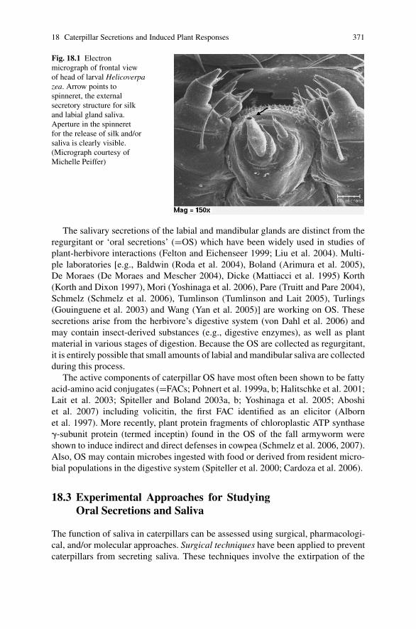
18 Caterpillar Secretions and Induced Plant Responses 371
Fig. 18.1 Electronmicrograph of frontal viewof head of larval Helicoverpazea. Arrow points tospinneret, the externalsecretory structure for silkand labial gland saliva.Aperture in the spinneretfor the release of silk and/orsaliva is clearly visible.(Micrograph courtesy ofMichelle Peiffer)
The salivary secretions of the labial and mandibular glands are distinct from theregurgitant or ‘oral secretions’ (=OS) which have been widely used in studies ofplant-herbivore interactions (Felton and Eichenseer 1999; Liu et al. 2004). Multi-ple laboratories [e.g., Baldwin (Roda et al. 2004), Boland (Arimura et al. 2005),De Moraes (De Moraes and Mescher 2004), Dicke (Mattiacci et al. 1995) Korth(Korth and Dixon 1997), Mori (Yoshinaga et al. 2006), Pare (Truitt and Pare 2004),Schmelz (Schmelz et al. 2006), Tumlinson (Tumlinson and Lait 2005), Turlings(Gouinguene et al. 2003) and Wang (Yan et al. 2005)] are working on OS. Thesesecretions arise from the herbivore’s digestive system (von Dahl et al. 2006) andmay contain insect-derived substances (e.g., digestive enzymes), as well as plantmaterial in various stages of digestion. Because the OS are collected as regurgitant,it is entirely possible that small amounts of labial and mandibular saliva are collectedduring this process.
The active components of caterpillar OS have most often been shown to be fattyacid-amino acid conjugates (=FACs; Pohnert et al. 1999a, b; Halitschke et al. 2001;Lait et al. 2003; Spiteller and Boland 2003a, b; Yoshinaga et al. 2005; Aboshiet al. 2007) including volicitin, the first FAC identified as an elicitor (Albornet al. 1997). More recently, plant protein fragments of chloroplastic ATP synthase�-subunit protein (termed inceptin) found in the OS of the fall armyworm wereshown to induce indirect and direct defenses in cowpea (Schmelz et al. 2006, 2007).Also, OS may contain microbes ingested with food or derived from resident micro-bial populations in the digestive system (Spiteller et al. 2000; Cardoza et al. 2006).
18.3 Experimental Approaches for StudyingOral Secretions and Saliva
The function of saliva in caterpillars can be assessed using surgical, pharmacologi-cal, and/or molecular approaches. Surgical techniques have been applied to preventcaterpillars from secreting saliva. These techniques involve the extirpation of the
372 G.W. Felton
salivary glands or cauterization of the spinneret to prevent salivation. Wroniszewska(1966) surgically removed the mandibular glands of Galleria mellonella and demon-strated that the loss of the glands causes a considerable decrease in the growthrate of larvae, although the specific cause for slower growth rates was not noted.Similarly Mossadegh (1978) surgically removed mandibular glands to examine therole of their secretions on silk filaments, and also cauterized the spinnerets (usinga heated needle) to prevent silk deposition. Cauterization of the spinneret has alsobeen used to examine the role of silk in trail following behavior in social caterpillarspecies (Fitzgerald 1993, 2003). Cauterization of the spinneret and surgical removalof the labial glands have been used to examine the function of labial saliva (Musseret al. 2002, 2005a, b, 2006; Bede et al. 2006) We have recently used a medicalcauterization tool which more consistently accomplishes a uniform cauterization(Peiffer and Felton 2005). Machida (1965) removed the Lyonet’s gland (Filippi’sgland) or severed its duct to determine the impact of the gland on silk secretion.
Surgical removal of salivary glands, particularly the mandibular glands that areoften relatively small, requires a high degree of skill to avoid killing larvae. Fur-thermore, the interpretation of results using surgically invasive procedures shouldbe cautioned because rupture of the cuticle causes a massive cascade of woundresponses (Jarosz 1993; Han et al. 1999). Cauterization of the spinneret avoids thisinvasive procedure, but is limited to examining labial gland secretions. In somespecies (e.g., Pieris rapae) cauterization may adversely affect larval feeding be-havior (Musser et al. 2006). Despite the robust size of Manduca sexta larvae, theirspinnerets are comparatively minute and difficult to cauterize without damaging themouthparts (Felton lab, personal observations).
Pharmacological techniques include directly treating plant tissues with OS orsalivary secretions. Typically OS is collected by inducing larvae to regurgitate whichis then applied to artificial plant wounds. Saliva has been prepared by surgical re-moval and homogenization of salivary glands with subsequent assay of their biolog-ical and/or chemical properties. These techniques have been widely used to studysalivary gland components in blood-feeding arthropods (Valenzuela 2002; Ribeiroet al. 2004) and in herbivorous insects (Musser et al. 2002; Shackel et al. 2005).Unfortunately, in many cases it has not been determined which components of OSor salivary gland homogenates are normally secreted during feeding. Without fur-ther studies showing that these components are actually deposited during feeding,pharmacological studies are of restricted value.
Fortunately, several approaches are available to identify secreted components.First, collection of saliva can be made directly from the spinneret (Liu et al. 2004)or saliva that is deposited on a feeding substrate can be collected and analyzed. Thelimitation of this approach is that at present only very minute quantities (ca. <1 nl)of saliva can be collected from individual larvae (personal observations). However,with greatly enhanced sensitivity of chemical and biochemical techniques, this isno longer a major impediment for analyses. Collecting saliva from a substrate af-ter feeding suffers from not knowing where the collected substance was secretedfrom—e.g., labial glands, mandibular glands, regurgitant, or other exocrine glands.Also, proteins may be degraded because of the presence of co-secreted proteases. In
18 Caterpillar Secretions and Induced Plant Responses 373
the case of the salivary enzyme glucose oxidase, we used a combination of molecu-lar and surgical techniques to demonstrate that the enzyme was produced primarilyin the labial glands with minor contribution from the mandibular glands (Eichenseeret al. 1999). Also, use of an antibody specific for glucose oxidase allowed us to notonly quantify the amount of enzyme secreted, but also to visualize where on the leafsurface caterpillars were secreting their saliva (Peiffer and Felton 2005).
To my knowledge, this approach has not been used to determine if specific com-ponents of OS are secreted. In one elegant example, Pare and colleagues labeledcorn seedlings with 14CO2 in situ, and Spodoptera exigua larvae were then allowedto feed on the labeled leaves to determine the production of volicitin. The OS wascollected from third-instars and contained approximately 120 pmol volicitin perlarva. When radio-labeled larvae were placed on unlabeled leaves, the amount ofvolicitin introduced to the feeding site was estimated at 100 pmol/larva (Truitt andPare 2004).
Finally molecular methods have proven extremely powerful for providing awealth of information about arthropod saliva (Valenzuela 2002). Preparation andhigh-throughput sequencing of full length cDNA salivary gland libraries combinedwith functional genomics will vastly improve our understanding of salivary glandfunction. More recently, proteomic approaches have been employed to identify pro-teins that are produced in the glands, many of which had eluded identification bystandard biochemical assays (Valenzuela 2002). Analysis of molecular sequencesby PSORT analysis (http://psort.hgc.jp/form2.html) can provide strong evidencefor proteins that may be secreted which then can be further verified by detec-tion with antibodies. RNA interference (RNAi) techniques are being employedto suppress specific salivary genes and then to examine their function (Aljamaliet al. 2003; Karim et al. 2004; Mutti et al. 2006). Although RNAi is an extremelypowerful tool for loss-of-function analysis, there are concerns over the possiblepleiotropic effects. If direct injection of the interfering RNAs into the hemocoelis required, the induction of undesired wound or immune responses may compli-cate interpretation of results. In other cases, specific expression of transgenes inthe salivary glands has been achieved using a vector such as the piggyBac vector(Royer et al. 2005), and offers great potential for understanding function of geneproducts.
18.4 Oral Secretions and Induced Defenses
Considerable progress has been made in the last decade in identifying elicitors of in-duced defenses in the oral secretions of caterpillars. Several groups of elicitors havebeen found including �-glucosidase (Mattiacci et al. 1995), FACs such as volicitin(Alborn et al. 1997), and most recently chloroplastic peptide fragments termed in-ceptins (Schmelz et al. 2006, 2007). The inceptins are formed when S. frugiperdaattack cowpea and partially digest a chloroplast protein, ATP-synthase. How plantsrecognize these herbivore-specific elicitors, and use defense signal transduction

374 G.W. Felton
pathways to regulate direct and indirect defenses is not well understood. CaterpillarOS (including FACs) are known to elicit bursts of the signaling compounds jasmonicacid and ethylene (Kahl et al. 2000). The inceptins trigger increases in ethylene,jasmonic acid, and salicylic acid in cowpea (Schmelz et al. 2006). Recently it wasshown in N. attenuata, that M. sexta OS/FACs activate MAP-kinases (an SIPK andWIPK; see Stratmann (this volume) for a more detailed discussion of the role ofthese two MPKs in defense signaling), which are upstream components regulat-ing salicylic acid, jasmonic acid, and ethylene biosynthesis (Wu et al. 2007). OSand FACs subsequently cause changes in gene expression (Korth and Dixon 1997;Halitschke et al. 2003; Roda et al. 2004), protein expression (Giri et al. 2006), andproduction of volatile organic compounds, as well as other secondary metabolites(Roda et al. 2004; von Dahl et al. 2006).
Most often OS has been shown to induce indirect defenses (Turlings et al. 1993;Rose and Tumlinson 2005). I refer the reader to Chapter 19 by Tumlinson andEngelberth for a more extensive coverage of FACs. The application of FACs towound sites frequently, but not always, mimics that observed with insect feeding.Interestingly, in the case of the S. frugiperda and cowpea interaction, the FACs areinactive as elicitors (Schmelz et al. 2006). Additional studies in particular systemsindicate that OS may not always play a major role in the indirect defenses. In oneexample, the generalist parasitoid Campoletis chlorideae, did not differentiate be-tween maize seedlings infested by the two different noctuid insects (Helicoverpaarmigera and Pseudaletia separata; Yan and Wang 2006). The parasitoid did notshow a difference in attraction to mechanically damaged plants treated with cater-pillar OS or water, although clear differences in volatile emissions were observedamong the various treatments (Yan and Wang 2006). In some instances, tobaccoleaves artificially damaged and then treated with caterpillar OS (H. armigera andHelicoverpa assulta) emitted the same levels of volatiles as leaves treated with water(Yan et al. 2005).
Moreover, although OS may elicit the production of volatiles, their productionmay not always result in enhanced plant defense. Besides serving as a cue for theattraction of natural enemies, volatiles may also attract other herbivores as wasshown in potato where Colorado potato beetles were more attracted to damagedplants (Bolter et al. 1997) or plants treated with beetle or cabbage looper OS than toundamaged plants (Landolt et al. 1999). Similarly fall armyworm larvae were moreattracted to odors from herbivore-damaged plants than undamaged plants (Carrollet al. 2006; but see De Moraes et al. 2001). These results suggest that althoughinduced volatiles may attract natural enemies of herbivores, they may inadvertentlyrecruit herbivores to damaged plants in some instances (Carroll et al. 2006). It is thusparticularly important to examine the role of induced volatiles in natural ecosystems(Kessler and Baldwin 2001). There are still other examples, where OS do not induceindirect but rather direct defenses (Korth and Dixon 1997; Roda et al. 2004; Majorand Constabel 2006; Schmelz et al. 2006). Furthermore, in N. attenuata, OS fromM. sexta was shown to suppress an inducible direct defense, the production of thealkaloid nicotine (Winz and Baldwin 2001). This finding may serve as an exampleof how the herbivore tries to escape plant defenses.

18 Caterpillar Secretions and Induced Plant Responses 375
18.5 Saliva and Induced Defenses
Considering the prominent role that saliva plays in host evasion by blood-feedingarthropods, it would not be surprising if herbivorous insects employed a similarcounter defense strategy. Our laboratory began studying the role of saliva in evad-ing induced defenses using the generalist noctuid H. zea as a model. We used bothcauterization of the spinneret and application of salivary gland homogenates to de-termine that labial gland saliva from H. zea mitigates induced resistance and theinduction of a direct defense (i.e., nicotine) in tobacco Nicotiana tabacum (Musseret al. 2002, 2005). Further evidence for saliva suppressing induced resistance wasobtained by surgical removal of salivary glands (Musser et al. 2006). Fractionationof the salivary gland homogenate showed that glucose oxidase (GOX) was primarilyresponsible for suppression of resistance (Musser et al. 2002). GOX is an abundantprotein in H. zea saliva and is expressed at highest levels during larval feeding(Eichenseer et al. 1999). The enzyme mediates the following reaction:
�-D-glucose + O2 → D-glucono-1,5-lactone + H2O2
The product of the reaction, H2O2, appears to have the most activity in sup-pression. Zong and Wang (2004) examined the role of saliva from labial glandhomogenates of three noctuid species, Helicoverpa armigera, Helicoverpa assulta,and Spodoptera litura in inducing nicotine production in tobacco. They reportedthat feeding by the two Helicoverpa species, or application of their labial glandhomogenates to plant wounds suppressed nicotine. GOX concentrations are excep-tionally high in H. armigera and somewhat lower in H. assulta.. In contrast, feedingby S. litura or application of its labial gland homogenates in which GOX was notdetectable, failed to suppress nicotine induction. Based upon these results and ex-periments with fungal GOX applied to wounded leaves, the authors concluded thatsalivary GOX (and H2O2) plays an important role in the suppression of wound-induced nicotine (Zong and Wang 2004).
GOX is found in many noctuid caterpillar species (Eichenseer and Felton, unpub-lished data) and may function in defense suppression in other systems. For instance,when tobacco leaves are fed on by tobacco budworms Heliothis virescens, there isno detectable volatile nicotine release; however if their spinnerets are ablated, then asignificant burst of volatile nicotine is observed (Delphia et al. 2006). The budwormhas comparatively high levels of GOX, but its role in suppressing the release ofvolatile nicotine was not tested in this case. Ablation of the spinnerets in the beetarmyworm Spodoptera exigua revealed that their saliva suppresses transcript levelsof two genes encoding early enzymes in terpenoid biosynthesis in Medicago trun-catula (Bede et al. 2006). GOX, which is found in the saliva of S. exigua, similarlycaused reduction in the transcript levels of these genes. Whether or not the reductionin these transcript levels is sufficient to alter the production of volatiles that mayfunction as indirect defenses is still unknown.
Further studies with H. zea suggest that the role of saliva in evading host defensesis more complex than what we previously thought. In the case of tomato, another

376 G.W. Felton
host of H. zea, GOX has a profoundly different effect than in tobacco. In this case,application of fungal GOX (and the resulting production of H2O2) causes a massiveincrease in defenses such as proteinase inhibitors (Orozco-Cardenas et al. 2001).We have confirmed these findings by applying purified GOX to mechanically-damaged leaves and showing that it significantly increases transcript levels ofproteinase inhibitor-II (PI-II) genes as measured by quantitative real time PCR(qRT-PCR) (Peiffer and Felton, unpublished data). Thus we expected that applica-tion of saliva (collected from the spinneret) would cause similar increases when ap-plied to mechanically-damaged leaves; even very small quantities of saliva (<1 nl)strongly induce PI-II expression (unpublished data). These findings may providean example of how a virulence factor of the insect (GOX and H2O2, respectively)can be exploited by the plant as an elicitor to induce appropriate counter measures(induction of proteinase inhibitors in tomato).
18.6 Dynamic Nature of Secretions
There may be considerable phenotypic variability in the composition of caterpillarsecretions which is dependent upon developmental and environmental influences.The developmental stage of the larva can have a profound effect on the compo-sition of the saliva. The synthesis of some salivary gland proteins such as GOXand lysozyme reaches maximal levels during the feeding interval of each molt(Eichenseer et al. 1999; Liu et al. 2004), whereas others such as chitinase reach peaklevels after larvae cease feeding and begin to molt (Zheng et al. 2003). The temporalexpression of a salivary gland protein may reveal important cues as to whether theprotein is secreted during feeding. As larvae reach the non-feeding prepupal stage,the labial glands undergo apoptosis and synthesis of most proteins ceases (Jochovaet al. 1997). Furthermore, some cellular proteins may be sloughed into the salivaduring this developmental stage.
Not only the stage of larval development, but also the diet of the larva canstrongly impact the composition and secretion of saliva. Labial GOX (and totalprotein of the gland) in H. zea was much higher in larvae reared on tobacco, ascompared to tomato or cotton (Peiffer and Felton 2005). The use of a GOX-specificantibody allowed direct quantification of GOX secreted onto the leaf surface, and itwas found that larvae secreted variable amounts of GOX with a trend toward lesssecretion on tomato compared to the other two hosts (Peiffer and Felton 2005). Lastinstar larvae secreted between 0.4 and 0.7 microgram of GOX per hour (Peiffer andFelton 2005). In contrast to GOX, lysozyme mRNA was much lower in tobacco-fedlarvae compared to those fed cotton or tomato (Liu et al. 2004). These findingsindicate that the amounts of specific proteins in the saliva are regulated differen-tially, and are not merely a function of the total protein contained in the glands.Labial glands from tobacco fed larvae are highest in protein, but lysozyme levelswere lowest (Peiffer and Felton 2005). Whether the differential rates of secretionfor different proteins on varying hosts have any adaptive value should be examined.

18 Caterpillar Secretions and Induced Plant Responses 377
The activity of labial GOX in the beet armyworm S. exigua and the bertha army-worm Mamestra configurata was much greater in artificial diet-fed larvae comparedto those ingesting the legume Medicago truncatula (Merkx-Jacques and Bede 2004,2005). In contrast, lysozyme expression was barely detectable in artificial diet fed-Hzea compared to those reared on host plants (Liu et al. 2004). These studies providean important precautionary note on generalizing the function of saliva from larvaereared on artificial diets.
The host plant has a profound impact on not only the composition and secretionof saliva, but also on the size of the salivary glands. Eichenseer et al. (2002) reportedthat the host plant of H. zea significantly affected the size of mandibular glands, aswell as the amount of carotenoids present in their tissues. Mandibular glands insoybean-fed larvae were 2-fold larger in length and/or diameter compared to larvaereared on tomato or cotton, despite the larvae being of identical developmental stage.The increased size of the glands is presumably due to increased cell size, becausein B. mori, the cell number of both labial and mandibular glands remain constant innumber (330 cells), yet the glands increase 1000x-fold in size during development(Parthasarathy and Gopinathan 2005).
Also the composition of OS may differ depending upon the food ingested. Thefact that different hosts respond differentially to M. sexta feeding, not only indicatesthat there may be host-specific differences in defense response, but also suggests thatthe composition of secretions may vary when feeding on different hosts (Schmidtet al. 2005). While different dietary sources of M. sexta or H. virescens larvae, in-cluding both host and nonhost plants, did not affect the amino acid compositionof the FACs in OS (Alborn et al. 2003), quantitative differences or differences inother OS components may occur. For example, Heliothis subflexa larvae fail to pro-duce volicitin if they feed exclusively on the fruits of Physalis angulata which lacklinolenic acid, a necessary component of the FAC (De Moraes and Mescher 2004).Only fall armyworm Spodoptera frugiperda larvae that ingested chloroplastic ATPsynthase �-subunit proteins and produced the peptide fragments termed ‘inceptins’significantly induced defenses in cowpea (Schmelz et al. 2006, 2007).
18.7 Saliva and Oral Secretions Should be ExaminedConcurrently
Oral secretions alone may not fully explain the specificity of caterpillar-inducedresponses. For instance, S. exigua feeding on maize inhibited the expression of somevolicitin-induced genes, despite the presence of volicitin in their OS (Lawrence andNovak 2004). This and other results suggest that additional components of caterpil-lar secretions may be involved in these responses. The Korth lab has examined theimpact of herbivory, OS, and saliva on genes controlling terpene production in themodel system of Medicago truncatula. They found that feeding by S. exigua, OS, orFACs (N-linolenoyl-glutamate or N-linoleoyl-glutamate) increased transcript levelsof a putative terpene synthase (MtTPS; Gomez et al. 2005). In a separate paper,

378 G.W. Felton
their lab examined the effect of saliva on the terpene biosynthesis pathway, and theyfound that transcripts coding for early pathway enzymes were lower in tissues thatsuffered herbivory (Bede et al. 2006). Larvae were ablated as previously described(Musser et al. 2002), and the authors concluded that a salivary factor, possibly GOX,was involved in reducing the transcript levels (Bede et al. 2006). These two studiesillustrate, that in addition to wounding, both insect OS and saliva are important inultimately determining the plant’s defensive response.
However, in some caterpillar/host plant systems, saliva and/or GOX may not beequally important. A microarray of 241-genes from Nicotiana attenuata differen-tially expressed in response to attack by M. sexta was used to compare the transcrip-tional response to FACs from Manduca regurgitant and fungal GOX (Halitschkeet al. 2003). GOX (and glucose) injection of unwounded leaf tissue up-regulated37 transcripts and down-regulated 41 transcripts within a 2 h period; howeveronly a handful of these genes were similarly regulated 1 h after treatment ofwounded leaves with Manduca regurgitant. The authors concluded that GOX-derived H2O2 plays only a minor role in elicitation of Manduca-specific plant re-sponses (Halitschke et al. 2003). Also salivary gland homogenates (both labial andmandibular) from M. sexta failed to amplify trypsin inhibitor expression in its hostN. attenuata (Roda et al. 2004). It is not surprising that fungal GOX did not producesimilar M. sexta-specific plant responses, considering that this species has virtuallyno detectable salivary GOX (Eichenseer and Felton, unpublished data).
Delphia et al. (2006) explored the role of H. virescens OS and salivary secre-tions on volatile induction in the commercial tobacco species, Nicotiana tabacum.Using the ablation technique previously described to prevent the release of labialsaliva, plants damaged by intact caterpillars were found to release 11 volatile com-pounds, whereas ablated caterpillars induced the same 11 compounds plus an addi-tional eight. Furthermore, plants damaged by ablated caterpillars generally releasedgreater amounts of volatile compounds, most notably volatile nicotine, as comparedto plants damaged by intact caterpillars. Apparently, the application of both salivaand OS induced less volatile nicotine as compared to the treatment with OS alone.These results show that both OS and saliva are necessary to produce the volatileprofile of H. virescens feeding on tobacco, and that saliva may mask the effect ofFACs found in the OS (Delphia et al. 2006). These studies suggest that herbivoresmay minimize their display of elicitors—either through minimizing the productionof elicitors or by masking their effects through secretion of salivary components.
18.8 Saliva and Oral Secretions and Host Range
Induced plant responses to insect herbivores are a widespread phenomenon; the de-gree to which these responses are specific to a given herbivore has been studied ina variety of examples (Hartley and Lawton 1987; Felton et al. 1994; Karban andBaldwin 1997; Stout et al. 1998; Agrawal 2000; Van Zandt and Agrawal 2004).The question whether herbivore diet breadth (or host specialization) is a factor in
18 Caterpillar Secretions and Induced Plant Responses 379
specificity has been examined in a few instances. The specificity of induction by twospecialist (narrow host range) and two generalist (broad host range) caterpillars wascompared in wild radish, where no general patterns in specificity between specialistsand generalists were observed (Agrawal 2000). The biochemical or molecular basesof the induced responses were not determined in this study.
A few representative examples where oral secretions and the host range of theherbivore may be important in determining the specificity of induced responses willbe presented. One example is ground cherry Physalis angulata, where the specialistHeliothis subflexa larvae suffers lower rates of parasitism by Cardiochiles nigricepsthan the extreme generalist H. virescens (Oppenheim and Gould 2002). The strengthof this experimental system is that it is a natural system, and that the two Heliothisspecies are closely related, sibling species representing both a generalist and a spe-cialist herbivore. H. subflexa larvae feed on ground cherry fruits lacking linolenicacid, a key component of volicitin, an elicitor of indirect defenses (De Moraes andMescher 2004). Volicitin is absent in the oral secretions of fruit-feeding caterpillarsand thus, the volatile profiles of plants induced by fruit feeding differ from thoseinduced by leaf feeding (De Moraes and Mescher 2004). In contrast, the general-ist H. virescens is unable to complete development on the fruit due to the lack oflinolenic acid. In this system H. subflexa gains a selective advantage by feedingon fruit, avoiding the production of an elicitor, and ultimately avoiding parasitism.Oppenheimer and Gould (2002) indicated that differences in parasitoid attractionto herbivore-induced plant volatiles did not contribute to H. subflexa’s compara-tively low parasitism rate, although their focus was on induction of volatiles byleaf-feeding. Continued study in this system is warranted.
The availability of microarrays for a growing number of plant species providespowerful tools for examining the specificity of induced responses to herbivores inthe context of host breadth. Herbivore host range and plant transcriptional responseshave been studied in the wild tobacco species Nicotiana attenuata where it wasfound that the transcriptional responses to feeding by the Solanaceous specialistManduca sexta were distinct from feeding by the generalists Heliothis virescensand Spodoptera exigua (Voelckel and Baldwin 2004). Differences in plant responseswere correlated with the profile of FAC elicitors in the regurgitant (Voelckel andBaldwin 2004); however the authors did not consider the possible contribution ofsalivary factors such as GOX (Voelckel and Baldwin 2004). We found that GOXis at high levels in the noctuid species (H. virescens and S. exigua), but virtuallyundetectable in M. sexta, as determined by enzyme activity and reactivity to anantibody (Eichenseer and Felton, unpublished data). Interestingly, the number ofdown-regulated genes correlates well with the presence of GOX: plants fed on byS. exigua and H. virescens had more down-regulated genes than plants fed on by M.sexta (Voelckel and Baldwin 2004).
The power of using the N. attenuata-Manduca system is that it is a natural sys-tem that has been extensively studied in terms of induced responses to herbivores(Baldwin et al. 2001; Kessler and Baldwin 2002). However, it is limited in thatthe microarray comprised only a restricted set of 240 M. sexta-responsive genes(Voelckel and Baldwin 2004). Arabidopsis offers a distinct advantage because of

380 G.W. Felton
the availability of a large, unbiased microarray containing in excess of 7200 uniquegenes. Using this microarray, Reymond et al. (2004) found that there were fewdifferences in the transcript profiles between plants fed on by Pieris rapae (nar-row host range) and Spodoptera littoralis (broader host range). The authors foundthat Pieris OS induced several genes not regulated by jasmonic acid and with fur-ther experiments concluded that the FACs are not responsible for the induction ofPieris-responsive genes (Reymond et al. 2004); standing in direct contrast to thestudy cited above (Voelckel and Baldwin 2004). The lack of differences betweenresponses to Pieris and Spodoptera is surprising, considering the likely differencesin both OS and saliva between these two species. However both species appear tolack salivary GOX. We were unable to detect GOX in Pieris larvae (Eichenseerand Felton, unpublished data) and it was also undetectable in S. littoralis (Zongand Wang 2004). Reymond et al. (2004) also reported that mechanical woundinginduces several genes that are not activated by Pieris feeding, suggesting that OS orsaliva may be responsible for suppression as shown with H. zea and tobacco (Musseret al. 2002). Nevertheless the transcript profiles show surprisingly few differencesbetween feeding by S. littoralis and P. rapae.
These cited studies illustrate the difficulty in generalizing across different hostplant-herbivore systems. Even M. sexta, a specialist on Solanaceous plants, elicitsdistinct transcript profiles when feeding on different hosts such as N. attenuata orblack nightshade Solanum nigrum (Schmidt et al. 2005). There are several possi-ble explanations for these disparate findings including that composition of salivaor OS may differ when caterpillars change host plants, thus influencing the induc-tion and/or suppression of defenses. Alternatively, there may be differences in howparticular plant species (or even cultivars) recognize herbivore secretions.
18.9 Future Directions and Perspectives
There are several critical needs that will shape the future direction of researchon herbivore secretions and induced responses. First, considering that insect her-bivore species likely number in excess of a million, the secretions of a preciousfew insect herbivores have been characterized. Improvements in proteomics andmetabolomics allowing the analysis of minute volumes will help uncover the bio-chemical and functional complexity of caterpillar secretions. A continued arrayof novel compounds will undoubtedly be identified as more species are examined(e.g., grasshoppers; Tumlinson and Engelberth, this volume).
With a few notable exceptions (Mattiacci et al. 1995; Schmelz et al. 2006, 2007),chemical characterization of Lepidopteran OS has mostly focused on FACs, whereasvirtually all of the research on saliva in larval Lepidoptera has focused on proteins.As far as I am aware, very little is known regarding small molecules present in theirsaliva (Howard and Baker 2004), although some have been reported in the salivaryglands: ascorbic acid (Kramer et al. 1981), carotenoids (Eichenseer et al. 2002),and even jasmonic acid (Tooker and De Moraes 2006) have been found at high

18 Caterpillar Secretions and Induced Plant Responses 381
levels in salivary glands; however it is unknown if these molecules are secreted.Jasmonic acid secretion would be of obvious significance in eliciting plant defenses.The most difficult experimental challenge has been collecting a sufficient volumeof saliva necessary for biochemical analyses, and for conducting pharmacological-type assays with plants. Nevertheless, the technology is now available to biochemi-cally characterize the small molecules present in saliva using HPLC/MS or GC/MSmethods.
Second, very few studies have included a dose-response analysis of OS or salivawhen determining effects on induced responses. There have been an inexplicablenumber of studies that have used an identical quantity of OS for testing, regardless ofthe relevance of species or stage of insect used. There needs to be greater emphasison quantifying the amount of specific elicitors released during feeding in order tomake further progress.
Third, there is the need for understanding the adaptive value of insect elicitors.If elicitation of induced indirect and direct defenses negatively impacts the fitnessof insect herbivores as proposed, then there should be strong selective pressure onherbivores to evade these host defenses. Several scenarios are possible includingminimizing the display of elicitors—achieved by producing less of the elicitor, orby masking the effect of the elicitor with other oral or salivary components (Delphiaet al. 2006). However, there may be physiological constraints on minimizing theproduction of elicitors if they perform vital physiological functions. In the case ofFACs their function(s) are unknown, but they may act as gut surfactants (Spitellerand Boland 2003b) and/or facilitate nutrient uptake. Elucidation of the genes reg-ulating their biosynthesis will facilitate the application of molecular approachessuch as RNA interference to examine their function in vivo. Another strategy forevading host defenses is to elicit a competing pathway—one that regulates negativecross talk with the jasmonate pathway (e.g., salicylate). The latter has been observedwith several pathovars of the bacterial pathogen Psuedomonas syringae that secretea jasmonate mimic, coronatine, which activates the jasmonate pathway at the ex-pense of salicylate-dependent systemic resistance (Nomura et al. 2005; Uppalapatiet al. 2005; Zhao et al. 2003). Whether herbivores employ such a strategy is un-known, but it has been suggested by some data (Musser et al. 2002).
Fourth, there is a need to understand the mode-of-action and target sites of elic-itors. Very little is understood at the molecular level about how plants recognizeelicitors such as FACs and then activate transcriptional responses. Evidence for anelicitor-protein interaction was presented from work on maize laves where it wasshown that a plasma membrane fractions bind volicitin (Truitt et al. 2004). Treat-ment of leaves with methyl jasmonate also enhanced the binding of volicitin tothe plasma membrane fraction. Further study is needed to determine if this plasmamembrane protein(s) functions as a membrane transporter and/or as a receptor toactivate secondary messenger cascades.
Finally, an additional glandular structure may be important in secretion duringcaterpillar feeding. Noctuids are ostensibly the largest family of Lepidoptera; oneof their defining larval characters is the ventral eversible gland (=VEG), a secre-tory structure that is found on the ventral surface of the larval thorax (Fig. 18.2;

382 G.W. Felton
Fig. 18.2 Light micrographof lateral view of head oflarval Helicoverpa zea.VEG = ventral eversiblegland, in distended view.(Micrograph courtesy ofMichelle Peiffer)
Speidel et al. 1996). The function of this gland is mostly unknown, but it is knownto secrete various lipids (Severson et al. 1991) and proteins (unpublished data). Con-sidering its close proximity to the mandibles and buccal cavity when distended, itssecretions would presumably be deposited onto the food substrate. Additionally,its secretions could be expected in regurgitant or OS when it is collected. We arein the initial stages of analyzing the VEG components, and testing their effect oninduced responses. Our preliminary findings indicate that these secretions will addyet another layer of complexity to the dynamic interactions between caterpillars andtheir host plants.
Acknowledgments The support of research by the National Science Foundation and the UnitedStates Department of Agriculture NRI programs is gratefully acknowledged. Unpublished datafrom Michelle Peiffer and Herb Eichenseer is appreciated.
References
Aboshi T, Yoshinaga N, Noge K, Nishida R, Mori N (2007) Efficient incorporation of unsaturatedfatty acids into volicitin-related compounds in Spodoptera litura (Lepidoptera: Noctuidae).Biosci Biotech Biochem 71:607–610
Agrawal AA (2000) Specificity of induced resistance in wild radish: causes and consequences fortwo specialist and two generalist caterpillars. Oikos 89:493–500
Alborn HT, Brennan MM, Tumlinson JH (2003) Differential activity and degradation of plantvolatile elicitors in regurgitant of tobacco hornworm (Manduca sexta) larvae. J Chem Ecol29:1357–1372
Alborn HT, Turlings TCJ, Jones TH, Stenhagen G, Loughrin JH, Tumlinson JH (1997) An elicitorof plant volatiles from beet armyworm oral secretion. Science 276:945–949
Aljamali MN, Bior AD, Sauer JR, Essenberg RC (2003) RNA interference in ticks: a study usinghistamine binding protein dsRNA in the female tick Amblyomma americanum. Insect Mol Biol12:299–305

18 Caterpillar Secretions and Induced Plant Responses 383
Arimura G, Kost C, Boland W (2005) Herbivore-induced, indirect plant defences. Biochim Bio-phys Acta 1734:91–111
Baldwin IT, Halitschke R, Kessler A, Schittko U (2001) Merging molecular and ecological ap-proaches in plant-insect interactions. Curr Opin Plant Biol 4:351–358
Bede J, Musser R, Felton G, Korth K (2006) Caterpillar herbivory and salivary enzymes decreasetranscript levels of Medicago truncatula genes encoding early enzymes in terpenoid biosynthe-sis. Plant Mol Biol 60:519–531
Bolter CJ, Dicke M, vanLoon JJA, Visser JH, Posthumus MA (1997) Attraction of Colorado potatobeetle to herbivore-damaged plants during herbivory and after its termination. J Chem Ecol23:1003–1023
Bordas L (1903) Mandibular glands of lepidoptera larvae. C R Hebd Seances Acad Sci 136:1273–1275
Cardoza YJ, Klepzig KD, Raffa KF (2006) Bacteria in oral secretions of an endophytic insectinhibit antagonistic fungi. Ecol Entomol 31:636–645
Carroll MJ, Schmelz EA, Meagher RL, Teal PEA (2006) Attraction of Spodoptera frugiperdalarvae to volatiles from herbivore-damaged maize seedlings. J Chem Ecol 32:1911–1924
Chauthan AR, Callahan PS (1967) Developmental morphology of alimentary canal of Heliothiszea (Lepidoptera – Noctuidae). Ann Entomol Soc Amer 60:1136–1967
De Moraes CM, Mescher MC (2004) Biochemical crypsis in the avoidance of natural enemies byan insect herbivore. Proc Natl Acad Sci USA 101:8993–8997
De Moraes CM, Mescher MC, Tumlinson JH (2001) Caterpillar-induced nocturnal plant volatilesrepel conspecific females. Nature 410:577–580
Delphia C, Mescher M, Felton GW, De Moraes CM (2006) The role of insect-derived cuesin eliciting indirect plant defenses in tobacco, Nicotiana tabacum. Plant Signal Behav1:243–250
Eichenseer H, Mathews MC, Bi JL, Murphy JB, Felton GW (1999) Salivary glucose oxidase:multifunctional roles for Helicoverpa zea? Arch Insect Biochem Physiol 42:99–109
Eichenseer H, Murphy JB, Felton GW (2002) Sequestration of host plant carotenoids in the larvaltissues of Helicoverpa zea. J Insect Physiol 48:311–318
Felton GW, Eichenseer H (1999) Herbivore saliva and its effects on plant defense against herbi-vores and pathogens. In: Agrawal AA, Tuzun S, Bent E (eds) Induced plant defenses againstpathogens and herbivores. APS Press, St. Paul, pp 19–36
Felton GW, Summers CB, Mueller AJ (1994) Oxidative responses in soybean foliage to herbivoryby bean leaf beetle and 3-cornered alfalfa hopper. J Chem Ecol 20:639–650
Fereres A (2007) The role of aphid salivation in the transmission of plant viruses. Phytoparasitica35:3–7
Fitzgerald TD (1993) Trail and arena marking by caterpillars of Archips cerasivoranus (Lepi-doptera, Tortricidae). J Chem Ecol 19:1479–1489
Fitzgerald TD (2003) Role of trail pheromone in foraging and processionary behavior of pineprocessionary caterpillars Thaumetopoea pityocampa. J Chem Ecol 29:513–532
Giri AP, Wunsche H, Mitra S, Zavala JA, Muck A, Svatos A, Baldwin IT (2006) Molecular interac-tions between the specialist herbivore Manduca sexta (Lepidoptera, Sphingidae) and its naturalhost Nicotiana attenuata. VII. Changes in the plant’s proteome. Plant Physiol 142:1621–1641
Gomez SK, Cox MM, Bede JC, Inoue K, Alborn HT, Tumlinson JH, Korth KL (2005) Lepi-dopteran herbivory and oral factors induce transcripts encoding novel terpene synthases inMedicago truncatula. Arch Insect Biochem Physiol 58:114–127
Gouinguene S, Alborn H, Turlings TCJ (2003) Induction of volatile emissions in maize by differentlarval instars of Spodoptera littoralis. J Chem Ecol 29:145–162
Halitschke R, Gase K, Hui DQ, Schmidt DD, Baldwin IT (2003) Molecular interactions betweenthe specialist herbivore Manduca sexta (Lepidoptera, Sphingidae) and its natural host Nicotianaattenuata. VI. Microarray analysis reveals that most herbivore-specific transcriptional changesare mediated by fatty acid-amino acid conjugates. Plant Physiol 131:1894–1902
Halitschke R, Schittko U, Pohnert G, Boland W, Baldwin IT (2001) Molecular interactions betweenthe specialist herbivore Manduca sexta (Lepidoptera, Sphingidae) and its natural host Nicotiana

384 G.W. Felton
attenuata. III. Fatty acid-amino acid conjugates in herbivore oral secretions are necessary andsufficient for herbivore-specific plant responses. Plant Physiol 125:711–717
Han YS, Chun JS, Schwartz A, Nelson S, Paskewitz SM (1999) Induction of mosquito hemolymphproteins in response to immune challenge and wounding. Develop Comp Immunol 23:553–562
Hartley SE, Lawton JH (1987) Effects of different types of damage on the chemistry of birchfoliage, and the responses of birch feeding insects. Oecologia 74:432–437
Howard RW, Baker JE (2004) Stage-specific surface chemicals of Plodia interpunctella: 2-acyl-1,3-cyclohexanediones from larval mandibular glands serve as cuticular lipids. Comp BiochemPhysiol B: Biochem Mol Biol 138:193–206
Jarosz J (1993) Induction kinetics of immune antibacterial proteins in pupae of Galleria mellonellaand Pieris brassicae. Comp Biochem Physiol B Biochem Mol Biol 106:415–421
Jochova J, Quaglin D, Zakeri Z, Woo K, Sikorska M, Weaver M, Lockshin RA (1997) Protein syn-thesis, DNA degradation, and morphological changes during programmed cell death in labialglands of Manduca sexta. Dev Genet 21:249–257
Kahl J, Siemens DH, Aerts RJ, Gabler R, Kuhnemann F, Preston CA, Baldwin IT (2000)Herbivore-induced ethylene suppresses a direct defense but not a putative indirect defenseagainst an adapted herbivore. Planta 210:336–342
Karban R, Baldwin IT (1997) Induced responses to herbivory. The University of Chicago PressKarim S, Ramakrishnan VG, Tucker JS, Essenberg RC, Sauer JR (2004) Amblyomma americanum
salivary glands: double-stranded RNA-mediated gene silencing of synaptobrevin homologueand inhibition of PGE2 stimulated protein secretion. Insect Biochem Mol Biol 34:407–413
Kessler A, Baldwin IT (2001) Defensive function of herbivore-induced plant volatile emissions innature. Science 291:2141–2144
Kessler A, Baldwin IT (2002) Plant responses to insect herbivory: the emerging molecular analysis.Ann Rev Plant Biol 53:299–328
Korth KL, Dixon RA (1997) Evidence for chewing insect-specific molecular events distinct froma general wound response in leaves. Plant Physiol 115:1299–1305
Kramer KJ, Spiers RD, Lookhart G, Seib PA, Liang YT (1981) Sequestration of ascorbic acidby the larval labial gland and hemolymph of the tobacco hornworm, Manduca sexta L (Lepi-doptera, Sphingidae). Insect Biochem 11:93–96
Lait CG, Alborn HT, Teal PEA, Tumlinson JH (2003) Rapid biosynthesis of N-linolenoyl-L-glutamine, an elicitor of plant volatiles, by membrane-associated enzyme(s) in Manduca sexta.Proc Natl Acad Sci USA 100:7027–7032
Landolt PJ, Tumlinson JH, Alborn DH (1999) Attraction of Colorado potato beetle (Coleoptera:Chrysomelidae) to damaged and chemically induced potato plants. Environ Entomol 28:973–978
Lawrence SD, Novak NG (2004) Maize genes induced by herbivory and volicitin. J Chem Ecol30:2543–2557
Lev E (2003) Traditional healing with animals (zootherapy): medieval to present-day Levantinepractice. J Ethnopharmacol 85:107–118
Liu F, Cui L, Cox-Foster D, Felton GW (2004) Characterization of a salivary lysozyme in larvalHelicoverpa zea. J Chem Ecol 30:2439–2457
Macgown MW, Sikorowski PP (1982) Anatomy of the digestive system of Heliothis zea larvae.Mississippi Agricultural and Forestry Experiment Station Bulletin 11:1–15
Machida Y (1965) Studies on the silk glands in the silkworm, Bombyx mori L. Morphological andfunctional studies of Filippi’s glands in the silkworm. Science Bulletin of Faculty of Journal ofChemical Ecology Agriculture, Kyushu University 22:95–108
Major IT, Constabel CP (2006) Molecular analysis of poplar defense against herbivory: comparisonof wound- and insect elicitor-induced gene expression. New Phytol 172:617–635
Mattiacci L, Dicke M, Posthumus MA (1995) Beta-Glucosidase: an elicitor of herbivore-inducedplant odor that attracts host-searching parasitic wasps. Proc Natl Acad Sci USA 92:2036–2040
Merkx-Jacques M, Bede JC (2004) Caterpillar salivary enzymes: ‘eliciting’ a response. Phytopro-tection 85:33–37
18 Caterpillar Secretions and Induced Plant Responses 385
Merkx-Jacques M, Bede JC (2005) Influence of diet on the larval beet armyworm Spodopteraexigua, glucose oxidase. J Insect Sci 5:48
Miles PW (1999) Aphid saliva. Biological Reviews 74:41–85Miles PW, Harrewijn P (1991) Discharge by aphids of soluble secretions into dietary sources.
Entomol Exp Appl 59:123–134Mithofer A, Wanner G, Boland W (2005) Effects of feeding Spodoptera littoralis on lima bean
leaves. II. Continuous mechanical wounding resembling insect feeding is sufficient to elicitherbivory-related volatile emission. Plant Physiol 137:1160–1168
Mossadegh MS (1978) Mechanism of secretion of the contents of the mandibular glands of Plodiainterpunctella larvae. Physiol Entomol 3:335–340
Musser RO, Cipollini DF, Hum-Musser SM, Williams SA, Brown JK, Felton GW (2005a) Evi-dence that the caterpillar salivary enzyme glucose oxidase provides herbivore offense in solana-ceous plants. Arch Insect Biochem Physiol 58:128–137
Musser RO, Farmer E, Peiffer M, Felton GW (2006) Ablation of caterpillar labial salivary glands:technique for determining the role of saliva in insect-plant interactions. J Chem Ecol 32:981–992
Musser RO, Hum-Musser SM, Eichenseer H, Peiffer M, Ervin G, Murphy JB, Felton GW (2002)Herbivory: caterpillar saliva beats plant defences – A new weapon emerges in the evolutionaryarms race between plants and herbivores. Nature 416:599–600
Musser RO, Kwon HS, Williams SA, White CJ, Romano MA, Holt SM, Bradbury S, Brown JK,Felton GW (2005a) Evidence that caterpillar labial saliva suppresses infectivity of potentialbacterial pathogens. Arch Insect Biochem Physiol 58:138–144
Mutti NS, Park Y, Reese JC, Reeck GR (2006) RNAi knockdown of a salivary transcript leadingto lethality in the pea aphid, Acyrthosiphon pisum. J Insect Sci 6:38
Nomura K, Melotto M, He SY (2005) Suppression of host defense in compatible plant-Pseudomonas syringae interactions. Curr Opin Plant Biol 8:361–368
Oppenheim SJ, Gould F (2002) Is attraction fatal? The effects of herbivore-induced plant volatileson herbivore parasitism. Ecology 83:3416–3425
Orozco-Cardenas ML, Narvaez-Vasquez J, Ryan CA (2001) Hydrogen peroxide acts as a secondmessenger for the induction of defense genes in tomato plants in response to wounding, sys-temin, and methyl jasmonate. Plant Cell 13:179–191
Parthasarathy R, Gopinathan KP (2005) Comparative analysis of the development of the mandibu-lar salivary glands and the labial silk glands in the mulberry silkworm, Bombyx mori. Gene ExpPatterns 5:323–339
Peiffer M, Felton GW (2005) The host plant as a factor in the synthesis and secretion of salivaryglucose oxidase in larval Helicoverpa zea. Arch Insect Biochem Physiol 58:106–113
Pohnert G, Jung V, Haukioja E, Lempa K, Boland W (1999a) New fatty acid amidesfrom regurgitant of lepidopteran (Noctuidae, Geometridae) caterpillars. Tetrahedron 55:11275–11280
Pohnert G, Koch T, Boland W (1999b) Synthesis of volicitin: a novel three-component Wittigapproach to chiral 17-hydroxylinolenic acid. Chem Commun 12:1087–1088
Reymond P, Bodenhausen N, Van-Poecke RMP, Krishnamurthy V, Dicke M, Farmer EE (2004)A conserved transcript pattern in response to a specialist and a generalist herbivore. Plant Cell16:3132–3147
Ribeiro JMC, Andersen J, Silva-Neto MAC, Pham VM, Garfield MK, Valenzuela JG (2004) Ex-ploring the sialome of the blood-sucking bug Rhodnius prolixus. Insect Biochem Mol Biol34:61–79
Roda A, Halitschke R, Steppuhn A, Baldwin IT (2004) Individual variability in herbivore-specificelicitors from the plant’s perspective. Mol Ecol 13:2421–2433
Rose USR, Tumlinson JH (2005) Systemic induction of volatile release in cotton: how specific isthe signal to herbivory? Planta 222:327–335
Royer C, Jalabert A, Da Rocha M, Grenier AM, Mauchamp B, Couble P, Chavancy G (2005)Biosynthesis and cocoon-export of a recombinant globular protein in transgenic silkworms.Transgenic Res 14:463–472
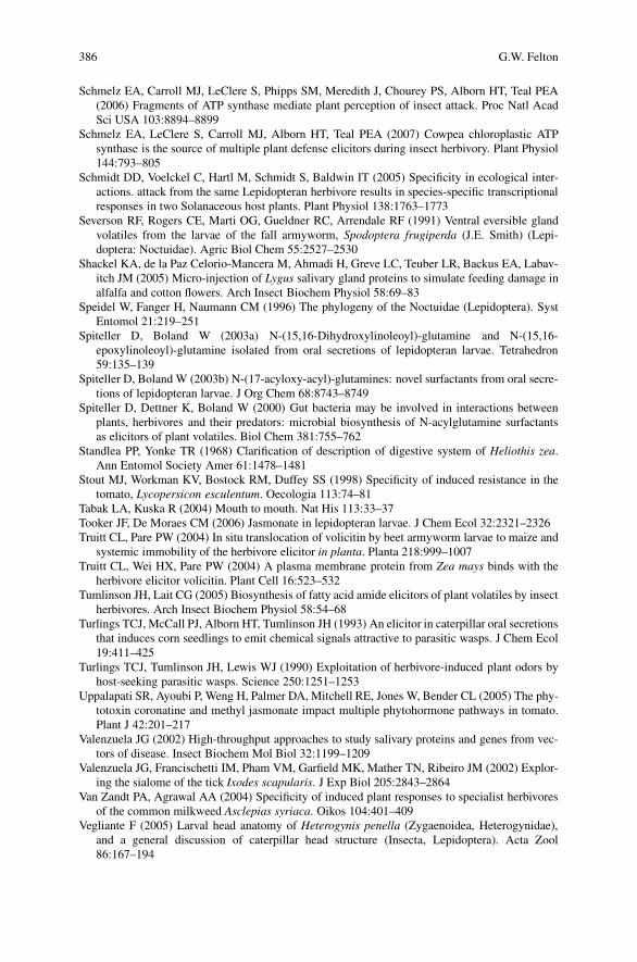
386 G.W. Felton
Schmelz EA, Carroll MJ, LeClere S, Phipps SM, Meredith J, Chourey PS, Alborn HT, Teal PEA(2006) Fragments of ATP synthase mediate plant perception of insect attack. Proc Natl AcadSci USA 103:8894–8899
Schmelz EA, LeClere S, Carroll MJ, Alborn HT, Teal PEA (2007) Cowpea chloroplastic ATPsynthase is the source of multiple plant defense elicitors during insect herbivory. Plant Physiol144:793–805
Schmidt DD, Voelckel C, Hartl M, Schmidt S, Baldwin IT (2005) Specificity in ecological inter-actions. attack from the same Lepidopteran herbivore results in species-specific transcriptionalresponses in two Solanaceous host plants. Plant Physiol 138:1763–1773
Severson RF, Rogers CE, Marti OG, Gueldner RC, Arrendale RF (1991) Ventral eversible glandvolatiles from the larvae of the fall armyworm, Spodoptera frugiperda (J.E. Smith) (Lepi-doptera: Noctuidae). Agric Biol Chem 55:2527–2530
Shackel KA, de la Paz Celorio-Mancera M, Ahmadi H, Greve LC, Teuber LR, Backus EA, Labav-itch JM (2005) Micro-injection of Lygus salivary gland proteins to simulate feeding damage inalfalfa and cotton flowers. Arch Insect Biochem Physiol 58:69–83
Speidel W, Fanger H, Naumann CM (1996) The phylogeny of the Noctuidae (Lepidoptera). SystEntomol 21:219–251
Spiteller D, Boland W (2003a) N-(15,16-Dihydroxylinoleoyl)-glutamine and N-(15,16-epoxylinoleoyl)-glutamine isolated from oral secretions of lepidopteran larvae. Tetrahedron59:135–139
Spiteller D, Boland W (2003b) N-(17-acyloxy-acyl)-glutamines: novel surfactants from oral secre-tions of lepidopteran larvae. J Org Chem 68:8743–8749
Spiteller D, Dettner K, Boland W (2000) Gut bacteria may be involved in interactions betweenplants, herbivores and their predators: microbial biosynthesis of N-acylglutamine surfactantsas elicitors of plant volatiles. Biol Chem 381:755–762
Standlea PP, Yonke TR (1968) Clarification of description of digestive system of Heliothis zea.Ann Entomol Society Amer 61:1478–1481
Stout MJ, Workman KV, Bostock RM, Duffey SS (1998) Specificity of induced resistance in thetomato, Lycopersicon esculentum. Oecologia 113:74–81
Tabak LA, Kuska R (2004) Mouth to mouth. Nat His 113:33–37Tooker JF, De Moraes CM (2006) Jasmonate in lepidopteran larvae. J Chem Ecol 32:2321–2326Truitt CL, Pare PW (2004) In situ translocation of volicitin by beet armyworm larvae to maize and
systemic immobility of the herbivore elicitor in planta. Planta 218:999–1007Truitt CL, Wei HX, Pare PW (2004) A plasma membrane protein from Zea mays binds with the
herbivore elicitor volicitin. Plant Cell 16:523–532Tumlinson JH, Lait CG (2005) Biosynthesis of fatty acid amide elicitors of plant volatiles by insect
herbivores. Arch Insect Biochem Physiol 58:54–68Turlings TCJ, McCall PJ, Alborn HT, Tumlinson JH (1993) An elicitor in caterpillar oral secretions
that induces corn seedlings to emit chemical signals attractive to parasitic wasps. J Chem Ecol19:411–425
Turlings TCJ, Tumlinson JH, Lewis WJ (1990) Exploitation of herbivore-induced plant odors byhost-seeking parasitic wasps. Science 250:1251–1253
Uppalapati SR, Ayoubi P, Weng H, Palmer DA, Mitchell RE, Jones W, Bender CL (2005) The phy-totoxin coronatine and methyl jasmonate impact multiple phytohormone pathways in tomato.Plant J 42:201–217
Valenzuela JG (2002) High-throughput approaches to study salivary proteins and genes from vec-tors of disease. Insect Biochem Mol Biol 32:1199–1209
Valenzuela JG, Francischetti IM, Pham VM, Garfield MK, Mather TN, Ribeiro JM (2002) Explor-ing the sialome of the tick Ixodes scapularis. J Exp Biol 205:2843–2864
Van Zandt PA, Agrawal AA (2004) Specificity of induced plant responses to specialist herbivoresof the common milkweed Asclepias syriaca. Oikos 104:401–409
Vegliante F (2005) Larval head anatomy of Heterogynis penella (Zygaenoidea, Heterogynidae),and a general discussion of caterpillar head structure (Insecta, Lepidoptera). Acta Zool86:167–194

18 Caterpillar Secretions and Induced Plant Responses 387
Voelckel C, Baldwin IT (2004) Generalist and specialist lepidopteran larvae elicit different tran-scriptional responses in Nicotiana attenuata, which correlate with larval FAC profiles. EcolLett 7:770–775
von Dahl CC, Havecker M, Schlogl R, Baldwin IT (2006) Caterpillar-elicited methanol emission:a new signal in plant-herbivore interactions? Plant J 46:948–960
Williams L, Rodriguez-Saona C, Pare PW, Crafts-Brandner SJ (2005) The piercing-sucking her-bivores Lygus hesperus and Nezara viridula induce volatile emissions in plants. Arch InsectBiochem Physiol 58:84–96
Winz RA, Baldwin IT (2001) Molecular interactions between the specialist herbivore Mand-uca sexta (Lepidoptera, Sphingidae) and its natural host Nicotiana attenuata. IV. Insect-induced ethylene reduces jasmonate-induced nicotine accumulation by regulating putrescineN-methyltransferase transcripts. Plant Physiol 125:2189–2202
Wroniszewska A (1966) Mandibular glands of the wax moth larva, Galleria mellonella (L.). J In-sect Physiol 12:509–514
Wu J, Hettenhausen C, Meldau S, Baldwin IT (2007) Herbivory rapidly activates MAPK signalingin attacked and unattacked leaf regions but not between leaves of Nicotiana attenuata. PlantCell 19:1096–1122
Yan ZG, Wang CZ (2006) Similar attractiveness of maize volatiles induced by Helicoverpaarmigera and Pseudaletia separata to the generalist parasitoid Campoletis chlorideae. EntomolExp Appl 118:87–96
Yan ZG, Yan YH, Wang CZ (2005) Attractiveness of tobacco volatiles induced by Helicoverpaarmigera and Helicoverpa assulta to Campoletis chlorideae. Chinese Sci Bull 50:1334–1341
Yokoyama T (1963) Sericulture. Annual Review Entomol 8:287–302Yoshinaga N, Kato K, Kageyama C, Fujisaki K, Nishida R, Mori N (2006) Ultraweak photon
emission from herbivory-injured maize plants. Naturwissenschaften 93:38–41Yoshinaga N, Morigaki N, Matsuda F, Nishida R, Mori N (2005) In vitro biosynthesis of volicitin
in Spodoptera litura. Insect Biochem Mol Biol 35:175–184Zheng Y-P, Retnakaran A, Krell P-J, Arif B-M, Primavera M, Feng Q-L (2003) Temporal, spatial
and induced expression of chitinase in the spruce budworm, Choristoneura fumiferana. J InsectPhysiol 49:241–249
Zhao Y, Thilmony R, Bender C L, Schaller A, He SY, Howe GA (2003) Virulence systems ofPseudomonas syringae pv. tomato promote bacterial speck disease in tomato by targeting thejasmonate signaling pathway. Plant J 36:485–99
Zong N, Wang CZ (2004) Induction of nicotine in tobacco by herbivory and its relation to glucoseoxidase activity in the labial gland of three noctuid caterpillars. Chinese Sci Bull 49:1596–1601

Chapter 19Fatty Acid-Derived Signals that Induceor Regulate Plant Defenses Against Herbivory
James H. Tumlinson and Juergen Engelberth
Jasmonic acid and other derivatives of linolenic acid produced in plants by theoctadecanoid pathway, as well as the six-carbon fatty acid derivatives called greenleaf compounds, play major roles in regulating plant defenses against herbivores.So do also conjugates of linolenic acid with glutamine and glutamate, found inthe regurgitant of several lepidopteran larvae, as well as in crickets and Drosophilalarvae. In all these cases, the linolenic acid precursor of the regulating compoundis produced by the plant on which the herbivore feeds. More recently elicitors ofplant volatiles containing a 16-carbon fatty acid moiety with either a saturated chainor a double bond at carbon six in the chain have been isolated and identified fromthe spit of grasshoppers. The origin of the grasshopper elicitors is not yet known.Also not known is how broadly active the herbivore-produced elicitors are across arange of plant species. Further, the mechanisms by which the green leaf compoundsand the elicitors induce defensive reactions in plants are not understood, although itappears that they affect the octadecanoid pathway in different ways.
19.1 Introduction
When a caterpillar chews on a leaf, a series of reactions are triggered that en-able the plant to defend against herbivory and limit damage. These biochemicaldefensive mechanisms of plants are highly sophisticated. They may act directlyagainst the herbivore by deterring feeding or inhibiting digestion. Alternatively,indirect defenses may be induced that result in the release of complex blends ofvolatile organic compounds (VOCs) for the recruitment of natural enemies, para-sites and predators, of the herbivores. The specificity of plant responses to herbivoryis amazing. Mechanical damage alone, for example, induces plant defensive reac-tions which in most cases are distinctly different from herbivory-induced defenseswhich involve the deposition of insect-derived substances on the damaged plant
J.H. TumlinsonDepartment of Entomology, Pennsylvania State University, University Park, PA 16802, USAe-mail: [email protected]
A. Schaller (ed.), Induced Plant Resistance to Herbivory, 389C© Springer Science+Business Media B.V. 2008

390 J.H. Tumlinson, J. Engelberth
tissues (Arimura et al. 2004; Degenhardt and Lincoln 2006; De Moraes et al. 2001;Dugravot et al. 2005; Litvak and Monson 1998; Pare and Tumlinson 1997a; Roseand Tumlinson 2005; Rodrigues-Saona et al. 2002; Schmelz et al. 2003; Turlingset al. 1998, 2000; Wei et al. 2006; Williams et al. 2005). The herbivore-inducedblends of VOCs released by plants during the light period differ dramatically fromthe similarly induced nocturnal blends (Loughrin et al. 1994). While the diurnalblends attract natural enemies, the nocturnal blends may deter oviposition by insectherbivores (De Moraes et al. 2001). Attack by different species of herbivores on thesame species/variety of plant induces release of distinctly different blends of VOCs,and natural enemies can distinguish between plants attacked by a host and those at-tacked by non-hosts on the basis of the blend composition (De Moraes et al. 1998).Further, plants respond systemically to herbivore attack, with undamaged leavesreleasing VOC blends that may differ in composition from the blends released byherbivore-damaged leaves (Rose et al. 1996).
Attack and counterattack are orchestrated by numerous chemical signals that reg-ulate the battle between plants and herbivores. Insect herbivores produce chemicalsin their saliva and/or regurgitant that trigger plant defensive reactions. The herbivoresmay also produce substances that counteract or alter plant defensive reactions (Feltonthis volume). Plants under attack by herbivores synthesize (Pare and Tumlinson 1997)and release VOC blends which may be perceived by and attract natural enemies of theherbivores. These blends, or components thereof, may also be perceived by neigh-boring plants and act as alarm signals that induce the receiving plants to prime theirdefenses against impending attack (Engelberth et al. 2004; Kessler et al. 2006; Tonet al. 2007). Internal signals also play a role in initiating and regulating plant defenses.In particular, the octadecanoid pathway is thought to play a major role in respondingto herbivory and initiating and regulating biochemical defensive mechanisms.
In each of these cases, fatty acid derivatives or metabolites function as key sig-nals, or components of the signals, that mediate the complex biochemical reactionsand interactions. The importance of fatty acids in these processes may simply bea result of their ubiquity in nature and their roles as sources of energy and precur-sors for biosynthesis of many important compounds in plants and animals. Theirability to be modified by conjugation with other molecules like amino acids, or byoxidation or reduction to change their solubility, reactivity, and other characteristicsmake them excellent substrates. As we illustrate in the following examples, theyare components of both internal and external signals that regulate plant defensivereactions against attack by insect herbivores.
19.2 Insect Herbivore-Produced Elicitors of Plant Defenses
19.2.1 Fatty Acid–Amino Acid Conjugates
The first non-protein elicitor of plant volatiles discovered in insect herbivoreswas isolated and identified from the regurgitant of beet armyworm, Spodopteraexigua, caterpillars. The isolation and identification of this compound, N-(17-hydroxylinolenoyl)-L-glutamine,named volicitin (Fig. 19.1), was based on its ability
19 Fatty Acid-Derived Signals 391
Fig. 19.1 Structures of linolenic acid–amino acid conjugates that have been found to be active aselicitors of VOC release in corn seedlings. These compounds and their linoleic acid analogs havebeen found in the regurgitant of the larvae of numerous lepidopteran species, and more recentlyin crickets and Drosophila larvae. The configuration at carbon 17 is reported to be (S) in volicitinisolated from insects, but both (17R)- and (17S)-volicitin, as well as the racemic compound, areequally active when applied to corn seedlings
to induce corn seedlings to release volatiles (Alborn et al. 1997, 2000; Turlingset al. 2000). Subsequently, other fatty acid–amino acid conjugates (FACs) have beendiscovered in the oral secretion or regurgitant of several caterpillar species (Pareet al. 1998; Pohnert et al. 1999; Alborn et al. 2000, 2003; Halitschke et al. 2001;Mori et al. 2001, 2003). Common features of the insect-derived FACs discov-ered thus far are the presence of either L-glutamine or L-glutamic acid linked viaan amide bond to linolenic acid, 17-hydroxylinolenic acid, or the correspondinglinoleic acid derivatives. The most potent elicitors in this group of FACs, based onbioassays conducted with corn seedlings and wild tobacco, are the linolenic acidanalogs (Fig. 19.1).
The three compounds shown in Fig. 19.1 were all demonstrated to be elicitorsof plant volatiles, albeit with varying activity (Alborn et al. 1997, 2000, 2003;Halitschke et al. 2001). On corn seedlings, we found the conjugates lacking thehydroxyl on carbon 17 of linolenic acid to be less than half as active as volicitin(Alborn et al. 2000). Consistently, Sawada et al. (2006) reported that N-linolenoyl-L-glutamine has only about 30% of the activity of volicitin, supporting the impor-tance of the C17-hydroxyl for bioactivity. There is little information available as tothe specific activity of these compounds or analogs on other plants, but in somespecies they have been reported to have little if any activity (Schmelz et al. 2006;Spiteller et al. 2001). The most potent elicitor on corn seedlings, volicitin, containstwo asymmetric carbons. Interestingly, synthetic volicitin made with D-glutaminedid not induce volatile emission when applied to plants (Alborn et al. 2000). In

392 J.H. Tumlinson, J. Engelberth
contrast, the configuration of carbon 17 in the linolenic acid chain, to which ahydroxyl group is attached in volicitin, does not seem critical for activity. In theoriginal work, although we did not determine the configuration at carbon 17 ofnatural volicitin from beet armyworm caterpillars, we found that synthetic volic-itin that is racemic at this position appears equal in potency to the natural elicitoras an inducer of corn seedling volatile release (Tumlinson et al. 2000). While theabsolute configuration of volicitin from several species of lepidopteran larvae hasrecently been determined to be (S) (Spiteller et al. 2001; Sawada et al. 2006), thisdoes not seem to be relevant for bioactivity. Sawada et al. (2006) found no signif-icant difference in the amount of volatiles released by corn seedlings induced with(17S)- or (17R)-volicitin. It is thus clear that in corn seedlings the configurationof the asymmetric carbon in glutamine is more critical than that of the hydroxy-lated C17 of linolenic acid in inducing biosynthesis and release of volatile organiccompounds.
In most cases described thus far, plants damaged by insect herbivores or treatedwith caterpillar regurgitant release greater quantities and/or different proportionsof VOCs than plants receiving only mechanical damage (Arimura et al. 2004;Degenhardt and Lincoln 2006; De Moraes et al. 2001; Dugravot et al. 2005;Litvak and Monson 1998; Pare and Tumlinson 1997; Rose and Tumlinson 2005;Rodrigues-Saona et al. 2002; Schmelz et al. 2003; Turlings et al. 1998, 2000; Weiet al. 2006; Williams et al. 2005). For some species however, like lima bean, ex-tensive mechanical damage has been reported to result in release of substantialquantities of VOCs (Mithofer et al. 2005), and this plant seems unresponsive toinsect-derived elicitors (Spiteller et al. 2001). Nevertheless, the fact that specialistparasitoids can distinguish volatiles induced by host larvae from those induced bynon-host larvae, even when both species of larvae are feeding on the same speciesand variety of plant in the same location (De Moraes et al. 1998), shows that in-sect herbivore-produced substances must play a critical role in induction of plantdefenses and herbivore-specific volatile blends.
While FACs appear to be found in most Lepidoptera studied thus far, it isnot yet known how widespread these elicitors are among other orders of insects.Very recently Yoshinaga et al. (2007) discovered N-linolenoyl- and N-linoleoyl-glutamates, as well as hydroxylated FACs and glutamine conjugates in the regurgi-tant of two closely related cricket species (Orthoptera: Gryllidae) and in Drosophilalarvae. Although Halitschke et al. (2001) reported that the FACs found in the oral se-cretions of Manduca sexta were ‘necessary and sufficient for herbivore specific plantresponses’, FAC elicitors are clearly not the only compounds produced by chewingcaterpillars with the potential to induce defensive reactions by plants or otherwiseaffect their biochemistry. Schmelz et al. (2006) demonstrated induced volatile re-lease in cowpea seedlings fed on by fall armyworm caterpillars. While volicitinand N-linolenoyl-L-glutamine were found in the regurgitant of these caterpillars,they failed to stimulate any significant response. This finding led to the discoveryof inceptin, a new peptide elicitor of cowpea volatiles from fall armyworm regur-gitant (Schmelz et al. 2006). When a caterpillar feeds on a plant, FACs, enzymes,and other caterpillar-derived, as well as plant-derived compounds that may not yet
19 Fatty Acid-Derived Signals 393
have been identified come into play, and have a concerted effect on the plant’sresponse. These factors may vary considerably among the different Lepidopteraspecies and this variability undoubtedly accounts for at least some of the diversityin plant responses, including observed differences in blends of emitted volatiles.Recent evidence indicates that glucose oxidase (GOX) occurs at higher concen-tration in the saliva of Helicoverpa zea than in Heliothis virescens, and this mayaffect the FAC-induced biosynthesis of volatile organic compounds in plants (DeMoraes and Felton personal communication, this volume). Oxidation of glucose inplants by H. zea salivary GOX leads to an increase in gluconic acid and also H2O2
(Eichenseer et al. 1999), a compound that triggers increases in free SA and ethylene(Chamnongpol et al. 1998). Ethylene has been shown to alter induced emission ofvolatiles (Schmelz et al. 2003). Therefore, high levels of GOX activity in H. zeasaliva could partially explain how its feeding stimulates plants to produce blends ofvolatiles that differ from those induced by H. virescens feeding (Mori et al. 2001).Thus, although the FAC elicitors are key factors, ultimately all the mediators ofplant-insect interactions must be elucidated and their concerted effect on plant bio-chemistry and gene expression determined before we can fully understand theseinteractions.
Considering the potency of some FACs as elicitors of volatile emission in plants,thus attracting the caterpillar’s natural enemies, the unanswered question arises as towhy caterpillars make FACs at all. It has been postulated that FACs might simply actas surfactants or emulsifiers in the caterpillar gut (Spiteller et al. 2000; Halitschkeet al. 2001). However, it is unclear why caterpillars would specifically synthe-size N-linolenoyl-L-glutamine when other amino acids (Yoshinaga et al. 2003)or fatty acids could potentially form suitable FAC surfactants without elicitinga defensive response in plants. Manifestly, the glutamine/glutamate and linolenicacid-based FACs must play a critical role in caterpillar metabolism or physiol-ogy, or else they would likely have been eliminated through evolution. It is in-teresting to note that dietary linolenic acid and glutamine are both essential forthe survival of most lepidopterous larvae (Stanley-Samuelson 1994; Kutlesa andCaveney 2001). The larvae can produce eicosanoid fatty acids from exogenous18-carbon polyunsaturated fatty acids (PUFAs) such as linolenic acid (Stanley-Samuelson et al. 1987). These 20-carbon PUFAs are crucial structural componentsof cellular and sub-cellular membranes and eicosanoid metabolites are biologicallyactive signaling and regulatory molecules (Stanley-Samuelson 1994, and referencestherein).
Recently, De Moraes and Mescher (2004) showed interesting differences in theFAC composition of oral secretions of Heliothis subflexa caterpillars fed on thefruit of their host plant, Physalis angulata, and conspecific larvae fed on leavesof the same plant species. The fruit lacks detectable linolenic acid, and larvae fedon fruit diet do not produce detectable amounts of volicitin or N-linolenoyl-L-glutamine, while those fed on leaves of the same plant, which do contain linolenicacid, produce both of these FAC elicitors. Thus, the composition of the FAC mix-ture is strongly influenced by the caterpillar diet, which is consistent with previousresearch showing that the fatty acid moiety of FAC elicitors is derived from the

394 J.H. Tumlinson, J. Engelberth
diet upon which caterpillars feed (Pare et al. 1998). The origin of the fatty acidand amino acid moieties of FAC elicitors was determined by feeding S. exiguacaterpillars on corn seedlings uniformly labeled with 13C by growing in a 13CO2
atmosphere (Pare et al. 1998). Volicitin in oral secretions from caterpillars that hadfed on the labeled seedlings was found to be synthesized by the insect by addinga hydroxyl group and glutamine to linolenic acid obtained directly from the planton which it fed (Pare et al. 1998). While the fatty acid precursor of volicitin isplant-derived, the bioactive product is formed specifically only in the caterpillar.This strongly suggests that these molecules play an important, yet still unknownrole either in the metabolism or some other process critical to the life of herbivorousinsects.
It has been suggested that one of several species of bacteria present in the gut ofcaterpillars may be responsible for synthesizing the FAC’s. It was in fact shown thatsuch microorganisms are capable of conjugating 12-phenyldodecanoic acid withglutamine in vitro, and they may therefore play a role in the synthesis of fatty acidamides in general (Spiteller et al. 2000). However, the rate of biosynthesis was ex-tremely slow, yielding only a small amount of N-12-phenyldodecanoylglutaminewithin the first hours, and requiring days for quantities to increase significantly(Spiteller et al. 2000). It was also reported that N-linolenoyl-L-glutamine was syn-thesized by a few of the bacteria isolated from S. exigua regurgitant, although nodata were provided as to the rate of biosynthesis or quantities produced. However,the slow rate (>24 hour) of bacterial volicitin biosynthesis (Spiteller et al. 2000)cannot account for the rapid biosynthesis required for elicitor accumulation. Therate of biosynthesis would need to be high enough to overcome the simultane-ous, and relatively rapid (<8 hour), enzymatic FAC hydrolysis inherent to themidgut lumen and regurgitant of caterpillars (Mori et al. 2001). All attempts inour laboratory to synthesize N-linolenoyl-L-glutamine using bacterial cultures de-rived from M. sexta caterpillar regurgitant, gut isolates, and tissue homogenatesyielded no evidence to support a role for bacteria in FAC biosynthesis(Lait et al. 2003).
In the Lepidoptera species we have studied thus far, it is clear that FACs areproduced by membrane embedded enzymes found in the crop and anterior midguttissues of the larvae (Lait et al. 2003; Tumlinson and Lait 2005). Since the crop isessentially an extension of the oral cavity, it seems likely that the elicitors producedin the crop are recirculated in some way during the feeding process to contact theleaf tissues that are damaged by the feeding caterpillar. In fact, in a recent study,in which S. exigua larvae were allowed to feed on 14C-labeled corn seedlings andthen transferred to unlabeled seedlings, approximately 100 pmol of volicitin percaterpillar was extracted from damaged leaves after nine hours of feeding (Truittand Pare 2004). This study established that FACs are ‘applied’ to damaged leaftissues by the feeding caterpillar, but that they apparently do not serve as mo-bile messengers within the plant to trigger systemic emission of volatile organiccompounds.
While there is much work yet to be done to fully understand the role of FACs inplant-insect interactions, it is clear that they play an important, yet undisclosed, role

19 Fatty Acid-Derived Signals 395
in caterpillar metabolism. Further, they are active in inducing volatile biosynthesisin some, but not all plants. It remains to be seen how widely active they are acrossthe spectrum of plant families and species, and whether the relative activity of thedifferent FACs varies with different species of plants.
19.2.2 The Caeliferins from Grasshoppers
A new class of insect herbivore-produced elicitors of plant volatiles has beenisolated and identified from the oral secretions of the grasshopper species Schis-tocerca americana (Alborn et al. 2007). Thus far, they have only been found inthe Orthoptera suborder Caelifera, and thus have been named caeliferins. While thebasic structure of the caeliferins is also a fatty acid, the structural differences fromthe FACs are very significant (Fig. 19.2). The caeliferin fatty acid chain may vary
Fig. 19.2 Structures of 16-carbon caeliferins isolated and identified from regurgitant ofSchistocerca americana. The most abundant component in S. americana regurgitant is caeliferinA16:0. The most active elicitor of VOC from corn seedlings is caeliferin A16:1
396 J.H. Tumlinson, J. Engelberth
in length from 15 to 19 carbons and thus far, only saturated and monounsaturatedcaeliferins have been discovered. Caeliferin A has sulfated hydroxyls on the �-and �-carbons of the fatty acid chain, while the caeliferin B molecules are �-hydroxylated diacids with the �-carboxyl conjugated to glycine, similar to theFACs. Further, the oral secretions of the grasshoppers contain a series of compoundsof each type with chain lengths varying from 15 to 19 carbons, but in S. americanathe 16-carbon compounds are most abundant in each class (Fig. 19.2).
The isolation and purification of the active caeliferins was monitored with a cornseedling volatile bioassay (Alborn et al. 2007). Caeliferin A16:1 is most activein inducing VOC emission, while caeliferin A16:0 and caeliferin B16:0 are con-siderably less active. Other compounds in this family, including caeliferin A17:0and 18:0, also appeared to have a low level of activity on corn seedlings. Thebiological activity of caeliferins on other plant species has not been tested yet.Given the polyphagous nature of grasshoppers, and the occurrence of the caelif-erins in all grasshopper species investigated so far, it will be interesting to seeif these compounds have a broad spectrum of activity across a range of plantspecies. It is also not known whether volatiles emitted by plants on which thegrasshoppers feed attract natural enemies of the grasshoppers, and if so, whatthe impact of induced volatile-attracted natural enemies would be on grasshopperpopulations. An alternative possibility is that plant volatiles induced by grasshop-per feeding attract other members of the species, and thus aid in grasshopperaggregation.
In the Lepidoptera, the fatty acid moiety of the FACs is obtained from theplant or diet on which the larvae feed (Pare and Tumlinson 1997a, b; De Moraesand Mescher 2004), and enzymes in the crop and midgut join the fatty acids toglutamine (Lait et al. 2003). The origin of the fatty acid moiety in caeliferins ingrasshoppers is not yet clear. Alborn et al. (2007) reported that regurgitant fromfield-collected grasshoppers differs in composition from that of lab-reared insects.However, when wild insects were brought to the lab, the composition changed tothat of lab-reared animals within a week, regardless of the diet with which theywere supplied. Whether these changes are due to unknown changes in diet or otherfactors, like crowding, is not known. Since the known caeliferins have chain lengthsfrom 15 to19 carbons, including chains with an odd number of carbons, and sincethe double bond in the chain is trans, it seems unlikely that these compounds arederived directly from plant fatty acids.
As with the FAC elicitors, the questions arises as to what function these com-pounds might serve in the grasshoppers. They may play a role in digestion sincesulfated and sulfonated alcohols and fatty acids are known to be good surfactantsand emulsifiers. Also, grasshoppers produce copious amounts of frothy spit whenhandled or disturbed and this is thought to function as a defensive mechanism.Several reports mention the importance of host plant compounds in the defensiveeffectiveness of grasshopper spit, and that unknown water soluble compounds havedeterrent activity against natural enemies (Ortego et al. 1997; Sword 2001; Calcagnoet al. 2004). The emulsifying properties of the caeliferins may be important in al-lowing the aqueous spit to carry lipophilic toxic compounds.

19 Fatty Acid-Derived Signals 397
Preliminary evidence indicates that the spit of several, if not all species ofgrasshoppers in the suborder Caelifera contains substantial quantities of one ormore caeliferins. Therefore, these compounds must have some importance in thephysiology and/or behavior of this group of insects, but their function(s) remains tobe determined.
19.3 The Green Leaf Volatiles
The blend of induced volatiles strongly depends on the herbivore and the plantspecies. However, the major components of these bouquets are mono- and sesquiter-penes as well as green leaf volatiles (GLVs). While most volatiles serve as signalsin tritrophic interactions, some also seem to exhibit other, more direct functionsin the plant defense response. In this context GLVs have emerged as a class ofcompounds, which now appears to play important roles in diverse plant defensestrategies. When GLVs were first characterized at the beginning of the last cen-tury (Curtius and Franzen 1911), they were considered as a shunt from a path-way leading to traumatin, the first wound hormone described for plants (Englishand Bonner 1937). This view has changed and the biosynthetic pathway for GLVproduction is now well understood (Hatanaka 1993; Matsui 2006). GLVs are fattyacid-derived products formed from linolenic and linoleic acids, which serve assubstrates for a pathway-specific 13-lipoxygenase. The resulting 13-hydroperoxyC18 fatty acid is then cleaved by the enzyme hydroperoxide lyase (HPL) produc-ing (Z )-3-hexenal (from 18:3 fatty acids) or hexanal (from 18:2 fatty acids) aswell as 12-oxo-(Z )-9-decenoic acid. Further processing of (Z )-3-hexenal by al-cohol dehydrogenase, acetylation and isomerization leads to the production of theremaining C6-components, like (Z )-3-hexenol, Z -3-hexenyl acetate, and the respec-tive (E)-2-enantiomers (Fig. 19.3). Recently, D’Auria et al. (2007) characterizeda BADH acyltransferase in Arabidopsis, which is responsible for the acetylationof (Z )-3-hexenol. GLVs are almost immediately released locally after wounding
Fig. 19.3 Structures ofcommon green leaf volatiles(GLVs) emitted by plants

398 J.H. Tumlinson, J. Engelberth
(Hatanaka 1993), but can also be produced and released systemically in responseto herbivore damage (Turlings and Tumlinson 1992; Pare and Tumlinson 1997;Rose et al. 1996). Besides their association with vegetative tissues, GLVs are alsomajor constituents of volatiles emitted by flowers and fruits.
19.3.1 GLVs and Plant Defense
The diverse functions of GLVs in plant defense include direct effects on attackingpathogens and herbivores, as well as roles as volatile signals between and withinplants. Several studies have shown that GLVs can act as anti-bacterial and anti-fungal agents, either directly or through the induction of phytoalexins (Matsui 2006,and references therein). For example, Prost et al. (2005) showed that (Z )-3- and(E)-2-hexenal are potent growth inhibitors of certain plant pathogens. Also, in astudy by Shiojiri et al. (2006) it was clearly demonstrated that GLVs affect the plantpathogen Botrytis cinerea by reducing its growth rate on Arabidopsis. Additionally,GLVs have been shown to affect aphid performance (Hildebrand et al. 1993). It wasfound that C6 aldehydes have a direct effect on aphid fecundity and leaf chemistry.C6 alcohols, on the other hand, did not affect the aphids directly, but caused changesin the chemical composition of the leaves. This result was confirmed by Vancanneytet al. (2001), who used genetically modified potato plants depleted in HPL to studythe effect of GLVs on aphid performance.
In addition to direct effects on microorganisms and aphids, GLVs play a sig-nificant role in plant–plant signaling. Communication between plants through theemission of certain volatiles was first described by Rhoades (1983) and Baldwin andSchultz (1983). They found independently that plants exposed to volatiles releasedfrom mechanically damaged plants are less attractive to insect herbivores. Later,Arimura et al. (2000) demonstrated convincingly for the first time that volatilesreleased from herbivore-infested plants can signal the induction of defense-relatedgenes in uninfested neighboring plants. Among the volatiles that induced defen-sive genes were certain terpenoids and GLVs. GLVs, applied as pure chemicals,had already been shown to induce defense-related genes in Arabidopsis (Bate andRothstein 1998). The potential of GLVs to induce the production of volatiles, evenin systemic leaves, was first reported by Farag and Pare (2002) for tomato. Finallyin 2005, Ruther and Kleier showed that GLVs applied together with ethylene, whichis also released upon herbivory, induced the release of a volatile blend compara-ble to that released in response to actual insect damage. While these examplesclearly demonstrate the potential of GLVs to act as signals in the plant defenseresponse, not much is known about the signaling pathways involved. It appearshowever, that the octadecanoid signaling pathway plays an important role in thisprocess. A closer look at the publicly available gene expression data reveals thatmost genes affected by GLVs are also responsive to JA application. More directevidence for the involvement of jasmonates in the GLV response was provided byKishimoto et al. (2006), who showed that JA signaling mutants are less responsiveto GLVs.

19 Fatty Acid-Derived Signals 399
19.3.2 Priming
While GLVs obviously induce gene expression and volatile release in plants, theseresponses are either incomplete or less pronounced than after actual herbivore at-tack or application of JA. The question therefore arises whether or not this weakinduction of defensive genes, or the low levels of volatiles released, are sufficientto effectively reduce insect herbivore damage. To answer that question, Engelberthet al. (2004) performed a comprehensive study of the effect of GLV treatment onsubsequent insect herbivory. Corn seedlings exposed to GLV responded with therapid but transient accumulation of JA which returned to resting levels after onlytwo to three hours. This accumulation of JA was accompanied by a very moder-ate release of volatiles, and thus confirmed previous studies. In a second set ofexperiments, the physiological effects of GLV exposure on subsequent elicitationby insect-derived elicitors was tested. Plants were exposed to GLVs overnight andthen treated with elicitors, at a time when JA levels had reached normal restinglevels again. These plants responded to elicitor treatment more rapidly and strongly,with respect to JA accumulation and volatile release, as compared to unexposed butotherwise similarly treated control plants. Interestingly, pre-treatment with GLVsspecifically affected elicitor-induced JA accumulation, but had no effect on otheraspects of the wound response. This was the first demonstration of a priming effectof GLVs against insect herbivory and shed new light on the potential function ofthese compounds in a plant community. Priming, meaning a process in which theresponse to a certain challenge is accelerated or enhanced by prior stimulation, iswell established for plant-pathogen interactions (Prime-A-Plant Group et al. 2006;Conrath et al. 2002). In the context of plant-insect interactions GLVs seem to serveas priming agents by preparing plants for impending herbivory.
Since its initial discovery, the priming effect of GLVs has been confirmed in amore natural environment by Kessler et al. (2006). Using a microarray enriched intobacco genes related to insect herbivory they found transcriptional responses inwild tobacco plants growing in the vicinity of clipped sagebrush but no increasein direct defenses like nicotine and proteinase inhibitor production was observed.However, when M. sexta caterpillars started feeding on these primed plants, an ac-celerated production of trypsin proteinase inhibitor occurred. This primed responseof wild tobacco plants exposed to clipped sagebrush also resulted in lower her-bivore damage as well as a higher mortality rate of young Manduca caterpillars.Among the volatiles responsible for this priming effect were (E)-2-hexenal, but alsomethacrolein and methyl jasmonate.
In a more recent study by Ton et al. (2007) the effect of priming by herbivore-induced volatiles on direct and indirect defenses in corn was shown on a molecular,chemical, and behavioral level. By use of a differential hybridization screen theywere able to identify 10 defense-related genes, which were inducible by caterpillarfeeding, mechanical wounding, application of caterpillar regurgitant, and JA. Expo-sure to volatiles from herbivore-infested plants did not activate these genes directly,but primed a subset of them for stronger and/or earlier induction upon subsequentdefense elicitation, resulting in reduced caterpillar damage, caterpillar development,

400 J.H. Tumlinson, J. Engelberth
and increased attraction of the natural enemies of the caterpillar, the parasitic waspCotesia marginiventris.
While most studies addressed the signaling role of volatiles, including GLVs, ininter-plant communication, it seems obvious that the same volatiles can also serve assignals in intra-plant communication (Karban et al. 2006). Karban et al. investigatedthe role of volatiles in induced resistance between individual sagebrush (Artemisiatridentata) plants, but also among branches within an individual plant. It was foundthat airflow was essential for the induction of induced resistance. Sagebrush, likemany other desert plants, is highly sectorial, which does not allow for a free ex-change of material, including signaling compounds, through vascular connectionsand thus, the coordinated induction of resistance to herbivores. Instead, volatiles areused to overcome this obstacle and take the role of the systemic signal preparingother parts of the plant to better fend off insect herbivores. The role GLVs in thisprocess is unclear, however, since the activity of individual volatiles has not beentested so far. Priming of indirect defenses through volatiles has also been shownfor extrafloral nectar production in lima beans (Heil and Kost 2006). Extrafloralnectar secretion attracts carnivorous arthropods thereby causing increased predatorpressure on attacking herbivores. In a field study, lima beans were exposed to arti-ficial blends of synthetic volatiles. It was found that those plants primed by volatileexposure produced almost three times as much extrafloral nectar in response tomechanical damage when compared to unprimed plants. The authors also reportthat the signaling function of herbivore-induced volatiles is not limited to inter-plant signaling, but extends to a role as systemic signal within the plant (Heil andBueno 2007), as originally described by Karban et al. (2006).
Because complex volatile blends were used in the studies by Ton et al. (2007),Karban et al. (2006), and Heil and Kost (2006), which contained GLVs among othertypical volatiles, it has to be clarified whether the whole blend or individual com-pounds are responsible for the observed priming effect. It can not be excluded thatother components of the volatile blend like certain terpenes can also function assignals in inter-plant communication.
As mentioned before, GLVs appear to activate JA-related signaling pathwaysand the GLV-induced production of JA was shown in corn seedlings. Yet, not muchis known about the GLV signaling pathway in other plant species. However, theactivation of typical JA-dependent defense genes supports a role for JA-related sig-naling pathways in the priming process initiated by GLVs. For corn, Engelberthet al. (2007) demonstrated that genes for the biosynthesis of JA are significantlyinduced further suggesting that the octadecanoid signaling pathway plays an impor-tant role in GLV-induced processes.
19.4 The Octadecanoid Signaling Pathway
When insect herbivores attack their host plant most of the countermeasures are sig-naled through the octadecanoid pathway with jasmonic acid (JA) and JA-conjugatesas the major internal plant signals. The octadecanoid signaling cascade is initiated
19 Fatty Acid-Derived Signals 401
by tissue damage resulting in the formation of 12-oxo-phytodienoic acid (OPDA)from polyunsaturated fatty acids in plastids. The final steps of the pathway occur inperoxisomes, including the reduction of the olefinic bond in the pentacyclic ring ofOPDA by 12-oxo-phytodienoate-10,11-reductase (OPR), followed by three cyclesof beta-oxidation resulting in JA formation (Schaller et al. 2004; Schaller and Stintzithis volume).
While much of the early work on octadecanoid signaling focused on JA, partic-ularly on its role in the activation of defense genes, it is now clear that pathwayintermediates like OPDA, and JA-conjugates exhibit their own distinct activity inthe plant. For example, OPDA has been shown to induce tendril coiling in Bryoniadioica more effectively than JA (Stelmach et al. 1998). Also, the OPDA inducedchanges in gene expression are distinctly different from those obtained with JA(Stintzi et al. 2001; Taki et al. 2005). Direct proof for the distinct activities of OPDAand JA came from the characterization of the Arabidopsis opr3 mutant, which isdefective in the reduction of OPDA and no longer able to form JA. The mutant ismale-sterile, indicating that JA is strictly required for male reproductive develop-ment. In contrast, octadecanoid-dependent defense responses are not impaired inopr3, showing that OPDA can substitute for JA in the induction of defense geneexpression (Stintzi and Browse 2000; Stintzi et al. 2001; Schaller and Stintzi thisvolume). Like JA, OPDA is massively up-regulated at the site of mechanical damageand it appears that its function is not only to serve as a precursor for JA, but also toplay an independent and important part in the immediate wound response.
It also has become clear in recent years, that mechanical wounding may not al-ways be a good mimic of herbivory. While wounding is a natural component ofinsect herbivore damage, significant differences were shown to occur when me-chanically damaged plants were compared to those actually damaged by herbivores(Reymond et al. 2000). These differences can be attributed to elicitors like volicitin(and other fatty acid amides), inceptins, and the caeliferins, which are essential com-ponents of the respective insect regurgitant. These compounds were first describedwhen analyses of VOC revealed significant differences between plants that wereonly mechanically damaged and those plants, where spit from caterpillars was ap-plied to the wound site. Qualitative and quantitative differences were observed andcould be attributed to factors in the regurgitant. By using purified or synthesizedelicitors, Schmelz et al. (2003) showed that those elicitors significantly affected JAlevels and VOC release. Engelberth et al. (2007) showed that in corn leaves notonly local JA and OPDA levels were increased after elicitor application, when com-pared to mechanical wounding, but also that JA distal from the elicitor applicationsite was up-regulated without measurable delay. Interestingly, while local OPDAis highly induced, no increases of OPDA were detected in the distal parts of thecorn leaf. This strongly suggests that OPDA and JA biosynthesis are differentiallyregulated and first results in our lab indicate that free OPDA is not involved in JAbiosynthesis in the wound response, which would further support the role of OPDAas an independent signaling compound. The activity of GLVs towards the inductionof JA adds to the list of stimuli that interact with the octadecanoid signaling pathway(Engelberth et al. 2004). Not only did exposure to GLV induce JA accumulation in

402 J.H. Tumlinson, J. Engelberth
corn seedlings, it also prepared them against impending insect herbivory expressedas increased JA production and VOC release when induced with insect elicitors. Onthe other hand, no effect of priming by GLVs on wound-induced JA accumulationwas observed, and JA levels were not increased further by a second applicationof GLVs, indicating a certain specificity of the GLV signal (Engelberth et al. 2004,2007). Additionally, the fact that GLV-induced elevated JA levels were not accompa-nied by a concomitant increase of OPDA bears close resemblance to the observed JAaccumulation in distal parts of the leaf after insect elicitor application, and suggestsa linking between these two events. It also suggested that either a highly regulatedturn-over, or a conjugated precursor exists within the pathway leading to the biosyn-thesis of JA, which does not allow for any free intermediate of the pathway to bedetected. So far all experiments performed to detect conjugated OPDA or JA, e.g.in the form of lipids as described by Stelmach et al. (2001) for OPDA or amino acidconjugates of JA, failed to produce any evidence for this kind of precursor.
Besides inducing increases of AOS and AOC transcript levels, GLV also appear toact as a major regulator for the induction of OPR genes in corn. Also, OPR activity issignificantly increased after 15 hours exposure to GLV, at a time when the primingeffect is most prominent (Engelberth et al. 2004, 2007). This increase in activitymight account for a higher turn-over activity when challenged with insect-derivedelicitors resulting in elevated JA levels when compared to equally challenged non-GLV exposed plants. Based on sequence identity with AtOPR3 and LeOPR3, onlyZmOPR8 seems likely to be involved in JA biosynthesis. However, the actual func-tions and the physiological consequences of distinct OPR induction in corn by GLVsand other stimuli like insect herbivore damage or mechanical wounding have yetto be established and a thorough biochemical characterization of individual OPRproteins in corn is needed to allow for the correct pathway-specific allocation of therespective enzyme.
The activation of the octadecanoid pathway appears to be the major signalingevent in plants with respect to insect herbivory. Wounding, insect-derived elicitors,and GLVs affect this pathway in a very sophisticated way, each inducing a distinctlydifferent response. These differences in the regulation of this pathway seem to beof critical importance not only for our understanding of how plants cope with theirenvironment, but also with regard to the development of future, environmentallysound pest management strategies.
19.5 Future Directions
There are several questions that need to be addressed to increase our understand-ing of the mechanisms by which plants defend themselves against herbivore attack.The knowledge gained from investigation of the basic biochemical and ecologicalplant/insect interactions may enable us to exploit these mechanisms to increase thecapabilities of crop plants to defend against insect pests, and thus create more sus-tainable pest management methods.

19 Fatty Acid-Derived Signals 403
Whether the various types of insect-derived elicitors are active across a broadrange of plant species is a question that has not yet been answered. Are certaintypes of elicitors active on certain types of plants only, and can this be correlatedwith the types of plants that the herbivores producing these elicitors feed on? Asyet, a clear pattern of activity is not apparent. Certainly, there are species of plants,like cowpeas, that do not appear to react to FAC elicitors. Also, the relative activityof the FAC elicitors has been determined on corn seedlings, but on no other plantspecies. Is volicitin more active than N-linolenoyl-L-glutamine and -glutamate onall species of plants that react to these compounds, or does the relative activity ofthe different FACs vary across the broad array of plant species. Similarly, is therelative activity of the caeliferins the same across all species of plants that respondto the grasshopper-produced elicitors? The answers to these questions may shedsome light on the question of the mechanism by which the elicitors induce plantdefenses. Is this mechanism the same for all herbivore-produced elicitors or does itdiffer with different types of elicitors?
Similar questions can be asked about priming. Is this a general phenomenon oris it restricted to a few species? How effective is it, and does it play a significantrole in protecting plants from herbivore attack or reducing plant damage in naturalecosystems? Are there other priming molecules in addition to the GLVs? Is therelative activity of the different GLV molecules the same for all plants, or doesit change with different plant species? What is the mechanism by which primingoccurs? Can we develop a method to use priming to protect crop plants againstimpending attack by insect pests?
Finally, there is the question of why the insect herbivores make the elicitorsand what role they play in insect herbivore physiology, metabolism, or defense.Since the FACs have been discovered in a broad range of Lepidopteran species, andmore recently in Orthoptera and Diptera, it seems possible that they play a criticalrole in these insects’ digestive processes and metabolism. Several experiments areplanned or in progress to decipher this role, including labeling studies to determinethe metabolic processes with which the FAC elicitors are involved. In addition, theenzymes in the gut of Lepidopteran caterpillars that synthesize and metabolize theseFACs are being purified and the genes cloned. Gene silencing experiments mayprovide information as to the importance of these molecules in digestion and othermetabolic processes.
References
Alborn HT, Brennan MM, Tumlinson JH (2003) Differential activity and degradation of plantvolatile elicitors present in the regurgitant of tobacco hornworm (Manduca sexta) larvae. JChem Ecol 29:1357–1372
Alborn HT, Hansen TV, Jones TH, Bennett DC, Tumlinson JH, Schmelz EA, Teal PEA (2007)Disulfooxy fatty acids from the American bird grasshopper Schistocerca americana, elicitorsof plant volatiles. Proc Natl Acad Sci USA 104:12976–12981
Alborn HT, Jones TH, Stenhagen GS, Tumlinson JH (2000) Identification and synthesis of volicitinand related components from beet armyworm oral secretion. J Chem Ecol 26:203–220
404 J.H. Tumlinson, J. Engelberth
Alborn HT, Turlings TCJ, Jones TH, Stenhagen G, Loughrin JH, Tumlinson JH (1997) An elicitorof plant volatiles identified from beet armyworm oral secretion. Science 276:945–949
Arimura G, Ozawa R, Kugimiya S, Takabayaashi J, Bohlman J (2004) Herbivore-induced defenseresponse in a model legume. Two-spotted spider mites induce emission of (E)-beta-ocimeneand transcript accumulaion of (E)-beta ocimene synthase in Lotus japonicus. Plant Physiol135:1976–1983
Arimura G, Ozawa R, Shimoda T, Nishioka T, Boland W, Takabayashi J (2000) Herbivory-inducedvolatiles elicit defense genes in lima bean leaves. Nature 406:512–515
Baldwin IT, Schultz JC (1983) Rapid changes in tree leaf chemistry induced by damage – evidencefor communication between plants. Science 221:277–279
Bate NJ, Rothstein SJ (1998) C6-volatiles derived from the lipoxygenase pathway induce a subsetof defense-related genes. Plant J 16:561–569
Calcagno MP, Avila JL, Rudman I, Otero LD, Alonso-Amelot ME (2004) Food-dependent regurgi-tate effectiveness in the defence of grasshoppers against ants: the case of bracken-fed Abracrisflavolineata (Orthoptera: Acrididae). Physiol Entomol 29:123–128
Chamnongpol S, Willekens H, Camp WV (1998) Defense activation and enhanced pathogen toler-ance induced by H2O2 in transgenic tobacco. Proc Natl Acad Sci USA 95:5818–5823
Conrath U, Pieterse CM, Mauch-Mani B (2002) Priming in plant-pathogen interactions. TrendsPlant Sci 7(5):210–216
Curtius T, Franzen H (1911) Aldehyde aus gruenen Pflanzenteilen. Chem Zentr II:1142–1143D’Auria JC, Pichersky E, Schaub A, Hansel A, Gershenzon J (2007) Characterization of a BAHD
acyltransferase responsible for producing the green leaf volatile (Z)-3-hexen-1-yl acetate inArabidopsis thaliana. Plant J 49:194–207
Degenhardt DC, Lincoln DE (2006) Volatile emissions from an odorous plant in response to her-bivory and methyl jasmonate exposure. J Chem Ecol 32:725–743
De Moraes CM, Lewis WJ, Pare PW, Alborn HT, Tumlinson JH (1998) Herbivore-infested plantsselectively attract parasitoids. Nature 393:570–573
De Moraes CM, Mescher MC (2004) Biochemical crypsis in the avoidance of natural enemies byan insect herbivore. Proc Natl Acad Sci USA 101:8993–8997
De Moraes CM, Mescher MC, Tumlinson JH (2001) Caterpillar-induced nocturnal plant volatilesrepel conspecific females. Nature 410:577–580
Dugravot S, Mondy N, Mandon N, Thibout E (2005) Increased sulfur precursors and volatilesproduction by the leek Allium porrum in response to specialist insect attack. J Chem Ecol31:1299–1314
Eichenseer H, Mathews MC, Bi JL, Murphy JB, Felton GW (1999) Salivary glucose oxidase:multifunctional roles for Helicoverpa zea? Arch Insect Biochem Physiol 42:99–109
Engelberth J, Alborn HT, Schmelz EA, Tumlinson JH (2004) Airborne signals prime plants againstinsect herbivore attack. Proc Natl Acad Sci USA 101:1781–1785
Engelberth J, Seidl-Adams I, Schulz JC, Tumlinson JH (2007) Insect elicitors and exposure togreen leafy volatiles differentially up-regulate major octadecanoids and transcripts of 12-oxophytodienoic acid reductases in Zea mays. Mol Plant Microbe Interact 20:707–716
English J, Bonner J (1937) The wound hormones of plants. I. Traumatin, the active principle of thebean test. J Biol Chem 121:791–799
Farag MA, Pare PW (2002) C6-green leaf volatiles trigger local and systemic VOC emissions intomato. Phytochemistry 61:545–554
Halitschke R, Schittko U, Pohnert G, Boland W, Baldwin IT (2001) Molecular interactions betweenthe specialist herbivore Manduca sexta (Lepidoptera, Sphingidae) and its natural host Nicotianaattenuata. III. Fatty acid–amino acid conjugates in herbivore oral secretions are necessary andsufficient for herbivore-specific plant responses. Plant Physiol 125:711–717
Hatanaka A (1993) The biogeneration of green odour by green leaves. Phytochemistry 34:1201–1218
Heil M, Bueno CS (2007) Within-plant signaling by volatiles leads to induction and priming of anindirect plant defense in nature. Proc Natl Acad Sci USA 104:5467–5472

19 Fatty Acid-Derived Signals 405
Heil M, Kost C (2006) Priming of indirect defenses. Ecol Lett 9:813–817Hildebrand DF, Brown GC, Jackson DM, Hamilton-Kemp TR (1993) Effects of some leaf-emitted
volatile compounds on aphid population increase. J Chem Ecol 19:1875–1887Karban R, Shiojiri K, Huntzinger M, McCall AC (2006) Damage-induced resistance in sagebrush:
volatiles are key to intra- and interplant communication. Ecology 87:922–930Kessler A, Halitschke R, Diezel C, Baldwin IT (2006) Priming of plant defense responses in na-
ture by airborne signaling between Artemisia tridentata and Nicotiana attenuata. Oecologia148:280–292
Kishimoto K, Matsui K, Ozawa R, Takabayashi J (2006) ETR1-, JAR1- and PAD2-dependentsignaling pathways are involved in C6-aldehyde-induced defense responses of Arabidopsis.Plant Sci 171:415–423
Kutlesa NJ, Caveney S (2001) Insecticidal activity of glufosinate through glutamine depletion in acaterpillar. Pest Manag Sci 57:25–32
Lait CG, Alborn HT, Teal PEA, Tumlinson JH (2003) Rapid biosynthesis of N-linolenoyl-L-glutamine, an elicitor of plant volatiles, by membrane associated enzyme(s) in Manduca sexta.Proc Natl Acad Sci USA 100:7027–7032
Litvak ME, Monson RK (1998) Patterns of induced and constitutive monoterpene production inconifer needles in relation to insect herbivory. Oecologia 114:531–540
Loughrin JH, Manukian A, Heath RR, Turlings TCJ, Tumlinson JH (1994) Diurnal cycle of emis-sion of induced volatile terpenoids by herbivore-injured cotton plants. Proc Natl Acad Sci USA91:11836–11840
Matsui K (2006) Green leaf volatiles: hydroperoxide lyase pathway of oxylipin metabolism. CurrOpin Plant Biol 9:274–280
Mithofer A, Wanner G, Boland W (2005) Effects of feeding Spodoptera littoralis on lima beanleaves. II. Continuous mechanical wounding resembling insect feeding is sufficient to elicitherbivory-related volatile emission. Plant Physiol 137:1160–1168
Mori N, Yoshinaga N, Sawada Y, Fukui M, Shimoda M, Fujisaki K, Nishida R, Kuwahara Y (2003)Identification odvolicitin-related compounds from the regurgitant of lepidopteran caterpillars.Biosci Biotech Biochem 67:1168–1171
Mori N, Alborn HT, Teal PEA, Tumlinson JH (2001) Enzymatic decomposition of elicitors of plantvolatiles in Heliothis virescens and Helicoverpa zea. J Insect Physiol 47:749–757
Ortego F, Evans PH, Bowers WS (1997) Enteric and plant-derived deterrents in regurgitate ofAmerican bird grasshopper, Schistocerca americana. J Chem Ecol 23:1941–1950
Pare PW, Alborn HT, Tumlinson JH (1998) Concerted biosynthesis of an insect elicitor of plantvolatiles. Proc Natl Acad Sci USA 95:13971–13975
Pare PW, Tumlinson JH (1997a) Induced synthesis of plant volatiles. Nature 385:30–31Pare PW, Tumlinson JH (1997b) De novo biosynthesis of volatiles induced by insect herbivory in
cotton plants. Plant Physiol 114:1161–1167Pohnert G, Jung V, Haukioja E, Lempa K, Boland W (1999) New fatty acid amides
from regurgitant of lepidopteran (Noctuidae, Geometridae) caterpillars. Tetrahedron 55:11275–11280
Prime-A-Plant Group, Conrath U, Beckers GJ, Flors V, Garcia-Agustin P, Jakab G, Mauch F,Newman MA, Pieterse CM, Poinssot B, Pozo MJ, Pugin A, Schaffrath U, Ton J, WendehenneD, Zimmerli L, Mauch-Mani B (2006) Priming: getting ready for the battle. Mol Plant MicrobeInteract 19:1062–1071
Prost I, Dhondt S, Rothe G, Vicente J, Rodriguez MJ, Kift N, Carbonne F, Griffiths G,Esquerre-Tugaye MT, Rosahl S, Castresana C, Hamberg M, Fournier J (2005) Evaluation ofthe antimicrobial activities of plant oxylipins supports their involvement in defense againstpathogens. Plant Physiol 139:1902–1913
Reymond P, Weber H, Damond M, Farmer EE (2000) Differential gene expression in response tomechanical wounding and insect feeding in Arabidopsis. Plant Cell 12:707–719
Rhoades DF (1983) Responses of alder and willow to attack by tent caterpillars and webworms –Evidence for pheromonal sensitivity of willows. ACS Symp Ser 208:55–68
406 J.H. Tumlinson, J. Engelberth
Rodrigues-Saona C, Crafts-Brandner SJ, Williams L, Pare PW (2002) Lygus hesperus feed-ing and salivary gland extracts induce volatile emissions in plants. J Chem Ecol 28:1733–1747
Rose USR, Manukian A, Heath RR, Tumlinson JH (1996) Volatile semiochemicals released fromundamaged cotton leaves: a systemic response of living plants to caterpillar damage. PlantPhysiol 111:487–495
Rose USR, Tumlinson JH (2005) Systemic induction of volatile release in cotton: how specific isthe signal to herbivory. Planta 222:327–335
Ruther J, Kleier S (2005) Plant–plant signaling: ethylene synergizes volatile emission in Zea maysinduced by exposure to (Z )-3-hexen-1-ol. J Chem Ecol 31:2217–2222
Sawada Y, Yoshinaga N, Fujisaka K, Nishida R, Kuwahara Y, Mori N (2006) Absolute configu-ration of volicitin from the regurgitant of lepidopteran caterpillars and biological activity ofvolicitin-related compounds. Biosci Biotechnol Biochem 70:2185–2190
Schaller F, Schaller A, Stintzi A (2004) Biosynthesis and metabolism of jasmonates. J Plant GrowthRegul 23(3):179–199
Schmelz EA, Alborn HT, Tumlinson JH (2003) Synergistic interactions between volicitin,jasmonic acid and ethylene mediate insect-induced volatile emission in Zea mays. Physiol Plant117:403–412
Schmelz EA, Carroll MJ, LeClere S, Phipps SM, Meredith J, Chourey PJ, Alborn HT, Teal PEA(2006) Fragments of ATP synthase mediate plant perception of insect attack. Proc Natl AcadSci USA 103:8894–8899
Shiojiri K, Kishimoto K, Ozawa R, Kugimiya S, Urashimo S, Arimura G, Horiuchi J, NishiokaT, Matsui K, Takabayashi J (2006) Changing green leaf volatile biosynthesis in plants: an ap-proach for improving plant resistance against both herbivores and pathogens. Proc Natl AcadSci USA 103:16672–16676
Spiteller D, Dettner K, Boland W (2000) Gut bacteria may be involved in interactions betweenplants, herbivores and their predators: microbial biosynthesis of N-acyclglutamine surfactantsas elicitors of plant volatiles. Biol Chem 381:755–762
Spiteller D, Pohnert G, Boland W (2001) Absolute configuration of volicitin, an elicitor of plantvolatile biosynthesis from lepidopteran larvae. Tetrahedron Lett 42:1483–1485
Stanley-Samuelson DW (1994) Prostaglandins and related eicosanoids in insects. Adv Insect Phys-iol 24:115–212
Stanley-Samuelson DW, Jurenka RA, Loher W, Blomquist GJ (1987) Metabolism of polyunsatu-rated fatty acids by larvae of the waxmoth, Galleria mellonella. Arch Insect Biochem Physiol6:141–149
Stelmach BA, Muller A, Hennig P, Gebhardt S, Schubert-Zsilavecz M, Weiler EW (2001) A novelclass of oxylipins, sn1-O-(12-oxophytodienoyl)-sn2-O-(hexadecatrienoyl)-monogalactosyldiglyceride, from Arabidopsis thaliana. J Biol Chem 276:28628–28628
Stelmach BA, Muller A, Hennig P, Laudert D, Andert L, Weiler EW (1998) Quantitation of theoctadecanoid 12-oxo-phytodienoic acid, a signaling compound in plant mechanotransduction.Phytochemistry 47:539–546
Stintzi A, Weber H, Reymond P, Browse J, Farmer EE (2001) Plant defense in the absence ofjasmonic acid: the role of cyclopentenones. Proc Natl Acad Sci USA 98: 12837–12842
Stintzi A, Browse J (2000) The Arabidopsis male-sterile mutant, opr3, lacks the 12-oxophytodienoic acid reductase required for jasmonate synthesis. Proc Natl Acad Sci USA97:10625–10630
Sword GA (2001) Tasty on the outside, but toxic in the middle: grasshopper regurgitation and hostplant-mediated toxicity to a vertebrate predator. Oecologia 128:416–421
Taki N, Sasaki-Sekimoto Y, Obayashi T, Kikuta A, Kobayashi K, Ainai T, Yagi K, SakuraiN, Suzuki H, Masuda T, Takamiya K, Shibata D, Kobayashi Y, Ohta H (2005) 12-Oxo-phytodienoic acid triggers expression of a distinct set of genes and plays a role in wound-induced gene expression in Arabidopsis. Plant Physiol 139:1268–1283

19 Fatty Acid-Derived Signals 407
Ton J, D’Alessandro MD, Jourdie V, Jakab G, Karlen D, Held M, Mauch-Mani B, Turlings TCJ(2007) Priming by airborne signals boosts direct and indirect resistance in maize. Plant J49:16–26
Truitt CL, Pare PW (2004) In situ translocation of volicitin by beet armyworm larvae to maize andsystemic immobility of the herbivore elicitor in planta. Planta 218:999–1007
Tumlinson JH, Alborn HT, Loughrin JH, Turlings TCJ, Jones TH (2000) Plant volatile elicitor frominsects. US Patent # 6,054,483; April 25, 2000 (M-3499); US Patent # 6,207,712 B1; March27, 2001
Tumlinson JH, Lait CG (2005) Biosynthesis of fatty acid amide elicitors of plant volatiles by insectherbivores. Arch Insect Biochem Physiol 58:54–68
Turlings TCJ, Alborn HT, Loughrin JH, Tumlinson JH (2000) Volicitin, an elicitor of maizevolatiles in oral secretion of Spodoptera exigua: isolation and bioactivity. J Chem Ecol26:189–202
Turlings TCJ, Bernasconi M, Bertossa R, Bigler F, Caloz G, Dorn S (1998) The induction ofvolatile emissions in maize by three herbivore species with different feeding habits: possibleconsequences for their natural enemies. Biol Control 11:122–129
Turlings TC, Tumlinson JH (1992) Systemic release of chemical signals by herbivore-injured corn.Proc Natl Acad Sci USA 89:8399–8402
Vancanneyt G, Sanz C, Farmaki T, Paneque M, Ortego F, Castanera P, Sanchez-Serrano JJ (2001)Hydroperoxide lyase depletion in transgenic potato plants leads to an increase in aphid perfor-mance. Proc Natl Acad Sci USA 98:8139–8144
Wei JN, Zhu JW, Kang L (2006) Volatiles released from bean plants in response to agromyzid flies.Planta 224:279–287
Williams L, Rodriguez-Saona C, Pare PW, Crafts-Brandner SJ (2005) Arch Biochem Phys58:84–96
Yoshinaga N, Aboshi T, Ishikawa C, Fukui M, Shimoda M, Nishida R, Lait CG, TumlinsonJH, Mori N (2007) Fatty acid amides, previously identified in caterpillars, found in thecricket Teleogryllus taewanemma and fruit fly Drosophila melanogaster larvae. J Chem Ecol33:1376–1381
Yoshinaga N, Sawada Y, Nishida R, Kuwahara Y, Mori N (2003) Specific incorporation ofL-glutamine into volicitin in the regurgitant of Spodoptera litura. Biosci Biotechnol Biochem67:2655–2657
Chapter 20Aromatic Volatiles and Their Involvementin Plant Defense
Anthony V. Qualley and Natalia Dudareva
More than one percent of secondary metabolites is represented by volatile com-pounds which are involved in plant reproduction and defense. About 20% ofthis volatile subset are aromatic constituents consisting of phenylpropanoids, ben-zenoids, phenylpropenes, and nitrogen-containing aromatics. Despite the wide di-versity of aromatic compounds only a small number are released, often at low levels,in response to and anticipation of attacks by herbivorous insect species. Scatteredamounts of information show that aromatic volatiles have roles in repulsion andintoxication of attacking organisms as well as in the attraction of beneficial insectspecies that control pest insect populations through predation and parasitization. Inthis chapter, we overview the current body of knowledge regarding biosynthesisof aromatic volatiles emitted from plants as well as their roles in plant-insect andplant–plant interactions and highlight the major unsolved questions awaiting furtherexploration.
20.1 Introduction
Volatile compounds represent approximately one percent of plant secondary metabo-lites whose primary functions include plant defense against herbivores andpathogens. These volatiles, when emitted from plant tissues as part of a defensesystem, can directly repel (De Moraes et al. 2001; Kessler and Baldwin 2001)or intoxicate (Vancanneyt et al. 2001) attacking organisms, and even attract natu-ral enemies of herbivores, indirectly protecting the plant via tritrophic interactions(Mercke et al. 2004; Arimura et al. 2004; Degen et al. 2004). By releasing volatiles,an herbivore-damaged plant can simultaneously reduce the number of attackers(Kessler and Baldwin 2001) while alerting neighboring plants of impending dan-ger (Shulaev et al. 1997). In neighboring plants, volatile warning signs induce theexpression of defense genes and emission of volatiles (Arimura et al. 2000; Birkett
N. DudarevaDepartment of Horticulture and Landscape Architecture, Purdue University, West Lafayette,IN 47907, USAe-mail: [email protected]
A. Schaller (ed.), Induced Plant Resistance to Herbivory, 409C© Springer Science+Business Media B.V. 2008
410 A.V. Qualley, N. Dudareva
et al. 2000; Ruther and Kleier 2005; Farag et al. 2005) and also, in some cases, primenearby plants to respond faster to future herbivore attack (Engelberth et al. 2004;Kessler et al. 2006). Plant volatiles are mainly represented by terpenoids, fatty acidderivatives, and amino acid derivatives, including those from L-phenylalanine (Phe),the phenylpropanoids, and benzenoids (Dudareva et al. 2006). Although the numberof volatile phenylpropanoids and benzenoids identified from plants is comparableto the volatile terpenoids, progress in research regarding their involvement in plant-plant and plant-insect interactions has been far from equal. In the last ten yearssome progress has been made elucidating the biosynthesis of volatile aromatic com-pounds, however, still very little is known about their contribution to plant defense.Our focus is on the biosynthesis of volatile aromatics and their involvement in plantdefense.
20.2 Biosynthesis of Volatile Aromatic Compounds
Aromatic metabolites are recognized as an important class of widespread secondarymetabolites possessing antiherbivory properties that actively participate in plantdirect defense. Their structural diversity ranges from hydroxycinnamic acids toflavonoids and stilbenes (Dixon et al. 2002). These compounds are mainly derivedfrom Phe with more complex metabolites (flavonoids, isoflavonoids, and stilbenes)formed by condensation of a phenylpropane unit with a unit derived from acetate viamalonyl coenzyme A. While most of these aromatic compounds are usually non-volatile, the volatile subset is represented by benzenoid (C6–C1), phenylpropanoid(C6–C3) and phenylpropanoid-related compounds (C6–C2) and by products synthe-sized directly from the shikimic acid pathway (e.g., indole or methyl anthranilate;Fig. 20.1). Although some of these compounds are already fairly volatile (aldehydes,alcohols, alkanes/alkenes, ethers, and esters), further modifications such as hydrox-ylation, acylation and methylation often enhance their volatilities as well as those ofnon-volatile compounds (Dudareva et al. 2004).
20.2.1 Aromatic Volatiles Derived from Phenylalanine
The first committed step in the biosynthesis of the majority of phenylpropanoidand benzenoid compounds is catalyzed by a well-known and widely distributedenzyme, L-phenylalanine ammonia-lyase (PAL). PAL catalyzes the deaminationof Phe to produce trans-cinnamic acid. Formation of the benzenoids (C6–C1)from cinnamic acid requires shortening of the side chain by a C2 unit, for whichseveral routes have been proposed (Fig. 20.1). This process could occur via aCoA-dependent – �-oxidative pathway, a CoA-independent –non-�-oxidative path-way, or a combination of both routes. The �-oxidative branchway is analogous tothat underlying �-oxidation of fatty acids and proceeds through the formation offour CoA-ester intermediates. Shortening of the trans-cinnamic acid side-chain by

20 Aromatic Volatiles in Plant Defense 411
Fig. 20.1 Proposed biosynthetic pathways leading to biosynthesis of volatile aromatic compoundsin plants. Solid arrows indicate established biochemical reactions, whereas broken arrows indi-cate possible steps not yet identified. AADC, amino-acid decarboxylase; ADT, arogenate de-hydratase; AMAT, anthraniloyl-coenzyme A (CoA):methanol acyltransferase; AS, anthranilatesynthase; BA2H, benzoic acid 2-hydroxylase; BALDH, benzaldehyde dehydrogenase; BEAT,acetyl-CoA:benzyl alcohol acetyltransferase; BPBT, benzoyl-CoA:benzyl alcohol/phenylethanolbenzoyltransferase; BSMT, S-adenosyl-l-Met:benzoic acid/salicylic acid carboxyl methyltrans-ferase; BZL, benzoate:CoA ligase; C4H, cinnamate 4-hydroxylase; CCMT, S-adenosyl-L-Met:coumarate/cinnamate carboxyl methyltransferase; CFAT, acetyl-CoA:coniferyl alcoholacetyltransferase; CM, chorismate mutase; EGS, eugenol synthase; ICS, isochorismate synthase;IGL, indole:glycerol-3-phosphate lyase; IGS, isoeugenol synthase; IPL, isochorismate pyruvatelyase; PAL, L-phenylalanine ammonia lyase; PAAS, phenylacetaldehyde synthase; PNT, prephen-ate aminotransferase; SAMT, S-adenosyl-L-Met:salicylate carboxyl methyltransferase

412 A.V. Qualley, N. Dudareva
�-oxidation has been shown in cucumber (Cucumis sativus) and Nicotiana atten-uata in feeding experiments using stable isotope-labeled (2H6, 18O) 3-hydroxy-3-phenylpropionic acid, which resulted in labeling of benzoic and salicylic acids butnot benzaldehyde, a key intermediate in the non-�-oxidative pathway (Fig. 20.1;Jarvis et al. 2000).
The CoA-independent – non-�-oxidative pathway involves hydration of free trans-cinnamic acid to 3-hydroxy-3-phenylpropionicacid and side-chain degradation via areverse aldol cleavage, leading to the formation of benzaldehyde which is oxidizedto benzoic acid by an NADP+- dependent enzyme, benzaldehyde dehydrogenase(BALDH). A non-�-oxidative mechanism for the formation of p-hydroxybenzoicacid was demonstrated using in vitro studies carried out with cell-suspension culturesof carrot (Schnitzler et al. 1992), cell-free extracts of Lithospermum erythrorhizon(Yazaki et al. 1991), and potato (French et al. 1976). This pathway is characterizedby the presence of p-hydroxybenzaldehyde as an important metabolic intermedi-ate formed prior to oxidation to p-hydroxybenzoic acid, and requires an aldehydedehydrogenase for the conversion of an aldehyde to its corresponding carboxylicacid. A non-oxidative conversion of trans-cinnamic acid to benzaldehyde followedby oxidation to benzoic acid was also shown in Hypericum androsaemum (Hyperi-caceae) cell culture using pathway-specific radiolabeled precursors (Abd El-Mawlaet al. 2001). Interestingly, this pathway was not found in Centaurium erythraea (Gen-tianaceae) cell culture wherein 3-hydroxybenzoic acid appears to originate directlyfrom the shikimate pathway (Abd El-Mawla et al. 2001). The contribution of both�-oxidative and non-�-oxidative pathways towards the formation of benzenoid com-pounds was recently shown in petunia (Petunia hybrida) flowers using in vivo stableisotope labeling and computer-assisted metabolic flux analysis (Boatright et al. 2004;Orlova et al. 2006). In addition, a recent discovery in the biosynthesis of salicylicacid suggests that it may be formed directly from isochorismate during the Ara-bidopsis thaliana pathogen response, raising the possibility of benzenoid biosyn-thesis from shikimate/chorismate pathway intermediates (Wildermuth et al. 2001).Overall, these results show that different routes to benzenoid compounds exist indifferent plant taxa and even in conspecifics depending on physiological conditions.The mechanism of side chain shortening remains an important unsolved questionand surprisingly little is known to date about the enzymes and genes responsiblefor biosynthesis of phenylpropanoid/benzenoid compounds. However, significantprogress has been made in the discovery of enzymes and genes involved in the fi-nal steps of benzenoid volatile formation. Two enzyme superfamilies were found togreatly contribute to the final biosynthetic steps of volatile benzenoids. They includethe BAHD superfamily of acyltransferases named according to the first four bio-chemically characterized enzymes of this family, BEAT (acetyl-CoA:benzyl alcoholacetyltransferase), AHCT (anthocyanin O-hydroxycinnamoyltransferase), HCBT(anthranilate N-hydroxycinnamoyl/ benzoyltransferase) and DAT (deacetylvindo-line 4-O-acetyltransferase; D’Auria, 2006) and the SABATH family of methyltrans-ferases, also named based on the first identified genes belonging to this family, SAMT(SAM:salicylic acid carboxyl methyltransferase), BAMT (SAM:benzoic acid car-boxyl methyltransferase), and theobromine synthase (D’Auria et al. 2003).

20 Aromatic Volatiles in Plant Defense 413
Acyltransferases belonging to BAHD superfamily catalyze the most commonmodification of secondary metabolites using alcohols and acyl CoAs as substratesto form acyl esters, many of which are volatile. Biosynthetic enzymes forming ben-zyl acetate (Dudareva et al. 1998), benzyl benzoate (D’Auria et al. 2002; Boatrightet al. 2004), phenylethyl benzoate (Boatright et al. 2004) and cinnamyl acetate(Beekwilder et al. 2004) have been isolated and characterized from different plantspecies including Clarkia breweri, petunia and banana (Musa sp.).
Members of SABATH family of methyltransferases form the volatile methylesters of salicylate, benzoate, cinnamate and p-coumarate. These enzymes useS-adenosyl-L-methionine to methylate the carboxyl groups of small molecules(Effmert et al. 2005). Eleven such carboxyl methyltransferases have been isolatedand characterized at the molecular, biochemical and structural levels within thepast decade. Two types of enzymes were distinguished based on their substratepreferences: the SAMT-type enzymes isolated from C. breweri, Stephanotis flori-bunda, Antirrhinum majus, Hoya carnosa, and petunia, which have higher cat-alytic efficiency and preference for salicylic acid, and the BAMT-type enzymesfrom A. majus, Arabidopsis thaliana, Arabidopsis lyrata, and Nicotiana suaveolenswhich prefer benzoic acid (reviewed in Effmert et al. 2005). Recently, a carboxylmethyltransferase that can methylate trans-cinnamate and p-coumarate with highefficiency was isolated from basil (Ocimum basilicum; Kapteyn et al. 2007). Surpris-ingly, this newly isolated carboxyl methyltransferase does not cluster with SAMT-and BAMT-type enzymes, having instead Arabidopsis indole-3-acetic acid carboxylmethyltransferase as its closest relative of known function (Kapteyn et al. 2007).
To date, none of the genes encoding the array of enzymes within the benzenoidnetwork that lead to the formation of volatile compound precursors have been iso-lated. However, the activities of two enzymes, benzoyl-coenzyme A ligase (BZL)and benzoic acid 2-hydroxylase (BA2H), responsible for the formation of benzoyl-CoA and salicylic acid, have been shown in C. breweri and tobacco, respectively(Beuerle and Pichersky 2002; Leon et al. 1995).
Formation of phenylpropanoid-related (C6–C2) compounds such as phenylac-etaldehyde and 2-phenylethanol from Phe does not occur via trans-cinnamic acidand competes with PAL for Phe utilization (Boatright et al. 2004; Kaminaga et al.2006; Tieman et al. 2006). Moreover, a quantitative explanation of the labelingkinetics of phenylacetaldehyde and 2-phenylethanol from deuterium-labeled Phesuggest that phenylacetaldehyde in petunia flowers is not the only precursor of2-phenylethanol and that the major flux to the latter goes through a differentroute (Orlova et al. 2006), possibly through phenylpyruvate and phenyllactic acidas recently reported in rose flowers (Watanabe et al. 2002). In contrast, neitherphenylpyruvate nor phenyllactate were detected in tomato (Solanum lycopersicum)fruits implying that in this system, 2-phenylethanol is mainly formed from pheny-lacetaldehyde via the action of recently identified phenylacetaldehyde reductases(Tieman et al. 2006, 2007). Biosynthesis of phenylacetaldehyde from Phe requiresthe removal of both the carboxyl and amino groups, which in petunia occurs by theaction of phenylacetaldehyde synthase (PAAS; Kaminaga et al. 2006). In tomato,Phe is first converted to phenylethylamine by aromatic amino acid decarboxylase

414 A.V. Qualley, N. Dudareva
(AADC) and requires the action of a hypothesized amine oxidase, dehydrogenase,or transaminase for phenylacetaldehyde formation (Tieman et al. 2006).
The volatile phenylpropenes (C6–C3), such as eugenol, isoeugenol, methyl-eugenol, isomethyleugenol, chavicol, and methylchavicol share the initial biosyn-thetic steps with the lignin biochemical pathway up to the phenylpropenol(monolignol) stage and then require two enzymatic reactions to eliminate theoxygen functionality at C-9 position (Koeduka et al. 2006; Dexter et al. 2007).Phenylpropene-forming enzymes have been isolated and characterized from basil,petunia and C. breweri (Koeduka et al. 2006, 2007). In these species, coniferyl al-cohol is first converted to coniferyl acetate by coniferyl alcohol acetyltransferase(CFAT; Dexter et al. 2007; D.R. Gang, personal communication) prior to its re-duction by eugenol synthase or isoeugenol synthase (EGS or IGS) to eugenol andisoeugenol, respectively. However, CFAT and its homologs have been identifiedonly in basil and petunia thus far. Although eugenol and isoeugenol differ exclu-sively by the position of the double bond in the propene side chain, in petunia theirformation is mediated by two different and highly diverged NADPH-dependentreductases (Koeduka et al. 2007). In contrast, C. breweri contains three distinctNADPH-dependent reductases, two of which are responsible for eugenol formationand the third possessing isoeugenol synthase activity. While one eugenol synthase(EGS) isoform is closely related to isoeugenol synthase (IGS), the other is highlydiverged (Koeduka et al. 2007). Similar to the role of coniferyl acetate in eugenoland isoeugenol formation in petunia, coumaryl acetate serves as the biosyntheticprecursor of chavicol in basil (Vassao et al. 2006). Often eugenol, isoeugenol, andchavicol undergo further methylation and O-methyltransferases responsible for thedownstream production of methyl eugenol, isomethyl eugenol and methyl chavi-col have been identified and characterized from C. breweri and basil plants (Wanget al. 1997; Gang et al. 2002).
20.2.2 Aromatic Volatiles Derived from Chorismate
One volatile benzenoid compound formed independently from Phe is methylanthranilate, whose precursor anthranilate is synthesized from chorismate(Radwanski and Last 1995). While anthranilate synthase has been isolated andcharacterized from several plant species (reviewed in Radwanski and Last 1995),an enzyme responsible for anthranilate carboxyl methylation has not yet been iden-tified. It is possible that a carboxyl methyltransferase belonging to SABATH fam-ily of methyltransferases is responsible for this process in some species, howevera member of the BAHD superfamily of acyltransferases, anthraniloyl-coenzymeA:methanol acyltransferase (AMAT), was found to be responsible for the formationof methyl anthranilate in Concord grape (Vitis labrusca; Wang and De Luca 2005).Anthraniloyl-CoA substrate used by AMAT could be synthesized by a BZL analogas was demonstrated for partially purified BZL from C. breweri, which possessedanthraniloyl-CoA ligase activity (Beuerle and Pichersky 2002).
20 Aromatic Volatiles in Plant Defense 415
Aromatic volatile plant defense compounds formed independently of Phe alsoinclude the nitrogen containing compound indole, a heterocyclic organic metabo-lite common to higher plants. Indole formation in plants occurs from chorismatevia the well-characterized tryptophan biosynthesis pathway, preceding tryptophanformation (Radwanski and Last 1995). The final step of indole biosynthesis is cat-alyzed by indole-3-glycerol phosphate lyase (IGL), which cleaves phosphorylatedglycerol from indole-3-glycerol phosphate with concurrent formation of the volatileindole (Frey et al. 2004). Maize IGL was likely derived from tryptophan synthasewhose alpha subunit is responsible for formation of indole that is channeled to abeta subunit, converting it to tryptophan. During evolution, divergence of IGL fromtryptophan synthase has allowed it to function efficiently independent of the betasubunit allowing for the release of volatile indole, an event that does not occur intryptophan biosynthesis. Moreover, the IGL gene is transcriptionally activated inmaize in response to herbivory and exogenous application of methyl jasmonate orvolicitin from pest regurgitant. Its induced expression pattern parallels the level ofvolatile indole emission (Frey et al. 2000).
20.3 Aromatic Volatiles and Plant Defense
In the past two decades it has been well documented that plants emit blends ofvolatile compounds from their tissues in response to herbivore damage. Odor blendsemitted by attacked plants are diverse; they are composed of more than 200 differ-ent compounds often present as minor constituents (Dicke and van Loon 2000).In many cases these minor constituents are aromatic compounds. Volatiles emittedcan directly affect herbivore physiology and behavior due to their potentially toxic,repellent, or deterrent properties (Bernasconi et al. 1998; De Moraes et al. 2001;Kessler and Baldwin 2001; Vancanneyt et al. 2001; Aharoni et al. 2003) and attractenemies of attacking herbivores such as parasitic wasps, flies or predatory mites,to protect the signaling plant from further damage (Dicke et al. 1990; Turlingset al. 1990; Vet and Dicke 1992; Pare and Tumlinson 1997; Drukker et al. 2000;Kessler and Baldwin 2001). Moreover, some volatile compounds can mediate bothdirect and indirect defenses, as is shown for methyl salicylate (see below).
20.3.1 Aromatic Volatiles in Plant Direct Defense
The involvement of volatile compounds in plant direct defense is widely accepted,though, to date, surprisingly little is known about the role of aromatic volatiles inthe intoxication, repulsion, or deterrence of herbivores. Only a small portion of 329known volatile phenylpropanoids (Knudsen and Gershenzon 2006) were found inthe headspaces of the herbivore-damaged plants and those were released from alimited range of plant taxa and often at very low levels (Tables 20.1 and 20.2).Moreover, biological activity toward insects has been shown for even fewer aromatic

416 A.V. Qualley, N. Dudareva
Table 20.1 Herbivore-induced volatile aromatics. Entries are grouped first by compound, thenplant species, pest species, and publication date
Compound PlantSpecies
Herbivore Reference
Indol Arachishypogaea
Spodopteraexigua
Cardoza et al. (2002)
Gossypiumherbaceum
Spodopteralittoralis
Gouinguene et al. (2005)
Gossypiumhirsutum
Spodopteraexigua
Loughrinet al. (1994, 1995a); Roseet al. (1996); Pare andTumlinson (1997, 1998)and Rodriguez-Saonaet al. (2003)
Phaseoluslunatus
Spodopteralittoralis
Mithofer et al. (2005)
Vigna un-guiculata
Spodopteralittoralis
Gouinguene et al. (2005)
Zea mays Rhopalosiphummaidis
Bernasconi et al. (1998)
Zea mays Spodopteraexigua
Frey et al. (2000); Schmelzet al. (2003) andD’Alessandroet al. (2006)
Zea mays Spodopteralittoralis
D’Alessandro andTurlings (2005) andGouinguene et al. (2005)
Methylsalicylate Capsicumannuum
Tetranychusurticae
Van Den Boom et al. (2004)
Daturastramo-nium
Tetranychusurticae
Van Den Boom et al. (2004)
Glycinemax
Tetranychusurticae
Van Den Boom et al. (2004)
Humuluslupulus
Tetranychusurticae
Van Den Boom et al. (2004)
Medicagotruncat-ula
Spodopteralittoralis
Leitner et al. (2005)
Robiniapseudo-acacia
Tetranychusurticae
Van Den Boom et al. (2004)
Vicia faba Aphis fabae Hardie et al. (1994)Vigna un-
guiculataSpodoptera
littoralisGouinguene et al. (2005)
Vigna un-guiculata
Tetranychusurticae
Van Den Boom et al. (2004)
Vitisvinifera
Tetranychusurticae
Van Den Boom et al. (2004)
Methylanthranilate Gossypiumherbaceum
Spodopteralittoralis
Gouinguene et al. (2005)
Zea mays Rhopalosiphummaidis
Bernasconi et al. (1998)

20 Aromatic Volatiles in Plant Defense 417
Table 20.1 (continued)
Compound PlantSpecies
Herbivore Reference
Zea mays Spodopteraexigua
D’Alessandro et al. (2006)
Zea mays Spodopteralittoralis
Degen et al. (2004);D’Alessandro andTurlings (2005) andGouinguene et al. (2005)
2-phenylethyl acetate Zea mays Rhopalosiphummaidis
Bernasconi et al. (1998)
Zea mays Spodopteraexigua
D’Alessandro et al. (2006)
Zea mays Spodopteralittoralis
Degen et al. (2004);D’Alessandro andTurlings (2005) andGouinguene et al. (2005)
Benzyl acetate Zea mays Rhopalosiphummaidis
Bernasconi et al. (1998)
Zea mays Spodopteraexigua
D’Alessandro et al. (2006)
Zea mays Spodopteralittoralis
Degen et al. (2004) andGouinguene et al. (2005)
Methylbenzoate Gossypiumherbaceum
Spodopteralittoralis
Gouinguene et al. (2005)
Malus sp. Tetranychusurticae
Takabayashi et al. (1991)
Vigna un-guiculata
Spodopteralittoralis
Gouinguene et al. (2005)
Zea mays Spodopteralittoralis
Gouinguene et al. (2005)
2-phenylethanol Malus sp. Popillajaponica
Loughrin et al. (1995b)
Robiniapseudo-acacia
Tetranychusurticae
Van Den Boom et al. (2004)
3-hexenyl benzoate Malus sp. Panonychusulmi
Takabayashi et al. (1991)
Malus sp. Tetranychusurticae
Takabayashi et al. (1991)
Cresol Medicagotruncat-ula
Tetranychusurticae
Leitner et al. (2005)
Phaseoluslunatus
Tetranychusurticae
Hopke et al. (1994)
Trimethylbenzene Medicagotruncat-ula
Spodopteralittoralis
Leitner et al. (2005)
Medicagotruncat-ula
Tetranychusurticae
Leitner et al. (2005)

418 A.V. Qualley, N. Dudareva
Table 20.1 (continued)
Compound PlantSpecies
Herbivore Reference
3,5 dimethoxytoluene Medicagotruncat-ula
Spodopteralittoralis
Leitner et al. (2005)
3,5 dimethylanisole Medicagotruncat-ula
Tetranychusurticae
Leitner et al. (2005)
Benzene acetonitrile Robiniapseudo-acacia
Tetranychusurticae
Van Den Boom et al. (2004)
Benzyl alcohol Phaseoluslunatus
Tetranychusurticae
Hopke et al. (1994)
Ethyl benzoate Malus sp. Tetranychusurticae
Takabayashi et al. (1991)
Phenylacetonitrile Malus sp. Popillajaponica
Loughrin et al. (1995)
volatiles. The repellent properties of methyl salicylate toward several aphid specieshave been observed both in olfactometer and field experiments. In behavioral stud-ies, methyl salicylate repelled the black bean aphid (Aphis fabae) and also inhibitedits attraction to host-specific cues from broad bean (Vicia faba; Hardie et al. 1994).In the field, exogenous application of methyl salicylate delayed the immigration andsetting of bird cherry oat aphids (Rhopalosiphum padi) on barley plants (Petterssonet al. 1994; Ninkovic et al. 2003).
Another aromatic compound possessing repellent activity is eugenol, a majorconstituent of cloves and some basil species. Eugenol is highly repellent to the fourbeetle species Sitophilus granaries, Sitophilus zeamais, Tribolium castaneum, andProstephanus truncates and inhibits the development of S. granaries and S. zeamaiseggs, larvae, and pupae inside grain kernels (Obeng-Ofori and Reichmuth 1997). Inaddition, insecticidal activities have been shown for a broad range of other aromaticcompounds such as benzyl benzoate, benzyl salicylate, isoeugenol, methyl eugenol,methyl cinnamate, cinnamaldehyde, safrole, and isosafrole (Ngoh et al. 1998; BinJantan et al. 2005). While the emission of these compounds in response to herbivoryhas yet to be reported in plants, many other aromatic compounds (summarized inTable 20.1) are emitted from herbivore-attacked plants, but their biological activitiestowards pest and beneficial insects remain to be determined.
20.3.2 Aromatic Volatiles in Plant Indirect Defense
In addition to their direct defenses, herbivore-attacked plants defend themselves in-directly by releasing volatiles that attract predatory or parasitic anthropods, naturalenemies of the attacking herbivores. The tritrophic plant-herbivore-predator/ par-asitoid interactions, since first suggested in 1980 (Price et al. 1980), are widely
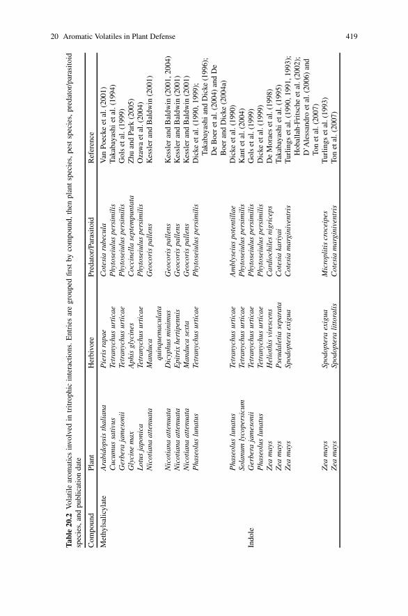
20 Aromatic Volatiles in Plant Defense 419
Tab
le20
.2V
olat
ile
arom
atic
sin
volv
edin
trit
roph
icin
tera
ctio
ns.E
ntri
esar
egr
oupe
dfir
stby
com
poun
d,th
enpl
ant
spec
ies,
pest
spec
ies,
pred
ator
/par
asit
oid
spec
ies,
and
publ
icat
ion
date
Com
poun
dPl
ant
Her
bivo
rePr
edat
or/P
aras
itoi
dR
efer
ence
Met
hyls
alic
ylat
eA
rabi
dops
isth
alia
naP
ieri
sra
pae
Cot
esia
rube
cula
Van
Poec
keet
al.(
2001
)C
ucum
ussa
tivu
sTe
tran
ychu
sur
tica
eP
hyto
seiu
lus
pers
imil
isTa
kaba
yash
iet
al.(
1994
)G
erbe
raja
mes
onii
Tetr
anyc
hus
urti
cae
Phy
tose
iulu
spe
rsim
ilis
Gol
set
al.(
1999
)G
lyci
nem
axA
phis
glyc
ines
Coc
cine
lla
sept
empu
ntat
aZ
huan
dPa
rk(2
005)
Lot
usja
poni
caTe
tran
ychu
sur
tica
eP
hyto
seiu
lus
pers
imil
isO
zaw
aet
al.(
2004
)N
icot
iana
atte
nuat
aM
andu
caqu
inqu
emac
ulat
aG
eoco
ris
pall
ens
Kes
sler
and
Bal
dwin
(200
1)
Nic
otia
naat
tenu
ata
Dic
yphu
sm
inim
usG
eoco
ris
pall
ens
Kes
sler
and
Bal
dwin
(200
1,20
04)
Nic
otia
naat
tenu
ata
Epi
trix
hert
ipen
nis
Geo
cori
spa
llen
sK
essl
eran
dB
aldw
in(2
001)
Nic
otia
naat
tenu
ata
Man
duca
sext
aG
eoco
ris
pall
ens
Kes
sler
and
Bal
dwin
(200
1)P
hase
olus
luna
tus
Tetr
anyc
hus
urti
cae
Phy
tose
iulu
spe
rsim
ilis
Dic
keet
al.(
1990
,199
9);
Taka
baya
shi
and
Dic
ke(1
996)
;D
eB
oer
etal
.(20
04)
and
De
Boe
ran
dD
icke
(200
4a)
Pha
seol
uslu
natu
sTe
tran
ychu
sur
tica
eA
mbl
ysei
uspo
tent
illa
eD
icke
etal
.(19
90)
Sola
num
lyco
pers
icum
Tetr
anyc
hus
urti
cae
Phy
tose
iulu
spe
rsim
ilis
Kan
teta
l.(2
004)
Indo
leG
erbe
raja
mes
onii
Tetr
anyc
hus
urti
cae
Phy
tose
iulu
spe
rsim
ilis
Gol
set
al.(
1999
)P
hase
olus
luna
tus
Tetr
anyc
hus
urti
cae
Phy
tose
iulu
spe
rsim
ilis
Dic
keet
al.(
1999
)Z
eam
ays
Hel
ioth
isvi
resc
ens
Car
dioc
hile
sni
gric
eps
De
Mor
aes
etal
.(19
98)
Zea
may
sP
seud
alet
iase
para
taC
otes
iaka
riya
iTa
kaba
yash
iet
al.(
1995
)Z
eam
ays
Spod
opte
raex
igua
Cot
esia
mar
gini
vent
ris
Tur
ling
set
al.(
1990
,199
1,19
93);
Hob
alla
h-Fr
itsc
heet
al.(
2002
);D
’Ale
ssan
dro
etal
.(20
06)
and
Ton
etal
.(20
07)
Zea
may
sSp
odop
tera
exig
uaM
icro
plit
iscr
ocei
pes
Tur
ling
set
al.(
1993
)Z
eam
ays
Spod
opte
rali
ttor
alis
Cot
esia
mar
gini
vent
ris
Ton
etal
.(20
07)
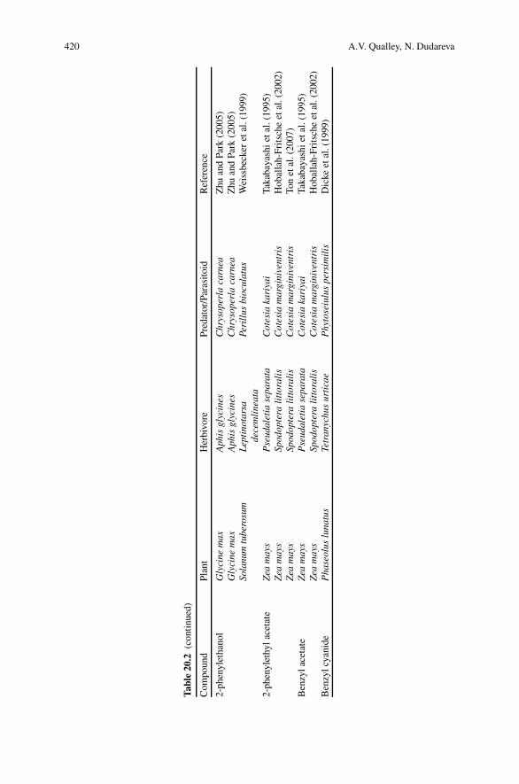
420 A.V. Qualley, N. Dudareva
Tab
le20
.2(c
onti
nued
)
Com
poun
dPl
ant
Her
bivo
rePr
edat
or/P
aras
itoi
dR
efer
ence
2-ph
enyl
etha
nol
Gly
cine
max
Aph
isgl
ycin
esC
hrys
oper
laca
rnea
Zhu
and
Park
(200
5)G
lyci
nem
axA
phis
glyc
ines
Chr
ysop
erla
carn
eaZ
huan
dPa
rk(2
005)
Sola
num
tube
rosu
mL
epti
nota
rsa
dece
mli
neat
aPe
rill
usbi
ocul
atus
Wei
ssbe
cker
etal
.(19
99)
2-ph
enyl
ethy
lace
tate
Zea
may
sP
seud
alet
iase
para
taC
otes
iaka
riya
iTa
kaba
yash
iet
al.(
1995
)Z
eam
ays
Spod
opte
rali
ttor
alis
Cot
esia
mar
gini
vent
ris
Hob
alla
h-Fr
itsc
heet
al.(
2002
)Z
eam
ays
Spod
opte
rali
ttor
alis
Cot
esia
mar
gini
vent
ris
Ton
etal
.(20
07)
Ben
zyla
ceta
teZ
eam
ays
Pse
udal
etia
sepa
rata
Cot
esia
kari
yai
Taka
baya
shi
etal
.(19
95)
Zea
may
sSp
odop
tera
litt
oral
isC
otes
iam
argi
nive
ntri
sH
obal
lah-
Frit
sche
etal
.(20
02)
Ben
zylc
yani
deP
hase
olus
luna
tus
Tetr
anyc
hus
urti
cae
Phy
tose
iulu
spe
rsim
ilis
Dic
keet
al.(
1999
)
20 Aromatic Volatiles in Plant Defense 421
spread in the plant kingdom. To date, this phenomenon has been reported in morethan 23 plant species with a diverse combination of plants, herbivores and naturalenemies (Dicke 1999), and aromatic volatiles have been reported in more than halfof these examples (Table 20.2). One of the best-studied tritrophic interactions isbetween lima bean plants (Phaseolus lunatus), herbivorous spider mites (Tetrany-chus urticae), and carnivorous mites (Phytoseiulus persimilis). Infestation of limabean plants by spider mites triggers the release of volatiles that attract predatorymites that prey on the pests (Takabayashi and Dicke 1996). One component ofthe volatile blend released by T. urticae-infested lima bean leaves is methyl salicy-late, an aromatic volatile commonly detected in the headspace of herbivore-infestedplants (Table 20.2; Dicke and van Poecke 2002). Methyl salicylate was not detectedfrom untreated or mechanically damaged lima bean plants, in contrast to some othervolatile compounds (Dicke et al. 1990). In addition, a dense spider mite infestation(40/leaf) leads to a larger methyl salicylate emission than infestation by smaller pestpopulations (10/leaf; De Boer et al. 2004). Thus, the level of methyl salicylate re-leased from infested lima beans appears to be positively correlated with the severityof T. urticae attack, allowing predator mites to select the plants hosting the largestnumbers of their prey.
Methyl salicylate attracts predatory mites in a dose-dependent manner, but atbiologically unrealistic amounts (200 �g) repels the carnivores (De Boer and Dicke2004a). Predators reared on spider mites infesting cucumber, which emits only atrace amount of methyl salicylate (Takabayashi et al. 1994), chose prey-infestedlima bean plants over cucumber when tested in an olfactometer. The addition ofexogenous methyl salicylate to the cucumber blend made it attractive to the car-nivorous mites (De Boer and Dicke 2004b). Moreover, when this single compoundwas offered as an alternative odor source in a dual-choice assays, the preferenceof the predators for a methyl salicylate-containing volatile blend from T. urticae-infested lima bean plants was lost suggesting an important role of this volatilein the foraging behavior of natural enemies of herbivorous arthropods (De Boeret al. 2004).
Interestingly, lima bean plants release unique volatile blends upon infestationby different pests, thus allowing natural enemies to avoid unproductive foragingif only non-prey species are present. When lima bean plants were infested bythe prey herbivore T. urticae or nonprey caterpillar Spodoptera exigua, carnivo-rous mites P. persimilis were able to locate their prey by detecting differences inthe volatile blends emitted in response to herbivory. Addition of methyl salicylateto the volatile blend released by plants infested with nonprey species eliminatedthe predatory mites’ ability to make this distinction, suggesting a specific role formethyl salicylate in this interaction (De Boer et al. 2004). The opposite situation wasfound in another legume Medicago truncatula, in which spider mite (T. urticae)feeding failed to induce methyl salicylate emission whereas Spodoptera littoralisherbivory did, highlighting the flexible roles of this ester in plant defense (Leitneret al. 2005).
In addition to P. lunatus and M. truncatula, at least 12 other plant speciesshow documented release of methyl salicylate upon herbivore damage, although
422 A.V. Qualley, N. Dudareva
at different levels (Tables 20.1 and 20.2). Slight induction of methyl salicylateemission following herbivory has been found in cucumber (Cucumus sativus) andgerbera (Gerbera jamesonii), where the levels of methyl salicylate were low com-pared to the other emitted volatiles (Takabayashi et al. 1994; Gols et al. 1999). Highlevels of methyl salicylate were detected relative to 38 total compounds releasedfrom Arabidopsis leaves infested with cabbage white butterfly larvae (Pieris rapae)but not from artificially damaged or undamaged plants (Van Poecke et al. 2001).Arabidopsis is the only cruciferous plant to date shown to emit methyl salicy-late in response to herbivore attack, with subsequent attraction of the larval par-asitoid Cotesia rubecula. In dual-choice bioassays, C. rubecula prefers volatileblends containing methyl salicylate from infested Arabidopsis plants versus mix-tures from artificially damaged or undamaged plants (Van Poecke et al. 2001). In-terestingly, caterpillar induced methyl salicylate emission was correlated with anincreased transcript abundance of AtPAL1 suggesting that flux through the path-way towards this compound was induced by herbivore infestation (Van Poeckeet al. 2001). In N. attenuata the level of methyl salicylate was significantly elevatedin volatile blends emitted from plants attacked by three prominent pest speciespresent during the field experiments: the caterpillars of Manduca quinquemacu-lata (Lepidoptera, Sphingidae), the leaf bug Dicyphus minimus (Heteroptera, Miri-dae), and the flea beetle Epitrix hirtipennis (Coleoptera, Chrysomelidae; Kesslerand Baldwin 2001, 2004), thus attracting the generalist predator Geocoris pallens(Kessler and Baldwin 2004).
Infestation of soybean (Glycine max), tomato, and Lotus japonicus with theherbivorous mite T. urticae resulted in release of volatile blends which containedmethyl salicylate and were more attractive to the predatory mite P. persimilis thanvolatiles from uninfested or artificially damaged plants (Ozawa et al. 2000; De Boerand Dicke 2004a, b; Kant et al. 2004; Van Den Boom et al. 2004). In soybean plants,methyl salicylate is also released after attack by soybean aphid (Aphis glycines), thesingle aphid species known to develop large colonies on soybean in North America.This is the only compound from the headspace of aphid-infested plants shown toelicit a significant gas chromatography-electroantennography (GC-EAG) responsein lady beetle Coccinella septempunctata, a species known to attack soybean aphidsin the field. In field tests, traps baited with methyl salicylate were highly attractiveto C. septempunctata, but not to the other common lady beetle Harmonia axyridis,suggesting that the former may use this volatile ester as an olfactory cue for preylocation (Zhu and Park 2005).
Infestation of cowpea (Vigna unguiculata) and hops (Humulus lupulus) withT. urticae also led to either a large increase or a novel release of methyl salicy-late while in other species including black locust (Robinia pseudo-acacia), sweetpepper (Capsicum annuum), thorn apple (Datura stramonium), and grapevine (Vitisvinifera) its emission was induced to a lesser extent (Van Den Boom et al. 2004).When attractiveness of methyl salicylate was tested in field experiments, it was seenthat the grape- and hop-yards containing sticky cards baited with this volatile es-ter captured significantly greater numbers of predatory insects including Chrysopanigricornis, Hemerobius sp., Deraeocoris brevis, Stethorus punctum picipes, and
20 Aromatic Volatiles in Plant Defense 423
Orius tristicolor than the yards with unbaited sticky cards (James and Price 2004).Moreover, this increase of predatory insects coincided with a dramatic reductionin the population of spider mites, the major arthropod pest of hops (James andPrice 2004).
Taken together, these results show that methyl salicylate can be a general indica-tor of herbivore damage in the context of the other volatiles also emitted followinginfestation. Surprisingly, in many of the tritrophic interactions described above, theherbivore-predator participants are the same while the attacked plants are differentand they emit different volatile blends with the exception of methyl salicylate. Anal-ysis of chemoreceptors from parasitoid wasps and predatory mites demonstratedthat insects are able to detect this compound within the volatile blend (De Bruyneet al. 1991; Van Poecke and Dicke 2002). However, methyl salicylate is not emittedby all plant taxa and moreover, its emission decreases in maize leaves after infes-tation by Pseudaletia separata larvae (Takabayashi et al. 1995), suggesting nicheroles for methyl salicylate in select tritrophic interactions.
Indole is another aromatic compound found in the volatile blends of someherbivore-damaged plants. Although the role of indole in tritrophic interactionslacks extensive study, induced indole emission subsequent to herbivory is well es-tablished and has been reported from many plant species including maize (Freyet al. 2000), cotton (Gossypium birsutum; Turlings et al. 1995; Pare and Tumlin-son 1997), gerbera (Gols et al. 1999), lima bean (Mithofer et al. 2005), and peanut(Cardoza et al. 2002). In maize, indole becomes a major constituent of the volatilespectrum released a few hours after feeding by the beet armyworm (S. exigua;Turlings et al. 1990). Its biosynthesis occurs de novo in response to insect damage(Pare and Tumlinson 1998) and positively correlates with the level of infestation(Schmelz et al. 2003). The level of indole emission is also affected by the devel-opmental stage of the infesting herbivores, as was shown during infestation by P.separata larvae (Takabayashi et al. 1995), suggesting that it is more advantageousfor the plant to attract parasitoids while the pest larvae are still small. Interestingly, abroad genetic variability was found with respect to herbivore-induced indole emis-sion among 31 maize inbred lines ranging from trace to >70% of total volatileemissions (Degen et al. 2004).
The most prominent and well-studied tritrophic systems involving indole con-sist of maize plants, armyworms, and parasitic wasps of the genus Cotesia. TheCotesia sub-species kariyai (Takabayashi et al. 1995) and marginiventris (Turlingset al. 1991) are known to visit maize plants emitting indole in response to dam-age by P. separata larvae and S. exigua larvae, respectively. However, the emis-sions of indole and other herbivore-induced volatile compounds are not alwayspest-species specific. In maize, regurgitates from five different caterpillar speciesas well as grasshoppers induce the same set and relative ratio of volatile com-pounds including indole, but at different total amounts, indicating that these highlydetectable cues may not always reliably signal the presence of a suitable host toforaging predators and parasitoids (Turlings et al. 1993). Surprisingly, attraction ofC . marginiventris was not affected by the presence or absence of indole in stud-ies where caterpillar-infested maize plants and those treated with glyphosate, an

424 A.V. Qualley, N. Dudareva
inhibitor of the 5-enolpyruvylshikimate-3-phosphate, were studied together. Fur-thermore, indole seemed to repel rather than attract the parasitoid Microplitis ru-fiventris (D’Alessandro et al. 2006) suggesting that two parasitoids with a compa-rable biology may employ different strategies in their use of plant-provided cues tolocate hosts and indole might play different roles in inducible defenses across plantand insect taxa.
Coincidence of indole emissions and C. marginiventris parasitoid activity hasalso been documented in cotton plants following damage by S. littoralis (Pare andTumlinson 1997) or S. exigua (Rose et al. 1996; Loughrin et al. 1994, 1995a).Similar to the situation in maize, indole emissions following herbivory have beenshown to differ greatly between cotton cultivars (Loughrin et al. 1995a). However,in contrast to maize, release of indole from cotton plants occurs only at the site ofcaterpillar damage and not systemically (Rose et al. 1996; Turlings and Tumlin-son 1992). Interestingly, simultaneous infestation of cotton plants by S. exigua andthe whitefly Bemisia tabaci led to a reduction of indole emission possibly weaken-ing the attraction strengths between parasitoids and their hosts (Rodriguez-Saonaet al. 2003).
When two types of attacking herbivores infested lima bean plants, only feed-ing by S. exigua induced indole emission but not T. urticae (De Boer et al. 2004).Even a high-density infestation by spider mites led to only low indole emissionwhereas feeding by moderate numbers of S. exigua larvae (two/leaf) resulted indrastic increases of indole release compared to control and spider mite test groups(De Boer et al. 2004). Neither artificial mechanical damage nor damage done bythe snail Cepaea hortensis caused a release of indole (Mithofer et al. 2005). Inter-estingly, only the jasmonic acid-related signaling pathway is involved in the pro-duction of caterpillar-induced volatiles in lima bean plants, while both the salicylicacid- and jasmonic acid-related signaling pathways are involved in the productionof T. urticae-induced volatiles (Ozawa et al. 2000).
The aromatic volatiles benzylacetate, 2-phenethyl acetate, benzyl cyanide, andmethyl anthranilate were also released following herbivore attack (Table 20.2).These compounds were emitted from maize after armyworm infestation with theexception of benzylcyanide and 2-phenylethanol, which were detected in lima beansand potato plants following spider mite and potato beetle damage, respectively(Takabayashi et al. 1995; Bernasconi et al. 1998; Hoballah-Fritsche et al. 2002;D’Alessandro and Turlings 2005; Dicke et al. 1999; Weissbecker et al. 1999). More-over, it has been shown that 2-phenylethanol, emitted exclusively following potatobeetle feeding, elicits very high GC-EAG responses from Perillus bioculatus, apredator of potato beetle (Weissbecker et al. 1999).
Release after herbivory of a majority of aromatic compounds at relatively lowlevels raises the question about their roles in the interactions between plants andarthropods. GC-EAG recordings show that the relative quantities of compoundswithin a volatile blend do not always correlate with their elicited EAG responses,indicating that minor compounds may provoke some of the strongest responses(Gouinguene et al. 2005; Zhu and Park 2005) and the key insect attractants remainto be determined.
20 Aromatic Volatiles in Plant Defense 425
20.4 Aromatic Volatiles and Plant–Plant Interactions
Volatiles released from herbivore-infestedplantsalso mediateplant–plant interactionsand may induce expression of defense genes and emission of volatiles in healthyleaves of the same plant or on neighboring, unattacked plants, thus increasing theirattractiveness to carnivores and decreasing their susceptibility to the damaging her-bivores (Dicke et al. 1990; Arimura et al. 2002, 2004; Ruther and Kleier 2005).Exposure of maize to the volatile blend emitted from S. littoralis-infested con-specifics reduces caterpillar feeding and development while significantly increasingattractiveness of the volatile-exposed plants to parasitic C. marginiventris wasps,thus boosting both their direct and indirect defenses (Ton et al. 2007). This effectcould partly be due to the enhanced production of the aromatic compounds indoleand 2-phenethyl acetate in plants following their exposure to caterpillar-inducedvolatiles. Also, neighboring lima bean plants became more attractive to predatorymites and less susceptible to spider mites when exposed to volatiles emitted fromconspecific leaves infested with T. urticae but not volatiles emitted by artificiallywounded leaves (Arimura et al. 2000). T. urticae-infestation induced transcript ac-cumulation of six defense-related genes in lima bean leaves, while exposure ofundamaged plants to volatiles from infested plants upregulated transcript levels ofonly five out of six genes investigated including PAL. Treatment of undamagedplants with gaseous methyl salicylate reproduced the expression patterns of sixdefense genes observed in the T. urticae-infested leaves, indicating the potentialimportance of this compound in plant–plant interactions (Arimura et al. 2000).
In addition to direct elicitation of defenses, exposure to volatile compounds fromattacked plants may lead to priming of plant defensive responses in their neighbors.Priming by volatiles prepares the plant to respond more rapidly and intensely againstsubsequent attack by herbivorous insects (Engelberth et al. 2004; Kessler et al. 2006;Tumlinson and Engelberth this volume). Priming by volatile compounds providesa different way of responding to the threat of insect herbivory via the incompleteactivation of defense-related processes, thus reducing biochemical investment to-wards defenses for the receiver plants until the onset of actual attack (Engelberthet al. 2004; Kessler et al. 2006). While priming by aromatic volatile compoundscould be one of the mechanisms involved in plant–plant signaling in nature, theroles of the aromatic volatiles in and the underlying molecular mechanisms andecological relevance of these particular interactions still remain to be determined.
20.5 Future Directions
To date, it is widely accepted that airborne volatile compounds play importantroles in plant defense, however, the contribution of aromatics to these processesstill requires further investigation. The most interesting unsolved question is whyaromatic volatiles lack the broad representation seen with terpenoids in herbivore-induced volatile blends. The ability of plants to synthesize a wide spectrum ofvolatile aromatic compounds, often at high levels, has been shown in floral organs

426 A.V. Qualley, N. Dudareva
(Verdonk et al. 2003; Boatright et al. 2004). However, in the limited number ofplant species studied, aromatic volatile compounds are comparatively underrepre-sented in plant defense. This discrepancy will be clarified through investigation ofplant-insect interactions occurring in a greater number of plants. Emission of lowerlevels of aromatic compounds may also indicate that their actions are synergistic andmay incorporate a level of specificity to the airborne signal, possibilities requiringfurther exploration at the sensory levels and in field experiments.
The importance of aromatic volatiles should not be overlooked based on theirsubtle presences since insects have an astonishing ability to detect low levels ofvolatile compounds that may provoke some of the strongest responses. Previousattempts to determine the effects of individual aromatic volatiles on insect behaviorwere typically accomplished by the addition of exogenous compounds to existingodor mixtures. Recent breakthroughs in gene discovery and metabolic engineeringnow make it possible to manipulate the levels of specific volatiles in planta, eitheromitting or increasing their emission. Although the contributions of some volatileterpenoids to plant defense have been explored via this method (Aharoni et al. 2003;Kappers et al. 2005; Schnee et al. 2006), it has yet to be applied to volatile aro-matics and will likely corroborate and clarify their previously conceived roles inplant-insect interactions. Of particular interest is how flux through the shikimatepathway, which provides precursors to aromatic volatiles, is regulated during plantdefense with the goal of determining whether higher emission of aromatics would bebeneficial for plant fitness and defense. Recent discovery of the MYB transcriptionfactor AtMYB15, which activates the shikimate pathway in response to wounding(Chen et al. 2006), will enable us to solve this question via metabolic engineering.
Another important unsolved question concerns the factors determining speci-ficity of plant response to a particular threat. An understanding of signal trans-duction pathways that lead to the herbivore-induced plant defenses will shed somelight on possible mechanisms for the exclusive release of certain compounds in re-sponse to given stimuli. In addition, the use of -omics-based approaches applied toboth plants and pests will be useful in elucidating general mechanisms involved inplant-insect and plant-plant interaction. Understanding of the mechanisms underly-ing plant-insect interaction and in particular the role of volatiles in biological controlof pests will allow us in the future to supplement and optimize plant volatile signal-ing and generate crops with enhanced attractiveness to natural enemies during her-bivory, providing a reduced need for chemical pesticide application in agriculture.
Acknowledgments This work was supported by grants from the National Science Foundation(Grant No. MCB-0615700), the USDA Cooperative State Research, Education, and ExtensionService (Grant No. 2005-35318-16207) and the Fred Gloeckner Foundation.
References
Abd El-Mawla AMA, Schmidt W, Beerhues L (2001) Cinnamic acid is a precursor of benzoic acidsin cell cultures of Hypericum androsaemum L. but not in cell cultures of Centaurium erythraeaRAFN. Planta 212:288–293

20 Aromatic Volatiles in Plant Defense 427
Aharoni A, Giri AP, Deuerlein S, Griepink F, De Kogel WJ, Verstappen FWA, Verhoeven HA,Jongsmaa MA, Schwab W, Bouwmeester HJ (2003) Terpenoid metabolism in wild-type andtransgenic Arabidopsis plants. Plant Cell 15:2866–2884
Arimura G, Ozawa R, Kugimiya S, Takabayashi J, Bohlmann J (2004) Herbivore-induced defenseresponse in a model legume: two-spotted spider mites, Tetranychus urticae, induce emissionof (E)-�-ocimene and transcript accumulation of (E)-�-ocimene synthase in Lotus japonicus.Plant Physiol 135:1976–1983
Arimura G, Ozawa R, Nishioka T, Boland W, Koch T, Kuhnemann F, Takabayashi J (2002)Herbivore-induced volatiles induce the emission of ethylene in neighboring lima bean plants.Plant J 29:87–98
Arimura G, Ozawa R, Shomoda T, Nishioka T, Boland W, Takabayashi J (2000) Herbivory-inducedvolatiles elicit defense genes in lima bean leaves. Nature 406:512–515
Beekwilder J, Alvarez-Huerta M, Neef E, Verstappen FWA, Bouwmeester HJ, Aharoni A (2004)Substrate usage by recombinant alcohol acyltransferases from various fruit species. Plant Phys-iol 135:1865–1878
Bernasconi M, Turlings TCJ, Ambrosetti L, Bassetti P, Dorn S (1998) Herbivore-induced emissionsof maize volatiles repel the corn leaf aphid, Rhopalosiphum maidis. Entomol Exp Appl 87:133–142
Beuerle T, Pichersky E (2002) Purification and characterization of benzoate: coenzyme A ligasefrom Clarkia breweri. Arch Biochem Biophys 400:258–264
Bin Jantan I, Yalvema MF, Ahmed NW, Jamal JA (2005) Insecticidal activities of the leaf oilsof eight Cinnamomum species against Aedes aegypti and Aedes albopictus. Pharm Biolog 43:526–532
Birkett MA et al (2000) New roles for cis-jasmone as an insect semiochemical and in plant defense.Proc Natl Acad Sci USA 97:9329–9334
Boatright J, Negre F, Chen X, Kish CM, Wood B, Peel G, Orlova I, Gang D, Rhodes D, DudarevaN (2004) Understanding in vivo benzenoid metabolism in petunia petal tissue. Plant Physiol135:1993–2011
Cardoza YJ, Alborn HT, Tumlinson JH (2002) In vivo volatile emissions from peanut plants in-duced by simultaneous fungal infection and insect damage. J Chem Ecol 28:161–174
Chen Y, Xiangbo Z, Wei W, Chen Z, Gu H, Qu LJ (2006) Overexpression of the wounding-responsive gene AtMYB15 activates the shikimate pathway in Arabidopsis. J Int Plant Biol48:1084–1095
D’Alessandro M, Held M, Triponez Y, Turlings TCJ (2006) The role of indole and other shikimicacid derived maize volatiles in the attraction of two parasitic wasps. J Chem Ecol 32:2733–2748
D’Alessandro M, Turlings TCJ (2005) In situ modification of herbivore-induced plant odors: anovel approach to study the attractiveness of volatile organic compounds to parasitic wasps.Chem Senses 30:739–753
D’Auria JC (2006) Acyltransferases in plants: a good time to be BAHD. Curr Opin Plant Biol9:331–340
D’Auria JC, Chen F, Pichersky E (2002) Characterization of an acyltransferase capable of syn-thesizing benzylbenzoate and other volatile esters in flowers and damaged leaves of Clarkiabreweri. Plant Physiol 130:466–476
D’Auria JC, Chen F, Pichersky E (2003) The SABATH family of methyltransferases in Arabidopsisthaliana and other plant species. Rec Adv Phytochem 37:253–283
De Boer JG, Dicke MA (2004a) The role of methyl salicylate in prey searching behavior of thepredatory mite Phytoseiulus persimilis. J Chem Ecol 30:255–271
De Boer JG, Dicke MA (2004b) Experience with methyl salicylate affects behavioural responsesof a predatory mite to blends of herbivore-induced plant volatiles. Entomol Exp Appl 110:181–189
De Boer JG, Posthumus MA, Dicke MA (2004) Identification of volatiles that are used in discrim-ination between plants infested with prey or nonprey herbivores by a predatory mite. J ChemEcol 30:2215–2230
De Bruyne M, Dicke MA, Tjallingii WF (1991) Receptor cell responses in the anterior tarsi ofPhytoseiulus persimilis to volatile kairomone components. Exp Appl Acarol 13:53–58
428 A.V. Qualley, N. Dudareva
Degen T, Dillmann C, Marion-Poll F, Turlings TCJ (2004) High genetic variability of herbivore-induced volatile emission within a broad range of maize inbred lines. Plant Physiol 135:1928–1938
De Moraes CM, Mescher MC, Tumlinson JH (2001) Caterpillar-induced nocturnal plant volatilesrepel conspecific females. Nature 210:577–580
Dexter R, Qualley A, Kish CM, Ma CJ, Koeduka T, Nagegowda DA, Dudareva N, Pichersky E,Clark D (2007) Characterization of a petunia acetyltransferase involved in the biosynthesis ofthe floral volatile isoeugenol. Plant J 49:265–275
Dicke MA (1999) Are herbivore-induced plant volatiles reliable indicators of herbivore identity toforaging carnivorous arthropods? Entomol Exp Appl 91:131–142
Dicke MA, Abelis MW, Takabayashi J, Bruin J, Posthumus MA (1990) Plant strategies of ma-nipulating predator-prey interactions through allelochemicals: prospects for application in pestcontrol. J Chem Ecol 16:3091–3117
Dicke MA, Gols R, Ludeking D, Posthumus MA (1999) Jasmonic acid and herbivory differentiallyinduce carnivore attracting plant volatiles in lima bean plants. J Chem Ecol 25:1907–1922
Dicke MA, Van Beek TA, Posthumus MA, Van Bokhoven H, De Groot AE (1990) Isolation andidentification of volatile kairomone that affects acarine predatorprey interactions Involvementof host plant in its production. J Chem Ecol 16:381–396
Dicke MA, Van Loon JJA (2000) Multitrophic effects of herbivore-induced plant volatiles in anevolutionary context. Entomol Exp Appl 97:237–249
Dicke MA, Van Poecke RMP (2002) Signaling in plant-insect interactions: signal transduction indirect and indirect plant defence. In: Scheel D, Wasternack C (eds) Plant signal transduction:frontiers in molecular biology. Oxford University Press, Oxford, pp 289–316
Dixon RA, Achnine L, Kota P, Liu CJ, Reddy MSS, Wang LJ (2002) The phenylpropanoid pathwayand plant defence – a genomics perspective. Mol Plant Pathol 3:371–390
Drukker B, Bruin J, Jacobs G, Kroon A, Sabelis MW (2000) How predatory mites learn to copewith variability in volatile plant signals in the environment of their herbivorous prey. Exp ApplAcarol 24:881–895
Dudareva N, D’Auria JC, Nam KH, Raguso RA, Pichersky E (1998) Acetyl-CoA: benzylalcoholacetyltransferase – an enzyme involved in floral scent production in Clarkia breweri. Plant J14:297–304
Dudareva N, Negre F, Nagegowda DA, Orlova I (2006) Plant volatiles: recent advances and futureperspectives. Crit Rev Plant Sci 25:417–440
Dudareva N, Pichersky E, Gershenzon J (2004) Biochemistry of plant volatiles. Plant Physiol135:1893–1902
Effmert U, Saschenbrecker S, Ross J, Negre F, Fraser CM, Noel JP, Dudareva N, Piechulla B(2005) Floral benzenoid carboxyl methyltransferases: from in vitro to in planta function. Phy-tochemistry 66:1211–1230
Engelberth J, Alborn HT, Schmelz EA, Tumlinson JH (2004) Airborne signals prime plants againstinsect herbivore attack. Proc Natl Acad Sci USA 101:1781–1785
Farag MA, Fokar M, Abd, H, Zhang H, Allen RD, Pare PW (2005) (Z )-3-Hexenol induces defensegenes and downstream metabolites in maize. Planta 220:900–909
French CJ, Vance CP, Towers GHN (1976) Conversion of p-coumaric acid to p-hydroxybenzoicacid by cell free extracts of potato tubers and Polyporus hispidus. Phytochem 15:564–566
Frey M, Spiteller D, Boland W, Gierl A (2004) Transcriptional activation of Igl, the gene for indoleformation in Zea mays: a structure-activity study with elicitor-active N-acyl glutamines frominsects. Phytochem 65:1047–1055
Frey M, Stettner C, Pare PW, Schmelz EA, Tumlinson JH, Gierl A (2000) An herbivore elicitoractivates the gene for indole emission in maize. Proc Natl Acad Sci USA 97:14801–14806
Gang DR, Lavid N, Zubieta C, Chen F, Beuerle T, Lewinsohn E, Noel JP, Pichersky E (2002)Characterization of phenylpropene O-methyltransferases from sweet basil: facile change ofsubstrate specificity and convergent evolution within a plant OMT family. Plant Cell 14:505–519
20 Aromatic Volatiles in Plant Defense 429
Gols R, Posthumus MA, Dicke MA (1999) Jasmonic acid induces the production of gerberavolatiles that attract the biological control agent Phytoseiulus persimilis. Entomol Exp Appl93:77–86
Gouinguene S, Pickett JA, Wadhams LJ, Birkett MA, Turlings TCJ (2005) Antennal electrophys-iological responses of three parasitic wasps to caterpillar-induced volatiles from maize (Zeamays Mays), cotton (Gossypium herbaceum), and cowpea (Vigna unguiculata). J Chem Ecol31:1023–1038
Hardie J, Isaacs R, Pickett JA, Wadhams LJ, Woodcock CM (1994) Methyl salicylate and (−)-(1R,5S)-myrtenal are plant-derived repellents for black bean aphid, Aphis fabae Scop. (Homoptera:Aphididae) J Chem Ecol 20:2847–2855
Hoballah-Fritsche ME, Tamo C, Turlings TCJ (2002) Differential attractiveness of induced odorsemitted by eight maize varieties for the parasitoid Cotesia marginiventris: is quality important?J Chem Ecol 28:951–968
Hopke J, Donath J, Blechert S, Boland W (1994) Herbivore-induced volatiles: the emission ofacyclic homoterpenes from Phaseolus lunatus and Zea mays can be triggered by a beta-glucosidase and jasmonic acid. FEBS Lett 352:146–150
James DG, Price TS (2004) Field-testing of methyl salicylate for recruitment and retention ofbeneficial insects in grapes and hops. J Chem Ecol 30:1613–1627
Jarvis AP, Schaaf O, Oldham NJ (2000) 3-Hydroxy-3-phenylpropanoic acid is an intermediatein the biosynthesis of benzoic acid and salicylic acid but benzaldehyde is not. Planta 212:119–126
Kaminaga Y, Schnepp J, Peel G, Kish CM, Ben-Nissan G, Weiss D, Orlova I, Lavie O, RhodesD, Wood K, Porterfield M, Cooper AJL, Schloss JV, Pichersky E, Vainstein A, Dudareva N(2006) Plant phenylacetaldehyde synthase is a bifunctional homotetrameric enzyme that cat-alyzes phenylalanine decarboxylation and oxidation. J Biol Chem 281:23357–23366
Kant MR, Ament K, Sabelis MW, Haring MA, Schuurink RC (2004) Differential timing of spidermite-induced direct and indirect defenses in tomato plants. Plant Physiol 135:483–495
Kappers IF, Aharoni A, Van Herpen TWJM, Luckerhoff LLP, Dicke MA, Bouwmeester HJ (2005)Genetic engineering of terpenoid metabolism attracts bodyguards to Arabidopsis. Science309:2070–2072
Kapteyn J, Qualley AV, Xie Z, Fridman E, Dudareva N, Gang DR (2007) Evolution of cinnamate/p-coumarate carboxyl methyltransferases and their role in the biosynthesis of methylcinnamate.Plant Cell 19:3212–3229
Kessler A, Baldwin IT (2001) Defensive function of herbivore-induced plant volatile emissions innature. Science 291:2142–2143
Kessler A, Baldwin IT (2004) Herbivore-induced plant vaccination. Part I. The orchestration ofplant defenses in nature and their fitness consequences in the wild tobacco Nicotiana attenuata.Plant J 38:639–649
Kessler A, Halitschke R, Diezel C, Baldwin IT (2006) Priming of plant defense responses in na-ture by airborne signaling between Artemisia tridentata and Nicotiana attenuata. Oecologia148:280–292
Knudsen JT, Gershenzon J (2006) The chemical diversity of floral scent. In: Dudareva N, PicherskyE (eds) Biology of floral scent. Taylor & Francis, Boca Raton, pp 27–52
Koeduka T, Fridman E, Gang DR, Vassao DG, Jackson BL, Kish CM, Orlova I, Spaaova SM, LewisNG, Noel JP, Baiga TJ, Dudareva N, Pichersky E (2006) Eugenol and isoeugenol, characteristicaromatic constituents of spices, are biosynthesized via reduction of a coniferyl alcohol ester.Proc Natl Acad Sci USA 103:10128–10133
Koeduka T, Orlova I, Kish CM, Ibdah M, Wilkerson CG, Baiga TJ, Noel JP, Dudareva N, PicherskyE (2007) The multiple phenylpropene synthases in both Clarkia breweri and Petunia hybridarepresent two distinct protein lineages and continue to evolve their product specificity. Plant J(in press)
Leitner M, Boland W, Mithofer A (2005) Direct and indirect defences induced by piercing-suckingand chewing herbivores in Medicago truncatula. New Phytol 167:597–606
430 A.V. Qualley, N. Dudareva
Leon J, Shulaev V, Yalpani N, Lawton MA, Raskin I (1995) Benzoic acid 2-hydroxylase, a sol-uble oxygenase from tobacco, catalyzes salicylic acid biosynthesis. Proc Natl Acad Sci USA92:10413–10417
Loughrin JH, Manukian A, Heath RR, Tumlinson JH (1995a) Volatiles emitted by different cottonvarieties damaged by feeding beet armyworm larvae. J Chem Ecol 21:1217–1227
Loughrin JH, Manukian A, Heath RR, Turlings TCJ (1994) Diurnal cycle of emission of in-duced volatile terpenoids by herbivore-injured cotton plants. Proc Natl Acad Sci USA 91:11836–11840
Loughrin JH, Potter DA, Hamilton-Kemp TR (1995b) Volatile compounds induced by herbivoryact as aggregation kairomones for the Japanese beetle (Popilla japonica Newman). J ChemEcol 21:1457–1467
Mercke P, Kappers IF, Verstappen FWA, Vorst O, Dicke MA, Bouwmeester HJ (2004) Combinedtranscript and metabolite analysis reveals genes involved in spider mite induced volatile forma-tion in cucumber plants. Plant Physiol 135:2012–2024
Mithofer A, Wanner G, Boland W (2005) Effects of feeding Spodoptera littoralis on lima beanleaves. II. Continuous mechanical wounding resembling insect feeding is sufficient to elicitherbivory-related volatile emission. Plant Physiol 137:1160–1168
Ngoh SP, Choo LEW, Pang FY, Huang Y, Kini MR, Ho SH (1998) Insecticidal and repellent prop-erties of nine volatile constituents of essential oils against the American cockroach. Periplanetaamericana (L.). Pestic Sci 54:261–268
Ninkovic V, Ahmed E, Glinwood R, Pettersson J (2003) Effects of two types of semiochemicalon population development of the bird cherry oat aphid Rhopalosiphum padi in a barley crop.Agric Forest Entomol 5:27–33
Obeng-Ofori D, Reichmuth CH (1997) Bioactivity of eugenol, a major component of essential oilof Ocimum suave (Wild.) against four species of stored-product Coleoptera. Int J Pest Manag43:89–94
Orlova I, Marshall-Colon A, Schnepp J, Wood B, Varbanova M, Fridman E, Blakeslee JJ, Peer WA,Murphy AS, Rhodes DR, Pichersky E, Dudareva N (2006) Reduction of benzenoid synthesis inpetunia flowers reveals multiple pathways to benzoic acid and enhancement in auxin transport.Plant Cell 18:3458–3475
Ozawa R, Shimoda T, Kawaguchi M, Arimura G, Horiuchi J, Nishioka T, Takabayashi J (2000)Lotus japonicus infested with herbivorous mites emits volatile compounds that attract predatorymites. J Plant Res 113:427–433
Ozawa R, Shiojiri K, Sabelis MW, Arimura G, Nishioka T, Takabayashi J (2004) Corn plantstreated with jasmonic acid attract more specialist parasitoids, thereby increasing parasitation ofthe common armyworm. J Chem Ecol 30:1797–1808
Pare PW, Tumlinson JH (1997) De novo biosynthesis of volatiles induced by insect herbivory incotton plants. Plant Physiol 114:1161–1167
Pare PW, Tumlinson JH (1998) Cotton volatiles synthesized and released distal to the site of insectdamage. Phytochem 47:521–526
Pettersson J, Pickett JA, Pye BJ, Quiroz A, Smart LE, Wadhams LJ, Woodcock CM (1994) Win-ter host component reduces colonization by bird-cherry-oat aphid, Rhopalosiphum padi (L.)(Homoptera, Aphididae), and other aphids in cereal fields. J Chem Ecol 20:2565–2574
Price PW, Bouton CE, Gross P, McPheron BA, Thompson JN, Weis AE (1980) Interactions among3 trophic levels – influence of plants on interactions between insect herbivores and naturalenemies. Ann Rev Ecol System 11:41–65
Radwanski ER, Last RL (1995) Tryptophan biosynthesis and metabolism – iochemical and molec-ular genetics. Plant Cell 7:921–934
Rodriguez-Saona C, Crafts-Brandner SJ, Canas LA (2003) Volatile emissions triggered by mul-tiple herbivore damage: beet armyworm and whitefly feeding on cotton plants. J Chem Ecol29:2539–2550
Rose USR, Manukian A, Heath RR, Tumlinson JH (1996) Volatile semiochemicals released fromundamaged cotton leaves. Plant Physiol 111:487–495

20 Aromatic Volatiles in Plant Defense 431
Ruther J, Kleier S (2005) Plant-plant signaling: ethylene synergizes volatile emission in Zea maysinduced by exposure to (Z )-3-hexen-1-ol. J Chem Ecol 31:2217–2222
Schmelz EA, Alborn HT, Banchio E, Tumlinson JH (2003) Quantitative relationships betweeninduced jasmonic acid levels and volatile emissions in Zea mays during Spodoptera exiguaherbivory. Planta 216:665–673
Schnee C, Kollner TG, Held M, Turlings TCJ, Gershenzon J, Degenhardt J (2006) The productsof a single maize sesquiterpene synthase form a volatile defense signal that attracts naturalenemies of maize herbivores. Proc Natl Acad Sci USA 103:1129–1134
Schnitzler JP, Madlung J, Rose A, Seitz HU (1992) Biosynthesis of p-hydroxybenzoic acid inelicitor-treated carrot cell cultures. Planta 188:594–600
Shulaev V, Silverman P, Raskin I (1997) Airborne signalling by methyl salicylate in plant pathogenresistance. Nature 385:718–721
Takabayashi J, Dicke MA (1996) Plant-carnivore mutualism through herbivore-induced carnivoreattractants. Trends Plant Sci 1:109–113
Takabayashi J, Dicke M, Posthumus MA (1991) Variation in composition in predator attractingallelochemicals emitted by herbivore-infested plants: relative influence of plant and herbivore.Chemoecology 2:1–6
Takabayashi J, Dicke MA, Takahashi S, Posthumus MA, Van Beek TA (1994) Leaf age affectscomposition of herbivore-induced synomones and attraction of predatory mites. J Chem Ecol20:373–386
Takabayashi J, Takahashi S, Dicke MA, Posthumus MA (1995) Developmental stage of herbivorePseudaletia separata affects production of herbivore-induced synomone by corn plants. J ChemEcol 21:273–287
Tieman DM, Loucas HM, Kim JY, Clark DG, Klee HJ (2007) Tomato phenylacetaldehydereductases catalyze the last step in the synthesis of the aroma volatile 2-phenylethanol. Phy-tochemistry 68:2660–2669
Tieman D, Taylor M, Schauer N, Fernie AR, Hanson AD, Klee HJ (2006) Tomato aromaticamino acid decarboxylases participate in synthesis of the flavor volatiles 2-phenylethanol and2-phenylacetaldehyde. Proc Natl Acad Sci USA 103:8287–8292
Ton J, D’Alessandro M, Jourdie V, Jakab G, Karlen D, Held M, Mauch-Mani B, Turlings TCJ(2007) Priming by airborne signals boosts direct and indirect resistance in maize. Plant J 49:16–26
Turlings TCJ, Loughrin JH, McCall PJ, Rose USR, Lewis WJ (1995) How caterpillar-damagedplants protect themselves by attracting parasitic wasps. Proc Natl Acad Sci USA 92:4169–4174
Turlings TCJ, McCall PJ, Alborn HT, Tumlinson JH (1993) An elicitor in caterpillar oral secretionsthat induces corn seedlings to emit chemical signals attractive to parasitic wasps. J Chem Ecol19:411–425
Turlings TCJ, Tumlinson JH (1992) Systemic release of chemical signals by herbivore-injuredcorn. Proc Natl Acad Sci USA 89:8399–8402
Turlings TCJ, Tumlinson JH, Heath RR, Proveaux AT, Doolittle RE (1991) Isolation and identifi-cation of allelochemicals that attract the larval parasitoid, Cotesia-marginiventris (Cresson), tothe microhabitat of one of its hosts. J Chem Ecol 17:2235–2251
Turlings TCJ, Tumlinson JH, Lewis WJ (1990) Exploitation of herbivore-induced plant odors byhost-seeking parasitoid wasps. Science 250:1251–1253
Vancanneyt G, Sanz C, Farmaki T, Paneque M, Ortego F, Castanera P, Sanchez-Serrano JJ (2001)Hydroperoxide lyase depletion in transgenic potato plants leads to an increase in aphid perfor-mance. Proc Natl Acad Sci USA 98:8139–8144
Van Den Boom CEM, Van Beek TA, Posthumus MA, De Groot AE, Dicke MA (2004) Qualitativeand quantitative variation among volatile profiles induced by Tetranychus urticae feeding onplants from various families. J Chem Ecol 30:69–89
Van Poecke RMP, Dicke MA (2002) Induced parasitoid attraction by Arabidopsis thaliana: in-volvement of the octadecanoid and the salicylic acid pathway. J Exp Bot 53:1793–1799
432 A.V. Qualley, N. Dudareva
Van Poecke RMP, Posthumus MA, Dicke MA (2001) Herbivore-induced volatile production byArabidopsis thaliana leads to attraction of the parasitoid Cotesia rubecula: chemical, behav-ioral, and gene-expression analysis. J Chem Ecol 27:1911–1928
Vassao DG, Gang DR, Koeduka T, Jackson B, Pichersky E, Davin LB, Lewis NG (2006) Chavicolformation in sweet basil (Ocimum basilicum): cleavage of an esterified C9 hydroxyl group withNAD(P)H-dependent reduction. Org Biomol Chem 4:2733–2744
Verdonk JC, De Vos CHR, Verhoeven HA, Haring MA, van Tunen AJ, Schuurink RC (2003)Regulation of floral scent production in petunia revealed by targeted metabolomics. Phytochem62:997–1008
Vet LEM, Dicke MA (1992) Ecology of infochemical use by natural enemies in a tritrophic context.Ann Rev Entomol 37:141–172
Wang J, De Luca V (2005) The biosynthesis and regulation of biosynthesis of Concord grape fruitesters, including ‘foxy’ methylanthranilate. Plant J 44:606–619
Wang J, Dudareva N, Bhakta S, Raguso RA, Pichersky E (1997) Floral scent production inClarkia breweri (Onagraceae). II. Localization and developmental modulation of the en-zyme SAM:(Iso)Eugenol O-methyltransferase and phenylpropanoid emission. Plant Physiol114:213–221
Watanabe S, Hayashi K, Yagi K, Asai T, Mactavish H, Picone J, Turnball C, Watanabe N (2002)Biogenesis of 2-phenylethanol in rose flowers: incorporation of [2H8]L-phenylalanine into 2-phenylethanol and its beta-D-glucopyranoside during the flower opening of Rosa ‘Hoh-Jun’and Rosa damascena. Mill Biosci Biotechnol Biochem 66:943–947
Weissbecker B, Van Loon JJA, Dicke MA (1999) Electroantennogram responses of a predator,Perillus bioculatus, and its prey, Leptinotarsa decemlineata, to plant volatiles. J Chem Ecol25:2313–2325
Wildermuth MC, Dewdney J, Wu G, Ausubel FM (2001) Isochorismate synthase is required tosynthesize salicylic acid for plant defence. Nature 414:562–565
Yazaki K, Heide L, Tabata M (1991) Formation of p-hydroxybenzoic acid from p-coumaric acidby cell free extract of Lithospermum erythrorhizon cell cultures. Phytochem 30:2233–2236
Zhu J, Park K (2005) Methyl salicylate, a soybean aphid-induced plant volatile attractive to thepredator Coccinella setempunctata. J Chem Ecol 31:1733–1746

Chapter 21Ecological Roles of Vegetative Terpene Volatiles
Jorg Degenhardt
With their enormous number and large structural diversity, terpenes dominate mostplant volatile blends. Terpenes were shown to mediate the interactions betweenplants and many organisms including arthropods, nematodes, and other plants.Often, these interactions are based on volatile mono- and sesquiterpenes which arereleased from the plant after damage by herbivores. The terpenes attract herbivoreenemies that attack the herbivore and thereby may reduce the damage to the plant.This interaction was termed ‘indirect defense’ and will often benefit the plant, butother organisms like parasitic plants or insects and can utilize the volatile signalsto their advantage. Despite the relatively low number of volatile terpene-mediatedinteractions identified today, many more are likely to be discovered with the advanceof volatile collection methods and molecular techniques.
21.1 Introduction
Terpenes form the largest group of volatile compounds among the natural productsof plants. Many of the 30,000 terpenes, especially monoterpenes, sesquiterpenes,and irregular terpenes of low molecular weight have high vapor pressures (Connollyand Hill 1991). High vapor pressure results in the emission of these compounds fromthe plant into the environment. Although terpene volatilization from flowers hasbeen known for many years (Knudsen et al. 2006), these compounds are also emittedfrom the vegetative tissues of plants. A detailed analysis of emission patterns inplants revealed that most tissues produce specific blends of terpenes (Connolly andHill 1991; for the model plants Arabidopsis and maize, see Chen et al. 2004; Kollneret al. 2004; Tholl et al. 2005). Some of these are emitted from specialized terpenestorage organs, for example the glandular trichomes of mints and the resin ducts inconifers, but many volatiles are not emitted from anatomically-specialized storagesites. Less than two decades ago, it was demonstrated that volatiles emitted from
J. DegenhardtDepartment of Biochemistry, Max Planck Institute for Chemical Ecology, D-07745 Jena, Germanye-mail: [email protected]
A. Schaller (ed.), Induced Plant Resistance to Herbivory, 433C© Springer Science+Business Media B.V. 2008
434 J. Degenhardt
plants after herbivore damage can attract enemies of the herbivore (Dicke et al. 1990;Turlings et al. 1990). Under favorable environmental conditions, this attraction ofherbivore enemies can limit herbivore damage to the plant. This review will focus onvolatile terpene signals from vegetative plant tissues and their roles in plant defenseagainst herbivores.
21.2 Terpenes as Volatile Plant Signals
Unlike fatty acid-derived signals and aromatic volatiles (see contributions byTumlinson and Engelberth this volume and Qualley and Dudareva this volume),terpenes are commonly emitted in complex blends with a large structural diversitybetween the compounds. Responsible for most of this terpene diversity are multi-product terpene synthases, the key enzymes of terpene biosynthesis (see Bohlmannthis volume; Gershenzon and Kreis 1999). Terpene synthases can produce blends ofup to 50 different compounds from one substrate and form mixtures with definedrelative ratios of products (Steele et al. 1998). In communication with other organ-isms, these mixtures might provide a more specific signal than signals consisting ofsingle terpenes.
While low concentrations of volatile terpenes are produced in almost everyplant tissue, the highest terpene concentrations from vegetative tissues are releasedin response to environmental cues. Terpene emission is increased in response todamage and contact with elicitors of herbivores (Mattiacci et al. 1995; Albornet al. 1997; Halitschke et al. 2001; Spiteller and Boland 2003; Tumlinson andLait 2005), and abiotic factors like wounding (Howe 2004; Schmelz et al. 2001;Mithofer et al. 2005), UV-radiation (Johnson et al. 1999), O3 and CO2 concentration(Vuorinen et al. 2004a, b; Jasoni et al. 2004; Beauchamp et al. 2005), nutritionalstatus of the plant (Schmelz et al. 2003), as well as temperature and light (Guentheret al. 1993; Takabayashi et al. 1994; Gouinguene and Turlings 2002). In contrast tomany compounds of the lipoxygenase pathway, terpenes are usually released afterde novo synthesis (Pare and Tumlinson 1997) involving gene transcription (Schneeet al. 2002, 2006; Gomez et al. 2005), and within a time window starting one to twohours after elicitation (Turlings et al. 1998).
Despite the enormous number of terpenes emitted by plants, relatively few ter-penes have been clearly identified as signals between organisms to date. In thischapter, we will present some of these interactions involving volatile terpenes anddiscuss the problems associated with the study of terpene-mediated interactions.
21.2.1 Tritrophic Interactions of Plants with Herbivoresand Natural Herbivore Enemies
Almost two decades ago, researchers in the Netherlands and the United States firstobserved that herbivore damage to certain plants induces the emission of volatile

21 Ecological Roles of Vegetative Terpene Volatiles 435
organic compounds that attract natural enemies of the herbivores (Dicke et al. 1990;Turlings et al. 1990). This phenomenon has been reported in more than 15 differ-ent plant species after feeding by an assortment of arthropod herbivores and wastermed ,indirect defense’ (Dicke 1999; Dicke and van Loon 2000; Kessler andBaldwin 2002; Meiners and Hilker 2000). The herbivore enemies that respond tovolatiles from herbivore-damaged plants include various carnivorous arthropods,both predators and parasitoids. Attraction of herbivore enemies has been shown tobenefit the plant by reducing subsequent herbivory and increasing reproductive fit-ness (Hoballah and Turlings 1999; van Loon et al. 2000; Kessler and Baldwin 2001),although such advantages are not realized in all cases (Coleman et al. 1999).
The attraction of the predatory mite Phytoseiulus persimilis to lima bean plantsinfested with the spider mite Tetranychus urticae has been studied in detail (Dickeet al. 1990). Olfactory assays testing the attraction of single compounds from thecomplex volatile blend demonstrated that the predatory mite was not only attractedto the aromatic compound methyl salicylate (De Boer et al. 2004; De Boer andDicke 2004; see also chapter of Qualley and Dudareva this volume), but also thesesquiterpene alcohol nerolidol. Transgenic Arabidopsis overexpressing a nerolidolsynthase from strawberry were used as volatile source in olfactometer experimentswith P. persimilis. Nerolidol-emitting transgenic plants were more attractive to thepredator than undamaged wild type plants (Kappers et al. 2005). In addition to aninnate attraction to compounds of the plant volatile bouquet, the predatory mites alsohave the ability to associate odors with host presence (De Boer and Dicke 2006). Thehomoterpene (3E ,7E)-4,8,12-trimethyl-1,3,7,11-tridecatetraene, a irregular C16 ter-pene olefin, is released from lima bean in response to feeding by T. urticae but notby the non-host organism Spodoptera exigua (Fig. 21.1A). After a series of expe-riences with the presence and absence of the prey, the predatory mite utilized thehomoterpene as an indicator for the presence of prey (De Boer et al. 2004). Theability for associative learning may guide the predatory mites to locate their preyunder natural conditions in a complex environment (De Boer and Dicke 2006). Notonly does this help the predator to identify plants infested with prey, but also allowsit to adapt when the prey switch between different host species during the season(Drukker et al. 2000).
Maize plants damaged by larvae of lepidopteran herbivores like Spodopteralittoralis emit a complex blend of volatiles dominated by mono- and sesquiterpenes(Turlings et al. 1991). These volatiles attract females of the parasitic wasp Cotesiamarginiventris which use the lepidopteran larvae as hosts (Turlings et al. 1990).The parasitation might benefit the maize plants under permitting circumstancessince the parasitized larvae feed less and will not procreate (Hoballah et al. 2004;Degenhardt et al. 2003). Since the herbivore-induced volatiles of maize consist of acomplex blend of compounds, it is difficult to demonstrate which of the compoundsare attractive to the parasitic wasp. The major sesquiterpenes of herbivore-inducedmaize are produced by the terpene synthase TPS10 which is strongly expressed af-ter herbivory by lepidopterans. TPS10 forms (E)-�-farnesene, (E)-�-bergamotene,and other herbivory-induced sesquiterpene hydrocarbons from the substrate far-nesyl diphosphate (Fig. 21.1B; Schnee et al. 2006). Overexpression of TPS10 in

436 J. Degenhardt
B
C D
-Sesquiphellandrene(E)- -Bergamotene(E)- -Farnesene
-Pinene -Myrcene -Phellandrene (E)- -Caryophyllene
OHA
(E)-Nerolidol (E)-4,8-dimethyl-1,3,7-nonatriene(DMNT)
Fig. 21.1 Structures of volatile plant terpenes involved in interactions with other organisms.(A) (E)-nerolidol and (E)-4,8-dimethyl-1,3,7-nonatriene (DMNT) are used by the predatorymite P. persimilis to locate spider mites. (B) (E)-�-farnesene, (E)-�-bergamotene and (E)-�-sesquiphellandrene attract parasitic wasps to maize damaged by lepidopteran larvae. (C)�-pinene, �-myrcene and �-phellandrene attract dodder (C. pentagona) to its host plant. (D) (E)-�-Caryophyllene guides entomophatogenic nematodes to maize roots damaged by D. v. virgifera
Arabidopsis thaliana resulted in plants emitting high quantities of TPS10 sesquiter-pene products identical to those released by maize. Using these transgenic Ara-bidopsis plants as odor sources in olfactometer assays showed that females of theparasitoid C. marginiventris learn to exploit the TPS10 sesquiterpenes to locate theirlepidopteran hosts after prior exposure to these volatiles in association with hosts(Schnee et al. 2006). This gene-based dissection of the herbivore-induced volatileblend demonstrates that a single gene such as tps10 can be sufficient to mediate theindirect defense of maize against herbivore attack. Furthermore, associative learn-ing can also adapt parasitoids to alterations of the herbivore-induced volatile blendby plant species, age and tissue of the plant, and abiotic conditions (Takabayashiet al. 1994; De Moraes et al. 1999; Schmelz et al. 2003; Van den Boom et al. 2004).However, females of C. marginiventris are also attracted to the full blend of maizevolatiles without prior association, indicating that the blend contains additional at-tractive compounds that elicit an innate response (Hoballah and Turlings 2005).Bioassay-guided fractionation of the maize volatiles has not yet identified suchcompounds (D’Alessandro and Turlings 2005, 2006). The combination of both in-nate and learned responses might allow this generalist parasitic wasp to locate awide range of hosts on different plant species in a natural, complex environment(Turlings and Wackers 2004; Degenhardt et al. 2003). Interestingly, the emission ofvolatiles in response to herbivore damage is not only beneficial for the maize plantsince larvae of lepidopteran Spodoptera frugiperda use these volatiles as a cue to
21 Ecological Roles of Vegetative Terpene Volatiles 437
find their food plants (Carroll et al. 2006). Further studies are required to determinewhether the benefit of the volatile signal outweighs its disadvantages under specificenvironmental conditions.
The function of terpenes as defense signals under field conditions was studiedon a wild tobacco species, Nicotiana attenuata. The release of terpenes was mim-icked by application of a lanolin paste that emitted physiological concentrations ofthe monoterpene linalool and the sesquiterpene (E)-�-bergamotene (Kessler andBaldwin 2001). The emission of exogenous linalool decreased lepidopteran ovipo-sition rates on N. attenuata plants while the release of (E)-�-bergamotene increasedegg predation rates by a generalist predator. These observations provided conclusiveevidence that indirect, terpene-based plant defenses can reduce the herbivore loadof a plant in a natural environment (Kessler and Baldwin 2001).
Terpene-mediated interactions were not only observed in response to damageof the leaves but also in response to root-feeding herbivores. Larvae of the beetleDiabrotica virgifera virgifera (Western corn rootworm) are an important pest ofmaize. In response to feeding by the larvae, maize roots release a signal that stronglyattracts the entomopathogenic nematode Heterorhabditis megidis (Boff et al. 2001;van Tol et al. 2001). The signal released by the maize roots was identified as(E)-�-caryophyllene, a sesquiterpene olefin (Fig. 21.1D; Rasmann et al. 2005).Most North American maize lines do not release (E)-�-caryophyllene, whereasEuropean lines and the wild maize ancestor, teosinte, do so in response to D. v.virgifera attack. Field experiments showed a five-fold higher nematode infectionrate of D. v. virgifera larvae on a maize variety that produces the signal than ona variety that does not. Spiking the soil near the latter variety with authentic (E)-�-caryophyllene decreased the emergence of adult D. v. virgifera to less than half(Rasmann et al. 2005).
Not only feeding, but also oviposition of the herbivore can induce terpene emis-sion in plants. The pine sawfly (Diprion pini) lays its eggs on pine twigs and woundsthe surface of the needles in the process. The volatiles emitted in response to ovipo-sition attract a wasp that parasitizes saw fly eggs (Hilker et al. 2002). The signalattracting the wasp is the sesquiterpene (E)-�-farnesene which is only recognizedin combination with other, constitutively released pine volatiles (Mumm et al. 2003;Mumm and Hilker 2005).
21.2.2 Interference of Plant Volatile Terpenes with InsectPheromones
Many species of aphids release a sesquiterpene alarm pheromone, (E)-�-farnesene,which lowers the risk of predation of other aphids in the vicinity, either by causingthem to move away (Hardie et al. 1999), or by increasing their proportion of wingedprogeny (Kunert et al. 2005). To determine if plant-produced (E)-�-farnesenecan mimic these effects, transgenic Arabidopsis plants overexpressing a (E)-�-farnesene synthase from Mentha x piperita were generated by Beale et al. (2006).

438 J. Degenhardt
These transgenic plants emitted high levels of (E)-�-farnesene and elicited potentalarm and repellent responses in the aphid Myzus persicae, and an arrestant re-sponse in the aphid parasitoid Diaeretiella rapae (Beale et al. 2006). In addition,(E)-�-farnesene attracts further predators and parasitoids which are natural ene-mies of aphids (Molck et al. 1999; Du et al. 1998; Al Abassi et al. 2000; Fosteret al. 2005). Thereby, the emission of (E)-�-farnesene after aphid-infestation pro-vides the plant with both direct and indirect defenses. The monoterpene alcohollinalool, albeit no alarm pheromone, also repelled M. persicae in experiments withtransgenic Arabidopsis plants overexpressing a terpene synthase from strawberry(Aharoni et al. 2003).
21.2.3 Terpene-Mediated Interactions Between Plants
Volatile-emitting plants can prime the defense metabolism in adjacent plants. Theprocess involves increased transcription of defense-related genes and allows theplant to respond faster and more vigorously to herbivore attack (Baldwin et al. 2006;Turlings and Ton 2006). Most of these interactions were shown to be based onvolatiles derived from the lipoxygenase pathway, the so called ‘green leaf volatiles’(Bate and Rothstein 1998; Arimura et al. 2001; Farag and Pare 2002; Engelberthet al. 2004; Farag et al. 2005; Ruther and Kleier 2005). Little is known about therole of terpenes in priming and plant–plant interaction. Only one study in lima bean(Phaseolus lunatus) suggests a role of terpenes in this interaction. When these plantswere attacked by the spider mite T. urticae, the neighboring plants became lesssusceptible to spider mites and more attractive to predatory mites like P. persimilis(Bruin et al. 1992). Lima bean infested by spider mite released a volatile blenddominated by (E)-�-ocimene and (E)-4,8-dimethyl-1,3,7-nonatriene (DMNT), amonoterpene and C11 homoterpene, respectively. In neighboring plants, each ofthese compounds induced the transcript level of pathogen-related proteins andphenylalanine ammonia lyase (Arimura et al. 2000). The volatiles also increased thetranscript concentrations of two enzymes involved in terpene biosynthesis: lipoxy-genase catalyses an early step in jasmonate biosynthesis, an important regulatorof terpene biosynthesis, and farnesyl diphosphate synthase, which is an enzyme ofterpene biosynthesis. Interestingly, the transcript levels of these genes were inducedmore quickly after exposure of lima bean to DMNT and a related homoterpene,(3E ,7E)-4,8,12-trimethyl-1,3,7,11-tridecatetraene (Arimura et al. 2000). In otherplant species, these terpenes were not effective in priming. In maize, for example,no effects were observed after exposure of the plant to exogenous DMNT (Rutherand Furstenau 2005).
Volatile terpenes also play an important role in host finding by parasitic plants.Seedlings of dodder (Cuscuta pentagona) display a host finding behavior that isguided by volatiles of the host plant. (Runyon et al. 2006). The seedling is ableto distinguish between the volatiles of tomato (Solanum lycopersicum), impatiens(Impatiens wallerana) and wheat (Triticum aestivum) as well as several syntheticmono- and sesquiterpenes (Fig. 21.1C). This indicates a finely tuned recognition ofhost plants by the composition of their terpenes emissions (Runyon et al. 2006).

21 Ecological Roles of Vegetative Terpene Volatiles 439
21.3 Perspectives
In the last decades, advances in sensitive, reliable methods for volatile collection andidentification have provided us with better insight into terpene-mediated interactionsamong plants and their enemies. Given the large numbers of terpenes emitted andthe large families of terpene synthase genes present in plants, the discovery of manymore such interactions is most likely. The advances in molecular biology facilitatethe research on genes of terpene biosynthesis and the regulation of terpene emissionin response to outside cues. The characterization of terpene biosynthesis genes alsoprovides tools to engineer transgenic plants with specifically altered volatile emis-sion. In the future, such plants will continue to be helpful to unravel the complexecological functions of terpene emission.
References
Aharoni A, Giri AP, Deuerlein S, Griepink F, de Kogel WJ, Verstappen FWA, Verhoeven HA,Jongsma MA, Schwab W, Bouwmeester HJ (2003) Terpenoid metabolism in wild-type andtransgenic Arabidopsis plants. Plant Cell 15:866–2884
Al Abassi S, Birkett MA, Pettersson J, Pickett JA, Wadhams LJ, Woodcock CM (2000) Responseof the seven-spot ladybird to an aphid alarm pheromone and an alarm pheromone inhibitor ismediated by paired olfactory cells. J Chem Ecol 26:1765–1771
Alborn HT, Turlings TCJ, Jones TH, Stenhagen G, Loughrin JH, Tumlinson JH (1997) An elicitorof plant volatiles from beet armyworm oral secretion. Science 276:945–949
Arimura G, Ozawa R, Shimoda T, Nishioka T, Boland W, Takabayashi J (2000) Herbivory-inducedvolatiles elicit defence genes in lima bean. Nature 406:512–515
Arimura G, Ozawa R, Nishioka T, Boland W, Koch T, Kuhnemann F, Takabayashi J (2001)Herbivore-induced volatiles induce the emission of ethylene in neighboring lima bean plants.Plant J 29:87–98
Baldwin IT, Halitschke R, Paschold A, von Dahl CC, Preston CA (2006) Volatile signaling inplant–plant interactions: ‘talking trees’ in the genomics era. Science 311:812–815
Bate NJ, Rothstein SJ (1998) C6-volatiles derived from the lipoxygenase pathway induce a subsetof defense-related genes. Plant J 16:561–569
Beale MH, Birkett MA, Bruce TJA, Chamberlain K, Field LM, Huttly AK, Martin JL, ParkerR, Phillips AL, Pickett JA, Prosser IM, Shewry PR, Smart LE, Wadhams LJ, Woodcock CM,Zhang YH (2006) Aphid alarm pheromone produced by transgenic plants affects aphid andparasitoid behavior. Proc Natl Acad Sci USA 103:10509–10513
Beauchamp J, Wisthaler A, Hansel A, Kleist E, Miebach M, Niinemets U, Schurr U, Wildt J (2005)Ozone induced emissions of biogenic VOC from tobacco: relationships between ozone uptakeand emission of LOX products. Plant Cell Environ 28:1334–1343
Boff MIC, Zoon FC, Smits PH (2001) Orientation of Heterorhabditis megidis to insect hosts andplant roots in a Y-tube sand olfactometer. Entomol Exp Appl 98:329–337
Bruin J, Dicke M, Sabelis MW (1992) Plants are better protected against spider-mites afterexposure to volatiles from infested conspecifics. Experientia 48:525–529
Carroll MJ, Schmelz EA, Meagher RL, Teal PEA (2006) Attraction of Spodoptera frugiperdalarvae to volatiles from herbivore-damaged maize seedlings. J Chem Ecol 32:1911–1924
Chen F, Ro DK, Petri J, Gershenzon J, Bohlmann J, Pichersky E, Tholl D (2004) Characterizationof a root-specific Arabidopsis terpene synthase responsible for the formation of the volatilemonoterpene 1,8-cineole. Plant Physiol 135:1956–1966
Coleman RA, Barker AM, Fenner M (1999) Parasitism of the herbivore Pieris brassicae L.(Lep., Pieridae) by Cotesia glomerata L. (Hym., Braconidae) does not benefit the host plant byreduction of herbivory. J Appl Entomol Z Ang Entomol 123:171–177

440 J. Degenhardt
Connolly JD, Hill RA (eds) (1991) Dictionary of terpenoids. Chapman and Hall, LondonD’Alessandro M, Turlings TCJ (2005) In situ modification of herbivore-induced plant odors: a
novel approach to study the attractiveness of volatile organic compounds to parasitic wasps.Chem Senses 30:739–753
D’Alessandro M, Turlings TCJ (2006) Advances and challenges in the identification of volatilesthat mediate interactions among plants and arthropods. Analyst 131:24–32
De Boer JG, Dicke M (2004) The role of methyl salicylate in prey searching behavior of thepredatory mite Phytoseiulus persimilis. J Chem Ecol 30:255–271
De Boer JG, Dicke M (2006) Olfactory learning by predatory arthropods. Anim Biol 56:143–155De Boer JG, Posthumus MA, Dicke M (2004) Identification of volatiles that are used in discrim-
ination between plants infested with prey or nonprey herbivores by a predatory mite. J ChemEcol 30:2215–2230
Degenhardt J, Gershenzon J, Baldwin IT, Kessler A (2003) Attracting friends to feast on foes:engineering terpene emission to make crop plants more attractive to herbivore enemies. CurrOpin Biotechnol 14:169–176
De Moraes CM, Lewis WJ, Pare PW, Alborn HT, Tumlinson JH (1999) Herbivore-infested plantsselectively attract parasitoids. Nature 393:570–573
Dicke M (1999) Are herbivore-induced plant volatiles reliable indicators of herbivore identity toforaging carnivorous arthropods? Entomol Exp Appl 91:131–142
Dicke M, Van Beek TA, Posthumus MA, Ben Dom N, Van Bokhoven H, De Groot AE (1990)Isolation and identification of voatile dairomone that affects acarine predator–prey interactions.J Chem Ecol 16:381–396
Dicke M, van Loon JJA (2000) Multitrophic effects of herbivore-induced plant volatiles in anevolutionary context. Ent Exp Appl 97:237–249
Drukker B, Bruin J, Jacobs G, Kroon A, Sabelis MW (2000) How predatory mites learn to copewith variability in volatile plant signals in the environment of their herbivorous prey. Exp ApplAcarol 24:881–895
Du Y, Poppy GM, Powell W, Pickett JA, Wadhams LJ, Woodcock CM (1998) Identification ofsemiochemicals released during aphid feeding that attract parasitoid Aphidius ervi. J ChemEcol 24:1355–1368
Engelberth J, Alborn HT, Schmelz EA, Tumlinson JH (2004) Airborne signals prime plants againstinsect herbivore attack. Proc Natl Acad Sci USA 101:1781–1785
Farag MA, Pare PW (2002) C6-Green leaf volatiles trigger local and systemic VOC emissions intomato. Phytochemistry 61:545–554
Farag MS, Fokar M, Zhang HA, Allen RD, Pare PW (2005) (Z)-3-Hexenol induces defense genesand downstream metabolites in maize. Planta 220:900–909
Foster SP, Denholm I, Thompson R, Poppy GM, Powell W (2005) Reduced response of insecticide-resistant aphids and attraction of parasitoids to aphid alarm pheromone; a potential fitnesstrade-off. Bull Entomol Res 95:37–46
Gershenzon J, Kreis W (1999) Biosynthesis of monoterpenes, sesquiterpenes, diterpenes,sterols, cardiac glycosides and steroid saponins. In: Wink M (ed) Biochemistry ofplant secondary metabolism, Ann Plant Rev, vol 2. Sheffield Academic Press, Sheffield,pp 222–299
Gomez SK, Cox MM, Bede JC, Inoue K, Alborn HT, Tumlinson JH, Korth KL (2005)Lepidopteran herbivory and oral factors induce transcripts encoding novel terpene synthasesin Medicago truncatula. Arch Insect Biochem Phys 58:114–127
Gouinguene SP, Turlings TCJ (2002) The effects of abiotic factors on induced volatile emissionsin corn plants. Plant Physiol 129:1296–1307
Guenther AB, Zimmerman PR, Harley PC, Monson RK, Fall R (1993) Isoprene and monoter-pene emission rate variability – model evaluations and sensitivity analyses. J Geophys Res98:12609–12617
Halitschke R, Schittko U, Pohnert G, Boland W, Baldwin IT (2001) Molecular interactions betweenthe specialist herbivore Manduca sexta (Lepidoptera, sphingidae) and its natural host Nicotiana

21 Ecological Roles of Vegetative Terpene Volatiles 441
attenuata III. Fatty acid–amino acid conjugates in herbivore oral secretions are necessary andsufficient for herbivore-specific plant responses. Plant Physiol 125:711–717
Hardie J, Pickett JA, Pow EM, Smiley DWM (1999) Aphids. In: Hardie J, Minks AK (eds)Pheromones of non-lepidopteran insects associated with agricultural plants. CAB International,Wallingford, pp 227–250
Hilker M, Kobs C, Varama M, Schrank K (2002) Insect egg deposition induces Pinus sylvestris toattract egg parasitoids. J Exp Biol 205:455–461
Hoballah MEF, Turlings TCJ (1999) Experimental evidence that plants under caterpillar attackmay benefit from attracting parasitoids. Evol Ecol Res 3:553–565
Hoballah ME, Turlings TCJ (2005) The role of fresh versus old leaf damage in the attraction ofparasitic wasps to herbivore-induced maize volatiles. J Chem Ecol 31:2003–2018
Hoballah ME, Kollner TG, Degenhardt J, Turlings TCJ (2004) Costs of induced volatile productionin maize. OIKOS 105:168–180
Howe GA (2004) Jasmonates as signals in the wound response. J Plant Growth Regul 33:223–237Jasoni R, Kane C, Green C, Peffley E, Tissue D, Thompson L, Payton P, Pare PW (2004) Altered
leaf and root emissions from onion (Allium cepa L.) grown under elevated CO2 conditions.Environ Exp Bot 51:273–280
Johnson CB, Kirby J, Naxakis G, Pearson S (1999) Substantial UV-B-mediated induction of es-sential oils in sweet basil (Ocimum basilicum L.). Phytochemistry 51:507–510
Kappers IF, Aharoni A, van Herpen T, Luckerhoff LLP, Dicke M, Bouwmeester HJ (2005) Ge-netic engineering of terpenoid metabolism attracts bodyguards to Arabidopsis. Science 309:2070–2072
Kessler A, Baldwin IT (2001) Defensive function of herbivore-induced plant volatile emissions innature. Science 1291:2141–2144
Kessler A, Baldwin IT (2002) Plant responses to insect herbivory: the emerging molecular analysis.Annu Rev Plant Biol 53:299–328
Kollner TG, Schnee C, Gershenzon J, Degenhardt J (2004) The sesquiterpene hydrocarbons ofmaize (Zea mays) form five groups with distinct developmental and organ-specific distribution.Phytochemistry 65:1895–1902
Knudsen JT, Eriksson R, Gershenzon J, Stahl B (2006) Diversity and distribution of floral scent.Bot Rev 72:1–120
Kunert G, Otto S, Rose USR, Gershenzon J, Weisser WW (2005) Alarm pheromone mediatesproduction of winged dispersal morphs in aphids. Ecol Lett 8:596–603
Mattiacci L, Dicke M, Posthumus MA (1995) Beta-glucosidase—an elicitor of herbivore-inducedplant odor that attracts host-searching parasitic wasps. Proc Natl Acad Sci USA 92:2036–2040
Meiners T, Hilker M (2000) Induction of plant synomones by oviposition of a phytophagous insect.J Chem Ecol 26:221–232
Molck G, Micha SG, Wyss U (1999) Attraction to odour of infested plants and learning behaviourin the aphid parasitoid Aphelinus abdominalis. J Plant Dis Protect 106:557–567
Mumm R, Schrank K, Wegener R, Schulz S, Hilker M (2003) Chemical analysis of volatiles emit-ted by Pinus sylvestris after induction by insect oviposition. J Chem Ecol 29:1235–1252
Mumm R, Hilker M (2005) The significance of background odour for an egg parasitoid to detectplants with host eggs. Chem Senses 30:337–343
Mithofer A, Wanner G, Boland W (2005) Effects of feeding Spodoptera littoralis on lima beanleaves. II. Continuous mechanical wounding resembling insect feeding is sufficient to elicitherbivory-related volatile emission. Plant Physiol 137:1160–1168
Pare PW, Tumlinson JH (1997) De novo biosynthesis of volatiles induced by insect herbivory incotton plants. Plant Physiol 114:1161–1167
Rasmann S, Kollner TG, Degenhardt J, Hiltpold I, Toepfer S, Kuhlmann U, Gershenzon J, TurlingsTCJ (2005) Recruitment of entomopathogenic nematodes by insect-damaged maize roots.Nature 434:732–737
Runyon JB, Mescher MC, De Moraes CM (2006) Volatile chemical cues guide host location andhost selection by parasitic plants. Science 313:1964–1967

442 J. Degenhardt
Ruther J, Furstenau B (2005) Emission of herbivore-induced volatiles in absence of a herbivore –response of Zea mays to green leaf volatiles and terpenoids. Z Naturforsch 60:743–756
Ruther J, Kleier S (2005) Plant–plant signaling: ethylene synergizes volatile emission in Zea maysinduced by exposure to (Z )-3-hexen-1-ol. J Chem Ecol 31:2217–2222
Schmelz EA, Alborn HT, Tumlinson JH (2001) The influence of intact-plant and excised-leafbioassay designs on volicitin- and jasmonic acid-induced sesquiterpene volatile release inZea mays. Planta 214:171–179
Schmelz EA, Alborn HT, Banchio E, Tumlinson JH (2003) Quantitative relationships betweeninduced jasmonic acid levels and volatile emission in Zea mays during Spodoptera exiguaherbivory. Planta 216:665–673
Schnee C, Kollner TG, Gershenzon J, Degenhardt J (2002) The maize gene terpene synthase 1encodes a sesquiterpene synthase catalyzing the formation of (E)-farnesene, (E)-nerolidol, and(E, E)-farnesol after herbivore damage. Plant Physiol 130:2049–2060
Schnee C, Kollner TG, Held M, Turlings TCJ, Gershenzon J, Degenhardt J (2006) The productsof a single maize sesquiterpene synthase form a volatile defense signal that attracts naturalenemies of maize herbivores. Proc Natl Acad Sci USA 103:1129–1134
Spiteller D, Boland W (2003) N-(17-acyloxy-acyl)-glutamines: novel surfactants from oral secre-tions of lepidopteran larvae. J Org Chem 68:8743–8749
Steele CL, Katoh S, Bohlmann J, Croteau R (1998) Regulation of oleoresinosis in grand fir(Abies grandis) – differential transcriptional control of monoterpene, sesquiterpene, and diter-pene synthase genes in response to wounding. Plant Physiol 116:1497–1504
Takabayashi J, Dicke M, Posthumus MA (1994) Volatile herbivore-induced terpenoids in plantmite interactions – variation caused by biotic and abiotic factors. J Chem Ecol 20:1329–1354
Tholl D, Chen F, Petri J, Gershenzon J, Pichersky E (2005) Two sesquiterpene synthases are re-sponsible for the complex mixture of sesquiterpenes emitted from Arabidopsis flowers. Plant J42:757–771
Tumlinson JH, Lait CG (2005) Biosynthesis of fatty acid amide elicitors of plant volatiles by insectherbivores. Arch Insect Biochem Physiol 58:54–68
Turlings TCJ, Tumlinson JH, Lewis WJ (1990) Exploitation of herbivore-induced plant odors byhost-seeking parasitic wasps. Science 250:1251–1253
Turlings TCJ, Tumlinson JH, Heath RR, Proveaux AT, Doolittle RE (1991) Isolation and identifi-cation of allelochemicals that attract the larval parasitoid, Cotesia marginiventris (Cresson), tothe microhabitat of one of its hosts. J Chem Ecol 17:2235–2251
Turlings TCJ, Lengwiler UB, Bernasconi ML, Wechsler D (1998) Timing of induced volatile emis-sions in maize seedlings. Planta 207:146–152
Turlings TCJ, Wackers FL (eds) (2004) Recruitment of Predators and parasitoids by Herbivore-damaged plants. Cambridge University Press, Cambridge
Turlings TCJ, Ton J (2006) Exploiting scents of distress: the prospect of manipulating herbivore-induced plant odours to enhance the control of agricultural pests. Curr Opin Plant Biol9:421–427
Van Den Boom CEM, Van Beek TA, Posthumus MA, De Groot A, Dicke M (2004) Qualitative andquantitative variation among volatile profiles induced by Tetranychus urticae feeding on plantsfrom various families. J Chem Ecol 30:69–89
van Loon JJA, De Boer JG, Dicke M (2000) Parasitoid-plant mutualism: parasitoid attack of her-bivore increases plant reproduction. Entomol Exp Appl 97:219–227
van Tol RWHM, van der Sommen ATC, Boff MIC, van Bezooijen J, Sabelis MW, Smits PH (2001)Plants protect their roots by alerting the enemies of grubs. Ecol Lett 4:292–294
Vuorinen T, Nerg AM, Holopainen JK (2004a) Ozone exposure triggers the emission of herbivore-induced plant volatiles, but does not disturb tritrophic signalling. Environ Pollut 131:305–311
Vuorinen T, Reddy GVP, Nerg AM, Holopainen JK (2004b) Monoterpene and herbivore-inducedemissions from cabbage plants grown at elevated atmospheric CO2 concentration. AtmosEnviron 38:675–682

Abbreviations
AADC - aromatic amino acid decarboxylaseABA - abscisic acidACA - Amaranthus caudatus agglutininACC - 1-aminocyclopropane-1-carboxylic acidACH - acyl-thioesteraseACS - 1-aminocyclopropane-1-carboxylate synthaseACX - acyl-CoA oxidaseADT - arogenate dehydrataseAHCT - anthocyanin O-hydroxycinnamoyltransferaseAMAT - anthraniloyl-coenzyme A:methanol acyltransferaseAOC - allene oxide cyclaseAOS - allene oxide synthaseAPA - Allium porrum lectinARG - ARGINASEAS - anthranilate synthaseASAL - Allium sativum agglutininAtPep - Arabidopsis defense-related peptideBA2H - benzoic acid 2-hydroxylaseBAHD - derived from BEAT, AHCT, HCBT, DATBALDH - benzaldehyde dehydrogenaseBAMT - SAM:benzoic acid carboxyl methyltransferaseBBI - Bowman-Birk inhibitorBEAT - acetyl-CoA:benzyl alcohol acetyltransferaseBPBT - benzoyl-CoA:benzyl alcohol/phenylethanol benzoyltransferaseBt toxin - Bacillus thuringiensis toxinBSMT - S-adenosyl-l-Met:benzoic acid/salicylic acid carboxyl methyltransferaseBZL - benzoyl-coenzyme A ligaseC3H - Coumarate-3-hydroxylaseC4H - cinnamate-4-hydroxylaseCAD - cinnamyl alcohol dehydrogenaseCCMT - S-adenosyl-L-Met:coumarate/cinnamate carboxyl methyltransferaseCCoAOMT - caffeoyl-CoA-O-methyltransferase
443

444 Abbreviations
CCR - cinnamoyl-CoA oxidoreductaseCFAT - coniferyl alcohol acetyltransferaseCHI - chalcone isomeraseCHS - chalcone synthase4-CL - 4-coumaryl-CoA ligaseCM - chorismate mutaseC/N - carbon/nutrientcoi1 - coronatine insensitive 1COMT - caffeic acid/5-hydroxyferulic acid O-methyltransferaseconA - concanavalin ACPA - carboxypeptidase ACPB - carboxypeptidase BCPC - CAPRICECPY - carboxypeptidase Y inhibitorcpr - constitutive expresser of PR genesCTS - COMATOSEDAHP - 3-deoxy-D-arabino-heptulosonate-7-phosphateDAT - deacetylvindoline 4-O-acetyltransferaseDGDG - digalactosyldiacylglycerolDHS - deoxyhypusine synthasednOPDA - dinor-OPDADMAPP - dimethylallyl diphosphateDMNT - (E)-4,8-dimethyl-1,3,7-nonatrieneDOPA - dihydroxy phenylalanineDXPS - deoxyxylulose phosphate synthaseEA - extracellular alkalinizationeIF5A - eukaryotic initiation factor 5AEFN - extrafloral nectarEGL - ENHANCER OF GLABRAEGS - eugenol synthase12,13-EOT - 12,13(S)-epoxy-octadecatrienoic acidESI-MS - electrospray mass spectrometryEST - expressed sequences tagET - ethyleneF5H - ferulate-5-hydroxylaseFAC - fatty acid-amino acid conjugateFC - fusicoccinFDP - farnesyl diphosphateFL-cDNA - full-length cDNAFuc - fucoseGal - galactoseGalNAc - N-acetylgalactosamineGC-EAG - gas chromatography-electroantennographyGC/MS - gas chromatography / mass spectrometryGDP - geranyl diphosphate
Abbreviations 445
GGDP - geranylgeranyl diphosphateGL - glabrousGlcNAc - N-acetylglucosamineGLVs - green leaf volatilesGNA - Galanthus nivalis agglutininGOGAT - glutamine 2-oxoglutarate amino transferaseGOX - glucose oxidaseGS - glutamine synthetaseGS-II - Griffonia simplicifolia lectin IIHCBT - anthranilate N-hydroxycinnamoyl/ benzoyltransferaseHHT - hydroxyfatty acid feruloyl-CoA transferaseHLH - helix-loop-helixHPL - hydroperoxide lyaseHPLC - high-pressure liquid chromatography13-HPOT - 13(S)-hydroperoxy-octadecatrienoic acidHSS - homospermidine synthaseHTH - hydroxycinnamoyl CoA:tyramine hydroxycinnamoyltransferaseHypSys - hydroxyproline-rich glycopeptidesIAA - indole acetic acidICS - isochorismate synthaseIDP - isopentenyl diphosphateIGL - indole-3-glycerol phosphate lyaseIGS - isoeugenol synthaseIL-1� - interleukin-1�IPL - isochorismate pyruvate lyaseISR - induced systemic resistanceJA - jasmonic acidjai1 - jasmonate insensitive 1jar1 - jasmonic acid resistent 1JAZ - jasmonate ZIM domainJIP -jasmonate-inducible proteinJMT - jasmonate methyl transferaseKAT - 3-ketoacyl-CoA thiolaseLOX - lipoxygenaseLRR - leucine-rich repeatMan - mannoseMAPK - mitogen-activated protein kinaseMeJA - methyl jasmonateMEP - methylerythritol phosphateMeSA - methyl salicylateMEV - mevalonic acidMFP - multifunctional proteinMGDG - monogalactosyldiacylglycerolMTI-2 - mustard trypsin inhibitorNMR - nuclear magnetic resonance

446 Abbreviations
NO - nitric oxideNOS - nitric oxide synthaseOGAs - oligogalacturonidesOPC-8:0 - 3-oxo-2-(2’(Z)-pentenyl)-cyclopentane-1-octanoic acidOPCL - OPC-8:0 CoA LigaseOPDA - oxo-phytodienoic acidOPR - 12-oxophytodienoate reductaseOS - oral secretionP450 - P450 dependent monooxygenasesPA - pyrrolizidine alkaloidPAAS - phenylacetaldehyde synthasePAL - phenylalanine ammonia-lyasePC - prohormone convertasePCI - potato carboxypeptidase inhibitorPG - polygalacturonasePI - proteinase inhibitorPLA2 - phospholipase A2
PMA - plasma membrane proton ATPasePMT - putrescine N-methyl transferasePNT - prephenate aminotransferasePP - Polyphenolic parenchymaPPO - polyphenol oxidasePR - pathogenesis-relatedProsys - prosysteminPUFAs - polyunsaturated fatty acidsPT - prenyltransferaseqRT-PCR - quantitative real time PCRQTL - quantitative trait lociRIP - ribosome-inactivating proteinROS - reactive oxygen speciesRNAi - RNA interferenceRTN - REDUCED TRICHOME NUMBERSA - salicylic acidSABATH - derived from SAMT, BAMT, theobromine synthaseSAMT - SAM:salicylic acid carboxyl methyltransferaseSAR - systemic acquired resistanceSIPK - stress-induced MAPKSnRK1 - SNF1-related kinase 1SOD - superoxide dismutaseSKTI - soybean Kunitz-type inhibitorSPR - Suppressed in 35S::prosystemin-mediated responsesSys - systeminTD - threonine deaminaseTPS - terpenoid synthasesTRD - traumatic resin duct

Abbreviations 447
THT-tyramine hydroxycinnamoyl transferaseTMOF - trypsin modulating oostatic factorTMV - tobacco mosaic virusTRY - TRIPTYCHONTTG1 - TRANSPARENT TESTA GLABRA1VEG - ventral eversible glandVIGS - virus-induced gene silencingVOC - volatile organic compoundVSP - vegetative storage proteinWIPK - wound-induced MAPKWGA - wheat germ agglutininWT - wild type
Subject Index
AAbiotic stress, 74, 75, 89, 90, 95, 100, 198,
259, 329, 338, 340Agriculture, 51, 426Agrochemicals, 107, 109, 119, 299Alkalinization response, 315, 323Alkaloids, 14, 15, 22, 36, 38, 41, 62, 69, 71, 90α-Amylase inhibitor, 237, 294Allelochemicals, 72, 148Allene oxide cyclase (AOC), 333, 351, 357,
358, 402crystal structure, 351stereochemical control, 351
Allocation costs, 62, 63, 71Anthocyanidin synthase, 204, 206, 207Antinutritive enzymes, 7Antixenosis, 2, 12Apparency, apparence, 63Arabidopsis defense-related peptides
(AtPeps), 321expression patterns, 322gene family, 322receptor protein (AtPepR1), 323
Arginase, 256, 271–276, 279, 281biochemical properties, 273depletion of Arg, 275pH optimum of, 274physiological function of, 273tissue-specific expression, 273
Aromatic volatiles, 409–426biological activity, 415biosynthesis of, 410derived from chorismate, 414derived from Phe, 410in plant direct defense, 415
insecticidal activity, 418repellent properties (or activity), 418
in plant indirect defense, 418tritrophic interactions, 418
in plant–plant interactions, 425Autotoxicity costs, 62
BBenzenoid compounds, 410, 412, 414
via non-β-oxidative pathway, 412via β-oxidative pathway, 412volatile benzenoids, 412
CCaeliferin, 395–397, 401, 403
biological activity of, 396chain lengths, 396function in the grasshoppers, 396origin of the fatty acid moiety, 396
Calcium oxalate, 148, 154, 155Callose, 12Cardenolides, 12(E)-β-Caryophyllene, 347, 436Cellulose, 12, 96, 111, 113Chalcone isomerase (CHI), 204–206Chalcone synthase (CHS), 204–206Cinnamate-4–hydroxylase (C4H), 194, 200,
201, 204, 411Cinnamoyl-CoA oxidoreductase (CCR), 136,
201, 204Community structure, 50Constitutive resistance, 10, 108, 112Coronatine, 358, 381Coronatine-insensitive1 (COI1), 18–21, 23,
350, 358–360Costs
of allocation, 62, 63, 70, 71of autotoxicity, 62of defense, 61, 64, 70, 74, 76, 78ecological, 62–64, 70evolutionary, 62physiological, 62of resistance, 66
449

450 Subject Index
Coumarate-3–hydroxylase (C3H), 200, 201,204, 205
4–Coumaroyl-CoA ligase (4CL), 136, 200,201, 204
Crop protection, 51, 109, 119, 300Cuticle, 10, 107–120
chemical composition, 108, 112, 114, 116microrelief, 109, 110optical properties, 107, 110, 112penetration, 119solvent extraction, 111ultrastructure, 108, 112
Cyanogenic glucosides, 14Cytochrome P450 dependent monooxygenase
(P450), 173–175, 177–183biochemical characterization of, 177evolution of, 178
DDefense
anti-nutritional, 8, 15anti-nutritive, 3, 253, 256chemical, 7, 13, 15, 61, 62, 96, 148, 157,175, 176, 196, 214, 217, 223, 225, 285,389, 390direct, 2, 7–23, 31, 32, 36, 39, 63, 68,70, 107, 112, 114, 116, 120, 183, 192,259, 350, 371, 374, 375, 381, 399, 418,410, 415ecological, 173indirect, 2, 3, 14, 31–52, 63, 70, 115, 117,118, 120, 181, 183, 330, 339, 374, 375,379, 389, 399, 400, 415, 418, 425, 433,435, 436, 438morphological, 7, 10physical, 7, 10, 14structural, 15, 96systemic, 16, 18, 22, 349
Deoxyhypusine synthase (DHS), 220–222Dihydroflavonol, 4–reductase, 204, 206, 207Dirigent proteins, 353
EEcological
costs, 62–64, 70relevance, 48–50, 425
Ecosystem, 8, 47, 50, 374, 403Elicitation studies, 61, 64–67Elicitor, 16, 20, 21, 38, 49, 51, 61, 65, 67, 72,
74, 117, 155, 174, 175, 196, 316, 322,330, 335, 336, 338, 350, 369, 371, 373,374, 376, 378, 379, 381, 389–396, 399,401–403, 434
Environmental stress, 74, 76–79, 208
Essential amino acids, 14, 235, 248, 256, 261,262, 271, 272
Ethylene (ET), 15, 17, 18, 30, 34, 38, 135, 140,141, 147, 163, 174, 175, 179, 180, 259,264, 322, 330, 333, 335, 336, 341, 342,374, 393, 398
Extrafloral nectar (EFN), 2, 31–33, 36, 37, 39,41–44, 50, 99, 400
F(E)-β-farnesene, 435–438Fatty acid–amino acid conjugates, (FACs), 38,
65, 329, 330, 339, 342, 371, 373, 374, 377,378, 380, 381, 390–396, 403common features of, 391influenced by the caterpillar diet, 393origin of the fatty acid moiety, 393role in caterpillar metabolism, 393–395synthesis by bacteria, 394
F-box protein, 18Feeding guild, 10, 34, 37, 38, 62, 95, 97,
330, 339Fitness
benefit, 2, 9, 10, 43, 62, 64, 65, 68, 72cost, 9, 39, 64–68, 70, 71, 73, 76, 99, 100Darwinian, 76, 78
Flavonoids, 110, 111, 119, 156, 189–192, 194,200, 204–208, 255, 410
Flavonoid 3’-hydroxylase, 204, 206
GGalactolipids, 359Gene
duplication, 178, 220, 279, 314, 315,320, 335neo-functionalization, 94, 178
Geneticbackground, 66, 67, 73linkage, 61, 69, 73modification, 48, 51, 236studies, 64, 78, 94variation, 9, 38, 64, 66, 94, 219,257, 423
Glabrous, 90, 94, 110Glucosinolate, 9, 10, 14, 15, 36, 42, 66, 111Grafting, 11, 18, 20, 21, 320Green leaf volatiles (GLVs), 35–37, 39,
397–403, 438activation of the octadecanoidpathway, 400biosynthetic pathway for, 397induction of JA, 401in intra-plant communication, 400and plant defense, 398
Subject Index 451
in plant–plant signaling, 398, 400priming effect of, 399
HHeadspace, 48, 415, 421, 422Hemolymph, 64, 224, 247, 297, 300Herbivore
performance, 13, 76, 98, 257, 271preference, 1, 9, 15
Homospermidine synthase (HSS), 218,220–222evolution of, 220
Hostacceptance, 118–120range, 13, 41, 378–380rejection, 120searching, 41, 42, 44
Hydrogen peroxide, 16, 17, 44Hydroxyproline-rich systemin glycopeptides
(HypSys), 314, 317, 320, 321, 324, 330,332, 338immunolocalization, 321overexpression of, 321processing events, 321
Hyperparasitoid, 41, 42, 45, 52, 118
IIndole, 34, 135, 410, 411, 413, 415, 419,
423–425Infochemicals, 33, 36, 39, 111, 112Insect
associative learning, 435, 436biting, chewing, 12generalist, 13, 38, 72, 96, 116, 219,244, 279, 331, 374, 375, 379, 422,436, 437metabolic adaptations, 225midgut chemistry, 264miners, 95, 97monophagous, 13, 113, 224phloem-feeding, 37, 291, 292, 295,300, 321piercing–sucking, 12, 36, 189,192, 193, 291, 292, 330, 331, 336,337, 370polyphagous, 13, 223–226, 395specialist, 12, 13, 38, 40, 66, 96, 148, 219,279, 331, 379, 380, 392
Interactionantagonistic, 89synergistic, 70, 72, 73tritrophic, 2, 36, 52, 89, 97, 100, 397, 409,419, 421, 423, 434
JJA-Ile, 19–21, 23, 278, 355, 356, 358–360Jasmonate ZIM domain proteins (JAZ
proteins), 23, 358–360Jasmonic acid (JA), or jasmonates, 7, 15,
16, 19–23, 30, 33, 35–38, 48, 50, 65, 70,72, 75, 89, 96, 116, 117, 193, 241, 258,274–279, 290, 294, 295, 314, 316, 318,320, 329, 330, 332–339, 342, 349–360,374, 380, 381, 389, 398–402, 424accumulation, 19, 21, 36, 48, 353, 355,357, 399, 401, 402biosynthesis, 11, 17–19, 21, 320, 333, 335,337, 351, 353–355, 358, 359, 401, 402as defense regulators, 350metabolites of, 355signaling properties, 353, 355
LLaccase, 207Latex (latices), 7, 12, 13, 288
lacticifers, 12Leaf toughness, 12Lectins
amaranthins, 286, 288, 290, 291carbohydrate-binding motifs, 299as a carrier for toxins, 300classical lectins, 285, 296Cucurbitaceae phloem lectins,286–288, 290GNA-related lectins, 286–288, 291, 292,296, 298, 299inducible lectins, 285, 287, 290, 294,298, 299insecticidal activity of, 285–301Jacalin-related lectins, 286–290, 292,293, 295jasmonate-inducible lectin, 294lectins composed of hevein domains,286–288, 291lectins with ricin-B domains, 286, 287,289, 294legume lectins, 286, 287, 289, 293, 296mechanism of action, 296seed lectins, 286, 288, 292, 293snowdrop lectin, 286, 288, 292, 293three-dimensional structure, 296
Lignin, lignification, 12, 13, 15, 132, 136, 148,189, 190, 192, 196, 200, 204, 255, 414
Linkagedisequilibrium, 69functional, 70, 72–74genetic, 61, 69, 73metabolic, 70–72

452 Subject Index
Linolenic acid, 17, 18, 34, 35, 48, 314, 319,336, 351, 377, 379, 389, 391–394
Lipocalin, 353Lipoxygenase, 50, 351, 397, 434, 438Locomotion, 107, 108, 112, 115, 117
MMAP kinase (MAPK), 17, 319, 320,
329–342, 374MEROPS database, 236, 237, 248Metabolites (metabolism)
primary, 13–15, 61, 67, 71, 74, 179,194, 281secondary, 7, 13–15, 16, 22, 35, 61–64, 67,68, 71, 72, 74, 90, 96, 99, 107, 108, 111,175, 177, 179, 192, 255, 265, 271, 272,281, 349, 374, 409, 410, 413
Methylerythritol phosphate (MEP) pathway,173, 175, 177
Methyl jasmonate (MeJA), 18–21, 34, 35,48, 49, 51, 65, 67, 70, 71, 78, 155, 174,175, 179, 180, 256, 259, 274, 276, 295,316–318, 321, 322, 335, 338, 349, 350,354, 359, 360, 381, 399, 415
Methyl salicylate (MeSA), 33, 36, 49, 415,418, 421–423, 425, 435
Mevalonic acid (MEV) pathway, 175–177, 181Microarray, 14, 15, 19, 22, 181, 190, 194, 208,
238, 322, 331, 338, 378–380, 399Mimicry, 118Moving-target model, 76Mustard trypsin inhibitor (MTI), 237–239,
241, 246Mutant studies, 61, 64, 67
NNectar, 2, 31–33, 36, 42–44, 99, 286, 400Nicotine, 10, 34, 42, 62, 64, 69, 70Nictaba, 287, 290Nitric oxide (NO) synthase (NOS), 273N-recycling pathway, 198
OOctadecanoid pathway, 17, 35, 36, 50, 147,
163, 319, 320, 323, 349–351, 356–358,389, 390, 402activation by GLVs, 401activation by insect-derived elicitors, 402compartmentation in plastids andperoxisomes, 353in JA biosynthesis, 319post-translational regulation, 358
Octadecanoid signaling, 175, 313, 314, 322,323, 398, 400, 401
Oleoresin, 173–175, 177–179, 181Olfactory assays (or olfactometer), 48, 418,
421, 435, 436Oligogalacturonides (OGAs), 16, 17, 356Optimal defense theory, 62Orysata, 290Oviposition, 7, 10, 39, 44, 48, 51, 96, 108,
112–114, 116–120, 164, 192, 224, 298,390, 437
Oxophytodienoate reductase (OPR3), 353,354, 358, 401, 402crystal structure, 358monomer/dimer equilibrium, 358reaction mechanism, 354stereospecificity, 354
12–Oxo-phytodienoic acid (OPDA), 19, 34,35, 49, 314, 319, 335, 338, 351–354, 356,358, 359, 401, 402in complex lipids, 359enantiomers, 351, 354spontaneous cyclization, 351
β-Oxidation, 18, 352–355, 401, 410, 412‘Oxylipin signature’, 36
PParasitoid, 3, 32, 39, 41, 42, 44, 45, 47, 48, 63,
64, 89, 98–100, 107, 108, 115, 118–120,213, 224, 225, 346, 374, 392, 418–420,422–424, 435, 438
Pathogen invasion, 131, 138, 142Perfuming, 47Periderm
anatomy, 132chemical composition, 133native, 132–134
Periderm development, 138hormonal involvement, 141temperature dependence of, 140
Peritrophic membrane, 191, 296, 297Pharmacophagy, 223Phellem, 133, 134, 137, 139, 141, 155
See also Periderm, anatomyPhellogen, 133, 134, 139–141, 155
See also Periderm, anatomyPhenolics
anti-microbial properties, 192anti-oxidative properties, 192induced biosynthesis of, 194as induced defense, 192as pre-formed (constitutive) defense, 192
Phenotypic plasticity, 33, 34, 66, 74, 78, 174Phenylalanine ammonia-lyase (PAL), 136,
139, 141, 153, 156, 164, 194, 196–198,200, 410, 411, 413, 425, 438
Subject Index 453
Phenylpropanoid pathway (or metabolism), 15,136, 189–208
Phenylpropenes, 409, 414Pheromone, 117, 157, 182, 183, 224–226, 353,
437, 438Phospholipase, 17, 314, 336, 358Pollination, pollinator, 30, 38, 39, 41–43, 46,
47, 51, 52, 66Polyphenolic parenchyma cells (PP cells), 147
activation of, 147, 156anatomy and development of, 149defensive role of, 156maturation of, 151
Polyphenol oxidase (PPO), 18, 190,253–265, 272activation, 260anti-sense suppression of, 259biochemistry of, 254defensive functions of, 259gene families, 259induction of, 259, 261mechanisms of action, 261overexpression of, 259, 260in pathogen defense, 263proteolytic processing, 260substrates, 255, 261, 264tissue-specific expression, 259
Post-ingestive defense, 272, 274, 278, 279Predator, 2, 31, 33, 36–38, 40, 43, 45–47, 50,
63, 72, 73, 89, 97–100, 107, 108, 115,118–120, 181, 213, 225, 235, 236, 248,291, 293, 299, 300, 389, 400, 418–424,435, 437, 438
Prenyltransferases (PT), 173–176Priming, 39, 78, 399, 400, 402, 403, 425, 438Prosystemin (ProSys), 17, 18, 21, 247,
313–320, 324, 334, 335, 337, 357amino acid composition of, 316compartmentalization, 318gene duplication, 314, 315, 320gene expression, 316proteolytic processing (or cleavage),315–317
Proteinase inhibitor (PI), 1, 3, 8, 9, 11, 14–18,20–23, 36, 43, 68, 69, 71, 72, 116, 236,241–243, 246, 247, 271, 272, 299, 315,332–335, 338, 339, 350, 376, 399antinutritional effects of, 235degradation of, 246determinants of specificity, 241families, 242, 243mechanism of inhibition, 241MEROPS database, 237, 248
multimeric associations, 244regulation of protease expression, 247resistance of insect proteases, 245species-specific, 237steric occlusion, 244structure-function relationship, 241tissue specificity, 241
Putrescine N-methyl transferase (PMT), 71Pyrrolizidine alkaloids (PAs), 39, 213
detoxification of, 216lycopsamine type, 214, 220occurrence of, 214, 222in plant defense, 219profile, 218, 219senecionine type, 214–216sequestration, 222–224site of synthesis, 218structural (or chemical) diversity of, 214tissue specificity, 219toxicity of, 216
RReproductive output (or success), 1, 7, 66,
67, 74Resin
cavities, 157, 160, 174cells, 157, 160, 161, 174ducts, 12, 13, 147–153, 157, 159–163, 165,174, 175, 178, 179, 182, 433
Resistanceconstitutive, 10, 108, 112, 157, 160,163, 164preformed, 7, 10
Resourceallocation, 70, 71mobilization, 22
Ribosome-inactivating proteins (RIPs), 289,290, 294
Ricin, 286, 287, 289, 290, 294, 299
SSalicylic acid (or salicylate) (SA), 31, 34, 75,
96, 238, 259, 264, 295, 299, 330, 332–334,337–339, 342, 374, 381, 393, 411–413,415, 418, 421–425, 435
Schizogenesis, 161Scion, 11, 19, 21, 319Scotophase, 42Seed production, 42, 65, 67, 71, 74Selection pressure, 33, 62, 220, 244Selective advantage, 62, 72, 379Senecionine, 214–216, 219Senecionine N−oxygenase, 213,
217–219, 224

454 Subject Index
Shikimate pathway, 194–198, 412, 426Sib analysis, 66Sieve element, 19, 318, 357Silica, 12, 22, 48Source–sink, 74Spines, 7, 8, 10Stone cells, 150, 152, 154, 165Suberin, 12, 13, 131–133, 135–141, 148, 189,
190, 192, 204, 205aliphatic domain, 135–137biosynthesis, 136as diffusion barrier, 135–138phenolic (or aromatic) domain, 138purification, 136See also Periderm, chemical composition
‘Supply-side hypothesis’, 63Systemic acquired resistance (SAR), 67, 264,
314, 338Systemic wound signaling (systemic wound
response), 9, 11, 16, 18–20, 22, 23, 256,314, 319–321, 324, 356, 357
Systemin (Sys), 7, 11, 16, 17, 19, 21–23, 117,247, 253, 256, 259, 313–315, 317–321,323–325, 329, 330, 332–340, 356, 357definition of, 323evolution, 324receptor, 324signaling events, 336turnover, 316
TTannins, 14, 156, 189–194, 198, 200, 204, 205,
207, 208, 271Terpene (terpenoids), 14, 22, 32, 36, 48, 111,
173–183, 214, 377, 398, 410, 425, 426,433–435, 437–439accumulation of, 175, 178, 179biosynthesis, 13, 174–179, 181, 182, 375,378, 434, 438
biochemistry of, 175subcellular compartmentation, 178
effects on insects, 180induction by MeJA, 180manipulation of, 182pheromones, 182, 183, 437
Terpene (or terpenoid) synthase (TPS), 47,174–181, 377, 434, 435, 439evolution of, 178, 179overexpression of, 438reaction mechanism, 176
Terpenoid volatiles, 175, 180–182, 412as alarm pheromone, 437in host finding, 438
in priming, 438in tritrophic interactions, 434
Thorns, 2, 7, 8, 10, 32, 100, 291, 422Threonine deaminase (TD), 256, 271, 272,
276, 277in anti-insect defense, 278biodegradative TDs, 276biosynthetic TDs, 276evolution of, 279expression pattern of, 279phylogenetic groups, 277proteolytic processing of, 278regulatory domain of, 278, 279stress-induced TD expression, 277
Tolerance, 2, 10, 15, 16, 23, 62, 63, 89, 90,160, 265
Traumatic resin ducts (TRDs), 147–150, 153,157, 160–164, 174, 175, 179, 182anatomy and development of, 160defensive roles of, 163
Trichome, 7, 9, 10, 12, 50, 89–100, 110, 111,114–116, 255, 260, 350, 433adaptive significance, 99density, 9, 12, 49, 89–92, 94, 95, 97–100development, 89, 350genetic basis of initiation, 94genetic variation, 94glandular, 12, 90, 94, 95–98, 116, 255, 260,350, 433initiation, 93, 94non-glandular, 12, 94–97polymorphism, 94spacing, 93
Trophic level, 35, 39, 40, 43, 47, 48, 51, 63,97, 118, 225
Trypsin modulating oostatic factor (TMOF),247, 317
UUbiquitin, 23, 358α, β-Unsaturated carbonyls, 354
VVascular system, 17, 19Vegetative storage proteins (VSPs), 16, 23,
271, 272, 280, 350growth-inhibiting effect of, 280phosphatase activity, 271
Volatile aromatic compounds, 410, 411, 425See also Aromatic volatiles
Volatile terpenes, 164, 433, 434, 437, 438See also Terpenoid volatiles
Volatile blend, 35, 39, 44–51, 392, 398, 400,421–425, 433, 435, 436, 438

Subject Index 455
Volicitin, 38, 371, 373, 377, 379, 381,390–394, 401, 403, 415absolute configuration of, 392
WWax
amorphous, 113compounds, 109, 110, 114, 136, 140crystal, 7, 110, 113, 117cuticular, 108, 109, 114, 117, 119, 120epicuticular, 10, 108–117, 119, 120film, 10, 109
Woundcallus, 13
healing, 13, 131, 135, 138–141, 147, 148,152, 153, 155, 190mechanical, 329periderm, 13, 131–142, 147, 148, 152, 153,155, 190response, 18, 20, 131, 141, 192, 194, 264,319, 320, 332–339, 358, 369, 370, 372,399, 401
local, 319systemic, 18, 20, 22, 314, 356, 357
signaling, 9, 11, 16, 18–21, 23, 131, 132,256, 319, 320, 329, 330, 332, 334, 356
XXylem mother cells, 163
Taxonomic Index
AAbies, 148, 158, 162Abies balsamea, 151, 161Abies grandis, 161, 176Acyrthosiphon pisum, 44, 115, 297, 298Adenostyles alliariae, 223Agaricus bisporus, 286Agathis, 158Alfalfa, 280, 332, 341Alliaceae, 288, 292Allium cepa, 291Allium porrum, 292, 299Allium sativum, 292, 299Alloxysta victrix, 42Alnus incana, 91Amaranthus caudatus, 291, 299Amphorophora idaei, 114Anagasta kuehniella, 293Angiosperms, 89, 147, 177, 178, 213, 214,
220–222Antirrhinum (majus), 413Ants, 2, 31, 32, 39, 40, 98, 115,
118, 224Aphidius ervi, 45, 115Aphids, 34, 40, 42, 44, 45, 99, 113–116,
118, 119, 223, 255, 291–293, 295, 297,330, 337, 339, 340, 370, 398, 418, 422,437, 438
Aphis fabae, 45, 416, 418Aphis glycines, 419, 420, 422Aphis gossypii, 291Aphis jacobaeae, 223Apium graveolens, 116Apocheima pilosaria, 198Apocynaceae, 214, 216, 221Apple, 98, 118, 259, 291, 422Arabidopsis, 9, 15, 23, 48, 89, 93, 94, 96,
165, 197, 198, 201, 202, 206, 236–239,246, 264, 276, 277, 280, 298, 313–318,
321–323, 329–332, 338, 339, 350, 351,354–356, 358, 359, 379, 397, 398, 401,413, 422, 433, 435–438
Arabidopsis, thaliana, 37, 47, 49, 51, 67, 69,89, 91, 93, 94, 96, 222, 236, 321, 412, 413,419, 436
Arabidopsis lyrata, 90, 94, 413Araceae, 288, 292Arachis hypogeae, 293Araucaria, 154, 158Araucariaceae, 148, 149, 157, 158Archaea, 220Arctiidae, 222, 223Arctiid moths, 223, 224Arisaema jacquemontii, 292Arthropods, 18, 29, 32, 33, 36, 40, 41, 47, 49,
96, 113, 238, 372, 373, 375, 400, 421, 423,424, 433, 435
Artocarpus integrifolia, 288Arum maculatum, 292Asclepiadacea, 12Asclepias, 97Asclepias syriaca, 97Ascogaster quadridentata, 118Asteraceae, 111, 214, 216, 220, 223Atherigona soccata, 113
BBacillus thuringiensis, 300Bactrocera cucurbitae, 292Bald Cypress, 151Banana, 254, 413Bark beetles, 12, 156, 157, 163, 164,
181–183Basil, 413, 414, 418Bauhinia monandra, 293Bauhinia purpurea, 293Bees, 42
457

458 Taxonomic Index
Beetle, 1, 12, 43, 46, 79, 95, 97, 100, 113,114, 117, 156, 157, 163, 164, 181–183,223, 224, 257, 262, 294, 374, 418, 422,424, 437
Bemisia tabaci, 424Beta vulgaris, 116, 118Betula pendula, 116Betula pubescens, 91Birch, 116, 198, 258Bluestain fungus (or fungi), 156Bombyx mori, 294, 370, 377Boraginaceae, 214, 216, 218–221, 223Botrytis cinerea, 398Brasilian soldier beetle, 223Brassicaceae, 111Brassica nigra, 91Brassica oleracea, 32, 46, 51Brassica rapa, 66Bromeliaceae, 288Brussels sprouts, 46Bumble bees, 42Butterflies, 39, 223, 369, 422
CCabbage, 38, 46, 113–115, 422Cabbage looper, 279, 374Callosobruchus chinensis, 294Callosobruchus maculatus, 291, 293, 294,
297, 298Calocedrus, 159Camponotus crassus, 118Camptotheca acuminata, 200Canavalia ensiformis, 293Capsicum annuum, 416, 422Cardiochiles nigriceps, 45, 379, 419Carrot, 412Cathaya, 158, 160Cedrus, 159, 162Centaurium erythraea, 412Cepaea hortensis, 424Cephalotaxaceae, 157, 158Ceratocysis polonica, 152, 153, 161Ceroplastes albolineatus, 223Chamaecyparis, 159Chauliognathus falax, 223Chickpea, 245, 276, 277, 298Choristoneura occidentalis, 175Chrysomelidae, 113, 114, 117, 422Chrysopa nigricornis, 422Cicadellidae, 292Cicer arietinum, 298Cinara pinea, 45Cinnamomum camphora, 294
Clarkia breweri, 413, 414Clavigralla tomentosicollis, 293Cnaphalocrocis medinalis, 300Cnidoscolus texanus, 92Coccinelids, 99, 224Coccinella septempunctata, 419, 422Coffee, 255, 257Coleoptera, 91, 92, 113, 114, 117, 223, 257,
258, 290, 422Concord grape, 414Conifers, 12, 13, 147–151, 153–158, 160, 161,
163–165, 173–183, 433Convolvulaceae, 214, 216Cotesia congregata, 64Cotesia glomerata, 32, 44, 46, 47Cotesia kariyai, 46, 419, 420, 423Cotesia marginiventris, 45, 47, 48, 400, 419,
420, 423–425, 435, 436Cotesia plutellae, 44Cotesia rubecula, 419, 422Cotton, 44, 45, 94, 257, 291, 292, 294, 295,
297, 376, 377, 423, 424Cowpea weevil, 291–294, 298Crotalariae, 214Cruciferae, 237Cryptomeria, 159Ctenarytaina spatulata, 113Cucumber, 412, 421, 422Cucumis sativus, 412, 419, 422Cucurbitaceae, 237, 286–288, 290Culex pipines pallens, 294Cupressaceae, 148, 149, 157, 158Cupressus, 159Cuscuta pentagona, 118Cydia pomonella, 118Cynoglossum officinale, 218, 219, 222
DDatura stramonium, 416, 422Datura wrightii, 94Delphacidae, 292Dendrocerus carpenteri, 42Dendroctonus ponderosa, 182Deraeocoris brevis, 422Diabrotica undecimpunctata, 291, 293Diabrotica virgifera virgifera, 436, 437Diaeratiella rapae, 115, 119, 438Diatraea saccharalis, 292Dicots, 20, 263, 332Dicyphus minimus, 419, 422Diprion pini, 183, 437Diptera, 113, 114, 257, 290, 403Dodder, 436, 438
Taxonomic Index 459
Douglas fir, 174, 175, 177, 178Drosophila, 216, 389, 391, 392Duckweed, 7
EElm, 46Empoasca fabae, 293Eoreuma loftini, 292Epitrix hirtipennis, 422Erwinia carotovora, 138Estigmene acrea, 224Eucalyptus, 8, 120Eukarya, 220Euneura augarus, 41, 42, 45Eupatorieae, 214, 216, 220European cinnabar moth, 222
FFabaceae, 214–216, 257, 287, 289Fall armyworm, 295, 371, 374, 377, 392Fire ant, 98Flea beetle, 113, 223, 422Flies, 41, 42, 117, 415Fusarium sambucinum, 138
GGalanthus nivalis, 288, 300Geocoris pallens, 73, 419, 422Gerbera, 422, 423Gerbera jamesonii, 419, 422Glechoma hederacea, 289Glycine max, 416, 419, 420, 422Gossypium birsutum, 423Gossypium herbaceum, 416, 417Grammia geneura, 224Grand fir, 161, 176–178Griffonia simplicifolia, 293, 295, 297Gryllidae, 392Gypsy moth, 193, 198
HHarmonia axyridis, 422H elianthus tuberosus, 293H eliothis (Helicoverpa) armigera, 245, 246,
260, 291, 292, 294, 295, 374, 375H eliothis (Helicoverpa) zea, 44, 198, 245,
256–259, 370, 371, 375–377, 380, 382, 393H eliothis subflexa, 377, 379, 393H eliothis virescens, 45, 244, 247, 294, 375,
377–379, 393, 419H eliotropium indicum, 218, 222H emerobius sp, 422Hemiptera, 223Hessian fly, 114, 295
Heteroptera, 97, 114, 257, 422H eterorhabditis megidis, 43, 437H evea brasiliensis, 288H omoptera, 113–116, 119, 257, 290–293H oya carnosa, 413H umulus lupulus, 416, 422Hymenoptera, 115, 118, 119H ypericum androsaemum, 412
IIlex aquifolium, 112Impatiens wallerana, 438Insecta, 1Ips pini, 182Iridaceae, 288
JJack bean, 289Jack fruit, 288Jasmine, 349Jerusalem artichoke, 200
KKeteleeria, 158K oelreuteria paniculata, 293
LLacanobia oleracea, 292, 293, 298, 300Lamiaceae, 287, 289Largus rufipennis, 223Larix, 148, 158, 160Leguminosae, 237Lepidium virginicum, 92Lepidoptera (or lepidopteran), 38, 65, 72, 91,
92, 96, 113, 114, 118, 213, 238, 247, 253,256–258, 261, 262, 264, 271, 272, 274,279, 290, 292, 293, 332, 369, 370, 380,381, 389, 391–394, 396, 403, 422, 435–437
Leptinotarsa decemlineata, 114, 257, 258,262, 420
Leucoptera coffeella, 257Liliaceae, 288Lilioceris lilii, 117Lilioceris merdigera, 117Liliopsida, 288Lima bean, 33, 35, 37, 39, 46, 50, 392, 400,
421, 423–425, 435, 438Lipaphis erysimi, 291, 292, 297Lithospermum erythrorhizon, 412Loblolly pine, 177, 178, 180Locusta migratoria, 114Longitarsus, 223Lotus japonicus, 422Lucilia cuprina, 291Lycopersicon hirsutum, 114

460 Taxonomic Index
Lycopersicon peruvianum, 30, 315, 317,336, 337
Lymantria dispar, 259, 260, 262Lysibia nana, 41
MMacaranga, 115, 120Maclura pomifera, 292, 293Macrosiphum euphorbiae, 42, 258Maize, 38, 43, 45, 114, 137, 165, 277, 354,
374, 377, 381, 415, 423–425, 433, 435–438Malacosoma disstria, 257, 260Malus sp., 417, 418Manduca quinquemaculata, 259, 419, 422Manduca sexta, 23, 64, 68, 72, 73, 118, 258,
274–279, 332, 337, 339, 342, 356, 372,374, 377–380, 392, 394, 399, 419
Marchantia polymorpha, 288Mayetiola destructor, 295Mechanitis polymnia, 118Medicago sativa, 114Medicago truncatula, 37, 198, 375, 377,
416–418, 421Melampsora medusae, 263Melanopus sanguinipes, 118Meligethes aeneus, 294Mentha x piperita, 437Mesobuthus tamulus, 300Metasequoia, 159Microplitis rufiventris, 48, 424Milkweed, 12, 50, 51, 97Miridae, 97, 422Mnesampela privata, 113Monocots, 220, 236–238, 263, 288, 291, 332Moraceae, 287, 292, 293Moths, 42, 46, 113, 118, 193, 198, 222–224,
292, 293, 369Musa sp., 413Mustard, 43, 113, 237, 238, 241, 291, 292, 297Mycorrhiza, 52Myzus nicotianae, 294Myzus persicae, 119, 291–294, 300, 438
NNematodes, 43, 44, 238, 331, 337, 433,
436, 437Nepenthes, 120Nicotiana, 64, 70, 94, 208Nicotiana attenuata, 10, 15, 20, 23, 38,
48–50, 62, 63, 65, 68–74, 276–278, 338,342, 350, 356, 374, 378–380, 412, 419,422, 437
Nicotiana plumbaginifolia, 276Nicotiana suaveolens, 413
Nicotiana sylvestris, 70Nicotiana tabacum, 46, 75, 222, 290, 375, 378Nilaparvata lugens, 291, 292, 297, 300Norway spruce, 149–155, 160–165, 174,
176–178, 180, 181, 205Nothotsuga, 158
OOcimum basilicum, 413Ophiostoma clavigerum, 182Ophiostoma ips, 182Orchidaceae, 214, 216, 218, 219, 288Oreina, 223Orgyia leucostigma, 260Orius tristicolor, 423Orthoptera, 114, 118, 223, 257, 258, 262, 392,
395, 403Oryza sativa, 236Ostrinia nubilalis, 291, 293, 297
PPacific yew, 154Panonychus ulmi, 417Papaver, 111Paropsis charybdis, 113Parsley, 200, 332Parsnip, 208Pastinaca sativa, 66Peanut, 247, 248, 289, 293, 423Perilampus fulvicornis, 118Perillus bioculatus, 420, 424Petunia hybrida, 412Phaedon cochleariae, 113, 117Phalaenopsis, 218, 219, 222Phaseolus lunatus, 416–421, 438Phaseolus vulgaris, 294Phratora vulgatissima, 97, 100Physalis angulata, 377, 379, 393Physcomitrella patens, 178Phytolacca americana, 291Phytoseiulus persimilis, 46, 47, 419–422, 435,
436, 438Picea, 148, 158, 160Picea abies, 152, 161, 174Picea glauca, 163, 174Picea sitchensis, 174Pieris rapae, 32, 44, 47, 372, 380, 419, 422Pinaceae, 148–151, 154, 155, 157, 158, 160,
173, 193, 198, 437Pine, Pinus, 45, 148, 158, 160, 163, 164, 173,
175, 177, 178, 180, 182, 183Pineapple, 237Pinellia ternata, 292Pinus longaeva, 148

Taxonomic Index 461
Pinus sylvestris, 183, 193Pinus taeda, 198Pissodes strobi, 163, 175Pisum, 120Pittocaulon (ex Senecio) praecox, 223Plantago major, 119Platyphora, 223Plutella xylostella, 44, 47, 113, 114,
119, 246Podocarpaceae, 148, 149, 157, 158Podocarpus, 159Pokeweed, 255Popilla japonica, 417–419Poplar, 9, 15, 192, 193, 196, 198, 200,
254–257, 259–261, 263, 277, 322, 331Populus tremuloides, 255Potato, 1, 8, 9, 16, 42, 79, 98, 114, 131–141,
192, 194, 196, 198, 204, 205, 237, 238,245, 257–259, 262, 264, 272, 276–278,280, 286, 288, 291–294, 297, 315, 317,321, 322, 324, 325, 355, 374, 398,412, 424
Predatory mites, 33, 46, 49, 98, 415, 421–423,425, 435, 436
Prostephanus truncates, 418Pseudaletia separata, 45, 374, 419, 420, 423Pseudolarix, 158, 162Pseudomonas syringae, 67, 241, 260, 263,
264, 381Pseudotsuga, 158, 160Pseudotsuga menziesii, 174Psophocarpus tetragonolobus, 293Pythium irregulare, 323, 356
RRadish, 9, 42, 50, 65, 97, 99, 379Raphanus raphanistrum, 92, 99Raphanus sativus, 99Red cotton bug, 297Red oak, 208, 263Reduvidae, 73Rhododendron, 114Rhopalosiphum maidis, 416, 417Rhopalosiphum padi, 295, 418Rice, 165, 236, 237, 277, 286, 290–292, 300,
331, 354Ricinus communis, 289Robinia pseudo-acacia, 422Rubus idaeus, 114Ruscaceae, 288
SSagebrush, 43, 50, 258, 399, 400Salix borealis, 92
Salix cinerea, 29, 95, 97Salix viminalis, 92, 97Sambucus nigra, 289Sawfly, 119, 183, 183, 437Schistocerca americana, 395, 396Scirpophaga incertulas, 300Scolytidae, 157Scots pine, 164, 183Segestria florentina, 300Senecio, 111, 213, 217–220, 222–224Senecio erucifolius, 218Senecio jacobaea, 39, 218–220, 222Senecioneae, 214, 220Senecio vernalis, 218, 222Senecio vulgaris, 218Sequoiadendron, 159Sequoia sempervirens, 147Sesamina noonagrioides, 198Sipha flava, 116Sitka spruce, 15, 174–178, 180–182, 194, 198,
200, 204, 205, 207Sitobion avenae, 42Sitodipolis mosellana, 198Sitophilus granaries, 418Sitophilus zeamais, 418Solanaceae (or solanaceous), 22, 114, 257,
258, 277, 313–315, 321, 323, 324Solanum berthaultii, 255Solanum lycopersicum, 273, 413, 419, 438Solanum nigrum, 22, 380Solanum tuberosum, 67, 134, 420Sorghum, 113, 114, 116Sorghum halepense, 116Soybean, 98, 198, 235, 247, 248, 257, 274,
280, 286, 289, 322, 350, 377, 422Sphenostylis stenocarpa, 293Spider mite, 33, 35–37, 46, 258, 421, 423–425,
435, 436, 438Spodoptera exigua, 45, 72, 258, 294, 373, 375,
377, 379, 390, 394, 416, 417, 419, 421,423, 424, 435
Spodoptera frugiperda, 114, 243, 295, 373,374, 377, 437
Spodoptera littoralis, 198, 292, 295, 373, 374,377, 437
Spodoptera litura, 114, 260, 375Spruce, 15, 149–155, 160–165, 173–182, 194,
198, 200, 204, 205, 207Spruce budworms, 175Squash bug, 99Stephanitis pyrioides, 114Stephanotis floribunda, 413Stethorus punctum picipes, 422
462 Taxonomic Index
Strawberry, 48, 435, 438Sweet potato, 196, 198, 280Symphytum officinale, 218, 222Syrphid flies, 42
TTaxaceae, 111, 149, 157, 158T axodium distichum, 151T axodium mucronatum, 147T axus brevifolia, 154Teosinte, 437Tephritid flies, 117T etranychus urticae, 46, 416–422, 424, 425,
435, 438T herioaphis maculata, 114T huja, 43, 159T huja occidentalis, 43Tiger moth, 223Tobacco, 34, 42, 43, 45, 47, 51, 64, 200, 236,
256, 258, 259, 274, 276, 290–292, 294,295, 299, 314, 315, 317–321, 323, 324,332–336, 339
Tobacco hornworm, 64, 274Tomato, 1, 7–9, 11, 16–20, 23, 34, 36,
50, 194, 196, 253, 257–261, 263–265,273–279, 286, 292, 313–321, 332,335–340
Tomato moth, 292T orreya, 159T ribolium castaneum, 418T richogramma ostriniae, 224T richoplusia ni, 258, 279T suga, 158, 162
T yria jacobaeae, 40,222–224
UUlmus minor, 46Urtica dioica, 92, 96
dioica, 92sondenii, 92
Utetheisa ornatrix, 224
VVicia faba, 45, 416, 418V itis labrusca, 414V itis vinifera, 416, 422
WWasps, 32, 33, 41, 44, 45, 64, 115, 183, 224,
300, 400, 415, 423, 425, 435, 437Weevil, 43, 163, 164, 175, 180–182, 194, 200,
204, 205, 207, 246, 291, 293, 294, 298Western corn rootworm, 437White pine weevil, 163, 164, 175White spruce, 163, 164, 174, 176, 177, 182Willow, 95, 97, 100, 119Willow warblers, 119Wireworms, 44W ollemia, 158
XXanthogaleruca lutteola, 46
ZZea mays, 44–47, 419, 420Zonocerus variegatus, 223