Increased epidermal tumors and increased skin wound healing in transgenic mice overexpressing the...
12
Eva Gonza ´ lez-Sua ´ rez, Enrique Samper, Angel Ramı ´rez 1 , Juana M.Flores 2 , Juan Martı´n-Caballero, Jose ´ L.Jorcano 1 and Marı ´a A.Blasco 3 Department of Immunology and Oncology, National Centre of Biotechnology, E-28049 Madrid, 1 Project on Cell and Molecular Biology and Gene Therapy, CIEMAT and 2 Department of Animal Pathology II, Facultad de Veterinaria, Universidad Complutense de Madrid, E-28040 Madrid, Spain 3 Corresponding author e-mail: [email protected] Telomerase transgenics are an important tool to assess the role of telomerase in cancer, as well as to evaluate the potential use of telomerase for gene therapy of age-associated diseases. Here, we have targeted the expression of the catalytic component of mouse telomerase, mTERT, to basal keratinocytes using the bovine keratin 5 promoter. These telomerase-trans- genic mice are viable and show histologically normal stratified epithelia with high levels of telomerase activity and normal telomere length. Interestingly, the epidermis of these mice is highly responsive to the mitogenic effects of phorbol esters, and it is more susceptible than that of wild-type littermates to the development skin tumors upon chemical carcino- genesis. The epidermis of telomerase-transgenic mice also shows an increased wound-healing rate compared with wild-type littermates. These results suggest that, contrary to the general assumption, telomerase actively promotes proliferation in cells that have suffi- ciently long telomeres and unravel potential risks of gene therapy for age-associated diseases based on telo- merase upregulation. Keywords: epithelial tumors/gene therapy/mTERT/skin carcinogenesis/telomerase-transgenic mouse Introduction Telomeres consist of tandem DNA repeats and specific proteins and protect the chromosome ends from degrad- ation, recombination and DNA repair activities (reviewed in Blackburn, 1991; Greider, 1996). In this regard, loss of telomeric function, either by loss of telomeric repeats or by mutation of telomeric proteins, is associated with in- creased chromosomal instability and loss of cell viability (Counter et al., 1992; Blasco et al., 1997; van Steensel et al., 1998; Samper et al., 2000). Telomerase is the cellular reverse transcriptase that synthesizes telomeric repeats de novo (Greider and Blackburn, 1985; reviewed in Nugent and Lundblad, 1998). It is composed of a catalytic subunit known as TERT ( telomerase reverse transcriptase) (Harrington et al., 1997a; Kilian et al., 1997; Lingner et al., 1997; Meyerson et al., 1997; Nakamura et al., 1997; Greenberg et al., 1998; Martı ´n-Rivera et al., 1998), an RNA molecule or TERC ( telomerase RNA component), which is used as template for the addition of new telomeric repeats (Greider and Blackburn, 1989; Singer and Gottschling, 1994; Blasco et al., 1995; Feng et al., 1995), and associated proteins (Collins et al., 1995; Harrington et al., 1997b; Nakayama et al., 1997; Gandhi and Collins, 1998; Mitchell et al., 1999). Telomerase activity is upregulated in the vast majority of human tumors as compared with normal somatic tissues. It has been shown that expression of the catalytic subunit of telomerase, TERT, in cultured human primary cells reconstitutes telomerase activity and allows immortal growth (Bodnar et al., 1998; Kiyono et al., 1998; Jiang et al., 1999; Morales et al., 1999). Furthermore, TERT- mediated telomerase activation is able to cooperate with oncogenes in transforming cultured primary human cells into neoplastic cells (Hahn et al., 1999). In addition, it has been shown recently that TERT-driven cell proliferation results in activation of the c-myc oncogene (Wang et al., 2000). These findings in cultured cells have opened up the possibility that telomerase upregulation, which occurs in >90% of all human tumors (reviewed in Shay and Bacchetti, 1997), may contribute actively to tumor growth (reviewed in Weitzman and Yaniv, 1999). On the other hand, telomerase activity reconstitution in adult somatic cells or tissues is envisaged as a potential approach for gene therapy of age-related diseases. To address directly if tissue-specific telomerase overexpression has an impact on tumor susceptibility or normal tissue biology, we targeted expression of the catalytic component of mouse telomer- ase, mTERT (Greenberg et al., 1998; Martı ´n-Rivera et al., 1998), to basal keratinocytes of stratified epithelia. Here, we describe the construction and characterization of these mice. Results and discussion In vivo reconstitution of telomerase activity in transgenic K5-mTERT mice We used the 5¢-regulatory region of the bovine keratin K5 gene (Ramı ´rez et al., 1994; Murillas et al., 1995) to target mTERT expression to basal keratinocytes (see Figure 1A for details of the bovine K5 promoter region used to express mTERT) (Materials and methods). Eight different founder K5-mTERT transgenic mice were identified (T1–T8) that showed increased telomerase activity in the skin of the tail compared with wild-type littermates (Figure 1B). Two of these founders, T1 = 8043 and T8 = 7664 (highlighted in red in Figure 1B) showed the highest in vivo reconstitution of telomerase activity in the tail skin. All the results described here were obtained using Increased epidermal tumors and increased skin wound healing in transgenic mice overexpressing the catalytic subunit of telomerase, mTERT, in basal keratinocytes The EMBO Journal Vol. 20 No. 11 pp. 2619–2630, 2001 ª European Molecular Biology Organization 2619
Transcript of Increased epidermal tumors and increased skin wound healing in transgenic mice overexpressing the...
Eva Gonza lez-Sua rez, Enrique Samper,Angel RamõÂrez1, Juana M.Flores2,Juan MartõÂn-Caballero, Jose L.Jorcano1 andMarõÂa A.Blasco3
Department of Immunology and Oncology, National Centre ofBiotechnology, E-28049 Madrid, 1Project on Cell and MolecularBiology and Gene Therapy, CIEMAT and 2Department of AnimalPathology II, Facultad de Veterinaria, Universidad Complutense deMadrid, E-28040 Madrid, Spain
3Corresponding authore-mail: [email protected]
Telomerase transgenics are an important tool to assessthe role of telomerase in cancer, as well as to evaluatethe potential use of telomerase for gene therapy ofage-associated diseases. Here, we have targeted theexpression of the catalytic component of mousetelomerase, mTERT, to basal keratinocytes using thebovine keratin 5 promoter. These telomerase-trans-genic mice are viable and show histologically normalstrati®ed epithelia with high levels of telomeraseactivity and normal telomere length. Interestingly, theepidermis of these mice is highly responsive to themitogenic effects of phorbol esters, and it is moresusceptible than that of wild-type littermates tothe development skin tumors upon chemical carcino-genesis. The epidermis of telomerase-transgenic micealso shows an increased wound-healing rate comparedwith wild-type littermates. These results suggest that,contrary to the general assumption, telomeraseactively promotes proliferation in cells that have suf®-ciently long telomeres and unravel potential risks ofgene therapy for age-associated diseases based on telo-merase upregulation.Keywords: epithelial tumors/gene therapy/mTERT/skincarcinogenesis/telomerase-transgenic mouse
Introduction
Telomeres consist of tandem DNA repeats and speci®cproteins and protect the chromosome ends from degrad-ation, recombination and DNA repair activities (reviewedin Blackburn, 1991; Greider, 1996). In this regard, loss oftelomeric function, either by loss of telomeric repeats or bymutation of telomeric proteins, is associated with in-creased chromosomal instability and loss of cell viability(Counter et al., 1992; Blasco et al., 1997; van Steenselet al., 1998; Samper et al., 2000).
Telomerase is the cellular reverse transcriptase thatsynthesizes telomeric repeats de novo (Greider andBlackburn, 1985; reviewed in Nugent and Lundblad,1998). It is composed of a catalytic subunit known asTERT (telomerase reverse transcriptase) (Harrington et al.,
1997a; Kilian et al., 1997; Lingner et al., 1997; Meyersonet al., 1997; Nakamura et al., 1997; Greenberg et al., 1998;MartõÂn-Rivera et al., 1998), an RNA molecule or TERC(telomerase RNA component), which is used as templatefor the addition of new telomeric repeats (Greider andBlackburn, 1989; Singer and Gottschling, 1994; Blascoet al., 1995; Feng et al., 1995), and associated proteins(Collins et al., 1995; Harrington et al., 1997b; Nakayamaet al., 1997; Gandhi and Collins, 1998; Mitchell et al.,1999).
Telomerase activity is upregulated in the vast majorityof human tumors as compared with normal somatictissues. It has been shown that expression of the catalyticsubunit of telomerase, TERT, in cultured human primarycells reconstitutes telomerase activity and allows immortalgrowth (Bodnar et al., 1998; Kiyono et al., 1998; Jianget al., 1999; Morales et al., 1999). Furthermore, TERT-mediated telomerase activation is able to cooperate withoncogenes in transforming cultured primary human cellsinto neoplastic cells (Hahn et al., 1999). In addition, it hasbeen shown recently that TERT-driven cell proliferationresults in activation of the c-myc oncogene (Wang et al.,2000). These ®ndings in cultured cells have opened up thepossibility that telomerase upregulation, which occurs in>90% of all human tumors (reviewed in Shay andBacchetti, 1997), may contribute actively to tumor growth(reviewed in Weitzman and Yaniv, 1999). On the otherhand, telomerase activity reconstitution in adult somaticcells or tissues is envisaged as a potential approach forgene therapy of age-related diseases. To address directly iftissue-speci®c telomerase overexpression has an impact ontumor susceptibility or normal tissue biology, we targetedexpression of the catalytic component of mouse telomer-ase, mTERT (Greenberg et al., 1998; MartõÂn-Rivera et al.,1998), to basal keratinocytes of strati®ed epithelia. Here,we describe the construction and characterization of thesemice.
Results and discussion
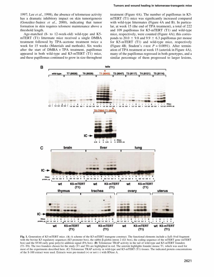
In vivo reconstitution of telomerase activity intransgenic K5-mTERT miceWe used the 5¢-regulatory region of the bovine keratin K5gene (RamõÂrez et al., 1994; Murillas et al., 1995) to targetmTERT expression to basal keratinocytes (see Figure 1Afor details of the bovine K5 promoter region used toexpress mTERT) (Materials and methods). Eight differentfounder K5-mTERT transgenic mice were identi®ed(T1±T8) that showed increased telomerase activity in theskin of the tail compared with wild-type littermates(Figure 1B). Two of these founders, T1 = 8043 andT8 = 7664 (highlighted in red in Figure 1B) showed thehighest in vivo reconstitution of telomerase activity in thetail skin. All the results described here were obtained using
Increased epidermal tumors and increased skinwound healing in transgenic mice overexpressingthe catalytic subunit of telomerase, mTERT,in basal keratinocytes
The EMBO Journal Vol. 20 No. 11 pp. 2619±2630, 2001
ã European Molecular Biology Organization 2619
the K5-mTERT mice derived from the T1 founder, K5-mTERT (T1) (asterisk in Figure 1B), and were con®rmed,where indicated, with the T8 founder-derived K5-mTERTtransgenics, K5-mTERT (T8), to rule out possible non-speci®c effects due to transgene insertion.
In agreement with the known expression pattern ofkeratin K5 (RamõÂrez et al., 1994; Murillas et al., 1995), thestrati®ed epithelia of K5-mTERT (T1) mice tested,including uterus, trachea and skin, showed signi®cantupregulation of telomerase activity as compared with thecorresponding littermate wild-type tissues (Figure 1B andC). This telomerase upregulation coincided with increasedmTERT mRNA levels in K5-mTERT transgenic skin(between 25- and 50-fold) compared with wild-typecontrols (data not shown). Other tissues of the K5-mTERT (T1) mice where the transgene is not expressed(i.e. liver, brain, lung, ovary and whole thymus) did notshow upregulation of telomerase activity as comparedwith wild-type littermates (Figure 1C).
Telomere length in wild-type and K5-mTERTtransgenic basal keratinocytesPrevious studies using cultured human cells showed thatTERT-mediated telomerase activity reconstitution re-sulted in extension or maintenance of telomere length(Bodnar et al., 1998; Kiyono et al., 1998; Hahn et al.,1999). We measured telomere length in K5-mTERT (T1)skin keratinocytes by quantitative ¯uorescence in situhybridization (Q-FISH) on skin sections as previouslydescribed (GonzaÂlez-SuaÂrez et al., 2000). Figure 2Ashows Q-FISH images of interphase nuclei in the skin ofage-matched (8-week-old) littermate wild-type and K5-mTERT (T1) mice; the basal layer of skin keratynocytes isindicated with a yellow arrow. Quanti®cation of the¯uorescence intensity of telomere dots showed thattelomeres in K5-mTERT (T1) epidermis are similar inlength to those of age-matched littermate wild-type mice(Figure 2B; black bars). Primary keratinocyte culturesderived from newborn wild-type and K5-mTERT (T1)mice also showed similar telomere length using Flow-FISH, an independent quantitative FISH technique tomeasure telomere length that is based on ¯ow cytometry(see Materials and methods) (Rufer et al., 1998)(Figure 2C). All together, these results suggest thatTERT-driven telomerase upregulation in basal skin
keratinocytes does not result in a signi®cant extension oftelomeres in the skin as compared with age-matched wild-type mice.
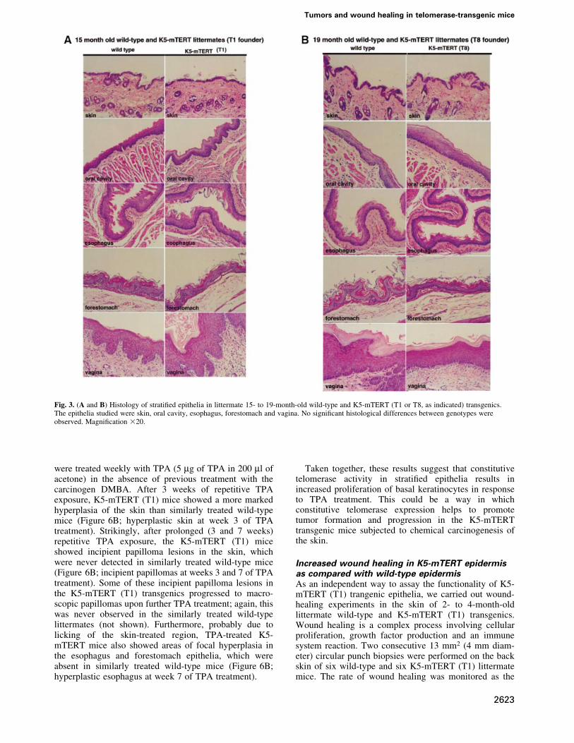
K5-mTERT strati®ed epithelia are histologicallynormalK5-mTERT (T1 and T8) mice, ranging from newborn to19 months old, were phenotypically indistinguishablefrom the corresponding age-matched wild-type littermatesand showed no pathologies, no spontaneous developmentof epithelial tumors and no loss of viability with age (notshown). In agreement with this, histopathological analysisof strati®ed epithelia (skin, oral cavity, esophagus, fore-stomach and vagina) from 15- to 19-month-old K5-mTERT (T1 and T8, as indicated) and the correspondingage-matched littermate wild-type controls revealed nomacroscopic or histological differences between geno-types, indicating that transgenic telomerase expressionper se does not alter the strati®ed epithelium structure(Figure 3). These results suggest that potential telomerasereconstitution for gene therapy of epithelial aging-associ-ated diseases would not have any deleterious effects on thestructure of strati®ed epithelia.
Increased tumor susceptibility and mortality ofK5-mTERT transgenics produced by chemicalcarcinogensTo study further the effects of telomerase overexpressionon the normal biology and function of strati®ed epithelia,we ®rst addressed whether transgenic telomerase expres-sion impacts on epithelial tumor formation upon exposureto chemical carcinogens. The stages of initiation, promo-tion and tumor progression in the classical skin chemicalcarcinogenesis model are well characterized (Heckneret al., 1982). In wild-type mice, initiation using 7,12-dimethylbenz[a]anthracene (DMBA) and subsequentpromotion with 12-o-tetradecanoylphorbol 13-acetate(TPA) leads to papillomas that are hyperplastic, well-differentiated, skin lesions. H-ras activation, loss of thep53 tumor suppressor and telomerase upregulation arereported in the majority of DMBA-initiated papillomas(Balmain et al., 1984, 1992; Quintanilla et al., 1986;Bednarek et al., 1997). Furthermore, when telomeres arecritically short, as in late generation mice de®cient for theRNA component of mouse telomerase, Terc (Blasco et al.,
E.Gonza lez-Sua rez et al.
2620
1997; Lee et al., 1998), the absence of telomerase activityhas a dramatic inhibitory impact on skin tumorigenesis(GonzaÂlez-SuaÂrez et al., 2000), indicating that tumorformation in skin requires telomere maintenance above athreshold length.
Age-matched (8- to 12-week-old) wild-type and K5-mTERT (T1) littermate mice received a single DMBAtreatment followed by TPA±acetone treatment twice aweek for 15 weeks (Materials and methods). Six weeksafter the start of DMBA + TPA treatment, papillomasappeared in both wild-type and K5-mTERT (T1) mice,and these papillomas continued to grow in size throughout
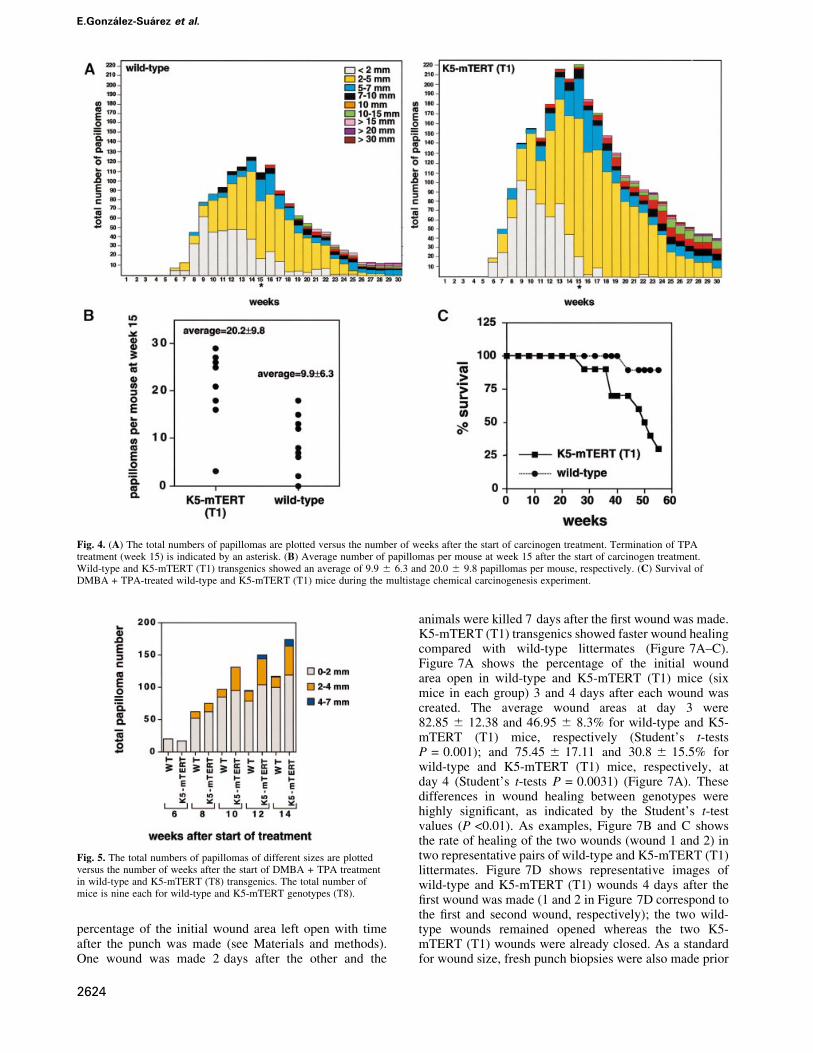
treatment (Figure 4A). The number of papillomas in K5-mTERT (T1) mice was signi®cantly increased comparedwith wild-type littermates (Figure 4A and B). In particu-lar, at week 15 (the end of TPA treatment), a total of 222and 109 papillomas for K5-mTERT (T1) and wild-typemice, respectively, were counted (Figure 4A); this corres-ponds to 20.0 6 9.8 and 9.9 6 6.3 papillomas per mousefor K5-mTERT (T1) and wild-type mice, respectively(Figure 4B; Student's t-test P = 0.0091). After termin-ation of TPA treatment at week 15 (asterisk in Figure 4A),many of the papillomas regressed in both genotypes, and asimilar percentage of them progressed to larger lesions,
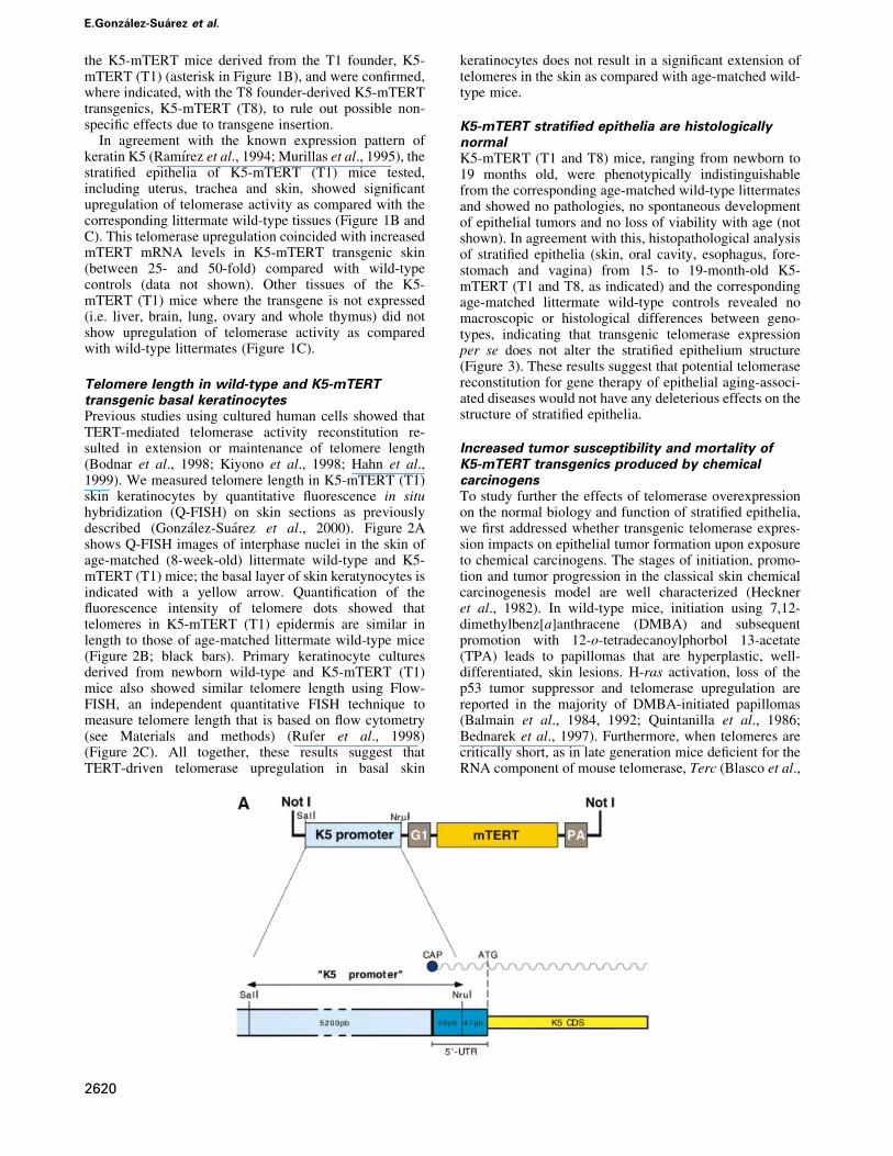
Fig. 1. Generation of K5-mTERT mice. (A) A scheme of the K5-mTERT transgene construct. The functional elements include a SalI±NruI fragmentwith the bovine K5 regulatory sequences (K5 promoter box), the rabbit b-globin intron 2 (G1 box), the coding sequence of the mTERT gene (mTERTbox) and the SV40 early gene poly(A) addition signal (PA box). (B) Telomerase TRAP activity in the tail of wild-type and K5-mTERT founders(T1±T8). The two founders chosen for the study (T1 and T8) are highlighted in red. The asterisk highlights founder mouse T1, which was used formost of the experiments described here. (C) Telomerase TRAP activity in wild-type and K5-mTERT (T1) tissues. The indicated protein concentrationsof the S-100 extract were used. Extracts were pre-treated (+) or not (±) with RNase A.
Tumors and wound healing in telomerase-transgenic mice
2621

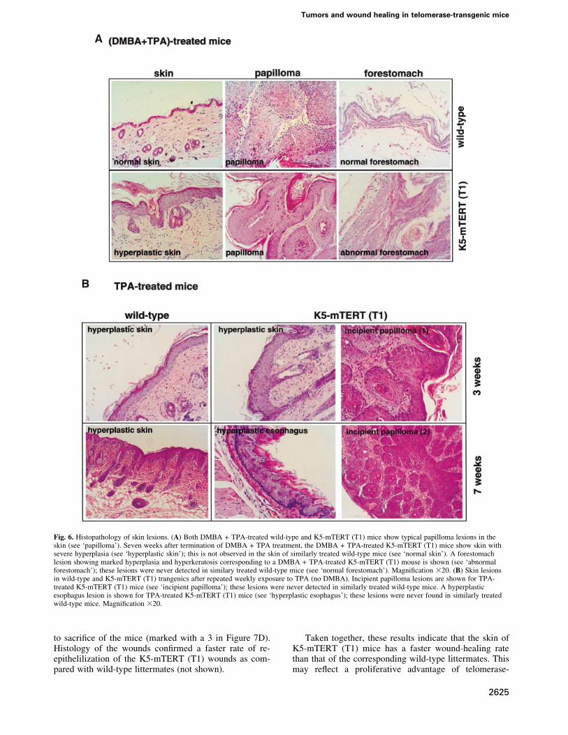
maintaining the difference in total number of papillomasbetween wild-type and K5-mTERT (T1) littermates. Inparticular, 6.4 and 7.2% of the papillomas at week 15progressed to lesions >1 cm at week 30, in wild-type andK5-mTERT (T1) mice, respectively (Figure 4A). Despitethe difference in total number of papillomas, no histo-logical differences were found between wild-type andK5-mTERT (T1) papillomas (Figure 6A; papilloma inwild-type and K5-mTERT T1 mice).
The increased susceptibility of transgenic K5-mTERT(T1) skin to develop papillomas due to chemicalcarcinogenesis was con®rmed using K5-mTERT micederived from a different founder, T8 (Figure 1B). K5-mTERT (T8) transgenics also showed an increasednumber of papillomas upon DMBA + TPA treatment ascompared with the corresponding wild-type littermates(Figure 5).
Strikingly, 80% of the DMBA + TPA-treated K5-mTERT (T1) mice (eight out of 10) died before week 60after the start of treatment, whereas 90% of the similarlytreated wild-type cohorts survived during this time (nineout of 10; Figure 4C). Death of the DMBA + TPA-treatedK5-mTERT (T1) mice was associated with severalpathologies (see Table I for quanti®cations) includingthe following. (i) Severe gastric hemorrhages resultingfrom an abnormal forestomach epithelium, which showeda marked hyperplasia and hyperkeratosis (Figure 6A;compare the normal forestomach epithelium inDMBA + TPA-treated wild-type mice with the abnormalforestomach in DMBA + TPA-treated K5-mTERT mice).This observation suggests that other K5-mTERT (T1)
strati®ed epithelia besides the skin are prone to tumor-igenesis upon DMBA + TPA treatment. (ii) Benign epi-dermal lessions: numerous keratoacanthomas and bluenevi (melanocyte accumulations in the skin). (iii) Pre-malignant epidermal lesions: severe dysplastic papillomas.(iv) Malignant epidermal tumors: basal cell carcinomas,malignant keratoacanthomas. (v) Tumors of non-epithelialorigin: subcutaneous ®brosarcoma, uterus histiocyticsarcoma, liver histiocytic sarcoma, cecum lymphoma, aswell as in®ltrations of tumoral lymphoid cells in varioustissues (multicentric lymphomas).
Hence, the increased mortality of K5-mTERT miceupon DMBA + TPA carcinogenesis coincides with ahigher susceptibility of these mice to develop neoplasias.
Telomeric Q-FISH analysis on skin sections fromDMBA + TPA-treated mice showed that telomeres weresimilar or slightly elongated in K5-mTERT (T1) mice ascompared with wild-type controls (Figure 2B; gray bars).
K5-mTERT skins are more sensitive to themitogenic effects of phorbol estersCuriously, whereas wild-type mice showed areas of histo-logically normal skin surrounding papillomas 7 weeksafter termination of DMBA + TPA treatment (Figure 6A;normal skin), the DMBA + TPA-treated K5-mTERT(T1) mice maintained a marked hyperplasia of the skin,with regions showing up to 10 layers of keratinocytes(Figure 6A; hyperplastic skin). These observations sug-gested an increased proliferative response of K5-mTERT(T1) basal keratinocytes to repetitive TPA treatment. Tostudy this further, wild-type and K5-mTERT (T1) mice
Fig. 2. Telomere ¯uorescence in skin sections. (A) Illustrative images showing telomere ¯uorescence in wild-type and K5-mTERT (T1) skin sections.The basal layer of skin keratinocytes is indicated (yellow arrow). (B) Quanti®cation of telomere ¯uorescence on untreated skin sections or on skinsections treated with DMBA + TPA, as indicated. More than 50 keratinocyte nuclei of each genotype were analyzed by Q-FISH. (C) Averagetelomere ¯uorescence as determined by Flow-FISH of three wild-type and six K5-mTERT primary keratinocyte cultures. Between 2000 and 3000nuclei were analyzed by ¯ow cytometry for each culture.
E.Gonza lez-Sua rez et al.
2622
were treated weekly with TPA (5 mg of TPA in 200 ml ofacetone) in the absence of previous treatment with thecarcinogen DMBA. After 3 weeks of repetitive TPAexposure, K5-mTERT (T1) mice showed a more markedhyperplasia of the skin than similarly treated wild-typemice (Figure 6B; hyperplastic skin at week 3 of TPAtreatment). Strikingly, after prolonged (3 and 7 weeks)repetitive TPA exposure, the K5-mTERT (T1) miceshowed incipient papilloma lesions in the skin, whichwere never detected in similarly treated wild-type mice(Figure 6B; incipient papillomas at weeks 3 and 7 of TPAtreatment). Some of these incipient papilloma lesions inthe K5-mTERT (T1) transgenics progressed to macro-scopic papillomas upon further TPA treatment; again, thiswas never observed in the similarly treated wild-typelittermates (not shown). Furthermore, probably due tolicking of the skin-treated region, TPA-treated K5-mTERT mice also showed areas of focal hyperplasia inthe esophagus and forestomach epithelia, which wereabsent in similarly treated wild-type mice (Figure 6B;hyperplastic esophagus at week 7 of TPA treatment).
Taken together, these results suggest that constitutivetelomerase activity in strati®ed epithelia results inincreased proliferation of basal keratinocytes in responseto TPA treatment. This could be a way in whichconstitutive telomerase expression helps to promotetumor formation and progression in the K5-mTERTtransgenic mice subjected to chemical carcinogenesis ofthe skin.
Increased wound healing in K5-mTERT epidermisas compared with wild-type epidermisAs an independent way to assay the functionality of K5-mTERT (T1) trangenic epithelia, we carried out wound-healing experiments in the skin of 2- to 4-month-oldlittermate wild-type and K5-mTERT (T1) transgenics.Wound healing is a complex process involving cellularproliferation, growth factor production and an immunesystem reaction. Two consecutive 13 mm2 (4 mm diam-eter) circular punch biopsies were performed on the backskin of six wild-type and six K5-mTERT (T1) littermatemice. The rate of wound healing was monitored as the
Fig. 3. (A and B) Histology of strati®ed epithelia in littermate 15- to 19-month-old wild-type and K5-mTERT (T1 or T8, as indicated) transgenics.The epithelia studied were skin, oral cavity, esophagus, forestomach and vagina. No signi®cant histological differences between genotypes wereobserved. Magni®cation 320.
Tumors and wound healing in telomerase-transgenic mice
2623
percentage of the initial wound area left open with timeafter the punch was made (see Materials and methods).One wound was made 2 days after the other and the
animals were killed 7 days after the ®rst wound was made.K5-mTERT (T1) transgenics showed faster wound healingcompared with wild-type littermates (Figure 7A±C).Figure 7A shows the percentage of the initial woundarea open in wild-type and K5-mTERT (T1) mice (sixmice in each group) 3 and 4 days after each wound wascreated. The average wound areas at day 3 were82.85 6 12.38 and 46.95 6 8.3% for wild-type and K5-mTERT (T1) mice, respectively (Student's t-testsP = 0.001); and 75.45 6 17.11 and 30.8 6 15.5% forwild-type and K5-mTERT (T1) mice, respectively, atday 4 (Student's t-tests P = 0.0031) (Figure 7A). Thesedifferences in wound healing between genotypes werehighly signi®cant, as indicated by the Student's t-testvalues (P <0.01). As examples, Figure 7B and C showsthe rate of healing of the two wounds (wound 1 and 2) intwo representative pairs of wild-type and K5-mTERT (T1)littermates. Figure 7D shows representative images ofwild-type and K5-mTERT (T1) wounds 4 days after the®rst wound was made (1 and 2 in Figure 7D correspond tothe ®rst and second wound, respectively); the two wild-type wounds remained opened whereas the two K5-mTERT (T1) wounds were already closed. As a standardfor wound size, fresh punch biopsies were also made prior
Fig. 4. (A) The total numbers of papillomas are plotted versus the number of weeks after the start of carcinogen treatment. Termination of TPAtreatment (week 15) is indicated by an asterisk. (B) Average number of papillomas per mouse at week 15 after the start of carcinogen treatment.Wild-type and K5-mTERT (T1) transgenics showed an average of 9.9 6 6.3 and 20.0 6 9.8 papillomas per mouse, respectively. (C) Survival ofDMBA + TPA-treated wild-type and K5-mTERT (T1) mice during the multistage chemical carcinogenesis experiment.
Fig. 5. The total numbers of papillomas of different sizes are plottedversus the number of weeks after the start of DMBA + TPA treatmentin wild-type and K5-mTERT (T8) transgenics. The total number ofmice is nine each for wild-type and K5-mTERT genotypes (T8).
E.Gonza lez-Sua rez et al.
2624
to sacri®ce of the mice (marked with a 3 in Figure 7D).Histology of the wounds con®rmed a faster rate of re-epithelilization of the K5-mTERT (T1) wounds as com-pared with wild-type littermates (not shown).
Taken together, these results indicate that the skin ofK5-mTERT (T1) mice has a faster wound-healing ratethan that of the corresponding wild-type littermates. Thismay re¯ect a proliferative advantage of telomerase-
Fig. 6. Histopathology of skin lesions. (A) Both DMBA + TPA-treated wild-type and K5-mTERT (T1) mice show typical papilloma lesions in theskin (see `papilloma'). Seven weeks after termination of DMBA + TPA treatment, the DMBA + TPA-treated K5-mTERT (T1) mice show skin withsevere hyperplasia (see `hyperplastic skin'); this is not observed in the skin of similarly treated wild-type mice (see `normal skin'). A forestomachlesion showing marked hyperplasia and hyperkeratosis corresponding to a DMBA + TPA-treated K5-mTERT (T1) mouse is shown (see `abnormalforestomach'); these lesions were never detected in similary treated wild-type mice (see `normal forestomach'). Magni®cation 320. (B) Skin lesionsin wild-type and K5-mTERT (T1) trangenics after repeated weekly exposure to TPA (no DMBA). Incipient papilloma lesions are shown for TPA-treated K5-mTERT (T1) mice (see `incipient papilloma'); these lesions were never detected in similarly treated wild-type mice. A hyperplasticesophagus lesion is shown for TPA-treated K5-mTERT (T1) mice (see `hyperplastic esophagus'); these lesions were never found in similarly treatedwild-type mice. Magni®cation 320.
Tumors and wound healing in telomerase-transgenic mice
2625

Fig. 7. Wound healing of wild-type and K5-mTERT (T1) skin. (A) Percentage of initial wound area left 3 and 4 days after the punch was created in agroup of six wild-type and six K5-mTERT (T1) mice. Statistical analysis showing that the healing rate is signi®cantly different between genotypes isalso shown (P-values). (B and C) Rate of wound healing in two wild-type and K5-TERT (T1) littermate pairs; the results are presented as thepercentage of open wound area versus time after wound creation. Closed circles, wild-type mice; open circles, K5-mTERT (T1) transgenics. (D) Grossappearance of healing wounds in a wild-type and a K5-mTERT (T1) mouse 5 and 2 days, for wounds 1 and 2, respectively, after wound creation. Justprior to sacri®ce of the mice, a third wound (wound 3) was created as an indicator of initial wound areas.
E.Gonza lez-Sua rez et al.
2626

transgenic skins as compared with controls in responseto proliferative signals associated with wound healing,in agreement with the greater hyperplasia seen inK5-mTERT epithelia as compared with wild-type litter-mates upon treatment with the mitogen TPA.
Normal p53, Ras and c-Myc levels inK5-mTERT skinp53 loss of function and H-ras upregulation are describedas occurring in multistage chemical carcinogenesis of theskin, and are thought to favor the appearance of tumors(Balmain et al., 1984, 1992; Quintanilla et al., 1986). Todetermine whether constitutive telomerase expressioninduces changes associated with transformation in the
skin, we studied several tumor markers such as p53,Ras and c-Myc in the skin of wild-type and littermateK5-mTERT (T1) mice. To analyze p53 status in skinkeratinocytes, we performed immunohistochemistry ofthe skin with anti-p53 antibody, as previously described(GonzaÂlez-SuaÂrez et al., 2000) (see Materials andmethods). There is no signi®cant variation in p53 levelsbetween wild-type and K5-mTERT (T1) untreated skin orDMBA + TPA-induced papillomas (Figure 8A), suggest-ing that transgenic telomerase expression does not affectnormal p53 levels; as a control, we show the skin of a p53-de®cient mouse (Figure 8A). Furthermore, there was nosigni®cant difference in Ras protein levels in nuclearextracts prepared from wild-type and K5-mTERT (T1)
Fig. 8. (A) p53 levels in wild-type and K5-mTERT (T1) skin keratinocytes. Immunohistochemistry with p53 antibody of untreated and DMBA +TPA-treated wild-type and K5-mTERT (T1) skin. The control untreated wild-type and K5-mTERT (T1) skin show similar p53 positivity in basalkeratinocyte nuclei; p53±/± mouse skin is shown as a negative control. Wild-type and K5-mTERT (T1) papillomas show decreased p53 staining ofbasal keratinocytes compared with wild-type skin. Magni®cation 320. (B) Ras protein levels detected by western blot in wild-type and K5-mTERT(T1) primary keratinocytes. No signi®cant differences in Ras protein levels were detected between genotypes. As a control for loading, nuclear proteinKu70 was also detected. Different numbers indicate different primary keratinocyte cultures. The arrows indicate the positions of theRas- and Ku86-speci®c bands. (C) c-myc mRNA levels detected by northern blot in the skin of four K5-mTERT (T1 or T8, as indicated) and the corresponding wild-type littermate skins. Mice 30, 32, 34 and 35 are littermates derived from founder T8. Mice 130, 131, 132 and 134 are littermates derived fromfounder T1. As a positive control for c-myc detection, total RNA from interleukin-2-stimulated Ba/FO3 cells was used (Hatakeyama et al., 1989).As control for loading, actin was also detected in the blots. Table II shows quanti®cation of c-myc levels.
Tumors and wound healing in telomerase-transgenic mice
2627
primary keratinocyte cultures, as determined by westernblot (Figure 8B). Detection of Ku70 nuclear protein wasused as a loading control (Figure 8B).
It has been shown recently that TERT-driven cellproliferation in cultured cells can result in the activation ofthe c-myc oncogene (Wang et al., 2000). We studied c-mycmRNA levels in the skin from several K5-mTERT (T1 andT8, as indicated) mice and corresponding wild-typelittermate controls. No signi®cant differences in thec-myc mRNA levels were observed between genotypes(see Materials and methods; Figure 8C and Table II forquantitation of c-Myc levels using a phosphoimager),suggesting that transgenic mTERT expression in the skindoes not result in c-myc upregulation.
Conclusions
We show here that constitutive high levels of telomeraseactivity in strati®ed epithelia do not alter the normalepithelium structure and are not associated with changes inp53, Ras or c-Myc levels, in agreement with previous
reports showing that TERT immortalized human culturedcells do not show changes associated with transformation(Jiang et al., 1999; Morales et al., 1999). However, follow-ing DMBA + TPA treatment of the skin, K5-mTERT (T1and T8) mice are signi®cantly more susceptible to thedevelopment of neoplasias than the corresponding wild-type littermates. This increased tumor susceptibility ofK5-mTERT transgenic skin is accompanied by a greaterproliferation of basal keratinocytes upon TPA treatment,as well as by an increased would-healing rate of theK5-mTERT skin compared with wild-type mice. All theseobservations are in agreement with a direct or indirect rolefor telomerase in signaling proliferation under mitogenicconditions. These results agree with previous observationsthat indicated that ®rst generation Terc±/± mice, which lacktelomerase activity but still have long telomeres, show alower incidence of papillomas than wild-type mice(GonzaÂlez-SuaÂrez et al., 2000). These ®ndings unravelthe potential risks of gene therapy for age-associateddiseases based on telomerase upregulation, and indicatethat telomerase upregulation, which occurs in the vast
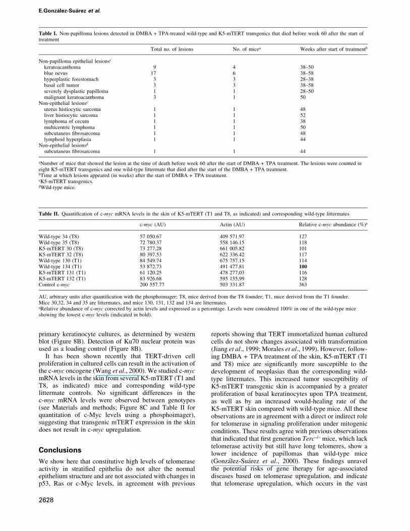
Table I. Non-papilloma lesions detected in DMBA + TPA-treated wild-type and K5-mTERT transgenics that died before week 60 after the start oftreatment
Total no. of lesions No. of micea Weeks after start of treatmentb
Non-papilloma epithelial lesionsc
keratoacanthoma 9 4 38±50blue nevus 17 6 38±58hyperplastic forestomach 3 3 28±38basal cell tumor 3 3 38±58severely dysplastic papilloma 1 1 28±50malignant keratoacanthoma 3 1 50
Non-epithelial lesionsc
uterus histiocytic sarcoma 1 1 48liver histiocytic sarcoma 1 1 52lymphoma of cecum 1 1 38multicentric lymphoma 1 1 50subcutaneus ®brosarcoma 1 1 48lymphoid hyperplasia 1 1 44
Non-epithelial lesionsd
subcutaneus ®brosarcoma 1 1 44
aNumber of mice that showed the lesion at the time of death before week 60 after the start of DMBA + TPA treatment. The lesions were counted ineight K5-mTERT transgenics and one wild-type littermate that died after the start of the DMBA + TPA treatment.bTime at which lesions appeared (in weeks) after the start of DMBA + TPA treatment.cK5-mTERT transgenics.dWild-type mice.
Table II. Quanti®cation of c-myc mRNA levels in the skin of K5-mTERT (T1 and T8, as indicated) and corresponding wild-type littermates
c-myc (AU) Actin (AU) Relative c-myc abundance (%)a
Wild-type 34 (T8) 57 050.67 409 571.97 127Wild-type 35 (T8) 72 780.37 558 146.15 118K5-mTERT 30 (T8) 73 277,28 661 005.82 101K5-mTERT 32 (T8) 80 397.53 622 336.42 117Wild-type 130 (T1) 84 549.74 675 757.15 114Wild-type 134 (T1) 53 872.73 491 477.81 100K5-mTERT 131 (T1) 61 120.25 478 277,03 116K5-mTERT 132 (T1) 83 926.68 595 155,99 128Control c-myc 200 557.77 503 331.87 363
AU, arbitrary units after quanti®cation with the phosphoimager; T8, mice derived from the T8 founder; T1, mice derived from the T1 founder.Mice 30,32, 34 and 35 are littermates, and mice 130, 131, 132 and 134 are littermates.aRelative abundance of c-myc corrected by actin levels and expressed as a percentage. Levels were considered 100% in one of the wild-type miceshowing the lowest c-myc levels (indicated in bold).
E.Gonza lez-Sua rez et al.
2628

majority of mouse and human tumors, actively promotestumor growth even in the presence of long telomeres.Since the average length of telomeres in wild-type and K5-mTERT skin was similar, it is tempting to speculate thattelomere conformation must be different whether or nottelomerase activity is present in the cell, and that telomeresthat are being `maintained' by telomerase could besignaling proliferation and/or survival under mitogenicconditions.
Finally, K5-mTERT mice constitute a new experimentalmodel to study the direct impact of transgenic telomeraseexpression in both skin tumorigenesis and aging in vivo.Tissue-speci®c telomerase-transgenic mice are essential tostudy the impact of potential use of telomerase for genetherapy of age-related diseases. K5-mTERT mice alsoconstitute a new animal model system to study theeffectiveness of anti-telomerase therapies in cancer.
Materials and methods
Generation and genotyping of transgenic K5-mTERT miceAn EcoRI fragment containing the full-length mTERT cDNA and 3¢-untranslated sequences was cloned 3¢ downstream of 5 kb of bovinekeratin K5 regulatory sequences (see Figure 1A) (RamõÂrez et al., 1994;MartõÂn-Rivera et al., 1998). The transgene was digested with NotI,puri®ed using Genclean (Bio 101 Inc.), adjusted to a ®nal concentration of~2 mg/ml and microinjected into (C57BL/6 3 DBA/2) mouse embryos,as described (Hogan et al., 1986). Eight different founder mice wereidenti®ed by Southern blot analysis of tail DNA, using the 5 kb fragmentcorresponding to the bovine keratin K5 promoter as a probe (not shown).The experiments described here were carried out with founder T1 andwere reproduced, as indicated, with founder T8, which shows a similarupregulation of telomerase activity (T1 = 8043 and T8 = 7664; high-lighted in red in Figure 1B). The other founders showing lowerreconstitution of telomerase activity in the skin were not included inthis study.
Handling of miceMice were housed in our barrier area, where pathogen-free procedures areemployed in all mouse rooms. Quarterly health monitoring reports havebeen negative for all pathogens in accordance with FELASA recommend-ations (Federation of European Laboratory Animal Science Associations).
Telomerase assaysTelomerase activity was measured with a modi®ed telomeric repeatampli®cation protocol (TRAP), as described (Blasco et al., 1997).
Tumor induction experimentsTen and 12 age-matched (8- to 12-week-old) mice of each genotype,wild-type and K5-mTERT (T1), respectively, were shaved and treatedwith a single dose of DMBA (0.1 mg/ml in acetone; Sigma). Nine age-matched (8-week-old) mice of each genotype, wild-type and K5-mTERT(T8), were shaved and treated with a single dose of DMBA (0.1 mg/ml inacetone; Sigma). In all cases, subsequently mice were treated twiceweekly with TPA (12.5 mg in 200 ml acetone each treatment; Sigma), for15 weeks. Four control mice of each genotype were treated with acetoneonly. In samples analyzed after weekly cumulative TPA doses, the TPAconcentration used was 12.3 mg in 200 ml of acetone.
Histopathological analyses and immunochemistryPapilloma, skin, esophagus, forestomach, oral mucosa and vaginasections from 15- to 19-month-old wild-type and K5-mTERT (T1 orT8, as indicated) mice were ®xed in 10% buffered formalin and stainedwith hematoxylin±eosin. Images were captured with an Olympus-Vanoxmicroscope at a 203 magni®cation.
For the DMBA + TPA-treated mice that died, necropsy of animalswas performed and all organs were examined macroscopically. Tumoraland normal tissues were ®xed in 10% buffered formalin and embeddedin paraf®n. Sections of 4 mm were stained with hematoxylin±eosin.Images were captured with an Olympus-Vanox microscope at a 203magni®cation.
Immunohistochemistry for p53 was performed on deparaf®natedsections (Vectabond slides) after pressure cooker processing. For p53detection, we used a streptavidin±biotin complex technique, with apolyclonal rabbit anti-mouse p53 antibody, CM5 (Novocastra LaboratoryLtd; 1:1500 dilution, overnight, 4°C), and a biotinylated anti-rabbit IgG(Vector Laboratory; 1:400 dilution, 30 min, room temperature), followedby streptavidin peroxidase (Zymed Laboratory, Inc.; 1:20 dilution,30 min, room temperature); the chromogen was developed withdiaminobenzidine, and slides were counterstained with hematoxylin.Control slides were obtained by replacing the primary antibody with TBS(not shown) (see also GonzaÂlez-SuaÂrez et al., 2000).
Wound-healing experimentsTwo to three full-thickness punch biopsies extending through theepidermis and dermis (punch diameter 4 mm; PFM, KoÈln, Germany)were performed in six wild-type and six K5-mTERT mice (2±4 months ofage) after depilation. Mice were anesthesized prior to wound creation.The wound-healing rate was calculated as the percentage of initial woundarea with time. Wound areas were calculated with the formula(area = pr 2; where r is the ratio of the wound).
Telomere length measurementsFor Q-FISH, paraf®n-embedded skin sections (untreated or treated withDMBA + TPA) were hybridized with a PNA-tel probe, and telomerelength was determined as described (Zijlmans et al., 1997;GonzaÂlez-SuaÂrez et al., 2000). Slides were deparaf®nated in three xylenewashes (3 min each), then treated for 3 min with a 100, 95 and 70%ethanol series. More than 50 keratinocyte nuclei from each genotype werecaptured at a 1003 magni®cation and the telomere ¯uorescence wasintegrated using spot IOD analysis in the TFL-TELO program. For Flow-FISH, primary mouse keratinocytes (105) were prepared (Hennings,1994) and hybridized using the Flow-FISH protocol (Rufer et al., 1998).Telomere ¯uorescence of 2000±3000 nuclei gated at the G1±G0 cell cyclestage was measured using a Coulter EPICS XL ¯ow cytometer withSYSTEM 2 software. The same cells were hybridized in parallel withoutthe PNA±¯uorescein isothiocyanate (FITC) probe to obtain background¯uorescence values, which were subtracted from the ¯uorescenceintensity of each sample. We used three wild-type and six K5-mTERT(T1) primary keratinocyte cultures.
Western blotsNuclear extracts were prepared from three primary wild-type and fourK5-mTERT (T1) keratinocyte cultures (Hennings, 1994), and westernblots were performed as previously described (GonzaÂlez-SuaÂrez et al.,2000). The protein concentration per well was 20 mg. For Ras detection,pan-ras antibody (Ab-3; Calbiochem) at a 1:1000 dilution was used. ForKu86 detection, anti-human p70 (Hamiya Biomedical Company, 1:000dilution) was used. The percentage of the acrylamide gel was 15%.
Northern blotsTotal RNA was extracted from the skin of a total of four wild-type andfour K5-mTERT (T1 or T8, as indicated) littermate mice essentially asdescribed (Chomczynski and Sacchi, 1987). A 50 mg aliquot of RNA wasused per lane in the northern blot. A 1500 nucleotide fragment of thehuman c-myc gene was used as probe for murine c-myc mRNA detection;as a positive control, total RNA from murine lymphocyte cell line Ba/FO3upon interleukin-2 stimulation was used (Hatakeyama et al., 1989). Ascontrol for loading, actin was also detected in the blots.
Acknowledgements
We are indebted to F.Larcher for help in isolating primary keratinocytes.We thank E.Santos and Rosa Serrano for mouse care and genotyping,P.Jurado for her contribution to the construction of the K5-mTERTtransgene, and M.Serrano and C.Mark for critical reading of themanuscript. E.G.-S. is a predoctoral fellow from the Fondo deInvestigaciones Sanitarias. E.S. is a predoctoral fellow from theRegional Government of Madrid (CAM). Research at the laboratory ofM.A.B. is funded by Swiss Bridge Award 2000, by grants PM97-0133from the Ministry of Science and Technology (MCT), Spain, 08.1/0030/98 from the CAM, FIGH-CT1999-00009, FIGH-CT-1999-00002 andQLG1-1999-01341 from the European Union, and by the Department ofImmunology and Oncology (DIO). Research at the laboratory of J.L.J.was funded by grant PM98-0039 from the MCT. The DIO was foundedand is supported by the Spanish Research Council (CSIC) and by thePharmacia Corporation.
Tumors and wound healing in telomerase-transgenic mice
2629
References
Balmain,A., Ramsden,M., Bowden,G.T. and Smith,J. (1984) Activationof the mouse Harvey-ras gene in chemically induced benign skinpapillomas. Nature, 307, 658±660.
Balmain,A. et al. (1992) Functional loss of tumor suppressor genes inmultistage chemical carcinogenesis. In Harris,C.C. et al. (eds),Multistage Carcinogenesis. Japan Scienti®c Society Press/CRCPress, Boca Raton, FL, pp. 97±108.
Bednarek,A.K., Chu,Y., Slaga,T.J. and Aldaz,C.M. (1997). Telomeraseand cell proliferation in mouse skin papillomas. Mol. Carcinog., 20,329±310.
Blackburn,E.H. (1991) Structure and function of telomeres. Nature, 350,569±573.
Blasco,M.A., Funk,W.D., Villeponteau,B. and Greider,C.W. (1995)Functional characterization and developmental regulation of mousetelomerase RNA. Science, 269, 1267±1270.
Blasco,M.A., Lee,H.-W., Hande,P., Samper,E., Lansdorp,P., DePinho,R.and Greider,C.W. (1997) Telomere shortening and tumor formation bymouse cells lacking telomerase RNA. Cell, 91, 25±34.
Bodnar,A.G. et al. (1998) Extension of life-span by introduction oftelomerase into normal human cells. Science, 279, 349±352.
Chomczynski,P and Sacchi,N. (1987) Single-step method of RNAisolation by acid guanidinium thiocyanate±phenol±chloroformextraction. Anal. Biochem., 162, 156±159.
Collins,K., Kobayashi,R. and Greider,C.W. (1995) Puri®cation ofTetrahymena telomerase and cloning of the genes for the twoprotein components of the enzyme. Cell, 81, 677±686.
Counter,C.M., Avilion,A.A., LeFeuvre,C.E., Stewart,N.G., Greider,C.W., Harley,C.B. and Bacchetti,S. (1992) Telomere shorteningassociated with chromosome instability is arrested in immortal cellswhich express telomerase activity. EMBO J., 11, 1921±1929.
Feng,J. et al. (1995) The RNA component of human telomerase. Science,269, 1236±1241.
Gandhi,L. and Collins,K. (1998) Interaction of recombinantTetrahymena telomerase proteins p80 and p95 with telomerase RNAand telomeric DNA substrates. Genes Dev., 12, 721±733.
GonzaÂlez-SuaÂrez,E., Samper,E., Flores,J.M. and Blasco,M.A. (2000)Telomerase-de®cient mice with short telomeres are resistant to skintumorigenesis. Nature Genet., 26, 114±117
Greenberg,R.A., Allsopp,R.C., Chin,L., Morin,G. and DePinho,R. (1998)Expression of mouse telomerase reverse transcriptase during develop-ment, differentiation and proliferation. Oncogene, 16, 1723±1730.
Greider,C.W. (1996) Telomere length regulation. Annu. Rev. Biochem.,65, 337±365.
Greider,C.W. and Blackburn,E.H. (1985) Identi®cation of a speci®ctelomere terminal transferase activity in Tetrahymena extracts. Cell,43, 405±413.
Greider,C.W. and Blackburn,E.H. (1989) A telomeric sequence in theRNA of Tetrahymena telomerase required for telomere repeatsynthesis. Nature, 337, 331±337.
Hahn,W.C., Counter,C.M., Lundberg,A.S., Beijersbergen,R.L.,Brooks,M.W. and Weinberg,R.A. (1999) Creation of human tumourcells with de®ned genetic elements. Nature, 400, 464±468.
Harrington,L., Zhou,W., McPhail,T., Oulton,R., Yeung,D., Mar,V.,Bass,M.B. and Robinson,M.O. (1997a) Human telomerase containsevolutionarily conserved catalytic and structural subunits. Genes Dev.,11, 3109±3115.
Harrington,L., McPhail,T., Mar,V., Zhou,W., Oulton,R., Bass,M.B.,Arruda,I. and Robinson,M.O. (1997b) A mammalian telomerase-associated protein. Science, 275, 973±977.
Hatakeyama,M., Mori,H., Doi,T. and Taniguchi,T. (1989) A restrictedcytoplasmic region of IL-2 receptor b chain is essential for growthsignal transduction but not for ligand binding and internalization. Cell,59, 837±845.
Heckner,E., Fusenig,N.E., Kunz,W., Marks,F. and Thielmann,H.W.(1982) Carcinogenesis: A Comprehensive Survey, Vol. 7. Co-carcinogenesis and Biological Effects of Tumor Promoters. RavenPress, New York, NY.
Hennings,H. (1994) Primary culture of keratinocytes from newbornmouse epidermis in medium with lowered levels of Ca2+. InLeigh,I.M. and Watt,F.M. (eds), Keratinocyte Methods. CambridgeUniversity Press, Cambridge, UK, pp. 21±23.
Hogan,B., Constantini,F. and Lacy,E. (1986) Manipulating the MouseEmbryo: A Laboratory Manual. Cold Spring Harbor Laboratory Press,Cold Spring Harbor, NY.
Jiang,X.-R. et al. (1999) Telomerase expression in human somatic cells
does not induce changes associated with a transformed phenotype.Nature Genet., 21, 111±114.
Kilian,A., Bowtell,D.D.L., Abud,H.E., Hime,G.R., Venter,D.J.,Keese,P.K., Duncan,E.L., Reddel,R.R. and Jefferson,R.A. (1997)Isolation of a candidate human telomerase catalytic subunit gene,which reveals complex splicing patterns in different cell types. Hum.Mol. Genet., 6, 2011±2019.
Kiyono,T., Foster,S.A., Koop,J.I., McDougall,J.K., Galloway,D.A. andKlingelhutz,A.J. (1998) Both Rb/p16INK4a inactivation and telomeraseactivity are required to immortalize human epithelial cells. Nature,396, 84±88.
Lee,H.-W., Blasco,M.A., Gottlieb,G.J., Horner,J.W., Greider,C.W. andDePinho,R.A. (1998) Essential role of mouse telomerase in highlyproliferative organs. Nature, 392, 569±574.
Lingner,J., Hughes,T.R., Schevchenko,A., Mann,M., Lundblad,V. andCech T. (1997) Reverse transcriptase motifs in the catalytic subunit oftelomerase. Science, 276, 561±567.
MartõÂn-Rivera,L., Herrera,E., Albar,J.P. and Blasco,M.A. (1998)Expression of mouse telomerase catalytic subunit in embryos andadult tissues. Proc. Natl Acad. Sci. USA, 95, 10471±10476.
Meyerson,M. et al. (1997) hEST2, the putative human telomerasecatalytic subunit gene, is upregulated in tumor cells and duringimmortalization. Cell, 90, 785±795.
Mitchell,J.R., Wood,E. and Collins,K. (1999) A telomerase component isdefective in the human disease dyskeratosis congenita. Nature, 402,551±555.
Morales,C.P., Holt,S.E., Ouellette,M., Kaur,K.J., Yan,Y., Wilson,K.S.,White,M.A., Wright,W.E. and Shay,J.W. (1999) Absence of cancer-associated changes in human ®broblasts immortalized withtelomerase. Nature Genet., 21, 115±118.
Murillas,R., Larcher,F., Conti,C.J., Santos,M., Ullrich,A. and Jorcano,J.L. (1995) Expression of a dominant negative mutant of epidermalgrowth factor receptor in the epidermis of transgenic mice elicitsstriking alterations on hair follicle development and skin structure.EMBO J., 14, 5216±5223.
Nakamura,T.M., Morin,G.B., Chapman,K.B., Weinrich,S.L., Andrews,W.H., Lingner,J., Harley,C.B. and Cech,T. (1997) Telomerasecatalytic subunit homologs from ®ssion yeast and human. Science,277, 955±959.
Nakayama,J., Saito,M., Nakamura,H., Matsuura,A. and Ishikawa,F.(1997) TLP1: a gene encoding a protein component of mammaliantelomerase is a novel member of WD repeat family. Cell, 88, 875±884.
Nugent,C.I. and Lundblad,V. (1998) The telomerase reversetranscriptase: components and regulation. Genes Dev., 12, 1073±1085.
Quintanilla,M., Brown,K., Ramsden,M. and Balmain,A. (1986)Carcinogen-speci®c mutation and ampli®cation of Ha-ras duringmouse skin carcinogenesis. Nature, 322, 78±80.
RamõÂrez,A., Bravo,A., Jorcano,J.L and Vidal,M. (1994) Sequences 5¢ ofthe bovine keratin 5 gene direct tissue- and cell-type-speci®cexpression of a LacZ gene in the adult and during development.Differentiation, 58, 53±64.
Rufer,N., Dragowska,W., Thornbury,G., Roosnek,E. and Lansdorp,P.M.(1998) Telomere length dynamics in human lymphocytesubpopulations measured by ¯ow cytometry. Nature Biotechnol., 16,743±147.
Samper,E., Goytisolo,F., Slijepcevic,P., van Buul,P. and Blasco,M.A.(2000) Mammalian Ku86 prevents telomeric fusions independently ofthe length of TTAGGG repeats and the G-strand overhang. EMBORep., 1, 244±252.
Shay,J.W. and Bacchetti,S. (1997) A survey of telomerase activity inhuman cancer. Eur. J. Cancer, 33, 787±791.
Singer,M.S. and Gottschling,D.E. (1994) TLC1: template RNAcomponent of the Saccharomyces cerevisiae telomerase. Science,266, 387±388.
van Steensel,B., Smogorzewska,A. and de Lange,T. (1998) TRF2protects human telomeres from end-to-end fusions. Cell, 92, 401±413
Wang,J., Hannon,G.J. and Beach,D.H. (2000) Risky immortalization bytelomerase. Nature, 405, 755±756.
Weitzman,J.B. and Yaniv,M. (1999) Rebuilding the road to cancer.Nature, 400, 401±402.
Zijlmans,J.M., Martens,U.M., Poon,S., Raap,A.K., Tanke,H.J., Ward,R.K. and P.M. Lansdorp. (1997) Telomeres in the mouse have largeinter-chromosomal variations in the number of T2AG3 repeats. Proc.Natl Acad. Sci. USA, 94, 7423±7428.
Received December 4, 2000; revised March 12, 2001;accepted April 9, 2001
E.Gonza lez-Sua rez et al.
2630