Increased diastolic time fraction as beneficial adjunct of 1-adrenergic receptor blockade after...
17
Downloaded from UvA-DARE, the institutional repository of the University of Amsterdam (UvA) http://dare.uva.nl/document/97769 File ID 97769 Filename Chapter 6: Increased diastolic time fraction as beneficial adjunct of alpha-adrenergic receptor blockade after percutaneous coronary intervention SOURCE (OR PART OF THE FOLLOWING SOURCE): Type Dissertation Title Analysis of pulsatile coronary pressure and flow velocity : looking beyond means Author C. Kolyva Faculty Faculty of Medicine Year 2008 Pages 150 ISBN 978-90-9022737-5 FULL BIBLIOGRAPHIC DETAILS: http://dare.uva.nl/record/268354 Copyright It is not permitted to download or to forward/distribute the text or part of it without the consent of the author(s) and/or copyright holder(s), other then for strictly personal, individual use. UvA-DARE is a service provided by the library of the University of Amsterdam (http://dare.uva.nl)
-
Upload
academicmedicalcentreuniversiteitvanamsterdam -
Category
Documents
-
view
0 -
download
0
Transcript of Increased diastolic time fraction as beneficial adjunct of 1-adrenergic receptor blockade after...

Downloaded from UvA-DARE, the institutional repository of the University of Amsterdam (UvA)http://dare.uva.nl/document/97769
File ID 97769Filename Chapter 6: Increased diastolic time fraction as beneficial adjunct of alpha-adrenergic receptor
blockade after percutaneous coronary intervention
SOURCE (OR PART OF THE FOLLOWING SOURCE):Type DissertationTitle Analysis of pulsatile coronary pressure and flow velocity : looking beyond meansAuthor C. KolyvaFaculty Faculty of MedicineYear 2008Pages 150ISBN 978-90-9022737-5
FULL BIBLIOGRAPHIC DETAILS: http://dare.uva.nl/record/268354
Copyright It is not permitted to download or to forward/distribute the text or part of it without the consent of the author(s) and/orcopyright holder(s), other then for strictly personal, individual use. UvA-DARE is a service provided by the library of the University of Amsterdam (http://dare.uva.nl)

Chapter 6
Increased diastolic time fraction as beneficial adjunct of alpha-adrenergic receptor blockade after percutaneous
coronary intervention
Christina Kolyva1
Bart-Jan Verhoeff2
Jos AE Spaan1
Jan J Piek2
Maria Siebes1
Departments of 1Medical Physics and 2Cardiology, Academic Medical Center, University of Amsterdam, Amsterdam, The Netherlands
Revision submitted

90 Chapter 6
Abstract Background: The effect of alpha1-receptor blockade with urapidil on coronary blood flow and left ventricular function has been attributed to relief of diffuse coronary vasoconstriction following percutaneous coronary intervention (PCI). We hypothesized that an increase in diastolic time fraction (DTF) contributes to this beneficial action of urapidil.
Methods: In eleven patients with a 63 ± 4% diameter stenosis, ECG, aortic pressure (Pa)and distal intracoronary pressure (Pd), and blood flow velocity were recorded at baseline and throughout adenosine-induced hyperemia. Measurements were obtained before and after PCI, and after subsequent 1-receptor blockade with urapidil (10 mg ic). DTF was determined from the ECG and the aortic pressure waveform. Functional parameters such as rate pressure product (RPP), coronary flow velocity reserve and fractional flow reserve were assessed, as well as an index of hyperemic microvascular resistance (HMR).
Results: Urapidil administration after PCI induced an upward shift in the DTF-heart rate relationship resulting in a heart rate-independent increase in hyperemic DTF by 3.1 ± 0.8% (P<0.005). Hyperemic Pa and Pd decreased, respectively, by 6.1 ± 2.0% (P<0.05) and 5.7 ± 1.8% (P<0.01) after 1-blockade and RPP was reduced by 5.9 ± 2.2% (P<0.05). Although on average no other functional parameter was altered by 1-blockade due to confounding changes in pressure and heart rate, individual changes in HMR revealed the concerted action of acute changes in distal pressure and DTF.
Conclusions: Alpha1-receptor blockade after PCI significantly prolonged DTF at a given heart rate, thereby providing an adjunctive beneficial mechanism for improving subendocardial perfusion, which critically depends on DTF.

Alpha-blockade and diastolic time fraction 91
6
Introduction Post-ischemic left ventricular dysfunction and impaired coronary flow reserve after percutaneous transluminal coronary angioplasty (PTCA) have been attributed to stimulation of -adrenergic pathways in the vessel wall [9, 11, 12, 15], leading to vasoconstriction not only of the manipulated coronary artery, but also of a normal control vessel [9, 18]. These adverse effects could be diminished by prior or subsequent -receptor blockade [9, 11, 12, 18, 25].
Coronary flow occurs predominantly during diastole [17, 29], and is therefore critically dependent on diastolic duration. Animal studies have demonstrated that in the maximally dilated coronary bed the duration of diastole is an important factor that limits subendocardial perfusion [1, 8]. The diastolic time fraction, DTF, decreases with increasing heart rate both in healthy humans and in patients with coronary artery disease [3]. Notably, diastolic perfusion time was shown to be closely related to stenosis severity at the onset of stress-induced myocardial ischemia in humans, while no correlation was found between DTF and heart rate at the ischemic threshold [7].
It has recently been demonstrated that DTF also depends inversely on coronary pressure [22], which is an important determinant of coronary perfusion, since it influences microvascular resistance in the vasodilated coronary bed [13, 32]. Wehypothesized that the reported improvement in coronary blood flow and cardiac function with -receptor blockade after PTCA may in part be due to a concomitant increase in the duration of diastole. Accordingly, the aim of this study was to assess the effect of -adrenergic receptor blockade after PTCA and stent placement on DTF.
Methods
Study population
Eleven patients (9 males; age 61 ± 2 years) scheduled for elective coronary angioplasty were enrolled in this study. All patients suffered from stable angina and had a single de novo stenosis in the target vessel. Exclusion criteria were diffuse or triple-vessel disease, left main coronary artery stenosis >50% or a subtotal lesion in the vessel targeted for angioplasty, recent myocardial infarction (< 6 weeks), prior cardiac surgery, serious heart valve abnormalities, hypertrophic cardiomyopathy, or visible collateral vessels. The local Medical Ethics Committee approved the study protocol and all patients gave written informed consent.
Hemodynamic measurements
All data were acquired during cardiac catheterization using a right femoral artery approach. Aortic pressure (Pa) was obtained via a 5F or 6F guiding catheter which was advanced into the coronary ostium. Intracoronary pressure (Pd) and blood flow velocity (v) distal to the target lesion were measured via a novel 0.014-in dual-sensor guidewire (Volcano Corp., Rancho Cordova, CA), equipped with a Doppler sensor at the tip and a pressure sensor 3 cm proximal to the tip. The position of the wire was manipulated until an optimum and stable velocity signal was obtained and attention was paid to maintain sensor position for measurements taken at different phases of

92 Chapter 6
the protocol. All signals were recorded on a personal computer at a sampling rate of 120 Hz after 12-bit A/D conversion for off-line analysis.
Protocol
The patients continued their antianginal and antiplatelet medication as clinically indicated and received 1 mg Lorazepam before cardiac catheterization. Heparin was administered at the beginning of the procedure (5000-7500 IU, intravenous bolus) followed by 0.1 mg intracoronary nitroglycerin in order to minimize vascular tone in the large epicardial vessels.
Hemodynamic measurements were obtained at resting conditions and during maximal hyperemia induced by 20-40 μg intracoronary adenosine, first in an angiographically normal reference vessel and then in the target vessel before and 3 min after stent placement. The measurements were repeated 10-15 min after stent placement, at a time when maximal -adrenergic coronary vasoconstriction had been reported [9, 18]. Finally, data were acquired 5-8 min following a 10 mg intracoronary bolus of the selective 1-receptor antagonist urapidil, when urapidil had reached its maximum effect [10, 11].
Data processing
Per-beat data analysis was performed using custom-made programs (Delphi, version 6.0, Borland Software Corporation). The duration of each heart cycle (TRR) was determined from consecutive ECG R-peaks. Systolic duration (TS) was defined as the time interval between the ECG R-peak and closure of the aortic valve as identified by the dicrotic notch in the aortic pressure signal, corresponding to the first local maximum of the first derivative of the aortic pressure signal after the ECG T-wave (Figure 6.1). From this, the duration of diastole was calculated as TD = TRR-TS and diastolic time fraction as DTF = TD/TRR.
Mid-systolic and end-diastolic hemodynamic values were derived during time intervals corresponding to the highest and lowest 10%, respectively, of the distal pressure signal as illustrated in Figure 6.1. Rate pressure product (RPP), calculated as the product of heart rate and systolic Pa served as an estimate of oxygen consumption. An index of cardiac contractility was derived as minimal end-diastolic Pa divided by the time interval to the preceding R-peak. Fractional flow reserve (FFR) was determined as the ratio of Pd/Pa at maximal hyperemia and coronary flow velocity reserve (CFVR) as hyperemic divided by resting flow velocity. An index of microvascular resistance during hyperemia was computed as HMR = Pd/v [28, 32].
Statistical analysis
All variables are expressed as mean ± SEM. Average per-beat data obtained at successive steps of the protocol were compared using analysis of variance with repeated measures, followed by contrast analysis. Paired Student’s t-test was used to compare means before and after -receptor blockade. Linear regressions were obtained for DTF and heart rate data obtained after PCI. The slopes of the regression lines obtained before and after urapidil administration were compared using a t-test. Analysis of covariance was performed to determine whether urapidil administration had a significant effect on the DTF-heart rate relationship (SPSS, version 12.0).

Alpha-blockade and diastolic time fraction 93
6
-0.5
0
0.5
1
EC
G (V
)
50
100
150
200
Pd (m
m H
g)
20
40
60
80
v (c
m/s
)
-1200
0
1200
dPa/
dt (m
m H
g/s)
50
100
150
200
Pa
(mm
Hg)
86.5 87 87.5 88 88.5time (s)
R-peak Dicroticnotch
Figure 6.1: Representative example of the hyperemic hemodynamic signals as a function of time in one patient 15 min after stent placement. Starting from the top panel, the ECG, aortic pressure (Pa) and its first derivative (dPa/dt), blood flow velocity (v) and coronary perfusion pressure (Pd) are plotted. The vertical solid lines show the position of the R-peak and the dashed lines the position of the dicrotic notch used to determine systolic duration. The thick tracings indicate those periods of the hemodynamic signals that were used to derive mid-systolic and end-diastolic values.
In order to remove effects of acute changes in heart rate prior to -blockade and at maximum action of urapidil, representative values of diastolic time fraction, DTFC, were derived from the respective regression lines at a common center heart rate halfway within the range of recorded heart rates. Statistical significance was assumed at P<0.05.

94 Chapter 6
ResultsClinical and angiographic features of the study population are listed in Table 6.1. Two lesions were located in the right coronary artery, six in the left anterior descending, and three in the left circumflex coronary artery. Diameter stenosis ranged from 43.0 to 86.8% prior to PCI. Treatment was successful with a residual stenosis of 0 ± 4%. No patient had diabetes mellitus and all but one patient received beta-blocking medication.
Table 6.1: Clinical and angiographic characteristics.
Age (yr) 61 ± 2 Gender (male/female) 9/2Coronary risk factors Diabetes mellitus 0 (0%) Hyperlipidemia 7 (64%) Hypertension 4 (36%) Positive family history 4 (36%) Smoking 4 (36%) Prior myocardial infarction >6 weeks 3 (27%) Prior angioplasty in different vessel 2 (18%) Medication ACE inhibitors 3 (27%) Aspirin 10 (91%)
-blockers 10 (91%) Calcium antagonists 5 (45%) Lipid-lowering drugs 9 (82%) Nitrates 6 (55%) % Diameter Stenosis Before PCI 62.9 ± 4.0 After PCI 0.0 ± 3.7 Minimum lumen diameter (mm) Before PCI 1.15 ± 0.12 After PCI 3.08 ± 0.13
PCI indicates balloon angioplasty and stent placement. Values are mean ± SEM or n (%).
Hemodynamic effects of PCI and -receptor blockade
Figure 6.2 depicts average hemodynamic parameters at each step of the protocol. Values obtained shortly and 15 min after stent placement, and after 1-blockade are listed in Table 6.2. On average, hemodynamic or functional parameters did not change in the period between stent placement and 15 min later. While hyperemic aortic pressure remained constant during the revascularization period, it decreased by 6.1 ± 2.0% after -blockade (P<0.05). Distal hyperemic coronary perfusion pressure increased with PCI (P<0.0001) and decreased by 5.7 ± 1.8% after urapidil administration (P<0.01). Mid-systolic and end-diastolic pressures followed the same pattern. Rate pressure product during maximal hyperemia decreased by 5.9 ± 2.2% after -blockade (P<0.05).

Alpha-blockade and diastolic time fraction 95
6
0
40
80
120
P (m
m H
g), v
(cm
/s)
0
1
2
3
4
5
CFV
R
0.5
0.75
1
FFR
0
1
2
3
HM
R (m
m H
g.s/
cm)
60
69
78
HR
(bpm
)
pre stent stent15 ura ref0.47
0.49
0.51
0.53
DTF
FFRCFVR
PaPdvbaseline v
DTFHR
Figure 6.2: Average hemodynamic parameters at each step of the procedure. All measurements were obtained under hyperemic conditions unless otherwise indicated. pre, before treatment; stent, shortly after stent placement; stent15, 15 min after stent placement; ura, after urapidil administration; ref, reference vessel. Pa, aortic pressure; Pd, distal pressure; v, velocity; FFR, fractional flow reserve; CFVR, coronary flow velocity reserve; HMR; hyperemic microvascular resistance index; DTF, diastolic time fraction; HR, heart rate. *P<0.05, †P<0.005 compared to the previous step.
These changes were similar at resting conditions. Flow velocity at rest remained constant throughout the protocol. Hyperemic blood flow velocity, CFVR, and FFR were restored to reference vessel values after PCI (P<0.0005), but remained unchanged thereafter. HMR decreased as perfusion pressure increased with treatment (P<0.05) and was not affected thereafter.

96 Chapter 6
Table 6.2: Hemodynamic data before and after -blockade.
Stent(n = 10)
Stent (15 min) (n = 11)
Urapidil(n = 11)
baseline v (cm/s) 22.8 ± 1.8 22.7 ± 3.3 22.3 ± 3.7 systolic 17.0 ± 2.2 16.9 ± 3.1 16.5 ± 3.4 diastolic 24.7 ± 2.0 25.5 ± 3.7 25.6 ± 4.2 hyperemic v (cm/s) 62.9 ± 4.4 63.0 ± 4.8 60.5 ± 5.7 systolic 54.9 ± 4.7 54.0 ± 5.5 49.6 ± 6.7 diastolic 69.7 ± 4.4 72.3 ± 4.8 71.2 ± 5.4 Pa (mm Hg) 98 ± 3 101 ± 4 95 ± 3* systolic 136 ± 7 139 ± 6 128 ± 4†
diastolic 78 ± 2 80 ± 3 76 ± 2* Pd (mm Hg) 92 ± 4 95 ± 4 89 ± 3†
systolic 131 ± 6 134 ± 6 123 ± 4†
diastolic 70 ± 3 73 ± 3 69 ± 3* contractility (mm Hg/s) 1338 ± 114 1372 ± 104 1298 ± 87 RPP (bpm·mm Hg) 9659 ± 503 10092 ± 592 9434 ± 464* FFR 0.93 ± 0.01 0.94 ± 0.01 0.94 ± 0.01 CFVR 2.84 ± 0.22 3.05 ± 0.29 2.95 ± 0.23 HMR (mm Hg·s/cm) 1.52 ± 0.13 1.58 ± 0.11 1.60 ± 0.15 heart rate (bpm) 72 ± 3 73 ± 3 74 ± 3 TRR (s) 0.85 ± 0.04 0.84 ± 0.04 0.83 ± 0.04 TS (s) 0.42 ± 0.01 0.42 ± 0.01 0.40 ± 0.01* TD (s) 0.43 ± 0.03 0.42 ± 0.03 0.42 ± 0.04 DTF 0.497 ± 0.016 0.497 ± 0.014 0.505 ± 0.015
All values are mean ± SEM and represent hyperemic conditions unless otherwise indicated. v, coronary blood flow velocity; Pa, aortic pressure; Pd, distal pressure; RPP, rate pressure product; FFR, fractional flow reserve; CFVR, coronary flow velocity reserve; HMR, hyperemic microvascular resistance index; T, duration of heart cycle (TRR), of systole (TS), of diastole (TD); DTF, diastolic time fraction. *P<0.05, †P<0.01 compared to previous condition.
DTF-heart rate relation before and after -receptor blockade
Changes in heart rate ranged from -7 to 9 bpm but were not in all cases associated with the expected inverse changes in DTF (Figure 6.3).
50 60 70 80 90 100Heart rate (bpm)
0.4
0.45
0.5
0.55
0.6
0.65
DTF
stent15urapidil
Figure 6.3: Individual changes in diastolic time fraction (DTF) and heart rate before (stent15) and after urapidil. The observed changes do not follow the expected inverse relationship between DTF and heart rate in many cases.
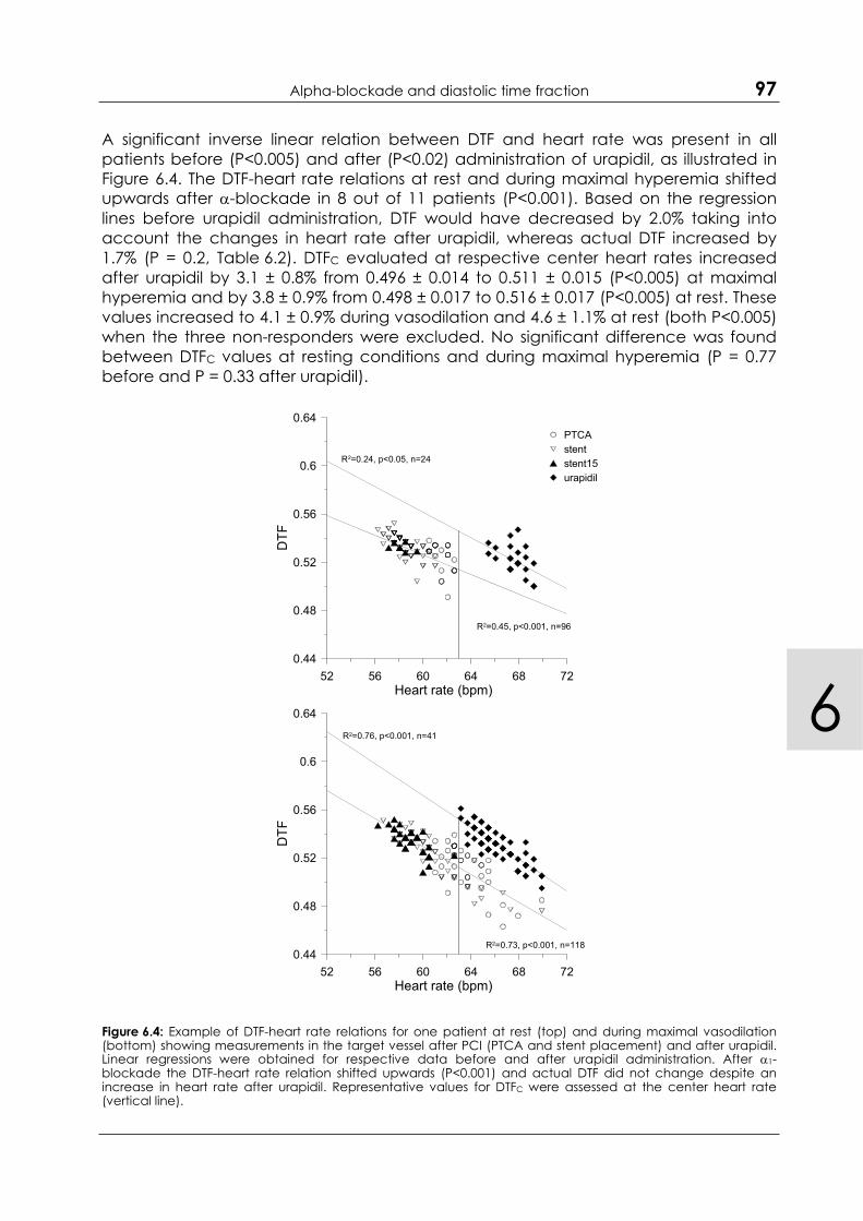
Alpha-blockade and diastolic time fraction 97
6
A significant inverse linear relation between DTF and heart rate was present in all patients before (P<0.005) and after (P<0.02) administration of urapidil, as illustrated in Figure 6.4. The DTF-heart rate relations at rest and during maximal hyperemia shifted upwards after -blockade in 8 out of 11 patients (P<0.001). Based on the regression lines before urapidil administration, DTF would have decreased by 2.0% taking into account the changes in heart rate after urapidil, whereas actual DTF increased by 1.7% (P = 0.2, Table 6.2). DTFC evaluated at respective center heart rates increased after urapidil by 3.1 ± 0.8% from 0.496 ± 0.014 to 0.511 ± 0.015 (P<0.005) at maximal hyperemia and by 3.8 ± 0.9% from 0.498 ± 0.017 to 0.516 ± 0.017 (P<0.005) at rest. These values increased to 4.1 ± 0.9% during vasodilation and 4.6 ± 1.1% at rest (both P<0.005) when the three non-responders were excluded. No significant difference was found between DTFC values at resting conditions and during maximal hyperemia (P = 0.77 before and P = 0.33 after urapidil).
52 56 60 64 68 72Heart rate (bpm)
0.44
0.48
0.52
0.56
0.6
0.64
DTF
PTCAstentstent15urapidil
R2=0.45, p<0.001, n=96
R2=0.24, p<0.05, n=24
52 56 60 64 68 72Heart rate (bpm)
0.44
0.48
0.52
0.56
0.6
0.64
DTF
R2=0.73, p<0.001, n=118
R2=0.76, p<0.001, n=41
Figure 6.4: Example of DTF-heart rate relations for one patient at rest (top) and during maximal vasodilation (bottom) showing measurements in the target vessel after PCI (PTCA and stent placement) and after urapidil. Linear regressions were obtained for respective data before and after urapidil administration. After 1-blockade the DTF-heart rate relation shifted upwards (P<0.001) and actual DTF did not change despite an increase in heart rate after urapidil. Representative values for DTFC were assessed at the center heart rate (vertical line).

98 Chapter 6
Effect of changes in pressure and DTF on microvascular resistance at maximal vasodilation
In order to separate concomitant changes in heart rate and perfusion pressure after urapidil administration, Figure 6.5 illustrates individual changes in HMR and hyperemic Pd and DTF after -receptor blockade. In four patients (open circles) where distal pressure decreased by 9.8 ± 1.3%, hyperemic microvascular resistance increased after urapidil at an almost unchanged DTF. In one patient (cross), HMR fell despite a 16.1% decrease in distal pressure. This patient had a marked 8.9% increase in DTF after urapidil. In all other patients (closed circles), distal pressure stayed constant and HMR decreased at a constant DTF. The reduction in HMR in these patients is therefore to a large extent due to the blocking action of urapidil following -receptor stimulation after PCI. The average decrease in HMR in this group was 10.5 ± 2.3% and was proportional to the value of HMR prior to urapidil administration (R2 = 0.93).
0.8 1.2 1.6 2.0 2.4 2.8HMR before (mm Hg s/cm)
0.8
1.2
1.6
2.0
2.4
2.8
HM
R a
fter (
mm
Hg
s/cm
)
60 80 100 120Pd before (mm Hg)
60
80
100
120
Pd
afte
r (m
m H
g)
0.40 0.45 0.50 0.55 0.60 0.65DTF before
0.40
0.45
0.50
0.55
0.60
0.65
DTF
afte
r
Figure 6.5: Individual patient data showing hyperemic microvascular resistance index (HMR), distal perfusion pressure (Pd) and DTF before and after 1-receptor blockade. The solid gray line in each graph is the identity line. Hyperemic Pd decreased after urapidil in four patients (open circles) resulting in an elevated HMR at an essentially unchanged DTF. In one patient (cross), HMR decreased despite a decrease in Pd, which is attributed to the substantial increase in DTF after urapidil. In all other patients (filled circles), Pd and DTF remained essentially constant and HMR decreased by about 10% due to the vasodilating action of urapidil.

Alpha-blockade and diastolic time fraction 99
6
DiscussionThe key finding of the present study is that intracoronary administration of the selective
1-receptor blocking agent urapidil after PCI induced an upward shift in the diastolic time fraction-heart rate relationship, which amounted to a 3.1% increase in DTFC
(range 1.4 to 8.2%). Changes in DTF and perfusion pressure after urapidil administration acted in concert to alter microvascular resistance of the maximally vasodilated bed.
Hemodynamic effects of 1-blockade after angioplasty
Coronary resistance vessels receive autonomic innervation and 1-adrenoreceptors are present in vessels with a diameter <300 μm [5, 15]. Although -adrenergic coronary constrictor tone is negligible under resting conditions, it has been shown to decrease hyperemic coronary resistance in normal coronary vessels [16, 21]. Pathophysiological conditions such as atherosclerosis sensitize resistance vessels to the constrictive effect of -receptors [2, 15].
Previous studies have furthermore demonstrated that -adrenergic pathways in the vessel wall were acutely stimulated in patients who underwent PTCA [9, 11, 18]. Gregorini et al. observed that 1-mediated vasoconstriction developed over time [9, 12] and reached a maximum 15 minutes after PTCA, resulting in a blunted flow reserve compared to shortly after PTCA [11].
Our results in terms of CFVR do not concur with these earlier findings in that CFVR was restored to values equal to the reference vessel after PCI and was not depressed 15 minutes later nor enhanced by subsequent administration of urapidil. These contrasting findings may be due to differences in adjunctive medication and initial outcomes of PCI. Patients in the former study were sedated with neuroleptic analgesia and CFVR was not improved by PCI [11]. In addition, vasoactive medication was not discontinued in the present study and all patients received an initial dose of intracoronary nitroglycerin, thereby attenuating the vasoconstrictive effects of PCI-induced -receptor stimulation as can be measured by CFVR.
In terms of hyperemic coronary resistance, 1-blockade with doxazosin demonstrated a reduction not only in normal humans [21] but also 5 days after PTCA [25]. In contrast, our measurements do not confirm an overall effect of urapidil on HMR. When studying the drug-induced effect on HMR one has to be aware of altered mechanical conditions that also influence HMR and in this respect especially DTF and perfusion pressure, which were shown to exert opposite effects on subendocardial conductance, are important [8]. Our data revealed interactive effects that occurred after -blockade. Administration of urapidil resulted in a significant elevation of the DTF-heart rate relationship, effectively prolonging diastolic perfusion time by 3.1% irrespective of heart rate. Additionally, coronary perfusion pressure, which has been shown to increase minimal microvascular resistance in humans, was reduced after urapidil [32]. Our observation of an inconsistent change in minimal coronary microvascular resistance after -blockade can therefore be explained as the result of concerted action of altered distending pressure and DTF. In cases where distal perfusion pressure decreased, HMR increased unless a substantial increase in DTF took place, as was the case for one patient. In all other patients, HMR decreased proportionally to the value before urapidil, indicating urapidil-induced release of -receptor mediated microvascular vasoconstriction in this subgroup.

100 Chapter 6
It should be noted that HMR represents the lumped resistance downstream from the measurement location including all layers of the cardiac muscle and that conductance in the subepicardial layer was not related to DTF or perfusion pressure in the study by Fokkema et al. [8]. HMR therefore only partially reflects a change in subendocardial conductance.
Potential mechanisms for DTF increase after -blockade
Pharmacologic agents can have a significant effect on diastolic time [4]. Urapidil is a selective 1-antagonist, which has a central hypotensive effect without reflex tachycardia due to serotonergic activation [2, 6, 27]. Urapidil has a 90-fold larger preference for 1- than for 2-receptors [6]. To some degree it causes presynaptic 2-blockade and thereby -receptor stimulation and increased norepinephrine release [26], which shortens the duration of systole and could allow for an increase in the duration of diastole at a given heart rate. However, all but one patient received -blocking medication, which limits the effectiveness of this mechanism.
Merkus et al. [22] demonstrated in an animal study an inverse nonlinear relation between DTF and intracoronary perfusion pressure and flow, suggesting a possible protective mechanism whereby DTF increases when coronary perfusion is impaired distal to a stenosis. However, this regulatory mechanism only takes action at perfusion pressure below 50-60 mm Hg, while average distal pressure after PCI was >90 mm Hg in our study group. There is no reference in the literature directly relating -adrenoreceptors and DTF. A possible link exists via stimulation of myocardial 1-adrenergic receptors, which has been shown to exert a positive inotropic effect in a large number of species, including humans [19, 24] and is associated with a prolongation of contraction [14, 20]. Hence, 1-receptor blockade by urapidil may counteract this prolongation and thereby increase DTF. Interestingly, cardiac myocytes have been shown to release a substance that stimulates production of the potent vasoconstrictor endothelin-1 in coronary microvessels in response to 1-adrenergic stimulation, which is blocked by administration of an -adrenergic antagonist [30] and augmented by decreased bioavailability of nitric oxide [23, 31], a hallmark of endothelial dysfunction associated with atherosclerotic coronary artery disease. Regulation of DTF may be an additional player in this scenario.
Methodological considerations
Because in the present study heart rate in individual patients did not change spontaneously for more than 20-25 bpm throughout the protocol, we fitted the heart rate-DTF relations with a linear rather than a curvilinear relationship as it is obtained over a larger range of heart rates that is usually achieved by pacing [3, 4].
We defined systole as the period between the ECG R-peak and the dicrotic notch on the Pa signal. In that way systolic time includes both the isovolumic contraction phase and the left ventricular ejection period. This method has been used throughout this study and while the absolute magnitudes of DTF might have been different for a different definition of systolic duration, it is unlikely that the relative changes in DTF would have been affected [22].
Because of the pressure dependence of DTF [22], we did not use any measurements obtained in the presence of a stenosis for the DTF-HR regression lines before -

Alpha-blockade and diastolic time fraction 101
6
blockade, in order to avoid mixing DTF values obtained at substantially lower perfusion pressures.
The size of our study group was small and although the effect of 1-blockade on heart rate-independent DTFC was significant, future studies in a larger population should be carried out to confirm this mechanism and further investigate potential implications for functional parameters.
Implications
Animal microsphere studies have shown a beneficial effect of prolonged DTF via an enhanced subendocardial perfusion [1, 8]. When extrapolated to the clinical setting, these studies provide sustaining evidence to the supposition that an increased DTF after 1-blockade augments improvement of impaired myocardial performance after PCI by increasing oxygen supply to the subendocardium. The observed increase in DTFC, although small, may well be of clinical importance, since it can be inferred from experimental studies [1] that a 1% increase in DTF increases subendocardial flow by 2.6 to 6.1%, which for the present 3.1% prolongation in DTFC translates to a potential 18.3% increase in subendocardial perfusion.
ConclusionsWe conclude that 1-receptor blockade after coronary angioplasty significantly prolongs DTF, independent of heart rate. This suggests a possible adjunctive mechanism to the well-established beneficial effect of urapidil after PCI via an improvement in subendocardial perfusion, which is critically dependent of DTF.
References [1] BACHE RJ and COBB FR, Effect of maximal coronary vasodilation on transmural myocardial
perfusion during tachycardia in the awake dog. Circ Res 41:648-653, 1977 [2] BAUMGART D, HAUDE M, GORGE G, LIU F, GE J, GROßE-EGGEBRECHT C, ERBEL R and HEUSCH G,
Augmented alpha-adrenergic constriction of atherosclerotic human coronary arteries.Circulation 99:2090-2097, 1999
[3] BOUDOULAS H, Diastolic time: The forgotten dynamic factor. Implications for myocardial perfusion. Acta Cardiol 46:61-71, 1991
[4] BOUDOULAS H, RITTGERS SE, LEWIS RP, LEIER CV and WEISSLER AM, Changes in diastolic time with various pharmacologic agents: Implication for myocardial perfusion. Circulation 60:164-169, 1979
[5] CHILIAN WM, Functional distribution of alpha 1- and alpha 2-adrenergic receptors in the coronary microcirculation. Circulation 84:2108-2122, 1991
[6] DULAS D, HOMANS D and BACHE RJ, Effects of urapidil on coronary blood flow during graded treadmill exercise. J Cardiovasc Pharmacol 24:1004-1009, 1994
[7] FERRO G, DUILIO C, SPINELLI L, LIUCCI GA, MAZZA F and INDOLFI C, Relation between diastolic perfusion time and coronary artery stenosis during stress-induced myocardial ischemia.Circulation 92:342-347, 1995
[8] FOKKEMA DS, VANTEEFFELEN JWGE, DEKKER S, VERGROESEN I, REITSMA JB and SPAAN JAE, Diastolic time fraction as a determinant of subendocardial perfusion. Am J Physiol Heart Circ Physiol288:H2450-2456, 2005

102 Chapter 6
[9] GREGORINI L, FAJADET J, ROBERT G, CASSAGNEAU B, BERNIS M and MARCO J, Coronary vasoconstriction after percutaneous transluminal coronary angioplasty is attenuated by antiadrenergic agents. Circulation 90:895-907, 1994
[10] GREGORINI L, MARCO J, BERNIES M, CASSAGNEAU B, POMIDOSSI G, ANGUISSOLA GB and FAJADET J, The alpha-1 adrenergic blocking agent urapidil counteracts postrotational atherectomy 'elastic recoil' where nitrates have failed. Am J Cardiol 79:1100-1103, 1997
[11] GREGORINI L, MARCO J, FARAH B, BERNIES M, PALOMBO C, KOZAKOVA M, BOSSI IM, CASSAGNEAU B,FAJADET J, DI MARIO C, ALBIERO R, CUGNO M, GROSSI A and HEUSCH G, Effects of selective alpha1- and alpha2-adrenergic blockade on coronary flow reserve after coronary stenting.Circulation 106:2901-2907, 2002
[12] GREGORINI L, MARCO J, PALOMBO C, KOZAKOVA M, ANGUISSOLA GB, CASSAGNEAU B, BERNIES M,DISTANTE A, MARCO I, FAJADET J and ZANCHETTI A, Postischemic left ventricular dysfunction is abolished by alpha-adrenergic blocking agents. J Am Coll Cardiol 31:992-1001, 1998
[13] HANLEY FL, MESSINA LM, GRATTAN MT and HOFFMAN JIE, The effect of coronary inflow pressure on coronary vascular resistance in the isolated dog heart. Circ Res 54:760-772, 1984
[14] HEUSCH G, Alpha-adrenergic mechanisms in myocardial ischemia. Circulation 81:1-13, 1990 [15] HEUSCH G, BAUMGART D, CAMICI P, CHILIAN W, GREGORINI L, HESS O, INDOLFI C and RIMOLDI O,
Alpha-adrenergic coronary vasoconstriction and myocardial ischemia in humans.Circulation 101:689-694, 2000
[16] HODGSON JM, COHEN MD, SZENTPETERY S and THAMES MD, Effects of regional alpha and beta-blockade on resting and hyperemic coronary blood flow in conscious, unstressed humans.Circulation 79:797-809, 1989
[17] HOFFMAN JI and SPAAN JA, Pressure-flow relations in coronary circulation. Physiol Rev 70:331-390, 1990
[18] INDOLFI C, PISCIONE F, RAPACCIUOLO A, ESPOSITO G, ESPOSITO N, CERAVOLO R, DI LORENZO E, MAIONE A, CONDORELLI M and CHIARIELLO M, Coronary artery vasoconstriction after successful single angioplasty of the left anterior descending artery. Am Heart J 128:858-864, 1994
[19] LANDZBERG JS, PARKER JD, GAUTHIER DF and COLUCCI WS, Effects of myocardial alpha 1-adrenergic receptor stimulation and blockade on contractility in humans. Circulation84:1608-1614, 1991
[20] LI K, HE H, LI C, SIROIS P and ROULEAU JL, Myocardial alpha 1-adrenoceptor: Inotropic effect and physiologic and pathologic implications. Life Sci 60:1305-1318, 1997
[21] LORENZONI R, ROSEN SD and CAMICI PG, Effect of alpha1-adrenoceptor blockade on resting and hyperemic myocardial blood flow in normal humans. Am J Physiol 271:H1302-1306, 1996
[22] MERKUS D, KAJIYA F, VINK H, VERGROESEN I, DANKELMAN J, GOTO M and SPAAN JAE, Prolonged diastolic time fraction protects myocardial perfusion when coronary blood flow is reduced.Circulation 100:75-81, 1999
[23] MERKUS D, SOROP O, HOUWELING B, BOOMSMA F, VAN DEN MEIRACKER AH and DUNCKER DJ, NO and prostanoids blunt endothelin-mediated coronary vasoconstrictor influence in exercising swine. Am J Physiol Heart Circ Physiol 291:H2075-2081, 2006
[24] MICHELOTTI GA, PRICE DT and SCHWINN DA, Alpha1-adrenergic receptor regulation: Basic science and clinical implications. Pharmacol Ther 88:281-309, 2000
[25] RIMOLDI O, SPYROU N, FOALE R, HACKETT DR, GREGORINI L and CAMICI PG, Limitation of coronary reserve after successful angioplasty is prevented by oral pretreatment with an alpha1-adrenergic antagonist. J Cardiovasc Pharmacol 36:310-315, 2000
[26] SAEED M, SOMMER O, HOLTZ J and BASSENGE E, Alpha-adrenoceptor blockade by phentolamine causes beta-adrenergic vasodilation by increased catecholamine release due to presynaptic alpha-blockade. J Cardiovasc Pharmacol 4:44-52, 1982

Alpha-blockade and diastolic time fraction 103
6
[27] SHEBUSKI RJ and ZIMMERMAN BG, Suppression of reflex tachycardia following alpha-adrenoceptor blockade in conscious dogs: Comparison of urapidil with prazosin. JCardiovasc Pharmacol 6:788-794, 1984
[28] SIEBES M, VERHOEFF B-J, MEUWISSEN M, DE WINTER RJ, SPAAN JAE and PIEK JJ, Single-wire pressure and flow velocity measurement to quantify coronary stenosis hemodynamics and effects of percutaneous interventions. Circulation 109:756-762, 2004
[29] SPAAN JAE, BREULS NPW and LAIRD JD, Diastolic-systolic coronary flow differences are caused by intramyocardial pump action in the anesthetized dog. Circ Res 49:584-593, 1981
[30] TIEFENBACHER CP, DEFILY DV and CHILIAN WM, Requisite role of cardiac myocytes in coronary alpha1-adrenergic constriction. Circulation 98:9-12, 1998
[31] TUNE JD, Withdrawal of vasoconstrictor influences in local metabolic coronary vasodilation.Am J Physiol Heart Circ Physiol 291:H2044-2046, 2006
[32] VERHOEFF B-J, SIEBES M, MEUWISSEN M, ATASEVER B, VOSKUIL M, DE WINTER RJ, KOCH KT, TIJSSEN JGP, SPAAN JAE and PIEK JJ, Influence of percutaneous coronary intervention on coronary microvascular resistance index. Circulation 111:76-82, 2005