
Homozygous missense and nonsense mutations in BMPR1B cause acromesomelic chondrodysplasia-type Grebe

In Silico Analysis of Missense Mutations in LPAR6 RevealsAbnormal Phospholipid Signaling Pathway Leading toHypotrichosisSyed Irfan Raza1,3, Dost Muhammad1, Abid Jan1, Raja Hussain Ali1, Mubashir Hassan2, Wasim Ahmad1,
Sajid Rashid2*
1 Department of Biochemistry, Faculty of Biological Sciences, Quaid-i-Azam University, Islamabad, Pakistan, 2 National Center for Bioinformatics, Faculty of Biological
Sciences, Quaid-i-Azam University, Islamabad, Pakistan, 3 Army Medical College, National University of Science and Technology NUST, Islamabad, Pakistan
Abstract
Autosomal recessive hypotrichosis is a rare genetic irreversible hair loss disorder characterized by sparse scalp hair, sparse toabsent eyebrows and eyelashes, and sparse axillary and body hair. The study, presented here, established genetic linkage infour families showing similar phenotypes to lysophosphatidic acid receptor 6 (LPAR6) gene on chromosome 13q14.11-q21.32. Subsequently, sequence analysis of the gene revealed two previously reported missense mutations includingp.D63V in affected members of one and p.I188F in three other families. Molecular modeling and docking analysis wasperformed to investigate binding of a ligand oleoyl-L-alpha-lysophosphatidic acid (LPA) to modeled protein structures ofnormal and mutated (D63V, G146R, I188F, N248Y, S3T, L277P) LPAR6 receptors. The mutant receptors showed a completeshift in orientation of LPA at the binding site. In addition, hydropathy analysis revealed a significant change in themembrane spanning topology of LPAR6 helical segments. The present study further substantiated involvement of LPAR6-LPA signaling in the pathogenesis of hypotrichosis/woolly hair and provided additional insight into the molecularmechanism of hair development.
Citation: Raza SI, Muhammad D, Jan A, Ali RH, Hassan M, et al. (2014) In Silico Analysis of Missense Mutations in LPAR6 Reveals Abnormal Phospholipid SignalingPathway Leading to Hypotrichosis. PLoS ONE 9(8): e104756. doi:10.1371/journal.pone.0104756
Editor: Maurice A.M. van Steensel, A*STAR, Singapore
Received April 17, 2014; Accepted July 16, 2014; Published August 13, 2014
Copyright: � 2014 Raza et al. This is an open-access article distributed under the terms of the Creative Commons Attribution License, which permits unrestricteduse, distribution, and reproduction in any medium, provided the original author and source are credited.
Data Availability: The authors confirm that all data underlying the findings are fully available without restriction. All relevant data are within paper and itssupporting information files.
Funding: The authors have no support or funding to report.
Competing Interests: The authors have declared that no competing interests exist.
* Email: [email protected]
Introduction
Autosomal recessive hypotrichosis is a rare form of alopecia
characterized by sparse hair on scalp, sparse to absent eyebrows
and eyelashes, and sparse auxiliary and body hair. Over the past
few years, mutations in at least eight genes causing variable
phenotypes of autosomal recessive form of hypotrichosis have been
reported. Except in two cases [1,2], the underlying disease causing
genes in rest of the six forms of hypotrichosis have been reported.
Mutations in two of these genes, LPAR6 and LIPH, result in
similar features including hypotrichosis and woolly hair [3].
Presence of woolly hair has also been reported in patients carrying
mutations in keratin-74 gene with phenotype segregating in
autosomal dominant fashion [4].
The LPAR6 is a nested gene residing within intron-17 of the
RB1 and encodes a heptaspan transmembrane protein that
belongs to an orphan G-protein-coupled receptor (GPCR). Both
LIPH and LPAR6 express in the inner root sheath (IRS) of hair
follicle [5,6,7] and are involved in the same pathway of hair
growth regulation and differentiation [8]. Oleoyl-Lalpha-lysopho-
sphatidic acid (LPA), a bioactive lipid and product of LIPH serves
as a ligand for LPAR6 receptor [8].
In the present study, screening LPAR6 in four consanguineous
Pakistani families led to the identification of two previously
reported missense mutations (p.D63V, p.I188F). Modeling and
mutagenesis studies of six missense mutations (Table 1), including
two identified here, in LPAR6 gene were performed to delineate
their significance at respective residual level. In addition, binding
of mutated LPAR6 with LPA was assessed to map the
conformational changes in ligand binding pocket.
Materials and Methods
Human subjectsThe study, presented here, describes clinical and molecular
analyses of four consanguineous families (A-D) segregating
autosomal recessive form of hypotrichosis/woolly hair phenotype.
The study was approved by the Institutional Review Board (IRB)
of Quaid-i-Azam University, Islamabad, Pakistan. Informed
consent was obtained from all those participated in the study. In
total, there were 27 affected and 83 unaffected individuals in the 4
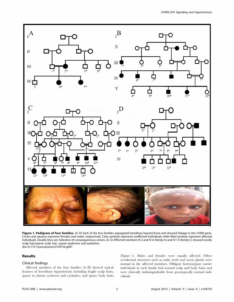
families investigated in the present study (Figure 1).
Extraction of genomic DNA and genotypingVenous blood samples from both affected and unaffected
members in each of the four families were collected in EDTA
containing vacutainer sets. DNA was extracted from the available
blood samples using GenElute Blood Genomic DNA Kit (Sigma-
PLOS ONE | www.plosone.org 1 August 2014 | Volume 9 | Issue 8 | e104756

Aldrich, MO, USA). Quantification of genomic DNA was carried
out by measuring optical density at 260 nm and diluted to 40–
50 ng/ml for amplification by polymerase chain reaction (PCR).
Genetic analysisBased on the clinical features observed in affected individuals,
all the four families were tested for linkage using microsatellite
markers mapped to the chromosomal regions harboring two genes
(LIPH, LPAR6) involved in autosomal recessive hypotrichosis.
The microsatellite markers used to test linkage in the families
included D3S2314, D3S1618, D3S3609, D3S3583, D3S3592 and
D3S1530 linked to LIPH gene on chromosome 3q27.2; and
D13S1312, D13S168, D13S164, D13S273, D13S284 and
D13S1807 linked to LPAR6 on chromosome 13q14.11-q21.323.
The amplification of microsatellite markers was performed
according to standard procedure in a total volume of 25 ml.
PCR was carried out in a 0.2 ml PCR tube with 40 ng human
genomic DNA, 2.5 ml reaction buffer (KCl 50 mM, Tris-Cl pH
8.3), 15–20 pmol of each primer, 1.5 mM MgCl2, 200 mM of each
deoxynucleoside triphosphate (dNTP), and 1 unit of Taq DNA
polymerase (MBI Fermentas, Life Sciences, UK). The standard
thermal conditions were used as described elsewhere [6]. The
PCR was performed in T3000 thermocycler (Biometra, Goettin-
gen, Germany) and PCR products were resolved on 8% non-
denaturing polyacrylamide gel. Gels were stained with ethidium
bromide and genotypes were assigned by visual inspection.
The entire 1032 bp coding sequence of LPAR6 gene was
sequenced in all available affected and unaffected members of the
four families. Purification of the PCR-amplified product was
performed with commercially available kits (Marligen Biosciences,
Ijamsville, MD, USA). Bidirectional DNA sequencing was
performed using DTCS Quick Start sequencing kit (Beckman
Coulter, Fullerton, CA, USA) according to the manufacturer’s
instructions. Sequence variants were identified via BIOEDIT
sequence alignment editor version 6.0.7.
3D structure predictionIn the absence of a well-defined or experimentally determined
structure, comparative modeling is one of the most accurate
computational approaches to generate a reliable tertiary protein
structure through sequence information [9]. Primary protein
sequence for LPAR6 (ID: ENST00000345941) was retrieved
through Ensembl Genome Browser (http://www.ensembl.org)
and subjected to BLAST search against protein data bank (http://
www.rcsb.org) for suitable template search. Three dimensional
(3D) structure of LPAR6WT was predicted through MODELLER
9V8 tool [10] using crystal structure of human kappa opoid
receptor (PDB ID: 4DJH) as template (resolution: 2.90 A). The
structures of mutated LPAR6 (D63V, G146R, I188F, N248Y,
S3T and L277P) were predicted through respective residue
function using MODELLER 9V8 tool. Subsequently, Ramachan-
dran plot, PROCHECK [11], ERRAT [12], VERIFY 3D [13]
and WHATIF [14] tools were utilized to validate these models,
followed by model refinement, editing and geometry optimization
using UCSF Chimera 1.7.0 [15] and VEGA ZZ [16] tools.
Hydropathy analysisIn order to assess and distinguish the transmembrane (TM)
helices of normal and mutated LPAR6 (D63V, G146R, I188F,
N248Y, L277P), hydrophobicity analyses of respective amino acids
were performed using Membrane Protein Explorer (MPEx) [17]
and TransMembrane protein Re-Presentation in 2 Dimensions
(TMRPres2D) tools [18]. MPEx tool uses experimental values of
hydrophobicity scales based on physical and biological analyses to
predict the TM regions. It is widely used to evaluate the effect of a
particular mutation in TM helices. TMRPres2D tool is used for
2D visualization of TM segments.
Molecular docking analysisMolecular docking assays for modeled LPAR6WT and LPA
interaction were carried out using AutoDock 4.0 tool [19] on
OpenSUSE 11.2 containing Intel(R) Core (TM) i5-2300 CPU
system. The structure of LPAR6 ligand designated as LPA was
retrieved through PDB database (PDB ID: NKP) and optimized
by performing energy minimization steps using MMFF94 force
field. Docking analysis was performed as described previously [20].
Briefly, polar hydrogen atoms were added and Kollman charges
were assigned for ligand and all torsions were set free to rotate in
order to perform docking experiments with a rigid receptor and
flexible ligand. Annealing parameters for hydrogen bonding and
Van der Waals interactions were set to 4.0 A and 2.5 A. A grid
map of 80680680 points with a spacing of 0.875 A was set on the
whole protein structure to generate the grid map. The number of
runs for each docking experiment was set to 100. The empirical
free energy function and Lamarckian genetic algorithm (LGA)
were applied with the following parameters: population of 150
randomly placed individuals, a maximum number of 27,000
generations, a mutation rate of 0.02, a crossover rate of 0.80 and
number of energy evaluations was 2.56106, the remaining docking
parameters were set to default. The program automatically
grouped potential receptor-ligand complex conformations into
clusters based on their RMSD, using the default threshold (2.0 A
RMSD). The best docked complex for LPAR6WT-LPA was
selected on the basis of binding free energy value and interactions
were monitored using UCSF Chimera 1.7.0 tool [21].
Table 1. Missense mutations and the resultant phenotypes observed in the families described in the present study.
S. No. Phenotype cDNA change Substitution Reference
1 Wooly hair c.188A.T D63V In family A [3,6,21]
2 Hypotrichosis/Woolly hair c.562A.T I188F In family B, C, D [3,6]
3 Hypotrichosis c.436G.A G146R [3,22]
4 Hypotrichosis c.8G .C S3T [22]
5 Hypotrichosis/Woolly hair c.565G.A E189K [6,22]
6 Hypotrichosis c.742A.T D248Y [25]
doi:10.1371/journal.pone.0104756.t001
LPAR6-LPA Signaling and Hypotrichosis
PLOS ONE | www.plosone.org 2 August 2014 | Volume 9 | Issue 8 | e104756
Results
Clinical findingsAffected members of the four families (A–D) showed typical
features of hereditary hypotrichosis including fragile scalp hairs,
sparse to absent eyebrows and eyelashes, and sparse body hairs
(Figure 1). Males and females were equally affected. Other
ectodermal structures such as nails, teeth and sweat glands were
normal in the affected members. Obligate heterozygous carrier
individuals in each family had normal scalp and body hairs and
were clinically indistinguishable from genotypically normal indi-
viduals.
Figure 1. Pedigrees of four families. (A–D) Each of the four families segregated hereditary hypotrichosis and showed linkage to the LPAR6 gene.Circles and squares represent females and males, respectively. Clear symbols represent unaffected individuals while filled symbols represent affectedindividuals. Double lines are indicative of consanguineous unions. (E–G) Affected members III-2 and IV-6 (family A) and IV-13 (family C) showed woollyscalp hair/sparse scalp hair, sparse eyebrows and eyelashes.doi:10.1371/journal.pone.0104756.g001
LPAR6-LPA Signaling and Hypotrichosis
PLOS ONE | www.plosone.org 3 August 2014 | Volume 9 | Issue 8 | e104756

LPAR6-LPA Signaling and Hypotrichosis
PLOS ONE | www.plosone.org 4 August 2014 | Volume 9 | Issue 8 | e104756

Genotyping and mutation analysisBased on clinical features exhibited by affected individuals, all
the four families were tested for linkage to genes LIPH and
LPAR6 mapped on chromosome 3q and 13q14.11-q21.32,
respectively. Genotyping data showed linkage of all the 4 families
to the LPAR6 gene. Subsequently, LPAR6 was sequenced in two
affected and one unaffected individual of each of the four families.
Sequence analysis of the LPAR6 was performed using a control
reference sequence obtained from the Ensembl database (Ensembl
accession ID: ENSG00000139679).
Mutation analysis of the LPAR6 in affected individuals of the
four families revealed two previously described mutations. In
family A, sequence analysis revealed a previously reported
missense mutation involving transition of A.T at nucleotide
position 188 (c.188A.T) [6,22]. This substitution resulted in
replacing the negatively charged aspartic acid residue with a
nonpolar aliphatic valine at position 63 (p.D63V) of LPAR6.
Sequence analysis of the LPAR6 in the affected members of other
three families (B, C, D) revealed the other reported missense
mutation involving substitution of Isoleucine with Phenylalanine at
amino acid position 188 (c.562A.T, p.I188F) (Figure 2) [3].
Hydropathy pattern analysisTo evaluate the functional impact of known mutations (D63V,
G146R, I188F, N248Y and L277P) in LPAR6, we first
characterized their structural placements. As shown in Figure 3A,
I188, N248 and L277 lie towards the extracellular part of helices,
while D63 and G146 residues are localized towards the cytosolic
part (Figures 2A and B).
In order to evaluate transmembrane helices for normal and
mutated LPAR6, hydrophobicity analysis of individual amino
acids was performed and helical conformations spanning the
membrane were carefully analyzed. By monitoring the favorable
protein transmembrane regions through hydropathy plot (Fig-
ure 2C), D63V and N248Y substitutions were found significant
and were chosen for detailed analysis. Evidently, placement of
helical segments in LPAR6D63V were modified, compared to
LPAR6WT (Table 2), resulting in intracellular shifting of a-helix1
and pushing the onward extracellular part to the membrane side
(Figure 2D). In case of LPAR6N248Y, we observed a slight increase
of DG score for a-helix6 (Table 2), compared to LPAR6WT.
However, there was no significant change in the topology of
membrane segments (Figure 2D). These results clearly demon-
Table 2. Hydropathy analysis results for LPAR6WT, LPAR6D63V and LPAR6N248Y proteins.
LPAR6 Hydropathy Segment Position DG score
Normal FVLGLISNCVAIYIFICVLKVRNETTTYMI 28–57 (30) 3.94
LLFVFTLPFRIFYFTTRNW 64–82 (19) 11.32
ISVMLFYTNMYGSILFLTCISVDRFLAIVYPF 91–122 (32) 11.22
IVCTGVWLTVIGGSAPAVF 135–153 (19) 3.6
IVIFIEIVGFFIPLILNVT 184–202 (19) 9.71
IFVhLIIFCFCFVPYNINLILYSLVRTQTF 231–260 (30) 16.5
ITLCIAVSNCCFDPIVYYF 275–293 (19) 4.59
D63V FVLGLISNCVAIYIFICVL 28–46 (19) 10.82
LAMSVLLFVFTLPFRIFYFTTRNWPFGDLL 59–88 (30) 12.02
ISVMLFYTNMYGSILFLTCISVDRFLAIVYPF 91–122 (32) 11.22
IVCTGVWLTVIGGSAPAVF 135–153 (19) 3.6
IVIFIEIVGFFIPLILNVT 184–202 (19) 9.71
IFVhLIIFCFCFVPYNINLILYSLVRTQTF 231–260 (30) 16.5
ITLCIAVSNCCFDPIVYYF 275–293 (19) 4.59
N248Y FVLGLISNCVAIYIFICVLKVRNETTTYMI 28–57 (30) 3.94
LLFVFTLPFRIFYFTTRNW 64–82 (19) 11.32
ISVMLFYTNMYGSILFLTCISVDRFLAIVYPF 91–122 (32) 11.22
IVCTGVWLTVIGGSAPAVF 135–153 (19) 3.6
IVIFIEIVGFFIPLILNVT 184–202 (19) 9.71
IFVhLIIFCFCFVPYNIYLILYSLVRTQTF 231–260 (30) 18.06
ITLCIAVSNCCFDPIVYYF 275–293 (19) 4.59
doi:10.1371/journal.pone.0104756.t002
Figure 2. Characterization of LPAR6 specific mutations at structure level. (A) 3D representation of LPAR6structure in ribbon form. Individual a-helices are indicated by distinct colors: a1, magenta; a2, green; a3, cornflower blue; a4, brown; a5, pink; a6, orange red and a7, olive. b-sheets are indicated byyellow color. (B) Schematic representation of LPAR6 secondary structures indicating the directionality of b-sheets to the extracellular part, while narrow end ofa-helices points to the cytosol, making a groove-like structure. Corresponding positions of known amino acids undergoing substitutions are indicated whichshow that I188, N248 and L277 lie to the extracellular part of a-helices, while D63 and G146 residues are near the cytosolic part. Surface view of groove-likestructure is shown to visualize the residual positions. (C) Hydropathy plot analysis of normal and mutated LPAR6 amino acids performed by MPEx tool. (D)Membrane spanning profile studies of individual a-helices for LPAR6WT, LPAR6D63V and LPAR6N248Y.doi:10.1371/journal.pone.0104756.g002
LPAR6-LPA Signaling and Hypotrichosis
PLOS ONE | www.plosone.org 5 August 2014 | Volume 9 | Issue 8 | e104756

strated that D63V substitution exerted more severe effect in
membrane spanning of LPAR6 helices.
Binding orientation and interaction mode studiesIn order to assess the conformational changes produced in
LPAR6 due to its interaction to ligand LPA, a predicted 3D
structure of LPAR6WT (D13-W307 residues) using human kappa
opoid receptor (PDB ID: 4DJH) as template (27% sequence
identity) was investigated. Further, the predicted structure was
refined and characterized using structure analysis tools available
online. Ramachandran plot for the predicted LPAR6WT model
indicated the presence of approximately 97.37% residues in
favorable region. In addition, other factors including peptide bond
planarity, non-bonded interactions, Ca tetrahedral distortion,
main chain H-bond energy, poor rotamers and overall G-factor
for the modeled structures lied within the favorable range. The
mutated LPAR6 structures were modeled using LPAR6WT
structure.
Docking analysis of LPAR6 and LPAThrough docking simulations, more pronounced changes were
observed in the b-sheets (N155-S159 and E166-E170), located at
the extracellular part of LPAR6. In LPAR6WT-LPA complex,
R73, Y76, F77, N248, L251, Q258, V261, C263, R270, Y273,
P274, L277 and C278 residues of LPAR6 were involved in LPA
binding (Figure 3). Interestingly, two residues N248 and L277,
found mutated (N248Y and L277P) in families with hypotrichosis/
woolly hair, were actively involved in binding LPA (Figure 4).
Overall, total binding energy for LPAR6WT-LPA interaction was -
7.94 Kcal/mol. A comparative binding analysis of interaction of
LPA with LPAR6WT and LPAR6MT proteins are described in
Table 3 and Figure S1.
Discussion
To date, several mutations in the hair-follicle specific epithelial
keratins have been reported which are associated with exhibition
of curly-wavy phenotype [23], woolly hair and hypotrichosis in
humans [4,24]. Notably, clinical features observed in the affected
members of these families were similar to those reported in the
patients carrying mutations in either LPAR6 or LIPH gene
(Table 1). It has been recently shown that epithelial keratins
colocalize with LPAR6 in the Henle’s (He) and Huxley’s (Hux)
layers of follicle-specific inner root sheath (IRS) [4]. Consequently,
hair follicle development requires an accurate binding of LPA (a
product of LIPH) to LPAR6 receptor for the subsequent
keratinization process, leading to the formation of hair shaft.
The missense mutations [9,25,26,27] and deletions [7,8,28] in
LPAR6 may disturb the underlying downstream signaling events
occurring in the inner root sheath (IRS) hair follicle.
A closer association between hair growth abnormalities and
spectrum of mutations reported in LPAR6 demands a detailed
characterization of underlying signaling events. Generally, LIPH
encoding enzyme, Lipase H is involved in the formation of LPA
from phosphatidic acid which elicits the signal transduction
mechanism (Figure 5) by binding to purine and pyrimidine
nucleotide receptor LPAR6 [7]. In this study, through 3D
structure prediction of LPAR6 and localization of missense
mutations followed by elucidation of LPA binding pattern to
normal and mutated LPAR6, we evaluated the residual contribu-
tions in mediating downstream signaling events.
Despite sharing a common binding region (Table 3), there exist
significant conformational transitions in the LPA binding pattern
to normal and mutated forms of the receptor. The four missense
mutations (S3T, G146R, I188F, L277P) completely shifted the
orientation of LPA at binding site of the receptor. These
substitutions altered the affinity of LPAR6 receptor to LPA ligand
Figure 3. Residual contributions explored through molecular dockings of LPAR6WT and LPA binding. Labels are indicated by blackcolor. Individual a-helices are indicated by distinct colors: a1, magenta; a2, green; a3, cornflower blue; a4, brown; a5, pink; a6, orange red and a7,olive. Loop regions and b-sheets are indicated by gold color.doi:10.1371/journal.pone.0104756.g003
LPAR6-LPA Signaling and Hypotrichosis
PLOS ONE | www.plosone.org 6 August 2014 | Volume 9 | Issue 8 | e104756

Figure 4. Comparative analysis of conformational changes in LPA-bound LPAR6 structures at residual level. Individual mutations areindicated by distinct colors. Binding residues are labeled in black color.doi:10.1371/journal.pone.0104756.g004
Table 3. Comparative binding analysis of LPAR6WT and LPAR6MT structures bound to LPA. Similar residues in multiple complexesare indicated in bold.
Structure RMSD (A) Binding energy (Kcal/mol) Interacting Residues
LPAR6WT 0 27.94 Y76, P77, R73, P274, Y273, N248, Y252, V269, L251, L277, V261
LPAR6D63V 0.447 27.78 R80, N81, W82, E166, Y273, M272, V269, N248, V261
LPAR6G146R 0.379 27.87 Y273, F169, V269, N248, N171, F172, Y252, L181, W177, E174
LPAR6I188F 0.379 26.24 R270, Y273, M272, V269, F169, N171, N248, L249, F172, L181, Y252, W177, E174
LPAR6L277P 0.253 28.28 R270, Y273, F169, N248, N171, Y252, F172, L181, W177, E174
LPAR6N248Y 0.274 28.4 R270, Y273, F169, N171, Y248, F172, W177, E174
LPAR6S3T 0.199 28.24 R270, V269, Y273, F169, N248, N171, Y252, F172, L181, W177
doi:10.1371/journal.pone.0104756.t003
LPAR6-LPA Signaling and Hypotrichosis
PLOS ONE | www.plosone.org 7 August 2014 | Volume 9 | Issue 8 | e104756

thereby disturbing the downstream signaling pathway, which
governs wooly hair formation in human. However, except D63V,
other mutations failed to alter the topology of transmembrane
helices. These findings largely underscore that regulation of lipid
metabolism resulting from coupling of LPA to its receptor is a key
factor in hair development and differentiation.
The comparative binding analyses of LPAR6 indicated the
predominant involvements of F172, W177, L181, N248, Y252,
V261, V269, R270 and Y273 residues in LPA interactions to its
receptor. Notably, N248 and L277 residues were frequently
observed in the receptor-mediated interactions, indicating N248Y
and L277P substitutions may have more severe effect in
development of phenotype. These findings are in good agreement
with the features of less dense and/or sparse hair observed in
families carrying such mutations (Table 1).
In binding of LPA to LPAR6 two aliphatic amino acids, V and
L, preferably localize in the middle of lipid bilayer, while two other
residues, W and Y, facing towards the membrane surface are
involved in contacting membrane spanning a-helices with long
chain fatty acid of LPA. Clearly, LPA acts as a multifunctional
signaling molecule due to incorporation of free hydroxyl,
phosphate moiety and hydrophilic nature [29]. However, it is
important to address any change occur in biological activity of
LPA as a result of transition of its conformational features,
particularly those related to binding to mutated LPAR6. Indeed,
LPA activity is dependent on the length of its acyl chains (C16 to
C20) and decreases by the shortening of chain length [30]. It has
been postulated that phosphate group of LPA is pivotal for its
activity and the acyl chains tend to anchor it in the hydrophobic
cavity for the optimal orientation of phosphate group to interact
with positively charged residues of receptor [30]. However, in
LPAR6-LPA model, we detected the participation of neutral
amino acids (F77, P274, L277) in phosphate group binding. A
somewhat different picture resulted for the mutated LPAR6 with
respect to orientation of phosphate group. It seems more likely that
the mutations may affect the bound LPA activity to act as an
intracellular messenger. Obviously, delineation of structure-
activity relationship of LPA would largely help in understanding
its precise biological function at molecular level. Generally,
LPAR6-LPA binding activates Rho-signaling pathway which is a
possible mediator of hair growth (Figure 5) [31]. Alternatively,
LPAR6 may implicate in the regulation of hair growth through b-
catenin dependent activity [32,33]. Overall, a continued study of
LPA in multiple independent pathways would assist in delineating
additional clues for designing novel therapeutic targets to treat hair
loss in humans.
Supporting Information
Figure S1 Ligplots showing comparative binding anal-ysis of LPAR6WT and LPAR6mut interactions with LPA.(TIF)
Author Contributions
Performed the experiments: SIR DM AJ RHA. Analyzed the data: WA
SR. Contributed reagents/materials/analysis tools: WA. Contributed to
the writing of the manuscript: SIR WA SR. Blood collection from families:
DM AJ RHA. Conceived and performed in silico analysis: SR MH.
Figure 5. Overview of LPAR6-LPA dependent pathwaysinvolved in hair follicle development. Three possible pathways(b-Catenin, Rho and EGFR) are known to occur in the inner root of hairsheath. A dotted line arrow indicates TGF-a inter-linkage with EGFRpathway. The b-catenin together with Cadherins may activate HK genesand help in hair development [34]. Rho and EGFR pathways are linkedwith each other by functional activity of TGF-a which is directly involvedin the activation of EGFR. Rho signaling cascade (GTP binding proteins)and EGFR proteins activate MEK/ERK which results in the developmentof hair follicle by regulating the downstream transcriptional activity[35,36]. HK, hair keratins; G-Protein abd, GTP binding proteins; Ga-12/13,Subunit of GTP binding protein alpha; RhoA, Ras homolog gene familymember A; ROCK, Rho associated protein kinase; PKC, Protein kinase C;
Pro TGF, Protein transforming growth factor; EGFR, Epidermal growthfactor receptor; SOS protein, son of seveless protein; GPR1, G-proteincouple receptor 1; MEK, MAPK/ERK kinases; ERK, extracellular signalregulated kinases.doi:10.1371/journal.pone.0104756.g005
LPAR6-LPA Signaling and Hypotrichosis
PLOS ONE | www.plosone.org 8 August 2014 | Volume 9 | Issue 8 | e104756

References
1. Naz G, Ali G, Naqvi SK, Azeem Z, Ahmad W (2010) Mapping of a novel
autosomal recessive hypotrichosis locus on chromosome 10q11.23-22.3. Hum
Genet 127: 395–401.
2. Basit S, Ali G, Wasif N, Ansar M, Ahmad W (2010) Genetic mapping of a novel
hypotrichosis locus to chromosome 7p21.3-p22.3 in a Pakistani family and
screening of the candidate genes. Hum Genet 128: 213–20.
3. Khan S, Habib R, Mir H, Umm-e-Kalsoom, Naz G, et al. (2011) Mutations in
the LPAR6 and LIPH genes underlie autosomal recessive hypotrichosis/woolly
hair in 17 consanguineous families from Pakistan. Clin Exp Dermatol 36: 652–
654.
4. Shimomura Y, Wajid M, Petukhova L, Kurban M, Christiano AM (2010)
Autosomal-dominant woolly hair resulting from disruption of keratin 74
(KRT74), a potential determinant of human hair texture. Am J Hum Genet
86: 632–638.
5. Kazantseva A1, Goltsov A, Zinchenko R, Grigorenko AP, Abrukova AV, et al.
(2006) Human hair growth deficiency is linked to a genetic defect in the
phospholipase gene LIPH. Science 314: 982–985.
6. Shimomura Y, Wajid M, Ishii Y, Shapiro L, Petukhova L, et al. (2008)
Disruption of P2RY5, an orphan G protein-coupled receptor, underlies
autosomal recessive woolly hair. Nat Genet 40: 335–339.
7. Pasternack SM, von Ku-gelgen I, Aboud KA, Lee YA, Ruschendorf F, et al.
(2008) G protein-coupled receptor P2Y5 and its ligand LPA are involved in
maintenance of human hair growth. Nat Genet 40: 329–34.
8. Pasternack SM, von Kugelgen I, Muller M, Oji V, Traupe H, et al. (2009) In
vitro analysis of LIPH mutations causing hypotrichosis simplex: evidence
confirming the role of lipase H and lysophosphatidic acid in hair growth. J Invest
Dermatol 129: 2772–2776.
9. Tramontano A (1998) Homology modeling with low sequence identity.
Methods. Mar 14: 293–300.
10. Eswar N, Webb B, Marti-Renom MA, Madhusudhan MS, Eramian D, et al.
(2006) Comparative protein structure modeling using Modeller. Curr Protoc
Bioinformatics 2006, Chapter 5: Unit 56.
11. Laskowski RA, MacArthur MW, Moss DS, Thornton JM (1993) PROCHECK:
a program to check the stereochemical quality of protein structures. J Appl Cryst
26: 283–291
12. Colovos C, Yeates TO (1993) Verification of protein structures: patterns of
nonbonded atomic interactions. Protein Sci 2: 1511–1519.
13. Eisenberg D, Luthy R, Bowie JU (1997) VERIFY3D: assessment of protein
models with three-dimensional profiles. Methods Enzymol 277: 396–404.
14. Vriend G (1990) WHAT IF: A molecular modeling and drug design program.
J Mol Graph 8: 52–56.
15. Meng EC, Pettersen EF, Couch GS, Huang CC, Ferrin TE (2006) Tools for
integrated sequence-structure analysis with UCSF Chimera. BMC Bioinfor-
matics 12: 339.
16. Pedretti A, Vila L, Vistoli G (2004) VEGA – an open platform to develop
chemo-bioinformatics application, using plug-in architecture and script
programming. J comput Aided Mater Des 18: 167–173.
17. Snider C, Jayasinghe S, Hristova K, White SH (2009) MPEx: A tool for
exploring membrane proteins. Protein Sci 18: 2624–2628.
18. Spyropoulos IC, Liakopoulos TD, Bagos PG, Hamodrakas SJ (2004)
TMRPres2D: high quality visual representation of transmembrane protein
models. Bioinformatics 22: 3258–60.
19. Morris GM, Huey R, Lindstrom W, Sanner MF, Belew RK, et al. (2009)
AutoDock4 and AutoDockTools4: Automated docking with selective receptorflexibility. J Comput Chem 30: 2785–91.
20. Bibi N, Parveen Z, Rashid S (2013) Identification of Potential Plk1 Targets in aCell-Cycle Specific Proteome through Structural Dynamics of Kinase and Polo
Box-Mediated Interactions. PLoS ONE 8: e70843.
21. Pettersen EF, Goddard TD, Huang CC, Couch GS, Greenblatt DM, et al.(2004) UCSF Chimera–a visualization system for exploratory research and
analysis. J Comput Chem 25: 1605–161.22. Azeem Z, Jelani M, Naz G, Tariq M, Wasif N, et al. (2008) Novel mutations in
G protein-coupled receptor gene (P2RY5) in families with autosomal recessive
hypotrichosis (LAH3). Hum Genet 123: 515–9.23. Fujimoto A, Farooq M, Fujikawa H, Inoue A, Ohyama M, et al. (2012) A
missense mutation within the helix initiation motif of the keratin K71 geneunderlies autosomal dominant woolly hair/hypotrichosis. J Invest Dermatol
132: 2342–9.24. Wasif N, Naqvi SK, Basit S, Ali N, Ansar M, et al. (2011) Novel mutations in the
keratin-74 (KRT74) gene underlie autosomal dominant woolly hair/hypotri-
chosis in Pakistani families. Hum Genet 129: 419–24.25. Tariq M, Ayub M, Jelani M, Basit S, Naz G, et al. (2009) Mutations in the
P2RY5 gene underlie autosomal recessive hypotrichosis in 13 Pakistani families.Br J Dermatol 160: 1006–10.
26. Shimomura Y, Ito M, Christiano AM (2009) Mutations in the LIPH gene in
three Japanese families with autosomal recessive woolly hair/hypotrichosis.J Dermatol Sci 56: 205–7.
27. Petukhova L, Sousa EC Jr, Martinez-Mir A, Vitebsky A, Dos Santos LG, et al.(2008) Genome-wide linkage analysis of an autosomal recessive hypotrichosis
identifies a novel P2RY5 mutation. Genomics 92: 273–8.28. Horev L, Saad-Edin B, Ingber A, Zlotogorski A (2010) A novel deletion
mutation in P2RY5/LPA(6) gene cause autosomal recessive woolly hair with
hypotrichosis. J Eur Acad Dermatol Venereol. 24: 858–9.29. Jalink K, van Corven EJ, Moolenaar WH (1990) Lysophosphatidic acid, but not
phosphatidic acid, is a potent Ca2 (+)-mobilizing stimulus for fibroblasts.Evidence for an extracellular site of action. J Biol Chem 265: 12232–9.
30. Jalink K, Hengeveld T, Mulder S, Postma FR, Simon MF, et al. (1995)
Lysophosphatidic acid-induced Ca2+ mobilization in human A431 cells:structure-activity analysis. Biochem J 307: 609–16.
31. Yanagida K, Masago K, Nakanishi H, Kihara Y, Hamano F, et al. (2009)Identification and Characterization of a Novel Lysophosphatidic Acid Receptor,
p2y5/LPA6. J Biol Chem 284: 17731–17741.32. Fuchs E (2007) Scratching the surface of skin development. Nature 445: 834–42.
33. Betz RC, Cabral RM, Christaino AM, Sprecher E (2012) Unveiling the Roots of
Monogenic Genodermatosis: Genotrichoses as a Paradigm. J Invest Dermatol132: 906–14.
34. Alexandrescu DT, Lisa Kauffman CL, Dasanu CA (2009) The cutaneousepidermal growth factor network: Can it be translated clinically to stimulate hair
growth? Dermatology Online J 3: 1.
35. Inoue A, Arima N, Ishiguro J, Prestwich GD, Arai H, et al. (2011) LPA-producing enzyme PA-PLA1a regulates hair follicle development by modulating
EGFR signalling. EMBO J 30: 4248–60.36. Doma E, Rupp C, Baccarini M (2013) EGFR-ras-raf signaling in epidermal stem
cells: roles in hair follicle development, regeneration, tissue remodeling andepidermal cancers. Int J Mol Sci 14: 19361–84.
LPAR6-LPA Signaling and Hypotrichosis
PLOS ONE | www.plosone.org 9 August 2014 | Volume 9 | Issue 8 | e104756
Copyright © 2022 FDOKUMEN


![in silico (BFA thesis), 2002 [español]](https://static.fdokumen.com/doc/165x107/631f4913dbf756400702aca8/in-silico-bfa-thesis-2002-espanol.jpg)