In silico identification and three-dimensional modelling of the missense mutation in ADAMTS2 in a...
8
USING e-ANNOTATION TOOLS FOR ELECTRONIC PROOF CORRECTION Once you have Acrobat Reader open on your computer, click on the Comment tab at the right of the toolbar: This will open up a panel down the right side of the document. The majority of tools you will use for annotating your proof will be in the Annotations section, pictured opposite. We’ve picked out some of these tools below: 1. Replace (Ins) Tool – for replacing text. Strikes a line through text and opens up a text box where replacement text can be entered. How to use it ‚ Highlight a word or sentence. ‚ Click on the Replace (Ins) icon in the Annotations section. ‚ Type the replacement text into the blue box that appears. 2. Strikethrough (Del) Tool – for deleting text. Strikes a red line through text that is to be deleted. How to use it ‚ Highlight a word or sentence. ‚ Click on the Strikethrough (Del) icon in the Annotations section. 3. Add note to text Tool – for highlighting a section to be changed to bold or italic. Highlights text in yellow and opens up a text box where comments can be entered. How to use it ‚ Highlight the relevant section of text. ‚ Click on the Add note to text icon in the Annotations section. ‚ Type instruction on what should be changed regarding the text into the yellow box that appears. 4. Add sticky note Tool – for making notes at specific points in the text. Marks a point in the proof where a comment needs to be highlighted. How to use it ‚ Click on the Add sticky note icon in the Annotations section. ‚ Click at the point in the proof where the comment should be inserted. ‚ Type the comment into the yellow box that appears.
Transcript of In silico identification and three-dimensional modelling of the missense mutation in ADAMTS2 in a...

USING e-ANNOTATION TOOLS FOR ELECTRONIC PROOF CORRECTION
Once you have Acrobat Reader open on your computer, click on the Comment tab at the right of the toolbar:
This will open up a panel down the right side of the document. The majority of
tools you will use for annotating your proof will be in the Annotations section,
pictured opposite. We’ve picked out some of these tools below:
1. Replace (Ins) Tool – for replacing text.
Strikes a line through text and opens up a text
box where replacement text can be entered.
How to use it
‚ Highlight a word or sentence.
‚ Click on the Replace (Ins) icon in the Annotations
section.
‚ Type the replacement text into the blue box that
appears.
2. Strikethrough (Del) Tool – for deleting text.
Strikes a red line through text that is to be
deleted.
How to use it
‚ Highlight a word or sentence.
‚ Click on the Strikethrough (Del) icon in the
Annotations section.
3. Add note to text Tool – for highlighting a section
to be changed to bold or italic.
Highlights text in yellow and opens up a text
box where comments can be entered.
How to use it
‚ Highlight the relevant section of text.
‚ Click on the Add note to text icon in the
Annotations section.
‚ Type instruction on what should be changed
regarding the text into the yellow box that
appears.
4. Add sticky note Tool – for making notes at
specific points in the text.
Marks a point in the proof where a comment
needs to be highlighted.
How to use it
‚ Click on the Add sticky note icon in the
Annotations section.
‚ Click at the point in the proof where the comment
should be inserted.
‚ Type the comment into the yellow box that
appears.
USING e-ANNOTATION TOOLS FOR ELECTRONIC PROOF CORRECTION
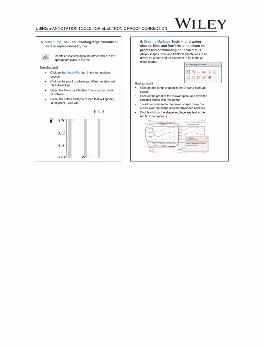
5. Attach File Tool – for inserting large amounts of
text or replacement figures.
Inserts an icon linking to the attached file in the
appropriate place in the text.
How to use it
‚ Click on the Attach File icon in the Annotations
section.
‚ Click on the proof to where you’d like the attached
file to be linked.
‚ Select the file to be attached from your computer
or network.
‚ Select the colour and type of icon that will appear
in the proof. Click OK.
6. Drawing Markups Tools – for drawing
shapes, lines and freeform annotations on
proofs and commenting on these marks.
Allows shapes, lines and freeform annotations to be
drawn on proofs and for comment to be made on
these marks.
How to use it
" Click on one of the shapes in the Drawing Markups
section.
" Click on the proof at the relevant point and draw the
selected shape with the cursor.
" To add a comment to the drawn shape, move the
cursor over the shape until an arrowhead appears.
" Double click on the shape and type any text in the
red box that appears.

In silico identification and three-dimensional modellingof the missense mutation in ADAMTS21 in a sheep flockwith dermatosparaxis
Luis V. Monteagudo*, Luis M. Ferrer†, Elena Catalan-Insa*†, Demetris Savva‡, Liam J. McGuffin‡ and
Maria T. Tejedor*
Department of *Anatomy, Embryology and Genetics, and †Animal Pathology, School of Veterinary Science, Miguel Servet 177, 50013, Zaragoza,
Spain
‡School of Biological Sciences, University of Reading, Whiteknights, Reading, Berkshire, RG6 6AH, UK2
Correspondence: Luis V. Monteagudo, Department of Anatomy, Embryology and Genetics, School of Veterinary Science, Miguel Servet 177,
50013 Zaragoza, Spain. E-mail: [email protected]
Background – Dermatosparaxis (Ehlers–Danlos syndrome in humans) is characterized by extreme fragility of
the skin. It is due to the lack of mature collagen caused by a failure in the enzymatic processing of procollagen I.
We investigated the condition in a commercial sheep flock.
Hypothesis/Objectives – Mutations in the ADAM metalloproteinase gene, specifically at the thrombospondin
type 1 motif 2 (ADAMTS2) locus, are involved in the development of dermatosparaxis in humans, cattle and the
dorper sheep breed; consequently, this locus was investigated in the flock.
Animals – A single affected lamb, its dam, the dam of a second affected lamb and the rams in the flock were
studied.
Methods – DNA was purified from blood, PCR primers were used to detect parts of the ADAMS2 gene and
nucleotide sequencing was performed using Sanger’s procedure. Skin samples were examined using standard
histology procedures.
Results – A missense mutation was identified in the catalytic domain of ADAMTS2. The mutation is predicted to
cause the substitution in the mature ADAMTS2 of a valine molecule by a methionine molecule (V15M) affecting
the catalytic domain of the enzyme. Both the ‘sorting intolerant from tolerant’ (SIFT) and the PolyPhen-2 method-
ologies predicted a damaging effect for the mutation. Three-dimensional modelling suggested that this mutation
may alter the stability of the protein folding or distort the structure, causing the protein to malfunction.
Conclusions and clinical importance – Detection of the mutation responsible for the pathology allowed us to
remove the heterozygote ram, thus preventing additional cases in the flock.
Introduction
Dermatosparaxis (Ehlers–Danlos syndrome type VIIC in
humans) is characterized by extreme fragility of the con-
nective tissue, resulting in severe skin tearing during nor-
mal life. It is an autosomal recessive inherited disease,
associated either with deletions or with single nucleotide
substitutions resulting in the appearance of premature
stop codons in the ‘a disintegrin6 and metalloproteinase
with thrombospondin motifs 2’ (ADAMTS2) gene in
humans, cattle and dorper sheep.1,2 Such mutations
impair the normal processing of procollagen I by ADAM-
TS2, resulting in accumulation of precursor molecules
which fail to provide the normal strength to fibrils.1 Con-
sequently, the skin tears following minimal tension during
normal management practices.
A case of dermatosparaxis in a crossbreed ovine flock
was referred to us. We had access to the affected lamb,
its mother, the rams in the flock and a second ewe which
had previously given birth to a lamb (not available) with
identical clinical signs. Affected lambs suffered extensive
wounds and presented with large skin flaps unrelated to
accidental causes. Attempts to suture the flaps were
unsuccessful because silk suture produced cuts and
failed to join the flaps to the rest of the skin. Severe
wound infections quickly led to a critical situation, and the
lambs were euthanized for humanitarian reasons. Post-
mortem examination showed extreme fragility of the skin
(Figure 1 and Video S1). Parents appeared normal, sup-
porting recessive inheritance.
Predictions of the nucleotide sequence coding for
ovine ADAMTS2 are available in GenBank (accession
NW_004080168).3 In cattle (accession NW_003103998),4
exon 2 corresponds to the ADAMTS2 pro-domain, while
the mature enzyme is encoded by subsequent exons,
starting with the catalytic domain (exons 3–8).2 BLAST
alignment shows that the DNA for this domain is
highly conserved in cattle and sheep.5 Given that
premature stop codons in ADAMST2 are responsible for
Accepted 9 August 2014
Sources of Funding: This study was self-funded.
Conflict of Interest: No conflicts of interest have been declared.
Luis V. Monteagudo and Luis M. Ferrer are joint first authors.
1
2
3
4
5
6
7
8
9
10
11
12
13
14
15
16
17
18
19
20
21
22
23
24
25
26
27
28
29
30
31
32
33
34
35
36
37
38
39
40
41
42
43
44
45
46
47
48
49
50
51
52
53
54
55
56
57
58
59
60
61
62
© 2014 ESVD and ACVD, Veterinary Dermatology 1
Vet Dermatol 2014 DOI: 10.1111/vde.12178
VD
E12178
Dispa
tch:
6.9.14
CE:Ash
okra
jD.
JournalCode
ManuscriptNo.
No.
ofpa
ges:
4PE:Sha
rmila
dermatosparaxis in dorper sheep and cattle, this was cho-
sen as the candidate gene to investigate this flock.1,2
Materials and methods
Animals
A flock of 200 crossbred sheep was initially formed by acquiring
ewes and rams from different farms; breeding animals were then
replaced by animals bred on the same farm.
DNA studies
DNA was isolated from peripheral blood. Briefly, 15 lL of blood was
lysed in 180 lL of TE (10 mM Tris–Cl, 0.1 mM EDTA, pH 8). After
centrifugation for 2 min (12,000g), pellets were resuspended in
180 lL of 5% w/v Chelex resin in TE, incubated for 20 min at 100°C
and recentrifuged; the supernatant was stored at �20°C until DNA
amplification (25–50% of the reaction volume).
PCR primers were designed for the conserved region of the first
segments of the ADAMTS2 catalytic domain (Table 1). Nucleotide
sequences of PCR products were determined using Sanger’s proce-
dure.
Histology
Tissues were fixed in formaldehyde, embedded in paraffin, cut in
sections (5 lm thick) and stained with haematoxylin and eosin after
paraffin removal.
In silico predictions and three-dimensional analysis
The ‘sorting intolerant from tolerant’ (SIFT) algorithm and the
PolyPhen-2 approach were applied to predict the effects of the
single nucleotide polymorphism (SNP) found in the ovine
ADAMTS2 gene.6–8 The three-dimensional (3D) structures of the
wild-type (WT) and mutant sequences were modelled on the
IntFOLD2 server, predicting disordered (natively unstructured)
regions, assigning putative domain boundaries and indicating
likely binding sites and ligand interactions;9–12 these models were
validated using the ModFOLD4 model quality assessment
method.13
Results
The epidermis was unchanged; a band of loose connec-
tive tissue rich in procollagen fibres showing different
sizes and orientations was visible in the deep dermis and
the hypodermis (Figure 2).
The sequencing of the ADAMST2 pro-domain (involved
in dermatosparaxis in dorper sheep) did not reveal any
premature stop codon in the lamb.2 Three sets of primers
were therefore designed to amplify the third, fourth and
fifth exon, according to precedent nomenclature
(Table 1).2
A missense SNP was detected in homozygosis in
the fourth exon in the affected lamb (g.1862883G>A
in ovine3 and g.2030792G>A in bovine4). This SNP
results in the substitution of valine (V) by methionine
(M) in the catalytic domain of the mature ADAMTS2
enzyme (V15M). Interestingly, both the ewe that gave
birth to this lamb and the ewe that gave birth to an
affected lamb previously were heterozygous (G/A) for
this SNP. A screening of the male reproducers in the
flock using microsatellite markers allowed identification
of the ram siring the affected lamb; ADAMST2
sequences showed that this was the only heterozy-
gous ram in the flock for this SNP. This provides
additional confirmation for recessive autosomal inheri-
tance; heterozygous G/A parents with normal pheno-
type produce homozygous A/A descendants affected
by dermatosparaxis.
The evaluation of the possible effect of the V15M
substitution using the SIFT algorithm and the PolyPhen-
2 approach produced consistent results. The SIFT algo-
rithm classified it as ‘damaging’, scoring it with 0.01 in
a 0–1 (damaging–tolerated) range. PolyPhen-2 indicated
that it was ‘probably damaging’, with scores of 1 and
0.98 on the 0–1 scales for the HumDiv and HumVar
method, respectively).6–8 Based on information available
at GenBank, the valine residue in the ADAMST2 cata-
lytic domain is conserved amongst mammals, marsupi-
als (Sarcophilus harrisii), birds (Gallus gallus) and fish
(Takifugu rubripes). However, this is the first time
methionine has been found at this position in any spe-
cies.
Further understanding on the effect of the mutation
was obtained from the 3D analysis of the protein. From
the initial run of the IntFOLD2 server,9 the ordered struc-
ture of the catalytic domain was shown to be flanked by
two natively unstructured regions (M1–R253 and R554–
F1205). As the mutation was in the catalytic domain, the
300 ordered residues (H254–K553) were selected for
higher quality modelling, and the models were validated
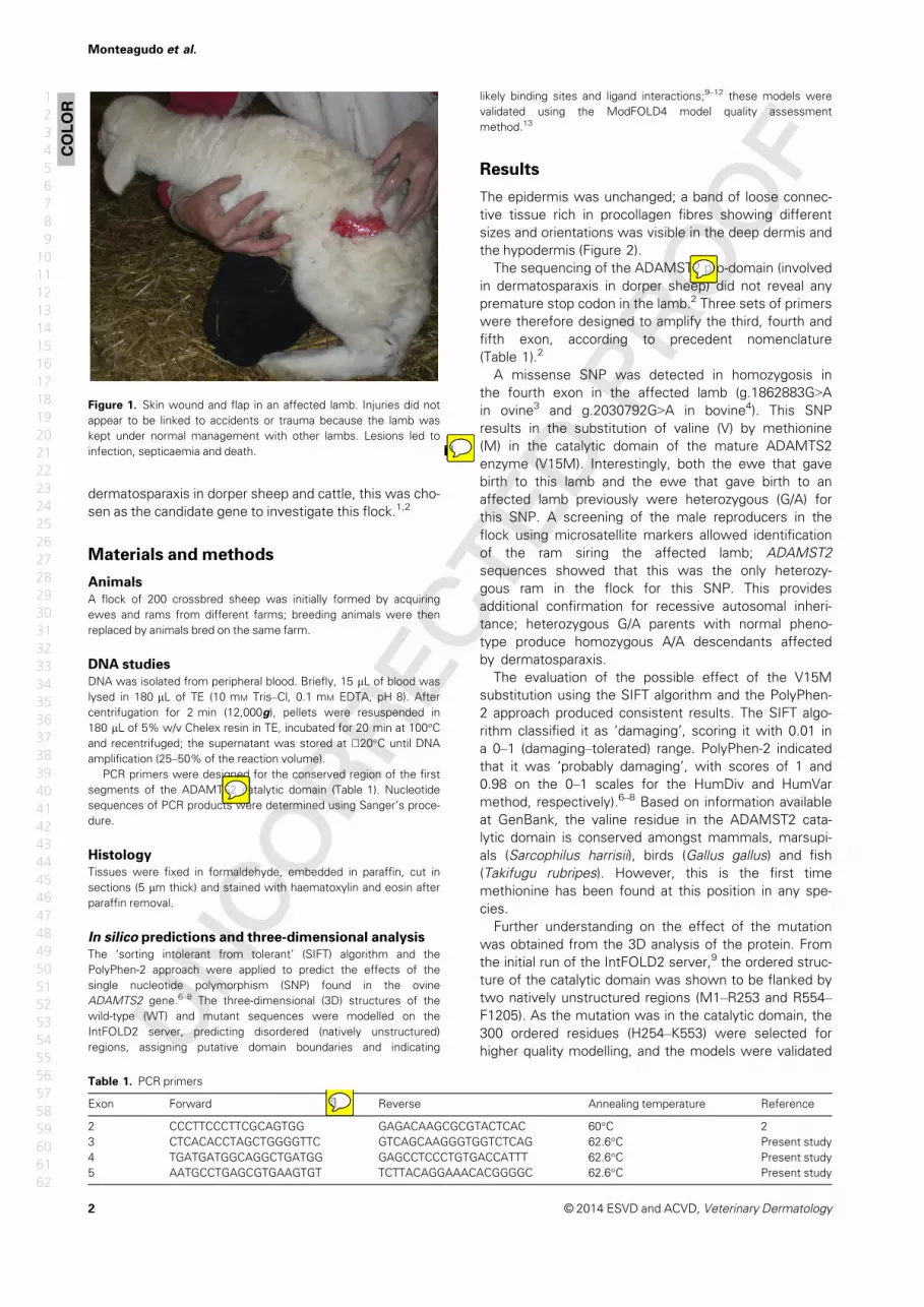
Figure 1. Skin wound and flap in an affected lamb. Injuries did not
appear to be linked to accidents or trauma because the lamb was
kept under normal management with other lambs. Lesions led to
infection, septicaemia and death 7.
Table 1. PCR primers
Exon Forward 8 Reverse Annealing temperature Reference
2 CCCTTCCCTTCGCAGTGG GAGACAAGCGCGTACTCAC 60°C 2
3 CTCACACCTAGCTGGGGTTC GTCAGCAAGGGTGGTCTCAG 62.6°C Present study
4 TGATGATGGCAGGCTGATGG GAGCCTCCCTGTGACCATTT 62.6°C Present study
5 AATGCCTGAGCGTGAAGTGT TCTTACAGGAAACACGGGGC 62.6°C Present study
COLOR
© 2014 ESVD and ACVD, Veterinary Dermatology2
Monteagudo et al.
1
2
3
4
5
6
7
8
9
10
11
12
13
14
15
16
17
18
19
20
21
22
23
24
25
26
27
28
29
30
31
32
33
34
35
36
37
38
39
40
41
42
43
44
45
46
47
48
49
50
51
52
53
54
55
56
57
58
59
60
61
62
admin
Nota adhesiva
ADAMTS2 must be written in italics at this point
admin
Nota adhesiva
ADAMTS2 in italics at this point
admin
Nota adhesiva
The Table is OK as it is now
admin
Nota adhesiva
All the figures are OK

using the ModFOLD4 model quality assessment
method.13 The results of the 3D modelling of the WT AD-
AMTS2 and mutant are shown in Figure 3a–h. In Fig-
ure 3a–c, the models of WT and mutant proteins show
similar global structures and have similarly high global
quality scores (P � 0.001; much <1 in 1000 chance of
the model being incorrect).13 Closer examination of the
site around the mutation shows the implications of the
mutation (Figure 3d–f). In Figure 3f, it can be seen that
the L32 side-chain is in a different orientation in the
mutant than in the WT; perhaps this is due to lack of
space caused by the longer M15 side-chain of the
mutant. In Figure 3g,h, the side-chains are shown as
spheres to indicate atomic clashes. In Figure 3h, we can
see overlapping spheres, indicating clashes occurring
in the mutant protein between the side-chains on the
b-strand and a-helix. However, in the WT protein (Fig-
ure 3g) no side-chain clashes are observed between the
b-strand and a-helix residues. The clashes observed in
the mutant protein may alter the stability of the fold or dis-
tort the structure, causing the protein to malfunction.
Discussion
The histology observations in this case are consistent
with previous descriptions of accumulation of immature
procollagen fibres leading to loose connective tissue that
lacks normal strength and resistance,1 resulting in skin
fragility, severe wounds and infections that lead to early
death9 .
Dermatosparaxis has been repeatedly reported in
sheep as an autosomal recessive trait.14,15 Recessive
inheritance is well known to be linked to defects in enzy-
matic activity.16 Insertions, deletions, nonsense muta-
tions and other changes, such as chromosome
inversions, have traditionally been identified as the origins
of inherited diseases. Nowadays, the analysis of the pos-
sible phenotypic consequences of missense SNP takes
advantage of the vast amount of information available on
genome sequences in humans and animals. State-of-the-
art methodologies, such as SIFT and PolyPhen, were orig-
inally employed to analyse human genome sequences.6–8
However, as these procedures were developed (among
other reasons) for the analysis of conservation in
sequences from different species, their use is spreading
to animal genomics analysis. Recently, SIFT and Poly-
Phen-2 were applied to the identification of deleterious
haplotypes in three different genes involved in bovine
prenatal death.17 For instance, a substitution affecting a
putative manganese binding site in the glycinamide ribo-
nucleotide transformylase protein (N290T) is considered a
critical mutation, with the authors remarking that this
asparagine-290 is conserved among eukaryotes, pointing
to a key role of this amino acid. A similar situation occurs
in the case of the V15M substitution in ADAMTS2, as
shown in the present study; the conservation of valine-15
in the active domain of the proteinase points to a critical
function of this amino acid in procollagen maturation.
Figure 2. Histology of the skin of the affected lamb. Arrows indicate
a band of connective tissue consisting of accumulated procollagen
fibres showing different sizes and orientations in the deep dermis
and the hypodermis. Epidermal and adnexal structures appear normal
(haematoxylin and eosin stain).
(a) (b)
(d)
(f)
(g) (h)
(e)
(c)
Figure 3. Three-dimensional (3D) modelling of single nucleotide
polymorphism mutation in ADAMTS2. (a) IntFOLD2 3D model of the
wild-type (WT) structure showing the 300 ordered residues in a car-
toon view. Amino acid residue V15 is shown in stick form and high-
lighted in dark blue. (b) IntFOLD2 3D model of the mutant structure
showing 300 ordered residues in cartoon view. The M15 mutation is
shown in stick form and highlighted in red. (c) Superposition of the
WT (cyan) and mutant (green) models. (d) Zoomed view of WT struc-
ture with key amino acids labelled as sticks. (e) Zoomed view of
mutant structure with key amino acids labelled as sticks. (f) Superpo-
sition of key residues in WT and mutant. (g,h) The same as (d,e),
respectively, but here the side-chains are represented as spheres to
indicate the van der Waals volume of the atoms and reveal overlaps/
clashes.
COLOR
COLOR
© 2014 ESVD and ACVD, Veterinary Dermatology 3
Inherited ovine dermatosparaxis
1
2
3
4
5
6
7
8
9
10
11
12
13
14
15
16
17
18
19
20
21
22
23
24
25
26
27
28
29
30
31
32
33
34
35
36
37
38
39
40
41
42
43
44
45
46
47
48
49
50
51
52
53
54
55
56
57
58
59
60
61
62
admin
Nota adhesiva
OK

Here, 3D analysis provides additional evidence of the
importance of the V15M substitution, because it causes
critical structural changes in the active domain of the
enzyme.
As in humans, this report demonstrates that ovine
dermatosparaxis may be due to different mutations in
the locus ADAMST2.1 Dorper is a synthetic breed cre-
ated in South Africa in the 1930s by crossing Dorset
and blackheaded Persian sheep.18 Reproductive isola-
tion can be assumed amongst affected dorper and
the flock studied here. It explains genetic heterogene-
ity for this pathology instead of a common origin for
all cases.
In conclusion, in the light of in silico approach results,
the V15M substitution is likely to be responsible for criti-
cal modifications in the ADAMTS2 enzymatic activity that
lead to ovine autosomal recessive inherited dermatospar-
axis.
Acknowledgements
The authors would like to acknowledge the use of Servi-
cio General de Apoyo a la Investigaci�on-SAI, Universidad
de Zaragoza, Jos�e. A. Garc�ıa de Jal�on for histological
analysis and Mar�ıa Pe~na and Ane Rivas for help in labora-
tory tasks.
References
1. Colige A, Sieron AL, Li SW et al. Human Ehlers-Danlos syn-
drome type VII C and bovine dermatosparaxis are caused by
mutations in the procollagen I N-proteinase gene. Am J Hum
Genet 1999; 65: 308–317.
2. Zhou H, Hickford JGH, Fang Q. A premature stop codon in the
ADAMTS2 gene is likely to be responsible for dermatosparaxis
in Dorper sheep. Anim Genet 2012; 43: 471–473.
3. Archibald AL, Cockett NE, Dalrymple BP et al. The sheep gen-
ome reference sequence: a work in progress. Anim Genet 2010;
41: 449–453.
4. Zimin AV, Delcher AL, Florea L et al. A whole-genome assem-
bly of the domestic cow, Bos taurus. Genome Biol 2009; 10:
R42.
5. Altschul SF, Gish W, Miller W et al. Basic local alignment search
tool. J Mol Biol 1990; 215: 403–410.
6. Kumar P, Henikoff S, Ng PC. Predicting the effects of coding
nonsynonymous variants on protein function using the SIFT algo-
rithm. Nat Protoc 2009; 4: 1073–1081.
7. Ramensky V, Bork P, Sunyaev S. Human non-synonymous
SNPs: server and survey. Nucleic Acids Res 2002; 30: 3894–
3900.
8. Adzhubei IA, Schmidt S, Peshkin L et al. A method and server
for predicting damaging missense mutations. Nat Methods
2010; 7: 248–249.
9. Roche DB, Buenavista MT, Tetchner SJ et al. The IntFOLD ser-
ver: an integrated web resource for protein folds recognition, 3D
model quality assessment, intrinsic disorder prediction, domain
prediction and ligand binding site prediction. Nucleic Acids Res
2011; 39(Suppl. 2): W171–W176.
10. Buenavista MT, Roche DB, McGuffin LJ. Improvement of 3D
protein models using multiple templates guided by single-tem-
plate model quality assessment. Bioinformatics 2012; 28: 1851–
1857.
11. McGuffin LJ. Intrinsic disorder prediction from the analysis of
multiple protein fold recognition models. Bioinformatics 2008;
24: 1798–1804.
12. Roche DB, Tetchner SJ, McGuffin LJ. FunFOLD: an improved
automated method for the prediction of ligand binding residues
using 3D models of proteins. BMC Bioinformatics 2011; 12: 160.
13. McGuffin LJ, Buenavista MT, Roche DB. The ModFOLD4 server
for the quality assessment of 3D protein models. Nucleic Acids
Res 2013; 41: W368–W372.
14. Fjolstad M, Helle O. A hereditary dysplasia of collagen tissues in
sheep. J Pathol 1974; 112: 183–188.
15. Wong S. Dermatosparaxis in Dorper sheep. Australian Society
for Veterinary Pathology. Vet Pathol Rep 2006; 72: 10–11. 10
16. Nicholas FW. Veterinary Genetics. Oxford, UK: Oxford Univer-
sity Press, 1987; 92–93.
17. Fritz S, Capitan A, Djari A et al. Detection of haplotypes associ-
ated with prenatal death in dairy cattle and identification of dele-
terious mutations in GART, SHBG and SLC37A2. PLoS ONE
2013; 8: e65550.
18. Database Breeds of Livestock, Oklahoma State University,
Department of Animal Science. Available at: http://www.ansi.
okstate.edu/breeds/. Accessed April 10, 2014.
Supporting Information
Additional Supporting Information may be found in the
online version of this article.
Video S1. Video recorded during the postmortem exami-
nation of an affected lamb immediately after euthanasia.
R�esum�e – Xxxxxxxx.
Resumen – Xxxxxxxx.
Zusammenfassung – Xxxxxxxx4 .
© 2014 ESVD and ACVD, Veterinary Dermatology4
Monteagudo et al.
1
2
3
4
5
6
7
8
9
10
11
12
13
14
15
16
17
18
19
20
21
22
23
24
25
26
27
28
29
30
31
32
33
34
35
36
37
38
39
40
41
42
43
44
45
46
47
48
49
50
51
52
53
54
55
56
57
58
59
60
61
62
admin
Texto insertado
Available at: http://www.asvp.asn.au/wp-content/uploads/2010/07/VPR72-Aug06.pdf Accessed September 8, 2014

Graphical Abstract
The contents of this page will be used as part of the graphical abstract
of html only. It will not be published as part of main.
Background – Dermatosparaxis (Ehlers–Danlos syndrome in humans) is characterized by extreme fragility of the skin. It is
due to the lack of mature collagen caused by a failure in the enzymatic processing of procollagen I. We investigated the
condition in a commercial sheep flock. Hypothesis/Objectives –Mutations in the ADAMmetalloproteinase gene, specifi-
cally at the thrombospondin type 1 motif 2 (ADAMTS2) locus, are involved in the development of dermatosparaxis in
humans, cattle and the dorper sheep breed; consequently, this locus was investigated in the flock. Conclusions and clin-
ical importance – Detection of the mutation responsible for the pathology allowed us to remove the heterozygote ram,
thus preventing additional cases in the flock.
© 2014 ESVD and ACVD, Veterinary Dermatology 5
Inherited ovine dermatosparaxis
admin
Tachado
admin
Texto insertado
ADAM metallopeptidase with thrombospondin type 1 motif, 2 (ADAMTS2) locus Note: ADAMTS2 must be written in italics at this point

Author Query Form
Journal: VDEArticle: 12178
Dear Author,
During the copy-editing of your paper, the following queries arose. Please respond to these by marking up
your proofs with the necessary changes/additions. Please write your answers on the query sheet if there is
insufficient space on the page proofs. Please write clearly and follow the conventions shown on the attached
corrections sheet. If returning the proof by fax do not write too close to the paper’s edge. Please remember
that illegible mark-ups may delay publication.
Many thanks for your assistance.
Query reference Query Remarks
1 AUTHOR: it is normal journal style for abbreviations of gene names to be
italic and proteins roman. Please confirm that this has been done correctly
and consistently throughout the manuscript or amend if necessary. Please
confirm that the gene is correct as ADAMTS2 or should it be Adamts2 for a
sheep gene?
2 AUTHOR: Please check that authors and their affiliations are correct.
3 AUTHOR: Please check the corresponding author’s details.
4 WILEY: Please provide French, Spanish and German abstract.
5 WILEY: Please confirm that the short abstract text is okay.
6 AUTHOR: is “a disintegrin” OK or does it need to be “A disintegrin” (as
opposed to “B disintegrin”, perhaps)?
7 AUTHOR: Please carefully check all the figures.
8 AUTHOR: do you need to specify 30–50 or vice versa?
9 AUTHOR: is “infections that lead to early death” OK now?
10 AUTHOR: Please check the abbreviated journal title for reference [15].