Pathology of severe urolithiasis in a flock of egg-laying hens
6
Brazilian Journal of Veterinary Pathology. www.bjvp.org.br . All rights reserved 2007-2021. Lopes et al.; Pathology of severe urolithiasis in a flock of egg-laying hens Braz J Vet Pathol, 2021, 14(1), 40 – 45 DOI: 10.24070/bjvp.1983-0246.v14i1p40-45 40 Case Report Pathology of severe urolithiasis in a flock of egg-laying hens Marcelo C. Lopes 1 , Clarissa S. Fonseca 1 , Camila I. Amaral 1 , Luis H. Saraiva 1 , Letícia B. Oliveira 1 , Dayse H. L. da Silva 1 , Matheus V. L. Moreira 1 , Fabrício J. M. da Silva 1 , Natalie K. Armour 2 , Roselene Ecco 1 * 1 Sector of Animal Pathology, Veterinary Clinic and Surgical Department, Veterinary School, Universidade Federal de Minas Gerais (UFMG), Belo Horizonte, Minas Gerais, Brazil. 2 Poultry Research and Diagnostic Laboratory, Department of Pathobiology and Population Medicine, College of Veterinary Medicine, Mississippi State University, Pearl, Mississippi, United States *Corresponding author: Sector of Animal Pathology, Veterinary Clinic and Surgical Department, Veterinary School, Universidade Federal de Minas Gerais, Belo Horizonte, Minas Gerais. Av. Antônio Carlos 6627, Belo Horizonte, MG 30123-970, Brazil. E-mail: [email protected] Submitted September, 09 th 2020, Accepted November, 27 th 2020 Abstract Fourteen, 31-week-old Lohmann white layers from a flock of 30,000 chickens had a history of apathy, and a drop in egg production. Clinical signs were observed in approximately 40% of the flock, and lasted for three months. Fourteen hens were euthanized for post-mortem examinations. Macroscopic findings included marked atrophy and loss of renal lobes along with compensatory renal hypertrophy of the contralateral lobe. Ureters were markedly dilated and filled with mucus and/or with molded white to yellow-grey uroliths that obliterated the lumen. At histopathology, the uroliths inside ureters and tubules were composed of concentrically arranged mineralized concretions, as well as urates associated with heterophilic infiltrations and epithelial hyperplasia. Renal parenchyma adjacent to obstructed ureters was compressed with tubules replaced by fibrous tissue. Multifocal interstitial lymphocytic nephritis, proteinuria and membranoproliferative glomerulonephritis were also found. Heterophilic and caseous ureteritis associated with numerous Gram-positive coccoid bacteria occurred in three chickens. Immunohistochemistry for avian coronavirus was negative. This negative result along with the case history indicated that water restriction was the most likely cause of mortality. This condition resulted in significant economic loss for this farmer. Key words: chickens, histopathology, layers, kidneys, uroliths. Introduction Flock health directly impacts productivity. Urolithiasis is a potentially significant disease of layers due to its propensity to reduce egg production and increase mortality (2, 5, 7, 16, 22, 24). Urolithiasis is an acquired and degenerative disease (24), characterized by the presence of uroliths in the urinary system (4). While a number of animal species have been affected, the condition is most frequently diagnosed in dogs, cats, swine, and cattle (19). In chickens, urolithiasis has been attributed to multiple etiologies. Non-infectious causes include excess dietary calcium, vitamin A deficiency, dietary electrolyte imbalances (25), mycotoxins (e.g. oosporein), treatment with sodium bicarbonate (5), high protein diet (14), and water restriction (10). Infectious causes, such as infection with nephropathogenic avian infectious bronchitis virus (IBV) (9) and avian nephritis virus (astrovirus) (3, 5) have also been implicated. Chickens with urolithiasis often show a drop in egg production (5), which may be accompanied by depression, dehydration (7), weight loss, and enlargement of the toes (22). Macroscopically, the ureters are distended by uroliths, with consequent renal atrophy, in addition to varying degrees of visceral urate deposition. At histopathology, urolithiasis is characterized by tubular degeneration or necrosis and dilation of the ureters, with cell cylinders and urate crystals in the lumen and varying degrees of fibrosis (5). In poultry, the early diagnosis of diseases of the urinary system is challenging, and treatment in cases of urolithiasis is seldom used (18). Considering the importance of urolithiasis in veterinary medicine, evidenced by numerous reports in
-
Upload
khangminh22 -
Category
Documents
-
view
0 -
download
0
Transcript of Pathology of severe urolithiasis in a flock of egg-laying hens
Brazilian Journal of Veterinary Pathology. www.bjvp.org.br . All rights reserved 2007-2021.
Lopes et al.; Pathology of severe urolithiasis in a flock of egg-laying hens Braz J Vet Pathol, 2021, 14(1), 40 – 45
DOI: 10.24070/bjvp.1983-0246.v14i1p40-45
40
Case Report
Pathology of severe urolithiasis in a flock of egg-laying hens Marcelo C. Lopes1, Clarissa S. Fonseca1, Camila I. Amaral1, Luis H. Saraiva1, Letícia B. Oliveira1,
Dayse H. L. da Silva1, Matheus V. L. Moreira1, Fabrício J. M. da Silva1, Natalie K. Armour2, Roselene Ecco1*1 Sector of Animal Pathology, Veterinary Clinic and Surgical Department, Veterinary School,
Universidade Federal de Minas Gerais (UFMG), Belo Horizonte, Minas Gerais, Brazil. 2 Poultry Research and Diagnostic Laboratory, Department of Pathobiology and Population Medicine,
College of Veterinary Medicine, Mississippi State University, Pearl, Mississippi, United States*Corresponding author: Sector of Animal Pathology, Veterinary Clinic and Surgical Department,
Veterinary School, Universidade Federal de Minas Gerais,Belo Horizonte, Minas Gerais. Av. Antônio Carlos 6627, Belo Horizonte, MG 30123-970, Brazil. E-mail: [email protected]
Submitted September, 09th 2020, Accepted November, 27th 2020
Abstract
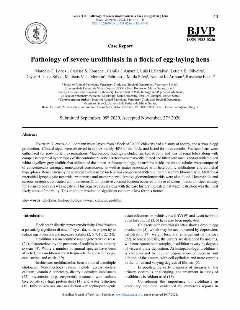
Fourteen, 31-week-old Lohmann white layers from a flock of 30,000 chickens had a history of apathy, and a drop in egg production. Clinical signs were observed in approximately 40% of the flock, and lasted for three months. Fourteen hens were euthanized for post-mortem examinations. Macroscopic findings included marked atrophy and loss of renal lobes along with compensatory renal hypertrophy of the contralateral lobe. Ureters were markedly dilated and filled with mucus and/or with molded white to yellow-grey uroliths that obliterated the lumen. At histopathology, the uroliths inside ureters and tubules were composed of concentrically arranged mineralized concretions, as well as urates associated with heterophilic infiltrations and epithelial hyperplasia. Renal parenchyma adjacent to obstructed ureters was compressed with tubules replaced by fibrous tissue. Multifocal interstitial lymphocytic nephritis, proteinuria and membranoproliferative glomerulonephritis were also found. Heterophilic and caseous ureteritis associated with numerous Gram-positive coccoid bacteria occurred in three chickens. Immunohistochemistry for avian coronavirus was negative. This negative result along with the case history indicated that water restriction was the most likely cause of mortality. This condition resulted in significant economic loss for this farmer.
Key words: chickens, histopathology, layers, kidneys, uroliths.
Introduction
Flock health directly impacts productivity. Urolithiasis is a potentially significant disease of layers due to its propensity to reduce egg production and increase mortality (2, 5, 7, 16, 22, 24).
Urolithiasis is an acquired and degenerative disease (24), characterized by the presence of uroliths in the urinary system (4). While a number of animal species have been affected, the condition is most frequently diagnosed in dogs, cats, swine, and cattle (19).
In chickens, urolithiasis has been attributed to multiple etiologies. Non-infectious causes include excess dietary calcium, vitamin A deficiency, dietary electrolyte imbalances (25), mycotoxins (e.g. oosporein), treatment with sodium bicarbonate (5), high protein diet (14), and water restriction (10). Infectious causes, such as infection with nephropathogenic
avian infectious bronchitis virus (IBV) (9) and avian nephritis virus (astrovirus) (3, 5) have also been implicated.
Chickens with urolithiasis often show a drop in egg production (5), which may be accompanied by depression, dehydration (7), weight loss, and enlargement of the toes (22). Macroscopically, the ureters are distended by uroliths, with consequent renal atrophy, in addition to varying degrees of visceral urate deposition. At histopathology, urolithiasis is characterized by tubular degeneration or necrosis and dilation of the ureters, with cell cylinders and urate crystals in the lumen and varying degrees of fibrosis (5).
In poultry, the early diagnosis of diseases of the urinary system is challenging, and treatment in cases of urolithiasis is seldom used (18).
Considering the importance of urolithiasis in veterinary medicine, evidenced by numerous reports in
Brazilian Journal of Veterinary Pathology. www.bjvp.org.br . All rights reserved 2007-2021.
Lopes et al.; Pathology of severe urolithiasis in a flock of egg-laying hens Braz J Vet Pathol, 2021, 14(1), 40 – 45
DOI: 10.24070/bjvp.1983-0246.v14i1p40-45
41
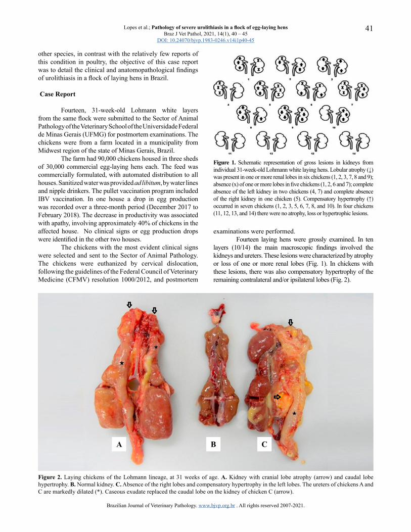
Figure 2. Laying chickens of the Lohmann lineage, at 31 weeks of age. A. Kidney with cranial lobe atrophy (arrow) and caudal lobe hypertrophy. B. Normal kidney. C. Absence of the right lobes and compensatory hypertrophy in the left lobes. The ureters of chickens A and C are markedly dilated (*). Caseous exudate replaced the caudal lobe on the kidney of chicken C (arrow).
Figure 1. Schematic representation of gross lesions in kidneys from individual 31-week-old Lohmann white laying hens. Lobular atrophy (↓) was present in one or more renal lobes in six chickens (1, 2, 3, 7, 8 and 9); absence (x) of one or more lobes in five chickens (1, 2, 6 and 7); complete absence of the left kidney in two chickens (4, 7) and complete absence of the right kidney in one chicken (5). Compensatory hypertrophy (↑) occurred in seven chickens (1, 2, 3, 5, 6, 7, 8, and 10). In four chickens (11, 12, 13, and 14) there were no atrophy, loss or hypertrophic lesions.
examinations were performed.Fourteen laying hens were grossly examined. In ten
layers (10/14) the main macroscopic findings involved the kidneys and ureters. These lesions were characterized by atrophy or loss of one or more renal lobes (Fig. 1). In chickens with these lesions, there was also compensatory hypertrophy of the remaining contralateral and/or ipsilateral lobes (Fig. 2).
other species, in contrast with the relatively few reports of this condition in poultry, the objective of this case report was to detail the clinical and anatomopathological findings of urolithiasis in a flock of laying hens in Brazil.
Case Report
Fourteen, 31-week-old Lohmann white layers from the same flock were submitted to the Sector of Animal Pathology of the Veterinary School of the Universidade Federal de Minas Gerais (UFMG) for postmortem examinations. The chickens were from a farm located in a municipality from Midwest region of the state of Minas Gerais, Brazil.
The farm had 90,000 chickens housed in three sheds of 30,000 commercial egg-laying hens each. The feed was commercially formulated, with automated distribution to all houses. Sanitized water was provided ad libitum, by water lines and nipple drinkers. The pullet vaccination program included IBV vaccination. In one house a drop in egg production was recorded over a three-month period (December 2017 to February 2018). The decrease in productivity was associated with apathy, involving approximately 40% of chickens in the affected house. No clinical signs or egg production drops were identified in the other two houses.
The chickens with the most evident clinical signs were selected and sent to the Sector of Animal Pathology. The chickens were euthanized by cervical dislocation, following the guidelines of the Federal Council of Veterinary Medicine (CFMV) resolution 1000/2012, and postmortem
A B C
Brazilian Journal of Veterinary Pathology. www.bjvp.org.br . All rights reserved 2007-2021.
Lopes et al.; Pathology of severe urolithiasis in a flock of egg-laying hens Braz J Vet Pathol, 2021, 14(1), 40 – 45
DOI: 10.24070/bjvp.1983-0246.v14i1p40-45
42
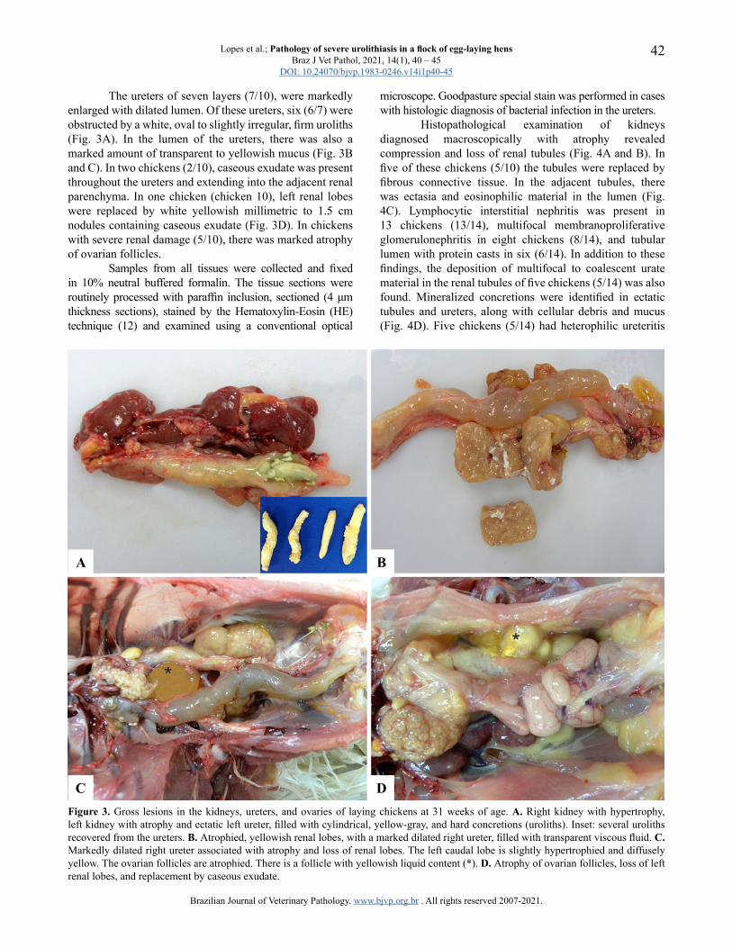
The ureters of seven layers (7/10), were markedly enlarged with dilated lumen. Of these ureters, six (6/7) were obstructed by a white, oval to slightly irregular, firm uroliths (Fig. 3A). In the lumen of the ureters, there was also a marked amount of transparent to yellowish mucus (Fig. 3B and C). In two chickens (2/10), caseous exudate was present throughout the ureters and extending into the adjacent renal parenchyma. In one chicken (chicken 10), left renal lobes were replaced by white yellowish millimetric to 1.5 cm nodules containing caseous exudate (Fig. 3D). In chickens with severe renal damage (5/10), there was marked atrophy of ovarian follicles.
Samples from all tissues were collected and fixed in 10% neutral buffered formalin. The tissue sections were routinely processed with paraffin inclusion, sectioned (4 μm thickness sections), stained by the Hematoxylin-Eosin (HE) technique (12) and examined using a conventional optical
microscope. Goodpasture special stain was performed in cases with histologic diagnosis of bacterial infection in the ureters.
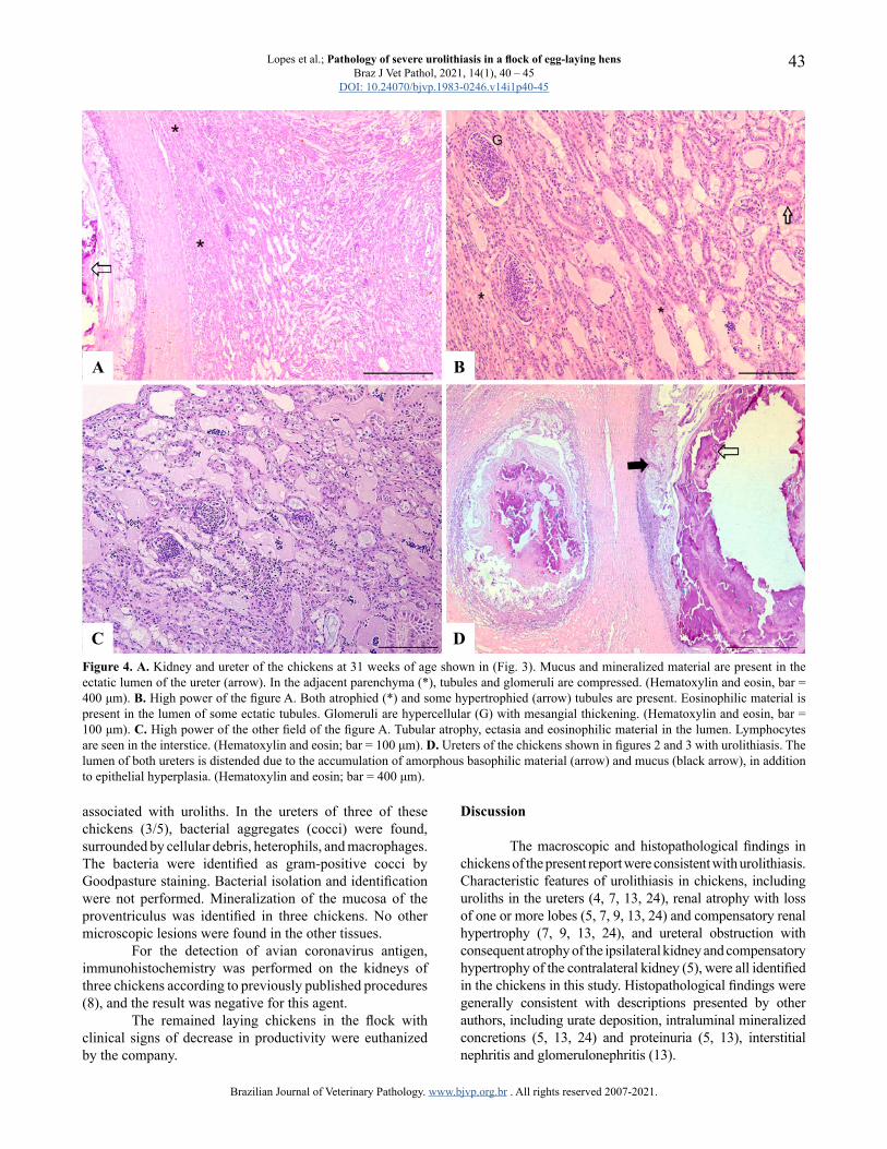
Histopathological examination of kidneys diagnosed macroscopically with atrophy revealed compression and loss of renal tubules (Fig. 4A and B). In five of these chickens (5/10) the tubules were replaced by fibrous connective tissue. In the adjacent tubules, there was ectasia and eosinophilic material in the lumen (Fig. 4C). Lymphocytic interstitial nephritis was present in 13 chickens (13/14), multifocal membranoproliferative glomerulonephritis in eight chickens (8/14), and tubular lumen with protein casts in six (6/14). In addition to these findings, the deposition of multifocal to coalescent urate material in the renal tubules of five chickens (5/14) was also found. Mineralized concretions were identified in ectatic tubules and ureters, along with cellular debris and mucus (Fig. 4D). Five chickens (5/14) had heterophilic ureteritis
Figure 3. Gross lesions in the kidneys, ureters, and ovaries of laying chickens at 31 weeks of age. A. Right kidney with hypertrophy, left kidney with atrophy and ectatic left ureter, filled with cylindrical, yellow-gray, and hard concretions (uroliths). Inset: several uroliths recovered from the ureters. B. Atrophied, yellowish renal lobes, with a marked dilated right ureter, filled with transparent viscous fluid. C. Markedly dilated right ureter associated with atrophy and loss of renal lobes. The left caudal lobe is slightly hypertrophied and diffusely yellow. The ovarian follicles are atrophied. There is a follicle with yellowish liquid content (*). D. Atrophy of ovarian follicles, loss of left renal lobes, and replacement by caseous exudate.
A B
C D
Brazilian Journal of Veterinary Pathology. www.bjvp.org.br . All rights reserved 2007-2021.
Lopes et al.; Pathology of severe urolithiasis in a flock of egg-laying hens Braz J Vet Pathol, 2021, 14(1), 40 – 45
DOI: 10.24070/bjvp.1983-0246.v14i1p40-45
43
associated with uroliths. In the ureters of three of these chickens (3/5), bacterial aggregates (cocci) were found, surrounded by cellular debris, heterophils, and macrophages. The bacteria were identified as gram-positive cocci by Goodpasture staining. Bacterial isolation and identification were not performed. Mineralization of the mucosa of the proventriculus was identified in three chickens. No other microscopic lesions were found in the other tissues.
For the detection of avian coronavirus antigen, immunohistochemistry was performed on the kidneys of three chickens according to previously published procedures (8), and the result was negative for this agent.
The remained laying chickens in the flock with clinical signs of decrease in productivity were euthanized by the company.
Discussion
The macroscopic and histopathological findings in chickens of the present report were consistent with urolithiasis. Characteristic features of urolithiasis in chickens, including uroliths in the ureters (4, 7, 13, 24), renal atrophy with loss of one or more lobes (5, 7, 9, 13, 24) and compensatory renal hypertrophy (7, 9, 13, 24), and ureteral obstruction with consequent atrophy of the ipsilateral kidney and compensatory hypertrophy of the contralateral kidney (5), were all identified in the chickens in this study. Histopathological findings were generally consistent with descriptions presented by other authors, including urate deposition, intraluminal mineralized concretions (5, 13, 24) and proteinuria (5, 13), interstitial nephritis and glomerulonephritis (13).
Figure 4. A. Kidney and ureter of the chickens at 31 weeks of age shown in (Fig. 3). Mucus and mineralized material are present in the ectatic lumen of the ureter (arrow). In the adjacent parenchyma (*), tubules and glomeruli are compressed. (Hematoxylin and eosin, bar = 400 μm). B. High power of the figure A. Both atrophied (*) and some hypertrophied (arrow) tubules are present. Eosinophilic material is present in the lumen of some ectatic tubules. Glomeruli are hypercellular (G) with mesangial thickening. (Hematoxylin and eosin, bar = 100 μm). C. High power of the other field of the figure A. Tubular atrophy, ectasia and eosinophilic material in the lumen. Lymphocytes are seen in the interstice. (Hematoxylin and eosin; bar = 100 μm). D. Ureters of the chickens shown in figures 2 and 3 with urolithiasis. The lumen of both ureters is distended due to the accumulation of amorphous basophilic material (arrow) and mucus (black arrow), in addition to epithelial hyperplasia. (Hematoxylin and eosin; bar = 400 μm).
C D
A B
Brazilian Journal of Veterinary Pathology. www.bjvp.org.br . All rights reserved 2007-2021.
Lopes et al.; Pathology of severe urolithiasis in a flock of egg-laying hens Braz J Vet Pathol, 2021, 14(1), 40 – 45
DOI: 10.24070/bjvp.1983-0246.v14i1p40-45
44
Clinical, epidemiological, and histopathological evidence of infectious diseases in the examined chickens was not found. Bacterial ureteritis in three chickens was interpreted as secondary and ascending infection.
In domestic chickens, urolithiasis is more frequently described in laying hens, but there are also reports in chicks (5), male broiler breeders (16), and other avian species such as passerines (14) and psittacines (6).
In avian species, several causes have been attributed to the occurrence of this disease. In the present report, negative laboratory results for IBV, together with the high ambient temperature during the period of manifestation of this condition (December to February) and the occurrence of the disease in only one house on a three-house farm raised the possibility of accidental water deprivation/restriction as a cause of this condition. Water deprivation is an important contributing factor for outbreaks of urolithiasis. In a previous report of urolithiasis in chickens, there was a decrease in mortality after ensuring that the chickens received sufficient amount of water (20).
Flocks deprived of water suffered an increase mortality as a result of kidney disease (20). Dehydration results in increased water reabsorption by kidney tubules, resulting in reduced urinary flow and uric acid secretion, with consequent precipitation of urates and obstruction of tubules and ureters. Due to their high metabolic demand for water, laying hens seem to be more susceptible to kidney damage due to water deprivation (10, 20).
Imbalances in the diet, such as high levels of protein, can also contribute to the formation of uroliths. Excess dietary protein can serve as an organic nucleus, facilitating mineral deposition and formation of uroliths (14), in addition to causing excessive production of uric acid (11). In an experimental study, chickens that fed high amounts of calcium and low amounts of phosphorus had macroscopic lesions associated with urolithiasis. High levels of dietary calcium can predispose to mineral deposition in kidneys, with cellular debris obstructing ducts and ureters, and predisposing to urolith formation (24). Feed analysis was not performed in the present case, but since all flocks on the farm received the same formulated diet and only chickens in one house had renal damage, a dietary imbalance was not considered a probable cause of this condition.
Deposition of urates in extrarenal tissues was not seen in the layers of this report. Gout is a metabolic disease characterized by the deposition of urates and uric acid in the kidneys and serosa of other tissues, such as heart, liver, air sacs (visceral gout), and joints. The disease occurs when there are failures in urinary excretion, resulting in an accumulation of metabolites in the body (5). Visceral gout in chickens typically involves multiple organs, including the kidneys, such as liver, spleen (15, 17, 20), pericardial sac (17, 20), heart, lungs and, air sacs (15). Urate deposition in the joints is referred to as articular gout (5).
Viral agents such as nephropathogenic strains of IBV (9) has also been implicated in outbreaks of urolithiasis.
IBV was considered unlikely to have caused urolithiasis in this case due to the negative immunohistochemistry results, as well as the involvement of chickens in only one house on the studied farm. IBV is a highly contagious disease (9), which can readily spread between houses on an affected farm. Avian nephritis virus (ANV) and chicken astrovirus (CAstV) have been implicated also as causative agents of renal gout (3). However, reports of urolithiasis in chickens due to ANV and CAstV infections have not been found in the current literature.
Laying hens affected by urolithiasis show a decrease in moderate egg production (5, 22). In the present report, the decrease in egg production was associated with atrophy of ovarian follicles. The relationship between urolithiasis and changes in the reproductive system of chickens has yet to be fully elucidated. In one study, urolithiasis and ovarian regression were reported in the same chicken (25). However, in another study of a large number of chickens, it was observed that despite the occurrence of urolithiasis, 93% of the affected chickens, including those that died and which were producing normally, did not have ovarian lesions (13).
Regarding the influence of water restriction on laying hens, chickens subjected to water deprivation eat less, resulting in lethargy and a drop in egg production (21). Thus, water restriction may have directly contributed to the decrease in egg production described in this report. In a previous report of urolithiasis, a reduction in eggshell quality in the absence of an egg production drop were described (13). In the present case, in contrast, urolithiasis was accompanied by an egg production drop with no change in eggshell quality.
In the present study, it is difficult to establish a link between glomerular lesions and the other renal lesions in the chickens evaluated. In chickens, glomerular lesions have been described associated with sodium chloride intoxications as well as high doses of deoxycorticosterone (1), neither of which was diagnosed in the present report. In chickens, membranoproliferative glomerulonephritis is considered among the most common glomerular lesions (23), and has been found in association with other diseases, such as infectious bursal disease and Mycoplasma synoviae infection (1). These infectious causes were not identified in the present case.
Some studies in commercial chickens demonstrate that urinary acidification techniques, involving ammonium sulfate and methionine (5) can be used as a treatment method in affected flocks, helping to dissolve uroliths, in addition to preventing new cases (24). Treatment for urolithiasis in commercial chickens is, however, seldom used and if considered economically viable, highlighting the importance of prevention of this potentially economically significant condition. In the present cases, slaughter of the flock was considered the most viable economic option, due to the chronic impairment of the kidneys and egg production losses.
Urolithiasis represents a serious problem in poultry, causing severe renal lesions and consequent loss of productivity. The clinical signs presented by affected
Brazilian Journal of Veterinary Pathology. www.bjvp.org.br . All rights reserved 2007-2021.
Lopes et al.; Pathology of severe urolithiasis in a flock of egg-laying hens Braz J Vet Pathol, 2021, 14(1), 40 – 45
DOI: 10.24070/bjvp.1983-0246.v14i1p40-45
45
chickens are often nonspecific, which makes early diagnosis difficult. Water deprivation is an important predisposing factor, and dietary imbalances and infectious bronchitis are other differentials. Further investigations concerning the causes and frequency of urolithiasis in layer hens may help to prevent this disease in the future.
Funding
Financial support and scholarships were provided by Coordenação de Aperfeiçoamento de Pessoal de Nível Superior (CAPES) - Finance Code 001 and Fundação de Amparo à Pesquisa do Estado de Minas Gerais (FAPEMIG), Brazil.
References1. Abdul-Aziz T, Fletcher OJ. Urinary System. In:
Abdul-Aziz T, Fletcher OJ, Barnes HJ, editors. Avian histopathology. 4th ed. Madison, WI: American Association of Avian Pathologists, Omni Press. 2016. p.423-68.
2. Brown TP, Glisson JR, Villegas GRP, Davis RB. Studies of avian urolithiasis associated with an infectious bronchitis virus. Avian Dis. 1987;31(3):629-36.
3. Bulbule NR, Mandakhalikar KD, Kapgate SS, Deshmukh VV, Schat KA, Chawak MM. Role of chicken astrovirus as a causative agent of gout in commercial broilers in India. Avian Pathol. 2013;42(5):464-73.
4. Cianciolo RE, Mohr FC, Urinary System. In: Maxie MG, editor. Jubb, Kennedy & Palmer’s Pathology of domestic animals. 6th ed., vol 2. Missouri: Elsevier. 2016. p.376-464.
5. Crespo R. Developmental, metabolic, and other noninfectious disorders. In: Bouliane M, Logue CM, Mc Dougald LR, Nair V, Suarez DL, editors. Diseases of Poultry. 14th ed. Iowa: Wiley-Blackwell. 2020. p.1286-329.
6. Dennis PM, Bennett RA. Ureterotomy for removal of two ureteroliths in a parrot. J Am Vet Med Assoc. 2000;217(6):865-68.
7. Fitz-coy SH, Edgar SA, Hoerr FJ. An outbreak of urolithiasis in single-comb white leghorn pullets. Avian Dis. 1988;32(3):563-66.
8. França M, Woolcock PR, Yu M, Jackwood MW, Shivaprasad HL. Nephritis Associated with Infectious Bronchitis Virus Cal99 Variant in Game Chickens. Avian Pathol. 2011;55(3):422-28.
9. Jackwood MW, De Wit S. Infectious Bronchitis. In: Bouliane M, Logue CM, Mc Dougald LR, Nair V, Suarez DL, editors. Diseases of Poultry. 14th ed. Iowa: Wiley-Blackwell. 2020. p.167-88.
10. Julian R. Water deprivation as a cause of renal disease in chickens. Avian Pathol. 1982;11(4):615-17.
11. Li XM, Deng MX, Li QY, Jia RY, Wang J. Experimental study on the clinical pathology of urate deposition in chickens. Chinese J Vet Sci. 1998;18:49-51.
12. Luna LG. Manual of histologic staining methods of the Armed Forces Institute of Pathology. 3rd ed. New York: McGraw-Hill. 1968. 258p.
13. Mallinson ET, Rothenba H, Wildeman RF, Snyder DB, Russek E, Zuckerman AI, Davidson JP. Epizootiology, Pathology, and Microbiology of an Outbreak of Urolithiasis in Chickens. Avian Dis. 1983;28(1):25-43.
14. Marietto-Gonçalves GA, Salgado BS. Postmortem lesions of urolithiasis in a lesser seed finch (Sporophila angolensis). Acta Vet Brasil. 2012;6(1):52-5.
15. Mir MS, Darzi MM, Khan AA, Ganaie NA, Gupta S, Nashiruddullah N, Kamil AS. Investigation of an outbreak of gout in kashmir favorella poultry. Indian J Vet Pathol. 2005;29(1):35-7.
16. Moyle JR, Wideman SM, Whipple DE, Yoho DE, Bramwell RK. Urolithiasis in male broiler breeders. Int J Poult Sci. 2011;10(11):839-41.
17. Mudasir M, Dogra S, Badroo GA, Nashirrudullah N, Dogra S, Batoo AS, Javaid M, Bhat MA. Pathomorphological study of visceral gout in desi fowl. Int J Curr Microbiol App. Sci. 2017;6(10):5039-42.
18. Pollock C. Diagnosis and treatment of avian renal disease. Vet Clin Exot Anim. 2006;9:107-28.
19. Robinson MR, Norris RD, Sur RL, Preminger GM. Urolithiasis: Not Just a 2-Legged Animal Disease. J Urol. 2008;179:46-52.
20. Rexhepi A, Brown C, Sherifi K, Behluli B. Visceral gout (Uricosis) and urolithiasis caused by dehydration in laying hen farm, necropsy and histopathology findings. Kafkas Univ Vet Fak Derg. 2014;21(2):291-294.
21. Sens DR, Rocha AS. Influência da restrição da água na produção e peso de ovos. Rev. Agrogeo. 2010;2:71-4.
22. Sid H, Fettah A, Lounas A. Descriptive study of an outbreak of avian urolithiasis in a large commercial egg complex in Algeria. Not Sci Biol. 2011;3(1):22-5.
23. Siller WG. Renal pathology of the fowl - A review. Avian Pathol. 1981;10(3):187-262.
24. Wideman Junior RF. Confirming the promise to prevent physiological disorders of organs: urolithiasis in laying hens. Poult Sci. 2015;25:292-304.
25. Wideman RF, Mallinson ET, Rothenbacher H. Kidney function of pullets and laying hens during outbreaks of urolithiasis. Poult Sci. 1983;62:1954-70.