Humoral immune response in hens naturally infected with Salmonella Enteritidis against outer...
8
291 Vet. Res. 35 (2004) 291–298 © INRA, EDP Sciences, 2004 DOI: 10.1051/vetres:2004011 Original article Humoral immune response in hens naturally infected with Salmonella Enteritidis against outer membrane proteins and other surface structural antigens Javier OCHOA-REPÁRAZ a , Begoña SESMA b , Miguel ÁLVAREZ c , M. Jesús RENEDO d , Juan M. IRACHE d , Carlos GAMAZO a * a Departamento de Microbiología, Universidad de Navarra, 31008 Pamplona, Spain b Instituto Salud Pública de Navarra, 31004 Pamplona, Spain c Lab. Municipal de Pamplona, 31004 Pamplona, Spain d Centro Galénico, Universidad de Navarra, 31008 Pamplona, Spain (Received 8 September 2003; accepted 19 December 2003) Abstract – A simple procedure for obtaining surface exposed antigens of Salmonella Enteritidis is described. A heat treatment of whole bacteria in saline solution induced the release of small mem- brane vesicles containing outer membrane components as well as surface appendage components, such as fimbriae and flagellin. The characterization of the structural components of this extract, called HE, was established by SDS-PAGE and immunoblotting using polyclonal and monoclonal specific antibodies. Five major groups of proteins were identified: flagellin, porins, OmpA, SEF21 and SEF14 fimbriae. The immunogenicity of these proteins was studied by immunoblotting with serum samples from naturally infected hens. Flagellin, porins, OmpA, SEF14 and SEF21 fimbriae were immuno- genic in the S. Enteritidis infected hens (frequency of reactants: 47.3, 97.3, 64.7, 50.0 and 60.8%, respectively); porins also reacted with sera from non infected hens (66.7%). The immunogenicity of these antigens in infected birds provide promise that they may serve as components of an effective subcellular vaccine for poultry salmonellosis. salmonellosis / immunoblotting / immunogenicity / surface antigens / hens 1. INTRODUCTION Salmonella enterica serovar Enteritidis (Salmonella Enteritidis, SE) is a major cause of human food-borne illness and is the most frequent serovar detected in out- breaks of human salmonellosis [18]. Poul- try products are known to be a significant reservoir for Salmonella and the most important source of SE infection in humans [7, 14, 19, 22]. Salmonella possess different surface struc- tures that can induce protective immune responses in experimentally infected chick- ens [16, 30]. Immunoblotting has been used previously to recognize antigenic polypep- tides of SE in experimentally infected chickens [1]. Other workers have also used experimentally infected birds, by ELISA and other serological techniques, to deter- mine the antigenicity of lipopolysaccharide (LPS), flagella and fimbriae [12, 23, 25, 28], * Corresponding author: [email protected]
Transcript of Humoral immune response in hens naturally infected with Salmonella Enteritidis against outer...
291Vet. Res. 35 (2004) 291–298© INRA, EDP Sciences, 2004DOI: 10.1051/vetres:2004011
Original article
Humoral immune response in hens naturally infected with Salmonella Enteritidis against outer membrane
proteins and other surface structural antigens
Javier OCHOA-REPÁRAZa, Begoña SESMAb, Miguel ÁLVAREZc, M. Jesús RENEDOd, Juan M. IRACHEd, Carlos GAMAZOa*
a Departamento de Microbiología, Universidad de Navarra, 31008 Pamplona, Spain b Instituto Salud Pública de Navarra, 31004 Pamplona, Spain
c Lab. Municipal de Pamplona, 31004 Pamplona, Spaind Centro Galénico, Universidad de Navarra, 31008 Pamplona, Spain
(Received 8 September 2003; accepted 19 December 2003)
Abstract – A simple procedure for obtaining surface exposed antigens of Salmonella Enteritidis isdescribed. A heat treatment of whole bacteria in saline solution induced the release of small mem-brane vesicles containing outer membrane components as well as surface appendage components,such as fimbriae and flagellin. The characterization of the structural components of this extract, calledHE, was established by SDS-PAGE and immunoblotting using polyclonal and monoclonal specificantibodies. Five major groups of proteins were identified: flagellin, porins, OmpA, SEF21 and SEF14fimbriae. The immunogenicity of these proteins was studied by immunoblotting with serum samplesfrom naturally infected hens. Flagellin, porins, OmpA, SEF14 and SEF21 fimbriae were immuno-genic in the S. Enteritidis infected hens (frequency of reactants: 47.3, 97.3, 64.7, 50.0 and 60.8%,respectively); porins also reacted with sera from non infected hens (66.7%). The immunogenicityof these antigens in infected birds provide promise that they may serve as components of an effectivesubcellular vaccine for poultry salmonellosis.
salmonellosis / immunoblotting / immunogenicity / surface antigens / hens
1. INTRODUCTION
Salmonella enterica serovar Enteritidis(Salmonella Enteritidis, SE) is a majorcause of human food-borne illness and isthe most frequent serovar detected in out-breaks of human salmonellosis [18]. Poul-try products are known to be a significantreservoir for Salmonella and the mostimportant source of SE infection in humans[7, 14, 19, 22].
Salmonella possess different surface struc-tures that can induce protective immuneresponses in experimentally infected chick-ens [16, 30]. Immunoblotting has been usedpreviously to recognize antigenic polypep-tides of SE in experimentally infectedchickens [1]. Other workers have also usedexperimentally infected birds, by ELISAand other serological techniques, to deter-mine the antigenicity of lipopolysaccharide(LPS), flagella and fimbriae [12, 23, 25, 28],
* Corresponding author: [email protected]
292 J. Ochoa-Repáraz et al.
or whole cells [6]. However, to our knowl-edge, there is not any report in the literatureshowing the reactivity by immunoblottingof the SE antigens during the course of anatural infection in hens.
For purposes of vaccine development,the present study was accomplished todetermine the main immunogenic outercomponents of SE in the course of a naturalinfection in hens. For this, a simple proce-dure for obtaining superficial antigens ofSE is also described.
2. MATERIALS AND METHODS
2.1. Serum samples
A total of 104 serum samples from lay-ing hens (7–11 months old) were studied.Serum samples in group 1 (n = 74) wereobtained from S. Enteritidis naturally infectedhens, from different flocks (as confirmed byrectal swab culture) and serum samples ingroup 2 (n = 30) were taken from salmonel-lae free hens (obtained from CESAC, Reus,Girona, Spain). Positive and negative con-trol serum sample pools were prepared bypooling ten individual sera from infectedhens and ten individual sera from salmonel-lae free hens, respectively. These controlswere employed to validate the immunoblot-ting procedure used in this study.
2.2. Bacterial strains and growth conditions
The antigenic extract was obtained fromthe clinically isolated S. Enteritidis strain3934 (Universitary Hospital of Navarra,Spain), that was grown in trypticase-soybroth (Biomérieux, Marcy-l’Étoile, France)in a rotary shaker at 37 °C for 24 h.
2.3. Antigenic extract of S. Enteritidis
A hot saline extract of S. Enteritidis (HE)was obtained following a procedure previ-
ously used for the extraction of some Bru-cella antigens [10]. Briefly, live cells weresuspended in physiological saline (10 g ofpacked cells per 100 mL) and heated inflowing steam for 15 min. After centrifuga-tion at 12 000 × g for 15 min, the superna-tant was dialyzed for two days at 4 °Cagainst several changes of deionized water.The dialyzed material was centrifuged for5 h at 100 000 × g, and the pellet (HE extract)was resuspended in deionized water,lyophilized and stored at room temperature.
Outer membrane proteins (OMPs) fromS. Enteritidis were prepared by sequentialdetergent extraction of cell envelopes [8].Briefly, after the disruption of cells by highpressure in a French Press (Aminco-SLMInstuments Inc, Urbana, Illinois, USA), theinner membranes of the bacteria were sol-ubilized after treatment with 1% Sarkosyl(N-Lauryl sarcosine, Sigma Chemical Co.,St. Louis, USA) and centrifuging (20 000 ×g; 30 min). The sediment was suspended in0.5 M Tris-HCl (pH 6.8) with 10% SDS(Lauryl sulfate, Sigma) and centrifuged(20 000 × g; 30 min). The OMPs of S. Enter-itidis were present in the final supernatant.
2.4. Electron microscopy
HE was resuspended in deionized waterand stained with 4% Uranyl acetate (Agarscientific) for 15 min and with lead citrate(Agar scientific) for 15 min and then wasexamined with a Hitachi 1100 transmis-sion electron microscope (Hitachi Scien-tific Instruments, California, USA) operat-ing at 100 kV.
2.5. Chemical analysis
Total protein content was determinedcolorimetrically [17], with bovine serumalbumin as the standard. The LPS contentof HE was estimated by colorimetric deter-mination of 2-keto-3-deoxyoctonate cor-rected for 2-deoxyaldoses, performed bythe method of Warren [32] as modified byOsborn [21].
Antibody response in hens to S. Enteritidis 293
2.6. SDS-PAGE and immunoblotting procedure
SDS-PAGE was performed in 15% acr-ylamide slabs by the method of Laemmli[15]. The gels were stained by the alkalinesilver-glutaraldehyde method for proteins.The apparent molecular masses of the pro-teins present in the antigenic extracts weredetermined by comparing their electro-phoretic mobility with that of the followingmolecular mass markers (Rainbow coloredprotein molecular weight marker, Amer-sham pharmacia biotech, Freiburg, Ger-many): myosin (220 kDa); phosphorylase b(97 kDa); bovine serum albumin (66 kDa);ovalbumin (45 kDa); carbonic anhydrase(30 kDa); trypsin inhibitor (20.1 kDa); lys-ozyme (14.3 kDa).
Immunoblotting was carried out asdescribed by Towbin [31] with the follow-ing modifications: after SDS-PAGE, thegel was transferred in a transfer buffer(0.2 M glycine; 24 mM Tris; 10% methanol[pH 8.3]) to PVDF (polyvinylidene fluoridepapers, pore size 0.45 µm, Schleicher andSchuell, Dassel, Germany) using a semidryelectroblotter (Bio-rad Laboratories, Rich-mond, USA) (200 mA; 5 V; 30 min). Theblots were placed in blocking buffer (3%skimmed milk and 0.15% Tween-20 in10 mM phosphate-buffered saline [pH 7.4])overnight at room temperature, and thenthey were incubated for 4 h at room tem-perature with hen serum sample diluted1:100 in blocking buffer without skimmedmilk. After four washes in blocking bufferwithout skimmed milk, the blots were incu-bated for one hour at room temperature withthe immunoconjugate: peroxidase-conju-gated rabbit anti-chicken IgG (Nordic Labs,Tilburg, The Netherlands), diluted 1:1000in the same buffer. The blots were washedfour times more, and were developed byincubation in a solution containing H2O2and 4-chloro, 1-naphtol for 20 min in thedark.
The frequency of the reactants to themain structural components of HE extractwas determined by computing the number
of sera of natural infected hens that reactedagainst these components. Positive andnegative control serum pools were used inall the experiments as an internal referenceto validate the comparison of the experi-ments.
2.7. Identification of the major components of HE
The presence of flagellin (FliC) andSEF14 were confirmed with specific mon-oclonal antibodies (kindly provided by Vet-erinary Laboratories Agency, Surrey, UK).SEF14 and SEF21 bands were recognizedby comparison with the correspondingpurified proteins obtained after a purifica-tion process based on a selective precipita-tion with (NH4)2SO4 (50% saturation); thestarting material was a crude extract enrichedin fimbriae and flagella, according to themethod of Freurier et al. [20]. LPS andporins were recognized with polyclonalserum from rabbits hyper-immunized withLPS and porins, respectively (sera obtainedfrom the Department of Microbiology, Uni-versity of Navarra, Spain). OmpA was iden-tified based on its different mobility inSDS-PAGE when the samples are incu-bated in SDS sample buffer at 100 ºC or atroom temperature [9].
3. RESULTS
3.1. Characterization of HE extract
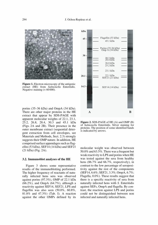
The electron microscopy studies suggestthat after a heat treatment of whole cellssmall spherical vesicles ranging from 15 to40 nm were released. Other filamentousappendages were seen (Fig. 1). This mate-rial was called the HE extract. The proteincontent of the HE extract obtained fromS. Enteritidis 3934 was 31.35 ± 4.55%, andthe LPS percentage was 69.14 ± 1.90% (n =10).
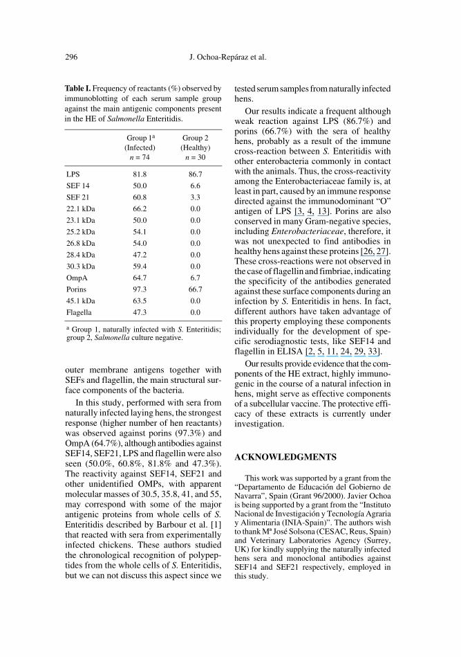
The SDS-PAGE profile of HE was sim-ilar to OMPs enriched fraction, containing
294 J. Ochoa-Repáraz et al.
porins (35–36 kDa) and OmpA (34 kDa).There are other major proteins in the HEextract that appear by SDS-PAGE withapparent molecular weights of 22.1, 23.1,25.2, 26.8, 28.4, 30.3 and 45.1 kDa(Figs. 2A and 2B). Their presence in theouter membrane extract (sequential deter-gent extraction from cell envelopes, seeMaterials and Methods, Sect. 2.3) stronglysuggests their OMP nature. In addition, HEcomprised surface appendages such as flag-ellin (53 kDa), SEF14 (14 kDa) and SEF21(21 kDa) (Fig. 2A).
3.2. Immunoblot analyses of the HE
Figure 3 shows some representativeresults of the immunoblotting performed.The higher frequency of reactants of natu-rally infected hens sera was observedagainst porins (97.3%), OMP of 22.1 kDa(66.2%), and OmpA (64.7%), although areactivity against SEF14, SEF21, LPS andflagellin was also seen (50.0%, 60.8%,81.8% and 47.3%) (Tab. I). A reactionagainst the other OMPs defined by its
molecular weight was observed between50.0% and 63.5%. There was a frequent butweak reactivity to LPS and porins when HEwas tested against the sera from healthyhens (86.7% and 66.7%, respectively), incontrast to the low percentage of seroposi-tivity against the rest of the components(SEF14, 6.6%; SEF21, 3.3%; OmpA, 6.7%;Flagella, 0.0%). These results suggest thatthere is a specific reactivity of sera fromnaturally infected hens with S. Enteritidisagainst SEFs, OmpA and flagella. By con-trast, the reaction against LPS and porinscould not be distinguished between noninfected and naturally infected hens.
Figure 2. SDS-PAGE of HE (A) and OMP (B)of Salmonella Enteritidis. Silver staining forproteins. The position of some identified bandsis indicated by arrows.
Figure 1. Electron microscopy of the antigenicextract (HE) from Salmonella Enteritidis.Negative staining (× 60 000).

Antibody response in hens to S. Enteritidis 295
4. DISCUSSION
In order to determine the most immuno-genic components of the surface of S. Enter-itidis, we studied by immunoblotting thereaction of sera from naturally infectedreproductive hens to an extract that containssurface proteins and external appendages ofthe bacteria. The immunogenicity of thesecomponents during natural infection wouldindicate their possible role as inductive
antigens in a subcellular vaccine for its usein poultry.
Salmonella possess surface structuresthat can induce protective humoral and cel-lular immune responses following experi-mental infection in poultry [16, 30]. Thesecomponents include LPS, OMPs, fimbriaeand flagellin. We obtained an antigenicextract of S. Enteritidis (called HE) by a sim-ple procedure that, instead of other antigenicextracts found in the literature, contains
Figure 3. Immunoblot analyses of sera from hens bacteriologically positive (A) and negative (B)for Salmonella Enteritidis against the HE components. Lane numbers correspond to animalreference numbers.
296 J. Ochoa-Repáraz et al.
outer membrane antigens together withSEFs and flagellin, the main structural sur-face components of the bacteria.
In this study, performed with sera fromnaturally infected laying hens, the strongestresponse (higher number of hen reactants)was observed against porins (97.3%) andOmpA (64.7%), although antibodies againstSEF14, SEF21, LPS and flagellin were alsoseen (50.0%, 60.8%, 81.8% and 47.3%).The reactivity against SEF14, SEF21 andother unidentified OMPs, with apparentmolecular masses of 30.5, 35.8, 41, and 55,may correspond with some of the majorantigenic proteins from whole cells of S.Enteritidis described by Barbour et al. [1]that reacted with sera from experimentallyinfected chickens. These authors studiedthe chronological recognition of polypep-tides from the whole cells of S. Enteritidis,but we can not discuss this aspect since we
tested serum samples from naturally infectedhens.
Our results indicate a frequent althoughweak reaction against LPS (86.7%) andporins (66.7%) with the sera of healthyhens, probably as a result of the immunecross-reaction between S. Enteritidis withother enterobacteria commonly in contactwith the animals. Thus, the cross-reactivityamong the Enterobacteriaceae family is, atleast in part, caused by an immune responsedirected against the immunodominant “O”antigen of LPS [3, 4, 13]. Porins are alsoconserved in many Gram-negative species,including Enterobacteriaceae, therefore, itwas not unexpected to find antibodies inhealthy hens against these proteins [26, 27].These cross-reactions were not observed inthe case of flagellin and fimbriae, indicatingthe specificity of the antibodies generatedagainst these surface components during aninfection by S. Enteritidis in hens. In fact,different authors have taken advantage ofthis property employing these componentsindividually for the development of spe-cific serodiagnostic tests, like SEF14 andflagellin in ELISA [2, 5, 11, 24, 29, 33].
Our results provide evidence that the com-ponents of the HE extract, highly immuno-genic in the course of a natural infection inhens, might serve as effective componentsof a subcellular vaccine. The protective effi-cacy of these extracts is currently underinvestigation.
ACKNOWLEDGMENTS
This work was supported by a grant from the“Departamento de Educación del Gobierno deNavarra”, Spain (Grant 96/2000). Javier Ochoais being supported by a grant from the “InstitutoNacional de Investigación y Tecnología Agrariay Alimentaria (INIA-Spain)”. The authors wishto thank Mª José Solsona (CESAC, Reus, Spain)and Veterinary Laboratories Agency (Surrey,UK) for kindly supplying the naturally infectedhens sera and monoclonal antibodies againstSEF14 and SEF21 respectively, employed inthis study.
Table I. Frequency of reactants (%) observed byimmunoblotting of each serum sample groupagainst the main antigenic components presentin the HE of Salmonella Enteritidis.
Group 1a
(Infected)n = 74
Group 2(Healthy)
n = 30
LPS 81.8 86.7
SEF 14 50.0 6.6
SEF 21 60.8 3.3
22.1 kDa 66.2 0.0
23.1 kDa 50.0 0.0
25.2 kDa 54.1 0.0
26.8 kDa 54.0 0.0
28.4 kDa 47.2 0.0
30.3 kDa 59.4 0.0
OmpA 64.7 6.7
Porins 97.3 66.7
45.1 kDa 63.5 0.0
Flagella 47.3 0.0
a Group 1, naturally infected with S. Enteritidis;group 2, Salmonella culture negative.

Antibody response in hens to S. Enteritidis 297
REFERENCES
[1] Barbour E.K., El Jurdi L.H., Faroo O.M.,Daghir N.J., Bouljihad M., Chronological rec-ognition by chicken of antigenic polypeptidesin Salmonella enteritidis with different plas-mid profiles: relationship to infection rate, J.Vet. Med. Sci. 62 (2000) 565–570.
[2] Barrow P.A., Serological diagnosis of Salmo-nella serotype enteritidis infections in poultryby ELISA and other tests, Int. J. Food Micro-biol. 21 (1994) 55–68.
[3] Barrow P.A., Berchieri A. Jr., al-Haddad O.,Serological response of chickens to infectionwith Salmonella gallinarum-S. pullorum detectedby enzyme-linked immunosorbent assay, AvianDis. 36 (1992) 227–236.
[4] Blanden R.V., Mackaness G.B., Collins F.M.,Mechanisms of acquired resistance in mousetyphoid, J. Exp. Med. 124 (1966) 585–600.
[5] Cooper G.L., Thorns C.J., Evaluation ofSEF14 fimbrial dot blot and flagellar westernblot tests as indicators of Salmonella enteri-tidis infection in chickens, Vet. Rec. 138(1996) 149–153.
[6] Darwin A.J., Miller V.L., Identification ofYersinia enterocolitica genes affecting sur-vival in an animal host using signature-taggedtransposon mutagenesis, Mol. Microbiol. 32(1999) 51–62.
[7] Fantasia M., Filetici E., Anastasio M.P.,Marcozzi M.D., Gramenzi M.P., Aureli P.,Italian experience in Salmonella enteritidis1978–1988: characterization of isolates fromfood and man, Int. J. Food Microbiol. 12(1991) 353–362.
[8] Filip C., Fletcher G., Wulff J.L., Earhart C.F.,Solubilization of the cytoplasmic membraneof Escherichia coli by the ionic detergentsodium-lauryl sarcosinate, J. Bacteriol. 115(1973) 717–722.
[9] Gamazo C., Vitas A.I., Moriyon I., Lopez-Goni I., Diaz R., Brucella group 3 outer mem-brane proteins contain a heat-modifiable pro-tein, FEMS Microbiol. Lett. 112 (1993) 141–146.
[10] Gamazo C., Winter A.J., Moriyon I., Riezu-Boj J.I., Blasco J.M., Diaz R., Comparativeanalyses of proteins extracted by hot saline orreleased spontaneously into outer membraneblebs from field strains of Brucella ovis andBrucella melitensis, Infect. Immun. 57 (1989)1419–1426.
[11] Gast R.K., Porter R.E. Jr., Holt P.S., Assess-ing the sensitivity of egg yolk antibody testing
for detecting Salmonella enteritidis infectionsin laying hens, Poult. Sci. 76 (1997) 798–801.
[12] Gast R.K., Nasir M.S., Jolley M.E., Holt P.S.,Stone H.D., Serologic detection of experi-mental Salmonella enteritidis infections inlaying hens by fluorescence polarization andenzyme immunoassay, Avian Dis. 46 (2002)137–142.
[13] Hormaeche C.E., Mastroeni P., Harrison J.A.,Demarco de Hormaeche R., Svenson S.,Stocker B.A., Protection against oral chal-lenge three months after i.v. immunization ofBALB/c mice with live Aro Salmonella typh-imurium and Salmonella enteritidis vaccinesis serotype (species)-dependent and only par-tially determined by the main LPS O antigen,Vaccine 14 (1996) 251–259.
[14] Humphrey T.J., Contamination of eggs andpoultry meat with Salmonella enterica sero-var Enteritidis, in: Saed A.M., Gast R.K.,Potter M.E., Wall P.G. (Eds.), Salmonellaenterica serovar Enteritidis in Humans andAnimals; Pathogenesis and Control, IowaState University Press, Ames, 1999, pp. 183–192.
[15] Laemmli U.K., Cleavage of structural pro-teins during the assembly of the head of bac-teriophage T4, Nature 227 (1970) 680–685.
[16] Liu W., Yang Y., Chung N., Kwang J., Induc-tion of humoral immune response and protec-tive immunity in chickens against Salmonellaenteritidis after a single dose of killed bacte-rium-loaded microspheres, Avian Dis. 45(2001) 797–806.
[17] Lowry O.H., Rosebrough N.J., Farr A.L., RandallR.J., Protein measurement by the Folin phenolreagent, J. Biol. Chem. 193 (1951) 265–275.
[18] Lu S., Manges A.R., Xu Y., Fang F.C., RileyL.W., Analysis of virulence of clinical isolatesof Salmonella enteritidis in vivo and in vitro,Infect. Immun. 67 (1999) 5651–5657.
[19] Meyer H., Animals as sources of infectionsin humans-salmonellosis, Dtsch. Tierarztl.Wochenschr. 106 (1999) 344–351.
[20] Ogunniyi A.D., Manning P.A., Kotlarski I., ASalmonella enteritidis 11RX pilin inducesstrong T-lymphocyte responses, Infect. Immun.62 (1994) 5376–5383.
[21] Osborn M.J., Studies on the gram negative cellwall. I. Evidence for the role of the 2-Keto,3-deoxyoctonate in the lipopolysaccharide ofSalmonella typhimurium, Proc. Natl. Acad.Sci. USA 50 (1963) 499–506.
[22] Palmer S., Parry S., Perry D., Smith R., EvansM., Nehaul L., Roberts R., Walapu M., Wright

298 J. Ochoa-Repáraz et al.
To access this journal online: www.edpsciences.org
D., The role of outbreaks in developing foodsafety policy: population based surveillanceof Salmonella outbreaks in Wales 1986–98,Epidemiol. Infect. 125 (2000) 467–472.
[23] Proux K., Jouy E., Houdayer C., Protais J.,Dibb-Fuller M., Boscher E., Gillard A.,Gracieux P., Gilbert F., Beaumont C., Duchet-Suchaux M., Reliable ELISAs showing dif-ferences between resistant and susceptiblelines in hens orally inoculated with Salmo-nella Enteritidis, Vet. Res. 33 (2002) 23–33.
[24] Rajashekara G., Munir S., Lamichhane C.M.,Back A., Kapur V., Halvorson D.A., NagarajaK.V., Application of recombinant fimbrialprotein for the specific detection of Salmo-nella enteritidis infection in poultry, Diagn.Microbiol. Infect. Dis. 32 (1998) 147–157.
[25] Seo K.H., Holt P.S., Brackett R.E., Gast R.K.,Stone H.D., Mucosal humoral immunity toexperimental Salmonella enteritidis infectionin the chicken crop, Avian Dis. 46 (2002)1015–1020.
[26] Simonet V., Mallea M., Fourel D., Bolla J.M.,Pages J.M., Crucial domains are conserved inEnterobacteriaceae porins, FEMS Microbiol.Lett. 136 (1996) 91–97.
[27] Singh S.P., Upshaw Y., Abdullah T., SinghS.R., Klebba P.E., Structural relatedness ofenteric bacterial porins assessed with mono-clonal antibodies to Salmonella typhimuriumOmpD and OmpC, J. Bacteriol. 174 (1992)1965–1973.
[28] Thiagarajan D., Thacker H.L., Saeed A.M.,Experimental infection of laying hens withSalmonella enteritidis strains that express dif-ferent types of fimbriae, Poult. Sci. 75 (1996)1365–1372.
[29] Thorns C.J., Sojka M.G., McLaren I.M.,Dibb-Fuller M., Characterisation of mono-clonal antibodies against a fimbrial structureof Salmonella enteritidis and certain otherserogroup D salmonellae and their applicationas serotyping reagents, Res. Vet. Sci. 53(1992) 300–308.
[30] Timms L.M., Marshall R.N., Breslin M.F.,Laboratory and field trial assessment of pro-tection given by a Salmonella enteritidis PT4inactivated, adjuvant vaccine, Br. Vet. J. 150(1994) 93–102.
[31] Towbin H., Staehelin T., Gordon J., Electro-phoretic transfer of proteins from polyacryla-mide gels to nitrocellulose sheets: procedureand some applications, Proc. Natl. Acad. Sci.USA 76 (1979) 4350–4354.
[32] Warren L., The thiobarbituric acid assay ofsialic acids, J. Biol. Chem. 243 (1959) 1971–1975.
[33] Zamora B.M., Hartung M., Hildebrandt G.,Simplified preparation of a specific S. enteri-tidis antigen for ELISA and other immunolog-ical techniques, Zentralbl. Veterinaermed. 46(1999) 1–7.


![Pulmonary Inflammation Induced by a Recombinant Brugia malayi [gamma]-glutamyl transpeptidase Homolog: Involvement of Humoral Autoimmune Responses](https://static.fdokumen.com/doc/165x107/631e10e40ff042c6110c2b14/pulmonary-inflammation-induced-by-a-recombinant-brugia-malayi-gamma-glutamyl-transpeptidase.jpg)