A performance comparison of current and previous generation ...

BioMed CentralBMC Infectious Diseases
ss
Open AcceResearch articleHumoral response to HspX and GlcB to previous and recent infection by Mycobacterium tuberculosisMarcelo Fouad Rabahi†1, Ana Paula Junqueira-Kipnis*2, Michelle Cristina Guerreiro dos Reis†2, Walter Oelemann3 and Marcus Barreto Conde4Address: 1Departamento de Clinica Medica, Faculdade de Medicina, Universidade Federal de Goiás, Goiania, Brazil, 2Departamento de Imunologia, Instituto de Patologia Tropical e Saúde Pública, Laboratório de Imunopatologia das Doenças Infecciosas Universidade Federal de Goiás, Goiania, Brazil, 3Departamento de Imunologia – Universidade Federal do Rio de Janeiro, Rio de Janeiro, Brazil and 4Instituto de Doenças do Tórax, Universidade Federal do Rio de Janeiro, Rio de Janeiro, Brazil
Email: Marcelo Fouad Rabahi - [email protected]; Ana Paula Junqueira-Kipnis* - [email protected]; Michelle Cristina Guerreiro dos Reis - [email protected]; Walter Oelemann - [email protected]; Marcus Barreto Conde - [email protected]
* Corresponding author †Equal contributors
AbstractBackground: Tuberculosis (TB) remains a major world health problem. Around 2 billions ofpeople are infected by Mycobacterium tuberculosis, the causal agent of this disease. This fact accountsfor a third of the total world population and it is expected that 9 million people will becomeinfected each year. Only approximately 10% of the infected people will develop disease. However,health care workers (HCW) are continually exposed to the bacilli at endemic sites presentingincreased chance of becoming sick. The objective of this work was to identify LTBI (latenttuberculosis infection) among all asymptomatic HCW of a Brazilian Central Hospital, in a three yearfollow up, and evaluate the humoral response among HCW with previous and recent LTBI torecombinant HspX and GlcB from M. tuberculosis.
Methods: Four hundred and thirty seven HCW were screened and classified into three differentgroups according to tuberculin skin test (TST) status: uninfected, previous LTBI and recent LTBI.ELISA test were performed to determine the humoral immune response to HspX and GlcB.
Results: The levels of IgG and IgM against the HspX and GlcB antigens were the same amongHCW with recent and previous LTBI, as well as among non infected HCW. However, the IgMlevels to HspX was significantly higher among HCW with recent LTBI (OD = 1.52 ± 0.40) thanamong the uninfected (OD = 1.09 ± 0.50) or subjects with previous LTBI (OD = 0.96 ± 0.51) (p <0.001).
Conclusion: IgG and IgM humoral responses to GlcB antigens were similar amongst all studiedgroups; nevertheless IgM levels against HspX were higher among the recent LTBI/HCW.
BackgroundTuberculosis (TB) remains one of the world's major pub-lic health problems. The World Health Organization
(WHO) estimated 8.9 million of new cases of TB in 2004and that one third of the world's population is infected byMycobacterium tuberculosis (M. tb) [1,2]. Although the
Published: 31 December 2007
BMC Infectious Diseases 2007, 7:148 doi:10.1186/1471-2334-7-148
Received: 1 June 2007Accepted: 31 December 2007
This article is available from: http://www.biomedcentral.com/1471-2334/7/148
© 2007 Rabahi et al; licensee BioMed Central Ltd. This is an Open Access article distributed under the terms of the Creative Commons Attribution License (http://creativecommons.org/licenses/by/2.0), which permits unrestricted use, distribution, and reproduction in any medium, provided the original work is properly cited.
Page 1 of 9(page number not for citation purposes)

BMC Infectious Diseases 2007, 7:148 http://www.biomedcentral.com/1471-2334/7/148
identification and cure of active, infectious cases of pul-monary TB is the most cost-effective public health meas-ure for the control of the disease, the detection andtreatment of individuals with latent TB infection (LTBI)may also provide an important role in the fight against theTB epidemic [2]. Different studies have demonstrated thatamong subjects with recent LTBI, the risk for developingactive TB in the first year of follow up was 8 times higherthan in the subsequent seven years and that 82% of TBcases developed active disease within 2 years of infection[3,4]. The most common test used to determine if a per-son has been infected by M. tb is the tuberculin skin test(TST). Although of low cost and relatively simple toadminister, the TST suffers from a number of well-docu-mented performance and logistical problems, such as theneed for individuals to return for test reading, the variabil-ity and subjectivity in test application and reading andlow specificity [5,6].
In 2005, the FDA approved an IFN-γ assay for the diagno-sis of LTBI. In this test, an enzyme-linked immunosorbentassay (ELISA) detects the IFN-γ in fresh blood of sensitizedindividuals when incubated with a specific antigen [7-9].A meta-analysis of 75 studies evaluating the IFN-γ assayfor LTBI diagnosis suggests that the use of RD1 ESAT-6and CFP-10 antigens can provide identification of LTBI inindividuals with high risk of active disease [8]. Further-more, less cross-reaction with BCG vaccine and othermycobacteria and the superior test management is in con-trast to the variability of administration and interpreta-tion of the TST. Even though the IFN-γ assay presentshigher specificity than the TST, this improved specificity isobserved only in low-TB-incidence settings. More over,both tests (TST and IFN-γ assay) are not capable of distin-guishing recent from previous (presumable old) latent M.tuberculosis infection.
Immunological methods are attractive as screening or asdiagnostic tests for TB disease or infection because theycould be relatively rapid and simple. Challenges for devel-opment of an effective immunological test include avoid-ing cross-reactivity with BCG or mycobacteria other thanM. tb; consistent performance in genetically and immuno-logically diverse populations; and the need to discrimi-nate active TB from LTBI, as well as recent LTBI fromprevious (old) LTBI. Serological ELISA tests can be candi-dates for the screening of individuals at higher risk todevelop active TB, because they are rapid, usually presenta higher sensitivity and could be performed at lowerincome countries due to their low cost. A significantnumber of M. tb proteins induce specific humoral and cel-lular immune responses and these antigens have beenshown to correlate with proven human M. tb infection ordisease [10-14]. Recent findings suggest that antigens like16-kDa (HspX, Rv2031c) and 80-kDa antigen (GlcB,
Rv1837c) could have a specific response in individualswith high risk of M. tb infection with recent LTBI [12-14].
Brazil presents one of the highest TB incidences in LatinAmerica (60/100,000) and is among the 22 countries withthe highest TB prevalence. Goiania, central Brazil, a midBrazilian city with one million people, presented in 2001an incidence rate of 22/100,000. Although Goiania had areduced incidence rate when compared to the rest of thecountry, the State Reference Hospital for Infectious dis-ease (HDT) was the only hospital admitting TB cases fromthe Central region of Brazil, which received a total of 350TB patients in 2001, where 77% of them were smear pos-itive [15,16]. Because of that, health care workers (HCW)caring TB patients at HDT could be at higher risk tobecome infected with TB and consequently with a supe-rior chance to develop active TB.
The purpose of the present study was to evaluate the spe-cific humoral response to HspX and GlcB among HCWwith previous and recent latent infection by M. tb.
MethodsSubject enrollmentThe study setting was the TB State Reference center forinfectious disease: Hospital de Doenças Tropicais (HDT).At the occasion of the enrollment, June 2001, a cross-sec-tional study was conducted to determine the prevalence ofLTBI among all asymptomatic HCW of HDT age 18 yearsand older without known history of active TB. After thescreening, HCW with no LTBI were prospectively enrolledin a cohort study and were followed through 2002, 2003and 2004 to determine the incidence LTBI within oneyear. The complete study was approved by the EthicsCommittee of HDT and an informed written consent wasobtained from all study participants.
Clinical evaluationDuring the cross-sectional evaluation, the medical historyof all HCW was obtained, including information on BCGvaccination (during childhood or re-vaccination) and pastTB diagnosis or treatment. A physical examination was per-formed. All HCW underwent a two-step TST applied to theforearm using the Mantoux technique and read by a trainedand certificated professional (degree of intra-reader reliabil-ity > 95% and inter-reader reliability ≥ 80%). All testing wasperformed using purified protein derivative (PPD), RT23(0.1 ml = 2 TU) (Staten Serum Institute, Copenhagen, Den-mark). The first TST was read 48–72 h after application.HCWs with a reading on the first TST of ≤ 9 mm indurationwere classified as negative and the subject was invited toreturn for a second TST applied 2 weeks after the first TST.Boosting reaction was defined as having a reaction of ≥ 10mm on the second TST with an increase in induration of atleast 6 mm compared to first TST [3,4]. Presence of an indu-
Page 2 of 9(page number not for citation purposes)

BMC Infectious Diseases 2007, 7:148 http://www.biomedcentral.com/1471-2334/7/148
ration of ≥ 10 mm in the first TST or a boosting in the sec-ond TST defined the HCW as a case of previous LTBI.Subjects with a negative result in the first TST and no boost-ing in the second were considered not infected by M. tb(uninfected HCWs) and were enrolled in the prospectivecohort. All enrolled participants of the cross-sectional eval-uation and of the prospective cohort had a sample ofvenous blood drawn and placed into glass tubes withoutpreservative or anticoagulant for serum separation and intoEDTA-coated tubes for whole blood and plasma separa-tion. Fasting venous blood samples (5 ml) were collectedfrom each subject at the time of enrollment, stored at -20°C, and thawed only once at the time of the serologyassays. All samples were tested for human immunodefi-ciency virus infection.
All HCWs enrolled in the prospective cohort were evalu-ated annually. During the annual evaluation a medicalhistory, physical examination and a new TST were per-formed. HCW presenting or referring to respiratory symp-toms ≥ 2 weeks underwent a chest radiograph and hadtwo sputum samples (spontaneous or induced) collectedand stained with Ziehl-Neelsen and cultured on Löwen-stein-Jensen media. We considered patients with a posi-tive culture for M. tb from a respiratory sample aspulmonary TB case. A 10 mm increase in the size of theinduration compared to the TST from the previous yearwas defined as a conversion. For the purpose of this studya HCW with a TST conversion within one year was classi-fied as a recent LTBI case. HCW with no conversion docu-mented and a TST induration ≤ 10 mm were invited toreturn for a new evaluation and TST annually for the fol-lowing 3 years (2002, 2003 and 2004). HCW with recentLBTI underwent isoniazid prophylaxis treatment duringsix months (300 mg/day). HCW that presented TB symp-toms and had a sputum stained positive by the ZiehlNeelsen method were treated according to the Brazilianstandard regimen I, which is composed of rifampin (600mg/day), isoniazid (300 mg/day) and pyrazinamide (400mg/day) [17].
Excluded from the study during the cross-sectional evalu-ation were HCWs who had a second TST ≥ 10 mm butwho did not meet criteria for booster phenomenon (anincrease in induration < 6 mm related to first TST). HCWsenrolled in the cohort who had no conversion docu-mented but TST induration ≥ 10 mm, those with positiveHIV test and those who missed the yearly evaluation ordid not come for TST reading were also excluded of thestudy.
Laboratory evaluationAntigen preparation: Purified recombinant antigen pro-teins HspX and GlcB from M. tuberculosis were obtained
from the Colorado State University TB Research (con-tract number: NIH, NO1-AI-75320). CSU protocolsprovide purified recombinant protein, free of other E.coli protein and LPS contaminations [18].
Serology testing was performed using an in houseELISA technique. Ninety-six-well microtiter plates(NUNC-immunoplate) were coated with 50 μl ofrecombinant HspX and or GlcB antigens from M. tuber-culosis, 2.5 μg/ml in 0.015 M carbonate buffer, pH 9.6(PA, Vetec). Plates were coated overnight at 4°C. Afterthis incubation period, the plates were blocked with 1%skim milk (Nestle, 100 μl/well) in buffered carbonate-bicarbonate pH 9.6 and incubated for another 2 hoursat 37°C. After washing 5 times with PBS 0.05% Tween20 (PA, Merck), sera samples were diluted to 1/100 inPBS and were added in duplicate to the wells and incu-bated for 2 hours. Next, after an exhaustive washingwas performed, 50 μl ml of conjugate solution (peroxi-dase-labeled anti-human IgM (1/15000, IgM-HRP,ZYMED LaboratoriesR) or peroxidase-labeled anti-human IgG conjugate (1/5000, IgG-HRP, BIO-RADR),was added into each well. Then the plate was incubatedfor one hour at 37°C, and after washing, 50 μl/well ofsubstrate solution (citrate-phosphate buffer pH 5.1(PA, Labsynth), OPD (1 μg/ml, Sigma, USA) andhydrogen peroxide 30 vol (PA, Vetec)) were added.Finally, after 15 min in the dark, 50 μl/well of stopsolution (sulfuric acid 4 N (PA, Vetec)) was pipettedinto each well and the optical density (OD) was meas-ured at 492 nm in ELISA microplate reader (ThermoLabsystem-Multiskan, Flow Laboratories, McLean, Va).Immunoenzymatic assays were performed blindly by alab technician, and were developed and standardized inour laboratory for the measurement of antibodies (IgMand IgG) against HspX and GlcB antigens. Each test wasperformed in duplicate, and the mean absorbance ofthe wells with no antigen was subtracted from those ofthe wells with the proteins antigens prior to analysis[19]. In order to exclude cross reactivity due to E. colicontamination or to the skim milk, the sera were preabsorbed with E. coli lysate (Promega) or with skimmilk for one hour at 37°C and then analyzed in our inhouse ELISA technique. There were no cross reactivityaffecting the ELISA results (data not shown).
Statistical analysisStudent's t-test was performed to compare the means ofcontinuous variables and Chi-square (or Fisher test whenapplicable) was used for dichotomous variables. One-wayANOVA was performed to compare the variance of opticaldensity (OD) from different groups. Differences were con-sidered significant at a P < 0.05. The analysis was per-formed using SPSS Version 11.0.
Page 3 of 9(page number not for citation purposes)

BMC Infectious Diseases 2007, 7:148 http://www.biomedcentral.com/1471-2334/7/148
ResultsFour hundred thirty seven HCW were initially evaluated.A flowchart of screened and followed HCW from June
2001 through June 2004 is presented in Figure 1.The exclusion rate for reasons described above was 13.5%(59/437).
Flow of health care workers evaluated and prospectively enrolled in the studyFigure 1Flow of health care workers evaluated and prospectively enrolled in the study. TST = Tuberculin Skin Test; LTBI = latent tuberculosis infection; HCW = health care workers.
HCWs evaluated in June 2001N= 437
First TST
TST ≥ 10 mm (LTBI)N=180
TST ≤ 9 mmN= 257
Second TST (2 weeks later)
Booster positive (LTBI)N=31
Uninfected HCW (prospectively enrolled)N=159
LostN=67
2002 2003 2004
HCW tested 131 90 40
TST positive
“conversion”
28
(21.3%)
12
(13.3%)
3
(7.5%)
Page 4 of 9(page number not for citation purposes)

BMC Infectious Diseases 2007, 7:148 http://www.biomedcentral.com/1471-2334/7/148
A total of 211 HCW were identified as infected by M. tbduring the cross-sectional evaluation and were classifiedas a case of previous LTBI. Sixty seven HCW were excludedbecause they refused to participate or did not return forthe second TST two weeks after the first one. One hundredfifty nine HCW were classified as uninfected and were pro-spectively enrolled in the study. During the three years offollow up a total of 27% (43/159) of the HCW convertedtheir TST and were classified as a recent LTBI according thestudy definition. Eighteen percent (28/159) of HCW werelost to follow-up in 2002, 13% (13/103) in 2003 and49% (38/78) in 2004. The yearly rate of TST conversionwas 21.3% (28/131), 13.3% (12/90) and 7.5% (3/40) in2002, 2003 and 2004, respectively. The demographic dataof all HCW is presented in Table 1. Around 80% of allHCW analyzed were female, with an average age of 40.9 ±10.2 years. Although the BCG vaccination status variedamong the groups, the statistical analysis showed that theHCW groups were equally distributed (P = 0.505).
Table 2 presents the response of IgM and IgG to therecombinant antigens in each group evaluated. Thehumoral response of IgM to HspX was statistically higheramong individuals with recent LTBI than among the unin-fected (P < 0.0001) as well as among individuals with pre-vious LTBI (P < 0.0001). There was no difference in thehumoral response of IgG to HspX and of IgM and IgG toGlcB among all three groups. Figure 2 shows a dot plot
distribution of the humoral response to different antigensamong previous LTBI, recent LTBI and uninfected HCW.
In order to verify if the BCG vaccination had any effect onthe IgM response to these antigens, the response to HspXand GlcB stratified according to their BCG vaccination sta-tus among the HCW groups was analyzed. There was nodifference between IgM humoral response to HspX (P =0.462) or GlcB (P = 0.607) (Table 3 and data not shown).The same inquiry was done regarding IgG responses andno difference was observed (data not shown). HCW thatrecently converted their TST (rLTBI) also presented IgMlevels that did not suffer BCG statuses influence (P =0,567). In addition, the time after BCG vaccinationamong HCW did not interfere with the humoralresponses (P = 0,608).
The sera from rLBTI individuals were analyzed before andafter TST conversion. To verify if the TST recent conversionreflected on the IgM response against HspX, a kinetic ofthe response of the 43 HCW was analyzed and is pre-sented on Figure 3. As expected the IgM levels againstHspX increased after TST conversion (P < 0.001).
DiscussionOur study was done in Goiania, a city with one of the bestBrazilian quality of life, which receives infectious diseasepatients from center and mid west region at its State
Table 1: Demographic data and vaccination status of 413 HCWs evaluated at least once during the period of the study.
Uninfected (n = 159) Previous LTBI (n = 211) Recent LTBI (n = 43)
Female/male 137/22 171/40 37/6Age – range 18–62 18–68 18–62
mean (SD) 39.78 (± 10.3) 41.82 (± 10.2) 39.91 (± 10.8)
Previous BCG 100 (62.9%) 163 (77.3%) 30 (59.8%)old application 92 (92%) 149 (91.4%) 29 (96.6%)recent application* 8 (8%) 14 (8.6%) 1 (3.4%)
SD = standard deviation; BCG = Bacilli Calmette-Guérin; LTBI = latent tuberculosis infection;*Vaccination in the last five years
Table 2: ELISA optical densities with HspX and GlcB recombinant antigens obtained for the different groups investigated.
HspX GlcBIgG m ± sd IgM m ± sd IgG m ± sd IgM m ± sd
Uninfected 0.231 ± 0.110 1.090 ± 0.504 0.975 ± 0.614 0.947 ± 0.263Previous LTBI 0.223 ± 0.121 0.957 ± 0.510 0.977 ± 0.595 0.942 ± 0.314Recent LTBI 0.252 ± 0.167 1.519 ± 0.394* 0.816 ± 0.574 1.053 ± 0.338
LTBI = latent tuberculosis infection; m = mean of optical density; sd = standard deviation*IgM against HspX specific humoral responses is significantly higher for recent LTBI than for uninfected (P < 0.0001), and LTBI (P < 0.0001) individuals. There was no statistical difference between IgG OD against HspX among the different groups. There was no statistical difference between IgG and IgM OD against GlcB.
Page 5 of 9(page number not for citation purposes)

BMC Infectious Diseases 2007, 7:148 http://www.biomedcentral.com/1471-2334/7/148
Page 6 of 9(page number not for citation purposes)
Table 3: Optical density of IgM ELISA against HspX obtained for the different groups according to their vaccination status.
IgM HspXBCG (-) m ± sd old BCG m ± sd recent BCGb m ± sd
Uninfected 1.154 ± 0.481 1.108 ± 0.511 0.927 ± 0.535
LTBI 1.035 ± 0.622 0.986 ± 0.445 0.991 ± 0.744r LTBI 1,580 ± 0.505 1,328 ± 0.477 1,560a
LTBI = latent tuberculosis infection; m = mean of optical density; sd = standard deviationa Only one HCW had recent BCG vaccination (last five years).b Vaccination in the last five years. There was no influence of BCG vaccination to the IgM responses for HspX.
Serologic response of study subjects by indirect ELISA to the recombinant antigensFigure 2Serologic response of study subjects by indirect ELISA to the recombinant antigens. A. Levels of HspX IgM and IgG antibodies in serum from HCW uninfected (n = 159), LTBI (n = 211) and recent LTBI (n = 43). B. Levels of GlcB IgM and IgG antibodies in serum from HCW uninfected, LTBI and recent LTBI. Horizontal bars represent the mean antibody levels in the groups. IgM levels in recent LTBI were significantly higher than in those uninfected and LTBI (P < 0.0001).
IgG ELISA-HspX
Uninfected LTBI recent LTBI0
1
2
3
Clinical Status
OD
IgM ELISA-HspX
Uninfected LTBI recent LTBI0
1
2
3 p<0,0001
p<0,0001
Clinical Status
OD
IgG ELISA-GlcB
Uninfected LTBI recent LTBI0
1
2
3
Clinical Status
OD
IgM ELISA-GlcB
Uninfected LTBI recent LTBI0
1
2
3
Clinical Status
OD
A
B
BMC Infectious Diseases 2007, 7:148 http://www.biomedcentral.com/1471-2334/7/148
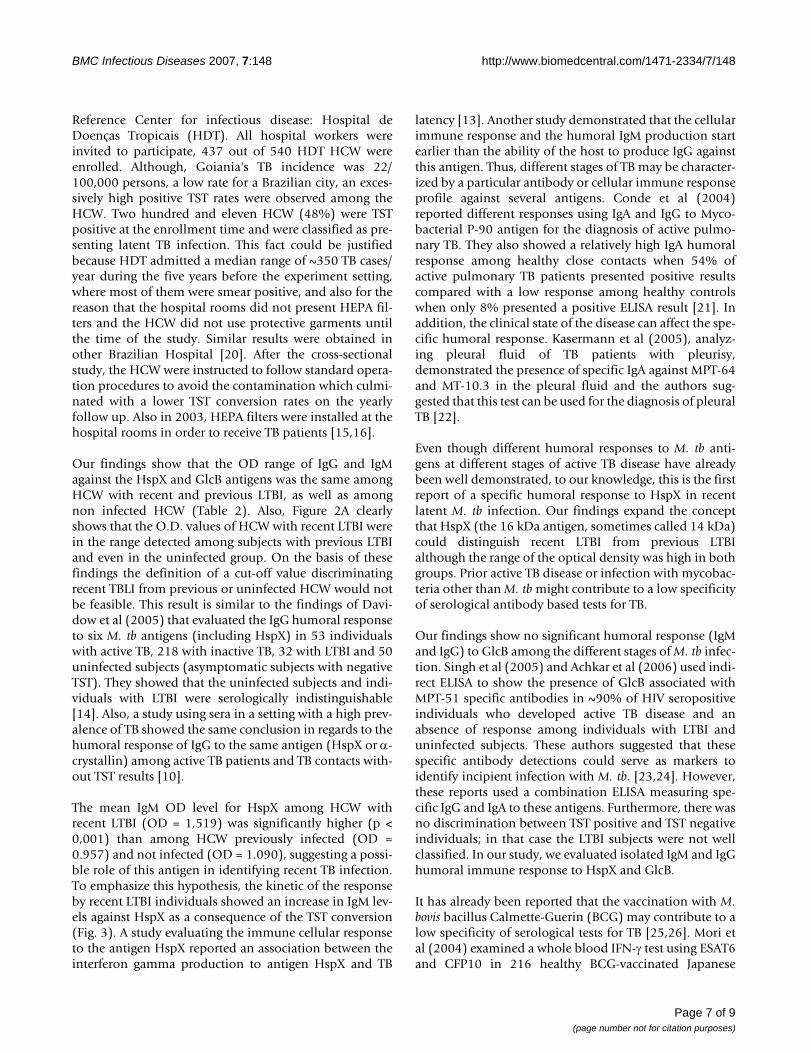
Reference Center for infectious disease: Hospital deDoenças Tropicais (HDT). All hospital workers wereinvited to participate, 437 out of 540 HDT HCW wereenrolled. Although, Goiania's TB incidence was 22/100,000 persons, a low rate for a Brazilian city, an exces-sively high positive TST rates were observed among theHCW. Two hundred and eleven HCW (48%) were TSTpositive at the enrollment time and were classified as pre-senting latent TB infection. This fact could be justifiedbecause HDT admitted a median range of ~350 TB cases/year during the five years before the experiment setting,where most of them were smear positive, and also for thereason that the hospital rooms did not present HEPA fil-ters and the HCW did not use protective garments untilthe time of the study. Similar results were obtained inother Brazilian Hospital [20]. After the cross-sectionalstudy, the HCW were instructed to follow standard opera-tion procedures to avoid the contamination which culmi-nated with a lower TST conversion rates on the yearlyfollow up. Also in 2003, HEPA filters were installed at thehospital rooms in order to receive TB patients [15,16].
Our findings show that the OD range of IgG and IgMagainst the HspX and GlcB antigens was the same amongHCW with recent and previous LTBI, as well as amongnon infected HCW (Table 2). Also, Figure 2A clearlyshows that the O.D. values of HCW with recent LTBI werein the range detected among subjects with previous LTBIand even in the uninfected group. On the basis of thesefindings the definition of a cut-off value discriminatingrecent TBLI from previous or uninfected HCW would notbe feasible. This result is similar to the findings of Davi-dow et al (2005) that evaluated the IgG humoral responseto six M. tb antigens (including HspX) in 53 individualswith active TB, 218 with inactive TB, 32 with LTBI and 50uninfected subjects (asymptomatic subjects with negativeTST). They showed that the uninfected subjects and indi-viduals with LTBI were serologically indistinguishable[14]. Also, a study using sera in a setting with a high prev-alence of TB showed the same conclusion in regards to thehumoral response of IgG to the same antigen (HspX or α-crystallin) among active TB patients and TB contacts with-out TST results [10].
The mean IgM OD level for HspX among HCW withrecent LTBI (OD = 1,519) was significantly higher (p <0,001) than among HCW previously infected (OD =0.957) and not infected (OD = 1.090), suggesting a possi-ble role of this antigen in identifying recent TB infection.To emphasize this hypothesis, the kinetic of the responseby recent LTBI individuals showed an increase in IgM lev-els against HspX as a consequence of the TST conversion(Fig. 3). A study evaluating the immune cellular responseto the antigen HspX reported an association between theinterferon gamma production to antigen HspX and TB
latency [13]. Another study demonstrated that the cellularimmune response and the humoral IgM production startearlier than the ability of the host to produce IgG againstthis antigen. Thus, different stages of TB may be character-ized by a particular antibody or cellular immune responseprofile against several antigens. Conde et al (2004)reported different responses using IgA and IgG to Myco-bacterial P-90 antigen for the diagnosis of active pulmo-nary TB. They also showed a relatively high IgA humoralresponse among healthy close contacts when 54% ofactive pulmonary TB patients presented positive resultscompared with a low response among healthy controlswhen only 8% presented a positive ELISA result [21]. Inaddition, the clinical state of the disease can affect the spe-cific humoral response. Kasermann et al (2005), analyz-ing pleural fluid of TB patients with pleurisy,demonstrated the presence of specific IgA against MPT-64and MT-10.3 in the pleural fluid and the authors sug-gested that this test can be used for the diagnosis of pleuralTB [22].
Even though different humoral responses to M. tb anti-gens at different stages of active TB disease have alreadybeen well demonstrated, to our knowledge, this is the firstreport of a specific humoral response to HspX in recentlatent M. tb infection. Our findings expand the conceptthat HspX (the 16 kDa antigen, sometimes called 14 kDa)could distinguish recent LTBI from previous LTBIalthough the range of the optical density was high in bothgroups. Prior active TB disease or infection with mycobac-teria other than M. tb might contribute to a low specificityof serological antibody based tests for TB.
Our findings show no significant humoral response (IgMand IgG) to GlcB among the different stages of M. tb infec-tion. Singh et al (2005) and Achkar et al (2006) used indi-rect ELISA to show the presence of GlcB associated withMPT-51 specific antibodies in ~90% of HIV seropositiveindividuals who developed active TB disease and anabsence of response among individuals with LTBI anduninfected subjects. These authors suggested that thesespecific antibody detections could serve as markers toidentify incipient infection with M. tb. [23,24]. However,these reports used a combination ELISA measuring spe-cific IgG and IgA to these antigens. Furthermore, there wasno discrimination between TST positive and TST negativeindividuals; in that case the LTBI subjects were not wellclassified. In our study, we evaluated isolated IgM and IgGhumoral immune response to HspX and GlcB.
It has already been reported that the vaccination with M.bovis bacillus Calmette-Guerin (BCG) may contribute to alow specificity of serological tests for TB [25,26]. Mori etal (2004) examined a whole blood IFN-γ test using ESAT6and CFP10 in 216 healthy BCG-vaccinated Japanese
Page 7 of 9(page number not for citation purposes)

BMC Infectious Diseases 2007, 7:148 http://www.biomedcentral.com/1471-2334/7/148
adults and 118 patients with TB [27]. The results demon-strated that this test was highly specific (98%) and sensi-tive (89%) for M. tb infection and was unaffected by BCGvaccination [27]. Although it has been shown elsewherethat HspX accumulates in long-term stationary-phase cul-tures of M. bovis- BCG [28], our findings suggest no inter-ference of BCG vaccination in the IgM humoral responseto HspX. Using the knowledge that BCG affects the TSTconversion during five years after the vaccination [3,4],the results of the groups studied here were comparedbased on the time of their BCG vaccination (Table 3).HCW with recent BCG vaccination (less than five years) ofuninfected, LTBI and rLTBI individuals did not changedthe humoral response to the recombinant antigens HspXand GlcB.
The present study has several limitations. The serologictests were performed retrospectively, using serum that hadbeen stored. Although no serum had been thawed morethan once, we cannot formally exclude the possibility thatserum storage conditions adversely affected the test per-formance, although we believe that this is unlikely. Thecross reactivity to E. coli proteins or to the skim milk wereexcluded during the standardization. The definition usedin the study for recent LTBI might not be appropriate as itis not possible to distinguish through a yearly TST if theM. tb infection occurred 2 weeks or 12 months prior to thetest. New studies evaluating the response to HspX amongTST conversion documented within 12 weeks after a neg-ative TST (such as during the evaluation of close contactsof an active pulmonary TB case) instead of 12 months
could help us to better understand the role of this antigenin recent LTBI.
ConclusionWe conclude that the OD range of IgG and IgM againstantigens HspX and GlcB was the same among subjectswith recent and previous LTBI, as well as among noninfected individuals. However, the OD mean of IgMresponse to HspX was significantly different among thedifferent stages of M. tb infection. This finding suggests apossible use of HspX as a marker for recent LTBI, even ina BCG vaccinated population, perhaps using differenttechniques to evaluate the antigen response. New studiesare necessary to evaluate this hypothesis.
Competing interestsThe author(s) declare that they have no competing inter-ests.
Authors' contributionsMFR and MCGR carried out the ELISA studies, partici-pated in the analysis of the results and drafted the manu-script. WO participated in the design of the study andperformed the statistical analysis. APJK and MBC con-ceived of the study, and participated in its design andcoordination and helped to draft the manuscript. Allauthors read and approved the final manuscript.
AcknowledgementsWe thank Lílian Kelly de Oliveira Lopes, Colombina da Silveira, Renner Caetano de Souza, and the staff of the Hospital de Doenças Tropicais for their support and help. We also thank Anne Efron (Johns Hopkins Univer-sity; Center for TB Research) and André Kipnis (Federal University of Goiás) for their critical review of the manuscript.
This work was supported by Conselho Nacional de Desenvolvimento Cien-tifico e Tecnológico-CNPq (grant numbers: 350543/2003-8 and 471348/2004-0), and FUNAPE-UFG, Brazil. MFR, APJK, MCGR, MBC received fel-lowship from CNPq.
References1. World Health Organization: Global tuberculosis control: sur-
veillance, planning, financing. WHO Report 2006 [http://www.who.int/tb/publications/global_report/2006/summary/en/index.html]. Geneva, Switzerland Accessed December 2006
2. The global plan to stop TB, 2006–2015 Geneva: Stop TBPartnership 2006:1–172 [http://www.stoptb.org/globalplan/assets/documents/SSP_DIAGWG.pdf]. Accessed December 2006
3. Ferebee SH: Controlled chemoprophylaxis trials in tuberculo-sis. A general review. Bibl Tuberc 1970, 26:28-106.
4. Targeted tuberculin testing and treatment of latent tuber-culosis infection. This official statement of the AmericanThoracic Society was adopted by the ATS Board of Direc-tors, July 1999. This is a Joint Statement of the AmericanThoracic Society (ATS) and the Centers for Disease Controland Prevention (CDC). This statement was endorsed by theCouncil of the Infectious Diseases Society of America.(IDSA), September 1999, and the sections of this statement.Am J Respir Crit Care Med 2000, 161(4 Pt 2):S221-S247.
5. Jasmer RM, Nahid P, Hopewell PC: Latent tuberculosis infection.N Engl J Med 2002, 347:1860-1866.
Kinetics of the IgM response against HspX from rLTBI study subjects by indirect ELISAFigure 3Kinetics of the IgM response against HspX from rLTBI study subjects by indirect ELISA. Negative TST are the OD of the serum samples obtained before the TST conversion and pos-itive TST are the OD at the time of conversion. The lines connects the same person (n = 43). HCW that converted to positive TST presented significantly higher levels of IgM than before conversion (P < 0.001).
negative TST positive TST0.0
0.5
1.0
1.5
2.0
2.5
Op
tica
lDen
sity
Page 8 of 9(page number not for citation purposes)

BMC Infectious Diseases 2007, 7:148 http://www.biomedcentral.com/1471-2334/7/148
Publish with BioMed Central and every scientist can read your work free of charge
"BioMed Central will be the most significant development for disseminating the results of biomedical research in our lifetime."
Sir Paul Nurse, Cancer Research UK
Your research papers will be:
available free of charge to the entire biomedical community
peer reviewed and published immediately upon acceptance
cited in PubMed and archived on PubMed Central
yours — you keep the copyright
Submit your manuscript here:http://www.biomedcentral.com/info/publishing_adv.asp
BioMedcentral
6. Andersen P, Munk ME, Pollock JM, Doherty TM: Specific immune-based diagnosis of tuberculosis. Lancet 2000, 356:1099-1104.
7. Brock I, Weldingh K, Lillebaeck T, Follmann , Andersen P: Compar-ison of tuberculin skin test and new specific blood test intuberculosis contacts. Am J Respir Crit Care Med 2004, 170:65-9.
8. Pai M, Riley LW, Colford JM: Interferon-assays in the immuno-diagnosis of tuberculosis: a systematic review. Lancet InfectiousDisease 2004, 4:761-76.
9. Mazurek GH, Villarino ME: MMWR January 31, 2003/52 (RR02).:15-18.
10. Beck ST, Leite OM, Arruda RS, Ferreira AW: Humoral responseto low molecular weight antigens of Mycobacterium tubercu-losis by tuberculosis patients and contacts. Braz J Med Biol Res2005, 38(4):587-596.
11. Silva VMC, Kanaujia G, Gennaro ML, Menzies D: Factors associ-ated with humoral response to ESAT-6, 38 kDa and 14 kDaantigens in patients with a spectrum of tuberculosis. Int JTuberc Lung Dis 2003, 7(5):478-484.
12. Smith CV, Huang CC, Andras Miczak A, David G, Russell DG, Sac-chettini JC, Bentrup KH: Biochemical and structural studies ofmalate synthase from Mycobacterium tuberculosis. Journal ofBiological Chemistry 2003, 278(3):1735-1743.
13. Demissie A, Leyten EMS, Abebe M, Wassie L, Aseffa A, Abate G,Fletcher H, Owiafe P, Hill PC, Brookes R, Rook G, Zumla A, ArendSM, Klein M, Ottenhoff TH, Andersen P, Doherty TM, the VACSELStudy Group: Recognition of stage-specific Mycobacterial anti-gens differentiates between acute and latent infections withMycobacterium tuberculosis. Clinical and Vaccine Immunology 2006,13(2):179-186.
14. Davidow A, Kanaujia GV, Shi L, Kaviar J, Guo X, Sung N, Kaplan G,Menzies D, Gennaro ML: Antibody profiles characteristic ofMycobacterium tuberculosis infection state. Infect Immun 2005,73:6846-6851.
15. Boletim Epidemiológico: Informe do Núcleo de Vigilancia Epide-miológica do Hospital de Doenças Tropicais Anuar Auad(HDT). Secretaria de Estado da Saúde de Goiás. Ano 1, número 1 2005.
16. Rabahi MF, Rodrigues AB, Queiroz de Mello F, de Almeida Netto JC,Kritski AL: Noncompliance with tuberculosis treatment bypatients at a tuberculosis and AIDS reference hospital inmidwestern Brazil. Braz J Infect Dis 2002, 6(2):63-73.
17. Ministério da Saúde, Brasil.- Datasus, Indicadores epidemi-ológicos e de morbidade [http://tabnet.datasus.gov.br/cgi/tabcgi.exe?idb2005/d0202.def]. Acessed August 2007
18. Department of Microbiology, Immunology and Pathology,Mycobacteria Research Laboratory [http://www.cvmbs.colostate.edu/mip/]
19. Steingart KR, Henry M, Laal S, Hopewell PC, Ramsay A, Menzies D,Cunningham J, Weldingh K, Pai M: Commercial serological anti-body detection tests for the diagnosis of pulmonary tubercu-losis: a systematic review. PLoS Med 2007, 12;4(6):e202.
20. Maciel ELN, Viana MC, Zeitoune RCG, Ferreira I, Fregona G, DietzeR: Prevalence and incidence of Mycobacterium tuberculosisinfection in nursing students in Vitoria, Espirito Santo. RevSoc Bras Med Trop 2005, 38(6):469-472.
21. Conde MB, Suffys P, Lapa E, Silva JR, Kritski AL, Dorman SE: Immu-noglobulin A (IgA) and IgG immune responses against P-90antigen for diagnosis of pulmonary tuberculosis and screen-ing for Mycobacterium tuberculosis infection. Clin Diagn LabImmunol 2004, 11(1):94-7.
22. Kaisermann MC, Sardella IG, Trajman A, Coelho LV, Kampfer S, JonasF, Singh M, Saad MHF: IgA antibody responses to Mycobacte-rium tuberculosis recombinant MPT-64 and MT-10.3 (Rv3019c) antigens in pleural fluid of patients with tuberculosispleurisy. Int J Tuberc Lung Dis 2005, 9(4):1-6.
23. Singh KK, Dong Y, Belisle JT, Harder J, Arora VK, Laal S: Antigensof Mycobacterium tuberculosis recognized by antibodies dur-ing incipient, subclinical tuberculosis. Clin Diagn Lab Immunol2005, 12:354-358.
24. Achkar JM, Dong Y, Holzman RS, Belisle J, Kourbeti IS, Sherpa T, Con-dos R, Rom WN, Laal S: Mycobacterium tuberculosis malate syn-thase and MPT51. based serodiagnostic assay as an adjunctto rapid identification of pulmonary tuberculosis. Clinical andVaccine Immunology 2006, 13(11):1291-1293.
25. Sepulveda RL, Ferrer X, Latrach C, Sorensen RU: The influence ofCalmette-Guérin bacillus immunization on the booster
effect of tuberculin testing in healthy young adults. Am RevRespir Dis 1990, 142(1):24-28.
26. Farhat M, Greenaway C, Pai M, Menzies D: False-positive tubercu-lin skin tests: what is the absolute effect of BCG and non-tuberculous mycobacteria? Int J Tuberc Lung Dis 2006,10(11):1192-11204.
27. Mori T, Sakatani M, Yamagishi F, Takashima T, Kawabe Y, Nagao K,Shigeto E, Harada N, Mitarai S, Okada M, Suzuki K, Inoue Y, Tsuyu-guchi K, Sasaki Y, Mazurek GH, Tsuyuguchi I: Specific detection oftuberculosis infection: an interferon-gamma-based assayusing new antigens. Am J Respir Crit Care Med 2004, 170(1):59-64.
28. Monahan IM, Betts J, Banerjee DK, Butcher PD: Differentialexpression of mycobacterial proteins following phagocytosisby macrophages. Microbiology 2001, 147:459-471.
Pre-publication historyThe pre-publication history for this paper can be accessedhere:
http://www.biomedcentral.com/1471-2334/7/148/prepub
Publish with BioMed Central and every scientist can read your work free of charge
"BioMed Central will be the most significant development for disseminating the results of biomedical research in our lifetime."
Sir Paul Nurse, Cancer Research UK
Your research papers will be:
available free of charge to the entire biomedical community
peer reviewed and published immediately upon acceptance
cited in PubMed and archived on PubMed Central
yours — you keep the copyright
Submit your manuscript here:http://www.biomedcentral.com/info/publishing_adv.asp
BioMedcentral
Page 9 of 9(page number not for citation purposes)
Copyright © 2022 FDOKUMEN