Rational Drug Designing Strategies & Inhibitor Optimization: Anthrax Lethal Toxin Factor
Upload
independentCategory
view
2download
0
RESEARCH ARTICLE
The early humoral immune response to Bacillus anthracis toxins in patients infected with cutaneous anthrax Karen E. Brenneman 1•
2, Mehmet Doganay3
, Arya Akmal 1, Stanley Goldman 1, Darrell R. Galloway 1•
2,
Alfred J. Mateczun 1, Alan S. Cross4 & Leslie W. Baillie5
1Biologtcal Defense Research Directorate, Naval Medical Research Center, Rockville, MD, USA; 2 Department of Microbiology, The Ohio State University, Columbus, OH, USA; 3Department of Infectious Diseases, Faculty of Medicine, Erciyes University, Kayseri, Turkey; 4Department of Medicine. Center for Vaccine Development, University of Maryland School of Medicine, Baltimore, MD. USA; and 5Medical Biotechnology Center, University of Maryland Biotechnology Institute, Baltimore, MD, USA
Correspondence: Alan S. Cross, Department of Medicine, Center for Vacc1ne Development. University of Maryland School of Medicine, 685 W. Baltimore Street, Baltimore, MD 21201, USA. Tel.: +1410 706 5328; fax: + 1 410 706 6205; e-mail: [email protected]
Received 4August 2010; revised 1 March 2011; accepted 1 March 2011. Final version published online 15 April 2011.
DOI:10.1111/j.1574-695X.2011.00800.x
Editor: Patrick Brennan
Keywords anthrax; lethal factor; edema factor; protective antigen.
Introduction
Abstract
Bacillus anthracis, the causative agent of anthrax, produces a tripartite toxin composed of two enzymatically active subunits, lethal factor (LF) and edema factor (EF), which, when associated with a cell-binding component, protective antigen (PA), form lethal toxin and edema toxin, respectively. In this preliminary study, we characterized the toxin-specific antibody responses observed in 17 individuals infected with cutaneous anthrax. The majority of the toxin-specific antibody responses observed following infection were directed against LF, with immunoglobulin G (lgG) detected as early as 4 days after the onset of symptoms in contrast to the later and lower EF- and FA-specific IgG responses. Unlike the case with infection, the predominant toxin-specific antibody response of those immunized with the US anthrax vaccine absorbed and UK anthrax vaccine precipitated licensed anthrax vaccines was directed against PA. We observed that the LF-specific human antibodies were, like anti-PA antibodies, able to neutralize toxin activity, suggesting the possibility that they may contribute to protection. We conclude that an antibody response to LF might be a more sensitive diagnostic marker of anthrax than to PA. The ability of human LF-spedfic antibodies to neutralize toxin activity supports the possible inclusion of LF in future anthrax vaccines.
Anthrax is a zoonotic disease caused by Bacillus anthracis, a Gram-positive spore-forming microorganism whose manifestations in humans depend on the route of infection. The cutaneous form of the disease accounts for more than 95% of reported cases (Shafazand et al., 1999) and, with treatment, does not usually pose a threat to human life (Little & Ivins, 1999). The gastrointestinal and inhalational forms of the disease, although not as common, are much more severe (Little & Ivins, 1999). The ability of the organism to form environmentally resistant spores, be dispersed as aerosols and cause lethal infection following inhalation has resulted in its development and use as a biological weapon.
they germinate into vegetative bacilli and escape from the macrophage (Lincoln et al., 1965; Dixon et al., 2000; GuidiRontani et al., 2001). In cutaneous anthrax, this results in a localized infection; in inhalational anthrax, the bacilli multiply in the lymphatic system and spread to the blood, resulting in massive bacteremia and toxemia (Fish & Lincoln, 1968).
Within 3 h of spore germination, the expression of the toxin proteins begins (Guidi-Rontani et al., 1999). The extracellular tripartite toxin of anthrax is composed of two enzymatically active subunits, lethal factor (LF) and edema factor (EF), and a cell-binding and translocation component, protective antigen (PA). Both lethal (PA + LF) and edema (PA + EF) toxins are able to suppress key parts of the innate immune response to the developing infection (O'Brien et al., 1985; Wright & Mandell, 1986; Duesbery
Following infection, spores are phagocytosed by macrophages and transported to the draining lymph nodes, where
© ZO 11 Federation of European Microbiological Societies Published by Blackwell Publishing ltd. All rights reserved
FEMS lmmunol Med M!crobiol62 (2011) 164-172

Report Documentation Page Form ApprovedOMB No. 0704-0188
Public reporting burden for the collection of information is estimated to average 1 hour per response, including the time for reviewing instructions, searching existing data sources, gathering andmaintaining the data needed, and completing and reviewing the collection of information. Send comments regarding this burden estimate or any other aspect of this collection of information,including suggestions for reducing this burden, to Washington Headquarters Services, Directorate for Information Operations and Reports, 1215 Jefferson Davis Highway, Suite 1204, ArlingtonVA 22202-4302. Respondents should be aware that notwithstanding any other provision of law, no person shall be subject to a penalty for failing to comply with a collection of information if itdoes not display a currently valid OMB control number.
1. REPORT DATE MAR 2011 2. REPORT TYPE
3. DATES COVERED 00-00-2011 to 00-00-2011
4. TITLE AND SUBTITLE The early humoral immune response to Bacillus anthracis toxins inpatients infected with cutaneous anthrax
5a. CONTRACT NUMBER
5b. GRANT NUMBER
5c. PROGRAM ELEMENT NUMBER
6. AUTHOR(S) 5d. PROJECT NUMBER
5e. TASK NUMBER
5f. WORK UNIT NUMBER
7. PERFORMING ORGANIZATION NAME(S) AND ADDRESS(ES) Naval Medical Research Center,Biologtcal Defense Research Directorate,Rockville,MD,20850
8. PERFORMING ORGANIZATIONREPORT NUMBER
9. SPONSORING/MONITORING AGENCY NAME(S) AND ADDRESS(ES) 10. SPONSOR/MONITOR’S ACRONYM(S)
11. SPONSOR/MONITOR’S REPORT NUMBER(S)
12. DISTRIBUTION/AVAILABILITY STATEMENT Approved for public release; distribution unlimited
13. SUPPLEMENTARY NOTES
14. ABSTRACT Bacillus anthracis, the causative agent of anthrax, produces a tripartite toxin composed of twoenzymatically active subunits, lethal factor (LF) and edema factor (EF), which, when associated with acell-binding component, protective antigen (PA), form lethal toxin and edema toxin, respectively. In thispreliminary study, we characterized the toxin-specific antibody responses observed in 17 individualsinfected with cutaneous anthrax. The majority of the toxin-specific antibody responses observed followinginfection were directed against LF, with immunoglobulin G (lgG) detected as early as 4 days after the onsetof symptoms in contrast to the later and lower EF- and FA-specific IgG responses. Unlike the case withinfection, the predominant toxin-specific antibody response of those immunized with the US anthraxvaccine absorbed and UK anthrax vaccine precipitated licensed anthrax vaccines was directed against PA.We observed that the LF-specific human antibodies were, like anti-PA antibodies, able to neutralize toxinactivity, suggesting the possibility that they may contribute to protection. We conclude that an antibodyresponse to LF might be a more sensitive diagnostic marker of anthrax than to PA. The ability of humanLF-spedfic antibodies to neutralize toxin activity supports the possible inclusion of LF in future anthrax vaccines.
15. SUBJECT TERMS
16. SECURITY CLASSIFICATION OF: 17. LIMITATION OF ABSTRACT Same as
Report (SAR)
18. NUMBEROF PAGES
9
19a. NAME OFRESPONSIBLE PERSON
a. REPORT unclassified
b. ABSTRACT unclassified
c. THIS PAGE unclassified
Standard Form 298 (Rev. 8-98) Prescribed by ANSI Std Z39-18

Human response to cutaneous anthrax
et al., 1998; Pellizzari et al., 1999; Erwin et al., 2001; Kalns et al., 2002; Popov et al., 2002; Moayeri et al., 2003). Later in the disease process, high levels of lethal toxin (LT) induce the cytokine independent shock-like death associated with anthrax (Moayeri et al., 2003).
Animal studies suggest that as the concentration of toxin increases the likelihood of successfully treating an infected individual decreases until it reaches a level at which antibiotics are no longer effective (Albrecht et al., 2007; Baillie, 2009). Given that the early detection of toxin is a key diagnostic marker, it surprising how little is known of the time course of toxin production in humans and thus we sought to characterize the early immune responses to individuals infected with anthrax (Baillie, 2009).
To achieve this aim, clinical serum samples previously obtained at various time points postinfection from indivi
duals who had contracted cutaneous anthrax were examined for the presence of toxin-specific immunoglobulin M (IgM) and IgG antibodies. In addition, we compared these antibody responses with those seen following immunization with the US anthrax vaccine absorbed (AVA) and UK anthrax vaccine precipitated (AVP) licensed human anthrax vaccines. Finally, the protective function of the toxinspecific antibody responses stimulated following infection were assessed using an assay that measures toxin neutralization, a recently demonstrated correlate of protection (Reuveny et al., 2001; Little et al., 2004).
Materials and methods
Expression and purification of toxin components
The PA, LF and EF genes were cloned into the Escherichia coli
expression vector pQE-30 (Qiagen) and confirmed by sequencing (Read et al., 2003). Proteins were expressed from either the MIS (PA) or the SG 13009 (LF and EF) strain of E.
coli. Host strains were grown in Luria-Bertani medium to an OD6oonm of0.55-0.65 and induced with 1 mM isopropyl-Po-thiogalactoside either 4 hat 37 oc (PA) or 20 hat 25 oc (LF and EF). Cells were pelleted and lysed by French press at 16 000 p.s.i.; lysates were cleared at 45 000 g for 15 min. Recombinant proteins were purified by cobalt affinity chromatography. Cleared lysate was batch bound to TALON resin ( Clontech) and then washed with 10 CV 300 mM NaCl, 50 mM Na2HP04 and 20 mM imidazole, pH 7.0. Proteins
were eluted in 5 CV 300 mM NaCl, 50 mM Na2HP04 and
150 mM imidazole, pH 7.0. Fractions containing protein
[determined by sodium dodecyl sulfate-polyacrylamide gel electrophoresis (SDS-PAGE)] were pooled and dialyzed into
10 mM HEPES and 50 mM NaCI, pH 7.5. Proteins purified using this procedure were approximately 90% pure as assessed by SDS-PAGE with Coomassie staining.
FEMS lmmunol Med Microbiol62 (2011) 164-172
165
Serum samples
Serum samples were obtained from volunteers who had received at least a priming series of the AVA (six Marylandbased volunteers) or AVP (four UK-based volunteers visiting Maryland) vaccines. Control samples were obtained from six nonimmunized, noninfected Maryland-based individuals. All samples were obtained under a protocol approved by the University of Maryland and the Naval Medical Research Center's Institutional Review Boards, as well as by the Ethics Committee at Erciyes University. Informed consent was obtained from all individuals. Clinical samples were obtained from 17 cutaneous anthrax
patients attending the infectious diseases clinic at Erciyes University in Turkey (Table 1). Serum samples were not collected prospectively from patients under a set protocol, but were instead collected when patients presented to the outpatient clinics for up to 21 days after the initial visit.
Anthrax was diagnosed by exposure history, clinical presentation consistent with anthrax, Gram stain and positive culture from the lesion.
Antitoxin lgG and lgM enzyme-linked immunosorbent assay (ELISA)
Antitoxin IgG and IgM levels were measured using an ELISA as described previously, with minor variations (Hepburn
et al., 2007). Data values were compared with a standard curve of purified human IgG or IgM (Sigma). Data in the
linear portion of the ELISA graph and within the range of the standard curve were used to calculate the quantitative titer (Jlg mL -I) for the serum sample. For each antigen, four to six naive serum samples were assayed and their titers were
averaged (geometric mean) and the 95% confidence interval of the distribution was calculated. Experimental data were scored as a positive result only if the calculated titer exceeded the upper limit of the confidence interval of the na'ive control samples.
LT neutralization assay
The toxin neutralization assay was performed on the mouse monocyte cell line J774A.l (ATCC) as described previously, with cell viability determined by the addition of DMEM containing sodium 3 '- [ 1-(phenylaminocarbonyl)-3,4-tetrazolium]-bis (4-methoxy-6-nitro) benzene sulfonic acid hydrate (XTT) (Roche) for 16 h. The assay was read at 480 nm. The dilution series data (A480 nm vs. toxin concentration)
were modeled with four-parameter logistic curves of the
form:
The data were fit using MATLAB software via a nonlinear
least-squares analysis, yielding the parameters (p 1, pz, p3,
© 2011 Federation of European Microbiological Societies Published by Blackwell Publishing Ltd. All rights reserved

~@ e-N Er~ ::,-_
~~ '< "'
~~ " 0 """" ::; 0
"' -=m -oc &a :-:""'0 ~ "' ::rw s· :::J
"';;:: ~ n· c_~ . 0 )>!2": =o ~-2 a~ rn ~ "'" < ;;;· ro ~-c_:J:
~
~ "' 3 3 c ::> Q_ ;;:: "' c_
;;:: r;· i3 C"
~ ~
-;;::; 0
~
! " N
Table 1. Details for the patients with cutaneous anthrax
•contact with +Antibiotic Incubation contaminated Site of tGram stain for tPositive culture Previous initiation day Given Duration of
Patient no. period (days) animal materials the lesion B. anthracis for B. anthracis antibiotic use of disease antibiotic therapy (days) Outcome
1 5 + Right eye lids - Yes Third Pen G 15 Left deep tissue scar, recovered
2 15 + Right arm Yes Fourth Pen G+Cipro 10 Recovered 3 1 + Right arm Yes Second PanG 10 Recovered 4 6 + Right hand Yes Fourth Pen G 10 Recovered 5 9 + Left hand No Sixth Pen G 7 Recovered 6 10 + Left forearm Yes Fifth Pen G 10 Recovered 7 5 + Right hand fingers + + No Fifth Pen G 7 Recovered 8 4 + Left hand finger + Yes Fourth Pen G 15 Recovered 9 12 + Both hands Yes Third Pen G 14 Recovered 10* 2 + Anterior neck + + No Second Pen G 14 Left deep tissue scar,
recovered 11 8 + The eye lid + + No Third Pen G 10 Recovered 12 8 + Left eye lids + Yes Third Pen G 14 Recovered 13 5 + Right wrist + Yes Fifth Pen G 10 Recovered 14 ? + Left elbow - Yes Tenth Pen G 10 Recovered 15 5 + Right face + Yes Fifth Pen G 10 Recovered 16 1 + Anterior neck + - No Seventh Pen G 7 Recovered 17 7 + Right arm and left Yes Fourth Pen G 10 Left deep tissue scar,
wrist recovered
*The patient gave a history of contact with an ill an1mal or contaminated animal materials (such as slaughtering of ill animals, skinning, chopping meat, carrying raw skin or splashing blood from dying animal during slaughtering). One case gave a history of fly bite. tThe swabs for Gram stain and culture were taken from the vesicles of fluid or under the crust if developed. 1Time from first symptoms to the diagnosis and initiation of antibiotiC treatment. §In this case, anthrax sepsis was developed from the cutaneous lesion. Pen G, penicillin G; Cipro, ciprofloxacin.
.... 0\ 0\
?'
"' (il :::J :::J
"' 3 "" :::J
~ "" :-

Human response to cutaneous anthrax
P4) of the best fit. The inflection point of the fit (p4
parameter) corresponds to the serum dilution that ensures the survival of 50% of the cells in the assay (ED50) (Quinn et al., 2004).
Results
Clinical characteristics
Cutaneous anthrax in the patients was diagnosed by the history of contact with ill animals and animal products, typical cutaneous lesions and demonstration of Grampositive bacilli (eight cases) and/or -positive cultures (three cases) from the lesion (Table 1). The upper body was the usual site of the cutaneous anthrax lesion. The finding that cultures were positive only in three cases was presumed to be due to the fact that many patients had taken an antibiotic before their initial visit. With cultures of both the lesion and the blood being positive, one patient was diagnosed with anthrax sepsis originating from the cutaneous lesion. The incubation period varied between 1 and 15 days. Seventeen patients gave a history of contact with an ill animal or contaminated materials of animals that died. The time from first symptom to the initiation of antibiotics ranged from 2 to 10 days, with 16 subjects receiving antibiotics, predominantly penicillin, within the first 7 days, while 10 patients received antibiotics within 4 days. Typically, the patients were treated for 7-15 days, and all recovered. Three subjects (patients 1, 10 and 17), however, were left with deep tissue scars.
Antitoxin lgM responses
In order to examine the early immune response to infection, the serum IgM levels against PA, LF and EF were measured and compared with the antitoxin IgM titer of six naive volunteers. Of the 17 infected individuals, six (35%) had an IgM response to at least one of the toxin components that was higher than that of the naive controls (Table 2). There was no statistical difference in the level of toxin-specific IgM between the remaining 11 patients and the naive controls. An anti-LF-specific IgM was detected in five individuals within the first and second weeks following symptoms, with the earliest response detected at day 4. In contrast, an antiPA IgM was detected in four individuals, but not until the second and third weeks following symptoms, with the earliest titers appearing at day 12. Finally, anti-EF titers were detected in only two patients with titers occurring in the second week after symptoms.
Antitoxin lgG responses
The serum IgG titers against the anthrax toxin proteins in 17 infected individuals were measured and compared with the
FEMS lmmunol Med Microbiol62 (2011) 164-172
167
Table 2. Comparison of serum antitoxin lgM levels in cutaneous anthrax patients
Days after onset Patient of symptoms Anti-PA lgM Anti-LF lgM Anti-EF lgM
Naive 1.2 (1.3) 4.7 (3.3) 2.2 (2.0) 5 < NB < NB < NB
12 7.1 (1.8) 11.3 (1.1) < NB 2 4 < NB 8.9 (0.2) < NB
13 2.7 (0.1) 10.2 (1.0) 4.7 (2.5) 5 6 < NB < NB < NB
13 < NB 9.3 (2.0) < NB 7 5 < NB < NB < NB
15 3.4(0.3) 8.6(0.8) < NB 10* 7 < NB 8.3 (0.1) < NB 16 14 < NB < NB 5.1 (2.3)
21 2.6(0.8) < NB < NB
Data are in 119 ml-1 and represent the mean titer ( ± SD) as determined by ELISA. *Patient developed septicemia from cutaneous lesion. < NB, less than the naive baseline (titer indistinguishable from naive controls).
geometric mean IgG titer (GMT) of six na'ive volunteers. In contrast to the IgM responses, 11 of the 17 patients ( 65%) demonstrated a measurable IgG titer to at least one of the toxin components on the days assayed (Table 3). While each positive patient generated an anti-LF IgG response with titers ranging from 13.6 to 817jlgmL-1
, only four individuals showed an anti-EF IgG response (patients 5, 6, 8 and 16) and only three patients (patients 2, 6 and 7) had IgG specific for PA. The anti-LF IgG response in all infected individuals exceeded that directed against PA and EF. During the second week of infection, the GMT of all responders to LF ( 69.3 jlg mL _,) was almost twice the
anti-EF titer (37.4jlgmL-1) and three times the anti-PA
titer (22.6jlgmL-1).
LF-specific IgG responses were detected as early as 4 days after onset of symptoms while anti-EF IgG responses were not seen until day 6 and anti-PA IgG responses first being detected at day 13. These preliminary results would appear to indicate that of the three toxins components, the antibody response to LF represents the most appropriate early diagnostic indicator.
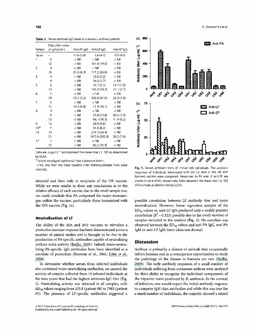
To further characterize the protective nature of the antibody response induced by infection, we compared the spectrum of the resulting toxin-specific IgG responses to those seen following immunization of healthy volunteers with the US AVA and the UK AVP licensed human anthrax vaccines (Pittman et al., 2002; Baillie et al., 2003, 2004). As can be seen from Fig. 1 a and b, all of the vaccinees mounted a PA-specific IgG response, the level of which varied depending on the vaccine used and the immunization schedule of each individual. In contrast to the responses observed following infection, little if any LF-specific IgG was
© 2011 Federation of European Microbiological Societies Published by Blackwell Publishing Ltd. All rights reserved
168
Table 3. Serum antitoxin lgG levels in cutaneous anthrax patients
Days after onset
Patient of symptoms Anti-PA lgG Anti-LF lgG Anti-EF lgG
Naive 11.6 (5.4) 5.6(4.1) 6.5 (6.0)
5 < NB < NB < NB
12 < NB 147.4(19.0) < NB
2 4 < NB < NB < NB
13 21.6 (6.4) 117.3 (26.8) < NB
3 4 < NB 13.6 (3.2) < NB
9 < NB 16.0 (3.7) < NB
5 6 < NB 15.7 (2.1) 14.7 (1.5)
13 < NB 141.0 (19.3) 27.1 (2.7)
6 5 < NB < NB < NB
20 26.1 (2.2) 430.8 (91.0) 24.3 (1.6)
7 5 < NB < NB < NB
15 19.3 (4.8) 71.9 (18.1) < NB
8 4 < NB < NB < NB
9 < NB 35.6 (13.6) 60.5 (7.9)
13 < NB 68.7 (18.5) 31.9 (8.2)
9 15 < NB 28.9 (9.6) < NB 10. 7 < NB 31.0 (8.2) < NB
16 14 < NB 214.3 (24.4) < NB
21 < NB 817.6 (202.8) 26.2 (7.6)
17 7 < NB < NB < NB
15 < NB 60.2 (10.9) < NB
Data are in J.l9 ml-1 and represent the mean titer ( ± SD) as determined
by ELISA.
•ratient developed septicemia from cutaneous lesion.
< NB, less than the na'lve baseline (titer indistinguishable from na'lve
controls).
detected and then only in rectptents of the UK vaccine. While we were unable to draw any conclusions as to the relative efficacy of each vaccine due to the small sample size, we could conclude that PA comprised the major immunogen within the vaccine, particularly those immunized with the AVA vaccine (Fig. 1a).
Neutralization of LT
The ability of the AVA and AVP vaccines to stimulate a protective immune response has been demonstrated across a number of animal studies and is thought to be due to the production of PA-specific antibodies capable of neutralizing anthrax toxin activity (Baillie, 2009). Indeed, toxin-neutralizing PA-specific IgG antibodies have been identified as a correlate of protection (Reuveny et al., 2001; Little et al., 2004).
To determine whether serum from infected individuals also contained toxin-neutralizing antibodies, we assayed the activity of samples collected from 10 infected individuals at the time point that had the highest antitoxin IgG titer (Fig. 2). Neutralizing activity was detected in all samples, with ED50 values ranging from 103.8 (patient #8) to 7983 (patient #5). The presence of LF-specific antibodies suggested a
© 2011 Federation of European Microbiological Societies Published by Blackwell Publishing Ltd. All rights reserved
K. Brenneman eta/.
(a) 800
1!1 Anti-PA
~ 600 E
"' 3 .. Cll 400 ~ -» "0 0 ..c 200 ;:;
~
0 f-:J~~.p ~ 1\~'!J~~~ ~~ ~ ~ ~ ~~ ~G:J ~G:J ~G:J ~ ~G:J ~G:)
(b) 75 &SJ Anti-LF
~ CJ Anti-EF E
"' 50 3 .. Cll ~ -» "0 25 0 ..c ;:;
~
Fig. 1. Serum antitoxin titers of immunized individuals. The antitoxin
responses of individuals immunized with the US AVA or the UK AVP
licensed vaccine were compared. Responses to PA and LF and EF are
shown in (a) and (b), respectively. Data represent the mean titer ( ± SD)
of four trials as determined by ELISA.
possible correlation between LF antibody titer and toxin neutralization. However, linear regression analysis of the ED50 values vs. anti-LF IgG produced only a weakly positive correlation (R2 = 0.323) possibly due to the small number of samples included in the analysis (Fig. 3). No correlate was observed between the ED50 values and anti-PA IgG, anti-PA IgM or anti-LF IgM titers (data not shown).
Discussion
Anthrax is primarily a disease of animals that occasionally infects humans and as a consequence opportunities to study the pathology of the disease in humans are rare (Baillie, 2009). The early antibody responses of a small number of individuals suffering from cutaneous anthrax were analyzed for their ability to recognize the individual components of the tripartite toxin produced by B. anthracis. In the context of infection, one would expect the initial antibody response to comprise IgM class antibodies and while this was true for a small number of individuals, the majority showed a mixed
FEMS lmmunol Med Microbial 62 (2011) 164-172

Human response to cutaneous anthrax
10000
-C)
"' Q
!!!. .. Ql
;!: ... Dl 1000 c ~ co .. -:I Q)
z
100 1 2 5 6 7 8 9 10 16 17
Patient
Fig. 2. Lethal toxin-neutralizing antibody titers of cutaneous anthrax
patients. For patients with more than one serum sample, neutralizing t1ters were measured at the time point with the highest lgG titer.
Neutralizing titer ED50 values ranged from 103.8 to 7983. Data represent
the ED50 (!34 parameter) generated by a four-parameter logistic fit of four
tests.
10
:::a "' 8
0 ~ $ 6 .. C) c . N
4 ~ 3 (!)
.s 2 c _J
0 0
R2 0.3227 •
·.~ ~··-• •
2 4 6
Ln [anti-LF titer {119 ml - 1)]
8
Fig. 3. Relationship of the anti-LF ELISA titer with the LT-neutralizing
ED50 in cutaneous anthrax patients. There was a weak positive correlation (~ = 0.3227) between anti-LF lgG levels and neutralizing titer in
infected individuals. No correlation was observed between anti-PA levels and neutralizing titer {R2 = 0.323).
IgM/IgG toxin-specific antibody response at the first sampling point. In general, the IgM responses were considerably lower than the corresponding IgG levels for all patients, a pattern similar to that observed in AVA-immunized primates (Ivins et al., 1998). Together, these data imply that class switching from IgM to IgG occurs early in the anthrax infection.
We observed differences in the ability of infected individuals to recognize toxin components. While 11 ( 65%) patients produced an LF-specific IgG response, only four (24%) mounted an IgG response to EF while only three (18%) demonstrated an IgG-specific immune response to PA. In those patients who showed the presence of anti-LF
FEMS lmmunol Med Microbiol62 (2011) 164-172
169
and anti-PA IgG antibodies (patients 2, 6 and 7), the LF titers exceeded the PA response by a factor of a least 3. Because both proteins are produced early in infection (Guidi-Rontani et al., 1999), this disparity in response may reflect fundamental differences in how each protein is processed by the immune system. For example, LF is known to elicit higher IgG antibody titers than PA when administered to experimental animals (Price et al., 2001; Hermanson et al., 2004; Flick-Smith et al., 2005).
Indeed, the overall disparity benveen anti-PA and anti-LF IgG responses observed in this study is likely to be the result of a combination of factors such as the virulence of the infecting strain, the route of infection and the health and genetic background of the individual. The initiation of antimicrobial therapy is also likely to have affected the level of toxin expression and may explain the absence of detectable response to any anthrax toxin component in six infected individuals (Stepanov et al., 1996; Athamna et al., 2004).
In an anthrax outbreak, it is crucial to identify infected individuals as quickly as possible to enable the initiation of timely treatment. The central role of the toxins in the progression of the infection has led to the proposal to use the antibody response to PA as a diagnostic marker of exposure to anthrax (Quinn et al., 2002, 2004). This recommendation was based on the analysis of the ?Aspecific antibody response of patients with bioterrorismrelated infections (Quinn et al., 2004). While individuals with inhalation anthrax demonstrated an IgG response to PA as early as 11 days following the onset of symptoms, those with the cutaneous infections did not develop anti-PA titers until21-34 days after the onset of symptoms and those titers did not peak until days 30-60 (Quinn et al., 2004), confirming our findings that the anti-PA immune response develops slowly. Unlike our study, Quinn and colleagues did not screen their samples for the presence of an anti-LF immune response. If they had, they may have also found that the LF-specific antibody response preceded that of the PA response. We found that the LF-specific IgG was detected as early as day 4 after the onset of symptoms, and that all patients had developed anti-LF titers by day 15. In contrast, PA-specific IgG was first detected 13 days after the onset of symptoms, and had appeared in only three patients by day 2l. The early production and prevalence of anti-LF antibodies after the development of symptoms suggest that the overall kinetics of the immune response is a rapid anti-LF response, followed by a slower, longer anti-PA response. The speed, strength and prevalence of the anti-LF response in infected patients clearly demonstrate that the presence of an anti-LF response might be a better diagnostic marker of infection than an anti-PA response.
We also sought to compare the response following immunization with the US AVA and UK AVP licensed anthrax vaccines. While both are subunit vaccines, they
2011 Federation of European Microbiological Societies Published by Blackwell Publishing Ltd. All rights reserved

170
differ primarily in their means of production. The current US licensed vaccine, AVA, is produced from a cell-free culture filtrate of an anaerobically grown B. anthracis strain V770-NP1-R (a nonencapsulated, nonproteolytic variant of a bovine strain isolated in Florida in 1951) and consists largely of PA adsorbed onto aluminum hydroxide (Baillie, 2001). In contrast, the UK vaccine, AVP, is produced from an alum precipitate of the cell-free culture filtrate of a static, aerobic culture of the B. anthracis Sterne strain 34F2. In addition to large amounts of PA, the vaccine also contains trace amounts of LF and other bacterially derived, immunogenic antigens, which have been shown to stimulate antibody responses in recipients and may contribute to protection (Baillie et al., 2003, 2004; Whiting et al., 2004). Indeed, the presence of these additional proteins may also account for the transient reactogenicity seen in some individuals (Turnbull, 2000). The efficacy of both vaccines has been demonstrated in numerous animal models including primates and has revealed a significant correlation between PA IgG titer and toxin-neutralizing activity, which is thought to be indicative of protection (Ivins et al., 1998; Reuveny et al., 2001; Little et al., 2004; Phipps et al., 2004; Quinn et al., 2004; Pittman et al., 2005).
In this study, we observed that the LF-specific human antibody was able to neutralize toxin activity. Several patients with high anti-LF IgG titers, but lacking anti-PA IgG titers had measurable toxin neutralization titers. This neutralization activity was not due to nonspecific protection as serum from six naive individuals failed to protect macrophages (data not shown). Neither could neutralization be attributed to the presence of anti-PA antibodies, as samples obtained from patients 5, 8, 9, 10 and 17 showed no detectable anti-PA IgM or IgG antibodies, but still had quantifiable neutralizing activity. To our knowledge, this is the first report of the production of LF-specific neutralizing antibodies by humans following infection. The levels of neutralizing antibody generated by these individuals were roughly equivalent to the levels generated by AVP-immunized individuals (Hepburn et al., 2007). The ability of LF to stimulate toxin-neutralizing antibodies is not surprising, given that LF must first bind to PA before it can be transported into the cytosol of the susceptible cell, where it exerts its toxic effects. Indeed, animal studies have demonstrated the ability of toxin-neutralizing LF-specific antibodies to protect rabbits against aerosol spore challenge (Zhao et al., 2003; Hermanson et al., 2004; Lim et al., 2005). While we found no such correlation among our patients for PA, we did observe a weak correlation between LF-specific IgG and toxin-neutralizing titers. Because little is known about either the human response to anthrax or the ability of LF to generate protective antibodies, further work is required to determine the factors mediating LF-specific toxin neutralization.
c9 2011 Federation of European Microbiological Societies Published by Blackwell Publishing Ltd. All rights reserved
K. Brenneman eta/.
Concerns over the ability to circumvent the current licensed vaccines by altering the antigenic structure of PA have spurred researchers to focus on additional vaccine targets (Schneerson et al., 2003; Hoffmaster et al., 2004). The immunogenicity of LF and its ability to stimulate the production of toxin-neutralizing antibodies makes a biologically inactive form of LF a strong candidate for inclusion in any future anthrax vaccine. We conclude from this preliminary study that an antibody response to LF might be a more sensitive diagnostic marker of anthrax than one to PA. In addition, the ability of human LF-specific antibodies to neutralize toxin activity supports the possible inclusion of LF in future anthrax vaccines.
Acknowledgements
We thank and acknowledge Dr Emine Alp for her assistance. For their assistance in providing samples and information, we acknowledge Dr Basak Dokuzoguz at Ankara Numune Clinical Research and Education Hospital (Ankara), Dr Mehmet Parlak at Atattirk University (Erzurum), Dr Ayhan Akbulut at Firat University (Elazig) and Dr Ilyas Dokrnetas at Cumhuriyet University (Sivas). We also acknowledge Stephanie Gray for her administrative efforts. Finally, we thank all the volunteers for their cooperation in donating their time and blood samples to this study. This work was supported by the Defense Threat Reduction Agency under the work unit number 80000.000.000.A0031 and NIH U54 AI057l68-0l. All authors have no conflicts.
Authors' contribution
K.E.B. and M.D. contributed equally to this work.
Disclaimer
The views in this article are those of the authors and do not necessarily reflect the official policy or position of the Department of the Navy, Department of Defense, nor of the US government.
References
Albrecht MT, Li H, Williamson ED et al. (2007) Human monoclonal antibodies against anthrax lethal factor and protective antigen act independently to protect against Bacillus anthracis infection and enhance endogenous immunity to anthrax. Infect Immun 75: 5425-5433.
Athamna A, Massalha M, Athamna M eta/. (2004) In vitro
susceptibility of Bacillus anthracis to various antibacterial agents and their time-kill activity. J Antimicrob Chemoth 53:
247-251.
FEMS Immune/ Med Microbio/62 (2011) 164-172

Human response to cutaneous anthrax
Baillie L (2001) The development of new vaccines against Bacillus
anthracis. I Appl Microbiol91: 609-613. Baillie L, Hebdon R, Flick-Smith H & Williamson D (2003)
Characterization of the immune response to the UK human
anthrax vaccine. FEMS Immunol Med Mic 36: 83-86. Baillie L, Townend T, Walker N, Erikson U & Williamson D
(2004) Characterization of the human immune response to the
UK anthrax vaccine. FEMS Immunol Med Mic 42: 267-270. Baillie LW (2009) Is new better than old? The development of
human vaccines for anthrax. Hum Vaccin 5: 806-816. Dixon TC, Fad! AA, Koehler TM, Swanson JA & Hanna PC
(2000) Early Bacillus anthracis-macrophage interactions:
intracellular survival and escape. Cell Microbial 2: 453-463. Duesbery NS, Webb CP, Leppla SH et al. (1998) Proteolytic
inactivation of MAP-kinase-kinase by anthrax lethal factor.
Science 280: 734-737. Erwin JL, DaSilva LM, Bavari S, Little SF, Friedlander AM &
Chanh C (2001) Macrophage-derived cell lines do not express
proinflammatory cytokines after exposure to Bacillus anthracis
lethal toxin. Infect Immun 69: 1175-1177. Fish DC & Lincoln RE (1968) In vivo-produced anthrax toxin. I
Bacteriol95: 919-924. Flick-Smith HC, Waters EL, Wlaker NJ et al. (2005) Mouse model
characterization for anthrax vaccine development: comparison
of one inbred and one outbred mouse strain. Microb
Pathogenesis 38: 33-40. Guidi-Rontani C, Weber-Levy M, Labruyere E & Mock M (1999)
Germination of Bacillus anthracis spores within alveolar
macrophages. Mol Microbiol31: 9-17. Guidi-Rontani C, Levy M, Ohayow H & Mock M (2001) Fate of
germinated Bacillus anthracis spores in primary murine
macrophages. Mol Microbiol42: 931-938. Hepburn MJ, Dyson EH, Simpson AJH et al. (2007) Immune
response to two different dosing schedules of the anthrax
vaccine precipitated (AVP) vaccine. Vaccine 25: 6089-6097. Hermanson G, Whitlow V, ParkerS et al. (2004) A cationic lipid
formulated plasmid DNA vaccine confers sustained antibody
mediated protection against aerosolized anthrax spores. P Natl
Acad Sci USA 101: 13601-13606. Hoffmaster AR, Ravel J, Rasko D et al. (2004) Identification of
anthrax toxin genes in a Bacillus cereus associated with an
illness resembling inhalation anthrax. P Natl Acad Sci USA
101: 8449-8454. Ivins BE, Pitt ML, Fellows PF et al. (1998) Comparative efficacy of
experimental anthrax vaccine candidates against inhalation
anthrax in rhesus macaques. Vaccine 16: 1141-1148.
Kalns J, Scruggs J, Millenbaugh N, Vivkananda J, Shealy D, Eggers
J & Kiel J (2002) TNF receptor l, ILl receptor, and iN OS
genetic knockout mice are not protected from anthrax
infection. Biochem Bioph Res Co 292: 41-44. Lim NK, Kim JH, OhMS et al. (2005) An anthrax lethal factor
neutralizing monoclonal antibody protects rats before and
after challenge with anthrax toxin. Infect Immun 73:
6547-6551.
FEMS lmmunol Med Microbial 62 (2011) 164-172
Lincoln RE, Hodges DR, Klein F et al. ( 1965) Role of the
lymphatics in the pathogenesis of anthrax. I Infect Dis 115:
481-494.
171
Little SF & Ivins BE (1999) Molecular pathogenesis of Bacillus
anthracis infection. Microbes Infect 1: 131-139. Little SF, Ivins BE, Fellows PF, Pitt ML, Norris SL & Andrews GP
(2004) Defining a serological correlate of protection in rabbits
for a recombinant anthrax vaccine. Vaccine 22: 422-430. Moayeri M, Haines D, Young HA & Leppla SH (2003) Bacillus
anthracis lethal toxin induces TNF-alpha-independent
hypoxia-mediated toxicity in mice. I Clin Invest 112: 670-682. O'Brien J, Friedlander A, Dreier T, Ezzell J & Leppla S (1985)
Effects of anthrax toxin components on human neutrophils.
Infect Immun 47: 306-310. Pellizzari R, Guidi-Rontani C, Vitale G, Mock M & Montecucco C
(1999) Anthrax lethal factor cleaves MKK3 in macrophages
and inhibits the LPS/IFNgamma-induced release of NO and
TNFalpha. FEES Lett 462: 199-204. Phipps AJ, Premanandan C, Barnewall RE & Lairmore MD
(2004) Rabbit and nonhuman primate models of toxin
targeting human anthrax vaccines. Microbial Mol Bioi R 68:
617-629. Pittman PR, Kim-Ahn G, Pifat DY et al. (2002) Anthrax vaccine:
immunogenicity and safety of a dose-reduction, route-change
comparison study in humans. Vaccine 20: 1412-1420. Pittman PR, Leitman SF, Oro JG et al. (2005) Protective antigen
and toxin neutralization antibody patterns in anthrax vaccines
undergoing serial plasmapheresis. Clin Diagn Lab Immun 12:
713-721. Popov SG, Villasmil R, Bernardin Jet al. (2002) Lethal toxin of
Bacillus anthracis causes apoptosis of macrophages. Biochem
Bioph Res Co 293: 349-355. Price BM, Liner AL, Park S, Leppla SH, Mateczun A & Galloway
DR (2001) Protection against anthrax lethal toxin challenge by
genetic immunization with a plasmid encoding the lethal
factor protein. Infect Immun 69: 4509-4515. Quinn CP, Semenova VA, Elie CM et al. (2002) Specific, sensitive,
and quantitative enzyme-linked immunosorbent assay for
human immunoglobulin G antibodies to anthrax toxin
protective antigen. Emerg Infect Dis 8: 1103-1110. Quinn CP, Dull PM, Semenova Vet al. (2004) Immune responses
to Bacillus anthracis protective antigen in patients with
bioterrorism-related cutaneous or inhalation anthrax. I Infect
Dis 190: 1228-1236. Read TD, Peterson SN, Tourasse Net al. (2003) The genome
sequence of Bacillus anthracis Ames and comparison to closely
related bacteria. Nature 423: 81-86. Reuveny S, White MD, Adar YY et al. (2001) Search for correlates
of protective immunity conferred by anthrax vaccine. Infect
Immun 69: 2888-2893. Schneerson R, Kubler-Kleib J, Liu TY et al. (2003) Poly(gamma-o
glutamic acid) protein conjugates induce IgG antibodies in
mice to the capsule of Bacillus anthracis: a potential addition
to the anthrax vaccine. P Natl Acad Sci USA 100: 8945-8950.
© 2011 Federation of European Microbiological Societies Published by Blackwell Publishing Ltd. All rights reserved
172
Shafazand S, Doyle R, Ruoss S, Weinacker A & Raffin TA (1999) Inhalational anthrax: epidemiology, diagnosis, and management. Chest 116: 1369-1376.
Stepanov AV, Marinin Ll, Pomerantsev AP & Staritsin NA (1996)
Development of novel vaccines against anthrax in man. I Biotechnol44: 155-160.
Turnbull PC (2000) Current status of immunization against anthrax: old vaccines may be here to stay for awhile. Curr Opin Infect Dis 13: 113-120.
2011 Federation of European Microbiological Societies Published by Blackwell Publishing ltd. All rights reserved
K. Brenneman eta/.
Whiting GC, Rijpkema S, Adams T & Corbel MJ (2004) Characterization of adsorbed anthrax vaccine by twodimensional gel electrophoresis. Vaccine 22: 4245-4251.
Wright GG & Mandell GL ( 1986) Anthrax toxin blocks priming
of neutrophils by lipopolysaccharide and by muramyl
dipeptide. I Exp Med 164: 1700-1709. Zhao P, Liang X, Kalbfleisch J et al. (2003) Neutralizing
monoclonal antibody against anthrax lethal factor inhibits
intoxication in a mouse model. Hum Antibodies 12: 129-135.
FEMS lmmunol Med Microbiol62 (2011) 164-172
Copyright © 2022 FDOKUMEN