Bacillus anthracis Interacts with Plasmin(ogen) to Evade C3b-Dependent Innate Immunity
Upload
independentCategory
view
2download
0
Bioorganic & Medicinal Chemistry 16 (2008) 8098–8108
Contents lists available at ScienceDirect
Bioorganic & Medicinal Chemistry
journal homepage: www.elsevier .com/locate /bmc
Molecular modeling and dynamics studies of Shikimate Kinasefrom Bacillus anthracis
Ivani Pauli a, Rafael Andrade Caceres a,b, Walter Filgueira de Azevedo Jr. a,*
a Faculdade de Biociências, Laboratório de Bioquímica Estrutural, Pontifícia Universidade Católica do Rio Grande do Sul, Av. Ipiranga, 6681,Porto Alegre, 90619-900 Rio Grande do Sul, CEP, Brazilb Programa de Pós Graduação em Medicina e Ciências da Saúde, Pontifícia Universidade Católica do Rio Grande do Sul, Porto Alegre, RS, Brazil
a r t i c l e i n f o a b s t r a c t
Article history:Received 18 June 2008Revised 17 July 2008Accepted 19 July 2008Available online 25 July 2008
Keywords:Molecular dynamicsMolecular modelingShikimate KinaseBacillus anthracisBioterrorism
0968-0896/$ - see front matter � 2008 Elsevier Ltd. Adoi:10.1016/j.bmc.2008.07.051
* Corresponding author. Tel.: +55 51 33203500.E-mail address: [email protected] (W.F. de A
Bacillus anthracis has been used as weapon in bioterrorist activities, with high mortality, despite anti-microbial treatment, which strongly indicates a need of new drugs to treat anthrax. Shikimate Pathwayis a seven-step biosynthetic route which generates chorismic acid. The shikimate pathway is essential formany pathological organisms, whereas it is absent in mammals. Therefore, these enzymes are potentialtargets for the development of non-toxic anti-microbial agents and herbicides and have been submittedto intensive structural studies. Shikimate Kinase is the fifth enzyme of shikimate pathway and catalyzesthe specific phosphorylation of the 3-hydroxyl group of shikimate using ATP as a co-substrate, resultingin shikimate-3-phosphate and ADP. The present work describes for the first time a structural model forthe Shikimate Kinase from B. anthracis using molecular modeling approach and molecular dynamics sim-ulations. This study was able to identify the main residues of the ATP-binding and the shikimate pocketsresponsible for ligand affinities. Analysis of the molecular dynamics simulations indicates the structuralfeatures responsible for the stability of the structure. This study may help in the identification of newinhibitors for this enzyme.
� 2008 Elsevier Ltd. All rights reserved.
1. Introduction (CoA) reductase7–12 and enzymes from shikimate pathway (SP) de-
Bacillus anthracis is an animal pathogen known since 1887,when was described by Koch and L. Pasteur. B. anthracis is the eti-ological agent of anthrax. Like all the members of the genus Bacil-lus, it is a Gram-positive bacterium, which produces spores inunfavorable environment conditions. Its natural habitat is the soil,where it can be viable for decades. The anthrax is a disease that oc-curs primary in herbivores. Humans are infected by exposure tosick animals or derivate products of these animals.1 The infectionoccurs by a contamination with bacteria spores that can go intothe organism through three ways: inoculation, inhalation andingestion.2
Recent events have strongly indicated the potential of B. anthra-cis as an agent of bioterrorism.3 In October 2001, four inhalationalanthrax cases in the United States were identified in a large mailprocessing and distribution center in Washington, DC, after enve-lopes containing B. anthracis spores were processed.4,5 The anthraxis related with high mortality, despite anti-microbial treatmentand therapy.6 Therefore, there is an urgent need of new therapiesto treat anthrax. Among potential targets, to be used on the devel-opment of drugs against bacterial diseases 2-trans-enoyl-ACP
ll rights reserved.
zevedo).
serve special attention.13–17
In plants and microorganisms, all the key aromatic compounds,involved on the primary metabolism, are produced by SP. It is a se-ven-step biosynthetic route which generates chorismic acid (themajor branch point in the synthesis of aromatic amino acids, ubi-quinone, and secondary metabolites) from phosphoenol pyruvateand erythrose-4-phosphate. This pathway is essential for algae,higher plants, bacteria, fungi, apicomplexan parasites, whereas itis absent in mammals.18 Therefore, these enzymes are potentialtargets for the development of non-toxic anti-microbial agents19
and herbicides.20 Several enzymes of the SP have been submittedto structural studies.21–27 Knowledge of three-dimensional struc-tures will undoubtedly aid the design of useful inhibitors.
The object of our study is the fifth enzyme of the SP, ShikimateKinase (SK), which catalyzes the specific phosphorylation of the 3-hydroxyl group of shikimate using ATP as a co-substrate, resultingin shikimate-3-phosphate (S3P), and ADP.28
It has been demonstrated that the gene aroK, is essential forbacteria, such as Mycobacterium tuberculosis.29 In addition, thisgene is not present in human genome, therefore we chose it as atarget for inhibitor development.
SK belongs to the same structural family of nucleoside mono-phosphate (NMP) kinases. A characteristic feature of the NMP ki-nases is that they undergo large conformational changes duringcatalysis.30
I. Pauli et al. / Bioorg. Med. Chem. 16 (2008) 8098–8108 8099
Previously MtSK and other NMP kinases were classified as con-sisting of three domains including an SB (Shikimate-Binding) orNMP-binding domain, a CORE domain and the LID domain. Basedon analysis of global movements accompanying conformationalchanges in agreement with the sequence of ligands entrance, fourdomains were identified in MtSK,31 the Extended Substrate-Bind-ing (ESB) site, the Nucleotide-Binding (NB) site, the LID domainand the Reduced Core (RC) domain. The ESB domain comprises res-idues 31–91 in BaSK and includes the SB sub-domain (residues 31–60). NB site contains the phosphate-binding (P-) loop (Walker-Amotif, 8–16), the AB-loop (145–152) and the segment 100–109including a6 (103–109). The LID domain is composed by the resi-dues from 111 to 123. The remaining part of the molecule formsthe RC domain. Fig. 1 shows these domains for BaSK.
The three-dimensional structure of SK from B. anthracis (BaSK)was modeled in both apo and complexed forms corresponding toindividual steps in the enzymatic reaction. We used as templatescrystallographic structures of SK from M. tuberculosis (MtSK) whichinclude the (unliganded) apo-form of the enzyme, binary com-plexes of the enzyme with MgATP and with shikimate (SKM),and a ternary complex with the products S3P and ADP.
Considerable progress in computer simulation has been made inrecent years. Improvement in force fields, simulation techniques, pro-tocols and increasing computer power are playing prominent roles ina wide area of research, including molecular dynamics (MD). The useof MD simulation allows monitoring the dynamics of individual atomsthereby giving a unique insight into the molecule behavior that cannotbe easily extracted from laboratory experiments.
MD simulations are becoming a powerful tool for analyzing bio-logical processes and proving to be a reliable way to understandthe static and dynamic aspects in solution, avoiding difficultiesthat can occur in a crystalline environment.32
The present work discusses the structural features of thehomology models obtained for BaSK, the structural stability as-sessed by MD simulation and the potential of these structures onthe studies of new inhibitors that may generate a new generationof drugs against B. anthracis.
Figure 1. This image illustrates the 4 domains for BaSK. The residues at both endsare indicated. Image generated by Pymol.58
2. Results and discussion
2.1. Quality of the models
There is no crystallographic structure available for BaSK. The se-quence of MtSK shows 32% of identity with the sequence of BaSK.This percentage of primary sequence identity indicates that thecrystallographic structures of MtSK are fair models to be used astemplates for modeling of BaSK. Furthermore, there are severalbinary complexes between MtSK and their ligands (SKM, ATP,ADP, and S3P), which make available templates to model binarycomplexes of BaSK against these ligands. The atomic coordinatesof crystallographic structures of templates were used as basicmodels for modeling of the BaSK. The analysis of the Ramachan-dran diagram / � w plots was used to compare the overall stereo-chemical quality of the BaSK structures against those of thetemplates solved by biocrystallographic methods. The homologymodels present over 89.3% of the residues in the most favorable re-gions (Table 1).
2.2. Overall description
BaSK is an a/b protein consisting of a mixed b-sheet surroundedby a helices. A central five stranded parallel b-sheet (b1–b5) pre-sents the strand order 2, 3, 1, 4, and 5 that consist of residues29–31, 73–75, 4–7, 96–100, and 143–146, respectively. The b-strands are flanked on either side by a helices (a1 and a8 on oneside, a4, a5, and a7 on the other). The residues making up the heli-ces are 14–25, 32–40, 44–51, 53–69, 84–90, 103–110, 122–134,and 152–163. Fig. 2 shows a schematic diagram of the BaSK struc-ture, with SKM and ADP bound to the structure.
The MtSK structure consists of 176 amino acids with a molecu-lar weight of 18,583.3 Da and a theoretical pI of 10.56. BaSK con-sists of 165 amino acids with a molecular weight of 19,130.8 Daand a theoretical pI of 5.38. Analysis of the structures of the SKsshows that some mutations were observed in both ATP- andSKM-binding sites. These differences are shown in the primary se-quence alignment of BaSK and MtSK (Fig. 3).
However, these mutations do not influence significantly in theaffinity of the ligands against BaSK. The pKds values, which indicatethe affinity of shikimate and ADP/ATP to the proteins MtSK andBaSK are shown in Table 2 We can observe that the BaSK modelsvalues do not change significantly from the templates (MtSK).
Fig. 4 shows the superposition of BaSK against MtSK.
2.3. Domain motions
Previous studies based on high resolution crystal structures ofMtSK that corresponds to individual steps in the enzymatic reac-tion show that the random sequential binding of SKM and nucleo-tides is associated with domain movements. Fig. 5 shows themodels obtained for BaSK binary complexes that were modeledwith the enzyme in a number of liganded states, including binarycomplexes of BaSK with SO4 or MgADP, and ternary complexes ofBaSK with shikimate (SKM) as one ligand and SO4, ADP or MgADPas a second ligand. The global motions which are associated withthe sequential binding for BaSK are described in the following withrespect to the RC domain.
The binding of nucleotides, ADP or ATP, induces a rotationalmovement of the NB domain (Fig. 6). The binding of SKM causesrotation of the ESB domain.
Previous studies indicate that a third type of global motioncausing the LID to flap over the active site is associated with thebinding of the first substrate, independent of whether it is shikim-ate or a nucleotide. For the LID, we observed two different confor-

Table 1This table shows the percentage of residues of MtSK (templates) and BaSK (models) in each of the Ramachandran plot regions
Enzyme (PDB access code) Most favorable regiona (%) Additional allowed regiona (%) Generously allowed regiona (%) Disallowed regiona (%)
MtSK (1WE2) 92.7 6.6 0.7 0.0BaSK (template 1WE2) 90.7 7.3 0.7 1.3MtSK (2IYU) 97.1 2.9 0.0 0.0BaSK (template 2IYU) 92.0 6.7 1.3 0.0MtSK (2IYV) 96.6 3.4 0.0 0.0BaSK (template 2IYV) 92.0 4.7 2.7 0.7MtSK (2IYW) 96.6 3.4 0.0 0.0BaSK (template 2IYW) 92.0 5.3 2.0 0.7MtSK (2IYS) 97.9 2.1 0.0 0.0BaSK (template 2IYS) 92.0 5.3 2.0 0.7MtSK (2IYR) 95.9 4.1 0.0 0.0BaSK (template 2IYR) 90.7 8.0 1.3 0.0MtSK (2IYQ) 98.6 1.4 0.0 0.0BaSK (template 2IYQ) 90.7 6.7 2.0 0.7MtSK (2IYY) 96.4 3.6 0.0 0.0BaSK (template 2IYY) 90.7 7.3 1.3 0.7MtSK (2IYZ) 96.4 3.6 0.0 0.0BaSK (template 2IYZ) 89.3 10.0 0.0 0.7
a The analysis of the Ramachandran diagram / � w plots was used to compare the overall stereochemical quality of the BaSK structures against those of the templatessolved by biocrystallographic methods.
Figure 2. Tertiary structure of the BaSK. The structure is presented as a ribbondiagram. The structure contains a central five stranded parallel b-sheet flanked oneither side by a helices. The ligands SKM and ADP are bound to the protein. Theimage was generated using Pymol.58
8100 I. Pauli et al. / Bioorg. Med. Chem. 16 (2008) 8098–8108
mations before the closure (Fig. 7), indicating the high flexibility ofthis domain.
2.4. Shikimate binding
The substrate shikimate binds to a pocket formed by the con-served residues Asp33, Arg57, Gly78 and Gly79, and Arg135 (Fig.8). The residues involved in the ADP/ATP- and SKM-binding sitesfor each binary complex are shown in Table 3. Analyzing all thestructures we could see that approximately 65% of the residues in-volved in the binding pocket are conserved in all SKs. The superpo-sition of the active sites of SKs from B. anthracis and M. tuberculosisgive us a value of 0.36 Å, indicating that they are very similar to
each other. We superposed the C-a of the residues that composethe shikimate-binding site which are Asp34, Arg58, Glu61, Gly79,Gly80, Gly81, and Arg136 for MtSK and Asp33, Arg57, Glu60,Glu77, Gly78, Gly79, and Arg135 for BaSK. The shikimate adoptsessentially the same orientation in all complexes of BaSK.
The intermolecular hydrogen bonds between BaSK and SKM aredescribed in Table 4. Most of the intermolecular hydrogen bonds ob-served in the binary complex MtSK and SKM are conserved in BaSK.
Experimental results for MtSK suggest that shikimate binding tothe enzyme involves two subsequent steps. In a first step, the sub-strate attaches to its binding site in its final orientation. In the ab-sence of molecular packing interactions stabilizing thisintermediate state, LID closure follows as a second step.9
2.5. Nucleotide binding
The binding of ATP or ADP to BaSK involves interactions withthe P-loop (residues 1–14 in BaSK), the AB-loop (residues 146–152 in BaSK), and a6 (residues 103–109 in BaSK), all three motifsbeing conserved in SKs. Similar as in the case of shikimate binding,LID closure appears to follow nucleotide binding as a subsequentstep.9 The intrinsic conformation of the P-loop is well preservedin all BaSK structures containing a nucleotide or sulfate that inter-acts with the P-loop. In particular, it is not affected by the bindingof shikimate or shikimate-3-phosphate. Fig. 9 shows the active siteof ATP in BaSK. Around 30% of the residues involved in this activesite are conserved in all SKs. The MtSK nucleotide-binding sitecomprises the residues Lys15, Ser16, Thr17, Asp32, Asp34,Arg110, Arg117, Val148, Asp149, Thr150, Asn151, Arg152,Arg153, Asn154, and Pro155. The correspondents for BaSK areLys14, Thr15, Thr17, Asp31, Asp33, Arg109, Arg116, Ile145,Asp146, Thr147, Thr148, Asn149, Lys150, Ser151, and Val152.Some of these residues such as Lys14, Asp33, and Arg109 are con-served in bacterial SKs. The intermolecular hydrogen bonds be-tween BaSK and ADP/ATP are shown in supplementary material.Most of the intermolecular hydrogen bonds observed in the binarycomplex BaSK structure are conserved in MtSK.
2.6. Attempts to confirm the pKd results
In order to confirm our results obtained for the affinities (pKd
values) of both ATP/ADP and SKM against BaSK, we used the datafrom BRENDA (BRaunschweig ENzyme DAtabase).33 There are Km
values for SK from the organisms B. anthracis, Escherichia coli,

10 20 30 40 50 60 ....|....|....|....|....|....|....|....|....|....|....|....|BaSK -MKSIYITGYMGAGKTTIGKALSKELHMDVVDTDQKIEEKQGKVIRDIFAEEGEMAFREYMtSK MAPKAVLVGLPGSGKSTIGRRLAKALGVGLLDTDVAIEQRTGRSIADIFATDGEQEFRRI
. :.* *:**:***: *:* * :.::*** **:: *: * **** :** **.
70 80 90 100 110 120 ....|....|....|....|....|....|....|....|....|....|....|....|BaSK ESEMLCSLPVDNVIITTGGGIVEREENRKWMKENGTVVYLYCDPHVIAERLREDTTRPLFMtSK EEDVVRAALADHDGVLSLGGGAVTSPGVRAALAGHTVVYLEISAAEGVRRTGGNTVRPLL
*.::: : .*: : : ** . . . : . ***** .. ..* :*.***:
130 140 150 160 170 ....|....|....|....|....|....|....|....|....|....|....|.BaSK QKKDIDAFVTKFESRRA--YYEEAHIHIDTTNKSVKQIMNELKEKINE--------MtSK AGPDRAEKYRALMAKRAPLYRRVATMRVDTNRRNPGAVVRHILSRLQVPSPSEAAT
* : ::** * . * :::**..:. ::..: .:::
Figure 3. Sequence alignment for MtSK and BaSK. The alignment was performed using ClustalW and edited with BioEdit. (*) and black boxes indicate positions which have asingle fully conserved, (:) and gray boxes indicate that one of the following ‘strong’ groups is fully conserved, and (.) indicates that one of the following ‘weaker’ groups is fullyconserved.
Table 2pKd values that indicate the affinity of the ligand against the proteins MtSK(templates) and BaSK (models)
Enzyme (PDB access code)/ligand ADP SKM ATP S3P
MtSK (1WE2) 5.6 (6.7) 4.8 (3.2)BaSK (template 1WE2) 3.3 (8.6) 3.7 (6.4)MtSK (2IYU) 5.4 (6.8)BaSK (template 2IYU) 5.4 (7.3)MtSK (2IYV) 5.7 (7.3)BaSK (template 2IYV) 5.5 (7.3)MtSK (2IYW) 5.7 (9.8)BaSK (template 2IYW) 5.7 (8.2)MtSK (2IYS) 5.0 (3.25)BaSK (template 2IYS) 5.0 (4.8)MtSK (2IYR) 4.9 (5.0)BaSK (template 2IYR) 4.7 (4.4)MtSK (2IYQ) 5.5 (7.6) 5.3 (4.5)BaSK (template 2IYQ) 5.5 (7.9) 5.3 (6.1)MtSK (2IYY) 5.2 (6.7)BaSK (template 2IYY) 5.3 (7.2)MtSK (2IYZ) 5.5 (8.3) 5.2 (4.5)BaSK (template 2IYZ) 5.4 (7.9) 5.3 (7.7)
All values are in pKd units.In parentheses we present the values obtained with PEARLS.
Figure 4. This figure illustrates the superposition of MtSK (light gray) against BaSK(dark gray). The image was generated using Pymol.58
I. Pauli et al. / Bioorg. Med. Chem. 16 (2008) 8098–8108 8101
Erwinia chrysanthemi, and Methanococcus jannaschii for ATP. To cal-culate the pKd values we modeled the structures of SKs from theseorganisms with the ligands ATP and SKM using molecular model-ing approach. As templates we used the structures deposited inthe Protein Data Bank, with PDB access codes 1WE2, 1U8A and2IYW in agreement with the molecular modeling procedure de-scribed in materials and methods. In a second moment, the pro-grams XSCORE34 and PEARLS35 were used to evaluate the bindingaffinity of the ligands ATP/ADP and SKM against these SK struc-tures. The calculated pKd values are shown in Table 5.
Comparing the results obtained with XSCORE and PEARLSagainst experimental determined affinities, we can see that thepKd values obtained by using PEARLS are slightly closer to theexperimental ones, however, they can be used only for a qualita-tive analysis. Using PEARLS results we may suggest that SKM pres-ent higher affinity for SK from B. anthracis. This result is alsoobserved using XSCORE. The high affinity of BaSK for SKM is prob-ably due to the higher number of hydrogen bonds between BaSKand SKM when comparing with the other three enzymes (Table6). E. coli presents the same number of intermolecular hydrogenbonds that B. anthracis; however, the intensity of electrostaticinteractions seems to be lower due to the hydrogen bonds distanceto be near to the cut off (�3.4 Å).
In order to estimate the solvent accessible surface (ASA)and contact surface of the proteins cited above, we had per-formed analysis using AREAIMOL,36 which is a program fromthe CCP4 package (Collaborative Computational Project, Num-ber 4, 1994),37 using default settings. The result does not showa correlation between the contact surface and the highest pKd
value for BaSK. However, for E. coli this relation can be ob-served (Table 6). Nevertheless, contact surface contributes toincrease protein–ligand affinity, as can be observed in theempirical scoring functions employed in the programs XSCOREand PEARLS.34,35
We used only the results for the ligand SKM because the ATPphosphate charges do not give us good results using PEARLS. Theresults for B. anthracis can be confirmed by enzymatic methods.
Figure 5. Representation of the models obtained for BaSK binary complexes that were modeled with the enzyme in a number of liganded states. (5a) BaSK(apo) with the LIDin open(A) conformation; (5b) BaSK:ADP LID with the LID in open(A) conformation; (5c) BaSK:ADP with the LID in open(B) conformation; (5d) BaSK:MgATP with the LID inopen(B) conformation; (5e) BaSK:SKM with the LID in open(A) conformation; (5f) BaSK:SKM with the LID in closed conformation; (5g) BaSK:SKM:ADP with the LID in closedconformation; (5h) BaSK:S3P:SO4 with the LID in closed conformation; (5i) BaSK:S3P:ADP with the LID in closed conformation. The images were generated using Pymol.57
8102 I. Pauli et al. / Bioorg. Med. Chem. 16 (2008) 8098–8108
2.7. Molecular dynamics simulation
The root mean square deviation (RMSD) from the startingstructure is an important criterion for the convergence of theprotein system. The RMSD values of all Ca of BaSK (Open (Sys-tem 1) and Closed (System 2) LID domain) and BaSK:SKM:ADP(System 3) structures are shown in Fig. 10, demonstrating thatthe 4 ns of simulation system appear to have been stabilizedafter 1000 ps of equilibration. Furthermore, the RMSD for thesystem 1 is higher than other systems (3.1 ± 0.5 Å), the system2 presents slightly larger (2.6 ± 0.4 Å) than that for the system3 (2.3 ± 0.4 Å), which indicates that the stability of BaSK de-creases upon binding the ligand.
Rg (Radius of gyration) as a function of time is shown in Fig. 11.Unfolding is often revealed by an increase of Rg. The mean valuesof the Rg averaged over the period from 0 to 4 ns were determined(16.0 ± 3.5, 15.7 ± 1.1, and 15.6 ± 1.2 for the systems 1, 2, and 3,respectively), remaining essentially constant after 2000 ps forthose systems, suggesting that the molecular conformation wassignificantly preserved as a whole.
2.8. Principal component analysis
To support our results and investigate the most significant col-lective modes of motion occurring during the simulations of theuncomplexed and complexed BaSK, the covariance matrix corre-sponding to the Ca-atom coordinates was calculated and PCAwas performed. By diagonalizing the covariance matrix, the anhar-monic and large-scale motions of the protein are isolated from themostly harmonic and small-scale motions. Because the large-scaleanharmonic motions in the essential subspace are often correlatedto the vital functions of the protein, we only focus on these move-ments. The 3N eigenvalues (495 eigenvalues) of the covariance ma-trix were ranked in a decreasing order of magnitude (data notshown). Ninety-two percent of the total positional fluctuationsare described by the first 50 eigenvectors of both models (datanot shown), but different from the usual case, in which the first10 eigenvectors represent �95% of the total motion for the otherprotein.38 This indicates that the fluctuations in BaSK are morecomplicated. Although the first 50 eigenvectors account for 94%of eigenvectors on the systems 1 and 2, and 92% on system 3, the

Figure 6. The binding of nucleotides, ADP or ATP, induces a rotational movement ofthe NB domain. The binding of SKM causes a rotation of the ESB domain. Thismotion is visible when we superposed the BaSK apo-enzyme structure—template2IYT—(light gray) to the BaSK:ADP:SKM complex—template 2IYQ—(dark gray). Theimage was generated using Pymol.58
Figure 7. This figure illustrates the high flexibility of the LID domain, indicating bythree different conformations. The image was generating using Pymol.58
Figure 8. This image presents the active site of the BaSK with SKM. The residues arepresented in light gray and the SKM is presented in dark gray. The bond lengths arepresent in ÅA
0
. The image was generated using Pymol.58
I. Pauli et al. / Bioorg. Med. Chem. 16 (2008) 8098–8108 8103
first 4 eigenvectors alone represent about 67.1%, 66.0%, and 57.4%for uncomplexed (system 1 and 2, respectively) and complexedBaSK (system 3), of the fluctuations. Thus, the displacements ofthe components of the first 4 eigenvectors for uncomplexed BaSK(Open and closed LID domain) and BaSK:SKM:ADP are shown inFig. 12. For uncomplexed BaSK, the significant fluctuations are
localized on the residues 1–16, 42–62, 106–36, and 150–166, aswell as the C-terminal regions. It is not surprising that the signifi-cant fluctuations occur in these regions, because they are generallylocated in the loop regions of the protein. Similarly, there also existthe fluctuations in these regions for the complexed BaSK. But thefluctuations in these residues decrease. Because the P-loop (1–14) moves toward the inhibitor and interacts with the ADP, thefluctuation in this region decreases. Correspondingly, the ESB(31–91) next to the P-Loop fluctuates less. Additionally, a signifi-cant fluctuation in residues 107–136, which are composed by LIDdomain, appears in the uncomplexed BaSK.
For comparison of the fluctuations between uncomplexed andcomplexed BaSK more clearly, we displayed the displacements ofresidues of eigenvector 1 for uncomplexed (Open and closed LIDdomain) and complexed BaSK in one plot (Fig. 13), due to fluctua-tions of eigenvector 1 being the major motion. It can be seen fromFig. 13 that ligand-binding reduces motion in residues 1–16, 44 –62, 103–109, 111–123, and 145–152, whereas increases fluctua-tions in segment 77–92. It is not surprising that the fluctuationin the LID reduces once the ligand binds, because the LID moves to-ward the ligand and forms hydrophobic interactions and hydrogenbonds with it, and the flexibility of the LID reduces accordingly.
3. Conclusions
The structures of BaSK modeled in different conformationalstates provided snapshots of the interactions of this protein withits natural substrates. The comparison of the enzyme in apo-formto the complexes collaborate with a better understanding of theinteractions between SK with its substrates. Structural analysesof BaSK were able to identify the main residues involved in ATP-and SKM-binding, which are responsible for ligand-binding affini-ties. The residues involved in BaSK- and MtSK-binding sites do notdiffer significantly. The residues Asp33, Arg57, Gly78, and Gly79present in BaSK SKM-binding site are conserved in all SKs and alsothe residues Gly13 and Lys14 from the BaSK ATP-binding site. S3P-binding involves conserved residues Lys14, Asp33, Arg57, andGly78.
The 4ns of MD simulations on three systems (two BaSK uncom-plexed and one BaSK complexed with SKM and ADP), were carriedout with the aim of revealing the possible mechanism of ligandrecognition and inhibition. Based on our models, dynamics simula-tion and conformation analysis, many useful results were obtained.First, upon binding of the ligand, the flexibility of the BaSK de-
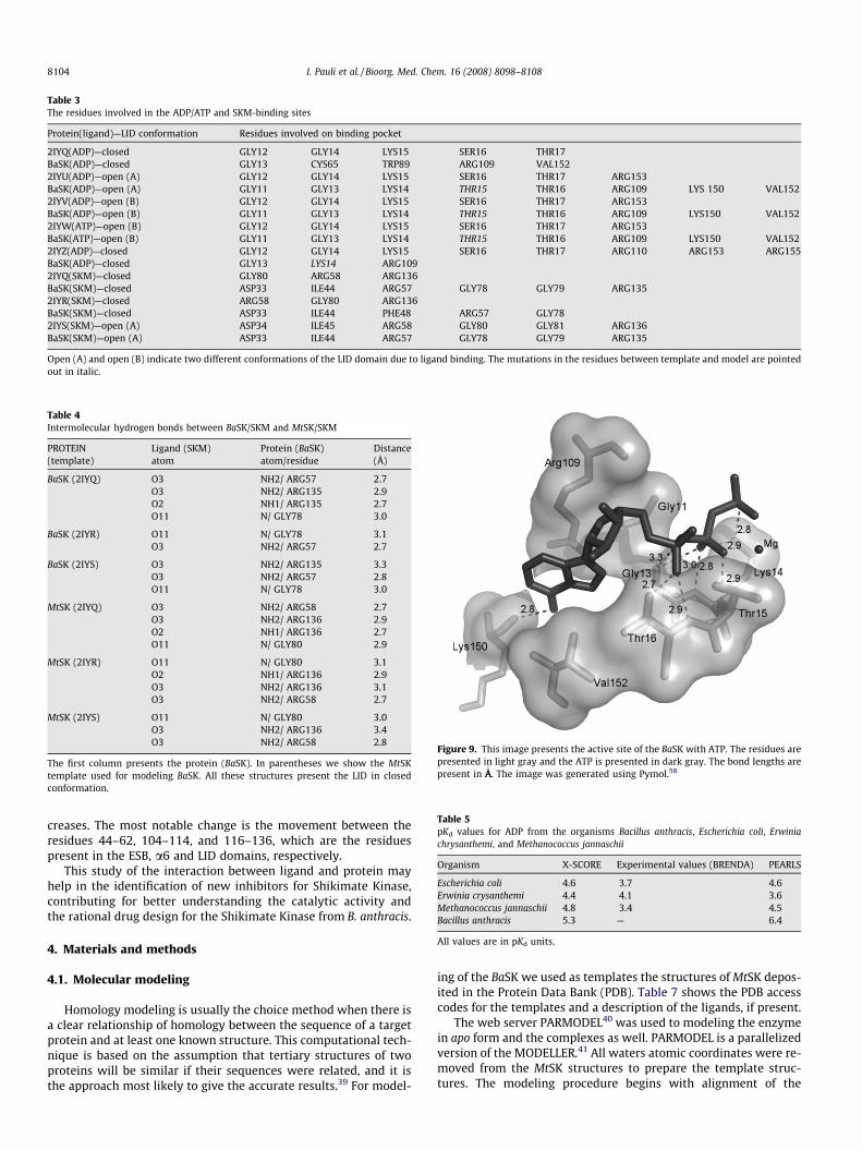
Table 3The residues involved in the ADP/ATP and SKM-binding sites
Protein(ligand)—LID conformation Residues involved on binding pocket
2IYQ(ADP)—closed GLY12 GLY14 LYS15 SER16 THR17BaSK(ADP)—closed GLY13 CYS65 TRP89 ARG109 VAL1522IYU(ADP)—open (A) GLY12 GLY14 LYS15 SER16 THR17 ARG153BaSK(ADP)—open (A) GLY11 GLY13 LYS14 THR15 THR16 ARG109 LYS 150 VAL1522IYV(ADP)—open (B) GLY12 GLY14 LYS15 SER16 THR17 ARG153BaSK(ADP)—open (B) GLY11 GLY13 LYS14 THR15 THR16 ARG109 LYS150 VAL1522IYW(ATP)—open (B) GLY12 GLY14 LYS15 SER16 THR17 ARG153BaSK(ATP)—open (B) GLY11 GLY13 LYS14 THR15 THR16 ARG109 LYS150 VAL1522IYZ(ADP)—closed GLY12 GLY14 LYS15 SER16 THR17 ARG110 ARG153 ARG155BaSK(ADP)—closed GLY13 LYS14 ARG1092IYQ(SKM)—closed GLY80 ARG58 ARG136BaSK(SKM)—closed ASP33 ILE44 ARG57 GLY78 GLY79 ARG1352IYR(SKM)—closed ARG58 GLY80 ARG136BaSK(SKM)—closed ASP33 ILE44 PHE48 ARG57 GLY782IYS(SKM)—open (A) ASP34 ILE45 ARG58 GLY80 GLY81 ARG136BaSK(SKM)—open (A) ASP33 ILE44 ARG57 GLY78 GLY79 ARG135
Open (A) and open (B) indicate two different conformations of the LID domain due to ligand binding. The mutations in the residues between template and model are pointedout in italic.
Table 4Intermolecular hydrogen bonds between BaSK/SKM and MtSK/SKM
PROTEIN(template)
Ligand (SKM)atom
Protein (BaSK)atom/residue
Distance(Å)
BaSK (2IYQ) O3 NH2/ ARG57 2.7O3 NH2/ ARG135 2.9O2 NH1/ ARG135 2.7O11 N/ GLY78 3.0
BaSK (2IYR) O11 N/ GLY78 3.1O3 NH2/ ARG57 2.7
BaSK (2IYS) O3 NH2/ ARG135 3.3O3 NH2/ ARG57 2.8O11 N/ GLY78 3.0
MtSK (2IYQ) O3 NH2/ ARG58 2.7O3 NH2/ ARG136 2.9O2 NH1/ ARG136 2.7O11 N/ GLY80 2.9
MtSK (2IYR) O11 N/ GLY80 3.1O2 NH1/ ARG136 2.9O3 NH2/ ARG136 3.1O3 NH2/ ARG58 2.7
MtSK (2IYS) O11 N/ GLY80 3.0O3 NH2/ ARG136 3.4O3 NH2/ ARG58 2.8
The first column presents the protein (BaSK). In parentheses we show the MtSKtemplate used for modeling BaSK. All these structures present the LID in closedconformation.
Figure 9. This image presents the active site of the BaSK with ATP. The residues arepresented in light gray and the ATP is presented in dark gray. The bond lengths arepresent in ÅA
0
. The image was generated using Pymol.58
Table 5pKd values for ADP from the organisms Bacillus anthracis, Escherichia coli, Erwiniachrysanthemi, and Methanococcus jannaschii
Organism X-SCORE Experimental values (BRENDA) PEARLS
Escherichia coli 4.6 3.7 4.6Erwinia crysanthemi 4.4 4.1 3.6Methanococcus jannaschii 4.8 3.4 4.5Bacillus anthracis 5.3 — 6.4
All values are in pKd units.
8104 I. Pauli et al. / Bioorg. Med. Chem. 16 (2008) 8098–8108
creases. The most notable change is the movement between theresidues 44–62, 104–114, and 116–136, which are the residuespresent in the ESB, a6 and LID domains, respectively.
This study of the interaction between ligand and protein mayhelp in the identification of new inhibitors for Shikimate Kinase,contributing for better understanding the catalytic activity andthe rational drug design for the Shikimate Kinase from B. anthracis.
4. Materials and methods
4.1. Molecular modeling
Homology modeling is usually the choice method when there isa clear relationship of homology between the sequence of a targetprotein and at least one known structure. This computational tech-nique is based on the assumption that tertiary structures of twoproteins will be similar if their sequences were related, and it isthe approach most likely to give the accurate results.39 For model-
ing of the BaSK we used as templates the structures of MtSK depos-ited in the Protein Data Bank (PDB). Table 7 shows the PDB accesscodes for the templates and a description of the ligands, if present.
The web server PARMODEL40 was used to modeling the enzymein apo form and the complexes as well. PARMODEL is a parallelizedversion of the MODELLER.41 All waters atomic coordinates were re-moved from the MtSK structures to prepare the template struc-tures. The modeling procedure begins with alignment of the
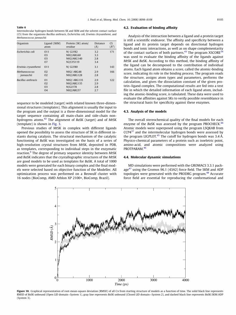
Table 6Intermolecular hydrogen bonds between SK and SKM and the solvent contact surface(CS) from the organisms Bacillus anthracis, Escherichia coli, Erwinia chrysanthemi, andMethanococcus jannaschii
Organism Ligand (SKM)atom
Protein (SK) atom/residue
Distance(Å)
CS(Å2)
Escherichia coli O11 N/ GLY82 3.2 171O3 NH2/ARG60 3.3O3 NH2/ARG140 2.8O7 N2/LYS119 3.4
Erwinia crysanthemi O11 N/ GLY80 3.1 143
Methanococcusjannaschii
O2 NH2/ ARG48 2.9 166O2 NH2/ARG128 2.8
Bacillus anthracis O1 NH2/ ARG135 2.9 122O2 NH2/ARG135 2.9O3 N/GLY78 2.8O4 NH2/ARG57 2.7
I. Pauli et al. / Bioorg. Med. Chem. 16 (2008) 8098–8108 8105
sequence to be modeled (target) with related known three-dimen-sional structures (templates). This alignment is usually the input tothe program and the output is a three-dimensional model for thetarget sequence containing all main-chain and side-chain non-hydrogens atoms.42 The alignment of BaSK (target) and of MtSK(template) is shown in Fig. 3.
Previous studies of MtSK in complex with different ligandsopened the possibility to assess the structure of SK in different in-stants during catalysis. The structural mechanism of the catalyticfunctioning of BaSK was investigated on the basis of a series ofhigh-resolution crystal structures from MtSK, deposited in PDB,as templates, corresponding to individual steps in the enzymaticreaction.9 The degree of primary sequence identity between MtSKand BaSK indicates that the crystallographic structures of the MtSKare good models to be used as templates for BaSK. A total of 1000models were generated for each binary complex and the final mod-els were selected based on objective function of the Modeller. Alloptimization process was performed on a Beowulf cluster with16 nodes (BioComp, AMD Athlon XP 2100+, BioComp, Brazil).
Figure 10. Graphical representation of root-mean-square deviation (RMSD) of all Ca froRMSD of BaSK unbound (Open LID domain—System 1), gray line represents BaSK unboun(System 3).
4.2. Evaluation of binding affinity
Analysis of the interaction between a ligand and a protein targetis still a scientific endeavor. The affinity and specificity between aligand and its protein target depends on directional hydrogenbonds and ionic interactions, as well as on shape complementarityof the contact surfaces of both partners.43 The program XSCORE34
was used to evaluate the binding affinity of the ligands againstMtSK and BaSK. According to this method, the binding affinity ofthe ligand can be decomposed to the contribution of individualatoms. Each ligand atom obtains a score, called the atomic-bindingscore, indicating its role in the binding process. The program readsthe structure, assigns atom types and parameters, performs thecalculation, and gives the dissociation constant of the given pro-tein–ligand complex. The computational results are fed into a textfile in which the detailed information of each ligand atom, includ-ing the atomic-binding score, is tabulated. These data were used toevaluate the affinities against SKs to verify possible resemblance inthe structural basis for specificity against these enzymes.
4.3. Analysis of the models
The overall stereochemical quality of the final models for eachenzyme of the BaSK was assessed by the program PROCHECK.44
Atomic models were superposed using the program LSQKAB fromCCP437 and the intermolecular hydrogen bonds were assessed bythe program LIGPLOT.45 The cutoff for hydrogen bonds was 3.4 Å.Physico-chemical parameters of a protein such as isoeletric point,amino-acid, and atomic compositions were analyzed usingPROTPARAM.46
4.4. Molecular dynamic simulations
MD simulations were performed with the GROMACS 3.3.1 pack-age47 using the Gromos 96.1 (43A2) force field. The SKM and ADPtopologies were generated with the PRODRG program.48 Accurateforce field are essential for reproducing the conformational and
m starting structure of models as a function of time. The solid black line representsd (Closed LID domain—System 2), and dashed black line represents BaSK:SKM:ADP
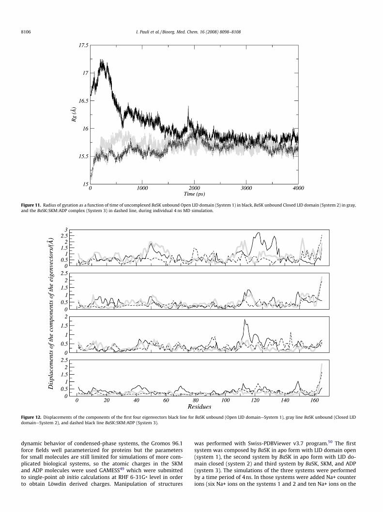
Figure 11. Radius of gyration as a function of time of uncomplexed BaSK unbound Open LID domain (System 1) in black, BaSK unbound Closed LID domain (System 2) in gray,and the BaSK:SKM:ADP complex (System 3) in dashed line, during individual 4 ns MD simulation.
Figure 12. Displacements of the components of the first four eigenvectors black line for BaSK unbound (Open LID domain—System 1), gray line BaSK unbound (Closed LIDdomain—System 2), and dashed black line BaSK:SKM:ADP (System 3).
8106 I. Pauli et al. / Bioorg. Med. Chem. 16 (2008) 8098–8108
dynamic behavior of condensed-phase systems, the Gromos 96.1force fields well parameterized for proteins but the parametersfor small molecules are still limited for simulations of more com-plicated biological systems, so the atomic charges in the SKMand ADP molecules were used GAMESS49 which were submittedto single-point ab initio calculations at RHF 6-31G* level in orderto obtain Löwdin derived charges. Manipulation of structures
was performed with Swiss-PDBViewer v3.7 program.50 The firstsystem was composed by BaSK in apo form with LID domain open(system 1), the second system by BaSK in apo form with LID do-main closed (system 2) and third system by BaSK, SKM, and ADP(system 3). The simulations of the three systems were performedby a time period of 4ns. In those systems were added Na+ counterions (six Na+ ions on the systems 1 and 2 and ten Na+ ions on the
Figure 13. Displacements of the components of first eigenvector black line for BaSK unbound (Open LID domain—System 1), gray line for BaSK unbound (Closed LID domain—System 2), and dashed black line for BaSK:SKM:ADP (System 3).
I. Pauli et al. / Bioorg. Med. Chem. 16 (2008) 8098–8108 8107
system 3) using Genion Program of the GROMACS simulation suiteto neutralize the negative charge density of the systems. Eachstructure was placed in the center of a truncated cubic box filledwith Extended Simple Point Charge (SPC/E) water molecules,51
containing 12,985 for the system 1, 9249 for the system 2, and9222 water molecules for the system 3. The initial simulation celldimensions were 33.34, 48.70, 55.39 Å for the system 1, 41.92,35.70, 45.14 Å for the system 2 and 3, and had the protein solvatedby a layer of water molecules of at least 10 Å in all directions inboth systems. During the simulations, bonds lengths within theproteins were constrained by using LINCS algorithm.52 The SETTLEalgorithm was used to constrain the geometry of water mole-cules.53 In the MD protocol, all hydrogen atoms, ions, and watermolecules were first subjected to 1500 steps of energy minimiza-tions by steepest descent to remove close van der Waals contacts.The systems were then submitted to a short molecular dynamicwith position restrains for a period of 20 ps and afterwards per-formed a full molecular dynamics without restrains. The tempera-ture of the system was then increased from 50 to 300 K in 5 steps(50 to 100, 100 to 150, 150 to 200, 200 to 250, 250 to 300 K), andthe velocities at each step were reassigned according to the Max-well–Boltzmann distribution at that temperature and equilibratedfor 10 ps except the last part of termalization phase where we useda period of 40 ps. Energy minimization and MD were carried outunder periodic boundary conditions. The simulation was computedin the NPT ensemble at 300 K with the Berendsen temperaturecoupling and constant pressure of 1 atm with isotropic molecule-based scaling.54 The LINCS algorithm, with a 10�5 Å tolerance,was applied to fix all bonds containing a hydrogen atom, allowingthe use of a time step of 2.0fs in the integration of the equations ofmotion. No extra restrains were applied after the equilibrationphase. The electrostatic interactions between non-ligand atomswere evaluated by the particle-mesh Ewald method55 with a
charge grid spacing of �1.0 Å and the charge grid was interpolatedon a cubic grid with the direct sum tolerance set to 1.0 � 10�5. TheLennard–Jones interactions were evaluated using a 9.0 Å atom-based cutoff.56 All analyses were performed on the ensemble ofsystem configurations extracted at 0.5-ps time intervals from thesimulation and MD trajectory collection was initiated after 1 nsof dynamics to guarantee a completely equilibrated evolution.The MD simulation and results analyses were performed on a per-sonal computer Intel Core 2 Duo E6300 – 1.86GHz and 4Gb RAM.The convergence of the different simulations were analyzed interms of the secondary structure, root mean-square deviation(RMSD) from the initial models structures and radius of gyration(Rg).
4.5. Essential dynamics analysis
Essential Dynamics (ED), also known as principal componentanalysis (PCA), is a method commonly used for dissecting thedynamics of proteins and their importance in biological processes,like protein folding or substrate binding. The ED analysis is a tech-nique that reduces the complexity of the data and extracts the con-certed motion in simulations that are essentially correlated andpresumably meaningful for biological function.57 In the ED analy-sis, a variance/covariance matrix was constructed from the trajec-tories after removal of the rotational and translational movements.A set of eigenvectors and eigenvalues was identified by diagonaliz-ing the matrix. The eigenvalues represented the amplitude of theeigenvectors along the multidimensional space, and the displace-ments of atoms along each eigenvector showed the concerted mo-tions of protein along each direction. An assumption of ED analysisis that the correlated motions for the function of the protein are de-scribed by eigenvectors with large eigenvalues. The movements ofprotein in the essential subspace were identified by projecting the

Table 7PDB access codes, resolution, description of the ligands and the LID conformation forMtSK structures (templates)
Enzyme PDB access code Resolution (Å) Ligands LID conformation
SK 1WE2 2.3 SKM + ADP Closed2IYT 1.5 Apo Open (A)2IYU 1.8 ADP Open (A)2IYV 1.3 ADP Open (B)2IYW 1.8 MgATP Open (B)2IYS 1.4 SKM Open (A)2IYR 2.0 SKM Closed2IYQ 1.8 SKM + ADP Closed2IYY 1.6 S3P + SO4 Closed2IYZ 2.3 S3P + ADP Closed1L4Y 2.0 MgADP Closed
Open (A) and open (B) indicate two different conformations of the LID domain dueto ligand binding.
8108 I. Pauli et al. / Bioorg. Med. Chem. 16 (2008) 8098–8108
Cartesian trajectory coordinates along the most important eigen-vectors from the analysis.
Acknowledgement
This work was supported by grants from Millennium Institute(CNPq-MCT). WFA is a researcher from the National ResearchCouncil of Brazil (CNPq).
Supplementary data
Supplementary data associated with this article can be found, inthe online version, at doi:10.1016/j.bmc.2008.07.051.
References and notes
1. Mock, M.; Fouet, A. Annu. Rev. Microbiol. 2001, 55, 647.2. Kalamas, A. G. Anesthes. Clin. North Am. 2004, 22, 533.3. Bagget, H. C.; Rhodes, J. C.; Fridkin, S. K.; Quinn, C. P.; Hageman, J. C.; Friedman,
C. R.; Dykewicz, C. A.; Semenova, V. A.; Romero-Steiner, S.; Elie, C. M.; Jernigan,J. A. Clin. Infect. Dis. 2005, 41, 991.
4. Jernigan, D. B.; Raghunathan, P. L.; Bell, B. P.; Brechner, R.; Bresnitz, E. A.; Butler,J. C.; Cetron, M.; Cohen, M.; Doyle, T.; Fischer, M.; Greene, C.; Griffith, K. S.;Guarner, J.; Hadler, J. L.; Hayslett, J. A.; Meyer, R.; Petersen, L. R.; Phillips, M.;Pinner, R.; Popovic, T.; Quinn, C. P.; Reefhuis, J.; Reissman, D.; Rosenstein, N.;Schuchat, A.; Shieh, W. J.; Siegal, L.; Swerdlow, D. L.; Tenover, F. C.; Traeger, M.;Ward, J. W.; Weisfuse, I.; Wiersma, S.; Yeskey, K.; Zaki, S.; Ashford, D. A.;Perkins, B. A.; Ostroff, S.; Hughes, J.; Fleming, D.; Koplan, J. P.; Gerberding, J. L.Emerg. Infect. Dis. 2002, 8, 1019.
5. Sanderson, W. T.; Stoddard, R. R.; Echt, A. S.; Piacitelli, C. A.; Kim, D.; Horan, J.;Davies, M. M.; McCleery, R. E.; Muller, P.; Schnorr, T. M.; Ward, E. M.; Hales, T.R. J. Appl. Microbiol. 2004, 96, 1048.
6. Casadevall, A. Front. Biosci. 2008, 13, 4009.7. Oliveira, J. S.; Sousa, E. H. S.; Basso, L. A.; Palaci, M.; Dietze, R.; Santos, D. S.;
Moreira, I. S. Chem. Commun. 2004, 7, 312.8. Oliveira, J. S.; Sousa, E. H. S.; Souza, O. N.; Moreira, I. S.; Santos, D. S.; Basso, L. A.
Curr. Pharm. Des. 2006, 12, 2409.9. Oliveira, J. S.; Vasconcelos, I. B.; Moreira, I. S.; Santos, D. S.; Basso, L. A. Curr.
Drug Targets 2007, 8, 399.10. Dias, M. V.; Vasconcelos, I. B.; Prado, A. M.; Fadel, V.; Basso, L. A.; de Azevedo,
W. F., Jr.; Santos, D. S. J. Struct. Biol. 2007, 159, 369.11. Vasconcelos, I. B.; Meyer, E.; Sales, F. A. M.; Moreira, I. S.; Basso, L. A.; Santos, D.
S. Anti-infective Agents Med. Chem. 2008, 7, 50.12. Tipparaju, S. K.; Jovasawal, S.; Forrester, S.; Mulhearn, D. C.; Pegan, S.; Johnson, M.
E.; Mesecar, A. D.; Kozikowski, A. P. Bioorg. Med. Chem. Lett. 2008, 18(12), 3565.13. Pereira, J. H.; Vasconcelos, J. B.; Oliveira, J. S.; Caceres, R. A.; de Azevedo, W. F.,
Jr.; Basso, L. A.; Santos, D. S. Curr. Drug Targets 2007, 8, 459.14. Dias, M. V.; Faı́m, L. M.; Vasconcelos, I. B.; de Oliveira, J. S.; Basso, L. A.; Santos,
D. S.; de Azevedo, W. F., Jr. Acta Crystallogr. Sect. F Struct. Biol. Crystallogr.Commun. 2007, 63, 1.
15. Silveira, N. J.; Uchôa, H. B.; Pereira, J. H.; Canduri, F.; Basso, L. A.; Palma, M. S.;Santos, D. S.; de Azevedo, W. F., Jr. J. Mol. Model. 2005, 11, 160.
16. Pereira, J. H.; de Oliveira, J. S.; Canduri, F.; Dias, M. V.; Palma, M. S.; Basso, L. A.;Santos, D. S.; de Azevedo, W. F., Jr. ActaCrystallogr. DBiol. Crystallogr. 2004, 60, 2310.
17. Segura-Cabrera, A.; Rodrı́guez-Pérez, MA. Bioorg. Med. Chem. Lett. 2008, 18,3152.
18. Bentley, R. Crit. Ver. Biochem. Mol. Biol. 1990, 25, 307.19. Davies, G. M.; Barret-Bee, K. J.; Jude, D. A.; Lehan, M.; Nichols, W. W.; Pinder, P. E.;
Thain, J. L.; Watkins, W. J.; Wilson, R. G. Antimicrob. AgentsChemother. 1994, 38, 403.20. De Azevedo, W. F. Curr. Drug Targets 2007, 8, 387.21. Pereira, J. H.; Canduri, F.; de Oliveira, J. S.; da Silveira, N. J. F.; Basso, L. A.; Palma,
M. S.; De Azevedo, W. F.; Santos, D. S. Biochem. Biophys. Res. Commun. 2003,312, 608.
22. Arcuri, H. A.; Borges, J. C.; Fonseca, I. O.; Pereira, J. H.; Neto, J. R.; Basso, L. A.;Santos, D. S.; de Azevedo, W. F., Jr. Proteins 2008, 72, 720.
23. Marques, M. R.; Pereira, J. H.; Oliveira, J. S.; Basso, L. A.; de Azevedo, W. F., Jr.;Santos, D. S.; Palma, M. S. Curr. Drug Targets 2007, 8, 445.
24. Borges, J. C.; Pereira, J. H.; Vasconcelos, I. B.; dos Santos, G. C.; Olivieri, J. R.;Ramos, C. H.; Palma, M. S.; Basso, L. A.; Santos, D. S.; de Azevedo, W. F., Jr. Arch.Biochem. Biophys. 2006, 452, 156.
25. Dias, M. V.; Borges, J. C.; Ely, F.; Pereira, J. H.; Canduri, F.; Ramos, C. H.; Frazzon,J.; Palma, M. S.; Basso, L. A.; Santos, D. S.; de Azevedo, W. F., Jr. J. Struct. Biol.2006, 154, 130.
26. Basso, L. A.; da Silva, L. H.; Fett-Neto, A. G.; de Azevedo, W. F., Jr.; Moreira, Ide.S.; Palma, M. S.; Calixto, J. B.; Astolfi, F. S.; dos Santos, R. R.; Soares, M. B.;Santos, D. S. Mem. Inst. Oswaldo Cruz 2005, 100, 475.
27. Ely, F.; Nunes, J. E.; Schroeder, E. K.; Frazzon, J.; Palma, M. S.; Santos, D. S.;Basso, L. A. BMC Biochem. 2008, 29, 9.
28. De Azevedo, W. F.; Canduri, F.; de Oliveira, J. S.; Basso, L. A.; Palma,M. S.; Pereira, J. H.; Santos, D. S. Biochem. Biophys. Res. Commun.2002, 295, 142.
29. Parish, T.; Stoker, N. G. Microbiology 2002, 148, 3069.30. Vonrhein, C.; Schlauderer, G. J.; Schulz, G. E. Structure 1995, 3, 483.31. Hartmann, M. D.; Bourenkov, G. P.; Strizhov, A. O. N.; Bartunik, H. D. J. Mol. Biol.
2006, 364, 411.32. Caceres, R. A.; Macedo Timmers, L. F.; Vivan, A. L.; Schneider, C. Z.; Basso, L. A.;
De Azevedo, W. F., Jr.; Santos, D. S. J. Mol. Model. 2008, 14, 427.33. Schomburg, I.; Chang, A.; Ebeling, C.; Hofmann, O.; Ebeling, C.; Ehrentreich, F.;
Schomburg, D. Trends Biochem. Sci. 2002, 27, 54.34. Wang, R.; Liu, L.; Lai, L.; Tang, Y. J. Mol. Model. 1998, 4, 379.35. Han, L. Y.; Lin, H. H.; Li, Z. R.; Zheng, C. J.; Cao, Z. W.; Xie, B.; Chen, Y. Z. J. Chem.
Inf. Model. 2006, 46, 445.36. Lee, B.; Richards, F. M. J. Mol. Biol. 1971, 55, 379.37. Collaborative Computation Project, Number 4. Acta Crystallogr. D Biol.
Crystallogr. 1994, 50, 760.38. Liu, G.; Tan, J.; Niu, C.; Shen, J.; Luo, X.; Shen, X.; Chen, K.; Jiang, H. Acta Pharm.
Sin. 2006, 27, 100.39. Kroemer, R. T.; Doughty, S. W.; Robinson, A. J.; Richards, W. G. Protein Eng.
1996, 9, 493.40. Uchoa, H. B.; Jorge, G. E.; Da Silveira, N. J. F.; Camera, J. C.; Canduri, F.; De
Azevedo, W. F. Biochem. Biophys. Res. Commun. 2004, 325, 1481.41. Sali, A.; Blundell, T. L. J. Mol. Biol. 1993, 234, 779.42. Canduri, F.; Uchoa, H. B.; De Azevedo, W. F., Jr. Biochem. Biophys. Res. Commun.
2004, 324, 661.43. De Azevedo, W. F.; Mueller-Dieckmann, J. H.; Schulze-Gahmen, U.; Worland, P.
J.; Sausville, E.; Kim, S. H. Proc. Natl. Acad. Sci. U.S.A. 1996, 93, 2735.44. Laskowski, R. A.; Macarthur, M. W.; Moss, D. S.; Thornton, J. M. J. Appl.
Crystallogr. 1993, 26, 283.45. Wallace, A. C.; Laskowski, R. A.; J.M.Thornton Protein Eng. 1995, 8, 127.46. Gasteiger, E.; Hoogland, C.; Gattiker, A.; Duvaud, S.; Wilkins, M. R.; Appel, R. D.;
Bairoch, A. In The Proteomics Protocols Handbook; Walker, John M., Ed.; HumanaPress, 2005; p 571.
47. Van der Spoel, D.; Lindahl, E.; Hess, B.; Groenhof, G.; Mark, A. E.; Berendsen, H.J. C. J. Comput. Chem. 2005, 26, 1701.
48. Van Aalten, D. M. F.; Bywater, B.; Findlay, J. B. C.; Hendlich, M.; Hooft, R. W. W.;Vriend, G. J. Comput. Aided Mol. Des. 1996, 10, 255.
49. Schmidt, M. W.; Baldridge, K. K.; Boatz, J. A.; Elbert, S. T.; Gordon, M. S.; Jensen,J. H.; Koseki, S.; Matsunaga, N.; Nguyen, K. A.; Su, S. J.; Windus, T. L.; Dupuis,M.; Montgomery, J. A. J. Comput. Chem. 1993, 14, 1347.
50. Guex, N.; Peitsch, M. C. Electrophoresis 1997, 18, 2714.51. Berendsen, H. J. C.; Postma, J. P. M.; van Gunsteren, W. F.; Hermans, J. In
Intermolecular Forces; Pullman, B., Ed.; Reidel D. Publishing Company:Dordrecht, The Netherlands, 1981; p 331.
52. Hess, B.; Bekker, H.; Berendsen, H. J. C.; Fraaije, J. G. E. M. J. Comput. Chem. 1997,18, 1463.
53. Miyamoto, S.; Kollman, P. A. J. Comput. Chem. 1992, 13, 952.54. Chowdhuri, S.; Ming-Liang, T.; Ichiye, T. J. Chem. Phys. 2006, 125, 144513.55. Darden, T.; York, D.; Pedersen, L. A. J. Chem. Phys. 1993, 98, 10089.56. De Souza, O. N.; Ornstein, R. L. J. Biomol. Struct. Dyn. 1999, 16, 1205.57. Amadei, A.; Linssen, A. B. M.; Berendsen, H. J. C. Proteins 1993, 17, 412.58. Delano, W. L.; Lam, J. W. Abstr. Pap. Am. Chem. Soc. 2005, 230, 1371.
Copyright © 2022 FDOKUMEN