Limited genetic diversity in Salmonella enterica serovar Enteritidis PT13
10
BioMed Central Page 1 of 10 (page number not for citation purposes) BMC Microbiology Open Access Research article Limited genetic diversity in Salmonella enterica Serovar Enteritidis PT13 Adam B Olson 1 , Ashleigh K Andrysiak 1,4 , Dobryan M Tracz 1 , Jean Guard- Bouldin 2 , Walter Demczuk 1 , Lai-King Ng 1,4 , Anne Maki 3 , Frances Jamieson 3 and Matthew W Gilmour* 1,4 Address: 1 National Microbiology Laboratory, Public Health Agency of Canada, Winnipeg, MB, Canada, 2 United States Department of Agricultural, Agricultural Research Service, Athens, GA, USA, 3 Ontario Central Public Health Laboratory, Ministry of Health and Long-Term Care, Toronto, ON, Canada and 4 Department of Medical Microbiology, University of Manitoba, Winnipeg, MB, Canada Email: Adam B Olson - [email protected]; Ashleigh K Andrysiak - [email protected]; Dobryan M Tracz - [email protected]; Jean Guard-Bouldin - [email protected]; Walter Demczuk - [email protected]; Lai-King Ng - [email protected]; Anne Maki - [email protected]; Frances Jamieson - [email protected]; Matthew W Gilmour* - [email protected] * Corresponding author Abstract Background: Salmonella enterica serovar Enteritidis has emerged as a significant foodborne pathogen throughout the world and is commonly characterized by phage typing. In Canada phage types (PT) 4, 8 and 13 predominate and in 2005 a large foodborne PT13 outbreak occurred in the province of Ontario. The ability to link strains during this outbreak was difficult due to the apparent clonality of PT13 isolates in Canada, as there was a single dominant pulsed-field gel electrophoresis (PFGE) profile amongst epidemiologically linked human and food isolates as well as concurrent sporadic strains. The aim of this study was to perform comparative genomic hybridization (CGH), DNA sequence-based typing (SBT) genomic analyses, plasmid analyses, and automated repetitive sequence-based PCR (rep-PCR) to identify epidemiologically significant traits capable of subtyping S. Enteritidis PT13. Results: CGH using an oligonucleotide array based upon chromosomal coding sequences of S. enterica serovar Typhimurium strain LT2 and the Salmonella genomic island 1 successfully determined major genetic differences between S. Typhimurium and S. Enteritidis PT13, but no significant strain-to-strain differences were observed between S. Enteritidis PT13 isolates. Individual loci (safA and fliC) that were identified as potentially divergent in the CGH data set were sequenced in a panel of S. Enteritidis strains, and no differences were detected between the PT13 strains. Additional sequence-based typing was performed at the fimA, mdh, manB, cyaA, citT, caiC, dmsA, ratA and STM0660 loci. Similarly, no diversity was observed amongst PT13 strains. Variation in plasmid content between PT13 strains was observed, but macrorestriction with BglII did not identify further differences. Automated rep-PCR patterns were variable between serovars, but S. Enteritidis PT13 strains could not be differentiated. Conclusion: None of the methods identified any significant variation between PT13 strains. Greater than 11,300 base pairs of sequence for each of seven S. Enteritidis PT13 strains were analyzed without detecting a single polymorphic site, although diversity between different phage types of S. Enteritidis was observed. These data suggest that Canadian S. Enteritidis PT13 strains are highly related genetically. Published: 1 October 2007 BMC Microbiology 2007, 7:87 doi:10.1186/1471-2180-7-87 Received: 12 June 2007 Accepted: 1 October 2007 This article is available from: http://www.biomedcentral.com/1471-2180/7/87 © 2007 Olson et al; licensee BioMed Central Ltd. This is an Open Access article distributed under the terms of the Creative Commons Attribution License (http://creativecommons.org/licenses/by/2.0 ), which permits unrestricted use, distribution, and reproduction in any medium, provided the original work is properly cited.
-
Upload
independent -
Category
Documents
-
view
0 -
download
0
Transcript of Limited genetic diversity in Salmonella enterica serovar Enteritidis PT13
BioMed CentralBMC Microbiology
ss
Open AcceResearch articleLimited genetic diversity in Salmonella enterica Serovar Enteritidis PT13Adam B Olson1, Ashleigh K Andrysiak1,4, Dobryan M Tracz1, Jean Guard-Bouldin2, Walter Demczuk1, Lai-King Ng1,4, Anne Maki3, Frances Jamieson3 and Matthew W Gilmour*1,4Address: 1National Microbiology Laboratory, Public Health Agency of Canada, Winnipeg, MB, Canada, 2United States Department of Agricultural, Agricultural Research Service, Athens, GA, USA, 3Ontario Central Public Health Laboratory, Ministry of Health and Long-Term Care, Toronto, ON, Canada and 4Department of Medical Microbiology, University of Manitoba, Winnipeg, MB, Canada
Email: Adam B Olson - [email protected]; Ashleigh K Andrysiak - [email protected]; Dobryan M Tracz - [email protected]; Jean Guard-Bouldin - [email protected]; Walter Demczuk - [email protected]; Lai-King Ng - [email protected]; Anne Maki - [email protected]; Frances Jamieson - [email protected]; Matthew W Gilmour* - [email protected]
* Corresponding author
AbstractBackground: Salmonella enterica serovar Enteritidis has emerged as a significant foodborne pathogenthroughout the world and is commonly characterized by phage typing. In Canada phage types (PT) 4, 8 and13 predominate and in 2005 a large foodborne PT13 outbreak occurred in the province of Ontario. Theability to link strains during this outbreak was difficult due to the apparent clonality of PT13 isolates inCanada, as there was a single dominant pulsed-field gel electrophoresis (PFGE) profile amongstepidemiologically linked human and food isolates as well as concurrent sporadic strains. The aim of thisstudy was to perform comparative genomic hybridization (CGH), DNA sequence-based typing (SBT)genomic analyses, plasmid analyses, and automated repetitive sequence-based PCR (rep-PCR) to identifyepidemiologically significant traits capable of subtyping S. Enteritidis PT13.
Results: CGH using an oligonucleotide array based upon chromosomal coding sequences of S. entericaserovar Typhimurium strain LT2 and the Salmonella genomic island 1 successfully determined majorgenetic differences between S. Typhimurium and S. Enteritidis PT13, but no significant strain-to-straindifferences were observed between S. Enteritidis PT13 isolates. Individual loci (safA and fliC) that wereidentified as potentially divergent in the CGH data set were sequenced in a panel of S. Enteritidis strains,and no differences were detected between the PT13 strains. Additional sequence-based typing wasperformed at the fimA, mdh, manB, cyaA, citT, caiC, dmsA, ratA and STM0660 loci. Similarly, no diversity wasobserved amongst PT13 strains. Variation in plasmid content between PT13 strains was observed, butmacrorestriction with BglII did not identify further differences. Automated rep-PCR patterns were variablebetween serovars, but S. Enteritidis PT13 strains could not be differentiated.
Conclusion: None of the methods identified any significant variation between PT13 strains. Greater than11,300 base pairs of sequence for each of seven S. Enteritidis PT13 strains were analyzed without detectinga single polymorphic site, although diversity between different phage types of S. Enteritidis was observed.These data suggest that Canadian S. Enteritidis PT13 strains are highly related genetically.
Published: 1 October 2007
BMC Microbiology 2007, 7:87 doi:10.1186/1471-2180-7-87
Received: 12 June 2007Accepted: 1 October 2007
This article is available from: http://www.biomedcentral.com/1471-2180/7/87
© 2007 Olson et al; licensee BioMed Central Ltd. This is an Open Access article distributed under the terms of the Creative Commons Attribution License (http://creativecommons.org/licenses/by/2.0), which permits unrestricted use, distribution, and reproduction in any medium, provided the original work is properly cited.
Page 1 of 10(page number not for citation purposes)

BMC Microbiology 2007, 7:87 http://www.biomedcentral.com/1471-2180/7/87
BackgroundSalmonella enterica serovar Enteritidis (S. Enteritidis) is afoodborne pathogen transmitted to humans predomi-nately through contaminated eggs and other poultry foodproducts [1]. S. Enteritidis was an infrequently reportedserovar until the mid- to late 1980's when it emerged as acommon cause of salmonellosis in European countriesand then worldwide [2-4]. By the 1990's S. Enteritidisreplaced S. enterica serovar Typhimurium (S. Typhimu-rium) as the most common serovar isolated from humansin many countries [5,3,6,4,7]. In Canada, S. Enteritidishas been among the top three reported Salmonella serovarsresulting in human disease since 1998, accounting forbetween 12–21% of infections caused by Salmonella [8]. S.Enteritidis is subtyped by a phage typing scheme imple-mented by Ward et al., [9], and this method has identifiedregionally endemic S. Enteritidis subtypes as well fluctua-tions in the predominate subtypes. For example, S. Enter-itidis phage type 13 (PT13) was the eighth most commonphage type identified in Canada from 1982–1992, but by1998 PT13 was ranked the third most common and hassubsequently remained one of the top five most prevalentphage types [8].
Phage typing of S. Enteritidis utilizes a set of sixteen bac-teriophage to generate a lytic pattern to group strains [9].Typing methods based upon comparisons of wholegenomic DNA, plasmid DNA or specific genetic determi-nants have also been used in place of phage typing or assupplementary techniques. The methods most frequentlyused to subtype S. Enteritidis include pulsed-field gel elec-trophoresis (PFGE), plasmid profiling and restriction-hybridization based ribotyping, which have each beenapplied with varying degrees of success [10-16]. A singlePFGE macrorestriction profile often predominated instrains of the same phage type or even amongst multiplephage types [10,13,17-19]. Alternatively, a group ofstrains of the same phage type can have multiple restric-tion patterns [20,21]. Ribotyping based upon restrictionanalysis of the rRNA operon has utilized several schemes,such as PvuII or a double digestion with PstI and SphI [22].The latter combination has been used to subtype strains ofthe same phage type, however, this discrimination wasnot always epidemiologically significant [17]. Plasmidprofiles have indicated divergence between strains repre-sented by the presence or absence of various low molecu-lar weight plasmids and the 55 to 60 kbp S. Enteritidisvirulence plasmid [11,12,23], which is similar to the 94kbp pSLT virulence plasmid of S. Typhimurium [24].Combinations of these methods have been successfullyused to subtype outbreak from non-outbreak strains of S.Enteritidis [25,11,13,15,18].
DNA microarray-based comparative genomic hybridiza-tion (CGH) and sequence-based typing (SBT) have
recently been used to determine genetic relatednessbetween S. Enteritidis strains, and there was generally alack of diversity for strains of the same phage type[26,24,27-29]. SBT of 24 clinical and poultry-related S.Enteritidis strains in a scheme comprised of segments ofthe 16S rRNA, manB, glnA and pduF loci detected 2, 37, 3and 3 polymorphic sites, respectively [28]. Similarly, aSBT scheme evaluating manB, mdh and fimA encoded by 7S. Enteritidis strains revealed 2, 2 and 0 polymorphic sites,respectively [26]. Typing of the misL, spaM and spaN lociencoding cell surface-associated proteins did not identifydifferences in four PT13 strains, but identified 2 polymor-phic sites in PT4 [29]. CGH analyses of a diverse collec-tion of 27 S. Enteritidis strains using a microarraycomposed of S. Typhimurium, S. Typhi, S. Paratyphi Aand S. Enteritidis PT4 probes identified few genetic differ-ences between these strains, including strains isolatedbefore the emergence of S. Enteritidis as a major food-borne Salmonella [24]. The genetic differences were pre-dominantly at phage-encoding loci (ST64B and Fels-2)but no significant delineation amongst S. Enteritidisstrains of the same phage type was observed [24]. CGHanalyses of PT13a and PT4 strains similarly identified nosignificant strain-to-strain variation, and these phagetypes were also distinguished by ST64B and Fels bacteri-ophage genetic content [27].
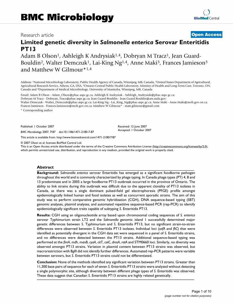
S. Enteritidis PT13 was identified in a large foodborneoutbreak in the Canadian province of Ontario in 2005.Over 700 cases of gastroenteritis were reported betweenOctober and December 2005 and these were associatedwith the consumption of contaminated mung beansprouts. A single PFGE profile (SENXAI.0038;SENBNI.0016) predominated amongst the outbreak-associated strains, and this profile was also seen in concur-rent and preceding sporadic human-clinical and poultry-related isolates. With the apparent clonality of PT13 iso-lates, it was difficult to support epidemiological links dur-ing the outbreak using PFGE-based typing. The aim of thisstudy was to perform CGH and SBT genomic analyses inparallel with other typing methodologies to identify epi-demiologically significant markers for subtyping S. Enter-itidis PT13.
ResultsIn response to the large outbreak of S. Enteritidis PT13, allS. Enteritidis PT13 strains with PFGE data reported to theNational Microbiology Laboratory were retrospectivelyanalyzed. This set of 32 strains comprised sporadichuman-clinical isolates and also poultry-related agri-foodisolates submitted by provincial public health laborato-ries in Alberta, British Columbia, Ontario and Québec.Two PFGE patterns were observed: 30 strains had the XbaImacrorestriction profile SENXAI.0038 and 2 strains pro-duced the related pattern SENXAI.0062 (Fig. 1). PFGE
Page 2 of 10(page number not for citation purposes)
BMC Microbiology 2007, 7:87 http://www.biomedcentral.com/1471-2180/7/87
with BlnI was performed for 19 of these strains and allwere pattern SENBNI.0016. The inability to differentiatestrains by PFGE necessitated the need to attempt addi-tional subtyping methodologies for the differentiation ofboth outbreak and sporadic S. Enteritidis PT13 strains.
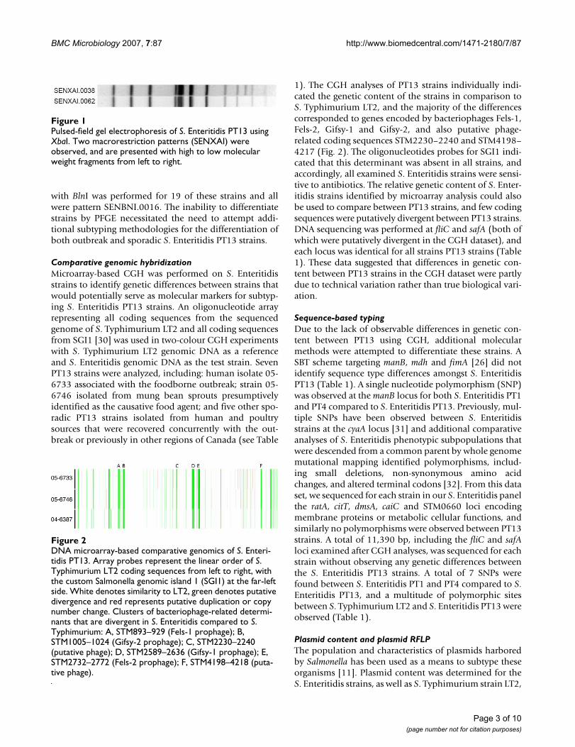
Comparative genomic hybridizationMicroarray-based CGH was performed on S. Enteritidisstrains to identify genetic differences between strains thatwould potentially serve as molecular markers for subtyp-ing S. Enteritidis PT13 strains. An oligonucleotide arrayrepresenting all coding sequences from the sequencedgenome of S. Typhimurium LT2 and all coding sequencesfrom SGI1 [30] was used in two-colour CGH experimentswith S. Typhimurium LT2 genomic DNA as a referenceand S. Enteritidis genomic DNA as the test strain. SevenPT13 strains were analyzed, including: human isolate 05-6733 associated with the foodborne outbreak; strain 05-6746 isolated from mung bean sprouts presumptivelyidentified as the causative food agent; and five other spo-radic PT13 strains isolated from human and poultrysources that were recovered concurrently with the out-break or previously in other regions of Canada (see Table
1). The CGH analyses of PT13 strains individually indi-cated the genetic content of the strains in comparison toS. Typhimurium LT2, and the majority of the differencescorresponded to genes encoded by bacteriophages Fels-1,Fels-2, Gifsy-1 and Gifsy-2, and also putative phage-related coding sequences STM2230–2240 and STM4198–4217 (Fig. 2). The oligonucleotides probes for SGI1 indi-cated that this determinant was absent in all strains, andaccordingly, all examined S. Enteritidis strains were sensi-tive to antibiotics. The relative genetic content of S. Enter-itidis strains identified by microarray analysis could alsobe used to compare between PT13 strains, and few codingsequences were putatively divergent between PT13 strains.DNA sequencing was performed at fliC and safA (both ofwhich were putatively divergent in the CGH dataset), andeach locus was identical for all strains PT13 strains (Table1). These data suggested that differences in genetic con-tent between PT13 strains in the CGH dataset were partlydue to technical variation rather than true biological vari-ation.
Sequence-based typingDue to the lack of observable differences in genetic con-tent between PT13 using CGH, additional molecularmethods were attempted to differentiate these strains. ASBT scheme targeting manB, mdh and fimA [26] did notidentify sequence type differences amongst S. EnteritidisPT13 (Table 1). A single nucleotide polymorphism (SNP)was observed at the manB locus for both S. Enteritidis PT1and PT4 compared to S. Enteritidis PT13. Previously, mul-tiple SNPs have been observed between S. Enteritidisstrains at the cyaA locus [31] and additional comparativeanalyses of S. Enteritidis phenotypic subpopulations thatwere descended from a common parent by whole genomemutational mapping identified polymorphisms, includ-ing small deletions, non-synonymous amino acidchanges, and altered terminal codons [32]. From this dataset, we sequenced for each strain in our S. Enteritidis panelthe ratA, citT, dmsA, caiC and STM0660 loci encodingmembrane proteins or metabolic cellular functions, andsimilarly no polymorphisms were observed between PT13strains. A total of 11,390 bp, including the fliC and safAloci examined after CGH analyses, was sequenced for eachstrain without observing any genetic differences betweenthe S. Enteritidis PT13 strains. A total of 7 SNPs werefound between S. Enteritidis PT1 and PT4 compared to S.Enteritidis PT13, and a multitude of polymorphic sitesbetween S. Typhimurium LT2 and S. Enteritidis PT13 wereobserved (Table 1).
Plasmid content and plasmid RFLPThe population and characteristics of plasmids harboredby Salmonella has been used as a means to subtype theseorganisms [11]. Plasmid content was determined for theS. Enteritidis strains, as well as S. Typhimurium strain LT2,
DNA microarray-based comparative genomics of S. Enteri-tidis PT13Figure 2DNA microarray-based comparative genomics of S. Enteri-tidis PT13. Array probes represent the linear order of S. Typhimurium LT2 coding sequences from left to right, with the custom Salmonella genomic island 1 (SGI1) at the far-left side. White denotes similarity to LT2, green denotes putative divergence and red represents putative duplication or copy number change. Clusters of bacteriophage-related determi-nants that are divergent in S. Enteritidis compared to S. Typhimurium: A, STM893–929 (Fels-1 prophage); B, STM1005–1024 (Gifsy-2 prophage); C, STM2230–2240 (putative phage); D, STM2589–2636 (Gifsy-1 prophage); E, STM2732–2772 (Fels-2 prophage); F, STM4198–4218 (puta-tive phage).
Pulsed-field gel electrophoresis of S. Enteritidis PT13 using XbaIFigure 1Pulsed-field gel electrophoresis of S. Enteritidis PT13 using XbaI. Two macrorestriction patterns (SENXAI) were observed, and are presented with high to low molecular weight fragments from left to right.
Page 3 of 10(page number not for citation purposes)
BM
C M
icro
biol
ogy
2007
, 7:8
7ht
tp://
ww
w.b
iom
edce
ntra
l.com
/147
1-21
80/7
/87
Page
4 o
f 10
(pag
e nu
mbe
r not
for c
itatio
n pu
rpos
es)
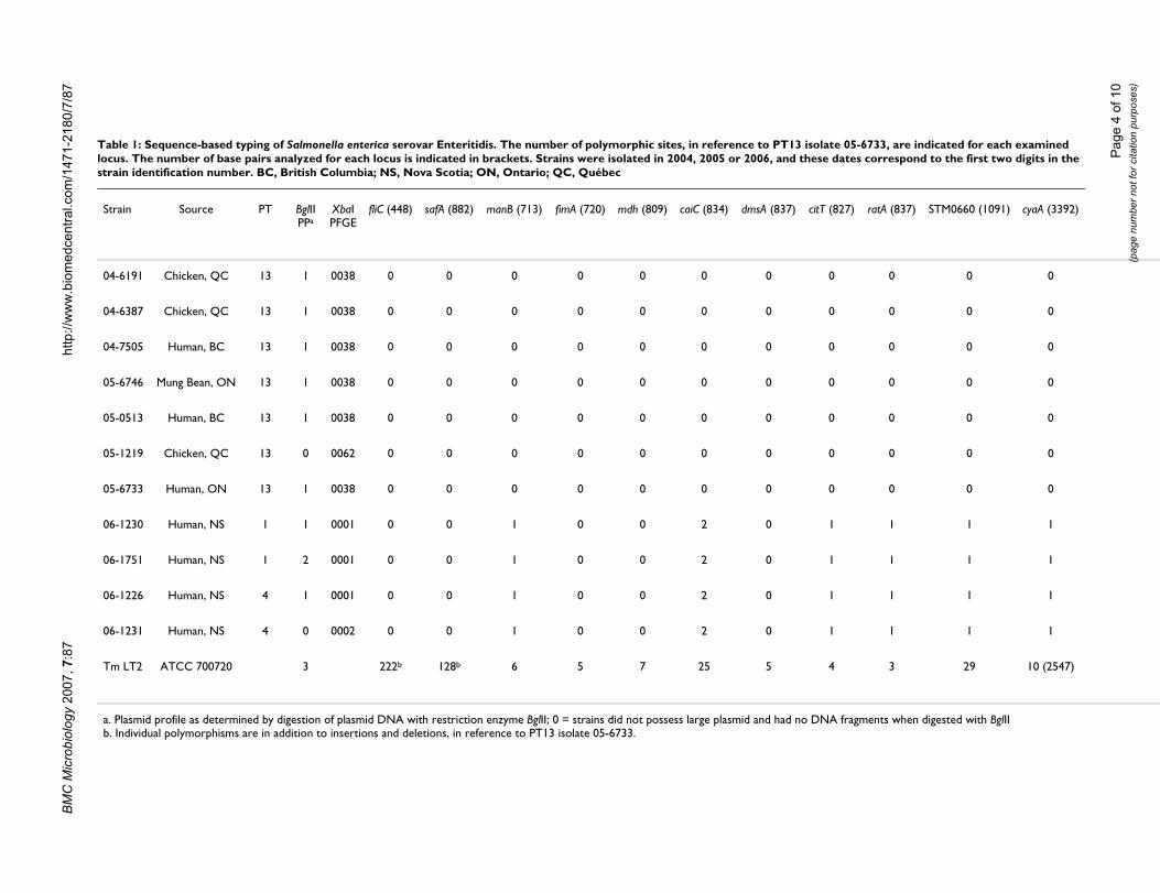
Table 1: Sequence-based typing of Salmonella enterica serovar Enteritidis. The number of polymorphic sites, in reference to PT13 isolate 05-6733, are indicated for each examined locus. The number of base pairs analyzed for each locus is indicated in brackets. Strains were isolated in 2004, 2005 or 2006, and these dates correspond to the first two digits in the strain identification number. BC, British Columbia; NS, Nova Scotia; ON, Ontario; QC, Québec
Strain Source PT BglII PPa
XbaI PFGE
fliC (448) safA (882) manB (713) fimA (720) mdh (809) caiC (834) dmsA (837) citT (827) ratA (837) STM0660 (1091) cyaA (3392)
04-6191 Chicken, QC 13 1 0038 0 0 0 0 0 0 0 0 0 0 0
04-6387 Chicken, QC 13 1 0038 0 0 0 0 0 0 0 0 0 0 0
04-7505 Human, BC 13 1 0038 0 0 0 0 0 0 0 0 0 0 0
05-6746 Mung Bean, ON 13 1 0038 0 0 0 0 0 0 0 0 0 0 0
05-0513 Human, BC 13 1 0038 0 0 0 0 0 0 0 0 0 0 0
05-1219 Chicken, QC 13 0 0062 0 0 0 0 0 0 0 0 0 0 0
05-6733 Human, ON 13 1 0038 0 0 0 0 0 0 0 0 0 0 0
06-1230 Human, NS 1 1 0001 0 0 1 0 0 2 0 1 1 1 1
06-1751 Human, NS 1 2 0001 0 0 1 0 0 2 0 1 1 1 1
06-1226 Human, NS 4 1 0001 0 0 1 0 0 2 0 1 1 1 1
06-1231 Human, NS 4 0 0002 0 0 1 0 0 2 0 1 1 1 1
Tm LT2 ATCC 700720 3 222b 128b 6 5 7 25 5 4 3 29 10 (2547)
a. Plasmid profile as determined by digestion of plasmid DNA with restriction enzyme BglII; 0 = strains did not possess large plasmid and had no DNA fragments when digested with BglIIb. Individual polymorphisms are in addition to insertions and deletions, in reference to PT13 isolate 05-6733.

BMC Microbiology 2007, 7:87 http://www.biomedcentral.com/1471-2180/7/87
and in all plasmid preparations a chromosomal DNAfragment was observed (Fig. 3). A high molecular weight(HWM) plasmid was observed for all strains except PT13strain 05-1219 and PT4 strain 06-1231. Accordingly, S.Typhimurium strain LT2 was known to harbor the 94 kbpvirulence plasmid pSLT [24] and S. Enteritidis commonlyharbors a ~60 kbp virulence plasmid [11]. Strains 05-1219 and 06-1231 that lacked the HMW virulence plas-mid had a different PFGE pattern than the other S. Enter-itidis PT13 and PT4 strains, respectively (Table 1 and Fig.1). The only strain from which low molecular weight(LMW) plasmids were isolated was PT1 strain 06-1751. To
confirm the presence of the Salmonella virulence plasmidin S. Enteritidis PT13, PCR screening for the spvC virulencedeterminant was performed. All strains encoded spvC(data not shown) except for the two S. Enteritidis strainsthat did not posses the HMW virulence plasmid.
Restriction enzyme digestion of the plasmid preparationswith BglII revealed that S. Enteritidis strains harboring theHMW plasmid had the same restriction fragment lengthpolymorphism (RFLP) pattern (Fig. 4). The S. EnteritidisPT1 strain with LMW plasmids had a different plasmidpattern due to additional DNA fragments (correspondingto the LMW plasmids) and the fragments in the patterncontributed by the HMW plasmid were otherwise identi-cal to those of strains that contained only the HMW plas-mid. Additionally, the RFLP pattern produced by the S.Typhimurium pSLT differed from the S. Enteritidis HMWplasmid.
Rep-PCRRepetitive sequence-based PCR (rep-PCR) methods tar-geting non-coding repetitive sequences are useful for bac-terial subtyping and this platform has been automatedand reproducibility improved through commercially-available reagents in conjunction with a microfluidics sta-tion [33]. Automated rep-PCR was performed on S. Enter-itidis PT13, PT1 and PT4 strains, and a small selection ofother serovars was also included to illustrate the scope ofdifferentiation at the serovar level. The rep-PCR ampliconpatterns for all S. Enteritidis PT13 strains clusteredtogether and were >98% related with no significant differ-ences (Fig. 5). The S. Enteritidis PT1 and PT4 strains pro-duced similar rep-PCR patterns as the PT13 strains (>95%relatedness) with a single amplicon being the predomi-
Plasmid profiles for S. Enteritidis strains used in this studyFigure 3Plasmid profiles for S. Enteritidis strains used in this study. Preparations were not digested with restriction endonucle-ase. Lanes 1–7: S. Enteritidis PT13 strains 04-6191, 04-6387, 04-7505, 05-6746, 05-0513, 05-1219 and 05-6733 respec-tively. Lanes 8 and 9: S. Enteritidis PT1 strains 06-1230 and 06-1751. Lanes 10 and 11: S. Enteritidis PT4 strains 06-1216 and 06-1231. Lane 12: plasmid extracted from S. Typhimu-rium LT2. Supercoiled DNA ladder molecular weights are to the left of lane 1. Arrow indicates a chromosomal DNA frag-ment.
Plasmid RFLP patterns for S. Enteritidis PT13, PT1 and PT4 strainsFigure 4Plasmid RFLP patterns for S. Enteritidis PT13, PT1 and PT4 strains. Restriction fragment patterns generated with BglII were analyzed in BioNumerics version 4 and a dendrogram was created using the UPGMA method with a coefficient of correlation, 2% optimization and 12% position tolerance. PP; RFLP plasmid pattern, PT; phage type.
Page 5 of 10(page number not for citation purposes)

BMC Microbiology 2007, 7:87 http://www.biomedcentral.com/1471-2180/7/87
nant difference. The other examined serovars werebetween 78–81% related to the S. Enteritidis strains (Fig.5).
DiscussionGenetic homogeneity has been repeatedly observed for S.Enteritidis, particularly between strains of the same phagetype [26,24,27]. Similarly, subtyping beyond phage typeutilizing methods such as ribotyping, PFGE and plasmidprofiling has generally only confirmed the clonal lineagesdiscerned by phage typing without providing any furtherdiscriminatory power [10,34,29]. This lack of subtypingcan prevent the establishment of absolute links to con-taminated agri-food sources during outbreak trace-backinvestigations. During an outbreak of S. Enteritidis PT13in Canada, the outbreak strain could not be distinguishedfrom concurrent and geographically and temporally dis-tinct isolates strains of S. Enteritidis PT13. CGH, SBT, plas-mid profiling and rep-PCR were performed in an attemptto identify genetic markers suitable for subtyping S. Enter-itidis PT13.
Clonally related phenotypic subpopulations of S. Enteri-tidis have been observed and discrete allelic variation atribosomal and metabolic loci corresponded to these
pathotypes [31,32]. In our CGH experiments, large clus-ters of genetic differences such as the absence of phageregions were discernible between S. Enteritidis and the ref-erence S. Typhimurium for which the array was designed.However, more subtle genetic differences such as allelicvariation between PT13 strains could not be confirmed,which was a finding similar to previous results [24,27].This may have resulted from a lack of S. Enteritidis specificprobes. Alternatively, identifying a reliable phenotypic orpathotype difference between PT13 strains could aid inthe identification of subtle but definitive genetic differ-ences that impact biology within a phage type.
Morales and colleagues hypothesized that genetic varia-tion between S. Enteritidis strains is better studied by per-forming DNA sequencing for SNPs than throughmicroarray-based comparative genomics [27]. Throughsequencing, it is possible to produce de novo data for eachstrain instead of attempting to discern genomic content inthe context of comparing hybridization data between twodifferent strains, or possibly different serovars, as in thisstudy (S. Typhimurium reference DNA versus S. Enteri-tidis test DNA). Although DNA sequencing provided ahigher level of detail than CGH, sequencing of the locithat were previously observed to be divergent between S.Enteritidis pathotypes and other loci commonly used forsequence-based typing (total 11,390 bp per strain) didnot identify any genomic differences between PT13strains. These results are similar to other published resultsin that discrimination between serovars was possible, butvariation within a phage type was not observed[29,26,35]. Alternatively, some loci were significantly var-iable between S. Enteritidis and S. Typhimurium (LT2),including safA (128 SNP), STM0660 (29 SNP) and caiC(25 SNP). These variable loci may prove to be useful astargets for molecular typing between Salmonella serovars.Our panel of strains was selected to include the outbreak-associated strains, concurrent sporadic clinical and agri-food strains, and for comparison to unrelated strains, iso-lates that were previously recovered in different Canadianprovinces. If the incidence of SNPs is less than 1 in 10,000bp it may be necessary to pursue a whole genomeapproach for detection of allelic variation because any onesection of the genome or group of genes could be similarbetween clonally related PT13 strains relevant to humanpublic health.
Plasmid profile analyses identified S. Enteritidis strainsthat lacked the HMW virulence plasmid or those that con-tained LMW plasmids, but these did not correspond to theoutbreak-associated strains. Carriage of the HMW viru-lence plasmid were confirmed by testing for the presenceof the spvC gene, which was previously observed on all S.Enteritidis plasmids of ~60 kbp [16]. The variation inplasmid content did influence the observed XbaI PFGE
Comparison of S. Enteritidis and select Salmonella serovars by automated rep-PCRFigure 5Comparison of S. Enteritidis and select Salmonella serovars by automated rep-PCR. The dendrogram represents the relatedness of strains based upon analyses of the amplifica-tion products using DiversiLab software. The vertical grey threshold line represents 95% similarity; PT = phage type.
Page 6 of 10(page number not for citation purposes)

BMC Microbiology 2007, 7:87 http://www.biomedcentral.com/1471-2180/7/87
patterns, which were otherwise identical. Additionally,RFLP analyses of the plasmid preparations did not iden-tify any further levels of genetic diversity within the plas-mids, but did support that both S. Enteritidis PT1 and PT4carry a similar HMW virulence plasmid as S. EnteritidisPT13. Furthermore, the usefulness of determining plas-mid carriage as a means of subtyping was limited since theoverall incidence of the HMW virulence plasmids in Can-ada was unknown and variation of this trait may be influ-enced by laboratory conditions (i.e. plasmid loss duringculturing).
Rep-PCR has the potential to represent genetic differencescontributed throughout the genome, unlike directed SBTand plasmid profiling, and targets different genetic fea-tures than PFGE. The S. Enteritidis PT13 strains examinedin this study were not differentiated with this method andwere greater than 97% related. Although there was somediscrimination between the S. Enteritidis PT1 and PT4strains and the S. Enteritidis PT13 strains (by a single banddifference), there was still greater than 95% relatednessamongst these strains. Alternatively, a select number ofother serovars were examined using automated rep-PCRand this method could differentiate between serovars,which is similar to a previous rep-PCR study [36].
ConclusionS. Enteritidis PT13 isolates in Canada were observed to beessentially genetically homogeneous after microarray-based comparisons did not identify overt differencesbetween strains and DNA sequencing of eleven loci and11,390 bp did not identify any polymorphisms. Further-more, PFGE and plasmid RFLP patterns were identical forthe majority of strains, except for some changes in plas-mid content that affected restriction patterns of strains notassociated with a large foodborne outbreak. Rep-PCRcould discriminate between serovars and phage types, butnot between outbreak-related and sporadic PT13 isolates.The apparent clonality of PT13 strains has implicationsfor the ability to subtype this pathogen during outbreaks.
MethodsBacterial strainsSalmonella enterica serovar Enteritidis strains were isolatedfrom human, food and animal sources by public healthlaboratories in Ontario, Québec, British Columbia andNova Scotia during 2004–2006 and submitted to theEnteric Diseases Program at the National MicrobiologyLaboratory. S. enterica serovar Typhimurium LT2 (ATCC700720) was included as a positive control for CGH andSBT because many of the utilized oligonucleotides weredesigned using the genomic sequence data from thisstrain (GenBank accession number NC_003197). Confir-mation of phage type was completed with bacteriophagestocks prepared at the National Microbiology Laboratory.
Antibiotic sensitivity testing was completed using theNational Antimicrobial Resistance Monitoring System(NARMS) recommended panel and the Sensititre brothdilution method.
Plasmid Isolation and AnalysisPlasmids were purified from overnight culture using aQiagen plasmid Midi kit (Qiagen, Mississauga, ON)according to manufacturer's directions with the followingmodification: plasmids were precipitated using 7.5 Mammonium acetate (Sigma-Aldrich, St. Louis, MO) incombination with isopropanol. Purified plasmid DNA(25 µl) was digested overnight at 37°C with 20 units ofBglII (New England Biolabs, Pickering, ON). Resultingplasmid fragments were separated by electrophoresis on0.7% Tris-acetate-EDTA agarose gels at 70 V for 6 hours in1 × Tris-acetate-EDTA buffer (Gibco BRL, Paisley, Scot-land). A 1 Kb Plus DNA Ladder (Invitrogen) and TrackIt™λ DNA/HindIII fragments (Invitrogen) were used asmolecular size standards. Gels were stained with ethidiumbromide (2 µg/ml) and digital images were obtainedusing a Bio-Rad Gel Doc XR (Bio-Rad).
PCR and SequencingTemplate DNA was prepared by centrifuging 1 mL of log-phase cultures grown in brain heart infusion (BHI) broth,the pellet was resuspended in 1 mL of TE buffer (Sigma,10 mM Tris-HCl, 1 mM EDTA, pH 8.0) and boiling for 15minutes. Boiled cells were pelleted and the supernatantwas removed and used as DNA template in PCR reactions.
Oligonucleotide primer sequences used for DNA amplifi-cation and/or sequencing are presented in Table 2. PCRwas performed with Platinum Taq (Invitrogen), followingthe manufacturer's directions. The thermocycling parame-ters for fimA, manB and mdh included an initial denatura-tion at 94°C for 5 minutes, 35 cycles of denaturation at94°C for 30 seconds, annealing at 50°C for 30 secondsand extension at 68°C for 45 seconds, with a final exten-sion at 68°C for 5 minutes. The annealing temperature forcaiC, dmsA, citT, ratA, STM0660, was 55°C for 40 secondsand an extension of 60 seconds. The annealing tempera-ture for safA and fliC was 50°C for 30 seconds and anextension of 30 seconds. For amplification of cyaA, theannealing temperature was 55°C for 60 seconds with anextension of 120 seconds. PCR products were purifiedusing the QIAquick PCR purification kit (Qiagen) andsequenced using the same primers that generated theseamplicons, with the exception of cyaA that required tenadditional oligonucleotides targeting internal segments toprovide complete coverage (Table 2). Sequencing was per-formed with an ABI3730 capillary electrophoresis instru-ment (Applied Biosystems, Foster City, CA). Multiplesequence alignments were completed using ClustalW [37]
Page 7 of 10(page number not for citation purposes)

BMC Microbiology 2007, 7:87 http://www.biomedcentral.com/1471-2180/7/87
and Boxshade [38], and these data were deposited in Gen-Bank with accession numbers EF113924–EF113956.
Rep-PCR typingTotal bacterial genomic DNA was isolated from selectedSE strains (Table 2) and a small selection of other Salmo-nella serovars (S. Agona strain 06-4852, S. Istanbul 05-1850, S. Newport S-444, and S. Paratyphi A 03-7699)using the Qiagen DNeasy extraction kit, following manu-facturer's instructions. The DiversiLab Salmonella DNAfingerprinting kit (Bacterial Barcodes) was used for auto-mated rep-PCR molecular typing using AmpliTaq DNApolymerase (Roche) and PCR conditions identified byBacterial Barcodes. The rep-PCR amplicons were sepa-rated using an Agilent 2100 Bioanalyzer on the DiversiLab
microfluidics DNA chip. DNA fingerprints were statisti-cally analyzed using DiversiLab software using manufac-turer's instructions.
Comparative Genomic HybridizationsDNA microarrays were constructed as previouslydescribed [39] from 4451 commercially-supplied oligo-nucleotides (Qiagen) representing coding sequences of S.Typhimurium LT2, with the custom addition of the puta-tive open reading frames from SGI1. Each oligonucleotidewas spotted in duplicate per slide. DNA was isolated fromovernight cultures grown in BHI broth at 37°C using thealkaline lysis protocol [40]. Proteins were removed usingphenol chloroform extractions in Phase lock gel tubes(Eppendorf, Westbury, NY) and purified DNA was frag-
Table 2: Oligonucleotides used in this study. For oligonucleotides used in PCR the amplicon size is listed, and for those oligonucleotides used solely for DNA sequencing no product size is listed
Oligonucleotide Target Sequence (5' to 3') Product size (bp) Reference
sfimAF fimA TCAGGGGAGAAACAGAAAACTAAT 760 [25]sfimAR TCCCCGATAGCCTCTTCC [25]smanBF manB CATAACCCGATGGACTACAACG 893 [25]smanBR ACCAGCAGCCACGGGATCAT [25]smdhF mdh GATGAAAGTCGCAGTCCTCG 849 [25]smdhR TATCCAGCATAGCGTCCAGC [25]ccmBF ccmB TCACCCTGTTTCCGTTAAGC 430 This studyccmBR AAAATCAGCACCGGGACAC This studyumuCF umuC TGACCACACTCGAGGAGATG 493 This studyumuCR CAAACGATTTCCTGCTTTGC This study
fljAF fljA GGCGAGAAGCTGAAATATGG 410 This studyfljAR ATTTACGCCTGTCGTTTTGC This study
safA2F safA TAAGAGGTGCTCTGATATATAG 959 This studysafA2R ATAGGGTAATTCTGCGGGTTG This studycaiC1F caiC GAATCGTTCGGCAGTTTAGC 874 This studycaiC1R GTTTCAGTCATACCATAAGAGG This studycitT1F citT GATGATTGTCGGTATGATCC 870 This studycitT1R GTAATATCTTTCCACGGCAC This study
dmsA1F dmsA ACTACGGTGATTACTCTTCC 869 This studydmsA1R CTGGTTAATCAAACAGTTGC This studyratA1F ratA GGCAAGATTCACAGCATTCAG 870 This studyratA1R TGGGCGGTATTTATCGTTCG This study
STM0660F STM0660 ACGATGTAGCCCATATTACG 1191 This studySTM0660R CCTGGCGAAAGTATTCATCC This study
spvC1 spvC AACTCCTTGCACAACCAAATG 230 This StudyspvC2 ACCATATCCCTGAGCACACTG This StudyCya1sF cyaA CATTGACCATCCTAACATCCTTATAGAGAG 3331 [31]Cya6sR ACTGGCGATATCACTCAATAGCGG [31]Cya1sR cyaA ATGCTGCGTAGAACCACAGTCTTC [31]Cya2sF cyaA TGGATATCTGGGTGTGCCATCAGT [31]Cya2sR cyaA GACTTTACCCGGCAACGCTTCAAA [31]Cya3sF cyaA CGATTACCGGCGTTTACACCAT [31]Cya3sR cyaA ATATCTTTCGCCAGCAGACGTG [31]Cya4sF cyaA CGCAGGATATCGGCGTACTGA [31]Cya4sR cyaA GACGGCAATTTCACCTGGTGGTT [31]Cya5sF cyaA GCGTCGGGAAGTATTAAGCCAGTT [31]Cya5sR cyaA AGGTCGACAATACCGTTGCCCTTA [31]Cya6sF cyaA TACGTCTTCCAGCACCCGTCA [31]
Page 8 of 10(page number not for citation purposes)

BMC Microbiology 2007, 7:87 http://www.biomedcentral.com/1471-2180/7/87
mented in a Nebulizer (Invitrogen) as per manufacturer'sprotocols, with the exception that DNA was nebulized for3 minutes at 10 psi to yield DNA fragments between 500and 700 bp. Probe DNA was created by labeling the frag-mented genomic DNA with Cy3- or Cy5-dCTP (Amer-sham, Baie d'Urfe, QC) by random priming using theBioprime Labeling Kit (Invitrogen) following the manu-facturer's directions. The concentration and labeling effi-ciencies were measured using the NanoDrop ND-1000spectrophotometer (NanoDrop technologies, Wilming-ton, DE). For each array a Cy3 or Cy5-labeled referenceprobe DNA (always S. Typhimurium LT2) was combinedwith a Cy3 or Cy5-labeled test PT13 strain along with 20µg of Salmon Sperm DNA and dried in a Vacufuge(Eppendorf). Each test-versus-reference comparison wasperformed in triplicate, with at least one of the slideshybridized as a dye swap. The DNA pellet was resus-pended in 10 µl of ddH20, denatured at 95°C for 5 min-utes, and snap cooled on ice for 5 minutes prior tohybridization to the array.
Slides were prepared as previously published [39] with theexception that they were pre-hybridized at 42°C in DIGEasy-Hyb buffer (Roche Diagnostic, Laval, QC) for 45minutes, followed by brief washes in dH2O and isopropa-nol. 50 µL of DIG Easy-Hyb buffer was added to eachprobe mixture and the entire volume was pipetted underan M-Series LifterSlip™ (Erie Scientific Co., Portsmouth,NH) onto the pre-hybridized slides. Slides were hybrid-ized at 42°C in a hybridization chamber (Genetix, Hamp-shire, UK). Slides were sequentially washed as follows:Buffer 1 (1× SSC and 0.2% SDS: Amersham) for 6 minutesat 56°C; Buffer 2 (0.1× SSC and 0.2% SDS) for 4 minutesat room temperature; two washes for 2 minute with Buffer3 (0.1× SSC). Slides were spun dry and scanned using theAgilent DNA microarray scanner (Agilent Technologies,Mississauga, ON).
Scanned slide images were analyzed as previously pub-lished [39] with the exception that following normaliza-tion and batch-effect removal, the data was antilog2-transformed to convert back to its original scale so that thelog2 ratio between test and reference could be measured.Log ratios were averaged across all replicates per spot foreach test versus reference comparison. GeneMaths XT soft-ware (Applied Maths, Austin, TX) was used for Hierarchi-cal clustering of the data to identify overall geneticrelatedness between examined strains, and identify spe-cific loci that were putatively absent of divergent in indi-vidual S. Enteritidis strains.
AbbreviationsComparative genomic hybridization (CGH), High molec-ular weight (HMW), Low molecular weight (LMW), Phagetype (PT), Pulsed field gel electrophoresis (PFGE), Restric-
tion fragment length polymorphism (RFLP), RFLP Plas-mid pattern (PP), Salmonella enterica serovar (S.),Sequence based typing (SBT), Single nucleotide polymor-phism (SNP).
Competing interestsThe author(s) declares that there are no competing inter-ests.
Authors' contributionsABO performed CGH, SBT and plasmid profile analysesfor this study, assisted in the study design, and drafted themanuscript. AKA and DMT participated in plasmid isola-tions and analysis, rep-PCR analysis, and assisted in draft-ing and revising the manuscript. JGB provided access towhole genomic analysis of S. Enteritidis subpopulationsand assisted in the sequence analysis. WD, AM, FJ andLKN assisted in conceiving the study, provided epidemio-logical data for strains and revised the manuscript. MWGsupervised the project and drafted and revised the manu-script. All authors read and approved the final manu-script.
AcknowledgementsWe would like to thank Michael Mulvey for providing oligonucleotides for the Salmonella microarray, Martin Wiedmann for sharing Salmonella SBT data, PulseNet Canada (Cynthia Misfeldt, Lorelee Tschetter and Jason Allen) for PFGE analysis, Melissa McCracken for technical help and Helen Tabor and Clifford Clark for helpful discussions. We also thank the Cana-dian Integrated Program from Antimicrobial Resistance Surveillance (CIPARS), British Columbia Centre for Disease Control, Nova Scotia Pub-lic Health Laboratory and the Laboratoire de Sante Publique du Québec for providing strains. Oligonucleotide synthesis, DNA sequencing and array printing was performed by the DNA Core facility and phage typing was per-formed by the Enteric Diseases Program at the National Microbiology Lab-oratory.
References1. St Louis ME, Morse DL, Potter ME, DeMelfi TM, Guzewich JJ, Tauxe
RV, Blake PA: The emergence of grade A eggs as a majorsource of Salmonella enteritidis infections. New implicationsfor the control of salmonellosis. JAMA 1988, 259:2103-2107.
2. Poppe C: Epidemiology of Salmonella enterica Serovar Enter-itidis. In Salmonella enterica Serovar Enteritidis in Humans and AnimalsEdited by: Saeed AM, Gast RK, Potter ME and Wall PG. Ames, IowaState University Press; 1999:3-18.
3. Munro DS, Girdwood RWA, Reilly WJ: Salmonella enterica Sero-var Enteritidis in Scotland. In Salmonella enterica Serovar Enteritidisin Humans and Animals Edited by: Saeed AM, Gast RK, Potter ME andWall PG. Ames, Iowa State University Press; 1999:27-31.
4. Cogan TA, Humphrey TJ: The rise and fall of Salmonella Enter-itidis in the UK. J Appl Microbiol 2003, 94 Suppl:114S-119S.
5. Tschape H, Liesegang A, Gericke B, Prager R, Rabsch W, Helmuth R:Ups and Downs of Salmonella enterica Serovar Enteritidis inGermany. In Salmonella enterica Serovar Enteritidis in Humans and Ani-mals Edited by: Saeed AM, Gast RK, Potter ME and Wall PG. Ames,Iowa State University Press; 1999:51-61.
6. Mishu B, Koehler J, Lee LA, Rodrigue D, Brenner FH, Blake P, TauxeRV: Outbreaks of Salmonella enteritidis infections in theUnited States, 1985-1991. J Infect Dis 1994, 169:547-552.
7. Angulo FJ, Swerdlow DL: Epidemiology of Human Salmonellaenterica Serovar Enteritidis Infections in the United States.In Salmonella enterica Serovar Enteritidis in Humans and Animals Edited
Page 9 of 10(page number not for citation purposes)
http://www.ncbi.nlm.nih.gov/entrez/query.fcgi?cmd=Retrieve&db=PubMed&dopt=Abstract&list_uids=3279240
http://www.ncbi.nlm.nih.gov/entrez/query.fcgi?cmd=Retrieve&db=PubMed&dopt=Abstract&list_uids=3279240
http://www.ncbi.nlm.nih.gov/entrez/query.fcgi?cmd=Retrieve&db=PubMed&dopt=Abstract&list_uids=3279240

BMC Microbiology 2007, 7:87 http://www.biomedcentral.com/1471-2180/7/87
Publish with BioMed Central and every scientist can read your work free of charge
"BioMed Central will be the most significant development for disseminating the results of biomedical research in our lifetime."
Sir Paul Nurse, Cancer Research UK
Your research papers will be:
available free of charge to the entire biomedical community
peer reviewed and published immediately upon acceptance
cited in PubMed and archived on PubMed Central
yours — you keep the copyright
Submit your manuscript here:http://www.biomedcentral.com/info/publishing_adv.asp
BioMedcentral
by: Saeed AM, Gast RK, Potter ME and Wall PG. Ames, Iowa StateUniversity Press; 1999:33-41.
8. Demczuk W, Woodward D, Ahmed R, Clark C, Tabor H, Dore K,Ciampa N, Muckle A: Laboratory surveillance data for entericpathogens in Canada: Annual summary 2002 and 2003. Win-nipeg, MB, Health Canada; 2005.
9. Ward LR, de Sa JD, Rowe B: A phage-typing scheme for Salmo-nella Enteritidis. Epidemiol Infect 1987, 99:291-294.
10. Thong KL, Ngeow YF, Altwegg M, Navaratnam P, Pang T: Molecularanalysis of Salmonella Enteritidis by pulsed-field gel electro-phoresis and ribotyping. J Clin Microbiol 1995, 33:1070-1074.
11. Pang JC, Chiu TH, Chiou CS, Schroeter A, Guerra B, Helmuth R, TsenHY: Pulsed-field gel electrophoresis, plasmid profiles andphage types for the human isolates of Salmonella entericaserovar Enteritidis obtained over 13 years in Taiwan. J ApplMicrobiol 2005, 99:1472-1483.
12. Millemann Y, Lesage MC, Chaslus-Dancla E, Lafont JP: Value of plas-mid profiling, ribotyping, and detection of IS200 for tracingavian isolates of Salmonella Typhimurium and S. Enteritidis.J Clin Microbiol 1995, 33:173-179.
13. Liebana E, Garcia-Migura L, Breslin MF, Davies RH, Woodward MJ:Diversity of strains of Salmonella enterica serotype Enteri-tidis from English poultry farms assessed by multiple geneticfingerprinting. J Clin Microbiol 2001, 39:154-161.
14. Liebana E, Garcia-Migura L, Clouting C, Clifton-Hadley FA, Breslin M,Davies RH: Molecular fingerprinting evidence of the contribu-tion of wildlife vectors in the maintenance of SalmonellaEnteritidis infection in layer farms. J Appl Microbiol 2003,94:1024-1029.
15. Liebana E, Clouting C, Garcia-Migura L, Clifton-Hadley FA, Lindsay E,Threlfall EJ, Davies RH: Multiple genetic typing of SalmonellaEnteritidis phage-types 4, 6, 7, 8 and 13a isolates from ani-mals and humans in the UK. Vet Microbiol 2004, 100:189-195.
16. Soto SM, Rodriguez I, Rodicio MR, Vila J, Mendoza MC: Detectionof virulence determinants in clinical strains of Salmonellaenterica serovar Enteritidis and mapping on macrorestric-tion profiles. J Med Microbiol 2006, 55:365-373.
17. Clark CG, Kruk TM, Bryden L, Hirvi Y, Ahmed R, Rodgers FG: Sub-typing of Salmonella enterica serotype Enteritidis strains bymanual and automated PstI-SphI ribotyping. J Clin Microbiol2003, 41:27-33.
18. Bakeri SA, Yasin RM, Koh YT, Puthucheary SD, Thong KL: Geneticdiversity of human isolates of Salmonella enterica serovarEnteritidis in Malaysia. J Appl Microbiol 2003, 95:773-780.
19. Ahmed R, Soule G, Demczuk WH, Clark C, Khakhria R, Ratnam S,Marshall S, Ng LK, Woodward DL, Johnson WM, Rodgers FG: Epi-demiologic typing of Salmonella enterica serotype Enteri-tidis in a Canada-wide outbreak of gastroenteritis due tocontaminated cheese. J Clin Microbiol 2000, 38:2403-2406.
20. Ridley AM, Threlfall EJ, Rowe B: Genotypic characterization ofSalmonella enteritidis phage types by plasmid analysis,ribotyping, and pulsed-field gel electrophoresis. J Clin Microbiol1998, 36:2314-2321.
21. Powell NG, Threlfall EJ, Chart H, Rowe B: Subdivision of Salmo-nella enteritidis PT 4 by pulsed-field gel electrophoresis:potential for epidemiological surveillance. FEMS Microbiol Lett1994, 119:193-198.
22. Landeras E, Gonzalez-Hevia MA, Alzugaray R, Mendoza MC: Epide-miological differentiation of pathogenic strains of Salmo-nella Enteritidis by ribotyping. J Clin Microbiol 1996,34:2294-2296.
23. Liebisch B, Schwarz S: Molecular typing of Salmonella entericasubsp. enterica serovar Enteritidis isolates. J Med Microbiol1996, 44:52-59.
24. Porwollik S, Santiviago CA, Cheng P, Florea L, McClelland M: Differ-ences in gene content between Salmonella enterica serovarEnteritidis isolates and comparison to closely related serov-ars Gallinarum and Dublin. J Bacteriol 2005, 187:6545-6555.
25. Tassios PT, Markogiannakis A, Vatopoulos AC, Katsanikou E,Velonakis EN, Kourea-Kremastinou J, Legakis NJ: Molecular epide-miology of antibiotic resistance of Salmonella Enteritidisduring a 7-year period in Greece. J Clin Microbiol 1997,35:1316-1321.
26. Sukhnanand S, Alcaine S, Warnick LD, Su WL, Hof J, Craver MP,McDonough P, Boor KJ, Wiedmann M: DNA sequence-based sub-
typing and evolutionary analysis of selected Salmonellaenterica serotypes. J Clin Microbiol 2005, 43:3688-3698.
27. Morales CA, Porwollik S, Frye JG, Kinde H, McClelland M, Guard-Bouldin J: Correlation of phenotype with the genotype of egg-contaminating Salmonella enterica serovar Enteritidis. ApplEnviron Microbiol 2005, 71:4388-4399.
28. Kotetishvili M, Stine OC, Kreger A, Morris JG Jr., Sulakvelidze A:Multilocus sequence typing for characterization of clinicaland environmental Salmonella strains. J Clin Microbiol 2002,40:1626-1635.
29. Hudson CR, Garcia M, Gast RK, Maurer JJ: Determination of closegenetic relatedness of the major Salmonella enteritidisphage types by pulsed-field gel electrophoresis and DNAsequence analysis of several Salmonella virulence genes.Avian Dis 2001, 45:875-886.
30. Boyd D, Peters GA, Cloeckaert A, Boumedine KS, Chaslus-Dancla E,Imberechts H, Mulvey MR: Complete nucleotide sequence of a43-kilobase genomic island associated with the multidrugresistance region of Salmonella enterica serovar Typhimu-rium DT104 and its identification in phage type DT120 andserovar Agona. J Bacteriol 2001, 183:5725-5732.
31. Morales CA, Musgrove M, Humphrey TJ, Cates C, Gast R, Guard-Bouldin J: Pathotyping of Salmonella enterica by analysis ofsingle-nucleotide polymorphisms in cyaA and flanking 23Sribosomal sequences. Environ Microbiol 2007, 9:1047-1059.
32. Comparative analyses of S. Enteritidis phenotypic subpopu-lations 2007 [http://www.ncbi.nlm.nih.gov/genomes/static/Salmonella_SNPS.html].
33. Healy M, Huong J, Bittner T, Lising M, Frye S, Raza S, Schrock R,Manry J, Renwick A, Nieto R, Woods C, Versalovic J, Lupski JR:Microbial DNA typing by automated repetitive-sequence-based PCR. J Clin Microbiol 2005, 43:199-207.
34. Olsen JE, Skov MN, Threlfall EJ, Brown DJ: Clonal lines of Salmo-nella enterica serotype Enteritidis documented by IS200-,ribo-, pulsed-field gel electrophoresis and RFLP typing. J MedMicrobiol 1994, 40:15-22.
35. Fakhr MK, Nolan LK, Logue CM: Multilocus sequence typinglacks the discriminatory ability of pulsed-field gel electro-phoresis for typing Salmonella enterica serovar Typhimu-rium. J Clin Microbiol 2005, 43:2215-2219.
36. Bennasar A, de Luna G, Cabrer B, Lalucat J: Rapid identification ofSalmonella typhimurium, S. enteritidis and S. virchow iso-lates by polymerase chain reaction based fingerprintingmethods. Int Microbiol 2000, 3:31-38.
37. ClustalW alignment tool 2007 [http://www.ebi.ac.uk/clustalw/].38. Boxshade 2007 [http://www.ch.embnet.org].39. Golding GR, Olson AB, Doublet B, Cloeckaert A, Christianson S,
Graham MR, Mulvey MR: The effect of the Salmonella genomicisland 1 on in vitro global gene expression in Salmonellaenterica serovar Typhimurium LT2. Microbes Infect 2007,9:21-27.
40. Sambrook J, Russell DW: Molecular Cloning: A Laboratory Manual Vol-ume 1. 3rd edition. Cold Spring Harbour, Cold Spring Harbour Labo-ratory Press; 2001:1.32-1.34.
Page 10 of 10(page number not for citation purposes)
http://www.ncbi.nlm.nih.gov/entrez/query.fcgi?cmd=Retrieve&db=PubMed&dopt=Abstract&list_uids=3315705
http://www.ncbi.nlm.nih.gov/entrez/query.fcgi?cmd=Retrieve&db=PubMed&dopt=Abstract&list_uids=3315705
http://www.ncbi.nlm.nih.gov/entrez/query.fcgi?cmd=Retrieve&db=PubMed&dopt=Abstract&list_uids=7615707
http://www.ncbi.nlm.nih.gov/entrez/query.fcgi?cmd=Retrieve&db=PubMed&dopt=Abstract&list_uids=7615707
http://www.ncbi.nlm.nih.gov/entrez/query.fcgi?cmd=Retrieve&db=PubMed&dopt=Abstract&list_uids=7615707
http://www.ncbi.nlm.nih.gov/entrez/query.fcgi?cmd=Retrieve&db=PubMed&dopt=Abstract&list_uids=7699037
http://www.ncbi.nlm.nih.gov/entrez/query.fcgi?cmd=Retrieve&db=PubMed&dopt=Abstract&list_uids=7699037
http://www.ncbi.nlm.nih.gov/entrez/query.fcgi?cmd=Retrieve&db=PubMed&dopt=Abstract&list_uids=9666012
http://www.ncbi.nlm.nih.gov/entrez/query.fcgi?cmd=Retrieve&db=PubMed&dopt=Abstract&list_uids=9666012
http://www.ncbi.nlm.nih.gov/entrez/query.fcgi?cmd=Retrieve&db=PubMed&dopt=Abstract&list_uids=9666012
http://www.ncbi.nlm.nih.gov/entrez/query.fcgi?cmd=Retrieve&db=PubMed&dopt=Abstract&list_uids=8039660
http://www.ncbi.nlm.nih.gov/entrez/query.fcgi?cmd=Retrieve&db=PubMed&dopt=Abstract&list_uids=8039660
http://www.ncbi.nlm.nih.gov/entrez/query.fcgi?cmd=Retrieve&db=PubMed&dopt=Abstract&list_uids=8039660
http://www.ncbi.nlm.nih.gov/entrez/query.fcgi?cmd=Retrieve&db=PubMed&dopt=Abstract&list_uids=8862603
http://www.ncbi.nlm.nih.gov/entrez/query.fcgi?cmd=Retrieve&db=PubMed&dopt=Abstract&list_uids=8862603
http://www.ncbi.nlm.nih.gov/entrez/query.fcgi?cmd=Retrieve&db=PubMed&dopt=Abstract&list_uids=8862603
http://www.ncbi.nlm.nih.gov/entrez/query.fcgi?cmd=Retrieve&db=PubMed&dopt=Abstract&list_uids=8544212
http://www.ncbi.nlm.nih.gov/entrez/query.fcgi?cmd=Retrieve&db=PubMed&dopt=Abstract&list_uids=8544212
http://www.ncbi.nlm.nih.gov/entrez/query.fcgi?cmd=Retrieve&db=PubMed&dopt=Abstract&list_uids=9163436
http://www.ncbi.nlm.nih.gov/entrez/query.fcgi?cmd=Retrieve&db=PubMed&dopt=Abstract&list_uids=9163436
http://www.ncbi.nlm.nih.gov/entrez/query.fcgi?cmd=Retrieve&db=PubMed&dopt=Abstract&list_uids=9163436
http://www.ncbi.nlm.nih.gov/entrez/query.fcgi?cmd=Retrieve&db=PubMed&dopt=Abstract&list_uids=7904649
http://www.ncbi.nlm.nih.gov/entrez/query.fcgi?cmd=Retrieve&db=PubMed&dopt=Abstract&list_uids=7904649