Comparative genomics of closely related Salmonella enterica serovar Typhi strains reveals genome...
20
RESEARCH ARTICLE Open Access Comparative genomics of closely related Salmonella enterica serovar Typhi strains reveals genome dynamics and the acquisition of novel pathogenic elements Kien-Pong Yap 1,2 , Han Ming Gan 4 , Cindy Shuan Ju Teh 2,3 , Lay Ching Chai 1,2 and Kwai Lin Thong 1,2* Abstract Background: Typhoid fever is an infectious disease of global importance that is caused by Salmonella enterica subsp. enterica serovar Typhi (S. Typhi). This disease causes an estimated 200,000 deaths per year and remains a serious global health threat. S. Typhi is strictly a human pathogen, and some recovered individuals become long-term carriers who continue to shed the bacteria in their faeces, thus becoming main reservoirs of infection. Results: A comparative genomics analysis combined with a phylogenomic analysis revealed that the strains from the outbreak and carrier were closely related with microvariations and possibly derived from a common ancestor. Additionally, the comparative genomics analysis with all of the other completely sequenced S. Typhi genomes revealed that strains BL196 and CR0044 exhibit unusual genomic variations despite S. Typhi being generally regarded as highly clonal. The two genomes shared distinct chromosomal architectures and uncommon genome features; notably, the presence of a ~10 kb novel genomic island containing uncharacterised virulence-related genes, and zot in particular. Variations were also detected in the T6SS system and genes that were related to SPI-10, insertion sequences, CRISPRs and nsSNPs among the studied genomes. Interestingly, the carrier strain CR0044 harboured far more genetic polymorphisms (83% mutant nsSNPs) compared with the closely related BL196 outbreak strain. Notably, the two highly related virulence-determinant genes, rpoS and tviE, were mutated in strains BL196 and CR0044, respectively, which revealed that the mutation in rpoS is stabilising, while that in tviE is destabilising. These microvariations provide novel insight into the optimisation of genes by the pathogens. However, the sporadic strain was found to be far more conserved compared with the others. Conclusions: The uncommon genomic variations in the two closely related BL196 and CR0044 strains suggests that S. Typhi is more diverse than previously thought. Our study has demonstrated that the pathogen is continually acquiring new genes through horizontal gene transfer in the process of host adaptation, providing novel insight into its unusual genomic dynamics. The understanding of these strains and virulence factors, and particularly the strain that is associated with the large outbreak and the less studied asymptomatic Typhi carrier in the population, will have important impact on disease control. Keywords: S. Typhi, Typhoid, Genomes, Comparative genomics, Pathogen, zot, SPI, T6SS, Phylogenetic, Enterobacteriaceae, Sequence, Evolution, Protein, Strain, Variation, Virulence, Infection, Protein modelling * Correspondence: [email protected] 1 Institute of Biological Sciences, Faculty of Science, University of Malaya, 50603 Kuala Lumpur, Malaysia 2 Laboratory of Biomedical Science and Molecular Microbiology, Institute of Graduate Studies, University of Malaya, 50603 Kuala Lumpur, Malaysia Full list of author information is available at the end of the article © 2014 Yap et al.; licensee BioMed Central Ltd. This is an Open Access article distributed under the terms of the Creative Commons Attribution License (http://creativecommons.org/licenses/by/2.0), which permits unrestricted use, distribution, and reproduction in any medium, provided the original work is properly credited. The Creative Commons Public Domain Dedication waiver (http://creativecommons.org/publicdomain/zero/1.0/) applies to the data made available in this article, unless otherwise stated. Yap et al. BMC Genomics 2014, 15:1007 http://www.biomedcentral.com/1471-2164/15/1007
Transcript of Comparative genomics of closely related Salmonella enterica serovar Typhi strains reveals genome...

Yap et al. BMC Genomics 2014, 15:1007http://www.biomedcentral.com/1471-2164/15/1007
RESEARCH ARTICLE Open Access
Comparative genomics of closely relatedSalmonella enterica serovar Typhi strains revealsgenome dynamics and the acquisition of novelpathogenic elementsKien-Pong Yap1,2, Han Ming Gan4, Cindy Shuan Ju Teh2,3, Lay Ching Chai1,2 and Kwai Lin Thong1,2*
Abstract
Background: Typhoid fever is an infectious disease of global importance that is caused by Salmonella entericasubsp. enterica serovar Typhi (S. Typhi). This disease causes an estimated 200,000 deaths per year and remains aserious global health threat. S. Typhi is strictly a human pathogen, and some recovered individuals become long-termcarriers who continue to shed the bacteria in their faeces, thus becoming main reservoirs of infection.
Results: A comparative genomics analysis combined with a phylogenomic analysis revealed that the strains from theoutbreak and carrier were closely related with microvariations and possibly derived from a common ancestor.Additionally, the comparative genomics analysis with all of the other completely sequenced S. Typhi genomes revealedthat strains BL196 and CR0044 exhibit unusual genomic variations despite S. Typhi being generally regarded as highlyclonal. The two genomes shared distinct chromosomal architectures and uncommon genome features; notably,the presence of a ~10 kb novel genomic island containing uncharacterised virulence-related genes, and zot in particular.Variations were also detected in the T6SS system and genes that were related to SPI-10, insertion sequences, CRISPRsand nsSNPs among the studied genomes. Interestingly, the carrier strain CR0044 harboured far more geneticpolymorphisms (83% mutant nsSNPs) compared with the closely related BL196 outbreak strain. Notably, the twohighly related virulence-determinant genes, rpoS and tviE, were mutated in strains BL196 and CR0044, respectively,which revealed that the mutation in rpoS is stabilising, while that in tviE is destabilising. These microvariations providenovel insight into the optimisation of genes by the pathogens. However, the sporadic strain was found to be far moreconserved compared with the others.
Conclusions: The uncommon genomic variations in the two closely related BL196 and CR0044 strains suggests that S.Typhi is more diverse than previously thought. Our study has demonstrated that the pathogen is continually acquiringnew genes through horizontal gene transfer in the process of host adaptation, providing novel insight into its unusualgenomic dynamics. The understanding of these strains and virulence factors, and particularly the strain that isassociated with the large outbreak and the less studied asymptomatic Typhi carrier in the population, will haveimportant impact on disease control.
Keywords: S. Typhi, Typhoid, Genomes, Comparative genomics, Pathogen, zot, SPI, T6SS, Phylogenetic,Enterobacteriaceae, Sequence, Evolution, Protein, Strain, Variation, Virulence, Infection, Protein modelling
* Correspondence: [email protected] of Biological Sciences, Faculty of Science, University of Malaya,50603 Kuala Lumpur, Malaysia2Laboratory of Biomedical Science and Molecular Microbiology, Institute ofGraduate Studies, University of Malaya, 50603 Kuala Lumpur, MalaysiaFull list of author information is available at the end of the article
© 2014 Yap et al.; licensee BioMed Central Ltd. This is an Open Access article distributed under the terms of the CreativeCommons Attribution License (http://creativecommons.org/licenses/by/2.0), which permits unrestricted use, distribution, andreproduction in any medium, provided the original work is properly credited. The Creative Commons Public DomainDedication waiver (http://creativecommons.org/publicdomain/zero/1.0/) applies to the data made available in this article,unless otherwise stated.

Yap et al. BMC Genomics 2014, 15:1007 Page 2 of 20http://www.biomedcentral.com/1471-2164/15/1007
BackgroundTyphoid fever is a human systemic infection that is causedby Salmonella enterica subsp. enterica serovar Typhi(S. Typhi). This human-restricted and highly adaptedpathogen is transmitted via the oral-faecal route. S.Typhi is responsible for 21.7 million infections and resultsin approximately 217,000 deaths worldwide annually [1].The disease primarily causes acute systemic infection withlife-threatening complications, and the recovering patientmay develop into a chronic carrier state [2].Typhoid is endemic, with periodic outbreaks and spor-
adic cases occurring in developing countries, particularlyin southeast Asia, south central Asia, Latin America andsouthern Africa where sanitary conditions are poor [1].Among the 13 states of Malaysia, Kelantan has a signifi-cantly higher incidence of typhoid fever [3]. A large ty-phoid outbreak occurred in Kelantan state, which resultedin 735 cases of infection and two deaths in a short periodof 3 months (from April to June of 2005) [3]. Previously,pulsed-field gel electrophoresis (PFGE) revealed close gen-etic relatedness between a S. Typhi strain that was isolatedfrom an asymptomatic carrier in 2007 and a strain thatoriginated from a patient during the 2005 outbreak inKelantan (unpublished data).Human carriers are the main reservoirs of S. Typhi
transmission, but the genetic basis, and the underlyingmechanisms in particular, are unclear [4]. It has beensuggested that a carrier strain will likely lack gene acqui-sition capabilities and have little fitness advantages com-pared with those strains causing symptomatic infectionsbecause the human reservoir is small and physiologicallyisolated [4,5]. Therefore, it is of great interest to know asto what extent these closely related strains differed orshared in its genomic contents despite being isolated fromthese two distinct epidemiological settings. In 2008, a S.Typhi strain was isolated from a sporadic case in KualaLumpur. This genome has been sequenced earlier [6], andPFGE analyses showed that this strain is more distantly re-lated to the outbreak and carrier strains, but the epidemio-logical link is unknown (unpublished data).Recent whole-genome sequencing of S. Typhi has dem-
onstrated that the pathogen shows limited genetic vari-ation with little evidence of purifying selection, antigenicvariation or recombination between isolates [4,7]. Thisclonal pathogen, however, is associated with varying de-grees of disease severity in different regions [8]. PreviousPFGE studies have also demonstrated genome size varia-tions and distinct PFGE patterns in relation with fatal andnon-fatal typhoid cases [9,10]. Although the health condi-tions of the host cannot be completely ruled out, variousreports have suggested that the gain and loss of genesthrough mutations and gene transfers that have occurredindependently in different lineages have markedly contrib-uted to the varying pathogenic potentials [4,11]. However,
these important factors are poorly understood becausethere is limited genomic information for S. Typhi, particu-larly involving strains that are associated with diverse epi-demiological settings. Its genomic heterogeneity is likelydue to the adaptation of the pathogen to the host and itsexposure to mobile elements, such as bacteriophages [12].Organisms having common core genomes could differ intheir dispensable (strain-specific) genes, reflecting theirunique physiological and virulence properties [13,14]. Al-though not all genetic variations are essential for adapta-tion, some dispensable genes are believed to be responsiblefor conferring fitness advantages to the pathogen to thrivein its host. Horizontal gene transfer is also thought to bethe predominant force in bacterial evolution, which con-tributes to novel gene acquisition. The acquired genesprovide new characteristics, which either aid in host adap-tation and persistence or enhance virulence capabilities[12-15]. A previous study on the pan-genome of Salmon-ella enterica revealed that the pan-genome (total knowngenomic content) of all strains will continue to increase asnew genomes are sequenced [16]. With the availability ofrobust next-generation sequencing technologies, highquality whole genome sequences can be generated andanalysed, which will be especially useful for capturing finevariations among highly conserved S. Typhi strains. Mul-tiple whole genome sequence comparisons of closely re-lated strains will not only lead to the better understandingof their relationships but also provide novel insights intothe functional roles of strain-specific genes.In this study, we performed detailed and comprehen-
sive comparative functional analyses of three previouslysequenced genomes of S. Typhi strains that were isolatedfrom typhoid patients during a large outbreak in 2005, asporadic case in 2008 and an asymptomatic carrier fromMalaysia, where typhoid is endemic. These Malaysian S.Typhi strains were compared with previously publishedS. Typhi genomes with the following aims: 1) to deter-mine and describe the genomes signatures and con-served and unique regions of the strains that were beingstudied; 2) to elucidate the phylogeny and genetic re-latedness of these Malaysian strains compared with 17other published strains using phylogenomic analysis; 3)to compare those strains that have been associated withvarious epidemiological settings (outbreak, carrier andsporadic cases), and particularly the regions of plasticitythat may contribute to the varying pathogenic potentials;4) to identify potential novel pathogenic factors that areharboured by the analysed strains; 5) to provide insightinto the possible differential functionalities of the genes,focusing mainly on virulence- and persistence-relatedgenes based on non-synonymous SNPs of closely relatedstrains and particularly on carrier strains to gain insightinto the persistence of the carrier state; and 6) to under-stand how the potential nsSNPs affect protein structures
Yap et al. BMC Genomics 2014, 15:1007 Page 3 of 20http://www.biomedcentral.com/1471-2164/15/1007
and functions. The data that are generated will be usefulfor the profiling of strains, marker development and theincreased understanding of outbreak, sporadic and lessstudied asymptomatic typhoid carriage infection.
Results and discussionGeneral genome signatures of S. Typhi in association withoutbreak, sporadic case and carrierThe genomes of the three previously sequenced MalaysianS. Typhi strains from different sources were compared toidentify potential genomic features that may help to eluci-date the different disease outcomes. One strain was iso-lated during the largest outbreak in the country, one froma carrier (food handler) during typhoid surveillance fol-lowing the outbreak, representing the carrier state, andone from a sporadic case in the metropolitan area of KualaLumpur, which has a relatively lower incidence of typhoid.However, because of the limited numbers of strains stud-ied and absence of information regarding the pan-genomeof the S.Typhi population, the genetic differences observedshould be taken with caution. The aim of the detailedcomparative analysis was to provide a better understand-ing and insights into the unusual genome dynamics of S.Typhi, a highly clonal organism.Previous genome sequencing analyses have generated
high-quality assemblies with an average genome coverageof 100× for the 3 Malaysian S. Typhi genomes, includingBL196 (an outbreak strain that was isolated from a bloodsample; Genbank accession number AJGK00000000.1)[17], CR0044 (a strain that was isolated from a stool sampleof a carrier; Genbank accession number AKZO00000000.1)[18] and ST0208 (a sporadic strain that was isolated froma stool sample of a typhoid case; Genbank accession num-ber AJXA00000000.1) [6]. The approximate predictedgenome sizes and average guanine-plus-cytosine (G +C)contents of all 3 genomes ranged from 4.7 Mb to 4.8 Mband 52.0% to 53.2%, respectively (Additional file 1). Thesegenomes form a single and circular chromosome with noplasmids detected. An in silico multi-locus sequence typ-ing (MLST) analysis classified both BL196 and CR0044 asST 1 and ST0208 as ST 2, which are the main sequencetypes that have been associated with the worldwide distri-bution of S. Typhi out of the 4 STs that have been identi-fied to date [19]. The predicted coding sequences (CDSs)of the genomes based on RAST subsystem-based annota-tions varied from 4,875 to 4,890 with an average codingpercentage of 86.0%. The average sizes of the CDSs weresimilar (ranging from 810 bp to 875 bp), indicating thatthe size differences among the genomes are largely attribut-able to a number of CDSs and intergenic regions. Approxi-mately 12% of the CDSs were annotated as uncharacterisedproteins (Additional file 1). Some of these genes (4.2%)were observed to vary from one strain to another. These“dispensable” genomes carried genes that were present in
one or more strains and could even be unique to a singlestrain [20], indicating a possible open pan-genome of S.Typhi. In general, the chromosomes of the three assem-bled genomes exhibited overall structural conservationand colinearity with each other as evidenced by the hom-ologous and conserved regions that were shared and thevery small strain-specific regions (Figure 1), which mayharbour genes that are relevant to the specific adaptationsand fitness advantages of each of the strains. These re-gions most likely represent DNA that was acquired duringevents of HGT that may provide the strains with greatermetabolic versatility or even virulence capabilities, as willbe further discussed in the plasticity section.
Comparative genomics of S. TyphiGenomic comparisons were performed on the threeMalaysian S. Typhi strains and the three completed S.Typhi genomes (the only full genomes available at theNCBI database to date), CT18 (Genbank accession num-ber AL513382) [19], Ty2 (Genbank accession numberAE014613) [21] and P-stx-12 (Genbank accession numberCP003278) [22]. The comparisons of the studied strainswith the reference genomes allowed for the elucidationof novel and additional genes that are carried by theMalaysian S. Typhi genomes that may be of significance.We have analysed the shared and unique genes of all sixgenomes to determine their distinct virulence and patho-genic features. As expected, the six genomes exhibitedhigh similarities and syntenies with each other with lim-ited evidence of genomic rearrangements, which collect-ively indicate stable genomic structures. The majority ofthe ORFs from the compared genomes were part of a con-served genomic core, in which 4532 ORFs were sharedamong all of the genomes. These shared ORFs provideclear evidence of conservation among the genomes ofthe S. Typhi strains. The rest of the unshared ORFs oraccessory genes are present in one or more strains,which represent the salient differences in the genomes.Most of these ORFs (4.2% to 4.9%) that were harbouredby each genome were annotated as hypothetical pro-teins (50% to 75%). Our extended homology analysis hasshown that the remaining unshared ORFs (25% to 50%)were likely to encode for functional proteins from di-verse categories, including virulence-related proteins,secretory proteins, conserved domain proteins, trans-porter proteins and phage proteins among others. Theseconsiderable portions of the genomes could provide im-portant functional clues for understanding the virulenceand persistence of the pathogen more clearly, anticipat-ing the need for extensive future studies focusing ontheir possible roles in bacterial pathogenesis. However,the numbers of shared and unshared ORFs may havebeen underestimated because the genomes were incom-plete. Among the shared genes that were found between
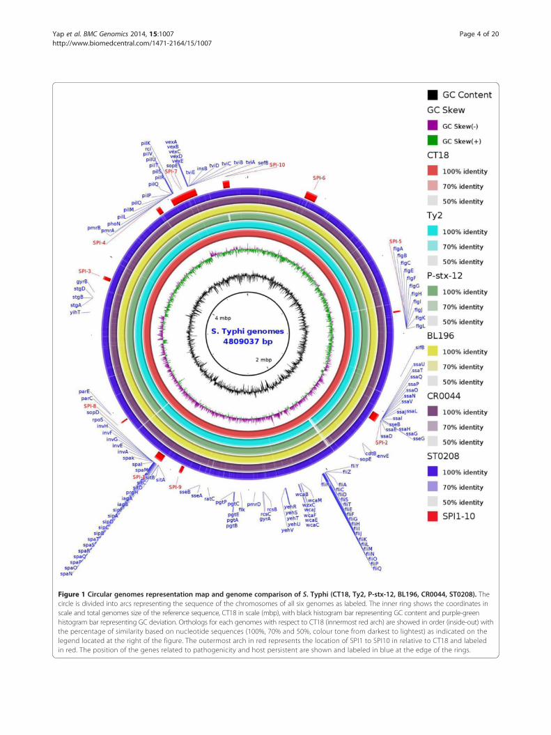
Figure 1 Circular genomes representation map and genome comparison of S. Typhi (CT18, Ty2, P-stx-12, BL196, CR0044, ST0208). Thecircle is divided into arcs representing the sequence of the chromosomes of all six genomes as labeled. The inner ring shows the coordinates inscale and total genomes size of the reference sequence, CT18 in scale (mbp), with black histogram bar representing GC content and purple-greenhistogram bar representing GC deviation. Orthologs for each genomes with respect to CT18 (innermost red arch) are showed in order (inside-out) withthe percentage of similarity based on nucleotide sequences (100%, 70% and 50%, colour tone from darkest to lightest) as indicated on thelegend located at the right of the figure. The outermost arch in red represents the location of SPI1 to SPI10 in relative to CT18 and labeledin red. The position of the genes related to pathogenicity and host persistent are shown and labeled in blue at the edge of the rings.
Yap et al. BMC Genomics 2014, 15:1007 Page 4 of 20http://www.biomedcentral.com/1471-2164/15/1007
Yap et al. BMC Genomics 2014, 15:1007 Page 5 of 20http://www.biomedcentral.com/1471-2164/15/1007
strains BL196 and CR0044, uncommon ORFs that encodedfor the VI Icm-F secretion protein, Icm-F-related proteinand type VI secretion protein EvpB were identified whoseproducts are related to the type VI secretion system. Thegenes shared 99% similarity with the type VI secretionprotein of Salmonella Typhimurium strain D23580 [23]and were only found in BL196 and CR0044. This proteinwas recently recognised as one of the main virulence de-terminants in Burkholderia pseudomallei, Legionella pneu-mophila and Vibrio cholerae, but its function in S. Typhiremains to be elucidated [24,25]. T6SS genes are believedto be involved in either structural components of thesecretory apparatus, secretory products or assisting withprotein translocation; for example, providing the energy topush substrates through the channel of the apparatus [26].These genes are also proposed to be involved in surfacereorganisation, enhancing adherence to epithelial cells,intracellular multiplication and human macrophage killing[26-28]. Other T6SS clusters were found intact as in refer-ence genomes. The high similarities of the genetic con-tents of BL196 and CR0044 with minor variations, andparticularly the presence of unique accessory genes (inaddition to SNPs, which are discussed in another section),are in agreement with the PFGE pulsotype data, which re-vealed that both strains are genetically similar, showing adifference of only one band (Additional file 2).The chromosome of Salmonella enterica is commonly
integrated with a large portion of horizontally acquiredDNA apart from its core, which are termed the Salmon-ella pathogenicity islands (SPIs) [29]. These acquiredSPIs have led to divergence and host restriction similarto those in S. Typhi. The identification of conserved SPIsand their variations have important implications in awide range of microbiological applications, such as anti-gen and marker discovery and the identification of es-sential genes and their respective traits. In this study, wehave annotated all SPIs (SPI-1 to SPI-10) and its geneticvariant of S. Typhi in the genome, which is characterisedby its deviated GC content, flanking by tRNA genes andthe presence of phages, integrases, recombinases andgenes that are related to DNA integration. Although allthe SPIs [1-10] (Figure 1) were detected in the genomes,there were marked variations. The presence of a largenumber of transposition-related genes in these SPIs sug-gests that the sites may be actively involved in the integra-tion and transposition of genetic elements, which drivegenetic variation. Interestingly, our comparative analysisrevealed that the carrier strain P-stx-12 lacks a ~10 kbprpZ cluster and adjacent gene clusters harbouring 14ORF with a deviated GC skew of 49.2 % at SPI-10 but re-mains fully intact in our carrier strain (Figure 1). Previ-ously, a prpZ cluster deletion study showed that themutant has a significantly lower survival rate comparedwith the parental strain, which may be due to a signalling
pathway that controls the long-term survival of S. Typhiin host cells, and particularly, the survival in human mac-rophages [30]. In fact, our results support this study withthe fine-tuned postulation that the deletion led to reducedvirulence that enabled the carrier strain to coexist withthe host; for example, in the tissue of gall bladder. Thispossibly explains why long-term survival in the macro-phage is no longer necessary, which is presumably becausethe pathogens have colonised and persisted in other cellsof the host during adaptation. However, the deletion wasnot detected in our carrier strain, suggesting that thegenes may not be the only factors that are relevant to acarrier state. Furthermore, the region is flanked by mul-tiple transposases, integrases, ligases and uncharacterisedproteins, which are known to be involved in transposition.Additionally, genes coding for the DNA mismatch repairprotein mutC and transposase were identified both up-stream and downstream of the cluster, suggesting that theregion could have possibly been acquired earlier duringhorizontal gene transfer. The presence of a DNA mis-match protein gene has been previously implicated to beinvolved in modulating recombination events by incorpor-ating or inhibiting the transfer of mobile genetic elements[31]. The deletion of the gene cluster and the presence ofa large number of genes that are related to transpositionindicate that SPI-10 may be unstable and prone to exci-sion similar to the precise excision of the crucial SPI-7,which has been recently reported in S. Typhi [32], suggest-ing that gene deletion may be important to the host adap-tation of this organism, although independent acquisitionor gene gain by other strains cannot be completely ruledout. These findings expand upon previous studies, whichhave reported that other SPIs are relatively stable in thegenome [4], highlighting the importance of a future evalu-ation of the stability of the other SPIs. Apart from theseremarkable differences, the comparison of the two carrierstrains, CR0044 and P-stx-12, revealed that CR0044 car-ries several additional genes that were not identified in P-stx-12 that encode for unknown functions and phages thatare present in the phage region. Almost all of the potentialmajor virulence and persistence-associated genes have ho-mologues in P-stx-12, suggesting that they are not specif-ically associated with the unique persistence of the carrierstrains but are common to S. Typhi. The genomic struc-ture of the sporadic strain ST0208 is more conserved incomparison and has relatively fewer dispensable ORFs,which are mainly genes that code for hypothetical proteinsand phages, indicating the conservation of large numbersof genes, which is essential for strict host adaptation andvirulence optimisation.
Phylogenomics of S. Typhi revealed shared common ancestryWe determined a core genome-based phylogeny by mapping20 query genomic sequences against CT18 into a single

Yap et al. BMC Genomics 2014, 15:1007 Page 6 of 20http://www.biomedcentral.com/1471-2164/15/1007
non-redundant alignment of 3,495,681 bp (Figure 2)(see Methods). The phylogenomic tree showed that theoutbreak strain BL196 and carrier strain CR0044 wereclosely related and could be differentiated by only 50SNPs. These data are in agreement with our PFGE re-sults that showed that both strains are highly related(Additional file 2). The observed close phylogenetic re-lationship between these two strains is consistent with
4.0E-4
E98-3139
Ty2
UJ308A
BL196
E01-6750
E98-0664
CR0044
UJ816A
P-stx-12
AG3
100%
100%
97%
100%
100%
99.8%
100%
100%
93.8%
Figure 2 Phylogenomic tree inferred by approximately-maximum-likealignments were generated by mapping genome sequences of the 20 gloanalysis using RealPhy [78]. Phylogenetic tree (unrooted) was inferred via aStrains studied were labelled in blue. Bootstrap support values shown at ea
our earlier speculation that the large outbreak that oc-curred in 2005 shared a common ancestor strain withthe typhoid carrier that may have been circulating for along period in the country. It is challenging to deter-mine how these two strains are related, consideringtheir short-term evolutionary relationship. However,three evolutionary postulations are possible. First, thecarrier strain may have been derived from the outbreak
E00-7866
J186
STH2370
E02-1180
ST0208
CR0063
M223
CT18
E98-2068
404ty
100%
100%
95.7%
100%
100%
100%
88%
99.9%
lihood method from the aligned core genomes. Multiple genomesbal S. Typhi strains against CT18 at all sites relevant for phylogenomicpproximately-maximum-likelihood method using FastTreeMP [79].ch node.

Yap et al. BMC Genomics 2014, 15:1007 Page 7 of 20http://www.biomedcentral.com/1471-2164/15/1007
strain. Second, the carrier strain may have long existedin a carrier who served as a reservoir and the source ofthe outbreak. Finally, both of these strains may have di-verged independently from a common ancestor, whichwas possibly harboured in a long existed carrier to giverise to two independent cases, considering the geo-graphical proximities. Notably, these two highly relatedstrains with unique gene repertoires clustered with 4 ep-idemiologically and geographically unrelated strainsfrom India (P-stx-12), Russia (TY2), Vietnam (AG3) andSenegal in west Africa (E01-6750) (Figure 2). Interestingly,all of these strains, including BL196, CR0044, P-stx-12,Ty2 and E01-6750, were subtyped as ST 1 (we couldnot establish the ST from the genomic sequence ofAG3). Conversely, the Malaysian sporadic S. Typhi strainST0208 clustered closely with the multidrug-resistantstrain CT18 from Vietnam together with other geograph-ically related strains from Indonesia (404ty and J185) andBangladesh (E98-2068) (Figure 2), which were all subtypedas ST 2, suggesting the possible movement of clonally re-lated strains among the southeast Asian countries. Suchclose genetic relatedness that is based on macrorestrictionand SNP typing analyses has been previously reported[33,34]. The rest of the genomes were clustered as ST 2.From the analysis, we found no temporal or geographicalsignals, but the sequence types was highly correlated withthe phylogenomic clustering. Apparently, the cluster sub-typed as ST 2 could be further differentiated into twoclusters according to the phylogenomic analysis but waslimited in the current MLST scheme for S. Typhi. Theseresults indicate that the phylogenomic analysis has muchbetter resolution power compared with MLST in separat-ing highly clonal strains in S. Typhi. This information isessential for devising a better set of MLST alleles for im-proved molecular typing. These data further support thewidespread distribution of ST 1 and ST 2 as the major ge-notypes that occur worldwide, although rarely, ST 3 andST 8 have also been isolated in a previous study [19]. Itis important to note that the two carriers, CR0044 andP-stx-12, belonged to two different subtypes, reflect thenon-universality of the genotypes relevant for carrierstate transformation.
Genome plasticity (insertion sequences, phages andCRISPRs)Our analyses of the relationships among the strains andtheir genomic variations are further supported and ex-tended to the study of the genome plasticity of S. Typhi. Inmany organisms, genomic plasticity is commonly observed,but S. Typhi has generally demonstrated few variationscompared with other Salmonella spp. [35]. However, inour study, we detected considerable genetic variations, pre-dominantly involving IS elements, phages and CRISPRs.Marked variations in the numbers and types of IS elements
were detected. IS200 (200, 200 F, 200C, 200G and 200H)and IS1541 (1541A, 1541B, 1541C and 1541D) were bothabundant in the three Malaysian S. Typhi genomes. TheIS elements, such as IS200, have been widely used as mo-lecular markers for subtyping due to their genome-widedistributions and high levels of diversity, but their rolesin modulating the gene expression in S. Typhi have yet tobe clarified [36]. Recent studies on enterohaemorrhagicEscherichia coli O157 have shown that the presence of IScould play a role in the gene inactivation and immobilisa-tion of incoming phages and plasmids, leading to the di-versification and evolution of the bacterial genome [37]. ISelements have also been shown to affect the expression ofneighbouring genes and induce genomic rearrangements(deletions, inversion and duplications) [38]. However, littleis understood about their roles in modulating virulenceand gene expression in S. Typhi. The variations that weredetected may provide clues on how these differences affectthe virulence and fitness strategies of the pathogens.The S. Typhi strains BL196, CR0044 and ST0208 carry
eight, seven and eight phages, respectively. We have iden-tified substantial phage variations among the S. Typhi ge-nomes. One of the differentiating features was a distinctset of prophages that were harboured by both BL196 andCR0044, which rendered them less unique compared withST0208 with the exception of a few ORFs that encodedfor phages and hypothetical proteins. Interestingly, thephages that were carried by ST0208 had relatively shorterin lengths (in bp) compared with the phages that wereidentified in the other strains. As expected, both of theclosely related strains (BL196 and CR0044) had highlysimilar phage contents and carried an additional intactSalmonella phage RE-2010 with uncommon ORFs, whichmainly contained genes coding for hypothetical proteins,phage proteins, prophage-like proteins, repressor proteins,excisionases, terminases and integrases. The variationsthat were detected in the numbers of predicted prophagesand prophage-like regions illustrated the dynamics ofphage gain and loss that distinguished one strain from theothers, indicating that phages may play important roles ingenomic diversity. By correlating phylogenomic analysisand whole genome sequence alignments, this region ap-pears to show the typical gain and loss of sequencesduring the course of genomic evolution. This evolution-ary relationship is consistent with the phage variations,providing a useful framework for investigating the rela-tionships of strains and their respective phenotypes. Thephage was likely acquired prior to the divergence of thecommon ancestor of both BL196 and CR0044 (Figure 2)through horizontal gene transfer rather than phage loss.Alternatively, due to the advantageous roles of the HGTevents, the phage proteins that were acquired by thestrains could promote their in vivo survival and pathogen-esis [39]. Among the detected phages, the two typical S.
Yap et al. BMC Genomics 2014, 15:1007 Page 8 of 20http://www.biomedcentral.com/1471-2164/15/1007
Typhi phages, Gifsy-2 and Fels-2, were both found to beintact and conserved in all six of the S. Typhi genomes,suggesting that these regions may play essential roles andprovide fitness advantages to the pathogen. Apart fromthe intact phages, 3 incomplete phages, including Bur-kholderia_phage_BcepMu, Enterobacteria_phage_cdtI andLactococcus_phage_bIL312, were also observed in the sixgenomes, suggesting that these common phages werelikely present in their common ancestors and may be rele-vant for host adaptation and survival.Clustered regularly interspaced short palindromic re-
peats (CRISPRs together with the cas genes) were recentlyfound to be important in bacteria as a primary defencestrategy against foreign nucleic acids, including phagesand conjugative plasmids [40]. In fact, CRISPR regionshave been found to be integrated in response to infectingphages. These regions are known to have hypervariablegenetic loci due to the high diversities of the interspacedregions between the palindromic repeats and frequentlymatch to phage and other extrachromosomal elements.The presence of CRISPRs in genomes may affect short-term phenotypic changes and mediate long-term subline-age divergences [40]. CRISPR regions were identified in allthree of the Malaysian S. Typhi genomes using CRISPRsFinder online (crispr.u-psud.fr/Server/). The identifiedregions were found to be located around the CRISPR-associated protein cas1 and flanked by genes codingfor alkaline phosphatase isoenzyme conversion amino-peptidase and clusters of CRISPR-associated genes, in-cluding cas and cse. CRISPR_1 was identified with thepalindromic repeats, showing strikingly high similar-ities among all of the analysed S. Typhi genomes withminor variations in spacers, indicating the importantevolutionary conservation of the S. Typhi strains. Appar-ently, the gene orders of CRISPRs are also conservedamong the genomes. The sporadic S. Typhi strain ST0208harbours a shorter CRISPR region (designated as con-firmed CRISPRs by CRISPRs Finder) of 333 bp in lengthcompared with CT18 (385 bp), Ty2 (394 bp) and P-stx-12(394 bp). The CRISPR region of ST0208 has identical pal-indromic repeats and a repeat length compared with theother genomes but lacks one spacer. Similar observationswere also observed in CT18, which had shorter spacerscompared with the rest, which is in agreement with phy-logenomic analysis, that CT18 is genetically related toST0208 and cluster together with Ty2 and P-stx-12,which were found to possess CRISPR regions of identi-cal lengths. A previous study showed that the additionor deletion of spacers is able to modify the phenotype ofphage resistance [41]. Alternatively, non-identity spacershave been suggested to mediate the interactions betweenCRISPRs and phages [42]. The diversity of spacers inCRISPRs of S. Typhi may be relevant to other interest-ing roles that are yet to be understood. Interestingly,
additional CRISPR-like regions (designated as possibleCRISPRs by CRISPRs Finder) were observed to beidentical in the two closely related strains, BL196 andCR0044. We found that all of the S. Typhi spacers thatwere analysed showed homology to many eukaryoticsequences, extrachromosomal sequences and phages,supporting the immunity roles of CRISPRs against phagesand other incoming DNA. Variations in CRISPR regionswere identified, including those in BL196 and CR0044,providing evidence of evolutionary relevance that is usefulfor distinguishing between closely related strains and in-ferring ancestral relationships.
Putative pathogenomic island harbouring zot, a potentialnovel virulence-determining factorA total of eight (7.7 kb; GC 42.3%) and 10 ORFs (9.6 kb;GC 40.5%) were identified in BL196 and CR0044, re-spectively, with eight commonly shared (100% sequencesimilarities) ORFs being identified (Figure 3) comparedwith the other genomes. These clusters were predictedto be Genomic Islands (GIs) using IslandViewer (pre-dicted by at least one method) [43]. GIs are non-self-mobilising elements that code for proteins with diversefunctions that may be integrated or excised, thus playingimportant roles in bacterial diversification and adaptation.The predicted features of the identified GIs resemblethose of the previously reported pathogenicity islands,such as the presence of ISs, integrases, transposasesand deviated GC contents [44]. The GIs were found toharbour a novel gene coding for the zonular occludentoxin family protein (zot), that is convergently orientedwith respect to its flanking genes coding for a conserveddomain protein and bacterial Type II and III secretionproteins respectively, suggesting their involvement inthe transportation of Zot toxin into the host intestine.Intriguingly, the genomic elements together with thehypothetical proteins shared remarkable sequence simi-larities of >90% with Yersinia pestis A1122 and Yersiniapestis CO92 [45,46], the causative agents of the BlackDeath (Figure 3). We further screened for the preva-lence of this gene in 41 S. Typhi strains from diverse lo-cations that were collected over a span of 25 years from1983–2008 and remarkably, zot is only present in strainsBL196 and CR0044 (Additional files 3, 4 and 5). This isin agreement with our speculations that both the clon-ally related strains are responsible for the outbreak andcarrier cases. Future studies could be carried out to fullycharacterise the GIs and their relevant roles in thepathogenesis of S. Typhi.Salmonella spp. including S. Typhi, that carry zot gene
are very limited (positive BLASTP hit only to Salmonellaenterica subsp. salamae and Salmonella enterica subsp.houtenae in NCBI nr database as of 10 May 2014) inour phylogenetic analysis. The S. Typhi zot homologue
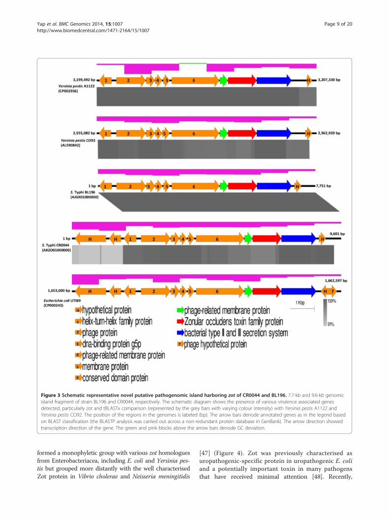
Figure 3 Schematic representative novel putative pathogenomic island harboring zot of CR0044 and BL196. 7.7-kb and 9.6-kb genomicisland fragment of strain BL196 and CR0044, respectively. The schematic diagram shows the presence of various virulence associated genesdetected, particularly zot and tBLASTx comparison (represented by the grey bars with varying colour intensity) with Yersinia pestis A1122 andYersinia pestis CO92. The position of the regions in the genomes is labeled (bp). The arrow bars denode annotated genes as in the legend basedon BLAST classification (the BLASTP analysis was carried out across a non-redundant protein database in GenBank). The arrow direction showedtranscription direction of the gene. The green and pink blocks above the arrow bars denode GC deviation.
Yap et al. BMC Genomics 2014, 15:1007 Page 9 of 20http://www.biomedcentral.com/1471-2164/15/1007
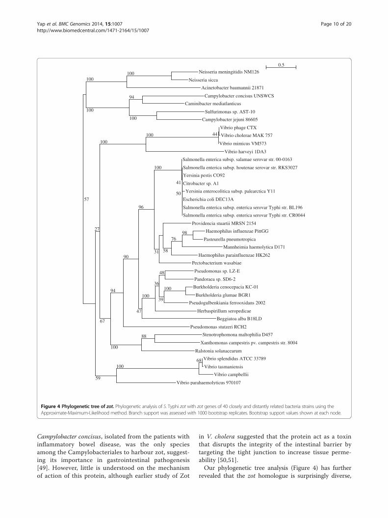
formed a monophyletic group with various zot homologuesfrom Enterobacteriacea, including E. coli and Yersinia pes-tis but grouped more distantly with the well characterisedZot protein in Vibrio cholerae and Neisseria meningitidis
[47] (Figure 4). Zot was previously characterised asuropathogenic-specific protein in uropathogenic E. coliand a potentially important toxin in many pathogensthat have received minimal attention [48]. Recently,
Vibrio parahaemolyticus 970107
Vibrio campbellii
Vibrio tasmaniensis
Vibrio splendidus ATCC 3378968100
Ralstonia solanacearum
Xanthomonas campestris pv. campestris str. 8004
Stenotrophomona maltophilia D45788
Pseudomonas stutzeri RCH2
Beggiatoa alba B18LD
Herbaspirillum seropedicae
Pseudogulbenkiania ferrooxidans 2002
Burkholderia glumae BGR1
Burkholderia cenocepacia KC-01100
Pandoraea sp. SD6-2
Pseudomonas sp. LZ-E
39
48
36
100
Pectobacterium wasabiae
Haemophilus parainfluenzae HK262
Mannheimia haemolytica D171
Pasteurella pneumotropica
Haemophilus influenzae PittGG9876
Providencia stuartii MRSN 2154
58
Salmonella enterica subsp. enterica serovar Typhi str. CR0044
Salmonella enterica subsp. enterica serovar Typhi str. BL196
Escherichia coli DEC13A
Yersinia enterocolitica subsp. palearctica Y11
Citrobacter sp. A1
Yersinia pestis CO92
Salmonella enterica subsp. houtenae serovar str. RKS3027
50
Salmonella enterica subsp. salamae serovar str. 00-0163
41
31
100
47
96
90
100
94
Vibrio harveyi 1DA3
Vibrio mimicus VM573
Vibrio cholerae MAK 757
Vibrio phage CTX44100
67
100
59
27
Campylobacter jejuni 86605
Sulfurimonas sp. AST-10
Caminibacter mediatlanticus
Campylobacter concisus UNSWCS
100
94
Acinetobacter baumannii 21871
Neisseria sicca
Neisseria meningitidis NM126100
100
100
57
0.5
Figure 4 Phylogenetic tree of zot. Phylogenetic analysis of S. Typhi zot with zot genes of 40 closely and distantly related bacteria strains using theApproximate-Maximum-Likelihood method. Branch support was assessed with 1000 bootstrap replicates. Bootstrap support values shown at each node.
Yap et al. BMC Genomics 2014, 15:1007 Page 10 of 20http://www.biomedcentral.com/1471-2164/15/1007
Campylobacter concisus, isolated from the patients withinflammatory bowel disease, was the only speciesamong the Campylobacteriales to harbour zot, suggest-ing its importance in gastrointestinal pathogenesis[49]. However, little is understood on the mechanismof action of this protein, although earlier study of Zot
in V. cholera suggested that the protein act as a toxinthat disrupts the integrity of the intestinal barrier bytargeting the tight junction to increase tissue perme-ability [50,51].Our phylogenetic tree analysis (Figure 4) has further
revealed that the zot homologue is surprisingly diverse,
Yap et al. BMC Genomics 2014, 15:1007 Page 11 of 20http://www.biomedcentral.com/1471-2164/15/1007
being present in the genomes of plant pathogens, suchas Ralstonia solanacearum and Xanthomonas campes-tris, and opportunistic pathogens that are found in soiland water, such as Providencia stuartii and Pandoraeaspp. Interestingly, this zot homologue was also found inlithoautotrophic bacteria, such as Caminibacter media-tlanticus, Sulphurimonas spp. that clustered closely withthe functionally validated Zot in Neisseria spp. andCampylobacter spp. but more distantly related to Enter-obacteriacea, including our S. Typhi. Other species suchas Pseudogulbenkiana ferrooxidans and Beggiatoa alba,which were isolated from extreme environments, such asdeep hydrothermal vents and sulphur springs were foundto be more closely related and shared a common ancestrywith S. Typhi (Figure 4). The importance of the necessityof gene targeting tight junctions in these extremophiles isyet to be understood but may be due to the role of thetoxins in facilitating the penetration of the tissue layersthat cover the host organisms that they colonise.Despite harboring four highly conserved domains lead-
ing to its putative assignment as a Zot toxin, the S.Typhi Zot lacks a previously identified active domain(FCIGRL) found in V. cholera. Nevertheless, a previousstudy has shown that the partial refolding of the dena-tured binding peptide of this domain did not prevent itsspecific binding to the Zot receptor on Caco-2 cells,demonstrating that the conformationally varied domainwas still able to induce toxin activity [52]. The possiblemechanism of Zot in S. Typhi may be similar to that ofV. cholerae, in which tight junction disassembly is in-duced through the activation of the proteinase-activatedreceptor 2 [53]. However, comparison of the predictedZot protein tertiary structures of S. Typhi with that of V.cholerae and N. meningitidis showed that they werehighly variable, suggesting that the toxin might have dif-ferent mechanisms of action despite maintaining somecore binding activities.The high similarities between the GIs of the BL196
and CR0044 strains further provide strong evidence ofthe common ancestry between the strains. Alarmingly,these strains are more diverse than was previously thought,emphasising our concern that such strains that have ac-quired new genes through HGT are circulating in thecountry or elsewhere.
Microvariations distinguishing closely related strainsSingle nucleotide polymorphisms (SNPs) were determinedfor the comparisons of the two highly related strains,BL196 and CR0044. We investigated the microvariationsof the high quality non-synonymous single nucleotidepolymorphism (nsSNPs) that were identified. In this study,only the potential functions altering the nsSNP mutationswere considered. Despite being highly related and similar,the genomes could be discretely distinguished from each
other by 29 nsSNPs (Additional file 6). Interestingly, the29 nsSNPs that were identified, two were found on twohighly related virulence determinant genes, RNA polymer-ase sigma factor rpoS and the Vi polysaccharide biosyn-thesis protein tviE. These two mutations are strain-specificand were not detected in any other S. Typhi genomes thatwere analysed. BL196 carried a mutant rpoS (P193L),which is 100% similar to Salmonella Pullorum S6702, thecausative agent of fowl typhoid [54]. Interestingly, unusualrpoS gene was also observed in P-stx-12. The rpoS ofP-stx-12 contains an additional 57 bp of a responseregulator gacA fragment that was fused at the 5’ end ofthe gene, resulting in a longer rpoS. The gacA fragmentwas highly similar to the transposons Tn10d tetA andtetR, which are associated with regulatory and tran-scription signals, suggesting the occurrence of trans-position events and possibly gene regulation. The rpoSmutant and its effects on S. Typhi were first reportedin the Ty2 genome and attenuated strain Ty21a. Thesemutants, which were partially derived from natural muta-tions in Ty2, were found to affect the stress response andother related functions significantly [21,55]. Mutations inrpoS are apparently advantageous to the strain for survivalin the host during prolonged stress, allowing for the selec-tion of more efficient transcription factors for survival andfitness during unfavourable conditions [56]. rpoS is com-monly associated with the virulence regulation of patho-gens because it regulates over 30 genes that are relatedto the stress response, such as the Spv protein, which isinvolved in host cell survival, and Vi-polysaccharide bio-synthesis proteins in different osmolarities [57].In contrast, CR0044 carried a mutant TviE (H53Y).
TviE, which is encoded by SPI-7, is required for viru-lence capsular formation and acts as a protective anti-gen. The antibody that responds to the Vi-positive strainhas been shown to be more virulent than that targetingthe Vi-negative strain [58,59]. A role of the RpoS proteinin fine-tuning the synthesis of the Vi polysaccharide in S.Typhi has also been reported [60], suggesting the pos-sible close regulation of these two genes in modulatingadaptation and virulence capabilities in different hostenvironments. The nsSNPs that were detected in thesetwo closely related genes may indicate that their adap-tive selection is important for host survival. To addressfalse-positive results, the nsSNPs were validated usinga high-resolution melt (HRM) analysis and direct se-quencing (Additional file 7). The unique HRM profilesof the wild-type strains and those containing the SNPtransition mutations in the normalised graph are shownin (Additional file 7a and b). Both SNPs showed uniquemelting profiles for the strains that were tested. For therpoS SNP, the transition mutation from C to T occurredin strain BL196, and the separation of the melting pro-file began at ~81°C and ended at 84°C. For the tviE SNP,

Yap et al. BMC Genomics 2014, 15:1007 Page 12 of 20http://www.biomedcentral.com/1471-2164/15/1007
the transition mutation from C to T occurred in strainCR0044, and the separation of the melting profile beganat ~74°C and ended at 78°C. The results of this analysiswere further confirmed by the direct sequencing of tar-geted loci (Additional file 7a and b). The primers andHRM profiles that were developed may be useful fordistinguishing between these two strains and as import-ant markers for future surveillance. Another 27 highquality nsSNPs were also identified that mainly encodedfor non-virulence factors (Additional file 6). Twenty-three genes had well defined functions, including fourthat were involved in metabolism and 12 that playedroles in cellular processes, signalling and transport. Theremainder were poorly characterised or had unknownfunctions. Additionally, out of 29 nsSNPs, 24 (83%)mutants were detected in the carrier strain CR0044,suggesting the possible functional adaptation of thiscarrier strain in the host cell relative to its closely re-lated strain.However, most of the mutant SNPs that were carried
by CR0044 was not detected in P-stx-12 with the excep-tion of the gene encoding the trehalose permease IICcomponent. In fact, these two carrier strains are genetic-ally different, with the presence of 253 SNPs, including159 nsSNP. Notably, the nsSNPs were mainly found inthe genes encoding proteins associated with virulence,metabolism, outer membrane, and other regulatory pro-teins. However, nsSNPs were also observed in a largenumber of uncharacterised proteins, suggesting that in-dependent genomic factors may have contributed to thecarrier state.It is important to note that the independent mutations
in rpoS and tviE as well as other genes in strains BL196and CR0044 support the postulation that these strainspossibly diverged from a common ancestor. We specu-late that the source of this ancestral strain may be stillcirculating in the country. Therefore, national surveil-lance program and epidemiologic study are pivotal foreffective microbial source tracking and disseminationcontrol.
Analysing the molecular effects of nsSNPs, proteinstructure modelling and molecular dynamics (MD)simulationPoint mutations that cause alterations in amino acids canhave profound effects on the structural stability of pro-teins; hence, a study of their effects is necessary to under-stand their functionalities. We have modelled both thenative and mutant structures of the proteins. Out ofthe 5 native protein structure models that were generatedby I-Tasser [61], the best structure with the highest confi-dence score (C-scores: RpoS, 0.64; TviE, −0.40) was col-lected and used for further investigations. The modellednative RpoS and mutant RpoS structures showed good
stereochemical properties, with 92.0% and 85.4% of theresidues being within the most favourable region of theRamachandran plot, respectively, whereas the nativeand mutant TviE showed 86.5% and 80.0% of the residuesin most favourable region of the plot, respectively. All ofthe structures passed ProSa model quality validation withZ-scores (RpoS native, −5.61; RpoS mutant, −5.57; TviEnative, −6.32; TviE mutant, −6.56) falling well within therange of those that are typically reported for native pro-teins of similar sizes from different sources (X-ray, NMR).Molecular dynamics (MD) simulation with a realistic
aqueous solvent environment was performed to revealthe explicit solvent behaviours of the native and mutantstructures, which could elicit the differences in their dy-namics and stabilities. The energy minimisation studieswere performed for both the native and mutant struc-tures, and the total energies of the native and mutantRpoS achieved were −4115.46 J/mol and −4804.66 J/mol,respectively, whereas those of the native and mutantTviE achieved were −3203.54 J/mol and −2906.31 J/mol,respectively. Energy minimisation assessments provideclues regarding protein stability. The deviation betweentwo structures can be evaluated by the root mean squaredeviation (RMSD). The higher the RMSD value is, thegreater the deviation will be between the native and mu-tant structures. The RMSD value between the native andmutant RpoS was found to be 0.39 Å and that betweenthe native and mutant TviE was 0.43 Å. The native, mu-tant, superimposed protein structures at their correspond-ing positions for RpoS and TviE are shown (Figure 5). TheRMSD values of the native and mutant structures weresignificantly similar for both RpoS and TviE, suggestingsimilar levels of protein folding alterations. We analysedthe molecular effects and functional modifications basedon several predictive tools (refer to Methods) targetingvarious aspects of protein dynamics with confidencescores (Additional file 8). Out of 9 predictive tools used,all found that the nsSNP in rpoS was deleterious (affectingprotein structure and function), whereas in tviE, 3 predict-ive tools found that the nsSNP to be deleterious, 5 to beneutral and one undetermined. The nsSNP in rpoS showedPSIC score of 1.0 (deleterious) with PolyPhen2 [62] inaddition to a probability score of 0 (<0.05, deleterious) withSIFT [63] and a Provean [64] score of −9.261 (<−2.5, dele-terious), suggesting that the nsSNP could affect the proteindrastically and result in functional modifications. It is im-portant to note that the deleterious effects could lead topositive or negative functional modifications. To furthervalidate the results, a method that was based on the hid-den Markov model (HMM) from PANTHER [65] wasused. The nsSNP in rpoS was found to be deleterious, butundetermined for tviE.The predictions of the nsSNP in rpoS by these tools are
in agreement and show strong correlations among various

Figure 5 Modelled protein structures of RpoS and TviE. A1 showed native RpoS with proline at position 193. A2 showed mutant RpoS withamino acid leucine at position 193. A3 showed superimposed structure of RpoS native structure (yellow) with mutant structure (pink). B1 showednative TviE with histidine at position 53. B2 showed mutant TviE with amino acid tyrosine at position 53. B3 showed superimposed structure ofTviE native structure (yellow) with mutant structure (pink).
Yap et al. BMC Genomics 2014, 15:1007 Page 13 of 20http://www.biomedcentral.com/1471-2164/15/1007
methodologies. The analysis with I-Mutant 3.0 [66], whichis based on the support vector machine (SVM) and DGGstability changes, revealed that the nsSNP in rpoS mayhave led to its greatly increased stability (DGG value of0.89 Kcal/mol, >0.5 kcal/mol indicates large increase inprotein stability), suggesting that the deleterious effectsof the nsSNP is favourable to the protein folding and
structure and possibly lead to enhanced functions. TheDDG value is calculated from the unfolding Gibbs freeenergy value of the mutated protein minus the unfoldingGibbs free energy value of the wild-type strain (Kcal/mol),which is based on a trained cross-validation procedureusing a comprehensive experimental database of proteinmutations [66]. These predicted results were consistent
Yap et al. BMC Genomics 2014, 15:1007 Page 14 of 20http://www.biomedcentral.com/1471-2164/15/1007
with the differences in the energy minimisation of MDsimulation, suggesting the favourable folding of the mu-tant structure. Native and mutant structures differ dueto the specific properties of the residues that could dis-rupt the structure and function of the protein. The mu-tant residue (leucine) of RpoS is molecularly larger insize than the wild-type residue (proline), which is ahighly rigid, small molecule that is required to induce aunique backbone conformation. However, the alterationfrom the small-sized secondary amine structure to alarger-sized primary aliphatic amine at this site can dis-turb its conformation and lead to bumps in the struc-ture, in which the mutant residue is not in the correctposition to make the typical hydrogen bond that isformed by the native residue (Figure 5). The hydropho-bicity of the wild type and mutant differ because themutation introduces a very high hydrophobic residue inplace of a less hydrophobic residue. This can result in aloss of hydrogen bonding and may disturb proper pro-tein folding. The wild-type proline residue in RpoS iswell conserved and is located at the discrete compactthree-helical domain within region 3 of the protein; how-ever, no known mutant residues with similar propertieswere observed at this position in the other homologoussequences. This region is the specific binding site of bac-terial promoters containing an extended −10-promoterelement and is primarily involved in the binding of thecore RNA polymerase in the holoenzyme. The mutationin this important site could affect protein functioning per-taining to the transcription efficiency.However, the predictions regarding the nsSNP in tviE
were contrasting, which suggest more benign effects, in-dicating that nsSNPs may have more modest effects onthe functioning of this protein. The nsSNP in tviE mayhave decreased its stability (DGG value of −0.54 Kcal/mol,with < −0.5 kcal/mol indicating large decrease in proteinstability) as predicted by I-Mutant 3.0 [66]. Unlike RpoS,the mutant residue (tyrosine) in TviE is not conservedat this position of the helical structure, and other non-similar residues were observed at this position with noknown protein binding sites in other homologous se-quences. However, the size of the mutant residue (tyro-sine) is larger compared with that of the native residue(histidine) despite the fact that both are polar and lo-cated at the surface of the protein. This mutation alsointroduces a more hydrophobic residue at this pos-ition, suggesting that it could possibly alter the correctfolding of the protein, subsequently affecting hydrogenbonding but with only modest effects. These novel mu-tations could have important impacts on the pathogenesisand persistence of strains in the host. Although we couldnot determine the true extent of the effects of the nsSNPson the protein functions, these data suggest that theyalter the protein structures (and possibly their functions)
considerably, potentially leading to the enhanced regula-tion of RpoS and stress response in BL196 and reduced ef-ficiency of TviE in the virulence capsular formation ofCR0044. The close regulation of these two genes may berelevant to the virulence and persistence capabilities ofthe closely related strains that lead to the different clinicaloutcomes. These data provide essential insights intothe underlying molecular mechanisms upon mutationsand serve as caveats for future functional gene knock-out studies.
ConclusionsWe have thoroughly dissected the genome of S. Typhi inassociation with three important epidemiological settings.Comparative genomics and phylogeny analyses have re-vealed that the strain that was associated with the largeoutbreak was highly related and shared common ances-try with the carrier strain. These findings are supportedby their common genomic features and uncommon generepertoires, including dispensable genes, phages andan additional putative pathogenomic island harbouringvirulence-related genes, and zot in particular. Apartfrom these, variations were also identified in T6SS andSPI-related genes, insertion sequences, CRISPRs andnsSNPs among the studied genomes, which may be novelfactors that contribute to the varied host adaptations andpathogenicities. Despite being highly similar, BL196 andCR0044 may be distinguished by microvariations in theirnsSNPs. Interestingly, the protein modelling and MDsimulation of the wild-type and mutant RpoS and TviEsuggest that the potential protein structure and func-tional modification was more stable in RpoS, whichplausibly leads to enhanced regulation and stress re-sponse. On the other hand, the mutation in TviE wasless stable than that of the wild type, which could po-tentially lead to lower capsular formation efficiency.The close association of these virulence-related genesare relevant for long-term host persistence and adapta-tion, which serve as important caveats for further func-tional studies. The analysis also revealed that SPI-10,which was previously thought to be relatively stable, ispossibly prone to excision. Moreover, multiple regionsof genomic plasticity were detected. In particular, thediscovery of new GIs in the outbreak strain and thehighly related carrier strain are of great concern epide-miologically. These results suggest the plasticity andopen pan-genome of S. Typhi, indicating that the patho-gen is more diverse than previously thought and thatgenes may have been acquired or transferred from one an-other through HGT, posing higher risk for effective dis-ease control. The genomic information that was obtainedin this study provides novel insights into the pathogenesisand control of S. Typhi, essentially, gene targets for vac-cine development.

Yap et al. BMC Genomics 2014, 15:1007 Page 15 of 20http://www.biomedcentral.com/1471-2164/15/1007
MethodsChoice of strainsThree Malaysian S. Typhi strains (BL196, ST0208 andCR0044) were selected for the comparative genomic ana-lysis that were based on previous PFGE data and reportedgenome sequences [6,17,18]. These strains are associatedwith diverse epidemiological settings. Strain BL196 wasisolated from a typhoid patient with diarrhoea during alarge outbreak in Kelantan, Malaysia that resulted in 735cases and 2 deaths in the year 2005. Strain ST0208 wasisolated from a typhoid patient, who was a sporadic case,at a local tertiary hospital in Kuala Lumpur, Malaysia.Strain CR0044 was isolated in 2007 from a carrier (foodhandler) following the large 2005 outbreak in Kelantan,Malaysia (Table 1). The initial molecular analysis showedthat both the CR0044 and BL196 strains were highly simi-lar with only one band difference as revealed by PFGE(Additional file 2). These 3 new genomes were comparedwith all three of the available S. Typhi full genomes at thetime of our analysis (CT18, Ty2 and P-stx-12). We com-pared our strains with CT18 and Ty2, the former is a fairlyrecent and geographically related multidrug-resistantstrain that was isolated from Vietnam, the latter was iso-lated from Russia in the early 1970s, a geographicallymore distant strain known to be used for oral typhoid vac-cine development. We also performed a comparison usinga carrier associated strain, P-stx-12, which was isolatedfrom a carrier in India. All of these strains represent S.Typhi from diverse temporal and spatial backgrounds in as-sociation with variable epidemiological settings. The detailsof the bacterial strains are provided in Table 1 [GenBankaccession number: BL196 (AJGK00000000.1), ST0208(AJXA00000000.1), CR0044 (AKZO00000000.1), CT18(AL513382), Ty2 (AE014613) and P-stx-12 (CP003278).
DNA sequencing, assembly and annotationPrevious sequencing was carried out on 3 S. Typhi strainsusing the Illumina Genome Analyser (GA2X, pipelineversion 1.6, insert size 300), generating >10 total giga-bytes of data. A de novo assembly and annotations werecarried out and further validated with various pipelinesas previously described [6,17,18].
Table 1 Details of bacterial strains used in this study
Strain namea Year of isolation Location of isolation
BL196 2005 Kelantan, Malaysia
CR0044 2007 Kelantan, Malaysia
ST0208 2008 Kuala Lumpur, Malaysia
CT18 1993 Mekong Delta, Vietnam
Ty2 1916 Russia
P-stx-12 2009 Varanassi, IndiaaS. Typhi strains and their Genbank accession numbers: BL196 (AJGK00000000.1) [1(AL513382) [20], Ty2 (AE014613) [21] and P-stx-12 (CP003278) [22]. bNA: Not availab
Multilocus sequence typingMultilocus sequence typing (MLST) housekeeping genesequences (thrA (aspartokinase + homoserine dehydrogen-ase), purE (phosphoribosylaminoimidazole carboxylase),sucA (alpha ketoglutarate dehydrogenase), hisD (histidinoldehydrogenase), aroC (chorismate synthase), hemD (uro-porphyrinogen III cosynthase) and dnaN (DNA polymeraseIII beta subunit) according to PubMLST were extractedfrom the genome sequences [19]. The alignments for eachof these genomic regions were bioinformatically extracted,trimmed and concatenated into final sequence lengths of3,336 bp using MEGA 5 [67]. The sequences were subse-quently submitted to the MLST database (http://mlst.warwick.ac.uk) and assigned existing or novel allele type num-bers. The composite sequence types (STs) were defined bythe database based on the allelic profile that was derivedfrom each of the seven loci. The STs from the fragmentedincomplete genomes were derived by comparing the 3 lessconserved alleles hemD, hisD and thrA, while assumingthat the other 4 alleles, aroC, dnaN, purE and sucA, wereconserved. The results with positive BLASTN hits of 100%query sequence coverage (E < 1 × 10⁻⁶) were only consid-ered in the analysis.
Comparative genomic analysisProtein coding gene predictions were performed usingProdigal [68]. The predicted genes were then subjectedto annotations using Blast2GO [69] (E < 1 × 10⁻³⁰). Thegenomic sequences and functional annotations of the CDSswere validated based on the results of homology searchesagainst the public non-redundant nucleotide and proteindatabases (http://www.ncbi.nlm.nih.gov/) using BLASTNand BLASTP [70] The genes were selected based onthe top BLAST hits (E < 1 × 10⁻³⁰, ≥60% query coverageand ≥60% protein identity). The open reading frames(ORFs) of the genomes were reciprocally compared (ORF-dependent comparisons) using RAST [71]. The subsystemcategory distributions were compared among the ge-nomes. The circular map of genes that was based on thesimilarities of the amino acid sequences of the BL196,CR0044, ST0208, TY2 and P-stx-12 genomes against thatof CT18 was generated using the BLAST Ring Image
Specimenb Epidemiological informationb (if available)
Blood Outbreak
Stool Carrier
Stool Sporadic
Blood NA
NA NA
Stool Carrier
7], CR0044 (AKZO00000000.1) [18], ST0208 (AJXA00000000.1) [6], CT18le.
Yap et al. BMC Genomics 2014, 15:1007 Page 16 of 20http://www.biomedcentral.com/1471-2164/15/1007
Generator (BRIG) [72]. A synteny-based analysis wasperformed by aligning the genomes with CT18 as a ref-erence, and the contigs were reordered with iterativerefinements using progressiveMauve [73] and Nucmer[74]. The best alignments were chosen for the multiplegenome alignments. The reference-ordered and -orientedgenomic scaffold that was used for the subsequent analysiswas generated by concatenating reordered contigs byinserting 5Ns between the contigs using an in-house py-thon script. A bioinformatic pipeline using the Pan-Genomes Analysis Pipeline (PGAP) [75] was utilised toidentify the homologous regions of the compared ORFsat an E value cut-off of 1 × 10⁻¹⁰. Then, the nucleotideand amino acid sequences of the query ORF and selectedtarget homologous regions were aligned and validatedusing BLAST against the NCBI redundant database. Theresulting matched and validated homologues, paraloguesand orthologues were used for the multiple alignmentcomparison. A genomic island analysis and predictionwere performed using IslandViewer [43], which in-cludes 3 methods (Island Pick, IslandPath-DIMOB andSGI-HMM). The IS elements were analysed by IS Finder(http://www-is.biotoul.fr). The phages were analysed usingPHAST [76]. The regions that were algorithmically identi-fied as intact and those sharing high similarities werecompared and analysed. The sequence content com-parison was performed using ACT [77] and MEGA 5[67]. The regions of interest were then manually curatedto improve the annotations and gene predictions. ThensSNP analysis was carried out using PGAP [75] bysorting them from synonymous SNPs, deletions and in-sertions, and the results were validated using the CLCGenomic Workbench version 5.1 (CLC Bio, Aarhus,Denmark).
Phylogenomic analysisThe genome sequences of 20 global [S. Typhi strainsand their Genbank accession numbers were as follows:BL196 (AJGK00000000.1), CR0044 (AKZO00000000.1),ST0208 (AJXA00000000.1), UJ308A (AJTD00000000.1),UJ816A (AJTE00000000.1), CR0063 (AKIC00000000.1),404ty (CAAQ00000000.1), E00-7866 (CAAR00000000.1),E01-6750 (CAAS00000000.1), E02-1180 (CAAT00000000.1),E98-0664 (CAAU00000000.1), E98-2068 (CAAV00000000.1),J185 (CAAW00000000.1), M223 (CAAX00000000.1), AG3(CAAY00000000.1), E98-3139 (CAAZ00000000.1), STH2370(JABZ00000000.1), CT18 (AL513382), Ty2 (AE014613) andP-stx-12 (CP003278) (Additional file 9). These sequenceswere submitted to the Reference Sequence Alignment-based Phylogeny Builder (RealPhy) [78] for the identifi-cation of sites that were relevant for the phylogenomicanalysis using the default parameters. The completegenome of S. typhi CT18 was chosen as the referencegenome, and all of the query genomic sequences were
divided into possible sequences of 50 bp (default) andsubsequently mapped to the reference genome viaBowtie2 with a default k-mer length of 22, allowing forone mismatch within the k-mers to maximise sensitivity.The generated multiple genome sequence alignmentswere subsequently used to construct an unrooted phylo-genetic tree that was inferred via the approximate max-imum likelihood method using FastTreeMP [79].
Phylogenetic analysis of zotThe zot amino acid sequence data from 40 closely anddistantly related bacterial strains were downloaded fromthe Genbank. The sequences from both BL196 andCR0044 were aligned with those of the 40 bacterial strainsthat were selected using the MAFFT [80] E-INS-I strategy.A phylogenetic analysis was subsequently performed usingmaximum likelihood phylogenetic algorithms with thePhyML module of SeaView V4.5 [81], which was sup-ported by 1000 bootstrap replicates. The Maximum Likeli-hood tree was constructed using the best substitutionmodel (Blosum62 algorithm) after being tested and opti-mised by ProtTest 2.4 [82].
PCR validation of selected genes and SNPsPCR was carried out to validate the identified high-quality nsSNPs. Genomic DNA for the sequencing reac-tions was extracted using the Wizard® Genomic DNAPurification Kit (Promega, Madison, WI, USA). Theamplification of the selected genes was performed usinga standard PCR protocol. Each 25 μl PCR reaction con-tained 150 μM (each) deoxynucleoside triphosphates, 1×PCR colourless buffer, 1.2 mM MgCl2, 0.2 μM of primerand 0.5 U of Go Taq Flexi DNA Polymerase (Promega,Madison, WI, USA). The PCR was performed under thefollowing conditions: initial denaturation at 95°C for30 s, 30 cycles of denaturation at 95°C for 30 s, 30 s atthe respective annealing temperature (Additional file 10)and an extension step at 72°C for 40 s; a final extensionwas performed at 72°C at 1 min. The reactions were car-ried out using a PCR Master Cycler (Eppendorf AG,Hamburg, Germany). The primer sets that were used forthe target genes are shown (Additional file 10).
Sequencing and high-resolution melting (HRM) analysisThe PCR products were purified using the PCR Clean-upKit (Qiagen, Valencia, CA) according to the manufac-turer’s instructions. The PCR products were then sentto a commercial sequencing facility (First BASE Labora-tory Sdn Bhd, Selangor, Malaysia) for direct sequencing.The nsSNP variations were validated with a pair ofprimers as described (Additional file 10) and subse-quently used for a high-resolution melting (HRM) analysisusing the Kapa HRM Fast PCR Kit (Kapa Biosystems,Boston, Massachusetts, USA) and Eco Real-Time qPCR
Yap et al. BMC Genomics 2014, 15:1007 Page 17 of 20http://www.biomedcentral.com/1471-2164/15/1007
System (Illumina, San Diego, California, USA) accordingto the manufacturer’s instructions. The melting curveprofiles that were generated were analysed with theEco-qPCR software using both homozygous and het-erozygous controls.
Analysis of molecular effects of nsSNPs and proteinstructure modellingThe nsSNPs of tviE and rpoS were selected for the pre-dictions and analyses of the further molecular effects.The Poly-Phen2 [62], SIFT [63], Provean [64], SNAP[83], I-Mutant 3.0 [66] and PredictSNP 1.0 (PredictSNP,MAPP, PhD-SNP and Panther) [84] tools were used toexamine the functional modifications and predictions ofthe tolerated and deleterious nsSNPs. The details of themethods and scores that were used for each tool are in-cluded in (Additional file 8). A combination of differentprediction methods were used to increase the predictionaccuracy and confidence. The protein structures of TviEand RpoS were modelled using the I-TASSER server[61]. The best model was selected based on the optimalC-score. Further, the native structure was mutated byintroducing a point mutation in the native RpoS proteinat P193L (proline to leucine) and native TviE protein atH53Y (histidine to tyrosine) using FixPDB with theNOMAD-Ref server [85] and validated with the SPDBviewer [86]. The native and mutant structures werechecked, fixed, refined and energetically optimised byMDWeb [87], ModRefiner [88], FG-MD [89] and theSPDB viewer [86]. The qualities of the model structureswere independently verified with the PROCHECK [90],WHATCHECK [91] and PROSA programs [92].
Molecular dynamics (MD) simulation and energyminimisationThe molecular dynamics (MD) simulations were carriedout using MDWeb [87]. The optimised structures of thenative and mutant RpoS and TviE proteins were used asinput data for the MD simulations. GROMACS topologieswere first generated by removing the crystallographicwater molecules and adding missing side chain and hydro-gen atoms. Histidine residues were protonated accordingto the protpKa program algorithm with the GROMACSpackage. Water molecules were added at energeticallyfavourable positions of the structure surfaces. Hydrogenatoms were energetically minimised for 500 steps ofhydrogen conjugate gradients, while the remainder ofthe structures were fixed and followed by energy minimi-sations for 500 steps of conjugate gradients, restrainingheavy atoms with a force constant of 500 KJ/mol.nm2 totheir initial positions. The system was solvated withsimple point charge (SPC) water molecules at spacingdistances of 15 Å around the molecules. Chloride (Cl-)and/or sodium (Na+) ions were added until the system
was neutralised at a concentration of 50 mM. The stepinvolving the minimisations of the structures for 500steps of conjugate gradients to restrain the heavy atomswith a force constant of 500 KJ/mol.nm2 to their initialpositions was repeated. The whole molecular system wassubjected to energy minimisations of 500 iterations by asteepest descent algorithm implementing a GROMOS9643a1 force field. The comparative analysis of structural de-viations between the native and mutant proteins of RpoSand TviE was assessed by their respective RMSD values.
Additional files
Additional file 1: Genomic features of S. Typhi strains.
Additional file 2: Pulsed-field gel electrophoresis of S. Typhi strainsBL196, CR0044 and ST0208.
Additional file 3: Strains used to detect prevalence of zot in S. Typhiusing PCR.
Additional file 4: Representative gel picture of zot prevalence inS. Typhi.
Additional file 5: Primers used for zot prevalence screening inS. Typhi strains.
Additional file 6: nsSNPs detected in S. Typhi strains BL196 andCR0044.
Additional file 7: a: High-resolution melting profile of rpoS fragment innormalised graph mode. b: High-resolution melting profile ofVi-polysaccharide biosynthesis tviE fragment in normalised graph mode.
Additional file 8: Prediction of nsSNP effects on rpoS and tviE usingmulti-predictive tools.
Additional file 9: Strain backgrounds used in phylogenomic analysis.
Additional file 10: Primers used for PCR, direct sequencing andhigh-resolution melt analysis for nsSNP detected in S. Typhi strainsBL196 and CR0044.
Competing interestsThe authors declare that they have no competing interests.
Authors’ contributionsKPY performed the full comparative genomic, bioinformatics analyses,experiments and drafted the manuscript. KPY and HMG analysed the dataand performed the experiments. KPY, KLT, HMG, CSJT and LCC criticallyrevised, reviewed and improved the manuscript. KLT conceived andmanaged the high-impact research project. KLT and LCC supervised thestudy. KPY, HMG, KLT, CSJT and LCC designed and fine-tuned the study. KLTprovided the information on strain background. KLT provided the additionallaboratory support and facilities. KLT supplied the reagents, kits and equipment.All of the authors were involved in compiling and approving the final version ofthe manuscript.
AcknowledgementsThis research was supported by a University of Malaysia High ImpactResearch Grant in Molecular Genetics (reference no. UM.C/625/1HIR/MOHE/-02[A000002-5000 1]), for which Kwai-Lin Thong is the grant holder for thehigh-impact research project under the title “Pathogenomic and Phenomic ofFood-Borne Disease.” Han Ming Gan would like to acknowledge the MonashUniversity Malaysia Tropical Medicine Biology platform for financial assistance.The first author, Kien-Pong Yap, was recipient and supported by His Majesty,the King’s Scholarship Award of Malaysia (Biasiswa Yang Dipertuan Agong). Thefunders had no role in the study design, data collection and analysis, decisionto publish or preparation of the manuscript. We gratefully acknowledge SafwanJusoh from the ICT department at the University of Malaya for assisting us withthe computing solutions and allowing us to use their servers and advancedcomputational facilities.

Yap et al. BMC Genomics 2014, 15:1007 Page 18 of 20http://www.biomedcentral.com/1471-2164/15/1007
Author details1Institute of Biological Sciences, Faculty of Science, University of Malaya,50603 Kuala Lumpur, Malaysia. 2Laboratory of Biomedical Science andMolecular Microbiology, Institute of Graduate Studies, University of Malaya,50603 Kuala Lumpur, Malaysia. 3Department of Medical Microbiology, Facultyof Medicine, University of Malaya, 50603 Kuala Lumpur, Malaysia. 4School ofScience, Monash University Malaysia, Jalan Lagoon Selatan, Bandar Sunway,46100 Selangor, Malaysia.
Received: 22 August 2013 Accepted: 6 November 2014Published: 20 November 2014
References1. Crump JA, Mintz ED: Global trends in typhoid and paratyphoid fever.
Clin Infect Dis 2010, 50:241–246.2. Gonzalez-Escobedo G, Marshall JM, Gunn J: Chronic and acute infection of
the gall bladder by Salmonella Typhi: understanding the carrier state.Nat Rev Microbiol 2010, 9:9–14.
3. Ministry of Health Malaysia: Epidemiology of foodborne diseases in Malaysia.Putrajaya, Malaysia: Director General Ministry of Health Malaysia TechnicalReport: Epidemiology of foodborne diseases in Malaysia; 2006:86–87.
4. Holt KE, Parkhill J, Mazzoni CJ, Roumagnac P, Weill FX, Goodhead I, Rance R,Baker S, Maskell DJ, Wain J, Dolecek C, Achtman M, Dougan G: High-throughputsequencing provides insights into genome variation and evolution inSalmonella Typhi. Nat Genet 2008, 40:987–993.
5. Vaishnavi C, Kochhar R, Singh G, Kumar S, Singh S, Singh K: Epidemiologyof typhoid carriers among blood donors and patients with biliary,gastrointestinal and other related diseases. Microbiol Immun 2005, 49:107.
6. Yap KP, Teh CSJ, Baddam R, Chai LC, Kumar N, Avasthi TS, Ahmed N, ThongKL: Insights from the genome sequence of a Salmonella enterica serovarTyphi strain associated with a sporadic case of typhoid fever in Malaysia.J Bacteriol 2012, 194:5124–5125.
7. Achtman M: Evolution, population structure, and phylogeography ofgenetically monomorphic bacterial pathogens. Annu Rev Microbiol 2008,62:53–70.
8. Parry CM, Hien TT, Dougan G, White NJ, Farrar JJ: Typhoid fever. N Engl JMed 2002, 347:1770–1782.
9. Thong KL, Puthucheary SD, Pang T: Genome size variation among recenthuman isolates of Salmonella typhi. Res Microbiol 1997, 148:229–235.
10. Thong KL, Passey M, Clegg A, Combs BG, Yassin RM, Pang T: Molecularanalysis of isolates of Salmonella typhi obtained from patients with fataland nonfatal typhoid fever. J Clin Microbiol 1996, 34:1029–1033.
11. Fraser-Liggett CM: Insights on biology and evolution from microbialgenome sequencing. Genome Res 2005, 15:1603–1610.
12. Dobrindt U, Hacker J: Whole genome plasticity in pathogenic bacteria.Curr Opin Microbiol 2001, 4:550–557.
13. Wren BW: Microbial genome analysis: insights into virulence, hostadaptation and evolution. Nature Rev 2000, 1:30–39.
14. Ahmed N, Dobrindt U, Hacker J, Hasnain SE: Genomic fluidity andpathogenic bacteria: applications in diagnostics, epidemiology andintervention. Nature Rev Microbiol 2008, 6:387–394.
15. Lawrence JG, Ochman H: Reconciling the many faces of lateral genetransfer. Trends Microbiol 2002, 10:1–4.
16. Medini D, Donati C, Tettelin H, Masignani V, Rappuoli R: The microbialpan-genome. Curr Opin Genet Dev 2005, 15:589–594.
17. Baddam R, Kumar N, Thong KL, Ngoi ST, Teh CS, Yap KP, Chai LC, Avasthi TS,N. Ahmed N: Genetic fine structure of a Salmonella enterica serovar Typhistrain associated with the 2005 outbreak of typhoid fever in Kelantan,Malaysia. J Bacteriol 2012, 194:3565–3566.
18. Yap KP, Gan HM, Teh CSJ, Baddam R, Chai LC, Kumar N, Avasthi TS, Ahmed N,Thong KL: Genome sequence and comparative pathogenomics analysis ofa Salmonella enterica serovar Typhi strain associated with a typhoid carrierin Malaysia. J Bacteriol 2012, 194:5970–5971.
19. Kidgell C, Reichard U, Wain J, Linz B, Torpdahl M, Dougan G, Achtman M:Salmonella typhi, the causative agent of typhoid fever, is approximately50,000 years old. Infect Genet Evol 2002, 2:39–45.
20. Parkhill J, Dougan G, James KD, Thomson NR, Pickard D, Wain J, Churcher C,Mungall KL, Bentley SD, Holden MTG, Sebaihia M, Baker S, Basham D, BrooksK, Chillingworth T, Connerton P, Cronin A, Davis P, Davies RM, Dowd L,White N, Farrar J, Feltwell T, Hamlin N, Haque A, Hien TT, Holroyd S, Jagels K,Krogh A, Larsen TS, Leather S, Moule S, Ó’Gaora P, Parry C, Quail M, Rutherford K,
Simmonds M, Skelton J, Stevens K, Whitehead S, Barrell BG: Completegenome sequence of a multiple drug resistant Salmonella enterica serovarTyphi CT18. Nature 2001, 413:848–852.
21. Deng W, Liou SR, Plunkett G III, Mayhew GF, Rose DJ, Burland V, Kodoyianni V,Schwartz DC, Blattner FR: Comparative genomics of Salmonella entericaserovar Typhi strains Ty2 and CT18. J Bacteriol 2003, 185:2330–2337.
22. Ong SY, Pratap CB, Wan X, Hou S, Rahman AYA, Saito JA, Nath G, Alam M:Complete genome sequence of Salmonella enterica subsp. entericaserovar Typhi P-stx-12. J Bacteriol 2012, 194:2115.
23. Kingsley RA, Msefula CL, Thomson NR, Kariuki S, Holt KE, Gordon MA, Harris D,Whitehead S, Sangal V, Marsh K, Achtman M, Molyneux ME, Cormican M,Parkhill J, MacLennan CA, Heyderman RS, Dougan G: Epidemic multiple drugresistant Salmonella Typhimurium causing invasive disease in sub-saharanAfrica have a distinct genotype. Genome Res 2009, 19:2279–2287.
24. Schell MA, Ulrich RL, Ribot WJ, Brueggemann EE, Hines HB, Chen D,Lipscomb L, Kim HS, Mrázek J, Nierman WC, DeShazer D: Type VI secretionis a major virulence determinant in Burkholderia mallei. Mol Microbiol2007, 64:1466–1485.
25. Pukatzki S, McAuley SB, Miyata ST: The type VI secretion system:translocation of effectors and effector-domains. Curr Opin Microbiol 2009,12:11–17.
26. Bingle LE, Bailey CM, Pallen MJ: Type VI secretion: a beginner’s guide. CurrOpin Microbiol 2008, 11:3–8.
27. Fernanda DP, Paiva JB, Nakazato G, Lancellotti M, Sircili MP, Stehling EG,Silveira WD, Sperandio V: Characterization of IcmF of the type VI secretionsystem in an avian pathogenic Escherichia coli (APEC) strain. Microbiol2011, 157:2954–2962.
28. Parsons DA, Heffron H: sciS, an icmF homolog in Salmonella entericaserovar Typhimurium, limits intracellular replication and decreasesvirulence. Infect Immun 2005, 73:4338–4345.
29. Ochman H, Groisman EA: Distribution of pathogenicity islands inSalmonella spp. Infect Immun 1996, 64:5410–5412.
30. Faucher SP, Viau C, Gros PP, Daigle F, Moual HL: The prpZ gene clusterencoding eukaryotic‐type Ser/Thr protein kinases and phosphatasesis repressed by oxidative stress and involved in Salmonella entericaserovar Typhi survival in human macrophages. FEMS Microbiol Lett2008, 281:160–166.
31. Schofield MJ, Hsieh P: DNA MISMATCH REPAIR: molecular mechanismsand biological function. Annu Rev Microbiol 2003, 57:579–608.
32. Bueno SM, Santiviago CA, Murillo AA, Fuentes JA, Trombert AN, Rodas PI,Youderian P, Mora GC: Precise excision of the large pathogenicity island,SPI7, in Salmonella enterica serovar Typhi. J Bacteriol 2004,186:3202–3213.
33. Roumagnac P, Weill FX, Dolecek C, Baker S, Brisse S, Chinh NT, Le TAH,Acosta CJ, Farrar J, Dougan G, Achtman M: Evolutionary history ofSalmonella Typhi. Science 2006, 314:1301–1304.
34. Viong V, Thong KL, Yusof MYM, Hanifah YA, Sam JIC, Hassan H:Macrorestriction analysis and antimicrobial susceptibility profiling ofSalmonella enterica at a university teaching hospital, Kuala Lumpur. Jpn JInfect Dis 2010, 63:317–322.
35. Edwards RA, Olsen GJ, Maloy SR: Comparative genomics of closely relatedsalmonellae. Trends Microbiol 2002, 10:94–99.
36. Threlfall EJ, Torre E, Ward LR, Dávalos-Pérez A, Rowe B, Gibert I: Insertionsequence IS200 fingerprinting of Salmonella typhi: an assessment ofepidemiological applicability. Epidemiol Infect 1994, 112:253–262.
37. Ooka T, Ogura Y, Asadulghani M, Ohnishi M, Nakayama K, Terajima J,Watanabe H, Hayashi T: Inference of the impact of insertion sequence (IS)elements on bacterial genome diversification through analysis of small-sizestructural polymorphisms in Escherichia coli O157 genomes. Genome Res2009, 19:1809–1816.
38. Kusumoto M, Ooka T, Nishiya Y, Ogura Y, Saito T, Sekine Y, Iwata T, Akiba M,Hayashi T: Insertion sequence-excision enhancer removes transposableelements from bacterial genomes and induces various genomic deletions.Nat Commun 2011, 2:152.
39. Casjens S: Prophages and bacterial genomics: what have we learned sofar? Mol Microbiol 2003, 49:277–300.
40. Barrangou R, Fremaux C, Deveau H, Richards M, Boyaval P, Moineau S,Romero DA, Horvath P: CRISPR provides acquired resistance againstviruses in prokaryotes. Science 2007, 315:1709–1712.
41. Horvath P, Barrangou R: CRISPR/Cas, the immune system of bacteria andarchaea. Science 2010, 327:167–170.

Yap et al. BMC Genomics 2014, 15:1007 Page 19 of 20http://www.biomedcentral.com/1471-2164/15/1007
42. Cady KC, O’Toole GA: Non-identity-mediated CRISPR-bacteriophage interactionmediated via the Csy and Cas3 proteins. J Bacteriol 2011, 193:3433–3445.
43. Langille MG, Brinkman FS: IslandViewer: an integrated interface forcomputational identification and visualization of genomic islands.Bioinformatics 2009, 25:664–665.
44. Michael H: Evolution of pathogenicity islands of Salmonella enterica. Int JMed Microbiol 2004, 294:95–102.
45. Radnedge L, Agron PG, Worsham PL, Andersen GL: Genome plasticity inYersinia pestis. Microbiol 2002, 148:1687–1698.
46. Rosso ML, Chauvaux S, Dessein R, Laurans C, Frangeul L, Lacroix C, SchiavoA, Dillies MA, Foulon J, Coppée JY, Médigue C, Carniel E, Simonet M,Marceau M: Growth of Yersinia pseudotuberculosis in human plasma:impacts on virulence and metabolic gene expression. BMC Microbiol2008, 8:211.
47. Hazen TH, Sahl JW, Redman JC, Morris CR, Daugherty SC, Chibucos MC,Sengamalay NA, Fraser-Liggett CM, Steinsland H, Whittam TS, Whittam B,Manning SD, Rasko DA: Draft genome sequences of the diarrheagenicEscherichia coli Collection. J Bacteriol 2012, 194:3026–3027.
48. Kurazono H, Yamamoto S, Nakano M, Nair GB, Terai A, Chaicumpa W,Hayashi H: Characterization of a putative virulence island in the chromosomeof uropathogenic Escherichia coli possessing a gene encoding auropathogenic-specific protein. Microb Pathog 2000, 28:183–189.
49. Mahendran V, Tan YS, Riordan SM, Grimm MC, Day AS, Lemberg DA,Octavia S, Lan R, Zhang L: The prevalence and polymorphisms of zonulaoccluden toxin gene in multiple campylobacter concisus strains isolatedfrom saliva of patients with inflammatory bowel disease and controls.PLoS One 2013, 8:75525.
50. Di Pierro M, Lu R, Uzzau S, Wang W, Margaretten K, Pazzani C, Maimone F,Fasano A: Zonula occludens toxin structure-function analysis. J Biol Chem2001, 276:19160–19165.
51. Groschwitz KR, Hogan SP: Intestinal barrier function: molecular regulationand disease pathogenesis. J Allergy Clin Immun 2009, 124:3–20.
52. Lee A, White N, van der Walle CF: The intestinal zonula occludens toxin(ZOT) receptor recognises non-native ZOT conformers and localises tothe intercellular contacts. FEBS Lett 2003, 555:638–642.
53. Goldblum SE, Rai U, Tripathi A, Thakar M, De Leo L, Di Toro N, Not T,Ramachandran R, Puche AC, Hollenberg MD, Fasano A: The active Zotdomain (aa 288–293) increases ZO-1 and myosin 1C serine/threoninephosphorylation, alters interaction between ZO-1 and its binding partners,and induces tight junction disassembly through proteinase activatedreceptor 2 activation. FASEB J 2011, 25:144–158.
54. Lu Y, Chen S, Dong H, Sun H, Peng D, Liu X: Identification of genes responsiblefor biofilm formation or virulence in Salmonella enterica Serovar Pullorum.Avian Dis 2012, 56:134–143.
55. Robbe‐saule V, Coynault C, Norel F: The live oral typhoid vaccine Ty21a isa rpoS mutant and is susceptible to various environmental stresses.FEMS Microbiol Lett 2006, 126:171–176.
56. Fang FC, Libby SJ, Buchmeier NA, Loewen PC, Switala J, Harwood J, Guiney DG:The alternative sigma factor katF (rpoS) regulates Salmonella virulence.Proc Natl Acad Sci U S A 1992, 89:11978–11982.
57. Santader J, Roland KL, Curtiss R: Regulation of Vi capsular polysaccharidesynthesis in Salmonella enterica serotype Typhi. J Infect Dev Ctries 2008,2:412–420.
58. Baker S, Sarwar Y, Aziz H, Haque A, Ali A, Dougan G, Wain J, Haque A:Detection of Vi-negative Salmonella enterica serovar typhi in theperipheral blood of patients with typhoid fever in the Faisalabad regionof Pakistan. J Clin Microbiol 2005, 43:4418–4425.
59. Arricau N, Hermant D, Waxin H, Ecobichon C, Duffey PS, Popoff MY: TheRcsB–RcsC regulatory system of Salmonella typhi differentially modulatesthe expression of invasion proteins, flagellin and Vi antigen in responseto osmolarity. Mol Microbiol 2002, 29:835–850.
60. Santader J, Wanda SY, Nickerson CA, Curtiss R: Role of RpoS in fine-tuningthe synthesis of Vi capsular polysaccharide in Salmonella entericaserotype Typhi. Infect Immun 2007, 75:1382–1392.
61. Zhang Y: I-TASSER server for protein 3D structure prediction. BMCBioinformatics 2008, 9:40.
62. Adzhubei IA, Schmidt S, Peshkin L, Ramensky VE, Gerasimova A, Bork P,Kondrashov AS, Sunyaev SR: A method and server for predictingdamaging missense mutations. Nat Methods 2010, 4:248–249.
63. Ng PC, Henikoff S: SIFT: predicting amino acid changes that affect proteinfunction. Nucleic Acids Res 2003, 31:3812–3814.
64. Choi Y, Sims GE, Murphy S, Miller JR, Chan AP: Predicting thefunctional effect of amino acid substitutions and indels. PLoS One2012, 7:e46688.
65. Thomas PD, Campbell MJ, Kejariwal A, Mi H, Karlak B, Daverman R, Diemer K,Muruganujan A, Narechania A: PANTHER: a library of protein families andsubfamilies indexed by function. Genome Res 2003, 13:2129–2141.
66. Capriotti E, Fariselli P, Rossi I, Casadio R: A three-state prediction ofsingle point mutations on protein stability changes. BMCBioinformatics 2008, 9:S6.
67. Kumar S, Nei M, Dudley J, Tamura K: MEGA: a biologist-centric software forevolutionary analysis of DNA and protein sequences. Brief Bioinform 2008,9:299–306.
68. Hyatt D, Chen GL, LoCascio P, Land M, Larimer F, Hauser L: Prodigal:prokaryotic gene recognition and translation initiation site identification.BMC Bioinformatics 2010, 11:119.
69. Conesa A, Götz S, García-Gómez JM, Terol J, Talón M, Robles M: Blast2GO: auniversal tool for annotation, visualization and analysis in functionalgenomics research. Bioinformatics 2005, 21:3674–3676.
70. Altschul SF, Gish W, Miller W, Myers EW, Lipman DJ: Basic local alignmentsearch tool. J Mol Biol 1990, 215:403–410.
71. Aziz RK, Bartels D, Best AA, DeJongh M, Disz T, Edwards RA, Formsma K,Gerdes S, Glass EM, Kubal M, Meyer F, Olsen GJ, Olson R, Osterman AL,Overbeek RA, McNeil LK, Paarmann D, Paczian T, Parrello B, Pusch GD,Reich C, Stevens R, Vassieva O, Vonstein V, Wilke A, Zagnitko O: TheRAST Server: rapid annotations using subsystems technology. BMCGenomics 2008, 9:75.
72. Alikha NF, Petty NK, Zakour NLB, Beatson SA: BLAST Ring Image Generator(BRIG): simple prokaryote genome comparisons. BMC Genomics 2011,12:402.
73. Darling AE, Mau B, Perna NT: progressiveMauve: multiple genomealignment with gene gain, loss and rearrangement. PLoS One 2010,5:e11147.
74. Delcher AL, Phillippy A, Carlton J, Salzberg SL: Fast algorithms forlarge-scale genome alignment and comparison. Nucleic Acids Res2002, 30:2478–2483.
75. Zhao Y, Wu J, Yang J, Sun S, Xiao J, Yu J: PGAP: pan-genomes analysispipeline. Bioinformatics 2012, 28:416–418.
76. Zhou Y, Liang Y, Lynch KH, Dennis JJ, Wishart DS: PHAST: a fast phagesearch tool. Nucleic Acids Res 2011, 39:W347–W352.
77. Carver TJ, Rutherford KM, Berriman M, Rajandream MA, Barrell BG, Parkhill J:ACT: the Artemis comparison tool. Bioinformatics 2005, 21:3422–3423.
78. Bertels F, Silander OK, Pachkov M, Rainey PB, van Nimwegen E: Automatedreconstruction of whole-genome phylogenies from short-sequencereads. Mol Biol Evol 2014, 31:1077–1088.
79. Price MN, Dehal PS, Arkin AP: FastTree 2–approximately maximum-likelihoodtrees for large alignments. PLoS One 2010, 5:e9490.
80. Katoh K, Standley DM: MAFFT multiple sequence alignment softwareversion 7: improvements in performance and usability. Mol Biol Evol 2013,30:772–780.
81. Gouy M, Guindon S, Gascuel O: SeaView version 4: a multiplatformgraphical user interface for sequence alignment and phylogenetic treebuilding. Mol Biol Evol 2010, 27:221–224.
82. Darriba D, Taboada GL, Doallo R, Posada D: ProtTest 3: fast selection ofbest-fit models of protein evolution. Bioinformatics 2011, 27:1164–1165.
83. Bromberg Y, Rost B: SNAP: predict effect of non-synonymous polymorphismson function. Nucleic Acids Res 2007, 35:3823–3835.
84. Bendl J, Stourac J, Salanda O, Pavelka A, Wieben ED, Zendulka J,Brezovsky J, Damborsky J: PredictSNP: robust and accurate consensusclassifier for prediction of disease-related mutations. PLoS Comput Biol2014, 1:e1003440.
85. Lindahl E, Azuara C, Koehl P, Delarue M: NOMAD-Ref: visualization, deformationand refinement of macromolecular structures based on all-atom normalmode analysis. Nucleic Acids Res 2006, 34:W52–W56.
86. Kaplan W, Littlejohn TG: Swiss-PDB viewer (deep view). Brief Bioinform2001, 2:195–197.
87. Andrio P, Fenollosa C, Cicin-Sain D, Orozco M, Gelpí JL: MDWeb andMDMoby: an integrated web-based platform for molecular dynamicssimulations. Bioinformatics 2012, 28:1278–1279.
88. Xu D, Zhang Y: Improving the physical realism and structural accuracy ofprotein models by a two-step atomic-level energy minimization. BiophysJ 2011, 101:2525–2534.
Yap et al. BMC Genomics 2014, 15:1007 Page 20 of 20http://www.biomedcentral.com/1471-2164/15/1007
89. Zhang J, Liang Y, Zhang Y: Atomic-level protein structure refinementusing fragment-guided molecular dynamics conformation sampling.Structure 2011, 19:1784–1795.
90. Laskowski RA, Rullmann JAC, MacArthur MW, Kaptein R, Thornton JM: AQUAand PROCHECK-NMR: programs for checking the quality of protein structuressolved by NMR. J Biomol NMR 1996, 8:477–486.
91. Hooft RWW, Vriend G, Sander C, Abola EE: Errors in protein structures.Nature 1996, 381:272.
92. Wiederstein M, Sippl MJ: ProSA-web: interactive web service for therecognition of errors in three-dimensional structures of proteins.Nucleic Acids Res 2007, 35:W407–W410.
doi:10.1186/1471-2164-15-1007Cite this article as: Yap et al.: Comparative genomics of closely relatedSalmonella enterica serovar Typhi strains reveals genome dynamics andthe acquisition of novel pathogenic elements. BMC Genomics2014 15:1007.
Submit your next manuscript to BioMed Centraland take full advantage of:
• Convenient online submission
• Thorough peer review
• No space constraints or color figure charges
• Immediate publication on acceptance
• Inclusion in PubMed, CAS, Scopus and Google Scholar
• Research which is freely available for redistribution
Submit your manuscript at www.biomedcentral.com/submit