Impact of Intracoronary Cell Therapy on Left Ventricular Function in the Setting of Acute Myocardial...
7
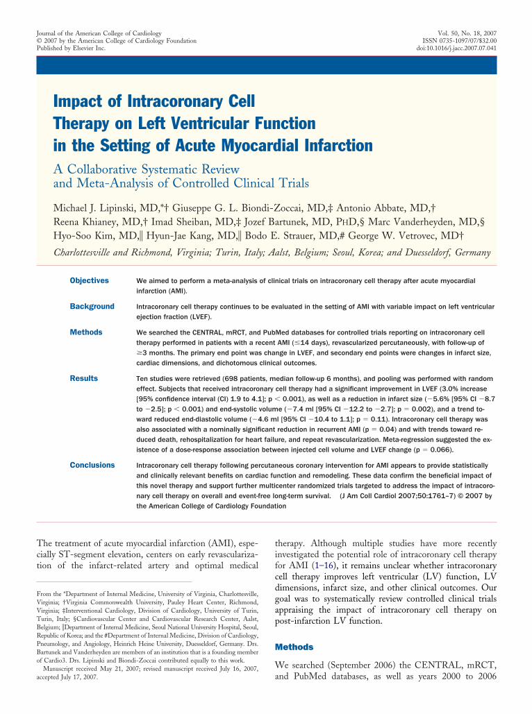
Impact of Intracoronary Cell Therapy on Left Ventricular Function in the Setting of Acute Myocardial Infarction A Collaborative Systematic Review and Meta-Analysis of Controlled Clinical Trials Michael J. Lipinski, MD,*† Giuseppe G. L. Biondi-Zoccai, MD,‡ Antonio Abbate, MD,† Reena Khianey, MD,† Imad Sheiban, MD,‡ Jozef Bartunek, MD, PHD,§ Marc Vanderheyden, MD,§ Hyo-Soo Kim, MD, Hyun-Jae Kang, MD, Bodo E. Strauer, MD,# George W. Vetrovec, MD† Charlottesville and Richmond, Virginia; Turin, Italy; Aalst, Belgium; Seoul, Korea; and Duesseldorf, Germany Objectives We aimed to perform a meta-analysis of clinical trials on intracoronary cell therapy after acute myocardial infarction (AMI). Background Intracoronary cell therapy continues to be evaluated in the setting of AMI with variable impact on left ventricular ejection fraction (LVEF). Methods We searched the CENTRAL, mRCT, and PubMed databases for controlled trials reporting on intracoronary cell therapy performed in patients with a recent AMI (14 days), revascularized percutaneously, with follow-up of 3 months. The primary end point was change in LVEF, and secondary end points were changes in infarct size, cardiac dimensions, and dichotomous clinical outcomes. Results Ten studies were retrieved (698 patients, median follow-up 6 months), and pooling was performed with random effect. Subjects that received intracoronary cell therapy had a significant improvement in LVEF (3.0% increase [95% confidence interval (CI) 1.9 to 4.1]; p 0.001), as well as a reduction in infarct size (5.6% [95% CI 8.7 to 2.5]; p 0.001) and end-systolic volume (7.4 ml [95% CI 12.2 to 2.7]; p 0.002), and a trend to- ward reduced end-diastolic volume (4.6 ml [95% CI 10.4 to 1.1]; p 0.11). Intracoronary cell therapy was also associated with a nominally significant reduction in recurrent AMI (p 0.04) and with trends toward re- duced death, rehospitalization for heart failure, and repeat revascularization. Meta-regression suggested the ex- istence of a dose-response association between injected cell volume and LVEF change (p 0.066). Conclusions Intracoronary cell therapy following percutaneous coronary intervention for AMI appears to provide statistically and clinically relevant benefits on cardiac function and remodeling. These data confirm the beneficial impact of this novel therapy and support further multicenter randomized trials targeted to address the impact of intracoro- nary cell therapy on overall and event-free long-term survival. (J Am Coll Cardiol 2007;50:1761–7) © 2007 by the American College of Cardiology Foundation The treatment of acute myocardial infarction (AMI), espe- cially ST-segment elevation, centers on early revasculariza- tion of the infarct-related artery and optimal medical therapy. Although multiple studies have more recently investigated the potential role of intracoronary cell therapy for AMI (1–16), it remains unclear whether intracoronary cell therapy improves left ventricular (LV) function, LV dimensions, infarct size, and other clinical outcomes. Our goal was to systematically review controlled clinical trials appraising the impact of intracoronary cell therapy on post-infarction LV function. Methods We searched (September 2006) the CENTRAL, mRCT, and PubMed databases, as well as years 2000 to 2006 From the *Department of Internal Medicine, University of Virginia, Charlottesville, Virginia; †Virginia Commonwealth University, Pauley Heart Center, Richmond, Virginia; ‡Interventional Cardiology, Division of Cardiology, University of Turin, Turin, Italy; §Cardiovascular Center and Cardiovascular Research Center, Aalst, Belgium; Department of Internal Medicine, Seoul National University Hospital, Seoul, Republic of Korea; and the #Department of Internal Medicine, Division of Cardiology, Pneumology, and Angiology, Heinrich Heine University, Duesseldorf, Germany. Drs. Bartunek and Vanderheyden are members of an institution that is a founding member of Cardio3. Drs. Lipinski and Biondi-Zoccai contributed equally to this work. Manuscript received May 21, 2007; revised manuscript received July 16, 2007, accepted July 17, 2007. Journal of the American College of Cardiology Vol. 50, No. 18, 2007 © 2007 by the American College of Cardiology Foundation ISSN 0735-1097/07/$32.00 Published by Elsevier Inc. doi:10.1016/j.jacc.2007.07.041
Transcript of Impact of Intracoronary Cell Therapy on Left Ventricular Function in the Setting of Acute Myocardial...
Tct
FVVTBRPBo
a
Journal of the American College of Cardiology Vol. 50, No. 18, 2007© 2007 by the American College of Cardiology Foundation ISSN 0735-1097/07/$32.00P
Impact of Intracoronary CellTherapy on Left Ventricular Functionin the Setting of Acute Myocardial InfarctionA Collaborative Systematic Reviewand Meta-Analysis of Controlled Clinical Trials
Michael J. Lipinski, MD,*† Giuseppe G. L. Biondi-Zoccai, MD,‡ Antonio Abbate, MD,†Reena Khianey, MD,† Imad Sheiban, MD,‡ Jozef Bartunek, MD, PHD,§ Marc Vanderheyden, MD,§Hyo-Soo Kim, MD,� Hyun-Jae Kang, MD,� Bodo E. Strauer, MD,# George W. Vetrovec, MD†
Charlottesville and Richmond, Virginia; Turin, Italy; Aalst, Belgium; Seoul, Korea; and Duesseldorf, Germany
Objectives We aimed to perform a meta-analysis of clinical trials on intracoronary cell therapy after acute myocardialinfarction (AMI).
Background Intracoronary cell therapy continues to be evaluated in the setting of AMI with variable impact on left ventricularejection fraction (LVEF).
Methods We searched the CENTRAL, mRCT, and PubMed databases for controlled trials reporting on intracoronary celltherapy performed in patients with a recent AMI (�14 days), revascularized percutaneously, with follow-up of�3 months. The primary end point was change in LVEF, and secondary end points were changes in infarct size,cardiac dimensions, and dichotomous clinical outcomes.
Results Ten studies were retrieved (698 patients, median follow-up 6 months), and pooling was performed with randomeffect. Subjects that received intracoronary cell therapy had a significant improvement in LVEF (3.0% increase[95% confidence interval (CI) 1.9 to 4.1]; p � 0.001), as well as a reduction in infarct size (�5.6% [95% CI �8.7to �2.5]; p � 0.001) and end-systolic volume (�7.4 ml [95% CI �12.2 to �2.7]; p � 0.002), and a trend to-ward reduced end-diastolic volume (�4.6 ml [95% CI �10.4 to 1.1]; p � 0.11). Intracoronary cell therapy wasalso associated with a nominally significant reduction in recurrent AMI (p � 0.04) and with trends toward re-duced death, rehospitalization for heart failure, and repeat revascularization. Meta-regression suggested the ex-istence of a dose-response association between injected cell volume and LVEF change (p � 0.066).
Conclusions Intracoronary cell therapy following percutaneous coronary intervention for AMI appears to provide statisticallyand clinically relevant benefits on cardiac function and remodeling. These data confirm the beneficial impact ofthis novel therapy and support further multicenter randomized trials targeted to address the impact of intracoro-nary cell therapy on overall and event-free long-term survival. (J Am Coll Cardiol 2007;50:1761–7) © 2007 bythe American College of Cardiology Foundation
ublished by Elsevier Inc. doi:10.1016/j.jacc.2007.07.041
tifcdgap
M
W
he treatment of acute myocardial infarction (AMI), espe-ially ST-segment elevation, centers on early revasculariza-ion of the infarct-related artery and optimal medical
rom the *Department of Internal Medicine, University of Virginia, Charlottesville,irginia; †Virginia Commonwealth University, Pauley Heart Center, Richmond,irginia; ‡Interventional Cardiology, Division of Cardiology, University of Turin,urin, Italy; §Cardiovascular Center and Cardiovascular Research Center, Aalst,elgium; �Department of Internal Medicine, Seoul National University Hospital, Seoul,epublic of Korea; and the #Department of Internal Medicine, Division of Cardiology,neumology, and Angiology, Heinrich Heine University, Duesseldorf, Germany. Drs.artunek and Vanderheyden are members of an institution that is a founding memberf Cardio3. Drs. Lipinski and Biondi-Zoccai contributed equally to this work.
aManuscript received May 21, 2007; revised manuscript received July 16, 2007,
ccepted July 17, 2007.
herapy. Although multiple studies have more recentlynvestigated the potential role of intracoronary cell therapyor AMI (1–16), it remains unclear whether intracoronaryell therapy improves left ventricular (LV) function, LVimensions, infarct size, and other clinical outcomes. Ouroal was to systematically review controlled clinical trialsppraising the impact of intracoronary cell therapy onost-infarction LV function.
ethods
e searched (September 2006) the CENTRAL, mRCT,
nd PubMed databases, as well as years 2000 to 2006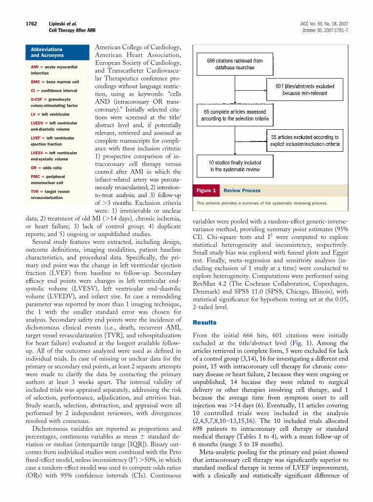
dor
ocmfesvptadtfuipwaioSpr
pvcfic(
vvCsStceRDs2
R
Feaopnudbi1(6m6
ts
1762 Lipinski et al. JACC Vol. 50, No. 18, 2007Cell Therapy After AMI October 30, 2007:1761–7
American College of Cardiology,American Heart Association,European Society of Cardiology,and Transcatheter Cardiovascu-lar Therapeutics conference pro-ceedings without language restric-tion, using as keywords: “cellsAND (intracoronary OR trans-coronary).” Initially selected cita-tions were screened at the title/abstract level and, if potentiallyrelevant, retrieved and assessed ascomplete manuscripts for compli-ance with these inclusion criteria:1) prospective comparison of in-tracoronary cell therapy versuscontrol after AMI in which theinfarct-related artery was percuta-neously revascularized; 2) intention-to-treat analysis; and 3) follow-upof �3 months. Exclusion criteriawere: 1) irretrievable or unclear
ata; 2) treatment of old MI (�14 days), chronic ischemia,r heart failure; 3) lack of control group; 4) duplicateeports; and 5) ongoing or unpublished studies.
Several study features were extracted, including design,utcome definitions, imaging modalities, patient baselineharacteristics, and procedural data. Specifically, the pri-ary end point was the change in left ventricular ejection
raction (LVEF) from baseline to follow-up. Secondaryfficacy end points were changes in left ventricular end-ystolic volume (LVESV), left ventricular end-diastolicolume (LVEDV), and infarct size. In case a remodelingarameter was reported by more than 1 imaging technique,he 1 with the smaller standard error was chosen fornalysis. Secondary safety end points were the incidence ofichotomous clinical events (i.e., death, recurrent AMI,arget vessel revascularization [TVR], and rehospitalizationor heart failure) evaluated at the longest available follow-p. All of the outcomes analyzed were used as defined inndividual trials. In case of missing or unclear data for therimary or secondary end points, at least 2 separate attemptsere made to clarify the data by contacting the primary
uthors at least 3 weeks apart. The internal validity ofncluded trials was appraised separately, addressing the riskf selection, performance, adjudication, and attrition bias.tudy search, selection, abstraction, and appraisal were allerformed by 2 independent reviewers, with divergencesesolved with consensus.
Dichotomous variables are reported as proportions andercentages, continuous variables as mean � standard de-iation or median (interquartile range [IQR]). Binary out-omes from individual studies were combined with the Petoxed-effect model, unless inconsistency (I2) �50%, in whichase a random-effect model was used to compute odds ratios
Abbreviationsand Acronyms
AMI � acute myocardialinfarction
BMC � bone marrow cell
CI � confidence interval
G-CSF � granulocytecolony-stimulating factor
LV � left ventricular
LVEDV � left ventricularend-diastolic volume
LVEF � left ventricularejection fraction
LVESV � left ventricularend-systolic volume
OR � odds ratio
PMC � peripheralmononuclear cell
TVR � target vesselrevascularization
ORs) with 95% confidence intervals (CIs). Continuous w
ariables were pooled with a random-effect generic-inverse-ariance method, providing summary point estimates (95%I). Chi-square tests and I2 were computed to explore
tatistical heterogeneity and inconsistency, respectively.mall study bias was explored with funnel plots and Eggerest. Finally, meta-regression and sensitivity analyses (in-luding exclusion of 1 study at a time) were conducted toxplore heterogeneity. Computations were performed usingevMan 4.2 (The Cochrane Collaboration, Copenhagen,enmark) and SPSS 11.0 (SPSS, Chicago, Illinois), with
tatistical significance for hypothesis testing set at the 0.05,-tailed level.
esults
rom the initial 666 hits, 601 citations were initiallyxcluded at the title/abstract level (Fig. 1). Among therticles retrieved in complete form, 5 were excluded for lackf a control group (3,14), 16 for investigating a different endoint, 15 with intracoronary cell therapy for chronic coro-ary disease or heart failure, 2 because they were ongoing ornpublished, 14 because they were related to surgicalelivery or other therapies involving cell therapy, and 1ecause the average time from symptom onset to cellnjection was �14 days (6). Eventually, 11 articles covering0 controlled trials were included in the analysis2,4,5,7,8,10–13,15,16). The 10 included trials allocated98 patients to intracoronary cell therapy or standardedical therapy (Tables 1 to 4), with a mean follow-up ofmonths (range 3 to 18 months).Meta-analytic pooling for the primary end point showed
hat intracoronary cell therapy was significantly superior totandard medical therapy in terms of LVEF improvement,
Figure 1 Review Process
This scheme provides a summary of the systematic reviewing process.
ith a clinically and statistically significant difference of
3Ib[i�iC
ia1trfarrb
paiw
tdLibtip
a(r(i54dn
sfpem
M
B lar ejer ll motio
P
A
1763JACC Vol. 50, No. 18, 2007 Lipinski et al.October 30, 2007:1761–7 Cell Therapy After AMI
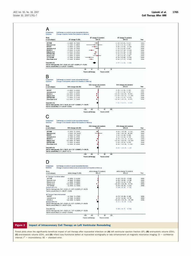
.0% (95% CI 1.9% to 4.1%; p � 0.00001; I2 � 73.2%).ntracoronary cell therapy was similarly found to haveenefit concerning LVESV (average difference �7.4 ml95% CI �12.2 to �2.7]; p � 0.002; I2 � 95.8%) andnfarct size (average difference � 5.6% [95% CI �8.7 to
2.5]; p � 0.0004; I2 � 92.6%). There was a trend formprovement in LVEDV (average difference �4.6 ml [95%I �10.4 to 1.1]; p � 0.11; I2 � 95.2%) (Fig. 2).Comparing dichotomous clinical end points (Table 4),
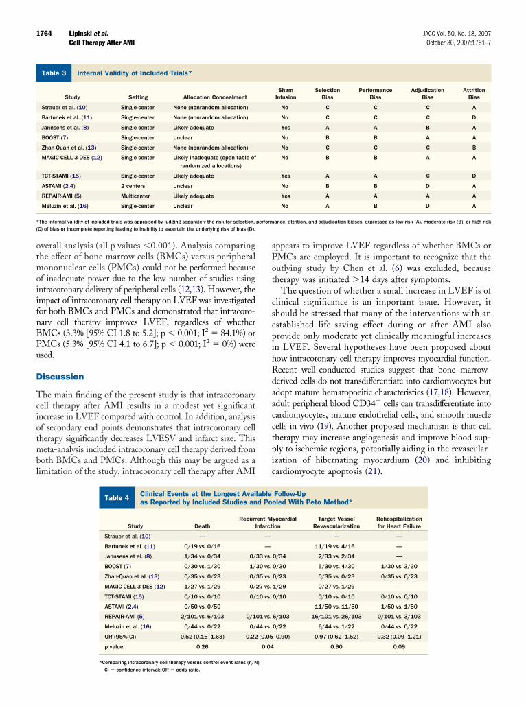
ntracoronary cell therapy proved to be notably safe, withoutny increase in the risk of TVR (OR 1.08 [95% CI 0.60 to.96]; p � 0.80; I2 � 25.3%). Conversely, intracoronary cellherapy tended to be associated, albeit nonsignificantly, witheductions in the risk of death or rehospitalization for heartailure. In addition, intracoronary cell therapy was associ-ted with a nominally statistically significant decrease inecurrent AMI (p � 0.04), but this finding should beegarded as hypothesis-generating only, given the low num-er of events in all but 1 of the studies (5).A number of exploratory meta-regression analyses were
erformed to appraise the impact of the following moder-tor or covariates on the changes in LVEF associated withntracoronary cell therapy. Specifically, at the overall analysise did not find statistically significant association between:
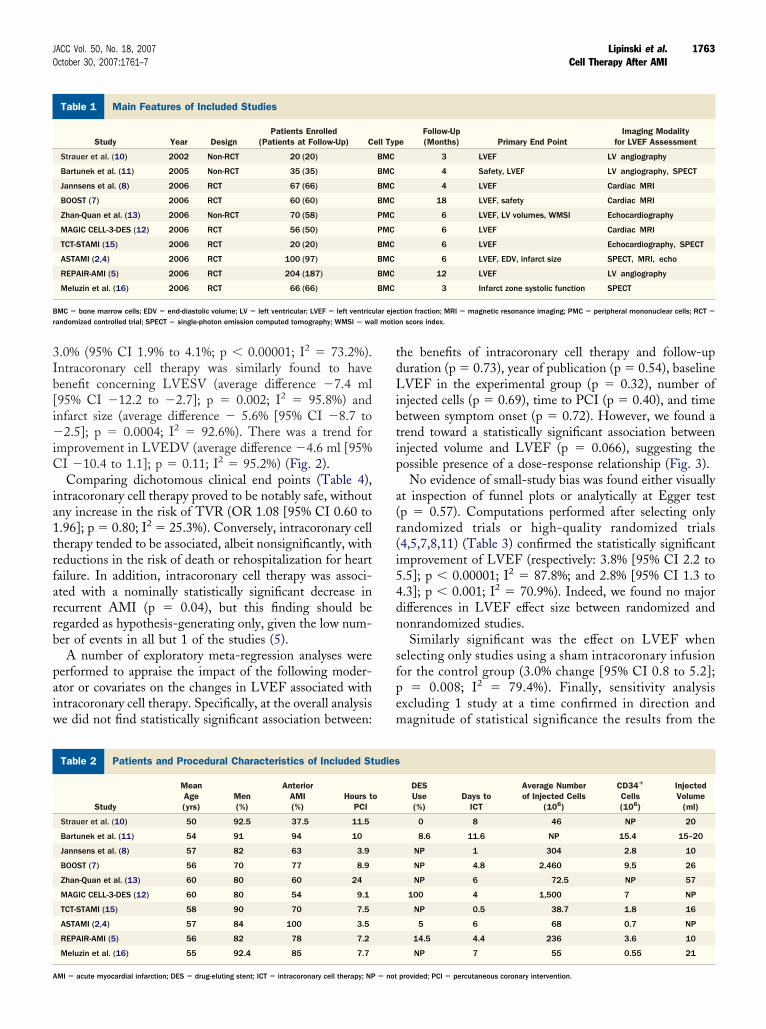
ain Features of Included Studies
Table 1 Main Features of Included Studies
Study Year DesignPatients Enrolled
(Patients at Follow-Up) C
Strauer et al. (10) 2002 Non-RCT 20 (20)
Bartunek et al. (11) 2005 Non-RCT 35 (35)
Jannsens et al. (8) 2006 RCT 67 (66)
BOOST (7) 2006 RCT 60 (60)
Zhan-Quan et al. (13) 2006 Non-RCT 70 (58)
MAGIC CELL-3-DES (12) 2006 RCT 56 (50)
TCT-STAMI (15) 2006 RCT 20 (20)
ASTAMI (2,4) 2006 RCT 100 (97)
REPAIR-AMI (5) 2006 RCT 204 (187)
Meluzin et al. (16) 2006 RCT 66 (66)
MC � bone marrow cells; EDV � end-diastolic volume; LV � left ventricular; LVEF � left ventricuandomized controlled trial; SPECT � single-photon emission computed tomography; WMSI � wa
atients and Procedural Characteristics of Included Studies
Table 2 Patients and Procedural Characteristics of Included St
Study
MeanAge(yrs)
Men(%)
AnteriorAMI(%)
Hours tPCI
Strauer et al. (10) 50 92.5 37.5 11.5
Bartunek et al. (11) 54 91 94 10
Jannsens et al. (8) 57 82 63 3.9
BOOST (7) 56 70 77 8.9
Zhan-Quan et al. (13) 60 80 60 24
MAGIC CELL-3-DES (12) 60 80 54 9.1
TCT-STAMI (15) 58 90 70 7.5
ASTAMI (2,4) 57 84 100 3.5
REPAIR-AMI (5) 56 82 78 7.2
Meluzin et al. (16) 55 92.4 85 7.7
MI � acute myocardial infarction; DES � drug-eluting stent; ICT � intracoronary cell therapy; NP � not
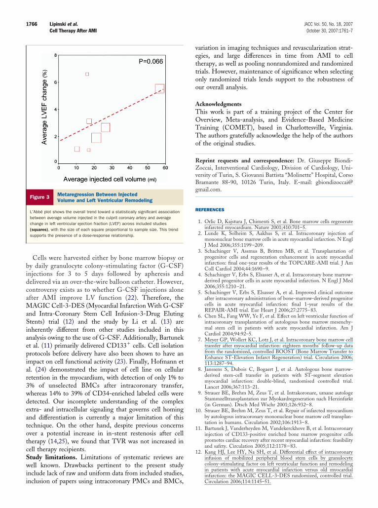
he benefits of intracoronary cell therapy and follow-upuration (p � 0.73), year of publication (p � 0.54), baselineVEF in the experimental group (p � 0.32), number of
njected cells (p � 0.69), time to PCI (p � 0.40), and timeetween symptom onset (p � 0.72). However, we found arend toward a statistically significant association betweennjected volume and LVEF (p � 0.066), suggesting theossible presence of a dose-response relationship (Fig. 3).No evidence of small-study bias was found either visually
t inspection of funnel plots or analytically at Egger testp � 0.57). Computations performed after selecting onlyandomized trials or high-quality randomized trials4,5,7,8,11) (Table 3) confirmed the statistically significantmprovement of LVEF (respectively: 3.8% [95% CI 2.2 to.5]; p � 0.00001; I2 � 87.8%; and 2.8% [95% CI 1.3 to.3]; p � 0.001; I2 � 70.9%). Indeed, we found no majorifferences in LVEF effect size between randomized andonrandomized studies.Similarly significant was the effect on LVEF when
electing only studies using a sham intracoronary infusionor the control group (3.0% change [95% CI 0.8 to 5.2];
� 0.008; I2 � 79.4%). Finally, sensitivity analysisxcluding 1 study at a time confirmed in direction andagnitude of statistical significance the results from the
eFollow-Up(Months) Primary End Point
Imaging Modalityfor LVEF Assessment
3 LVEF LV angiography
4 Safety, LVEF LV angiography, SPECT
4 LVEF Cardiac MRI
18 LVEF, safety Cardiac MRI
6 LVEF, LV volumes, WMSI Echocardiography
6 LVEF Cardiac MRI
6 LVEF Echocardiography, SPECT
6 LVEF, EDV, infarct size SPECT, MRI, echo
12 LVEF LV angiography
3 Infarct zone systolic function SPECT
ction fraction; MRI � magnetic resonance imaging; PMC � peripheral mononuclear cells; RCT �
n score index.
DESUse(%)
Days toICT
Average Numberof Injected Cells
(106)
CD34�
Cells(106)
InjectedVolume
(ml)
0 8 46 NP 20
8.6 11.6 NP 15.4 15–20
NP 1 304 2.8 10
NP 4.8 2,460 9.5 26
NP 6 72.5 NP 57
100 4 1,500 7 NP
NP 0.5 38.7 1.8 16
5 6 68 0.7 NP
14.5 4.4 236 3.6 10
NP 7 55 0.55 21
ell Typ
BMC
BMC
BMC
BMC
PMC
PMC
BMC
BMC
BMC
BMC
udies
o
provided; PCI � percutaneous coronary intervention.
otmoiifnBPu
D
Tciotmbl
aPot
csepihRdaacctpic
I
* perform(
1764 Lipinski et al. JACC Vol. 50, No. 18, 2007Cell Therapy After AMI October 30, 2007:1761–7
verall analysis (all p values �0.001). Analysis comparinghe effect of bone marrow cells (BMCs) versus peripheralononuclear cells (PMCs) could not be performed because
f inadequate power due to the low number of studies usingntracoronary delivery of peripheral cells (12,13). However, thempact of intracoronary cell therapy on LVEF was investigatedor both BMCs and PMCs and demonstrated that intracoro-ary cell therapy improves LVEF, regardless of whetherMCs (3.3% [95% CI 1.8 to 5.2]; p � 0.001; I2 � 84.1%) orMCs (5.3% [95% CI 4.1 to 6.7]; p � 0.001; I2 � 0%) weresed.
iscussion
he main finding of the present study is that intracoronaryell therapy after AMI results in a modest yet significantncrease in LVEF compared with control. In addition, analysisf secondary end points demonstrates that intracoronary cellherapy significantly decreases LVESV and infarct size. Thiseta-analysis included intracoronary cell therapy derived from
oth BMCs and PMCs. Although this may be argued as aimitation of the study, intracoronary cell therapy after AMI
nternal Validity of Included Trials*
Table 3 Internal Validity of Included Trials*
Study Setting Allocation Concealment
Strauer et al. (10) Single-center None (nonrandom allocation)
Bartunek et al. (11) Single-center None (nonrandom allocation)
Jannsens et al. (8) Single-center Likely adequate
BOOST (7) Single-center Unclear
Zhan-Quan et al. (13) Single-center None (nonrandom allocation)
MAGIC-CELL-3-DES (12) Single-center Likely inadequate (open table ofrandomized allocations)
TCT-STAMI (15) Single-center Likely adequate
ASTAMI (2,4) 2 centers Unclear
REPAIR-AMI (5) Multicenter Likely adequate
Meluzin et al. (16) Single-center Unclear
The internal validity of included trials was appraised by judging separately the risk for selection,C) of bias or incomplete reporting leading to inability to ascertain the underlying risk of bias (D).
Clinical Events at the Longest Available Follow-as Reported by Included Studies and Pooled Wi
Table 4 Clinical Events at the Longest Avaias Reported by Included Studies an
Study DeathRecurr
I
Strauer et al. (10) —
Bartunek et al. (11) 0/19 vs. 0/16
Jannsens et al. (8) 1/34 vs. 0/34 0/
BOOST (7) 0/30 vs. 1/30 1/
Zhan-Quan et al. (13) 0/35 vs. 0/23 0/
MAGIC-CELL-3-DES (12) 1/27 vs. 1/29 0/
TCT-STAMI (15) 0/10 vs. 0/10 0/
ASTAMI (2,4) 0/50 vs. 0/50
REPAIR-AMI (5) 2/101 vs. 6/103 0/1
Meluzin et al. (16) 0/44 vs. 0/22 0/
OR (95% CI) 0.52 (0.16–1.63) 0.22
p value 0.26
*Comparing intracoronary cell therapy versus control event rates (n/N).CI � confidence interval; OR � odds ratio.
ppears to improve LVEF regardless of whether BMCs orMCs are employed. It is important to recognize that theutlying study by Chen et al. (6) was excluded, becauseherapy was initiated �14 days after symptoms.
The question of whether a small increase in LVEF is oflinical significance is an important issue. However, ithould be stressed that many of the interventions with anstablished life-saving effect during or after AMI alsorovide only moderate yet clinically meaningful increasesn LVEF. Several hypotheses have been proposed aboutow intracoronary cell therapy improves myocardial function.ecent well-conducted studies suggest that bone marrow-erived cells do not transdifferentiate into cardiomyocytes butdopt mature hematopoeitic characteristics (17,18). However,dult peripheral blood CD34� cells can transdifferentiate intoardiomyocytes, mature endothelial cells, and smooth muscleells in vivo (19). Another proposed mechanism is that cellherapy may increase angiogenesis and improve blood sup-ly to ischemic regions, potentially aiding in the revascular-zation of hibernating myocardium (20) and inhibitingardiomyocyte apoptosis (21).
ShamInfusion
SelectionBias
PerformanceBias
AdjudicationBias
AttritionBias
No C C C A
No C C C D
Yes A A B A
No B B A A
No C C C B
No B B A A
Yes A A C D
No B B D A
Yes A A A A
No A B D A
ance, attrition, and adjudication biases, expressed as low risk (A), moderate risk (B), or high risk
to Method*
Follow-Upoled With Peto Method*
yocardialion
Target VesselRevascularization
Rehospitalizationfor Heart Failure
— —
11/19 vs. 4/16 —
0/34 2/33 vs. 2/34 —
0/30 5/30 vs. 4/30 1/30 vs. 3/30
0/23 0/35 vs. 0/23 0/35 vs. 0/23
1/29 0/27 vs. 1/29 —
0/10 0/10 vs. 0/10 0/10 vs. 0/10
11/50 vs. 11/50 1/50 vs. 1/50
6/103 16/101 vs. 26/103 0/101 vs. 3/103
0/22 6/44 vs. 1/22 0/44 vs. 0/22
–0.90) 0.97 (0.62–1.52) 0.32 (0.09–1.21)
0.90 0.09
Upth Pe
labled Po
ent Mnfarct
—
—
33 vs.
30 vs.
35 vs.
27 vs.
10 vs.
—
01 vs.
44 vs.
(0.05
0.04
1765JACC Vol. 50, No. 18, 2007 Lipinski et al.October 30, 2007:1761–7 Cell Therapy After AMI
Figure 2 Impact of Intracoronary Cell Therapy on Left Ventricular Remodeling
Forest plots show the significantly beneficial impact of cell therapy after myocardial infarction on (A) left ventricular ejection fraction (EF), (B) end-systolic volume (ESV),(C) end-diastolic volume (EDV), and (D) infarct size/functional defect at myocardial scintigraphy or late enhancement at magnetic resonance imaging. CI � confidenceinterval; I2 � inconsistency; SE � standard error.
bidcaMaSiaepiar3wdeatotcSwii
vettoo
ATOTTo
RZvBg
R
1
1
1
1766 Lipinski et al. JACC Vol. 50, No. 18, 2007Cell Therapy After AMI October 30, 2007:1761–7
Cells were harvested either by bone marrow biopsy ory daily granulocyte colony-stimulating factor (G-CSF)njections for 3 to 5 days followed by apheresis andelivered via an over-the-wire balloon catheter. However,ontroversy exists as to whether G-CSF injections alonefter AMI improve LV function (22). Therefore, the
AGIC Cell-3-DES (Myocardial Infarction With G-CSFnd Intra-Coronary Stem Cell Infusion-3-Drug Elutingtents) trial (12) and the study by Li et al. (13) are
nherently different from other studies included in thisnalysis owing to the use of G-CSF. Additionally, Bartunekt al. (11) primarily delivered CD133� cells. Cell isolationrotocols before delivery have also been shown to have anmpact on cell functional activity (23). Finally, Hofmann etl. (24) demonstrated the impact of cell line on cellularetention in the myocardium, with detection of only 1% to% of unselected BMCs after intracoronary transfer,hereas 14% to 39% of CD34-enriched labeled cells wereetected. Our incomplete understanding of the complexxtra- and intracellular signaling that governs cell homingnd differentiation is currently a major limitation of thisechnique. On the other hand, despite previous concernsver a potential increase in in-stent restenosis after cellherapy (14,25), we found that TVR was not increased inell therapy recipients.tudy limitations. Limitations of systematic reviews areell known. Drawbacks pertinent to the present study
nclude lack of raw and uniform data from included studies,
Figure 3 Metaregression Between InjectedVolume and Left Ventricular Remodeling
L’Abbé plot shows the overall trend toward a statistically significant associationbetween average volume injected in the culprit coronary artery and averagechange in left ventricular ejection fraction (LVEF) across included studies(squares), with the size of each square proportional to sample size. This trendsupports the presence of a dose-response relationship.
nclusion of papers using intracoronary PMCs and BMCs,
ariation in imaging techniques and revascularization strat-gies, and large differences in time from AMI to cellherapy, as well as pooling nonrandomized and randomizedrials. However, maintenance of significance when selectingnly randomized trials lends support to the robustness ofur overall analysis.
cknowledgmentshis work is part of a training project of the Center forverview, Meta-analysis, and Evidence-Based Medicineraining (COMET), based in Charlottesville, Virginia.he authors gratefully acknowledge the help of the authorsf the original studies.
eprint requests and correspondence: Dr. Giuseppe Biondi-occai, Interventional Cardiology, Division of Cardiology, Uni-
ersity of Turin, S. Giovanni Battista “Molinette” Hospital, Corsoramante 88-90, 10126 Turin, Italy. E-mail: [email protected].
EFERENCES
1. Orlic D, Kajstura J, Chimenti S, et al. Bone marrow cells regenerateinfarcted myocardium. Nature 2001;410:701–5.
2. Lunde K, Solheim S, Aakhus S, et al. Intracoronary injection ofmononuclear bone marrow cells in acute myocardial infarction. N EnglJ Med 2006;355:1199–209.
3. Schachinger V, Assmus B, Britten MB, et al. Transplantation ofprogenitor cells and regeneration enhancement in acute myocardialinfarction: final one-year results of the TOPCARE-AMI trial. J AmColl Cardiol 2004;44:1690–9.
4. Schachinger V, Erbs S, Elsasser A, et al. Intracoronary bone marrow-derived progenitor cells in acute myocardial infarction. N Engl J Med2006;355:1210–21.
5. Schachinger V, Erbs S, Elsasser A, et al. Improved clinical outcomeafter intracoronary administration of bone-marrow-derived progenitorcells in acute myocardial infarction: final 1-year results of theREPAIR-AMI trial. Eur Heart J 2006;27:2775–83.
6. Chen SL, Fang WW, Ye F, et al. Effect on left ventricular function ofintracoronary transplantation of autologous bone marrow mesenchy-mal stem cell in patients with acute myocardial infarction. Am JCardiol 2004;94:92–5.
7. Meyer GP, Wollert KC, Lotz J, et al. Intracoronary bone marrow celltransfer after myocardial infarction: eighteen months’ follow-up datafrom the randomized, controlled BOOST (Bone Marrow Transfer toEnhance ST-Elevation Infarct Regeneration) trial. Circulation 2006;113:1287–94.
8. Janssens S, Dubois C, Bogaert J, et al. Autologous bone marrow-derived stem-cell transfer in patients with ST-segment elevationmyocardial infarction: double-blind, randomised controlled trial.Lancet 2006;367:113–21.
9. Strauer BE, Brehm M, Zeus T, et al. Intrakoronare, umane autologeStammzelltransplantation zur Myokardregeneration nach Herzinfarkt(in German). Dtsch Med Wschr 2001;126:932–8.
0. Strauer BE, Brehm M, Zeus T, et al. Repair of infarcted myocardiumby autologous intracoronary mononuclear bone marrow cell transplan-tation in humans. Circulation 2002;106:1913–8.
1. Bartunek J, Vanderheyden M, Vandekerckhove B, et al. Intracoronaryinjection of CD133-positive enriched bone marrow progenitor cellspromotes cardiac recovery after recent myocardial infarction: feasibilityand safety. Circulation 2005;112:1178–83.
2. Kang HJ, Lee HY, Na SH, et al. Differential effect of intracoronaryinfusion of mobilized peripheral blood stem cells by granulocytecolony-stimulating factor on left ventricular function and remodelingin patients with acute myocardial infarction versus old myocardial
infarction: the MAGIC CELL-3-DES randomized, controlled trial.Circulation 2006;114:1145–51.
1
1
1
1
1
1
1
2
2
2
2
2
2
1767JACC Vol. 50, No. 18, 2007 Lipinski et al.October 30, 2007:1761–7 Cell Therapy After AMI
3. Li ZA, Zhang M, Jing YZ, et al. The clinical study of autologousperipheral blood stem cell transplantation by intracoronory infusion inpatients with acute myocardial infarction (AMI). Int J Cardiol 2007;115:52–6.
4. Assmus B, Schachinger V, Teupe C, et al. Transplantation ofProgenitor Cells and Regeneration Enhancement in Acute MyocardialInfarction (TOPCARE-AMI). Circulation 2002;106:3009–17.
5. Ge J, Li Y, Qian J, et al. Efficacy of emergent transcatheter transplan-tation of stem cells for treatment of acute myocardial infarction(TCT-STAMI). Heart 2006;92:1764–7.
6. Meluzin J, Mayer J, Groch L, et al. Autologous transplantation ofmononuclear bone marrow cells in patients with acute myocardialinfarction: the effect of the dose of transplanted cells on myocardialfunction. Am Heart J 2006;152:975e9–15.
7. Balsam LB, Wagers AJ, Christensen JL, Kofidis T, Weissman IL,Robbins RC. Haematopoietic stem cells adopt mature haematopoieticfates in ischaemic myocardium. Nature 2004;428:668–73.
8. Murry CE, Soonpaa MH, Reinecke H, et al. Haematopoietic stemcells do not transdifferentiate into cardiac myocytes in myocardialinfarcts. Nature 2004;428:664–8.
9. Yeh ET, Zhang S, Wu HD, Korbling M, Willerson JT, Estrov Z.Transdifferentiation of human peripheral blood CD34�-enriched cellpopulation into cardiomyocytes, endothelial cells, and smooth muscle
cells in vivo. Circulation 2003;108:2070–3.0. Boyle AJ, Whitbourn R, Schlicht S, et al. Intra-coronary high-doseCD34� stem cells in patients with chronic ischemic heart disease: a12-month follow-up. Int J Cardiol 2006;109:21–7.
1. Kocher AA, Schuster MD, Szabolcs MJ, et al. Neovascularization ofischemic myocardium by human bone-marrow–derived angioblastsprevents cardiomyocyte apoptosis, reduces remodeling and improvescardiac function. Nat Med 2001;7:430–6.
2. Nienaber CA, Petzsch M, Kleine HD, Eckard H, Freund M, Ince H.Effects of granulocyte-colony-stimulating factor on mobilization ofbone-marrow–derived stem cells after myocardial infarction in hu-mans. Nat Clin Pract Cardiovasc Med 2006;3 Suppl 1:S73–7.
3. Seeger FH, Tonn T, Krzossok N, Zeiher AM, Dimmeler S. Cellisolation procedures matter: a comparison of different isolation proto-cols of bone marrow mononuclear cells used for cell therapy in patientswith acute myocardial infarction. Eur Heart J 2007;28:766–72.
4. Hofmann M, Wollert KC, Meyer GP, et al. Monitoring of bonemarrow cell homing into the infarcted human myocardium. Circula-tion 2005;111:2198–202.
5. Kang HJ, Kim HS, Zhang SY, et al. Effects of intracoronary infusionof peripheral blood stem-cells mobilised with granulocyte-colonystimulating factor on left ventricular systolic function and restenosisafter coronary stenting in myocardial infarction: the MAGIC CELL
randomised clinical trial. Lancet 2004;363:751–6.