Investigation of amino-tail translocation by the conserved YidC ...

10.1128/MCB.02478-05.
2006, 26(16):6261. DOI:Mol. Cell. Biol. B. Sacks, Warren S. Pear and Jon C. AsterMonideepa Roy, Yunsun Nam, Stephen C. Blacklow, David Mark Y. Chiang, Mina L. Xu, Gavin Histen, Olga Shestova, Leukemogenic Activity of NOTCH1Regulatory Sequence That Influences the Identification of a Conserved Negative
http://mcb.asm.org/content/26/16/6261Updated information and services can be found at:
These include:
REFERENCEShttp://mcb.asm.org/content/26/16/6261#ref-list-1at:
This article cites 44 articles, 26 of which can be accessed free
CONTENT ALERTS more»articles cite this article),
Receive: RSS Feeds, eTOCs, free email alerts (when new
http://journals.asm.org/site/misc/reprints.xhtmlInformation about commercial reprint orders: http://journals.asm.org/site/subscriptions/To subscribe to to another ASM Journal go to:
on Decem
ber 24, 2013 by guesthttp://m
cb.asm.org/
Dow
nloaded from
on Decem
ber 24, 2013 by guesthttp://m
cb.asm.org/
Dow
nloaded from

MOLECULAR AND CELLULAR BIOLOGY, Aug. 2006, p. 6261–6271 Vol. 26, No. 160270-7306/06/$08.00�0 doi:10.1128/MCB.02478-05Copyright © 2006, American Society for Microbiology. All Rights Reserved.
Identification of a Conserved Negative Regulatory Sequence ThatInfluences the Leukemogenic Activity of NOTCH1
Mark Y. Chiang,1† Mina L. Xu,2† Gavin Histen,2 Olga Shestova,3 Monideepa Roy,2 Yunsun Nam,2Stephen C. Blacklow,2 David B. Sacks,2 Warren S. Pear,3 and Jon C. Aster2*
Department of Hematology/Oncology, Abramson Family Cancer Research Institute, University of Pennsylvania, Philadelphia,Pennsylvania 191041; Department of Pathology, Brigham and Women’s Hospital and Harvard Medical School,
Boston, Massachusetts 021152; and Department of Pathology and Laboratory Medicine,Abramson Family Cancer Research Institute, Institute for Medicine and Engineering,
University of Pennsylvania, Philadelphia, Pennsylvania 191043
Received 28 December 2005/Returned for modification 8 February 2006/Accepted 26 May 2006
NOTCH1 is a large type I transmembrane receptor that regulates normal T-cell development via asignaling pathway that relies on regulated proteolysis. Ligand binding induces proteolytic cleavages inNOTCH1 that release its intracellular domain (ICN1), which translocates to the nucleus and activatestarget genes by forming a short-lived nuclear complex with two other proteins, the DNA-binding factorCSL and a Mastermind-like (MAML) coactivator. Recent work has shown that human T-ALL is frequentlyassociated with C-terminal NOTCH1 truncations, which uniformly remove sequences lying between res-idues 2524 and 2556. This region includes the highly conserved sequence WSSSSP (S4), which based onits amino acid content appeared to be a likely site for regulatory serine phosphorylation events. We showhere that the mutation of the S4 sequence leads to hypophosphorylation of ICN1; increased NOTCH1signaling; and the stabilization of complexes containing ICN1, CSL, and MAML1. Consistent with thesein vitro studies, mutation of the WSSSSP sequence converts nonleukemogenic weak gain-of-functionNOTCH1 alleles into alleles that cause aggressive T-ALLs in a murine bone marrow transplant model.These studies indicate that S4 is an important negative regulatory sequence and that the deletion of S4likely contributes to the development of human T-ALL.
NOTCH receptors and downstream mediators participate ina signaling pathway that variously regulates the specification ofcell fate, proliferation, self-renewal, survival, and apoptosis ina dose- and context-dependent fashion (2). Like other mem-bers of the NOTCH receptor family, human NOTCH1 is alarge multimodular type I transmembrane glycoprotein (Fig.1A). Newly synthesized NOTCH1 is cleaved by furin at a sitetermed S1 just external to the transmembrane domain (21),yielding two noncovalently associated extracellular (NEC) andtransmembrane (NTM) subunits (5, 21, 29). Binding of ligandsto NEC triggers two sequential proteolytic events within theNTM subunit at sites S2 and S3. S2 cleavage occurs just externalto the transmembrane domain and is catalyzed by ADAM-typemetalloproteases (6, 24). This creates a short-lived intermedi-ate, NTM*, which is recognized by nicastrin (33), a componentof the protease complex called �-secretase (8, 18, 32). Addi-tional cleavages by �-secretase free the intracellular domain ofNOTCH1 (ICN1), allowing it to translocate to the nucleus,where it activates transcription through the formation of aternary complex with the DNA-binding factor CSL (16, 19, 36,44) and coactivator proteins of the Mastermind-like (MAML)family (27, 28, 43).
Nuclear ICN1 is short-lived. One mechanism that appearsto promote the rapid turnover of the CSL/ICN1/MAMLtranscription complex involves the recruitment of mediator
complexes and CycC-CDK8 through the C-terminal tail ofMAML1 (13). Phosphorylation of ICN1 on multiple C-ter-minal serine residues by CycC-CDK8 is hypothesized tocreate recognition sites for E3 ligases such as FBW7/Sel10(13), which has been implicated in the ubiquitylation andsubsequent degradation of ICN (38, 39). Some of the sitestargeted by CycC-CDK8 lie in the far C-terminal portion ofNOTCH1 (13), an unstructured region that is enriched forthe amino acids proline, glutamate, serine, and threonine(PEST). PEST sequences regulate the degradation of anumber of proteins (30), sometimes by serving as substratesfor phosphorylation events that mark a protein for degra-dation (22). In the case of CycC-CDK8, phosphorylation ofICN is hypothesized to couple MAML-dependent transcrip-tional activation to rapid ICN degradation (12, 13). How-ever, there is evidence that NOTCH stability is also regu-lated at other levels. For example, phosphorylation byGSK� appears to promote the degradation of the intracel-lular domain of NOTCH2 (9), and E3 ligases of the Itchfamily have been implicated in the ubiquitylation and reg-ulation of membrane-associated NOTCH receptors (23, 34).Thus, inputs from multiple pathways regulate NOTCH atthe level of protein stability during different stages of re-ceptor activation and trafficking.
Increased NOTCH1 signaling plays a central part in thepathogenesis of T-cell acute lymphoblastic leukemia (T-ALL), a tumor derived from T-cell progenitors. We ob-served that human T-ALLs commonly harbor frameshiftand stop codon mutations that delete various numbers ofC-terminal residues from NOTCH1 (41), a finding that was
* Corresponding author. Mailing address: Department of Pathology,Brigham and Women’s Hospital, Boston, MA 02115. Phone: (617)278-0032. Fax: (617) 264-5169. E-mail: [email protected].
† M.Y.C. and M.L.X. contributed equally to this study.
6261
on Decem
ber 24, 2013 by guesthttp://m
cb.asm.org/
Dow
nloaded from

presaged by the detection of retroviral insertions in murineT-ALLs that cause similar truncations (10, 14). Althoughthese mutations are scattered throughout the 3� end of exon34, all of the deletions found to date eliminate at leastresidues 2524 to 2556, suggesting that this minimal regioncontains at least one important motif that negatively regu-lates NOTCH1 signal strength. Here, we analyze the role ofa short conserved sequence, WSSSSP (referred to as S4),found within the minimal deleted region that influences notonly the function and stability of activated NOTCH1 butalso its leukemogenic activity.
MATERIALS AND METHODS
Expression plasmids. A diagram depicting the various forms of NOTCH1used in these studies is shown in Fig. 1. Expression constructs that encodefull-length human NOTCH1 (residues M1 to K2556); �EGF, a form bearing adeletion that removes the coding region for epidermal growth factor (EGF)-likerepeats 1 to 36 (residues R23 to I1446); �EGF�LNR, a form bearing a deletionthat removes the coding region for EGF-like repeats 1 to 36 and the threeLin12/NOTCH repeats (residues R23 to C1562); and ICN1 (residues 1762 to2556) have been described (31). Expression constructs for forms of ICN1 bearinga N-terminal FLAG-tag were created by PCR with primers containing a consen-sus Kozak start codon followed by the coding sequence for the FLAG epitope.A C-terminal deletion removing residues 2473 to 2556 (originally identified inthe cell line ALL-SIL) has been described (41). NOTCH1-GAL4 chimericcDNAs were creating by ligating a PCR product encoding the DNA-bindingdomain of GAL4 in frame to ICN1 cDNA cut with the restriction enzymesBsu36I and NcoI, which removes sequences encoding the RAM and ANK do-mains. In other constructs, premature stop codons and point mutations wereintroduced by using the QuikChange kit (Stratagene). cDNAs were assembledvariously in pcDNA3 (Invitrogen); pcDNA5 (Invitrogen); or the retroviral vectorMSCV-GFP, which drives expression of NOTCH1 and green fluorescence pro-tein (GFP) from a single bicistronic RNA containing an internal ribosomal entrysequence (IRES). Expression plasmids for CSL-MYC (4), MAML1-GFP (43),and dominant-negative MAML1-GFP (42) have all been described. A pCMV2plasmid (Sigma) encoding a “kinase-dead” dominant-negative form of CDK8was kindly provided by Andrew Rice, Baylor University.
Cell culture. U2OS and 293T cells (American Type Culture Collection) weremaintained in Dulbecco modified Eagle medium (DMEM; Invitrogen) supple-
mented with 10% fetal bovine serum (Invitrogen), 2 mM L-glutamine (Invitrogen),100 U of penicillin (Invitrogen)/ml, and 100 �g of streptomycin (Invitrogen)/ml. 293TRex cells were obtained from Invitrogen. Cells were grown at 37°C under 5% CO2.
Transcriptional activation assays. NOTCH1 expression plasmids were intro-duced into U2OS cells by transient transfection with Lipofectamine Plus(Invitrogen) and assessed for their ability to activated a NOTCH-sensitive lucif-erase reporter gene, as described previously (4). Briefly, cells in 24-well disheswere cotransfected in triplicate with 10 ng of various pcDNA3-NOTCH1 expres-sion constructs, a NOTCH-sensitive firefly luciferase reporter gene (15), and aninternal control Renilla luciferase plasmid (Promega). Experiments involvingNOTCH1-GAL4 fusion constructs used a GAL4-luciferase reporter gene (Clon-tech). Total introduced DNA was kept constant by adding empty pcDNA3plasmid. Normalized firefly luciferase activities were measured in whole-cellextracts prepared 44 to 48 h after transfection using the Dual Luciferase kit(Promega) and a specially configured luminometer (Turner Systems). In someexperiments, the cells were treated posttransfection with the �-secretase inhibi-tor compound E (kindly provided by Michael Wolfe) at 1 �M or with carrieralone (0.01% dimethyl sulfoxide [DMSO]).
ICN1 immunoprecipitation. 293T cells transfected with pcDNA plasmids en-coding various NOTCH signaling components were lysed in 50 mM Tris (pH8.0), containing 1% NP-40, 100 mM NaCl, 30 mM NaF, 20 mM Na pyrophos-phate, 2 mM Na vanadate, 2 mM Na molybdate, and 5 mM Na EDTA (buffer A).ICN1 polypeptides were immunoprecipitated with a rabbit polyclonal antibodyraised against the transcriptional activation domain of NOTCH1, as describedpreviously (3). In some experiments, the immunoprecipitation polypeptides weretreated with lambda phosphatase (New England Biolabs) according to the man-ufacturer’s recommendations. In other experiments, complexes containing CSL-MYC were immunoprecipitated with the mouse monoclonal antibody 9E10, asdescribed previously (3).
Phosphoamino acid analysis. 293 TRex cells (Invitrogen) were cotransfectedwith pcDNA5-FLAG-ICN1 plasmids and pOGG44, which expresses Flp recom-binase. Isogenic 293 recombinants were selected with hygromycin B and thensplit into 100-mm dishes. After the induction of ICN1 expression by the additionof tetracycline (1 �g/ml) for 24 h, cells were incubated twice for 1 h in phosphate-free DMEM (Invitrogen) containing 10% dialyzed fetal calf serum (depletionmedium) and then grown overnight in a 9:1 mixture of depletion medium andcomplete medium containing 2.5 mCi of [32P]orthophosphate (New EnglandNuclear). After three washes with ice-cold phosphate-buffered saline, the cellswere lysed in ice-cold buffer A for 15 min and centrifuged at 14,000 � g for 15min. Proteins in the resulting supernatants were immunoprecipitated by addingFLAG-M2-antibody agarose beads (Sigma), followed by mixing for 2 h at 4°C.The beads were washed four times with buffer A, and bound proteins werereleased by adding sodium dodecyl sulfate-polyacrylamide gel electrophoresis(SDS-PAGE) loading buffer and heating the mixture for 10 min to 100°C. ICN1proteins were separated by SDS-PAGE in 8% gels, which were dried and ana-lyzed by autoradiography.
After digestion of phosphorylated ICN1 with trypsin-TPCK (tolylsulfonyl phe-nylalanyl chloromethyl ketone; Worthington), acid hydrolysis was carried out in6 M HCl at 110°C for 2 h. Phosphoamino acids were separated by thin-layerelectrophoresis at pH 1.9 as described previously (17).
Phosphopeptide analysis. 32P-labeled ICN1 polypeptides were prepared from293 TRex cells by immunoprecipitation, followed by SDS-PAGE in 8% gels asdescribed above. Portions of the dried gels containing phosphorylated ICN1polypeptides were excised, rehydrated, oxidized with performic acid, and di-gested with trypsin (Worthington) as described previously (17). Peptides werethen resuspended in 5 �l of formic acid (96%) and separated by thin-layerelectrophoresis on cellulose plates (Sigma) for 5 h at 400 V in formic acid-aceticacid-water (10:31:359 [pH 1.9]). After drying the cellulose plate, ascending chro-matography was performed in butanol-pyridine-acetic acid-water (50:33:10:40).32P-labeled peptides were visualized by autoradiography.
Pulse-chase analysis. 293 cells in 60-mm dishes were transfected withpcDNA3-FLAG-ICN1 plasmids (1 �g) on day 1, split into six-well dishes on day2, and then subjected to pulse-chase labeling on day 3 as follows. Cells wereincubated twice for 1 h in DMEM without L-methionine containing 10% dialyzedfetal calf serum, followed by incubation in the same medium containing 2.5 mCiof 35S-labeled L-methionine (New England Nuclear) for 30 min. After twowashes with Hanks buffered saline, the cells were either harvested immedi-ately or incubated for up to 6 additional hours in replete DMEM containing10% fetal calf serum and 2 mM cold L-methionine. 35S-labeled ICN1 polypep-tides were immunoprecipitated from whole-cell detergent lysates on FLAG-M2-antibody beads (Sigma). After elution from the beads by heating for 10
FIG. 1. NOTCH1 expression constructs. A schematic representa-tion of the mature full-length human NOTCH1 receptor and of ex-pression constructs bearing N-terminal NOTCH1 deletions is shown.Furin cleavage in the extracellular domain creates NEC (NOTCH1extracellular) and NTM (NOTCH1 transmembrane) subunits that re-main associated through noncovalent interactions between the N-ter-minal and C-terminal portions of the heterodimerization domain(HD). Other important functional domains include the LNR domain,which comprises the three LIN12/Notch repeats; the transmembranesegment (TM); the intracellular domain (ICN); the RAM domain; theankyrin repeat domain (ANK); the transactivation domain (TAD);and the PEST domain. Some experiments used chimeric polypep-tides, in which the RAM and ANK domains of NOTCH1 werereplaced with the DNA-binding domain of the transcription factorGAL4.
6262 CHIANG ET AL. MOL. CELL. BIOL.
on Decem
ber 24, 2013 by guesthttp://m
cb.asm.org/
Dow
nloaded from

min at 100°C in SDS-PAGE loading buffer, the proteins were separated bySDS-PAGE in 10% polyacrylamide gels, and detected within dried gels byautoradiography.
Western blot analysis. Whole-cell extracts and immunoprecipitated polypep-tides were resolved by SDS-PAGE in 8% gels and transferred to polyvinylidenedifluoride membranes (Millipore) as described previously (3). Membranes werestained with rabbit polyclonal antibodies against the intracellular domain ofNOTCH1 (3) or ICN1 (V1744 antibody, Cell Signaling) or with mouse mono-clonal antibodies against GFP (Clontech), the MYC epitope (clone 9E10), or theFLAG epitope (Sigma).
Murine bone marrow transplantation assays. All experiments were performedas described previously (1, 4), in accordance with National Institutes of Healthguidelines for the care and use of animals, and with an approved animal protocolfrom the University of Pennsylvania Animal Care and Use Committee. Briefly,cDNAs cloned into the MigRI vector were packaged into retroviruses by tran-sient transfection of 293T cells. After the virus titers were determined on NIH3T3 cells, GFP-normalized retroviral supernatants were used to “spinoculate”5-fluorouracil-treated bone marrow cells from female 4- to 8-week-old C57BL/6mice (Taconic Farms). Transduction was performed over 48 h in a cocktailconsisting of DMEM, 10% heat-inactivated fetal bovine serum (Gibco-BRL,Gaithersburg, MD), 5% WEHI-conditioned medium, 6 U of recombinant mouseinterleukin-3 (Genzyme Corp., Cambridge, MA)/ml, 10,000 U of recombinantmouse interleukin-6 (Genzyme)/ml, 5 U of recombinant mouse stem cell factor
(Genzyme)/ml, 1 �g of Polybrene (Sigma Chemical Co., St. Louis, MO)/ml, 100U of streptomycin (Gibco-BRL)/ml, 100 U of penicillin (Gibco-BRL)/ml, and 2mM L-glutamine (Gibco-BRL). The retrovirally transduced bone marrow cellswere then injected into lethally irradiated (900 rads) 4- to 8-week-old femalesyngeneic recipients.
Flow cytometry. Peripheral blood samples and tumor cell suspensions wereassessed for the presence of GFP� immature T cells by flow cytometric analysis(FACSCalibur; Becton Dickinson). Cells were incubated with phycoerythrin-labeled anti-CD8� (53-6.7), biotinylated anti-TCR� (H57-597), and allophyco-cyanin-labeled anti-CD4 (RM4-5) antibodies (Pharmingen). Biotinylated anti-bodies were revealed with streptavidin-PerCP. Dead cells, identified by forwardscatter and side scatter, were excluded from the analysis. fluorescence-activatedcell sorting results were analyzed by using Flowjo software.
Southern blot analysis. High-molecular-weight DNA was isolated from fresh orsnap-frozen spleen tissue. A total of 10 �g of DNA was digested with the appropriaterestriction enzymes overnight, fractionated by electrophoresis on a 0.8% agarose gel,and blotted overnight onto Nytran membrane (Schleicher & Schuell, Keene, NH)via alkaline transfer. Blots were hybridized overnight with gel-purified 32P-labeledprobes corresponding to the IRES or GFP fragments of MigR1.
Histology and immunohistochemistry. To assess histology, paraffin-embeddedsections of mouse tissues fixed in 10% phosphate-buffered formalin were stainedwith hematoxylin and eosin. For immunohistochemistry, sections were deparaf-finized in xylene and graded alcohols, subjected to antigen retrieval in citrate
FIG. 2. Functional effects of C-terminal NOTCH1 deletions and mutations. (A) Conservation of C-terminal NOTCH sequences. Residues inboldface highlight the highly conserved S4 sequence; amino acids in boldface italics correspond to S residues that are phosphorylated byCycC:CDK8; and the asterisk denotes S2524, which is the site of the most C-terminal mutation yet detected in human T-ALL. The arrows denotethe positions of nested deletions engineered to test the function of residues in this region. Key: hN1, human NOTCH1; mN1, mouse NOTCH1;cN1; chicken NOTCH1; xN1, Xenopus NOTCH1; fN, Drosophila NOTCH; hN2, human NOTCH2; Con., consensus. (B to D) Effects of C-terminaldeletions and mutations on NOTCH1 signal strength. In each set of experiments, NOTCH1 signaling was assessed by cotransfection of U2OS cellswith 10 ng of pcDNA3-NOTCH1 plasmid, 250 ng of CSLx4-luciferase reporter plasmid (15), and 5 ng of pRL-TK-Renilla luciferase internal controlplasmid. Normalized luciferase activities were measured in triplicate and expressed relative to an empty plasmid control. Error bars representstandard deviations. (B) Relative effects of deletions removing residues 2473 to 2556 (�2473), 2520 to 2556 (�2520), 2535 to 2556 (�2535), or 2545to 2556 (�2545) and the A4 mutation on signals produced by NOTCH1 polypeptides bearing a point mutation in the heterodimerization domain,L1601P, which causes modest activation of NOTCH1 signaling (41). (C) Relative effects of a deletion removing residues 2473 to 2556 (D2473) andthe A4 mutation on signals produced by �EGF�LNR, a form of NOTCH1 bearing a deletion removing the extracellular EGF repeats and LNRdomain (31). (D) Relative effects of the indicated point mutations in the S4 sequence on signals produced by �EGF�LNR.
VOL. 26, 2006 NEGATIVE REGULATION OF NOTCH1 6263
on Decem
ber 24, 2013 by guesthttp://m
cb.asm.org/
Dow
nloaded from
buffer using a pressure cooker, and then stained with rabbit polyclonal antibodiesspecific for the intracellular domain of NOTCH1 (3), CD3 (Dako), and terminaldeoxytransferase (Dako). Antibody staining was developed by using the DakoEnvision kit, per the manufacturer’s instructions, and the horseradish peroxidasesubstrate diaminobenzamidine.
RESULTSIdentification of WSSSSP as a NOTCH1 negative regulatory
sequence. The commonly deleted region in T-ALL contains anumber of conserved residues (Fig. 2A). We commenced our
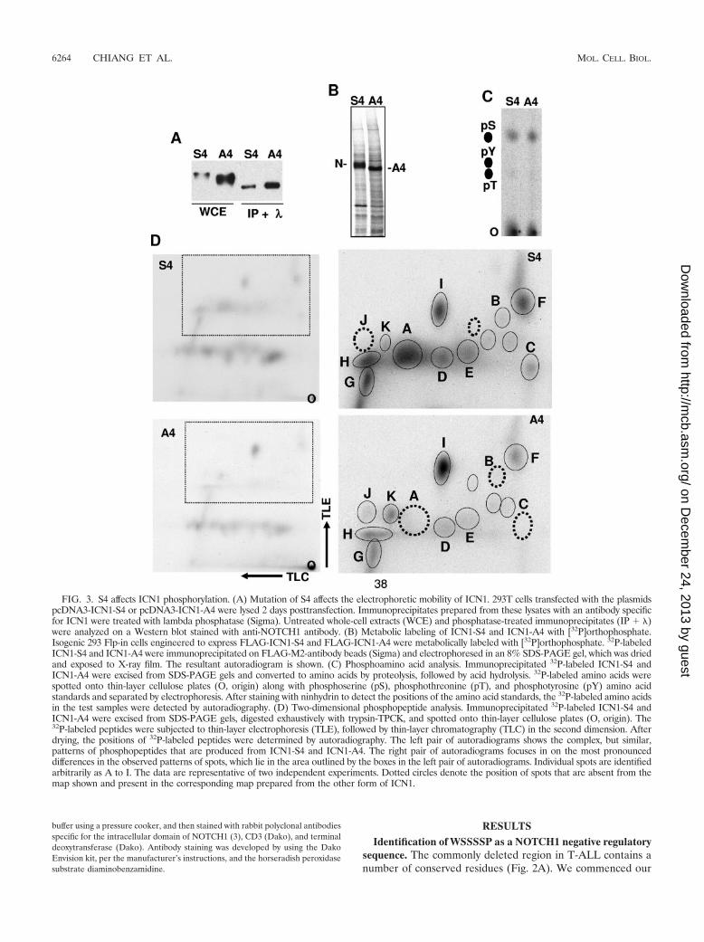
FIG. 3. S4 affects ICN1 phosphorylation. (A) Mutation of S4 affects the electrophoretic mobility of ICN1. 293T cells transfected with the plasmidspcDNA3-ICN1-S4 or pcDNA3-ICN1-A4 were lysed 2 days posttransfection. Immunoprecipitates prepared from these lysates with an antibody specificfor ICN1 were treated with lambda phosphatase (Sigma). Untreated whole-cell extracts (WCE) and phosphatase-treated immunoprecipitates (IP � )were analyzed on a Western blot stained with anti-NOTCH1 antibody. (B) Metabolic labeling of ICN1-S4 and ICN1-A4 with [32P]orthophosphate.Isogenic 293 Flp-in cells engineered to express FLAG-ICN1-S4 and FLAG-ICN1-A4 were metabolically labeled with [32P]orthophosphate. 32P-labeledICN1-S4 and ICN1-A4 were immunoprecipitated on FLAG-M2-antibody beads (Sigma) and electrophoresed in an 8% SDS-PAGE gel, which was driedand exposed to X-ray film. The resultant autoradiogram is shown. (C) Phosphoamino acid analysis. Immunoprecipitated 32P-labeled ICN1-S4 andICN1-A4 were excised from SDS-PAGE gels and converted to amino acids by proteolysis, followed by acid hydrolysis. 32P-labeled amino acids werespotted onto thin-layer cellulose plates (O, origin) along with phosphoserine (pS), phosphothreonine (pT), and phosphotyrosine (pY) amino acidstandards and separated by electrophoresis. After staining with ninhydrin to detect the positions of the amino acid standards, the 32P-labeled amino acidsin the test samples were detected by autoradiography. (D) Two-dimensional phosphopeptide analysis. Immunoprecipitated 32P-labeled ICN1-S4 andICN1-A4 were excised from SDS-PAGE gels, digested exhaustively with trypsin-TPCK, and spotted onto thin-layer cellulose plates (O, origin). The32P-labeled peptides were subjected to thin-layer electrophoresis (TLE), followed by thin-layer chromatography (TLC) in the second dimension. Afterdrying, the positions of 32P-labeled peptides were determined by autoradiography. The left pair of autoradiograms shows the complex, but similar,patterns of phosphopeptides that are produced from ICN1-S4 and ICN1-A4. The right pair of autoradiograms focuses in on the most pronounceddifferences in the observed patterns of spots, which lie in the area outlined by the boxes in the left pair of autoradiograms. Individual spots are identifiedarbitrarily as A to I. The data are representative of two independent experiments. Dotted circles denote the position of spots that are absent from themap shown and present in the corresponding map prepared from the other form of ICN1.
6264 CHIANG ET AL. MOL. CELL. BIOL.
on Decem
ber 24, 2013 by guesthttp://m
cb.asm.org/
Dow
nloaded from
functional analysis by creating a series of nested deletions infull-length NOTCH1 cDNAs bearing the leukemia-associatedheterodimerization domain mutation L1601P, which causes amodest activation of NOTCH1 signaling (Fig. 2B). We ob-served that the deletion of residues 2545 to 2556 had littleeffect on NOTCH1 signaling, whereas larger deletions span-ning residues 2535 to 2556, 2520 to 2556, and 2473 to 2556increased NOTCH1 signal strength in a stepwise fashion. Priorwork from Jones’ group showed that serine residues at posi-tions 2514, 2517, and 2538 can be phosphorylated by CycC:CDK8, an event that is hypothesized to mark ICN1 for tran-scription-dependent degradation (13). However, the largeststepwise increase in activity was associated with the deletion ofresidues 2520 to 2534, a region where none of the serines havebeen reported to be phosphorylated by CycC-CDK8. This re-gion encompasses the position of the most C-terminal deletionwe have yet identified in human T-ALL (a stop codon muta-tion at residue 2524 in the cell line MOLT-15 [41]; Fig. 2B) andincludes a short sequence, WSSSSP (designated S4), that isrestricted in the protein sequence database to a subset ofNOTCH receptors. Specifically, WSSSSP is 100% conservedwithin fly NOTCH and vertebrate NOTCH1 and NOTCH2receptors (Fig. 2A) but has diverged in NOTCH3 (WSDSTP)and is completely absent from NOTCH4.
To test the idea that the S4 sequence might regulateNOTCH1 function, we mutated S4 to AAAA (A4). In thecontext of either full-length NOTCH1 L1601P (Fig. 2B) or asecond relatively weak gain-of-function form of NOTCH1,
�EGF�LNR (Fig. 2C), the A4 mutation stimulated signalingto an extent comparable to the �2473-2556 deletion. We alsoinvestigated the effects of mutating individual S4 residues.Each single S-to-A substitution produced a modest stimulationin NOTCH1 activity in the context of the �EGF�LNRpolypeptide, but no single mutation was as strong as the A4substitution (Fig. 2D). Thus, multiple S residues within the S4sequence appear to contribute to negative regulation ofNOTCH1 signaling.
S4 affects ICN1 phosphorylation. The S4 sequence is distinctfrom previously identified sites of NOTCH1 phosphorylation.To determine whether the S4 sequence influences ICN1 phos-phorylation, we first compared the electrophoretic mobilitiesof ICN1-S4 and ICN1-A4. The mean electrophoretic mobilityof ICN1-A4 was consistently greater than that of ICN1-S4 (Fig.3A), a finding that could be explained by ICN-A4 being un-derphosphorylated relative to ICN-S4. In support of this inter-pretation, the broad bands corresponding to ICN1-S4 andICN1-A4 on Western blots were resolved into tight bands offaster, but identical, electrophoretic mobility by treatment withlambda phosphatase (Fig. 3A), suggesting that the S4 sequenceinfluences the phosphorylation of ICN1 by one or more pro-tein kinases.
To further characterize the effect of the S4 sequence onphosphorylation, we compared the phosphoamino acid contentand the phosphopeptide maps of ICN1-S4 and ICN1-A4 pre-pared from metabolically labeled isogenic 293 TRex cells. 32P-labeled ICN1-S4 and ICN-A4 again demonstrated a difference
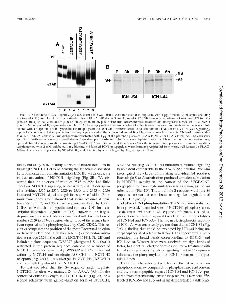
FIG. 4. S4 influences ICN1 stability. (A) U2OS cells in 6-well dishes were transfected in duplicate with 1 �g of pcDNA3 plasmids encodinginactive �EGF (lanes 1 and 2), constitutively active �EGF�LNR (lanes 3 and 4), or �EGF�LNR bearing the deletion of residues 2473 to 2556(lanes 5 and 6) or the A4 mutation (lanes 7 and 8). Immediately posttransfection, cells were refed medium containing 0.1% DMSO or 0.1% DMSOplus 1 �M compound E, a �-secretase inhibitor. At two days posttransfection, whole-cell extracts were prepared and analyzed on Western blotsstained with a polyclonal antibody specific for an epitope in the NOTCH1 transcriptional activation domain (TAD) or anti-V1744 (Cell Signaling),a polyclonal antibody that is specific for a neo-epitope created at the N-terminal end of ICN1 by �-secretase cleavage. (B) ICN1-A4 is more stablethan ICN1-S4. 293 cells in 60-mm dishes were transfected with 1 �g of the pcDNA3 plasmids FLAG-ICN1-S4 or FLAG-ICN1-A4. The cells weresplit 24 h posttransfection into six-well dishes. Two days posttransfection, the cells were depleted twice for 1 h in medium lacking methionine,“pulsed” for 30 min with medium containing 2.5 mCi of [35S]methionine, and then “chased” for the indicated time periods with complete mediumsupplemented with 2 mM unlabeled L-methionine. 35S-labeled ICN1 polypeptides were immunoprecipitated from whole-cell lysates on FLAG-M2-antibody beads, separated by SDS-PAGE, and detected by autoradiography. NS, nonspecific band.
VOL. 26, 2006 NEGATIVE REGULATION OF NOTCH1 6265
on Decem
ber 24, 2013 by guesthttp://m
cb.asm.org/
Dow
nloaded from

in electrophoretic mobility consistent with underphosphoryla-tion of the A4 mutant (Fig. 3B). Phosphoserine was the majorphosphoamino acid in both ICN1-S4 and ICN1-A4 (Fig. 3C), afinding consistent with previous analysis (7). Two-dimensionaltryptic maps prepared from ICN1-S4 and ICN1-A4 revealed acomplex, but reproducible, set of 32P-labeled peptides (Fig.3D). Of these phosphopeptides, one major peptide (desig-nated peptide A) and two minor peptides (peptides B and C)were absent from digests prepared from ICN1-A4. We alsonoted that the labeling of a number of other peptides (such as
peptides D to H in Fig. 3D) was reduced in ICN1-A4 com-pared to ICN1-S4. This tendency toward a global decrease inlabeling of ICN1-A4 was consistent with the results of countingof the excised ICN1-S4 and ICN1-A4 bands prior to trypticdigestion, which typically revealed that ICN1-A4 incorporated40 to 50% less 32P (data not shown). On the other hand, whilethe overall effect of the A4 mutation was to decrease ICN1phosphorylation, one major phosphopeptide (designated I)and several minor phosphopeptides (designated J and K) re-producibly showed relatively increased labeling in ICN1-A4
FIG. 5. CDK8 is unlikely to be the S4 kinase. (A) Dominant-negative CDK8 fails to affect the difference in ICN1-S4 and ICN1-A4 phosphor-ylation. 293 cells in 60-mm dishes were transfected with the indicated combinations of pcDNA3-ICN1-S4 or pcDNA3-ICN1-A4 (100 ng),pCMV2-FLAG-dominant-negative CDK8 (DN-CDK8) (1 �g), and pcDNA3-CSL-MYC (1 �g) plasmids. At 2 days posttransfection, whole-celldetergent extracts were prepared from each dish of transfected cells. Extracts prepared from cells cotransfected with CSL-MYC (lanes 5 to 8) werefurther subjected to immunoprecipitation with anti-MYC 9E10 antibody. The upper panel shows a Western blot containing whole-cell extracts thatwas stained with the FLAG M2 antibody, which recognizes FLAG-DN-CDK8. The lower panel shows a blot stained with anti-NOTCH1. In thisblot, lanes 1 to 4 contain whole-cell extracts, while lanes 5 to 8 contain NOTCH1 polypeptides that were coprecipitated with CSL-MYC.(B) Altered MAML1 function fails to abrogate the difference in ICN1-S4 and ICN1-A4 phosphorylation. 293T cells in six-well format werecotransfected with pcDNA3-CSL-MYC (1 �g), pEGFP-MAML1-GFP or dominant-negative MAML1-GFP (0.5 �g), and pcDNA3-ICN1 orICN1-A4 (0.1 �g). At 2 days posttransfection, whole-cell detergent extracts and CSL-MYC immunoprecipitates were prepared as describedpreviously (3). Lanes 1 to 6 contain 1% of total protein inputted into the immunoprecipitates; lanes 7 to 12 show 20% of the proteins in thecorresponding immunoprecipitates. The blot was sequentially stained with anti-MYC (9E10), anti-GFP (Clontech), and anti-NOTCH1, using anantibody against the intracellular transcriptional activation domain (3). HC and LC, immunoglobulin heavy chain and light chains, respectively.(C) The A4 mutation increases the function of NOTCH1-GAL4 fusion proteins lacking the ankyrin repeats. U2OS cells in 24-well format werecotransfected with 10 ng of empty pcDNA3 plasmid or pcDNA3 plasmids encoding GAL4 fusion proteins in which the RAM and ankyrin repeatdomains of NOTCH1 were replaced with the DNA-binding domain of GAL4. N1 corresponds to a full-length NOTCH1-GAL4 construct, whereas�EGF�LNR corresponds to a construct bearing a deletion removing the EGF and LNR repeat coding regions of NOTCH1. �EGF�LNR-A4 isa GAL4 fusion construct in which the S4 site has been mutated to A4. These plasmids were cotransfected with 250 ng of a GAL4-luciferase reporterplasmid bearing four GAL4 binding sites and 5 ng of the pRL-TK-Renilla luciferase internal control plasmid. Normalized luciferase activities weremeasured in triplicate and are expressed relative to an empty plasmid control. Error bars represent standard deviations.
6266 CHIANG ET AL. MOL. CELL. BIOL.
on Decem
ber 24, 2013 by guesthttp://m
cb.asm.org/
Dow
nloaded from

compared to ICN1-S4 (Fig. 3D). Thus, these data indicate thatthe A4 mutation has complex effects on ICN1 phosphorylationthat are generally, but not uniformly, inhibitory.
S4 influences ICN1 stability. The simplest way for the A4mutation to stimulate ICN1 activity is by increasing ICN1 pro-tein levels. We first addressed this by comparing the levels ofICN1 that are present in cells expressing various forms of�EGF�LNR, a NOTCH1 polypeptide that is susceptible toligand-independent S2 and S3 cleavages due to the absence ofthe protective LNR domain (31). Relative to intact �EGF�LNR,�EGF�LNR polypeptides bearing the deletion �2473-2556 orthe A4 mutation generated substantially higher steady-state levelsof ICN1 in a fashion that was sensitive to a �-secretase inhibitor,compound E (Fig. 4A).
To determine directly whether the A4 mutation stabilizedICN1, pulse-chase experiments were performed with FLAG-tagged ICN1-S4 and ICN1-A4 (Fig. 4B). This revealed that theA4 mutation has a modest, but appreciable, stabilizing effecton ICN1. Together with experiments, such as those in Fig. 4A,showing that the A4 mutation allows ICN1 to accumulate tohigher levels in cells, these results provide a likely explanationfor the stimulatory effect of the A4 mutation on ICN1 function.
S4 is unlikely to be a target sequence for CDK8. Three sitesin the C terminus of ICN1 near the S4 site (S2514, S2517, andS2538) can be phosphorylated by CDK8, which appears to berecruited to ICN1 through the C terminus of MAML cofactors(13). We thus performed a series of experiments to explore therelationship of the S4 site to CDK8. Most directly, we first
FIG. 6. S4 influences the development of T-ALL. (A) �EGF�LNR�P(�2473-2556) and �EGF�LNR-A4 cause the development of leukemia,whereas �EGF�LNR causes only a transient lymphocytosis of CD4� CD8� T cells. Representative flow cytometric analyses of peripheral bloodsamples drawn from mice at 6 and 13 weeks posttransplant with bone marrow cells transduced with the indicated MigRI retroviruses are shown.(B) Kaplan-Meier curve showing that leukemia develops in mice reconstituted with bone marrow cells expressing �EGF�LNR�P(�2473-2556),�EGF�LNR-A4, and ICN1 NOTCH1 polypeptides but not in �EGF�LNR or MigRI animals. Each group in this experiment contained at leastfive mice.
VOL. 26, 2006 NEGATIVE REGULATION OF NOTCH1 6267
on Decem
ber 24, 2013 by guesthttp://m
cb.asm.org/
Dow
nloaded from

investigated whether a “kinase-dead” dominant-negative formof CDK8 altered the difference in phosphorylation betweenICN1-S4 and ICN1-A4, as judged by electrophoretic mobility.We observed that ICN1-A4 remained underphosphorylatedrelative to ICN1-S4 in whole-cell extracts and in CSL com-plexes even in the presence of a 10-fold excess of dominant-negative CDK8 plasmid (Fig. 5A). Additional experimentswere performed in cells overexpressing CSL and full-lengthMAML1 (which should enhance CDK8 recruitment), or CSLand a dominant-negative form of MAML1 (DN-MAML1).DN-MAML consists of a 62-amino-acid kinked �-helix thatforms a stable ternary complex through contacts on both CSLand the ankyrin repeats of NOTCH1 (25) but which lacks theC-terminal portions of MAML1 that are responsible for re-cruitment of p300 and CycC:CDK8 (12, 13, 40). Thus, if S4-targeted phosphorylation is dependent on CycC:CDK8, DN-MAML should render ICN1-S4 equivalent to ICN1-A4, bothin terms of stability and phosphorylation, by preventing therecruitment of CycC:CDK8. In these experiments, we adjustedthe input of the ICN1-A4 and ICN1-S4 plasmids, relative tothe CSLmyc and MAML1 plasmids, to make ICN1 the limitingfactor for complex assembly (Fig. 5B). Whether judged byWestern blots of whole-cell extracts (lanes 1 to 6) or CSLimmunoprecipitates (lanes 7 to 12), we observed differences inelectrophoretic mobility consistent with underphosphorylationof ICN1-A4 in the presence of endogenous MAMLs (comparelanes 1 and 2 and lanes 7 and 8), overexpressed full-lengthMAML1 (compare lanes 3 and 4 and lanes 9 and 10), andDN-MAML1 (compare lanes 5 and 6 and lanes 11 and 12). Wealso observed a stabilizing effect of the A4 mutation on full-length MAML1 and CSL when these proteins were coex-pressed (compare the recovery of MAML1 in CSL complexesimmunoprecipitated in lanes 9 and 10). The ability of overex-pressed MAML1 to promote degradation of CSL and ICN1 isconsistent with prior data implicating assembly of the CSL/ICN/MAML ternary complex in ICN1 turnover (12, 13, 40).
We also studied whether the stabilizing effect of the A4mutation could be transferred to other types of transcriptionalactivation complexes. Experiments were performed with chi-meric �EGF�LNR polypeptides in which the RAM andankyrin repeat domains of ICN1 were replaced with the DNA-binding domain of GAL4 (an approach used first by Struhl andAdachi [35]). This form of NOTCH1 activates GAL4 reportergenes in a �-secretase-dependent fashion but fails to assemblea ternary complex and cannot stimulate transcription fromCSL-dependent promoters (data not shown). As seen in Fig.5C, the A4 mutation strongly stimulated the activity of�EGF�LNR-GAL4 on a GAL4-reporter gene. Taken to-gether, these data suggest that the S4 site is phosphorylated bya kinase or kinases other than CycC-CDK8 and that this phos-phorylation event does not depend on the entry of ICN1 intothe ternary complex.
S4 influences leukemogenesis. A striking feature of theNOTCH1 tumor-associated mutations is that extracellular HDmutations frequently occur in cis with deletions of the intra-cellular PEST region (41). The data described above suggestedthat loss of S4-associated regulation of ICN1 might enhancethe leukemogenic activity of weak gain-of-function forms ofactivated NOTCH1, which are not themselves leukemogenic,and might thus mimic the effect of tumor-associated mutations
found in cis. To assess this possibility, we compared the leu-kemogenic activity of the weak gain-of-function �EGF�LNRform of NOTCH1 with �EGF�LNR-A4 in a murine bonemarrow transplantation assay. The selection of �EGF�LNRfor these experiments was based on the observations showingthat although this form of NOTCH1 generated signals of suf-ficient strength to drive T-cell development (31), it did notinduce T-ALL (Fig. 6A). Typically, mice reconstituted withbone marrow cells expressing �EGF�LNR developed a GFP�
CD4� CD8� immature “double-positive” T-cell population by6 weeks after transplantation that disappeared by 13 weeksposttransplantation. None of these animals have developedleukemia at times greater than 1 year posttransplantation (Fig.6B and data not shown).
In contrast, the double-positive T-cell count in the periph-eral blood of mice reconstituted with bone marrow cells ex-pressing the �EGF�LNR-A4 continued to rise (Fig. 6A), andall of these animals eventually became moribund and suc-cumbed to disseminated leukemia (Fig. 6B). The disease la-tency in �EGF�LNR-A4 mice was similar to that seen inanimals reconstituted with bone marrow cells expressing�EGF�LNR�PEST (which bears a deletion removing the 73C-terminal amino acids of NOTCH1) and ICN1, a strong gain-of-function form of NOTCH1 (Fig. 6B). At necropsy, leukemicblasts replaced the bone marrow and heavily infiltrated thespleen, liver, lymph nodes, and viscera such as the kidneys
FIG. 7. Pathology of �EGF�LNR-A4-induced T-ALL. Sectionsstained with hematoxylin and eosin (H�E) show lymphoblasts infil-trating the liver and the kidney. In the bottom four panels, tumor cellsare shown in sections of liver stained with H�E or antibodies specificfor CD3, terminal deoxytransferase (TdT), and NOTCH1. Immuno-staining was developed by method that produces a brown color (he-matoxylin counterstain).
6268 CHIANG ET AL. MOL. CELL. BIOL.
on Decem
ber 24, 2013 by guesthttp://m
cb.asm.org/
Dow
nloaded from

(Fig. 7). Immunohistochemical stains confirmed that theseblasts were immature T cells expressing CD3, terminal de-oxytransferase (TdT), and NOTCH1 (Fig. 7). Western blotsprepared from heavily infiltrated spleens confirmed the pres-ence of NOTCH1 polypeptides of the expected size of theprecursor and furin-processed forms of �EGF�LNR-A4 (Fig.8). Further workup of �EGF�LNR-A4 tumors included flowcytometry, which revealed that the tumors expressed surfaceCD3 and variable levels of CD4 and CD8 (data not shown). Asanticipated given the results of Western blotting, the tumorscontained intact proviruses and were monoclonal or oligo-clonal based on the presence of one or several dominant pro-viral insertions (Fig. 9 and data not shown).
DISCUSSION
Our studies indicate that the sequence WSSSSP (S4), whichspans residues 2521 to 2526 of human NOTCH1, exerts afunctionally important restraint on NOTCH1 signal strength,and influences the development of T-ALL in a murine model.It follows that the C-terminal NOTCH1 deletions that areoften seen in human T-ALL contribute to leukemogenesis, atleast in part, by removing this sequence. Consistent with this
possibility, the most C-terminal deletion that we have yet ob-served in T-ALL starts within this sequence at residue 2524(41). The gains in ICN1 function that occur when the S4sequence is mutated are correlated with changes in ICN1 phos-phorylation, making it probable that this sequence acts byaltering the recognition of ICN1 by one or more kinases. Be-cause it is necessary to mutate multiple S residues within S4 tomaximally activate NOTCH1, it is possible that one or morekinases phosphorylate more than one site within this sequence.More broadly, mutation of the S4 site appears to affect thephosphorylation of multiple ICN1 tryptic peptides, suggestingthat the phosphorylation status of S4 influences the recognitionof ICN1 by other kinases. NOTCH signaling is tightly coordi-nated with other signaling pathways, and it is possible that S4phosphorylation may serve to prime ICN1 for recognition byother kinases. The existence of multiple kinases that targetICN1 for degradation through S4 and adjacent residues such asthose recognized by CDK8 provides a reasonable explanationfor why deletions involving the C terminus of NOTCH1 arecommon in T-ALL, whereas point mutations are rare (41).
In addition to previously recognized retroviral insertions(10, 14), several recent reports describe acquired frameshift orstop codon mutations in diverse murine T-ALL models that
FIG. 8. Detection of NOTCH1 polypeptides in �EGF�LNR�P(�2473-2556) and �EGF�LNR-A4-induced T-ALLs. Detergent extracts fromspleens heavily involved by T-ALLs arising in �EGF�LNR�P(�2473-2556) and �EGF�LNR-A4 mice were analyzed on Western blots stainedwith a polyclonal antibody specific for the intracellular domain of NOTCH1. (A) Cross-reactive NOTCH1 polypeptides are observed in both typesof tumors of the expected size for pro-�EGF�LNR�, pro-�EGF�LNRA4, and the furin-processed mature forms of each of these polypeptides(NTM�PEST and NTM-A4, respectively). (B) The size of the Notch1 polypeptides in panel A is compared to NOTCH1 polypeptides expressed inthe cell line ALL-SIL, which has a normal NOTCH1 allele that gives rise to NTM, and a mutated NOTCH1 allele containing the same PESTdeletion (residues 2473 to 2556) that has been engineered into the �EGF�LNR� expression plasmid (41). The deleted allele gives rise to NTM�Pafter furin processing.
VOL. 26, 2006 NEGATIVE REGULATION OF NOTCH1 6269
on Decem
ber 24, 2013 by guesthttp://m
cb.asm.org/
Dow
nloaded from

produce C-terminal truncations of NOTCH1 (20, 26). Themost C-terminal deletion noted in murine tumors to date fallsat amino acid residue 2490 in murine NOTCH1 (20), which isthe equivalent of residue 2516 in human NOTCH1; thus, themutations that occur in murine T-ALL also consistently deletethe S4 sequence. While these correlations (and the studiesdirected at the S4 sequence described here) do not precludeadditional important contributions of other sequences in theC-terminal tail to the negative regulation of normal and patho-physiologic NOTCH1 signaling, they are consistent with a ma-jor role for the S4 sequence in T-ALL.
Several questions arise from this work, the most immediateof which concerns the identity of the kinase(s) that targetsICN1 through S4. After MAML-dependent recruitment to theNOTCH1 transcription complex on DNA, CycC-CDK8 canphosphorylate at least three serine residues in the far C-ter-minal region of NOTCH1, including one within the minimaldeleted region (41). However, ICN1-A4 is still underphosphor-ylated relative to ICN1-S4 even under a variety of conditions inwhich ternary complex formation and recruitment of CDK8are defective. These findings suggest that the S4 kinase is likelyto be different than CycC-CDK8. Other candidates include theGSK3� (9, 11) and MEK/ERK kinases, based on the well-recognized, but complex, functional interactions between theRAS and NOTCH signaling pathways (37). However, experi-
ments conducted with pharmacologic inhibitors of MEK andGSK3�, RNAi directed against GSK3�, and constitutively ac-tive forms of MEK1, have failed to detect evidence of epistasisbetween these kinases and the S4 sequence (J. C. Aster, datanot shown). It is likely that unbiased functional screens will benecessary to identify the kinase(s) that is responsible for S4-dependent ICN1 phosphorylation.
We have also failed to see evidence of epistasis betweendominant-negative forms of FBW7 (a homolog of Sel-10) andS4 (J. C. Aster, data not shown). In this regard, it is relevantthat the stability of mammalian NOTCH4 is regulated byFBW7/Sel-10 (38, 39) despite the absence of the S4 sequencefrom this Notch receptor. We thus favor the idea that S4-dependent modulation of ICN1 levels depends on factors otherthan FBW7/Sel-10. It will be of interest to determine whethermutations in the kinases or the destruction machinery respon-sible for S4-dependent ICN1 degradation will be identified inhuman T-ALLs lacking C-terminal NOTCH1 deletions.
The C-terminal NOTCH1 deletions that are found in humanT-ALL often occur in concert with mutations involving theheterodimerization domain of NOTCH1 that lead to increasedICN1 production (41). We have also further characterized herea form of NOTCH1 with a mutation affecting the ectodomain,�EGF�LNR, which drives abnormal double positive T-celldevelopment (31) without causing T-ALL. This demonstratesfor the first time that the threshold dose of NOTCH1 signalsthat is required for efficient induction of T-ALL developmentis higher than that which is required to induce T-cell develop-ment. This raises the question of whether certain heterodimer-ization domain mutations will suffice to create signals that arestrong enough to induce T-ALL, or whether they will requireadditional events in cis or in trans. The “hypoleukemic” phe-notype induced in vivo by weak gain-of-function NOTCH1alleles such as �EGF�LNR should be useful in identifying notonly cis-acting elements such as the S4 sequence but also ele-ments that act in trans. Such trans-acting factors could eitherelevate NOTCH1 signaling tone directly or, by complementingNOTCH1 functions that require strong signals, lower the doseof NOTCH1 that is required for efficient induction of T-ALL.
ACKNOWLEDGMENTS
We thank Hong Sai and Zhigang Li for excellent technical assis-tance. We are also grateful to the John Morgan and Stemmler ASU(University of Pennsylvania), the Abramson Cancer Center Flow Cy-tometry Core (University of Pennsylvania), the AFCRI Core (Univer-sity of Pennsylvania), and the Hematopathology Core of the DanaFarber/Harvard Cancer Center.
This study was supported by grants from the National Institutes ofHealth to W.S.P. (CA93615 and AI47833), J.C.A. (CA82308), S.C.B.(CA92433), and D.B.S. (CA75205) and from the Leukemia and Lym-phoma Society SCOR Program. M.L.X. was the recipient of an Amer-ican Society of Hematology Medical Student Trainee award. M.C. wassupported by a training grant from the NIH/NIDDK (T32-DK007780-07).
REFERENCES
1. Allman, D., F. G. Karnell, J. A. Punt, S. Bakkour, L. Xu, P. Myung, G. A.Koretzky, J. C. Pui, J. C. Aster, and W. S. Pear. 2001. Separation of Notch1promoted lineage commitment and expansion/transformation in developingT cells. J. Exp. Med. 194:99–106.
2. Artavanis-Tsakonas, S., M. D. Rand, and R. J. Lake. 1999. Notch signaling:cell fate control and signal integration in development. Science 284:770–776.
3. Aster, J. C., E. S. Robertson, R. P. Hasserjian, J. R. Turner, E. Kieff, and J.Sklar. 1997. Oncogenic forms of NOTCH1 lacking either the primary bind-ing site for RBP-J or nuclear localization sequences retain the ability to
FIG. 9. Analysis of proviral integrity and integration in A4-inducedT-ALLs. (A) Southern blot of EcoRV-digested genomic DNAs iso-lated from four spleens involved by A4-induced T-ALLs. EcoRV,which cleaves twice within the provirus at sites flanking the�EGF�LNR-A4 cDNA, is expected to release a proviral fragment of5.9 kb from the intact �EGF�LNR-A4 provirus when hybridized to aprobe specific for the IRES sequence. The correctly sized fragment isshown in the right-hand lane, which contains MigRI-�EGF�LNR-A4and MigRI-NOTCH1 plasmid DNAs digested with Eco RV. Each ofthe left-hand four lanes contain genomic DNA cut with EcoRV obtainedfrom spleens heavily infiltrated by different MigRI-�EGF�LNR-A4 T-ALLs. (B) Genomic DNAs from the same four spleens shown in panel Awere analyzed by digestion with EcoRI, which cleaves once within theprovirus; thus, each EcoR1-digested provirus will produce a different sizedDNA fragment. The Southern blot hybridized to a 592-bp ECMV IRESprobe. Single proviruses are readily seen in tumors 1, 4, and 5, whereas asingle faint proviral band is seen in tumor 3 (arrowheads). NS, nonspecificband. In both panels A and B, the numbers correspond to sizes in kilo-bases.
6270 CHIANG ET AL. MOL. CELL. BIOL.
on Decem
ber 24, 2013 by guesthttp://m
cb.asm.org/
Dow
nloaded from

associate with RBP-J and activate transcription. J. Biol. Chem. 272:11336–11343.
4. Aster, J. C., L. Xu, F. G. Karnell, V. Patriub, J. C. Pui, and W. S. Pear. 2000.Essential roles for ankyrin repeat and transactivation domains in inductionof T-cell leukemia by notch1. Mol. Cell. Biol. 20:7505–7515.
5. Blaumueller, C. M., H. Qi, P. Zagouras, and S. Artavanis-Tsakonas. 1997.Intracellular cleavage of Notch leads to a heterodimeric receptor on theplasma membrane. Cell 90:281–291.
6. Brou, C., F. Logeat, N. Gupta, C. Bessia, O. LeBail, J. R. Doedens, A.Cumano, P. Roux, R. A. Black, and A. Israel. 2000. A novel proteolyticcleavage involved in Notch signaling: the role of the disintegrin-metallopro-tease TACE. Mol. Cell 5:207–216.
7. Cagan, R. L., and D. F. Ready. 1989. Notch is required for successive celldecisions in the developing Drosophila retina. Genes Dev. 3:1099–1112.
8. De Strooper, B., W. Annaert, P. Cupers, P. Saftig, K. Craessaerts, J. S.Mumm, E. H. Schroeter, V. Schrijvers, M. S. Wolfe, W. J. Ray, A. Goate, andR. Kopan. 1999. A presenilin-1-dependent �-secretase-like protease medi-ates release of Notch intracellular domain. Nature 398:518–522.
9. Espinosa, L., J. Ingles-Esteve, C. Aguilera, and A. Bigas. 2003. Phosphory-lation by glycogen synthase kinase-3� down-regulates Notch activity, a linkfor Notch and Wnt pathways. J. Biol. Chem. 278:32227–32235.
10. Feldman, B. J., T. Hampton, and M. L. Cleary. 2000. A carboxy-terminaldeletion mutant of Notch1 accelerates lymphoid oncogenesis in E2A-PBX1transgenic mice. Blood 96:1906–1913.
11. Foltz, D. R., M. C. Santiago, B. E. Berechid, and J. S. Nye. 2002. Glycogensynthase kinase-3� modulates notch signaling and stability. Curr. Biol. 12:1006–1011.
12. Fryer, C. J., E. Lamar, I. Turbachova, C. Kintner, and K. A. Jones. 2002.Mastermind mediates chromatin-specific transcription and turnover of theNotch enhancer complex. Genes Dev. 16:1397–1411.
13. Fryer, C. J., J. B. White, and K. A. Jones. 2004. Mastermind recruits CycC:CDK8 to phosphorylate the Notch ICD and coordinate activation withturnover. Mol. Cell 16:509–520.
14. Hoemann, C. D., N. Beaulieu, L. Girard, N. Rebai, and P. Jolicoeur. 2000.Two distinct Notch1 mutant alleles are involved in the induction of T-cellleukemia in c-myc transgenic mice. Mol. Cell. Biol. 20:3831–3842.
15. Hsieh, J. J., T. Henkel, P. Salmon, E. Robey, M. G. Peterson, and S. D.Hayward. 1996. Truncated mammalian Notch1 activates CBF1/RBPJk-re-pressed genes by a mechanism resembling that of Epstein-Barr virusEBNA2. Mol. Cell. Biol. 16:952–959.
16. Jarriault, S., C. Brou, F. Logeat, E. H. Schroeter, R. Kopan, and A. Israel.1995. Signalling downstream of activated mammalian Notch. Nature 377:355–358.
17. Joyal, J. L., and D. B. Sacks. 1994. Insulin-dependent phosphorylation ofcalmodulin in rat hepatocytes. J. Biol. Chem. 269:30039–30048.
18. Kimberly, W. T., W. P. Esler, W. Ye, B. L. Ostaszewski, J. Gao, T. Diehl, D. J.Selkoe, and M. S. Wolfe. 2003. Notch and the amyloid precursor protein arecleaved by similar �-secretase(s). Biochemistry 42:137–144.
19. Kopan, R., E. H. Schroeter, H. Weintraub, and J. S. Nye. 1996. Signaltransduction by activated mNotch: importance of proteolytic processing andits regulation by the extracellular domain. Proc. Natl. Acad. Sci. USA 93:1683–1688.
20. Lin, Y. W., R. A. Nichols, J. J. Letterio, and P. D. Aplan. 2006. Notch1mutations are important for leukemic transformation in murine models ofprecursor-T leukemia/lymphoma. Blood 107:2540–2543.
21. Logeat, F., C. Bessia, C. Brou, O. LeBail, S. Jarriault, N. G. Seidah, and A.Israel. 1998. The Notch1 receptor is cleaved constitutively by a furin-likeconvertase. Proc. Natl. Acad. Sci. USA 95:8108–8112.
22. MacKichan, M. L., F. Logeat, and A. Israel. 1996. Phosphorylation of p105PEST sequence via a redox-insensitive pathway up-regulates processing ofp50 NF-B. J. Biol. Chem. 271:6084–6091.
23. McGill, M. A., and C. J. McGlade. 2003. Mammalian numb proteins pro-mote Notch1 receptor ubiquitination and degradation of the Notch1 intra-cellular domain. J. Biol. Chem. 278:23196–23203.
24. Mumm, J. S., E. H. Schroeter, M. T. Saxena, A. Griesemer, X. Tian, D. J.Pan, W. J. Ray, and R. Kopan. 2000. A ligand-induced extracellular cleavageregulates �-secretase-like proteolytic activation of Notch1. Mol. Cell 5:197–206.
25. Nam, Y., P. Sliz, L. Song, J. C. Aster, and S. Blacklow. 2006. Structural basisfor cooperativity in recruitment of the mastermind coactivator to Notchtranscription complexes. Cell 124:973–983.
26. O’Neil, J., J. Calvo, K. McKenna, V. Krishnamoorthy, J. C. Aster, C. H.Bassing, F. W. Alt, M. Kelliher, and A. T. Look. 2006. Activating Notch1mutations in mouse models of T-ALL. Blood 107:781–785.
27. Petcherski, A. G., and J. Kimble. 2000. LAG-3 is a putative transcriptionalactivator in the Caenorhabditis elegans Notch pathway. Nature 405:364–368.
28. Petcherski, A. G., and J. Kimble. 2000. Mastermind is a putative activator forNotch. Curr. Biol. 10:R471–R473.
29. Rand, M. D., L. M. Grimm, S. Artavanis-Tsakonas, V. Patriub, S. C. Blacklow,J. Sklar, and J. C. Aster. 2000. Calcium depletion dissociates and activatesheterodimeric notch receptors. Mol. Cell. Biol. 20:1825–1835.
30. Rechsteiner, M. 1989. PEST regions, proteolysis, and cell cycle progression.Revis. Biol. Celular. 20:235–253.
31. Sanchez-Irizarry, C., A. C. Carpenter, A. P. Weng, W. S. Pear, J. C. Aster,and S. C. Blacklow. 2004. Notch subunit heterodimerization and preventionof ligand-independent proteolytic activation depend, respectively, on a noveldomain and the LNR repeats. Mol. Cell. Biol. 24:9265–9273.
32. Schroeter, E. H., J. A. Kisslinger, and R. Kopan. 1998. Notch-1 signallingrequires ligand-induced proteolytic release of intracellular domain. Nature393:382–386.
33. Shah, S., S. F. Lee, K. Tabuchi, Y. H. Hao, C. Yu, Q. LaPlant, H. Ball, C. E.Dann III, T. Sudhof, and G. Yu. 2005. Nicastrin functions as a �-secretase-substrate receptor. Cell 122:435–447.
34. Shaye, D. D., and I. Greenwald. 2005. LIN-12/Notch trafficking and regula-tion of DSL ligand activity during vulval induction in Caenorhabditis elegans.Development 132:5081–5092.
35. Struhl, G., and A. Adachi. 1998. Nuclear access and action of notch in vivo.Cell 93:649–660.
36. Struhl, G., and I. Grenwald. 1999. Presenilin is required for activity andnuclear access of Notch in Drosophila. Nature 398:522–525.
37. Sundaram, M. V. 2005. The love-hate relationship between Ras and Notch.Genes Dev. 19:1825–1839.
38. Tetzlaff, M. T., W. Yu, M. Li, P. Zhang, M. Finegold, K. Mahon, J. W.Harper, R. J. Schwartz, and S. J. Elledge. 2004. Defective cardiovasculardevelopment and elevated cyclin E and Notch proteins in mice lacking theFbw7 F-box protein. Proc. Natl. Acad. Sci. USA 101:3338–3345.
39. Tsunematsu, R., K. Nakayama, Y. Oike, M. Nishiyama, N. Ishida, S.Hatakeyama, Y. Bessho, R. Kageyama, T. Suda, and K. I. Nakayama. 2004.Mouse Fbw7/Sel-10/Cdc4 is required for notch degradation during vasculardevelopment. J. Biol. Chem. 279:9417–9423.
40. Wallberg, A. E., K. Pedersen, U. Lendahl, and R. G. Roeder. 2002. p300 andPCAF act cooperatively to mediate transcriptional activation from chromatintemplates by notch intracellular domains in vitro. Mol. Cell. Biol. 22:7812–7819.
41. Weng, A. P., A. A. Ferrando, W. Lee, J. P. T. Morris, L. B. Silverman, C.Sanchez-Irizarry, S. C. Blacklow, A. T. Look, and J. C. Aster. 2004. Activat-ing mutations of NOTCH1 in human T-cell acute lymphoblastic leukemia.Science 306:269–271.
42. Weng, A. P., Y. Nam, M. S. Wolfe, W. S. Pear, J. D. Griffin, S. C. Blacklow,and J. C. Aster. 2003. Growth suppression of pre-T acute lymphoblasticleukemia cells by inhibition of notch signaling. Mol. Cell. Biol. 23:655–664.
43. Wu, L., J. C. Aster, S. C. Blacklow, R. Lake, S. Artavanis-Tsakonas, and J. D.Griffin. 2000. MAML1, a human homologue of Drosophila mastermind, is atranscriptional coactivator for NOTCH receptors. Nat. Genet. 26:484–489.
44. Ye, Y., N. Lukinova, and M. E. Fortini. 1999. Neurogenic phenotypes andaltered Notch processing in Drosophila Presenilin mutants. Nature 398:525–529.
VOL. 26, 2006 NEGATIVE REGULATION OF NOTCH1 6271
on Decem
ber 24, 2013 by guesthttp://m
cb.asm.org/
Dow
nloaded from
Copyright © 2022 FDOKUMEN