Notch1 activation reduces proliferation in the multipotent hematopoietic progenitor cell line...
10
Notch1 activation reduces proliferation in the multipotent hematopoietic progenitor cell line FDCP-mix through a p53-dependent pathway but Notch1 effects on myeloid and erythroid differentiation are independent of p53 K Henning 1,6 , J Heering 1,6,7 , R Schwanbeck 1,6 , T Schroeder 2,6 , H Helmbold 3 , H Scha ¨fer 4 , W Deppert 3 , E Kim 5 and U Just* ,1 Signaling mediated by activation of the transmembrane receptor Notch influences cell-fate decisions, differentiation, proliferation, and cell survival. Activated Notch reduces proliferation by altering cell-cycle kinetics and promotes differentiation in hematopoietic progenitor cells. Here, we investigated if the G 1 arrest and differentiation induced by activated mNotch1 are dependent on tumor suppressor p53, a critical mediator of cellular growth arrest. Multipotent wild-type p53-expressing (p53 wt ) and p53-deficient (p53 null ) hematopoietic progenitor cell lines (FDCP-mix) carrying an inducible mNotch1 system were used to investigate the effects of proliferation and differentiation upon mNotch1 signaling. While activated Notch reduced proliferation of p53 wt -cells, no change was observed in p53 null -cells. Activated Notch upregulated the p53 target p21 cip/waf in p53 wt -cells, but not in p53 null -cells. Induction of the p21 cip/waf gene by activated Notch was mediated by increased binding of p53 to p53-binding sites in the p21 cip/waf promoter and was independent of the canonical RBP-J binding site. Re-expression of p53 wt in p53 null cells restored the inhibition of proliferation by activated Notch. Thus, activated Notch inhibits proliferation of multipotent hematopoietic progenitor cells via a p53-dependent pathway. In contrast, myeloid and erythroid differentiation was similarly induced in p53 wt and p53 null cells. These data suggest that Notch signaling triggers two distinct pathways, a p53-dependent one leading to a block in proliferation and a p53-independent one promoting differentiation. Cell Death and Differentiation (2008) 15, 398–407; doi:10.1038/sj.cdd.4402277; published online 30 November 2007 Notch proteins are a family of highly conserved transmem- brane receptors that transduce signals involved in the control of cell fate; differentiation, proliferation, and apoptosis in a cell context-dependent manner (reviewed by Wilson and Radtke, Lai, and Hurlbut et al. 1–3 ). Depending on the cell type and other signals present for the receiving cell, Notch signaling can inhibit or delay differentiation, or promote differentiation. Similarly, Notch activation can induce cell cycle progression thereby increasing cell proliferation, while under different conditions it blocks cell cycle progression leading to growth arrest. Further, Notch activation can influence apoptosis positively or negatively. Notch receptors are activated by specific transmembrane ligands of the Delta and Serrate/Jagged family. After ligand binding, the intracellular domain of Notch (Notch IC ) is proteolytically cleaved from the transmembrane region and translocates to the nucleus, where it associates with the transcriptional repressor RBP-J also termed CBF1. 4 After binding of Notch IC , RBP-J is converted to a transcriptional activator and in conjunction with chromatin remodeling Received 22.2.07; revised 05.10.07; accepted 19.10.07; Edited by M Oren; published online 30.11.07 1 Department of Biochemistry, Christian-Albrechts-University of Kiel, Olshausenstrae 40, Kiel, Germany; 2 Department of Stem Cell Research, GSF, Ingolstaedter Landstr. 1, Munich, Germany; 3 Department of Tumor Virology, Heinrich-Pette-Institute, Martinistr. 52, Hamburg, Germany; 4 Laboratory of Molecular Gastroenterology, 1st Department of Medicine, Christian-Albrechts-University of Kiel, Schittenhelmstr. 12, Kiel, Germany and 5 Laboratory of Neurooncology, Department of Neurosurgery, Georg-August-University of Go ¨ ttingen, Robert-Koch-Str. 40, Go ¨ ttingen, Germany *Corresponding author: U Just, Department of Biochemistry, Christian-Albrechts-University of Kiel, Olshausenstrae 40, Kiel 24098, Germany. Tel: 49 431 880 2513; Fax: 49 431 880 2609; E-mail: [email protected] 6 These authors contributed equally to this work. 7 Present address: Department for Cell Biology and Immunology, University of Stuttgart, Allmandring 31, Stuttgart 70569, Germany. Keywords: Notch; p53; proliferation; hematopoiesis; FDCP-mix cells Abbreviations: p53 wt , wild type p53; p53 null , p53-deficient; FDCP-mix, factor-dependent cell established at the Paterson Institute with mixed differentiation potential; mNotch1, murine Notch1; Notch IC , intracellular domain of Notch; RBP-J, recombination recognition sequence binding protein at the Jk site; Hes, hairy and enhancer of split; Hey/Herp, Hes-related repressor; mN1 IC , intracellular domain of murine Notch1; NERT, mN1 IC fused to the hormone binding domain of the human estrogen receptor tamoxifen-sensitive mutant; ERT, estrogen receptor tamoxifen-sensitive mutant; rneo, retroviral control vector carrying the neomyin resistance gene; rNERTneo, retroviral control vector carrying the NERT and neomyin resistance gene; rNERTneo FDCP-mix, FDCP-mix cells expressing an OHT-inducible form of Notch1 IC ; rneo FDCP-mix, FDCP-mix cells carrying the retroviral control vector conferring neomycin resistance; G418, neomycin; OHT, 4-hydroxytamoxifen; CFSE, carboxyfluorescein diacetate succinimidyl ester; p21RBP-Jmut promoter, p21 promoter compromised in RBP-J binding; IMDM, Iscove’s modified Dulbecco’s medium; FCS, fetal calf serum; PE, Phycoerytrin; APC, allophycocyanin; PBS, phosphate-buffered saline; RT, room temperature; p21luc, reporter plasmid carrying a luciferase gene under the control of the p21 promoter; p21Dp53BS1luc, p21 promoter lacking the first p53 binding site; p21Dp53BS2luc, p21 promoter lacking the second p53 binding site; p21Dp53BS1 þ 2luc, p21 promoter lacking both p53 binding sites; ChIP, chromatin immunoprecipitation Cell Death and Differentiation (2008) 15, 398–407 & 2008 Nature Publishing Group All rights reserved 1350-9047/08 $30.00 www.nature.com/cdd
-
Upload
independent -
Category
Documents
-
view
3 -
download
0
Transcript of Notch1 activation reduces proliferation in the multipotent hematopoietic progenitor cell line...
Notch1 activation reduces proliferation in themultipotent hematopoietic progenitor cell lineFDCP-mix through a p53-dependent pathway butNotch1 effects on myeloid and erythroid differentiationare independent of p53
K Henning1,6, J Heering1,6,7, R Schwanbeck1,6, T Schroeder2,6, H Helmbold3, H Schafer4, W Deppert3, E Kim5 and U Just*,1
Signaling mediated by activation of the transmembrane receptor Notch influences cell-fate decisions, differentiation,proliferation, and cell survival. Activated Notch reduces proliferation by altering cell-cycle kinetics and promotes differentiationin hematopoietic progenitor cells. Here, we investigated if the G1 arrest and differentiation induced by activated mNotch1 aredependent on tumor suppressor p53, a critical mediator of cellular growth arrest. Multipotent wild-type p53-expressing (p53wt)and p53-deficient (p53null) hematopoietic progenitor cell lines (FDCP-mix) carrying an inducible mNotch1 system were used toinvestigate the effects of proliferation and differentiation upon mNotch1 signaling. While activated Notch reduced proliferation ofp53wt-cells, no change was observed in p53null-cells. Activated Notch upregulated the p53 target p21cip/waf in p53wt-cells, but notin p53null-cells. Induction of the p21cip/waf gene by activated Notch was mediated by increased binding of p53 to p53-bindingsites in the p21cip/waf promoter and was independent of the canonical RBP-J binding site. Re-expression of p53wt in p53null
cells restored the inhibition of proliferation by activated Notch. Thus, activated Notch inhibits proliferation of multipotenthematopoietic progenitor cells via a p53-dependent pathway. In contrast, myeloid and erythroid differentiation was similarlyinduced in p53wt and p53null cells. These data suggest that Notch signaling triggers two distinct pathways, a p53-dependent oneleading to a block in proliferation and a p53-independent one promoting differentiation.Cell Death and Differentiation (2008) 15, 398–407; doi:10.1038/sj.cdd.4402277; published online 30 November 2007
Notch proteins are a family of highly conserved transmem-brane receptors that transduce signals involved in the controlof cell fate; differentiation, proliferation, and apoptosis in a cellcontext-dependent manner (reviewed by Wilson and Radtke,Lai, and Hurlbut et al.1–3). Depending on the cell type andother signals present for the receiving cell, Notch signalingcan inhibit or delay differentiation, or promote differentiation.Similarly, Notch activation can induce cell cycle progressionthereby increasing cell proliferation, while under differentconditions it blocks cell cycle progression leading to growth
arrest. Further, Notch activation can influence apoptosispositively or negatively.
Notch receptors are activated by specific transmembraneligands of the Delta and Serrate/Jagged family. After ligandbinding, the intracellular domain of Notch (NotchIC) isproteolytically cleaved from the transmembrane region andtranslocates to the nucleus, where it associates with thetranscriptional repressor RBP-J also termed CBF1.4 Afterbinding of NotchIC, RBP-J is converted to a transcriptionalactivator and in conjunction with chromatin remodeling
Received 22.2.07; revised 05.10.07; accepted 19.10.07; Edited by M Oren; published online 30.11.07
1Department of Biochemistry, Christian-Albrechts-University of Kiel, Olshausenstra�e 40, Kiel, Germany; 2Department of Stem Cell Research, GSF, IngolstaedterLandstr. 1, Munich, Germany; 3Department of Tumor Virology, Heinrich-Pette-Institute, Martinistr. 52, Hamburg, Germany; 4Laboratory of Molecular Gastroenterology,1st Department of Medicine, Christian-Albrechts-University of Kiel, Schittenhelmstr. 12, Kiel, Germany and 5Laboratory of Neurooncology, Department of Neurosurgery,Georg-August-University of Gottingen, Robert-Koch-Str. 40, Gottingen, Germany*Corresponding author: U Just, Department of Biochemistry, Christian-Albrechts-University of Kiel, Olshausenstra�e 40, Kiel 24098, Germany.Tel: 49 431 880 2513; Fax: 49 431 880 2609; E-mail: [email protected] authors contributed equally to this work.7Present address: Department for Cell Biology and Immunology, University of Stuttgart, Allmandring 31, Stuttgart 70569, Germany.Keywords: Notch; p53; proliferation; hematopoiesis; FDCP-mix cellsAbbreviations: p53wt, wild type p53; p53null, p53-deficient; FDCP-mix, factor-dependent cell established at the Paterson Institute with mixed differentiation potential;mNotch1, murine Notch1; NotchIC, intracellular domain of Notch; RBP-J, recombination recognition sequence binding protein at the Jk site; Hes, hairy and enhancer ofsplit; Hey/Herp, Hes-related repressor; mN1IC, intracellular domain of murine Notch1; NERT, mN1IC fused to the hormone binding domain of the human estrogenreceptor tamoxifen-sensitive mutant; ERT, estrogen receptor tamoxifen-sensitive mutant; rneo, retroviral control vector carrying the neomyin resistance gene;rNERTneo, retroviral control vector carrying the NERT and neomyin resistance gene; rNERTneo FDCP-mix, FDCP-mix cells expressing an OHT-inducible form ofNotch1IC; rneo FDCP-mix, FDCP-mix cells carrying the retroviral control vector conferring neomycin resistance; G418, neomycin; OHT, 4-hydroxytamoxifen; CFSE,carboxyfluorescein diacetate succinimidyl ester; p21RBP-Jmut promoter, p21 promoter compromised in RBP-J binding; IMDM, Iscove’s modified Dulbecco’s medium;FCS, fetal calf serum; PE, Phycoerytrin; APC, allophycocyanin; PBS, phosphate-buffered saline; RT, room temperature; p21luc, reporter plasmid carrying a luciferasegene under the control of the p21 promoter; p21Dp53BS1luc, p21 promoter lacking the first p53 binding site; p21Dp53BS2luc, p21 promoter lacking the second p53binding site; p21Dp53BS1þ 2luc, p21 promoter lacking both p53 binding sites; ChIP, chromatin immunoprecipitation
Cell Death and Differentiation (2008) 15, 398–407& 2008 Nature Publishing Group All rights reserved 1350-9047/08 $30.00
www.nature.com/cdd
enzymes, components of the transcriptional machinery andthe activity of other cofactors induces transcription of down-stream target genes, including genes of the Hes (Enhancer ofSplit) and Hey (also called Hes-related repressor Herp, Hesr,Hrt, CHF, gridlock) family.1,2 Recently, two proteins of theHes/Hey basic helix-loop-helix transcription factor family,Hes1 and Hey1, were found in a screen for regulators ofp53 activity.5 However, little is known about the biologicalsignificance of Hes1- and Hey1-dependent regulation of p53activity in regard to Notch signaling.
The tumor suppressor protein p53 plays a critical role inmaintaining cellular homeostasis both in normal developmentand following various cellular stresses including DNA damageand oncogene activation.6 After activation, it initiates atranscriptional program that can result in cell cycle arrest orapoptosis depending on the type and extent of the stimulus.Recently, we reported that activated Notch1 inhibits prolifera-tion of hematopoietic progenitor cells due to cell cycle arrest inG0/G1
7 and promotes myeloid differentiation.8,9 In this study,we examine whether p53 is required for these Notch-mediatedeffects. Our results show that Notch signaling inhibitsproliferation via a p53-dependent mechanism involvingupregulation of p21 expression while induction of myeloidand erythroid differentiation is p53 independent.
Results
Generation of p53null FDCP-mix cell lines expressing aconditional activated mNotch1IC. To determine whetherp53 has a role in mediating the Notch1-induced cell cycleblock and/or myeloid differentiation of hematopoieticprogenitor cells, we generated p53null FDCP-mix cellsclones expressing an inducible form of the constitutivelyactive intracellular part of murine Notch1. Like the FDCP-mixcells lines A4 or A7, p53null FDCP-mix cells are strictlyfactor-dependent hematopoietic progenitor cell lines thatdifferentiate in a multilineage response to physiologicalregulators for differentiation, such as stromal cells orhematopoietic cytokines.10–12 Both were established frommurine long-term cultures in the same way and share manycharacteristics with primitive hematopoietic progenitorcells.10,13 First, we confirmed that the p53 pathway wasfunctional in FDCP-mix A7 cells and not responsive in p53null
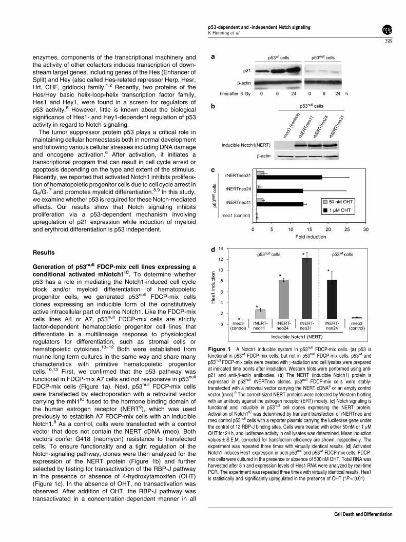
FDCP-mix cells (Figure 1a). Next, p53null FDCP-mix cellswere transfected by electroporation with a retroviral vectorcarrying the mN1IC fused to the hormone binding domain ofthe human estrogen receptor (NERT9), which was usedpreviously to establish A7 FDCP-mix cells with an inducibleNotch1.8 As a control, cells were transfected with a controlvector that does not contain the NERT cDNA (rneo). Bothvectors confer G418 (neomycin) resistance to transfectedcells. To ensure functionality and a tight regulation of theNotch-signaling pathway, clones were then analyzed for theexpression of the NERT protein (Figure 1b) and furtherselected by testing for transactivation of the RBP-J pathwayin the presence or absence of 4-hydroxytamoxifen (OHT)(Figure 1c). In the absence of OHT, no transactivation wasobserved. After addition of OHT, the RBP-J pathway wastransactivated in a concentration-dependent manner in all
Figure 1 A Notch1 inducible system in p53null FDCP-mix cells. (a) p53 isfunctional in p53wt FDCP-mix cells, but not in p53null FDCP-mix cells. p53wt andp53null FDCP-mix cells were treated with g-radiation and cell lysates were preparedat indicated time points after irradiation. Western blots were performed using anti-p21 and anti-b-actin antibodies. (b) The NERT (inducible Notch1) protein isexpressed in p53null rNERTneo clones. p53null FDCP-mix cells were stably-transfected with a retroviral vector carrying the NERT cDNA9 or an empty controlvector (rneo).9 The correct-sized NERT proteins were detected by Western blottingwith an antibody against the estrogen receptor (ERT) moiety. (c) Notch signaling isfunctional and inducible in p53null cell clones expressing the NERT protein.Activation of Notch1IC was determined by transient transfection of rNERTneo andrneo control p53null cells with a reporter plasmid carrying the luciferase gene underthe control of 12 RBP-J binding sites. Cells were treated with either 50 nM or 1mMOHT for 24 h, and luciferase activity in cell lysates was determined. Mean inductionvalues±S.E.M. corrected for transfection efficiency are shown, respectively. Theexperiment was repeated three times with virtually identical results. (d) ActivatedNotch1 induces Hes1 expression in both p53null and p53wt FDCP-mix cells. FDCP-mix cells were cultured in the presence or absence of 500 nM OHT. Total RNA washarvested after 8 h and expression levels of Hes1 RNA were analyzed by real-timePCR. The experiment was repeated three times with virtually identical results. Hes1is statistically and significantly upregulated in the presence of OHT (*Po0.01)
p53-dependent and -independent Notch signalingK Henning et al
399
Cell Death and Differentiation
clones used in this report (Figure 1c). Control rneo clones didnot express the NERT protein and did not transactivate theRBP-J pathway regardless of the addition of OHT (Figure 1band c). To confirm that the Notch1-signaling pathway isactivated by the addition of OHT, we determined theexpression of endogenous Notch1 target genes of the Hesand Hey family in response to OHT treatment. In NERT-expressing p53null FDCP-mix cells, Hes1, and Hey1expression was upregulated after the addition of OHT,whereas in control p53null FDCP-mix cells, the expressionof Hes1 and Hey1 remained unaltered (Figure 1d and datanot shown)
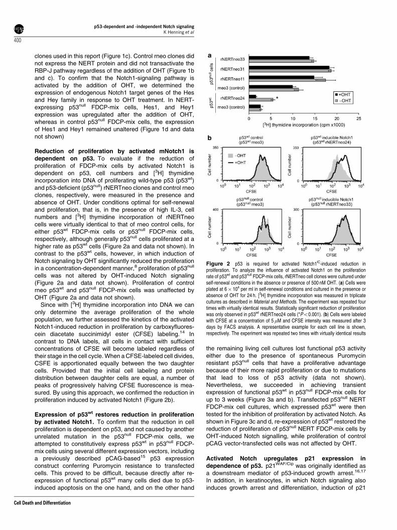
Reduction of proliferation by activated mNotch1 isdependent on p53. To evaluate if the reduction ofproliferation of FDCP-mix cells by activated Notch1 isdependent on p53, cell numbers and [3H] thymidineincorporation into DNA of proliferating wild-type p53 (p53wt)and p53-deficient (p53null) rNERTneo clones and control rneoclones, respectively, were measured in the presence andabsence of OHT. Under conditions optimal for self-renewaland proliferation, that is, in the presence of high IL-3, cellnumbers and [3H] thymidine incorporation of rNERTneocells were virtually identical to that of rneo control cells, foreither p53wt FDCP-mix cells or p53null FDCP-mix cells,respectively, although generally p53null cells proliferated at ahigher rate as p53wt cells (Figure 2a and data not shown). Incontrast to the p53wt cells, however, in which induction ofNotch signaling by OHT significantly reduced the proliferationin a concentration-dependent manner,8 proliferation of p53null
cells was not altered by OHT-induced Notch signaling(Figure 2a and data not shown). Proliferation of controlrneo p53wt and p53null FDCP-mix cells was unaffected byOHT (Figure 2a and data not shown).
Since with [3H] thymidine incorporation into DNA we canonly determine the average proliferation of the wholepopulation, we further assessed the kinetics of the activatedNotch1-induced reduction in proliferation by carboxyfluores-cein diacetate succinimidyl ester (CFSE) labeling.14 Incontrast to DNA labels, all cells in contact with sufficientconcentrations of CFSE will become labeled regardless oftheir stage in the cell cycle. When a CFSE-labeled cell divides,CSFE is apportionated equally between the two daughtercells. Provided that the initial cell labeling and proteindistribution between daughter cells are equal, a number ofpeaks of progressively halving CFSE fluorescence is mea-sured. By using this approach, we confirmed the reduction inproliferation induced by activated Notch1 (Figure 2b).
Expression of p53wt restores reduction in proliferationby activated Notch1. To confirm that the reduction in cellproliferation is dependent on p53, and not caused by anotherunrelated mutation in the p53null FDCP-mix cells, weattempted to constitutively express p53wt in p53null FDCP-mix cells using several different expression vectors, includinga previously described pCAG-based15 p53 expressionconstruct conferring Puromycin resistance to transfectedcells. This proved to be difficult, because directly after re-expression of functional p53wt many cells died due to p53-induced apoptosis on the one hand, and on the other hand
the remaining living cell cultures lost functional p53 activityeither due to the presence of spontaneous Puromycinresistant p53null cells that have a proliferative advantagebecause of their more rapid proliferation or due to mutationsthat lead to loss of p53 activity (data not shown).Nevertheless, we succeeded in achieving transientexpression of functional p53wt in p53null FDCP-mix cells forup to 3 weeks (Figure 3a and b). Transfected p53null NERTFDCP-mix cell cultures, which expressed p53wt were thentested for the inhibition of proliferation by activated Notch. Asshown in Figure 3c and d, re-expression of p53wt restored thereduction of proliferation of p53null NERT FDCP-mix cells byOHT-induced Notch signalling, while proliferation of controlpCAG vector-transfected cells was not affected by OHT.
Activated Notch upregulates p21 expression independence of p53. p21WAF/Cip was originally identified asa downstream mediator of p53-induced growth arrest.16,17
In addition, in keratinocytes, in which Notch signaling alsoinduces growth arrest and differentiation, induction of p21
Figure 2 p53 is required for activated Notch1IC-induced reduction inproliferation. To analyze the influence of activated Notch1 on the proliferationrate of p53wt and p53null FDCP-mix cells, rNERTneo cell clones were cultured underself-renewal conditions in the absence or presence of 500 nM OHT. (a) Cells wereplated at 6� 104 per ml in self-renewal conditions and cultured in the presence orabsence of OHT for 24 h. [3H] thymidine incorporation was measured in triplicatecultures as described in Material and Methods. The experiment was repeated fourtimes with virtually identical results. Statistically significant reduction of proliferationwas only observed in p53wt rNERTneo24 cells (*Po0.001). (b) Cells were labeledwith CFSE at a concentration of 5 mM and CFSE intensity was measured after 3days by FACS analysis. A representative example for each cell line is shown,respectively. The experiment was repeated two times with virtually identical results
p53-dependent and -independent Notch signalingK Henning et al
400
Cell Death and Differentiation
expression by activated Notch was described.18 Thus, apossible underlying mechanism for the Notch1-induced, p53-dependent, cell cycle arrest could be that Notch1 upregulatesp21 expression in a p53-dependent manner. First, wedetermined the protein levels of p21 in p53wt and p53null
NERTneo and rneo FDCP-mix clones in the presence orabsence of OHT. After addition of OHT, p21 proteinexpression rapidly increased in p53wt NERTneo FDCP-mixcells, but not in p53null NERTneo FDCP-mix cells (Figure 4a).In control p53wt and p53null rneo FDCP-mix cells, the p21protein levels remained unchanged by the addition of OHT(Figure 4a). Thus, Notch signaling upregulates p21expression only in p53wt cells. Nucleotide sequenceanalysis indicated that the promoter region of p21 containstwo p53 binding site and one canonical RBP-J binding site(Figure 5a). To test whether activation of Notch1 signalingcould induce the p21 promoter and further, whether thisinduction would require the presence of p53wt, wedetermined the transactivation of the p21 promotercontaining the p53 and RBP-J-binding sites in p53wt
rNERTneo and p53null rNERTneo FDCP-mix cells,
respectively, in the presence or absence of OHT. In theabsence of OHT, no transactivation of the p21 promoter ineither p53wt or p53null rNERTneo FDCP-mix cells wasobserved. After the addition of OHT, the p21 promoter wasonly induced in p53wt cells but not in p53null rNERTneoFDCP-mix cells (Figure 4b).
The 2.4 kb region of the p21 promoter contains a sequencethat fully matches the consensus binding site of RBP-J19 andtwo conserved p53 binding sites.17 To test whether activatedNotch1 transactivates the p21 promoter via the canonicalRBP-J-binding site, luciferase reporter constructs containingeither the functional RBP-J binding site of the p21 promoter ora mutated sequence (Figure 5a) that does not bind RBP-J19
were transiently transfected into p53wt rNERTneo and rneoFDCP-mix cells and luciferase activity was determined in thepresence or absence of OHT. Induction of Notch signaling bythe addition of OHT resulted in the transactivation of theconstruct containing the canonical RBP-J binding site afterinduction (Figure 5b). Addition of OHT to control rneo FDCP-mix cells did not activate transcription (Figure 5b). Unexpect-edly, the mutant p21RBP-Jmut promoter compromised in
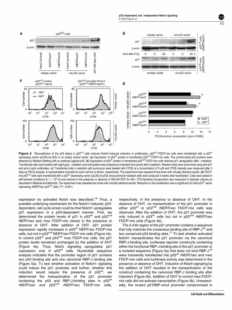
Figure 3 Reconstitution of the p53 status in p53null cells restores Notch1-induced reduction in proliferation. p53null FDCP-mix cells were transfected with a p53wt
expressing vector (pCAG-wt p53) or an empty control vector. (a) Expression of p53wt protein in transfected p53null FDCP-mix cells. The correct-sized p53 proteins weredetected by Western Blotting with an antibody against p53. (b) Expression of p53wt protein in transfected p53null FDCP-mix cells restores p21 upregulation after g-radiation.Transfected cells were treated with eight gray g-radiation and cell lysates were prepared at indicated time points after irradiation. Western blots were performed using anti-p21and anti-b-actin antibodies. (c) Transfected cells in selection with puromycin were labeled with CFSE at a concentration of 5mM and CFSE intensity was measured after 3days by FACS analysis. A representative example for each cell line is shown, respectively. The experiment was repeated three times with virtually identical results. (d) FDCP-mix p53null cells were transfected with a p53wt expressing vector (pCAG-wt p53) and puromycin-resistant cells were analyzed 3 weeks after transfection. Cells were plated inself-renewal conditions at 1� 105 ml and cultured in the presence or absence of 500 nM OHT for 48 h. [3H] thymidine incorporation was measured in triplicate cultures asdescribed in Material and Methods. The experiment was repeated two times with virtually identical results. Reduction in the proliferation rate is significant for both p53wt vectorexpressing rNERTneo p53null cells (*Po0.001)
p53-dependent and -independent Notch signalingK Henning et al
401
Cell Death and Differentiation

RBP-J binding was transactivated by induction of Notch1signaling to a similar extent as the construct containing thecanonical RBP-J binding site (Figure 5b). These data suggestthat the canonical RBP-J site is not involved in mediating theNotch1-dependent transactivation of the p21 promoter. Next,we tested whether the transactivation of the p21 promoter byactivated Notch1 would depend on the p53 binding sites. Asshown in Figure 5c, deletion of either one of the p53 bindingsite led to a reduction in Notch1-dependent transactivationof the p21 promoter and deletion of both p53 binding sitescompletely abolished Notch1-induced transactivation of thep21 promoter. In line with these results, activated Notch1 alsotransactivated the mdm2 promoter, which does contain twop53 binding sites but no canonical RBP-J binding site(Figure 4c).
To test whether activated Notch would enhance bindingof p53 to the endogenous promoter, chromatin preparations
from p53wt rNERTneo FDCP-mix cells in the presence orabsence of OHT were crosslinked to cellular DNA andimmunoprecipitated with anti-p53 antibodies, followed byPCR amplifications of specific p21 promoter regions. Asshown in Figure 6, the two fragments of the mouse p21promoter containing the p53 binding sites, in particular bindingsite 2, were of higher abundance in the immunoprecipitatesfrom rNERTneo FDCP-mix cells treated with OHT comparedto rNERTneo FDCP-mix cells kept without OHT. As expected,no difference was observed after OHT treatment in theamount of immunoprecipitated fragments of a p21 promoterproximal control region that does not harbor p53 binding sites(Figure 6). These results were also confirmed by real-timePCR (data not shown). Taken together, these results provideevidence that Notch-signaling upregulates p21 expression bya p53-dependent mechanism involving enhanced binding ofp53 to the p21 promoter.
Figure 4 p21 expression is only activated by Notch1 signaling in p53wt cells. (a) Activated Notch1IC increases the level of p21 protein in p53wt, but not in p53null FDCP-mixcells. p53wt rneo control, p53null rneo control, p53wt rNERTneo and p53null rNERTneo clones were treated with 500 nM OHT for up to 3 days (three independent experiments).Cell lysates were prepared at indicated time points after induction. Western blots were carried out using antibodies against p21 and actin. A representative Western blotanalysis is shown. (b) Activated Notch1 induces p21 promoter activity in p53wt FDCP-mix cells, but not in p53null FDCP-mix cells. p53wt rneo control, p53null rneo control, p53wt
rNERTneo and p53null rNERTneo clones were transiently transfected in triplicates with a reporter plasmid (p21luc) carrying a luciferase gene under the control of a 2.3 kbfragment of the p21 promoter shown in Figure 5a. Cells were cultured under self-renewal conditions in the absence or presence of 250 nM OHT for 16 h, and luciferase activitywas measured and normalized to the activity of renilla luciferase. The means±S.D. corrected for transfection efficiency are shown. The experiment was repeated three timeswith virtually identical results. Statistically significant upregulation of p21 promoter activity was only observed in p53wt rNERTneo24 cells (*Po0.02). (c) Activated Notch1induces MDM2 promoter activity in p53wt FDCP-mix cells, but not in p53null FDCP-mix cells. p53wt rneo control, p53null rneo control, p53wt rNERTneo and p53null rNERTneoclones were transiently transfected in triplicates with a reporter plasmid (mdm2luc) carrying a luciferase gene under the control of a 0.4 kb fragment of the mdm2 promoter.Cells were cultured under self-renewal conditions in the absence or presence of 250 nM OHT for 16 h, and luciferase activity was measured and normalized to the activity ofrenilla luciferase. The means±S.D. corrected for transfection efficiency are shown. The experiment was repeated three times with virtually identical results. Statisticallysignificant upregulation of p21 promoter activity was only observed in p53wt rNERTneo24 cells (*Po0.001)
p53-dependent and -independent Notch signalingK Henning et al
402
Cell Death and Differentiation
Activated Notch promotes myeloid and erythroiddifferentiation independently of p53. Recently, we haveshown that activated Notch1 induced myeloid and erythroiddifferentiation in p53wt rNERTneo FDCP-mix cells.8,20
Therefore, we asked whether differentiation into myeloidand erythroid cells by the induction of activated Notch1 wassimilarly accelerated in p53null rNERTneo FDCP-mix cells inthe presence of lineage-affiliated cytokines. In the presenceof GM-CSF and G-CSF (myeloid differentiation conditions)differentiation is directed along the myeloid lineage and in thepresence of erythropoietin and low amounts of IL-3 along theerythroid lineage. Thus, p53null rNERTneo FDCP-mix cells,p53wt rNERTneo FDCP-mix cells, p53null control rneo FDCP-mix cells and p53wt control rneo FDCP-mix cells werecultured under conditions that promote either myeloiddifferentiation or erythroid differentiation in the presence orabsence of OHT and were monitored for changes inmorphology and cell surface phenotype. As shown recentlyfor p53wt rNERTneo FDCP-mix cells,8,20 differentiation alongthe myeloid as well as erythroid lineages was accelerated in
the p53null rNERTneo FDCP-mix cells (Tables 1 and 2).Differentiation of p53wt and p53null rneo control cells wasunaltered by the addition of OHT (Tables 1 and 2). Sinceactivated Notch1 accelerated myeloid and erythroiddifferentiation in both p53wt and p53null rNERTneo FDCP-mix cells, we analyzed whether activated Notch1 altersproliferation in differentiation conditions. While the activationof Notch1-signaling considerably reduced proliferation ofp53wt rNERTneo FDCP-mix cells (Table 3) in differentiationconditions,8 Notch1 signaling did not alter proliferation duringthe first 4 days in differentiation medium in p53null cells(Table 3). Only with the appearance of terminallydifferentiated cells, proliferation also started to decrease inp53null rNERTneo FDCP-mix cells to a similar extent as inp53null rneo FDCP-mix cells (data not shown). Takentogether, our data show that activated Notch1 promotesdifferentiation of p53wt and p53null FDCP-mix cells along themyeloid and erythroid lineages, but activated Notch1 reducescell proliferation only in p53wt FDCP-mix cells and not inp53null FDCP-mix cells.
Discussion
In the present work, we provide evidence that p53 is a criticaldeterminant for Notch-mediated inhibition of proliferation:depending on the presence of functional p53, proliferation ofmultipotent hematopoietic progenitor cells is inhibited byactivated Notch1. Recently, we showed that this inhibition ofproliferation is related to a G0/G1 cell cycle arrest.7 Inductionof p21 expression is one of the earliest cell cycle regulatory
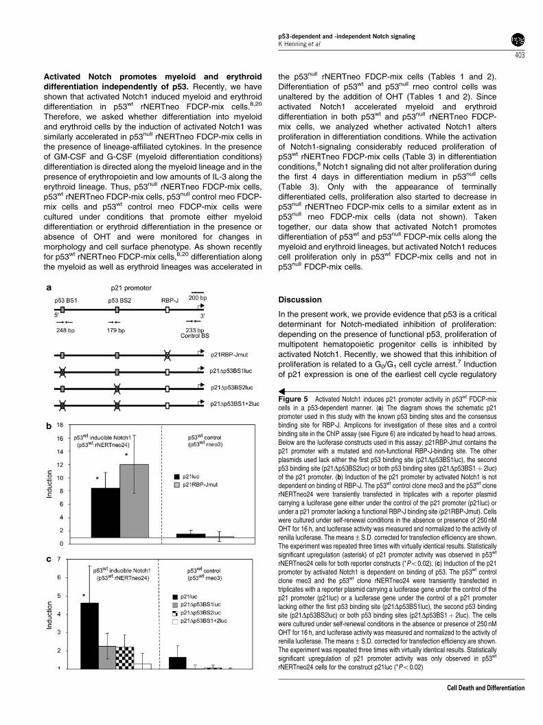
Figure 5 Activated Notch1 induces p21 promoter activity in p53wt FDCP-mixcells in a p53-dependent manner. (a) The diagram shows the schematic p21promoter used in this study with the known p53 binding sites and the consensusbinding site for RBP-J. Amplicons for investigation of these sites and a controlbinding site in the ChIP assay (see Figure 6) are indicated by head to head arrows.Below are the luciferase constructs used in this assay: p21RBP-Jmut contains thep21 promoter with a mutated and non-functional RBP-J-binding site. The otherplasmids used lack either the first p53 binding site (p21Dp53BS1luc), the secondp53 binding site (p21Dp53BS2luc) or both p53 binding sites (p21Dp53BS1þ 2luc)of the p21 promoter. (b) Induction of the p21 promoter by activated Notch1 is notdependent on binding of RBP-J. The p53wt control clone rneo3 and the p53wt clonerNERTneo24 were transiently transfected in triplicates with a reporter plasmidcarrying a luciferase gene either under the control of the p21 promoter (p21luc) orunder a p21 promoter lacking a functional RBP-J binding site (p21RBP-Jmut). Cellswere cultured under self-renewal conditions in the absence or presence of 250 nMOHT for 16 h, and luciferase activity was measured and normalized to the activity ofrenilla luciferase. The means±S.D. corrected for transfection efficiency are shown.The experiment was repeated three times with virtually identical results. Statisticallysignificant upregulation (asterisk) of p21 promoter activity was observed in p53wt
rNERTneo24 cells for both reporter constructs (*Po0.02). (c) Induction of the p21promoter by activated Notch1 is dependent on binding of p53. The p53wt controlclone rneo3 and the p53wt clone rNERTneo24 were transiently transfected intriplicates with a reporter plasmid carrying a luciferase gene under the control of thep21 promoter (p21luc) or a luciferase gene under the control of a p21 promoterlacking either the first p53 binding site (p21Dp53BS1luc), the second p53 bindingsite (p21Dp53BS2luc) or both p53 binding sites (p21Dp53BS1þ 2luc). The cellswere cultured under self-renewal conditions in the absence or presence of 250 nMOHT for 16 h, and luciferase activity was measured and normalized to the activity ofrenilla luciferase. The means±S.D. corrected for transfection efficiency are shown.The experiment was repeated three times with virtually identical results. Statisticallysignificant upregulation of p21 promoter activity was only observed in p53wt
rNERTneo24 cells for the construct p21luc (*Po0.02)
p53-dependent and -independent Notch signalingK Henning et al
403
Cell Death and Differentiation
events underlying growth arrest. We have shown here thatNotch1 signaling upregulates p21 expression. This upregula-tion required the presence of p53. Because p53 can alsocause cell cycle arrest by transcriptionally upregulating p21,21
one possibility could be that activated Notch1 upregulates p53expression. In several mammalian cell lines including humanhepatocellular carcinoma cells, mouse neural progenitor cells,
and human cervical cancer cells, expression of activatedNotch1IC elevated the levels of nuclear p53 and its target genetranscription.22–24 In addition, two well known direct Notchtarget genes, Hes1 and Hey1, upregulate p53 activity throughrepression of HDM2 transcription.5 However, since we did notsee an upregulation of p53 mRNA and protein in p53wt FDCP-mix cells after activation of Notch signaling (data not shown), itseems unlikely that the Notch1-induced proliferation arrest ismediated by an upregulation of p53 expression.
The p21 promoter used in this study contains two p5317 andone RBP-J binding site. Although binding of the RBP-J proteinto this RBP-J site as well as an involvement of RBP-J inNotch-dependent transactivation was recently shown inkeratinocytes,18 transactivation of the p21 promoter by
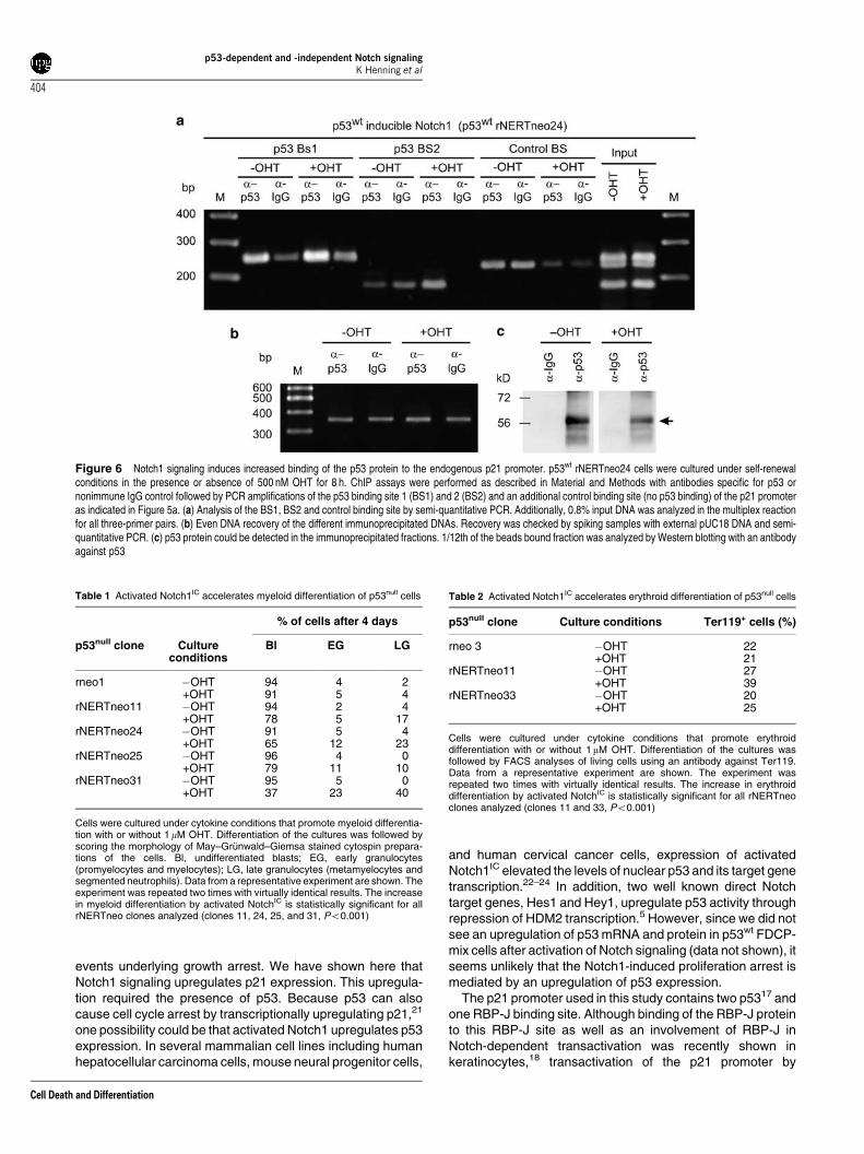
Figure 6 Notch1 signaling induces increased binding of the p53 protein to the endogenous p21 promoter. p53wt rNERTneo24 cells were cultured under self-renewalconditions in the presence or absence of 500 nM OHT for 8 h. ChIP assays were performed as described in Material and Methods with antibodies specific for p53 ornonimmune IgG control followed by PCR amplifications of the p53 binding site 1 (BS1) and 2 (BS2) and an additional control binding site (no p53 binding) of the p21 promoteras indicated in Figure 5a. (a) Analysis of the BS1, BS2 and control binding site by semi-quantitative PCR. Additionally, 0.8% input DNA was analyzed in the multiplex reactionfor all three-primer pairs. (b) Even DNA recovery of the different immunoprecipitated DNAs. Recovery was checked by spiking samples with external pUC18 DNA and semi-quantitative PCR. (c) p53 protein could be detected in the immunoprecipitated fractions. 1/12th of the beads bound fraction was analyzed by Western blotting with an antibodyagainst p53
Table 1 Activated Notch1IC accelerates myeloid differentiation of p53null cells
% of cells after 4 days
p53null clone Cultureconditions
Bl EG LG
rneo1 �OHT 94 4 2+OHT 91 5 4
rNERTneo11 �OHT 94 2 4+OHT 78 5 17
rNERTneo24 �OHT 91 5 4+OHT 65 12 23
rNERTneo25 �OHT 96 4 0+OHT 79 11 10
rNERTneo31 �OHT 95 5 0+OHT 37 23 40
Cells were cultured under cytokine conditions that promote myeloid differentia-tion with or without 1mM OHT. Differentiation of the cultures was followed byscoring the morphology of May–Grunwald–Giemsa stained cytospin prepara-tions of the cells. Bl, undifferentiated blasts; EG, early granulocytes(promyelocytes and myelocytes); LG, late granulocytes (metamyelocytes andsegmented neutrophils). Data from a representative experiment are shown. Theexperiment was repeated two times with virtually identical results. The increasein myeloid differentiation by activated NotchIC is statistically significant for allrNERTneo clones analyzed (clones 11, 24, 25, and 31, Po0.001)
Table 2 Activated Notch1IC accelerates erythroid differentiation of p53null cells
p53null clone Culture conditions Ter119+ cells (%)
rneo 3 �OHT 22+OHT 21
rNERTneo11 �OHT 27+OHT 39
rNERTneo33 �OHT 20+OHT 25
Cells were cultured under cytokine conditions that promote erythroiddifferentiation with or without 1 mM OHT. Differentiation of the cultures wasfollowed by FACS analyses of living cells using an antibody against Ter119.Data from a representative experiment are shown. The experiment wasrepeated two times with virtually identical results. The increase in erythroiddifferentiation by activated NotchIC is statistically significant for all rNERTneoclones analyzed (clones 11 and 33, Po0.001)
p53-dependent and -independent Notch signalingK Henning et al
404
Cell Death and Differentiation
activated Notch was not mediated via this RBP-J-binding sitein hematopoietic progenitor cells. A recent report hasindicated that Notch activity on RBP-J-responsive promoterscritically depends on protein modules and promoter context.25
Thus, one possibility to explain this discrepancy could be thatactivated Notch1 transactivates the p21 promoter via theRBP-J binding site in keratinocytes, but not in hematopoieticprogenitor cells due to the lack of other essential cofactors inhematopoietic cells.
In addition to the RBP-J-dependent mechanism,Mammucari et al.26 identified a second Calcineurin-depen-dent mechanism for transactivation of p21 by activatedNotch1 in keratinocyte growth and differentiation control,acting on the p21 TATA box proximal region. Whiletransactivation of the p21 promoter by increased Calcineur-in/NFAT activity induced by Notch signaling required RBP-Jand implies a crosstalk between these two signaling path-ways, we show here a novel, RBP-J independent mechanismfor transactivation of the p21 promoter by activated Notch1that critically depends on the p53 binding sites of the p21promoter. Deletion of both p53 binding sites of the p21promoter completely abolished transactivation, suggestingthat p53 is required for Notch1-mediated transactivation ofp21. The activated Notch1-induced, increased binding of p53to these sites further suggests that enhanced binding of p53 tothe p21 promoter after activation of Notch1 signaling isinvolved in the activation of p21 transcription, thereby inducinga cell cycle arrest.
Post-translational modifications of the p53 protein enhancethe ability of p53 to activate transcription.27 Phosphorylationof p53 at various sites can lead to either stabilization of p53,activation of p53 activity or recruitment of CBP/p300 or PCAFand p53 acetylation. p53 acetylation can be involved inenhancing p53 DNA binding, activating p53 transcriptionalactivities and in p53 stabilization. Furthermore, specific
combinations of post-transcriptional modifications generatedistinct p53 cassettes that direct p53 towards precise cellularfunctions.28 While acetylation at K320 suppresses theapoptotic program and activates promoters such as p21leading to a temporary cell cycle arrest, acetylation at K373together with phosphorylation at S46 and S15 stabilizes theinteraction with p300 and activates promoters of proapoptoticgenes.28 Recently, it was shown that Notch1 interacts withp53 and inhibits its phosphorylation at S15 and S4629 siteswhich were implicated in the study by Knights et al.28 indirecting p53 to promoters for proapoptotic genes therebyactivating the proapoptotic program. In line with the lack of p53modifications,28 activated Notch1-inhibited p53-dependenttransactivation and suppressed p53-dependent apoptosis ofhuman colon cancer cells.29 In addition, several other reportshave indicated that Notch signaling may negatively regulatep53 function and inhibit apoptosis.30–32 These data seem tobe in contrast to other studies18,22–24 and our data shown hereand previously7,8 that activated Notch1 reduces proliferationof hematopoietic progenitor cells through a p53-dependentpathway by blocking the cell cycle in G0/G1 but does notinfluence apoptosis. However, Notch effects as well as p53modifications depend largely on the cell type, and consideringthe direct interaction of Notch with p53, it is tempting tospeculate, that activated Notch may increase levels of p53acetylated at K320, thereby blocking the cell cycle via p21.
Irrespective of the presence or absence of p53, Notch1-signaling promoted myeloid and erythroid differentiation ofFDCP-mix cells as described previously.8,20 This suggeststhat the Notch1-induced cell-cycle block and the induction ofdifferentiation are unlinked events and most likely mediated bydistinct biochemical mechanisms. Consistent with its functionin temporarily halting the cell cycle under stress conditions,mice with a deficiency in p53 as well as mice with a deficiencyin p21 have increased numbers of cycling hematopoietic stemcells under stress conditions, while hematopoietic differentia-tion is entirely normal.33–35 Thus, activated Notch1 mayreduce proliferation in dependence of p53 via p21 upregula-tion, however, the induction of myeloid and erythroiddifferentiation by increased Notch1-signaling occurs by adifferent p53-independent pathway. Recently, we have shownthat activated Notch1 directly and specifically upregulates thehematopoietic transcription factor PU.1 in correlation with itsinduction of myeloid differentiation.8 Along this line, we haveidentified b-globin as a direct Notch1 target gene involvedin erythroid maturation.20 This suggests that the induction ofdifferentiation by Notch1 signaling involves hematopoietictranscription factors as well as functional proteins but not cellcycle regulators. The role of Notch signaling for self renewal,maintenance, and proliferation of adult hematopoietic stemand progenitor cells is controversial.1 This may reflect the factthat the hematopoietic stem and progenitor compartmentconsists of several diverse cell types with different molecularsignatures that are likely to respond in a different way to Notchsignals. In the present study, we have used the multipotentcell line FDCP-mix that, although being an immortal cell line,shares many characteristics with primary common myeloidprogenitor cells. It will be of considerable interest todetermine, which of the hematopoietic stem and progenitorcell types respond in a similar way to Notch and p53 signaling
Table 3 Activated Notch1IC does not affect proliferation of p53null cells indifferentiation conditions
Cell number� 105
Cell clone �OHT +OHT
p53null
rneo1 1.8±0.3 1.7±0.4rNERTneo11 1.6±0.3 1.9±0.4rNERTneo24 1.5±0.3 1.2±0.4rNERTneo25 1.6±0.3 1.6±0.4rNERTneo31 1.5±0.3 1.2±0.4
p53wt
A7rneo1 1.3±0.4 1.2±0.3A7rNERTneo24 1.3±0.3 0.6±0.2A7rNERTneo25 0.7±0 0.1±0A7rNERTneo26 1.2±0.4 0.4±0.1
p53null and p53wt FDCP-mix cells were cultured under cytokine conditions thatpromote myeloid differentiation with or without 1 mM OHT. Cell numbers weredetermined after 3 days (p53wt cells) or 4 days (p53null cells). Data from arepresentative experiment are shown. The experiment was repeated two timeswith virtually identical results. The reduction in proliferation is statisticallysignificant for p53wt A7rNERTneo cells (all p53wt A7rNERTneo clones analyzed(clones 24, 25 and 26, Po0.001) but not for p53null rNERTneo cells. The slightreduction of cell numbers in p53null rNERTneo24 and p53null rNERTneo31clones results from terminally differentiated cells in the presence of OHT(compare Table 1)
p53-dependent and -independent Notch signalingK Henning et al
405
Cell Death and Differentiation
as the FDCP-mix cells. In line with our work with FDCP-mixcells, Notch-signaling triggers two distinct pathways inkeratinocytes, involving a p21 and a RBP-J independentmechanism, respectively, leading to growth arrest anddifferentiation.18 It remains to be tested whether p53 playsalso a role for the Notch-induced growth arrest in keratino-cytes.
Materials and MethodsCell culture. FDCP-mix cells were maintained (self renewal conditions) inIscove’s modified Dulbecco’s medium (IMDM) supplemented with 20% pretestedhorse serum and mouse IL-3 conditioned medium at a concentration that stimulatedoptimal cell growth, which corresponds to 100 U rIL-3 per ml. FDCP-mix cells werekept in a density between 6� 104 to 106 cells/ml and were carefully controlled fornormal growth rates, factor dependency and differentiation. For activation of theOHT-inducible mN1IC FDCP-mix cells, 4-Hydroxy-Tamoxifen (OHT, RBI, USA) wasadded to the medium at the concentrations indicated. All cells were regularlychecked to be free of mycoplasma contamination using a Mycoplasma PCR ElisaKit (Roche, Germany).
Erythroid differentiation of cells was induced by washing the cells once in IMDMand plating 1� 105 cells/ml in IMDM containing 20% pretested fetal calf serum(FCS), 5 U/ml erythropoietin (Roche, Germany) and 5 U/ml IL-3 (Roche, Germany).Myeloid differentiation of cells was induced by washing the cells once in IMDM andplating 1� 105 cells/ml in IMDM containing 20% pretested FCS, 1000 U/ml G-CSF(Neupogen, Amgen, USA), 2 U/ml IL-3 (Roche, Germany) and 250 U recombinantmurine GM-CSF per ml.36 Aliquots were removed for analysis at time pointsindicated. To ensure optimal growth and differentiation conditions, the cells weresplit to constant density and fed with fresh differentiation medium every fourth day.Viable cells were counted by trypan blue dye exclusion. Differentiation of FDCP-mixcells was monitored by FACS analyses and by morphological scoring of May-Grunwald-Giemsa and O-Dianiside stained cytospin preparations. Considerablecare was taken to validate accurate differential counts. All differential counts weredone on 100–200 cells in a blinded fashion by TS and UJ.
Plasmids, stable transduction and selection procedures. Toobtain clones of the p53null multipotent hematopoietic progenitor cell line,11 inwhich translocation of the constitutive active intracellular domain of mNotch1 intothe nucleus and RBP-J-dependent transactivation of target genes can be regulated,p53null FDCP-mix cells were transfected by electroporation with a retroviral vectorcarrying an intracellular domain of the murine Notch1 fused to the hormone-bindingdomain of the human estrogen receptor (rNERTneo9) and stable cell lines wereestablished by G418 selection. As a control, cells were transfected with the retroviralvector alone (rneo9). Cell clones derived from three independent transfections wereused in this study. To obtain p53null NERTneo cells, which express functional p53wt,a DNA fragment containing the p53wt gene was subcloned into the pCAG-expression vector carrying the puromycin resistance gene,15 and cells weretransfected by electroporation or nucleofection (Amaxa, Germany). In brief, 5� 106
cells were either electroporated as described previously9 or nucleofected with 1 mgpCAG-p53 in solution R with program W-01. Mass cultures were selected with 0.5–2.0mg Puromycin for 3–21 days and clonal cell lines were established after 3 weeksby cloning in soft agar in the presence of 2.0mg Puromycin. Mass cultures wereanalyzed 3 days (CFSE staining) or 3 weeks ([3H] thymidine incorporation) aftertransfection.
FACS analyses. Phycoerytrin (PE)- and Allophycocyanin (APC)-conjugatedmonoclonal antibodies directed against Ly76 (clone Ter119) and CD11b (Mac-1,clone M1/70), respectively, or their respective isotype controls were used (allPharmingen, Europe). Cells were harvested by centrifugation and resuspended inPBS containing 3% FCS. Fc-Block (#01241D, Pharmingen, Europe) was added at adilution of 1 : 100 for 5 min at room temperature (RT). Subsequently, antibodieswere added at a dilution of 1 : 100. After an incubation for 20 min at RT in the dark,cells were washed and resuspended in PBS containing 1% FCS and 1mg/mlpropidium iodide or 7AAD for dead cell exclusion. FACS analysis was done with aBecton Dickinson FACSCalibur machine and Cell Quest software, or a BectonDickinson FACSCanto machine and FACSDiva software using standardprocedures.
Reporter constructs, transient transfections and luciferaseassays. Cells (5� 106) were transiently transfected by electroporation9 andluciferase reporter assays8 were done as described. The following constructs wereused: For control of Notch activation: 1mg of (RBP-J RE)12-Luc (pGa981-6, fireflyluciferase reading frame under the control of a minimal b-globin promoter and 12RBP-J-binding sites37); for transactivation of the p21 promoter: 18 mg of p21luc,which carries a firefly luciferase gene under the control of the �2. 2 kb human p21promoter;38 for transactivation of the mdm promoter: 18mg of mdm2luc, whichcarries a firefly luciferase gene under control of the �350 bp human mdm2promoter;38 and either 0.1mg phRL-CMV plasmids or 3 mg pTK-renilla plasmids(constitutive expression of Renilla-luciferase for transfection efficiency control),respectively. Cells were treated with different concentrations of OHT as indicatedand measurements of luciferase activities were performed using the Dual LuciferaseKit (Promega) according to manufacturers instructions.
Mutation of the RBP-J binding site. Mutation of the RBP-J binding sitewithin the p21-promoter (p21RBP-Jmut) was performed by a three-step site-directed PCR mutagenesis using the primers p21mut for (50-gagggatcacaccgtctagaggtgatattgtggg-30), p21mut rev (50-tcacctctagacggtgtgatccctcactaggtca-30),NheI for (50-cggtcccggaacctcgcgtgctgcagagg-30) and PstI rev (50-tacgcgtgctagcccgggctc-30) and the plasmid p21luc as a template. Deletion of the p53 bindingsites was performed by the same method using the primers p53BS1 for(50-ggccattagagctctggcatagaagag-30), p53BS1 rev (50-gccagagctctaatggccagaaagcca-30), XhoI for (50-tcttacgcgtgctagcccgggctcgagatc-30), and AvaIII rev (50-caagcacacatgcatcagatcc-30) for deletion of the first p53 binding site and primers p53BS2for (50-agaggaagagagatttccagactctga-30), p53BS2 rev (50-ggaaatctctcttcctctaacgcagct-30), PstI rev and NheI for for deletion of the second p53 binding site. Mutation ofthe RBP-J binding site and deletion of the p53 binding sites were confirmed bysequencing.
CFSE-assay. Cells (5� 106) were incubated with CFSE (Sigma, Munich) at afinal concentration of 5mM in 1 ml PBS containing 0.1% BSA for 5 min at 371C. Thereaction was stopped by adding 10 ml cold-medium containing 20% horse serumand incubation on ice for 5 min. The cells were washed two times and resuspendedat a concentration of 2� 105 cells/ml in self-renewal medium in the presence orabsence of 500 nM OHT. Proliferation was analyzed after 3 days by FACS analysis.
[3H]-thymidine incorporation. Cells were plated in self-renewal medium ateither 6� 104 or 1� 105 per ml in the absence or presence of 500 nM OHT. After24 or 48 h, [3H] thymidine incorporation was measured as described previously.7
Data are expressed as mean counts per minute of triplicate wells±S.D.
ChIP assay. The ChIP was performed as described previously39 with slightmodifications. Briefly, 6.5� 107 FDCP-mix cells (A7rNERTneo24 cells) wereinduced or not induced for 8 h with 500 nM OHT, washed with PBS andresuspended in 1.5 ml SDS lysis buffer (1% SDS, 10 mM EDTA, 50 mM Tris–HCl,pH 8.1) plus Complete protease inhibitor (Roche). To confirm the induction of theNotch-signaling pathway, the upregulation of Hes1 and Hey1 transcripts waschecked by real-time PCR as described previously40 (data not shown). 2� 400mlof the lysate were sonified and precleared with 70 ml Protein A (Upstate,Charlottesville, VA, USA)/Protein G (Active Motif, Carlsbad, CA, USA) agarosebeads mix (1 : 1, Salmon Sperm saturated) for 1 h at 41C. 10mg of anti-p53 antibody(Pab240) or 10mg anti-mouse IgG (Active Motif) were added as a control, incubatedfor 1 h at 41C, and 150ml Protein A/G slurry mix were added overnight. Additionally,samples were spiked with 100 ng of pUC18 vector DNA as an external control forrecovery. The beads were washed and crosslinks were reversed as described in theUpstate ChIP protocol. DNA was purified using the ChIP-IT purification columns(Active Motif) and eluted in a 100ml H2O. Five microliter of this DNA were measuredeither in a semi-quantitative PCR using the Multiplex PCR Kit-100 (Qiagen, Hilden,Germany) in 50ml or in a real-time PCR using FastStart Universal SYBR GreenMaster Mix (Rox) (Roche Diagnostics, Mannheim, Germany) in a 10 ml volume.Primers for p53-binding site within the p21 promoter at a final concentrationof 100 nM were: BS1 forward: 50-cccttggatttcctttctatcag-30; BS1 reverse:50-gtagttgggtatcatcaggtctcc-30; BS2 forward: 50-tgtgtttctgaacaggatgagg-30; BS2reverse: 50-tgagttctgacatctgctctcc-30; control BS (no p53 binding) forward: 50-gttcatagatgtatgtggctctgc-30; control BS reverse: 50-gtcgagctgcctccttatagc-30; pUCforward: 50-aagttggccgcagtgttatc-30; pUC reverse: 50-tttgccttcctgtttttgct-30. For semi-quantitative PCR BS1, BS2, and control BS were cycled 36 times and pUC wascycled 28 times (protocol on request).
p53-dependent and -independent Notch signalingK Henning et al
406
Cell Death and Differentiation
Western Blotting. Cell were cultured under self-renewal conditions and in thepresence or absence of 500 nM OHT. Harvesting, electrophoresis and WesternBlotting of protein extracts were performed as described previously.9 Tenmicrograms of protein was separated per lane. Antibodies specific for p21(sx-118, Pharmingen/BectonDickinson, Heidelberg, Germany), b-actin (C2, SantaCruz Biotechnology, CA, USA) or estrogen receptor (SC-8002, Santa Cruz) wereused. The detected proteins were visualized by the ECL system (AmershamPharmacia Biotec, UK). For control of the ChIP assay 25 ml of the Protein A/G slurrywere incubated in SDS loading buffer and half of it were used for a Western blot.Pab240 anti-p53 antibody (1 ng/ml) was used to detect the precipitated p53.
Statistical analysis. Statistical differences were assessed using the Student’st-test for paired data.
Acknowledgements. We thank Silke Horn, Cornelia Kuklik-Roos andGabriele Warnecke for expert technical support and Elaine Spooncer andMichael Dexter for the p53null FDCP-mix cell lines. This work was supported bythe Deutsche Forschungsgemeinschaft (Priority Program 1109 ‘Stem Cells’ andSFB 415 ‘Signal Transduction’ project B8 to UJ). This report represents a part of thedoctoral thesis by KH and of the diploma thesis of JH.
1. Wilson A, Radtke F. Multiple functions of Notch signaling in self-renewing organs andcancer. FEBS Lett 2006; 580: 2860–2868.
2. Lai EC. Notch signaling: control of cell communication and cell fate. Development 2004;131: 965–973.
3. Hurlbut GD, Kankel MW, Lake RJ, Artavanis-Tsakonas S. Crossing paths with Notch in thehyper-network. Curr Opin Cell Biol 2007; 19: 166–175.
4. Honjo T. The shortest path from the surface to the nucleus: RBP-J kappa/Su(H)transcription factor. Genes Cells 1996; 1: 1–9.
5. Huang Q, Raya A, DeJesus P, Chao S-H, Quon K, Caldwell J et al. Identification ofp53 regulators by genome-wide functional analysis. Proc Natl Acad Sci USA 2004; 101:3456–3461.
6. Vogelstein B, Lane D, Levine A. Surfing the p53 network. Nature 2000; 408: 307–310.7. Schroeder T, Just U. mNotch1 signaling reduces proliferation of myeloid progenitor cells by
altering cell-cycle kinetics. Exp Hematol 2000; 28: 1206–1213.8. Schroeder T, Kohlhof H, Rieber N, Just U. Notch signaling induces multilineage myeloid
differentiation and up-regulates Pu.1 expression. J Immunol 2003; 170: 5538–5548.9. Schroeder T, Just U. Notch signalling via RBP-J promotes myeloid differentiation. EMBO J
2000; 19: 2558–2568.10. Spooncer E, Heyworth CM, Dunn A, Dexter TM. Self-renewal and differentiation of
interleukin-3-dependent multipotent stem cells are modulated by stromal cells and serumfactors. Differentiation 1986; 31: 111–118.
11. Pierce A, Spooncer E, Wooley S, Dive C, Francis J, Miyan J et al. Bcr-Abl protein tyrosinekinase activity induces a loss of p53 protein that mediates a delay in myeloid differentiation.Oncogene 2000; 19: 5487–5497.
12. Just U, Stocking C, Spooncer E, Dexter TM, Ostertag W. Expression of the GM-CSF geneafter retroviral transfer in hematopoietic stem cell lines induces synchronous granulocyte-macrophage differentiation. Cell 1991; 64: 1163–1173.
13. Just U, Boettiger D, Kan O, Dexter TM, Spooncer E. Insertional mutagenesis as a route toidentifying genes involved in self renewal of haemopoietic stem cells. Curr Top MicrobiolImmunol 2000; 251: 27–34.
14. Lyons A, Parish C. Determination of lymphocyte division by flow cytometry. J ImmunolMethods 1994; 171: 131–137.
15. Niwa H, Masui S, Chambers I, Smith AG, Miyazaki J. Phenotypic complementationestablishes requirements for specific POU domain and generic transactivation function ofOct-3/4 in embryonic stem cells. Mol Cell Biol 2002; 22: 1526–1536.
16. Sherr C, Roberts J. CDK inhibitors:positive and negative regulators of G1-phaseprogression. Genes Dev 1999; 13: 1501–1512.
17. El-Deiry WS, Tokino T, Velculescu VE, Levy DB, Parsons R, Trent JM et al. WAF1, apotential mediator of p53 tumor suppression. Cell 1993; 75: 817–825.
18. Rangarajan A, Talora C, Okuyama R, Nicolas M, Mammucari C, Oh H et al. Notch signalingis a direct determinant of keratinocyte growth arrest and entry into differentiation. EMBO J2001; 20: 3427–3436.
19. Tun T, Hamaguchi Y, Matsunami N, Furukawa T, Honjo T, Kawaichi M. Recognitionsequence of a highly conserved DNA binding protein RBP-J. Nucleic Acids Res 1994; 22:965–971.
20. Henning K, Schroeder T, Schwanbeck R, Rieber N, Bresnick EH, Just U. mNotch1signalling and erythropoietin cooperate in erythroid differentiation of multipotent progenitorcells by upregulation of b-globin. Exp Hematol 2007; 35: 1321–1332.
21. Allan LA, Duhig T, Read M, Fried M. The p21(WAF1/CIP1) promoter is methylated inRat-1 cells; stable restoration of p53-dependent p21 (WAF1/CIP1) expression aftertransfection of a genomic clone containing the p21(WAF1/CIP1) gene. Mol Cell Biol 2000;
20: 1291–1298.22. Qi R, An H, Yu Y, Zhang M, Liu S, Xu H et al. Notch1 signaling inhibits growth of human
hepatocellular carcinoma through induction of cell cycle arrest and apoptosis. Cancer Res
2003; 63: 8323–8329.23. Talora C, Cialfi S, Segatto O, Morrone S, Kim Choi J, Frati L et al. Constitutively active
Notch1 induces growth arrest of HPV-positive cervical cancer cells via separate signaling
pathways. Exp Cell Res 2005; 305: 343–354.24. Yang X, Klein R, Tian X, Cheng HT, Kopan R, Shen J. Notch activation induces apoptosis
in neural progenitor cells through a p53-dependent pathway. Dev Biol 2004; 269: 81–94.25. Ong C-T, Cheng H-T, Chang L-W, Ohtsuka T, Kageyama R, Stormo G et al. Target
selectivity of vertebrate Notch proteins: collaboration between discrete domains andCSL-binding site architecture determines activation probability. J Biol Chem 2006; 281:5106–5119.
26. Mammucari C, Tommasi di Vignano A, Sharov A, Neilson J, Havrda M, Roop D et al.Integration of Notch1 and calcineurin/NFAT signaling pathways in keratinocyte growth anddifferentiation control. Dev Cell 2005; 8: 665–676.
27. Xu Y. Regulation of p53 responses by post-translational modifications. Cell Death Diff2003; 10: 400–403.
28. Knights CD, Catania J, Di Giovanni S, Muratoglu S, Perez R, Swartzbeck A et al. Distinct
p53 acetylation cassettes differentially influence gene-expression patterns and cell fate.J Biol Chem 2007; 173: 533–544.
29. Kim SB, Chae GW, Park J, Tak H, Chung JH, Park TG et al. Activated Notch1 interacts with
p53 to inhibit its phosphorylation and transactivation. Cell Death Diff 2007; 14: 982–991.30. Nair P, Somasundaram K, Krishna S. Activated Notch1 inhibits p53-induced apoptosis and
sustains transformation by human papillomavirus type 16 E6 and E7 oncogenes through a
PI3K-PKB/Akt-dependent pathway. J Virol 2003; 77: 7106–7112.31. Mungamuri S, Yang X, Thor A, Somasundaram K. Survival signaling by
Notch1:mammalian target of rapamycin (mTOR)-dependent inhibition of p53. Cancer
Res 2006; 66: 4715–4724.32. Beverly L, Felsher D, Capobianco A. Suppression of p53 by Notch in lymphomagenesis:
implications for initiation and regression. Cancer Res 2005; 65: 7159–7168.33. Cheng T, Rodrigues N, Shen H, Yang Y, Dombkowski D, Sykes M et al. Hematopoietic
stem cell quiescence maintained by p21cip1/waf1. Science 2000; 287: 1804–1808.34. van Os B, Kamminga L, Ausema A, Bystrykh L, Draijer D, van Pelt K et al. A limited role for
p21cip1/waf1 in maintaining normal hematopoietic stem cell function. Stem Cells 2007; 25:836–843.
35. TeKippe M, Harrison DE, Chen J. Expansion of hematopoietic stem cell phenotype and
activity in Trp53-null mice. Exp Hematol 2003; 31: 521–527.36. Schroeder T, Lange C, Strehl J, Just U. Generation of functionally mature dendritic cells
from the multipotential stem cell line FDCP-mix. Br J Haematol 2000; 111: 890–897.37. Strobl LJ, Hofelmayr H, Stein C, Marschall G, Brielmeier M, Laux G et al. Both Epstein-Barr
viral nuclear antigen 2 (EBNA2) and activated Notch1 transactivate genes by interactingwith the cellular protein RBP-J kappa. Immunobiology 1997; 198: 299–306.
38. Kim E, Rohaly G, Heinrichs S, Gimnopoulos D, Mei�ner H, Deppert W. Influence ofpromoter DNA topology on sequence-specific DNA binding and transactivation by tumorsuppressor p53. Oncogene 1999; 18: 7310–7318.
39. Kim E, Gunther W, Yoshizato K, Meissner H, Zapf S, Nusing RM et al. Tumor suppressorp53 inhibits transcriptional activation of invasion gene thromboxane synthase mediated bythe proto-oncogenic factor ets-1. Oncogene 2003; 22: 7716–7727.
40. Schroeder T, Meier-Stiegen F, Schwanbeck R, Eilken H, Nishikawa S, Hasler R et al.
Activated Notch1 alters differentiation of embryonic stem cells into mesodermal celllineages at multiple stages of development. Mech Dev 2006; 123: 570–579.
p53-dependent and -independent Notch signalingK Henning et al
407
Cell Death and Differentiation