Gene expression profile based classification models of psoriasis
Upload
independentCategory
view
1download
0
AU
TH
OR
PR
OO
F
Erythroid Disturbances Before and After Treatmentof Portuguese Psoriasis Vulgaris PatientsA Cross-Sectional and Longitudinal Study
Susana Coimbra,1,2,3 Hugo Oliveira,4 Flavio Reis,5 Luıs Belo,1,2 Susana Rocha,1,2 Alexandre Quintanilha,2,6
Americo Figueiredo,4 Frederico Teixeira,5 Elisabeth Castro,1,2 Petronila Rocha-Pereira2,7 and Alice Santos-Silva1,2
1 Department of Biological Sciences, Biochemistry, Faculty of Pharmacy, University of Porto, Porto, Portugal
2 Institute of Cellular and Molecular Biology (IBMC), University of Porto, Porto, Portugal
3 Center of Investigation of Technologies of Health (CITS) – IPSN-CESPU, Gandra-Paredes, Portugal
4 Dermatology Service, University Hospitals of Coimbra, Coimbra, Portugal
5 Institute of Experimental Pharmacology and Therapeutics, IBILI, Faculty of Medicine, University of Coimbra, Coimbra, Portugal
6 Institute for Biomedical Sciences Abel Salazar (ICBAS), University of Porto, Porto, Portugal
7 Center of Investigation of Health Sciences (CICS), University of Beira Interior, Covilha, Portugal
Abstract Background: A few studies in psoriasis vulgaris patients have reported changes suggesting red blood cell
(RBC) damage is linked to neutrophil activation, oxidative stress, and psoriasis worsening.
Objective: The aim of this study was to evaluate erythroid disturbances in Portuguese psoriasis vulgaris
patients, before, during, and after treatment.
Methods:Across-sectional study (n= 73 patients vs 40 healthy control subjects) followed by a longitudinal study
(n= 47 patients) was performed, with assessments before, and at 3, 6, and 12weeks of therapy (10 patients started
topical treatment, 17 narrow-band UVB, and 20 photochemotherapy [psoralen plus UVA; PUVA]).
Evaluations included hematologic data, total bilirubin levels, membrane-bound hemoglobin (MBH), membrane
protein band 3 profile, total plasma antioxidant status (TAS), lipid peroxidation (thiobarbituric acid [TBA]
assay), elastase, lactoferrin, and C-reactive protein (CRP).
Results: Before treatment, patients presented with higher leukocyte/neutrophil and reticulocyte counts,
elastase, lactoferrin, TBA, TBA/TAS, reticulocyte production index, total bilirubin andMBH values, lower
RBC and hematocrit, higher percentages of high-molecular-weight aggregates, and lower percentages of
band 3 monomer. After treatment, we observed a reversal in most of the parameters. However, patients still
presented with values suggestive of accelerated RBC damage, removal, and production, as most of the
parameters were still higher than those in the control group; the same occurred with CRP.
Conclusion: Our data suggest that psoriasis vulgaris triggers an inflammatory response, with release of acute-
phase reactants, reactive oxygen species, cationic proteins, and proteases, leading to enhanced RBC damage/aging and, ultimately, to enhancedRBC removal. These assumptions were strengthened by the observation that,
with treatment, all of these changeswere reversed, the inflammationwas reduced, the production of reticulocytes
was increased, and the RBCs presented changes usually observed in younger/less damaged RBCs. These
erythroid changes were enhanced with PUVA therapy, probably due to the more pronounced clearing of the
lesions, as suggested by Psoriasis Area and Severity Index (PASI) scores. Finally, after treatment, a residual
inflammation still persisted that might contribute to the observed erythroid disturbances.
Introduction
Psoriasis vulgaris is a chronic and recurrent inflammatory
skin disease that has been associated with oxidative stress and
with marked inflammatory changes in the epidermis and der-
mis, as well as in plasmatic markers of inflammatory re-
sponse.[1] This response is linked to the release of chemotactic
substances that trigger the mobilization and activation of
Approval for publication Signed Date Number of amended pages returned
ORIGINAL RESEARCH ARTICLEAm J Clin Dermatol 2012; 13 (1): 1-11
1175-0561/12/0001-0001/$49.95/0
ª 2012 Adis Data Information BV. All rights reserved.
AU
TH
OR
PR
OO
F
inflammatory cells, such as neutrophils, which seem to play a
crucial role in the evolution of psoriasis.[2,3] A significant rise in
leukocytes, due to an increase in neutrophils and lymphocytes,
has been reported in the earliest stages of psoriatic lesions.[4]
This rise has been associated with an increase in elastase and
lactoferrin, both products of neutrophil activation, and with
the severity of the disease, as defined by the Psoriasis Area and
Severity Index (PASI).[1,5] Along the inflammatory process,
several cytokines are released by inflammatory cells that lead to
a rise in C-reactive protein (CRP).[6] This acute-phase protein
has emerged as a risk factor for atherosclerosis and it has been
proposed to also have an active role in inflammation,[7-10] by
inhibiting neutrophil chemotaxis and endothelial cell adhesion,
reducing transmigration to the site of inflammation and, there-
fore, increasing the number of circulating activated neutrophils.
When activated, neutrophils are able to release oxygen me-
tabolites and their granular constituents, both of which may
induce oxidative and proteolytic modifications to plasma
constituents and to other blood cells, namely erythrocytes.[11,12]
A strong association between psoriasis, overweight/obesity,and cardiovascular co-morbidities has been proposed.[13-16]
Obesity is a proinflammatory state, presenting as a low-grade,
chronic inflammatory condition that might further enhance
inflammation in psoriatic patients;[17,18] it might also stimulate
erythropoiesis to increase the number of circulating red cells[19]
to face the higher oxygen requirements, due to the increase in
body mass.
The erythrocyte has a limited biosynthesis capacity, suffer-
ing and accumulating physical and chemical changes, that
become more pronounced with cell aging, and whenever an un-
usual physical or chemical stress develops.[20] The inflammatory
process in psoriasis, in part the result of leukocyte activation,
seems to impose oxidative and proteolytic changes in red blood
cells (RBCs) that lead to accelerated RBC aging and premature
removal.[12] The removal of senescent or damaged RBCs
involves the development of a neoantigen on the membrane
surface that is immunologically related to band 3,[21-23] a
transmembrane RBC protein that seems to have a key role in
erythrocyte function, and membrane deformability.[24] Mod-
ifications in this protein trigger the binding of specific natural
anti-band 3 autoantibodies and complement activation, mark-
ing the cell for death.[25,26] Actually, the degradation of the
RBC metabolism and failure of the antioxidant defences in
damaged or senescent RBCs favours the development of oxi-
dative stress within the cell, allowing hemoglobin oxidation
and its linkage to the cytoplasmatic domain of band 3, pro-
moting the aggregation of band 3 and the binding of anti-band 3
autoantibodies.[27] Membrane-bound hemoglobin (MBH) and
the band 3 profile were proposed as good cumulative markers
of erythrocyte aging and/or damage in several inflammatory
and oxidative stress conditions.[28-35] Differences in the band 3
profile (high-molecular-weight aggregates [HMWAg], band 3
monomer, and proteolytic fragments) have been associated
with the age and condition of RBCs.[12,29,30,35,36] Older and
damaged RBCs have higher band 3 aggregation and lower
fragmentation, whereas younger RBCs showed reduced ag-
gregation and higher fragmentation.
Other studies have reported erythrocyte membrane changes
in psoriasis, namely, in membrane lipid peroxidation,[37,38] in
the sodium-potassium adenosine triphosphatase pump,[39] in the
bindingof cyclic adenosinemonophosphate to proteinkinaseA,[40]
and in the expression and distribution ofmembrane protein 4.1.[41]
Previous studies in our laboratory[12] showed that leukocyte
activation seems to induce, especially in the active forms of
psoriasis, an altered erythrocyte band 3 profile, similar to that
observed in older and/or damaged erythrocytes; we also ob-
served a lower RBC count accompanied by changes suggesting
a slight increase in RBC removal and RBC production. How-
ever, few longitudinal studies have been performed to examine
the erythrocyte changes and their relationship with neutrophil
activation, oxidative stress, and inflammation, before, during,
and after treatment for psoriasis. These studies could contrib-
ute to a better understanding of psoriasis pathogenesis, and of
the efficacy of currently used therapies.
Topical agents, appropriate wavelengths of UV radiation,
and systemic therapies are treatments commonly used, singly or
in combination, in psoriasis vulgaris.[42] Topical agents are
usually sufficient to control mild psoriasis, while moderate to
severe psoriasis often requires phototherapy (narrow-band
UVB [NB-UVB], photochemotherapy [psoralen plus UVA;
PUVA]) or even systemic agents.
Our aim was to study erythrocyte changes alongside neu-
trophil activation, oxidative stress, and inflammatory markers
in Portuguese psoriasis vulgaris patients, before, during, and
after treatment, by performing a cross-sectional and a longi-
tudinal study (before and 3, 6, and 12 weeks after therapy).
Indeed, by performing a longitudinal study, we aimed to eval-
uate the changes occurring during the course of treatment, with
improvement of the disease and clearing of the lesions. The
results were also analyzed according to the type of therapy used,
i.e. topical, NB-UVB, and PUVA.
Materials and Methods
The protocol was approved by the Committee on Ethics of
the University Hospitals of Coimbra, Coimbra, Portugal.
2 Coimbra et al.
ª 2012 Adis Data Information BV. All rights reserved. Am J Clin Dermatol 2012; 13 (1)
AU
TH
OR
PR
OO
F
Subjects and Treatment
All of the 73 patients presented with chronic psoriasis vulgaris
during an exacerbation phase (33 women and 40 men, with a
median PASI score of 18.0 [interquartile range: 10.8–29.1]) and
gave informed consent to participate in the cross-sectional
study; 47 of the 73 patients gave their informed consent to
participate in a 12-week follow-up study.
Clinical evaluation of psoriasis was performed using the
PASI score[43] and was measured by the same dermatologist to
diminish subjectivity. Psoriasis was diagnosed between 2 months
and 55 years before the study (duration of the disease) and the
age at onset of psoriasis was 3–65 years. All patients were
clinically and analytically studied in the active phase of the
disease, meaning during an exacerbation of psoriatic lesions,
before starting therapy. The 47 patients who agreed to partic-
ipate in the follow-up study were also evaluated 3, 6, and
12 weeks after starting treatment. Of these, 20 patients received
PUVA therapy, 17 patients were treated with NB-UVB radia-
tion, and 10 patients received topical treatment (calcipotriene
[calcipotriol] or betamethasone dipropionate, or a combination
of the two). The type of treatment was decided by the patient’s
dermatologist, according to the severity of the disease pre-
sentation and the clinical and therapeutic history of the patient.
After a treatment had been chosen, patients were invited to
participate in the study.
NB-UVB irradiation (311 – 2 nm) was administered using
a Waldmann 7001K cabin (UVA/UVB-TL01; Waldmann
Medizintechnik, Villigen-Schwenningen, Germany). The initial
dose, dependent on the phototype of the patient, was 0.1–
0.3 J/cm2; an increasing dose schedule was used, based on an
increase of 0.1 J/cm2 at every session (three times weekly) until a
maximum dose of 2.5 J/cm2 was reached. UVA irradiation (320–
400 nm) was administered using the same cabin; 2 hours earlier,
methoxsalen (8-methoxypsoralen) was administered (0.6mg/kgbodyweight). The initial dose of UVAwas 2–3 J/cm2, according
to the patient’s phototype; an increasing dose schedule was used,
based on an increase of 0.5 J/cm2 at every session (three times
weekly) until a maximum dose of 12 J/cm2 was reached. Eyes and
genitals were shielded during the irradiation procedures.
Patients in the topical, NB-UVB, and PUVA groups pre-
sented with similar baseline characteristics with regard to age,
duration of the disease, sex, and body mass index (BMI) [data
not shown]. However, patients in the topical treatment group
differed from those in the NB-UVB (p £ 0.001) and PUVA
(p £ 0.001) groups with regard to the PASI score; NB-UVB and
PUVA groups did not present with significantly different PASI
values (p = 0.125).
The control group included 40 apparently healthy volunteers
(18 women and 22 men) without psoriasis or other skin disease,
with normal hematologic and biochemical values, andmatched
with patients for sex, age (41 – 14 and 45 – 15 years in control
and patient groups, respectively; p = 0.147), and smoking habits
(20% and 19% smokers, respectively).
Patients and control subjects presenting at the start of the
study with other skin diseases, anemia, diabetes mellitus, or
cardiovascular, liver, kidney, inflammatory, or infectious
diseases were excluded from the study. None of the selected
patients received any psoriatic treatment for at least 1 month
prior to the start of the study. None of the selected patients was
receiving regular therapy that could interfere with our results or
that could result in a flare of psoriasis (e.g. ACE inhibitors,
b-adrenoceptor antagonists [b-blockers], lithium, andNSAIDs).
Additionally, we evaluated in all subjects the serum levels of
vitamin B12, folic acid, and iron, and excluded all individuals
showing any hematologic condition. Patients were also asked
about their alcohol consumption and excluded when it was high.
Moreover, patients were asked tomaintain their nutritional habits
and physical exercise activity during the treatment.
Collection and Preparation of Blood Samples
Blood from non-fasted subjects was collected in order
to obtain whole blood, plasma (the anticoagulant used was
edetic acid [ethylenediaminetetraacetic acid or EDTA]), and
serum. None of the collected samples was icteric or he-
molysed. Blood samples were processed within 2 hours after
collection.
To isolate and prepare RBC membranes, leukocytes and
plasma were isolated from RBCs and discarded after cen-
trifugation on a double-density gradient (700 g for 30 minutes
at room temperature; Histopaque�-1.077; Histopaque�-1.119;
Sigma-Aldrich Co. LLC, St. Louis, MO, USA). RBCs were
washed with saline solution and submitted to hypotonic lysis,
according to the method by Dodge et al.[44] The membrane
suspensions were carefully washed inDodge buffer, using in the
first twowashes a protease inhibitor, phenylmethylsulfonylfluoride
(final concentration of 0.1mmol/L in Dodge buffer). The pro-
tein concentration of membrane suspensions was determined
by the Bradford method.[45]
Assays
Total and differential leukocyte count, RBC count, hemo-
globin level, hematocrit, and hematimetric indices were evaluated
by using an automatic blood cell counter (Sysmex XT-1800i;
Erythroid Disturbances: Psoriasis Vulgaris Therapy 3
ª 2012 Adis Data Information BV. All rights reserved. Am J Clin Dermatol 2012; 13 (1)
AU
TH
OR
PR
OO
F
Sysmex Europe GMBH, Norderstedt, Germany). Cell mor-
phology was evaluated in Wright-stained blood films.
Reticulocyte count was performed in supravital newmethylene
blue-stained blood films (reticulocyte stain; Sigma, St Louis,
MO, USA) and the reticulocyte production index (RPI) was
calculated.
As an index ofRBC removal and destruction, the serum level
of total bilirubin was evaluated by using a commercially
available kit (Bil-T; Roche, Basel, Switzerland).
Plasma levels of elastase and lactoferrin were evaluated by
enzyme immunoassays (human PMN elastase ELISA; Bender
MedSystems, Vienna, Austria; and Bioxytech Lactof EIA;
OxisResearch, Foster City, CA, USA, respectively).
The serum levels of CRP were evaluated by immuno-
turbidimetry (CRP [latex] High-Sensitivity; RocheDiagnostics,
Basel, Switzerland).
To study the oxidative stress we evaluated total antioxidant
status (TAS) [colorimetric assay; Randox Laboratories, Crumlin,
UK] and lipid peroxidation (thiobarbituric acid reactivity
[TBA] assay),[46] and calculated the ratio between the two
parameters.
MBH was measured spectrophotometrically after protein
dissociation of membrane components with octoxynol (Triton
X-100; Sigma, St. Louis, MO, USA) [5% in Dodge buffer] at
415 nm, and this value was corrected by subtracting the ab-
sorbance of the background at 700 nm; the value obtained and
the membrane protein concentration were used to calculate the
percentage of MBH.
To study the erythrocyte membrane band 3 profile, RBC
membranes were treated with an equal volume of a solubiliza-
tion buffer containing 0.125mol/LTris HCl pH 6.8, 4% sodium
laurilsulfate (sodium dodecyl sulfate, SDS), 20% glycerol, 10%2-mercaptoethanol, heat denatured and submitted to poly-
acrylamide gel electrophoresis (SDS-PAGE) [20 mg protein/lane], using the discontinuous Laemmli system[47] (a 9% sepa-
rating gel and a 4.5% stacking gel). Membrane proteins
were electrophoretically transferred from SDS gels to a nitro-
cellulose sheet with a porosity of 0.2 mm (Sigma).[48] Additional
reactive sites on the nitrocellulose were blocked by incubation
in a low-fat milk phosphate buffered solution (PBS) pH 7 (5%in 0.1% octoxynol) for 1 hour at room temperature and under
gentle rotation. Band 3 immunoblot was then performed to
determine whether erythrocyte band 3 was modified; mono-
clonal antibodies anti-human band 3, produced in mouse,
recognizing an epitope located in the cytoplasmic pole of the
band 3 molecule[49] (Sigma) were added (dilution 1:3000) and
incubated for 4 hours; washing of the nitrocellulose was fol-
lowed by the addition and incubation with anti-mouse Ig per-
oxidase linked (Sigma) for 1 hour (dilution 1:4000). The
incubations were carried out at room temperature; the dilutions
of the antibodies were prepared with PBS pH 7.0 containing
0.1% octoxynol and 0.5% low-fat drymilk. The washes used the
same buffer without low-fat dry milk. Hydrogen peroxide and
a-cloronaphtol (Sigma) were used to develop the immunoblot.[35]
The immunoblots were scanned (Darkroom CN UV/wl,BioCaptMW version 99; Vilber Lourmat, Marne-La-Vallee,
France) and the relative amount of band 3 monomer, of
HMWAg and of proteolytic fragments (as a percentage of
total), was quantified by densitometry (Bio1D++ version 99;
Vilber Lourmat, Marne-La-Vallee, France).
Statistical Analysis
The statistical analysis was performed using the Statistical
Package for Social Sciences (SPSS, version 16 for Windows,
Chicago, IL, USA). To evaluate the differences between con-
trol and psoriatic groups, at baseline (T0) or after 12 weeks of
treatment (T12), we used the unpaired Student’s t-test for the
determinations presenting aGaussian distribution, and theMann-
Whitney U test for those presenting a non-Gaussian dis-
tribution. To evaluate the differences between the active stage
of disease and the end of treatment we used the paired Student’s
t-test for the determinations presenting a Gaussian distribu-
tion, and the Wilcoxon signed-rank test for those presenting a
non-Gaussian distribution. A p-value lower than 0.05 was con-
sidered statistically significant. The adjustment for confounding
factors used analysis of covariance, after transformation of var-
iables (when necessary). The correlation analysis was performed
by calculating the Pearson or the Spearman coefficient correla-
tion, according to Gaussian distribution of the substances.
Results
The weight of patients did not change during and after
treatment (data not shown). Psoriatic patients had significantly
higher median [interquartile range] BMI values, when com-
pared with matched control subjects (27.18 [23.79–29.73] and
25.26 [21.83–28.52], respectively; p = 0.046). Thus, psoriatic
patients seem to be more prone to overweight and obesity,
because 67% of them presented with a BMI >25 kg/m2 com-
pared with 53% in the control group.
We observed that psoriatic patients presented with signif-
icantly higher leukocyte and neutrophil counts, as well as sig-
nificantly higher elastase, lactoferrin, and CRP levels (table I).
Concerning the oxidative stress markers, psoriatic patients
presented with significantly higher TBA and TBA/TAS values;
4 Coimbra et al.
ª 2012 Adis Data Information BV. All rights reserved. Am J Clin Dermatol 2012; 13 (1)
AU
TH
OR
PR
OO
F
however, after BMI adjustment, TBA/TAS results lost sta-
tistical significance. No statistically significant difference was
observed between groups in TAS.
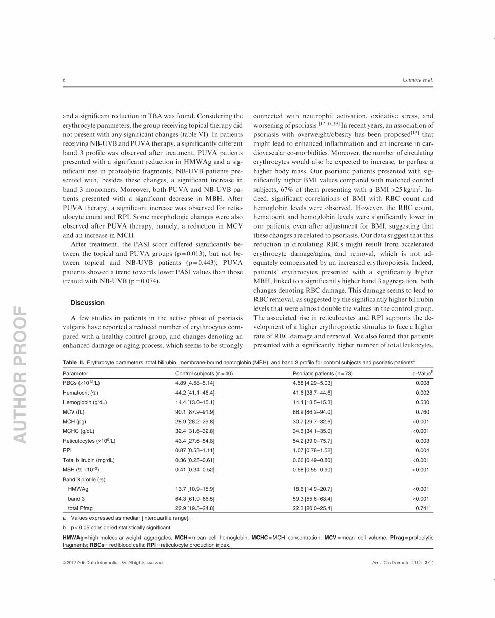
In table II we present the qualitative and quantitative RBC
studies that were performed, the plasma levels of total bilirubin
(as a marker of RBC removal), and the values of reticulocytes
and RPI (as markers of erythroid bone marrow production).
Psoriatic patients presented with significantly lower values of
RBC and hematocrit, and higher values of mean cell hemo-
globin (MCH) andMCH concentration (MCHC) than control
subjects. The reticulocyte count, RPI, and the total bilirubin
levels were also significantly higher in psoriatic patients. Con-
cerning erythrocyte damage,MBH (amarker of oxidative stress
in RBCs) showed significantly higher values, and the band 3
profile presented a significantly different pattern for psoriatic
patients, with higher percentages of HMWAg and lower per-
centages of band 3 monomer. All the statistical significances
observed persisted after adjustment for BMI.
For psoriatic patients we observed a significant positive
correlation of BMI with RBC count (r = 0.279, p= 0.017) and
with hemoglobin level (r= 0.232, p = 0.048). Furthermore, we
found that CRP presented positive significant correlations with
PASI score (r = 0.310, p= 0.008), leukocyte count (r = 0.489,
p < 0.001), neutrophil count (r = 0.522, p< 0.001), elastase
(r = 0.455, p< 0.001), and lactoferrin level (r = 0.323, p= 0.005).
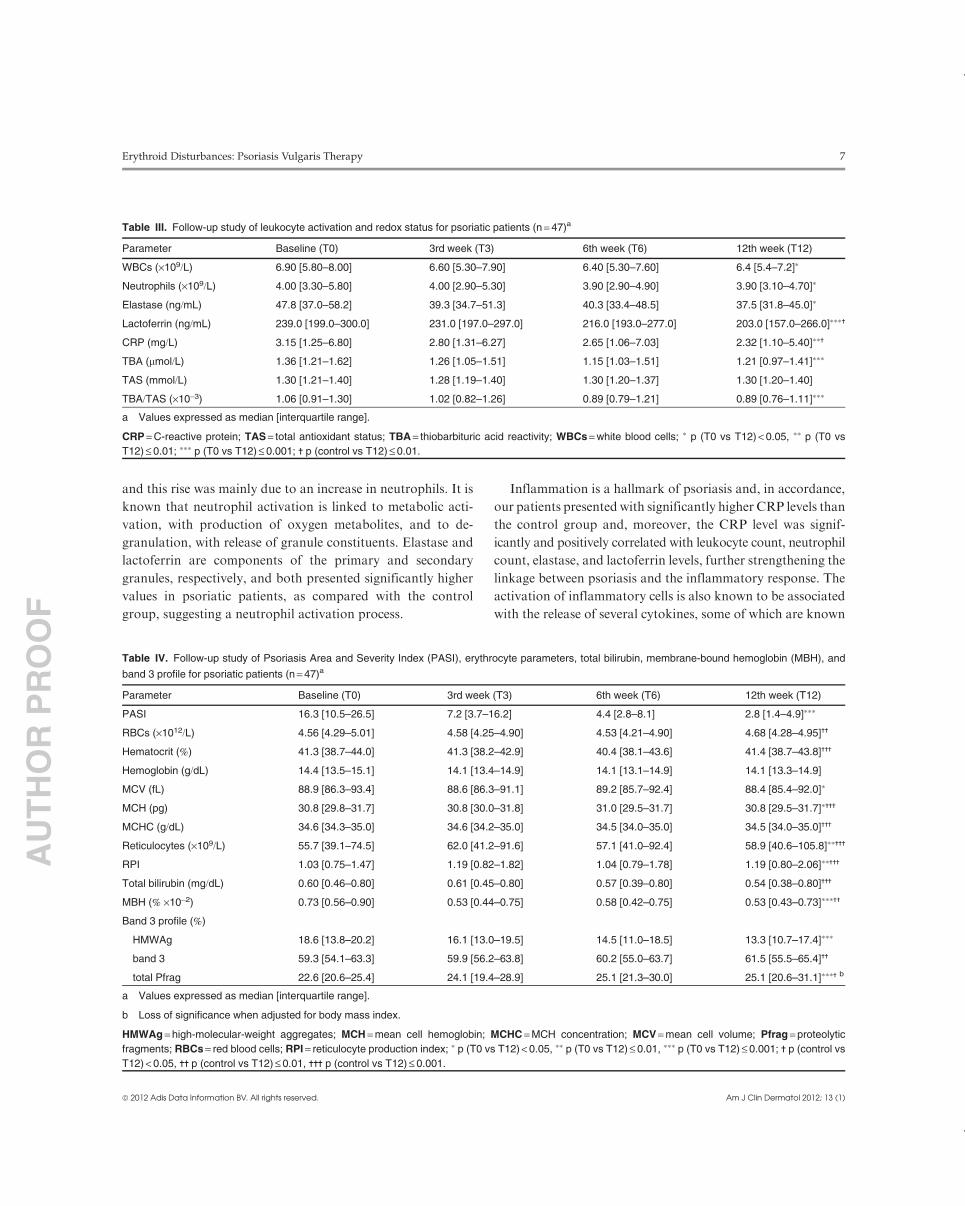
Concerning the follow-up study (n = 47), we observed slight
changes in the studied parameters during the course of treat-
ment, which reached statistical significance at the end of treat-
ment (table III). Indeed, we found a significant decrease in
leukocyte and neutrophil counts, and in the neutrophil acti-
vation products, elastase and lactoferrin. CRP also showed a
significant decrease, as did TBA and the TBA/TAS ratio. We
should note that at the end of treatment, with clearing of the
lesions (as shown by PASI), the CRP and lactoferrin levels were
still statistically significantly higher than in the control group
(table III).
The erythrocyte parameters also showed slight changes with
treatment, some of them reaching statistical significance at the
end of treatment (table IV), namely, mean cell volume (MCV)
and MCH decreased significantly and reticulocyte count and
RPI showed a significant rise. No significant changes were
observed for total bilirubin levels. Considering the erythrocyte
damagemarkers, a significant decrease was observed forMBH,
and a different band 3 profile was found, with a decrease in
HMWAg and a rise in proteolytic fragments at the end of
treatment.
As shown, several changes occurred during treatment, with
improvement of the disease; however, some of the changes in-
duced by treatment did not reach values similar to those pre-
sented by the control group. Thus, the RBC count, hematocrit,
MCH,MCHC, reticulocyte count, RPI and bilirubin level were
still significantly higher than in the control subjects. MBH was
also still significantly higher, and the band 3 profile was also
different, with a decreased percentage of band 3 monomer and
increased percentage of proteolytic fragments compared with
control subjects (that lost significance after statistical adjust-
ment for BMI).
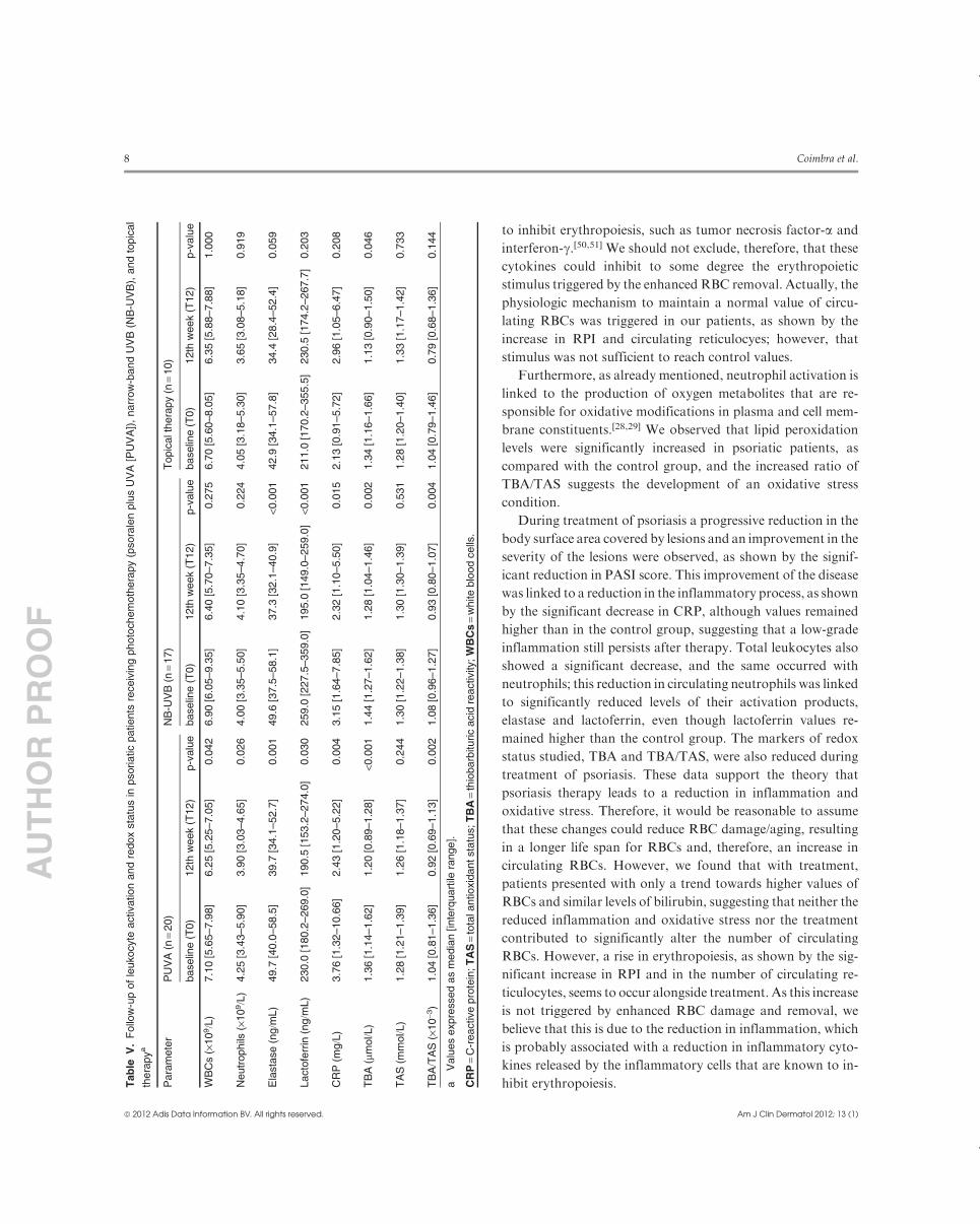
Analysing the results according to the therapy used, we
found that patients receiving PUVA treatment presented with a
significant reduction in leukocyte and neutrophil counts, elas-
tase, lactoferrin, CRP, TBA, and TBA/TAS (table V). Patients
treated with NB-UVB showed a significant decrease in the
values of elastase, lactoferrin, CRP, TBA, and TBA/TAS.With
topical therapy, only a trend towards reduced values of elastase
Table I. Leukocyte activation and redox status for control subjects and psoriatic patientsa
Parameter Control subjects (n = 38) Psoriatic patients (n = 73) p-Value
WBCs (·109/L) 6.70 [5.20–7.50] 7.00 [5.75–8.70] 0.043
Neutrophils (·109/L) 3.95 [2.98–4.60] 4.30 [3.40–5.95] 0.016
Elastase (ng/mL) 40.5 [32.8–45.6] 52.5 [42.8–67.8] <0.001
Lactoferrin (ng/mL) 182.5 [128.0–203.8] 239.0 [201.0–349.5] <0.001
CRP (mg/L) 1.51 [0.60–2.75] 4.18 [1.59–9.12] <0.001
TBA (mmol/L) 1.18 [1.00–1.41]b 1.36 [1.21–1.64] 0.004
TAS (mmol/L) 1.30 [1.09–1.61]b 1.30 [1.22–1.43] 0.228
TBA/TAS (·10-3) 0.90 [0.73–1.13]b 1.05 [0.89–1.31] 0.047c
a Values expressed as median [interquartile range].
b n = 37.
c Loss of significance when adjusted for body mass index.
CRP = C-reactive protein; TAS = total antioxidant status; TBA = thiobarbituric acid reactivity; WBCs = white blood cells.
Erythroid Disturbances: Psoriasis Vulgaris Therapy 5
ª 2012 Adis Data Information BV. All rights reserved. Am J Clin Dermatol 2012; 13 (1)
AU
TH
OR
PR
OO
F
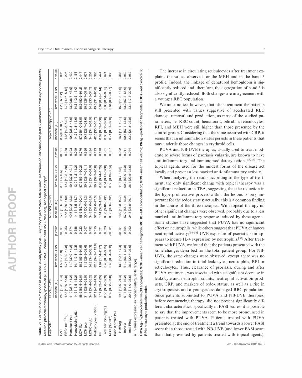
and a significant reduction in TBA was found. Considering the
erythrocyte parameters, the group receiving topical therapy did
not present with any significant changes (table VI). In patients
receivingNB-UVB and PUVA therapy, a significantly different
band 3 profile was observed after treatment; PUVA patients
presented with a significant reduction in HMWAg and a sig-
nificant rise in proteolytic fragments; NB-UVB patients pre-
sented with, besides these changes, a significant increase in
band 3 monomers. Moreover, both PUVA and NB-UVB pa-
tients presented with a significant decrease in MBH. After
PUVA therapy, a significant increase was observed for retic-
ulocyte count and RPI. Some morphologic changes were also
observed after PUVA therapy, namely, a reduction in MCV
and an increase in MCH.
After treatment, the PASI score differed significantly be-
tween the topical and PUVA groups (p = 0.013), but not be-
tween topical and NB-UVB patients (p = 0.443); PUVA
patients showed a trend towards lower PASI values than those
treated with NB-UVB (p = 0.074).
Discussion
A few studies in patients in the active phase of psoriasis
vulgaris have reported a reduced number of erythrocytes com-
pared with a healthy control group, and changes denoting an
enhanced damage or aging process, which seems to be strongly
connected with neutrophil activation, oxidative stress, and
worsening of psoriasis.[12,37,38] In recent years, an association of
psoriasis with overweight/obesity has been proposed[15] that
might lead to enhanced inflammation and an increase in car-
diovascular co-morbidities. Moreover, the number of circulating
erythrocytes would also be expected to increase, to perfuse a
higher body mass. Our psoriatic patients presented with sig-
nificantly higher BMI values compared with matched control
subjects, 67% of them presenting with a BMI >25 kg/m2. In-
deed, significant correlations of BMI with RBC count and
hemoglobin levels were observed. However, the RBC count,
hematocrit and hemoglobin levels were significantly lower in
our patients, even after adjustment for BMI, suggesting that
these changes are related to psoriasis. Our data suggest that this
reduction in circulating RBCs might result from accelerated
erythrocyte damage/aging and removal, which is not ad-
equately compensated by an increased erythropoiesis. Indeed,
patients’ erythrocytes presented with a significantly higher
MBH, linked to a significantly higher band 3 aggregation, both
changes denoting RBC damage. This damage seems to lead to
RBC removal, as suggested by the significantly higher bilirubin
levels that were almost double the values in the control group.
The associated rise in reticulocytes and RPI supports the de-
velopment of a higher erythropoietic stimulus to face a higher
rate of RBC damage and removal. We also found that patients
presented with a significantly higher number of total leukocytes,
Table II. Erythrocyte parameters, total bilirubin, membrane-bound hemoglobin (MBH), and band 3 profile for control subjects and psoriatic patientsa
Parameter Control subjects (n = 40) Psoriatic patients (n = 73) p-Valueb
RBCs (·1012/L) 4.89 [4.58–5.14] 4.58 [4.29–5.03] 0.008
Hematocrit (%) 44.2 [41.1–46.4] 41.6 [38.7–44.6] 0.002
Hemoglobin (g/dL) 14.4 [13.0–15.1] 14.4 [13.5–15.3] 0.530
MCV (fL) 90.1 [87.9–91.9] 88.9 [86.2–94.0] 0.760
MCH (pg) 28.9 [28.2–29.8] 30.7 [29.7–32.6] <0.001
MCHC (g/dL) 32.4 [31.6–32.8] 34.6 [34.1–35.0] <0.001
Reticulocytes (·109/L) 43.4 [27.6–54.8] 54.2 [39.0–75.7] 0.003
RPI 0.87 [0.53–1.11] 1.07 [0.78–1.52] 0.004
Total bilirubin (mg/dL) 0.36 [0.25–0.61] 0.66 [0.49–0.80] <0.001
MBH (% ·10-2) 0.41 [0.34–0.52] 0.68 [0.55–0.90] <0.001
Band 3 profile (%)
HMWAg 13.7 [10.9–15.9] 18.6 [14.9–20.7] <0.001
band 3 64.3 [61.9–66.5] 59.3 [55.6–63.4] <0.001
total Pfrag 22.9 [19.5–24.8] 22.3 [20.0–25.4] 0.741
a Values expressed as median [interquartile range].
b p < 0.05 considered statistically significant.
HMWAg = high-molecular-weight aggregates; MCH = mean cell hemoglobin; MCHC = MCH concentration; MCV = mean cell volume; Pfrag = proteolytic
fragments; RBCs = red blood cells; RPI = reticulocyte production index.
6 Coimbra et al.
ª 2012 Adis Data Information BV. All rights reserved. Am J Clin Dermatol 2012; 13 (1)
AU
TH
OR
PR
OO
F
and this rise was mainly due to an increase in neutrophils. It is
known that neutrophil activation is linked to metabolic acti-
vation, with production of oxygen metabolites, and to de-
granulation, with release of granule constituents. Elastase and
lactoferrin are components of the primary and secondary
granules, respectively, and both presented significantly higher
values in psoriatic patients, as compared with the control
group, suggesting a neutrophil activation process.
Inflammation is a hallmark of psoriasis and, in accordance,
our patients presented with significantly higher CRP levels than
the control group and, moreover, the CRP level was signif-
icantly and positively correlated with leukocyte count, neutrophil
count, elastase, and lactoferrin levels, further strengthening the
linkage between psoriasis and the inflammatory response. The
activation of inflammatory cells is also known to be associated
with the release of several cytokines, some of which are known
Table III. Follow-up study of leukocyte activation and redox status for psoriatic patients (n = 47)a
Parameter Baseline (T0) 3rd week (T3) 6th week (T6) 12th week (T12)
WBCs (·109/L) 6.90 [5.80–8.00] 6.60 [5.30–7.90] 6.40 [5.30–7.60] 6.4 [5.4–7.2]*
Neutrophils (·109/L) 4.00 [3.30–5.80] 4.00 [2.90–5.30] 3.90 [2.90–4.90] 3.90 [3.10–4.70]*
Elastase (ng/mL) 47.8 [37.0–58.2] 39.3 [34.7–51.3] 40.3 [33.4–48.5] 37.5 [31.8–45.0]*
Lactoferrin (ng/mL) 239.0 [199.0–300.0] 231.0 [197.0–297.0] 216.0 [193.0–277.0] 203.0 [157.0–266.0]***-
CRP (mg/L) 3.15 [1.25–6.80] 2.80 [1.31–6.27] 2.65 [1.06–7.03] 2.32 [1.10–5.40]**-
TBA (mmol/L) 1.36 [1.21–1.62] 1.26 [1.05–1.51] 1.15 [1.03–1.51] 1.21 [0.97–1.41]***
TAS (mmol/L) 1.30 [1.21–1.40] 1.28 [1.19–1.40] 1.30 [1.20–1.37] 1.30 [1.20–1.40]
TBA/TAS (·10-3) 1.06 [0.91–1.30] 1.02 [0.82–1.26] 0.89 [0.79–1.21] 0.89 [0.76–1.11]***
a Values expressed as median [interquartile range].
CRP = C-reactive protein; TAS = total antioxidant status; TBA = thiobarbituric acid reactivity; WBCs = white blood cells; * p (T0 vs T12) < 0.05, ** p (T0 vs
T12) £ 0.01; *** p (T0 vs T12) £ 0.001; - p (control vs T12) £ 0.01.
Table IV. Follow-up study of Psoriasis Area and Severity Index (PASI), erythrocyte parameters, total bilirubin, membrane-bound hemoglobin (MBH), and
band 3 profile for psoriatic patients (n = 47)a
Parameter Baseline (T0) 3rd week (T3) 6th week (T6) 12th week (T12)
PASI 16.3 [10.5–26.5] 7.2 [3.7–16.2] 4.4 [2.8–8.1] 2.8 [1.4–4.9]***
RBCs (·1012/L) 4.56 [4.29–5.01] 4.58 [4.25–4.90] 4.53 [4.21–4.90] 4.68 [4.28–4.95]--
Hematocrit (%) 41.3 [38.7–44.0] 41.3 [38.2–42.9] 40.4 [38.1–43.6] 41.4 [38.7–43.8]---
Hemoglobin (g/dL) 14.4 [13.5–15.1] 14.1 [13.4–14.9] 14.1 [13.1–14.9] 14.1 [13.3–14.9]
MCV (fL) 88.9 [86.3–93.4] 88.6 [86.3–91.1] 89.2 [85.7–92.4] 88.4 [85.4–92.0]*
MCH (pg) 30.8 [29.8–31.7] 30.8 [30.0–31.8] 31.0 [29.5–31.7] 30.8 [29.5–31.7]*---
MCHC (g/dL) 34.6 [34.3–35.0] 34.6 [34.2–35.0] 34.5 [34.0–35.0] 34.5 [34.0–35.0]---
Reticulocytes (·109/L) 55.7 [39.1–74.5] 62.0 [41.2–91.6] 57.1 [41.0–92.4] 58.9 [40.6–105.8]**---
RPI 1.03 [0.75–1.47] 1.19 [0.82–1.82] 1.04 [0.79–1.78] 1.19 [0.80–2.06]**---
Total bilirubin (mg/dL) 0.60 [0.46–0.80] 0.61 [0.45–0.80] 0.57 [0.39–0.80] 0.54 [0.38–0.80]---
MBH (% ·10-2) 0.73 [0.56–0.90] 0.53 [0.44–0.75] 0.58 [0.42–0.75] 0.53 [0.43–0.73]***--
Band 3 profile (%)
HMWAg 18.6 [13.8–20.2] 16.1 [13.0–19.5] 14.5 [11.0–18.5] 13.3 [10.7–17.4]***
band 3 59.3 [54.1–63.3] 59.9 [56.2–63.8] 60.2 [55.0–63.7] 61.5 [55.5–65.4]--
total Pfrag 22.6 [20.6–25.4] 24.1 [19.4–28.9] 25.1 [21.3–30.0] 25.1 [20.6–31.1]***- b
a Values expressed as median [interquartile range].
b Loss of significance when adjusted for body mass index.
HMWAg = high-molecular-weight aggregates; MCH = mean cell hemoglobin; MCHC = MCH concentration; MCV = mean cell volume; Pfrag = proteolytic
fragments; RBCs = red blood cells; RPI = reticulocyte production index; * p (T0 vs T12) < 0.05, ** p (T0 vs T12) £ 0.01, *** p (T0 vs T12) £ 0.001; - p (control vs
T12) < 0.05, -- p (control vs T12) £ 0.01, --- p (control vs T12) £ 0.001.
Erythroid Disturbances: Psoriasis Vulgaris Therapy 7
ª 2012 Adis Data Information BV. All rights reserved. Am J Clin Dermatol 2012; 13 (1)
AU
TH
OR
PR
OO
F
to inhibit erythropoiesis, such as tumor necrosis factor-a and
interferon-g.[50,51] We should not exclude, therefore, that these
cytokines could inhibit to some degree the erythropoietic
stimulus triggered by the enhanced RBC removal. Actually, the
physiologic mechanism to maintain a normal value of circu-
lating RBCs was triggered in our patients, as shown by the
increase in RPI and circulating reticulocyes; however, that
stimulus was not sufficient to reach control values.
Furthermore, as already mentioned, neutrophil activation is
linked to the production of oxygen metabolites that are re-
sponsible for oxidative modifications in plasma and cell mem-
brane constituents.[28,29] We observed that lipid peroxidation
levels were significantly increased in psoriatic patients, as
compared with the control group, and the increased ratio of
TBA/TAS suggests the development of an oxidative stress
condition.
During treatment of psoriasis a progressive reduction in the
body surface area covered by lesions and an improvement in the
severity of the lesions were observed, as shown by the signif-
icant reduction in PASI score. This improvement of the disease
was linked to a reduction in the inflammatory process, as shown
by the significant decrease in CRP, although values remained
higher than in the control group, suggesting that a low-grade
inflammation still persists after therapy. Total leukocytes also
showed a significant decrease, and the same occurred with
neutrophils; this reduction in circulating neutrophils was linked
to significantly reduced levels of their activation products,
elastase and lactoferrin, even though lactoferrin values re-
mained higher than the control group. The markers of redox
status studied, TBA and TBA/TAS, were also reduced during
treatment of psoriasis. These data support the theory that
psoriasis therapy leads to a reduction in inflammation and
oxidative stress. Therefore, it would be reasonable to assume
that these changes could reduce RBC damage/aging, resultingin a longer life span for RBCs and, therefore, an increase in
circulating RBCs. However, we found that with treatment,
patients presented with only a trend towards higher values of
RBCs and similar levels of bilirubin, suggesting that neither the
reduced inflammation and oxidative stress nor the treatment
contributed to significantly alter the number of circulating
RBCs. However, a rise in erythropoiesis, as shown by the sig-
nificant increase in RPI and in the number of circulating re-
ticulocytes, seems to occur alongside treatment. As this increase
is not triggered by enhanced RBC damage and removal, we
believe that this is due to the reduction in inflammation, which
is probably associated with a reduction in inflammatory cyto-
kines released by the inflammatory cells that are known to in-
hibit erythropoiesis.Tab
leV
.F
ollo
w-u
pof
leuko
cyte
activation
and
redox
sta
tus
inpsoriatic
patients
receiv
ing
photo
chem
oth
era
py
(psora
len
plu
sU
VA
[PU
VA
]),
narr
ow
-band
UV
B(N
B-U
VB
),and
topic
al
thera
pya
Para
mete
rP
UV
A(n
=20)
NB
-UV
B(n
=17)
Topic
alt
hera
py
(n=
10)
baselin
e(T
0)
12th
week
(T12)
p-v
alu
ebaselin
e(T
0)
12th
week
(T12)
p-v
alu
ebaselin
e(T
0)
12th
week
(T12)
p-v
alu
e
WB
Cs
(·10
9/L
)7.1
0[5
.65–7.9
8]
6.2
5[5
.25–7.0
5]
0.0
42
6.9
0[6
.05–9.3
5]
6.4
0[5
.70–7.3
5]
0.2
75
6.7
0[5
.60–8.0
5]
6.3
5[5
.88–7.8
8]
1.0
00
Neutr
ophils
(·10
9/L
)4.2
5[3
.43–5.9
0]
3.9
0[3
.03–4.6
5]
0.0
26
4.0
0[3
.35–5.5
0]
4.1
0[3
.35–4.7
0]
0.2
24
4.0
5[3
.18–5.3
0]
3.6
5[3
.08–5.1
8]
0.9
19
Ela
sta
se
(ng/m
L)
49.7
[40.0
–58.5
]39.7
[34.1
–52.7
]0.0
01
49.6
[37.5
–58.1
]37.3
[32.1
–40.9
]<0
.001
42.9
[34.1
–57.8
]34.4
[28.4
–52.4
]0.0
59
Lacto
ferr
in(n
g/m
L)
230.0
[180.2
–269.0
]190.5
[153.2
–274.0
]0.0
30
259.0
[227.5
–359.0
]195.0
[149.0
–259.0
]<0
.001
211.0
[170.2
–355.5
]230.5
[174.2
–267.7
]0.2
03
CR
P(m
g/L
)3.7
6[1
.32–10.6
6]
2.4
3[1
.20–5.2
2]
0.0
04
3.1
5[1
.64–7.8
5]
2.3
2[1
.10–5.5
0]
0.0
15
2.1
3[0
.91–5.7
2]
2.9
6[1
.05–6.4
7]
0.2
08
TB
A(m
mol/L
)1.3
6[1
.14–1.6
2]
1.2
0[0
.89–1.2
8]
<0.0
01
1.4
4[1
.27–1.6
2]
1.2
8[1
.04–1.4
6]
0.0
02
1.3
4[1
.16–1.6
6]
1.1
3[0
.90–1.5
0]
0.0
46
TA
S(m
mol/L
)1.2
8[1
.21–1.3
9]
1.2
6[1
.18–1.3
7]
0.2
44
1.3
0[1
.22–1.3
8]
1.3
0[1
.30–1.3
9]
0.5
31
1.2
8[1
.20–1.4
0]
1.3
3[1
.17–1.4
2]
0.7
33
TB
A/T
AS
(·10
-3)
1.0
4[0
.81–1.3
6]
0.9
2[0
.69–1.1
3]
0.0
02
1.0
8[0
.96–1.2
7]
0.9
3[0
.80–1.0
7]
0.0
04
1.0
4[0
.79–1.4
6]
0.7
9[0
.68–1.3
6]
0.1
44
aV
alu
es
expre
ssed
as
media
n[inte
rquart
ilera
nge].
CR
P=
C-r
eactive
pro
tein
;T
AS
=to
tala
ntioxid
antsta
tus;T
BA
=th
iobarb
ituric
acid
reactivity
;W
BC
s=
white
blo
od
cells
.
8 Coimbra et al.
ª 2012 Adis Data Information BV. All rights reserved. Am J Clin Dermatol 2012; 13 (1)
AU
TH
OR
PR
OO
F
The increase in circulating reticulocytes after treatment ex-
plains the values observed for the MBH and in the band 3
profile. Indeed, the linkage of denatured hemoglobin is sig-
nificantly reduced and, therefore, the aggregation of band 3 is
also significantly reduced. Both changes are in agreement with
a younger RBC population.
We must notice, however, that after treatment the patients
still presented with values suggestive of accelerated RBC
damage, removal and production, as most of the studied pa-
rameters, i.e. RBC count, hematocrit, bilirubin, reticulocytes,
RPI, and MBH were still higher than those presented by the
control group. Considering that the same occurred with CRP, it
seems that an inflammation status persists in these patients that
may underlie those changes in erythroid cells.
PUVA and NB-UVB therapies, usually used to treat mod-
erate to severe forms of psoriasis vulgaris, are known to have
anti-inflammatory and immunomodulatory actions.[52,53] The
topical agents used for the mildest forms of the disease act
locally and present a less marked anti-inflammatory activity.
When analyzing the results according to the type of treat-
ment, the only significant change with topical therapy was a
significant reduction in TBA, suggesting that the reduction in
the hyperproliferative process within the lesions is very im-
portant for the redox status; actually, this is a common finding
in the course of the three therapies. With topical therapy no
other significant changes were observed, probably due to a less
marked anti-inflammatory response induced by these agents.
Some studies have suggested that PUVA has no significant
effect on neutrophils, while others suggest that PUVA enhances
neutrophil activity;[54-56] UVB exposure of psoriatic skin ap-
pears to induce IL-4 expression by neutrophils.[57] After treat-
ment with PUVA, we found that the patients presented with the
same changes described for the total patient group. For NB-
UVB, the same changes were observed, except there was no
significant reduction in total leukocytes, neutrophils, RPI or
reticulocytes. Thus, clearance of psoriasis, during and after
PUVA treatment, was associated with a significant decrease in
leukocyte and neutrophil counts, neutrophil activation prod-
ucts, CRP, and markers of redox status, as well as a rise in
erythropoiesis and a younger/less damaged RBC population.
Since patients submitted to PUVA and NB-UVB therapies,
before commencing therapy, did not present significantly dif-
ferent characteristics, specifically in PASI scores, it is possible
to say that the improvements seem to be more pronounced in
patients treated with PUVA. Patients treated with PUVA
presented at the end of treatment a trend towards a lower PASI
score than those treated with NB-UVB (and lower PASI score
than that presented by patients treated with topical agents),Tab
leV
I.F
ollo
w-u
pstu
dy
ofP
soriasi
sA
rea
and
Severity
Index
(PA
SI)
,ery
thro
cyte
para
mete
rs,t
ota
lbili
rubin
,mem
bra
ne-b
ound
hem
oglo
bin
(MB
H),
and
band
3pro
file
inpsoriatic
patients
receiv
ing
photo
chem
oth
era
py
(psora
len
plu
sU
VA
[PU
VA
]),narr
ow
-band
UV
B(N
B-U
VB
),and
topic
alt
hera
py
a
Para
mete
rP
UV
A(n
=20)
NB
-UV
B(n
=17)
Topic
alth
era
py
(n=
10)
baselin
e(T
0)
12th
week
(T12)
p-v
alu
ebaselin
e(T
0)
12th
week
(T12)
p-v
alu
ebaselin
e(T
0)
12th
week
(T12)
p-v
alu
e
PA
SI
24.8
[13.2
–33.4
]1.5
[0.7
–3.8
]<0
.001
17.2
[12.6
–25.0
]3.1
[1.6
–6.6
]<0
.001
8.6
[6.9
–10.5
]4.2
[2.6
–6.2
]0.0
05
RB
Cs
(·10
12/L
)4.5
8[4
.30–5.0
9]
4.7
6[4
.30–4.9
8]
0.2
83
4.5
5[4
.36–4.9
9]
4.5
7[4
.22–4.8
0]
0.2
88
4.6
6[4
.23–5.2
7]
4.7
3[4
.16–5.1
2]
0.2
26
Hem
ato
crit(%
)41.9
[38.7
–44.2
]41.9
[39.9
–44.2
]0.7
74
40.5
[39.2
–42.3
]40.1
[37.5
–42.1
]0.1
03
40.9
[36.8
–45.0
]43.0
[36.7
–43.9
]0.4
32
Hem
oglo
bin
(g/d
L)
14.5
[13.5
–15.2
]14.4
[13.8
–15.3
]0.6
86
14.1
[13.5
–14.7
]13.9
[13.2
–14.2
]0.0
49
14.2
[12.8
–15.6
]14.6
[12.5
–15.0
]0.1
53
MC
V(f
L)
88.9
[86.9
–95.0
]88.5
[85.8
–94.5
]0.0
23
88.9
[86.7
–90.4
]87.9
[86.1
–90.2
]0.2
76
87.9
[84.0
–91.5
]88.9
[83.6
–91.2
]0.4
47
MC
H(p
g)
31.1
[29.9
–33.5
]31.2
[29.8
–32.6
]0.0
47
30.7
[30.0
–31.2
]30.3
[29.7
–31.1
]0.8
66
30.7
[28.7
–31.6
]30.7
[28.7
–31.9
]0.6
77
MC
HC
(g/d
L)
34.7
[34.4
–35.2
]34.8
[34.3
–35.3
]0.4
92
34.5
[34.0
–35.0
]34.5
[34.0
–34.9
]0.4
84
34.6
[34.1
–34.9
]34.5
[33.5
–34.7
]0.2
01
Reticulo
cyte
s(·
10
9/L
)57.1
[41.3
–91.5
]82.5
[54.2
–113.8
]0.0
15
57.9
[39.0
–77.3
]50.2
[39.5
–90.8
]0.1
93
42.0
[30.5
–55.7
]45.5
[31.1
–60.8
]0.3
86
RP
I1.1
7[0
.82–1.8
5]
1.6
7[1
.04–2.2
7]
0.0
21
1.1
4[0
.69–1.5
7]
0.9
8[0
.74–1.7
5]
0.1
70
0.8
2[0
.59–1.0
8]
0.9
7[0
.49–1.1
4]
0.4
44
Tota
lbili
rubin
(mg/d
L)
0.5
5[0
.38–0.6
9]
0.4
6[0
.34–0.7
5]
0.6
23
0.8
0[0
.40–0.8
2]
0.5
4[0
.41–0.8
0]
0.8
01
0.8
0[0
.53–0.8
0]
0.8
0[0
.54–0.8
5]
0.6
71
MB
H(%
·10
-2)
0.6
9[0
.58–0.9
0]
0.4
6[0
.34–0.6
3]
<0.0
01
0.8
5[0
.56–0.9
2]
0.5
3[0
.49–0.7
4]
0.0
07
0.7
1[0
.51–0.8
3]
0.6
4[0
.46–0.7
7]
0.3
86
Band
3pro
file
(%)
HM
WA
g19.3
[16.0
–21.4
]13.3
[10.7
–17.4
]<0
.001
18.0
[13.3
–19.7
]11.6
[8.7
–18.3
]0.0
02
16.1
[11.1
–19.1
]15.0
[11.8
–16.6
]0.3
86
band
361.3
[54.2
–66.2
]61.2
[56.1
–65.8
]0.6
54
57.0
[54.1
–62.9
]60.2
[55.0
–66.3
]0.0
11
60.5
[57.1
–64.5
]63.2
[57.3
–65.6
]0.9
59
tota
lP
frag
20.9
[15.0
–25.5
]25.1
[20.7
–28.6
]0.0
02
24.3
[21.5
–26.1
]26.7
[22.5
–33.6
]0.0
44
23.0
[21.8
–23.8
]23.1
[17.3
–30.6
]0.9
59
aV
alu
es
expre
ssed
as
media
n[inte
rquart
ilera
nge].
HM
WA
g=
hig
h-m
ole
cula
r-w
eig
hta
ggre
gate
s;M
CH
=m
ean
cell
hem
oglo
bin
;MC
HC
=M
CH
concentr
ation;M
CV
=m
ean
cell
volu
me;P
frag
=pro
teoly
ticfr
agm
ents
;RB
Cs
=re
dblo
od
cells
;
RP
I=re
ticu
locyte
pro
duction
index.
Erythroid Disturbances: Psoriasis Vulgaris Therapy 9
ª 2012 Adis Data Information BV. All rights reserved. Am J Clin Dermatol 2012; 13 (1)

AU
TH
OR
PR
OO
F
suggesting that a better improvement of the disease might ex-
plain these differences.
Conclusions
Our data suggest that psoriasis vulgaris triggers an inflam-
matory response with release of acute-phase reactants, reactive
oxygen species, cationic proteins, and proteases, leading to
enhanced RBC damage/aging and, ultimately, to enhanced
RBC removal. These assumptions were strengthened by the
observation that with the treatment of psoriasis some of these
changes reversed, the inflammation was reduced, the pro-
duction of reticulocytes was increased, and the RBCs showed
changes usually observed in younger/less damaged RBCs.
These erythroid changes were enhanced with PUVA therapy,
probably due to the more pronounced clearing of the lesions, as
suggested by PASI scores. Finally, after treatment, a residual
inflammation still persisted that might contribute to the ob-
served erythroid disturbances.
Acknowledgments
This study was supported by FCT (POCI/SAU –OBS/58600/2004) andby Fundacao para a Ciencia e Tecnologia (FCT: POCI/SAU – OBS/58600/2004) and Fundo Europeu de Desenvolvimento Regional (FEDER).
The authors have no conflicts of interest that are directly relevant to the
content of this study.
References1. Rocha-Pereira P, Santos-Silva A, Rebelo I, et al. The inflammatory response in
mild and in severe psoriasis. Br J Dermatol 2004 May; 150 (5): 917-28
2. Krueger JG, Bowcock A. Psoriasis pathophysiology: current concepts of
pathogenesis. Ann Rheum Dis 2005 Mar; 64 Suppl. 2: ii30-6
3. Krueger G, Ellis CN. Psoriasis: recent advances in understanding its patho-
genesis and treatment. J AmAcadDermatol 2005 Jul; 53 (1 Suppl. 1): S94-100
4. Chowaniec O, Jablonska S, Beutner EH, et al. Earliest clinical and histological
changes in psoriasis. Dermatologica 1981; 163 (1): 42-51
5. Orem A, Deger O, Cimsit G, et al. Plasma polymorphonuclear leukocyte
elastase levels and its relation to disease activity in psoriasis. Clin Chim Acta
1997 Aug 8; 264 (1): 49-56
6. Arican O, Aral M, Sasmaz S, et al. Serum levels of TNF-alpha, IFN-gamma,
IL-6, IL-8, IL-12, IL-17, and IL-18 in patients with active psoriasis and
correlation with disease severity. Mediators Inflamm 2005 Oct 24; 2005 (5):
273-9
7. Scirica BM, Morrow DA. Is C-reactive protein an innocent bystander or
proatherogenic culprit? The verdict is still out. Circulation 2006 May 2; 113
(17): 2128-34; discussion 51
8. Yeh ET. CRP as a mediator of disease. Circulation 2004 Jun 1; 109 (21 Suppl. 1):
II11-4
9. Friedewald VE,Cather JC,Gelfand JM, et al. AJC editor’s consensus: psoriasis
and coronary artery disease. Am J Cardiol 2008 Dec 15; 102 (12): 1631-43
10. Friedewald Jr VE, Cather JC, Gordon KB, et al. The editor’s roundtable:
psoriasis, inflammation, and coronary artery disease. Am J Cardiol 2008 Apr
15; 101 (8): 1119-26
11. Pelle E, Mammone T, Maes D, et al. Keratinocytes act as a source of reactive
oxygen species by transferring hydrogen peroxide to melanocytes. J Invest
Dermatol 2005 Apr; 124 (4): 793-7
12. Rocha-Pereira P, Santos-Silva A, Rebelo I, et al. Erythrocyte damage in mild
and severe psoriasis. Br J Dermatol 2004 Feb; 150 (2): 232-44
13. Setty AR, Curhan G, Choi HK. Obesity, waist circumference, weight change,
and the risk of psoriasis in women: Nurses’ Health Study II. Arch InternMed
2007 Aug 13-27; 167 (15): 1670-5
14. Shirai K.Obesity as the core of themetabolic syndrome and themanagement of
coronary heart disease. Curr Med Res Opin 2004 Mar; 20 (3): 295-304
15. Sterry W, Strober BE, Menter A. Obesity in psoriasis: the metabolic, clinical
and therapeutic implications. Report of an interdisciplinary conference and
review. Br J Dermatol 2007 Oct; 157 (4): 649-55
16. Coimbra S, Oliveira H, Reis F, et al. Circulating levels of adiponectin, oxidized
LDL and C-reactive protein in Portuguese patients with psoriasis vulgaris,
according to body mass index, severity and duration of the disease. J Der-
matol Sci 2009 Sep; 55 (3): 202-4
17. Panagiotakos DB, Pitsavos C, Yannakoulia M, et al. The implication of obe-
sity and central fat on markers of chronic inflammation: the ATTICA study.
Atherosclerosis 2005 Dec; 183 (2): 308-15
18. Wellen KE, Hotamisligil GS. Obesity-induced inflammatory changes in adi-
pose tissue. J Clin Invest 2003 Dec; 112 (12): 1785-8
19. Rao GM, Morghom LO. Effect of obesity on erythrocyte count and hemo-
globin levels in Libyan diabetic patients. Clin Physiol Biochem 1986; 4 (4):
277-80
20. Lutz HU. Erythocyte clearance. In: Harris J, editor. Blood cell biochemistry.
New York (NY): Plenum Press, 1990: 81
21. KayMM.Role of physiologic autoantibody in the removal of senescent human
red cells. J Supramol Struct 1978; 9 (4): 555-67
22. KayMM. Localization of senescent cell antigen on band 3. Proc Natl Acad Sci
U S A 1984 Sep; 81 (18): 5753-7
23. Kay MM, Goodman SR, Sorensen K, et al. Senescent cell antigen is im-
munologically related to band 3. Proc Natl Acad Sci U S A 1983 Mar; 80 (6):
1631-5
24. Bosman GJ, Werre JM, Willekens FL, et al. Erythrocyte ageing in vivo and
in vitro: structural aspects and implications for transfusion. Transfus Med
2008 Dec; 18 (6): 335-47
25. Lutz HU, Bussolino F, Flepp R, et al. Naturally occurring anti-band-3 anti-
bodies and complement together mediate phagocytosis of oxidatively stressed
human erythrocytes. Proc Natl Acad Sci U S A 1987 Nov; 84 (21): 7368-72
26. Lutz HU. Naturally occurring anti-band 3 antibodies. Transfus Med Rev 1992
Jul; 6 (3): 201-11
27. Low PS. Role of hemoglobin denaturation and band 3 clustering in initiating
red cell removal. In: Magnani M, De Flora A, editors. Red blood cell aging.
New York (NY): Plenum Press, 1991: 173-83
28. Santos-Silva A, Rebelo MI, Castro EM, et al. Leukocyte activation, eryth-
rocyte damage, lipid profile and oxidative stress imposed by high competition
physical exercise in adolescents. Clin Chim Acta 2001 Apr; 306 (1-2): 119-26
29. Santos-Silva A, Rebelo I, Castro E, et al. Erythrocyte damage and leukocyte
activation in ischemic stroke. Clin Chim Acta 2002 Jun; 320 (1-2): 29-35
30. Belo L, Rebelo I, Castro EM, et al. Band 3 as a marker of erythrocyte changes
in pregnancy. Eur J Haematol 2002 Sep; 69 (3): 145-51
31. Coimbra S, Castro E, Rocha-Pereira P, et al. The effect of green tea in oxidative
stress. Clin Nutr 2006 Oct; 25 (5): 790-6
32. Catarino C, Rebelo I, Belo L, et al. Erythrocyte changes in preeclampsia:
relationship between maternal and cord blood erythrocyte damage. J Perinat
Med 2009; 37 (1): 19-27
33. Rocha S, Vitorino RM, Lemos-Amado FM, et al. Presence of cytosolic per-
oxiredoxin 2 in the erythrocyte membrane of patients with hereditary sphero-
cytosis. Blood Cells Mol Dis 2008 Jul-Aug; 41 (1): 5-9
10 Coimbra et al.
ª 2012 Adis Data Information BV. All rights reserved. Am J Clin Dermatol 2012; 13 (1)
AU
TH
OR
PR
OO
F
34. Costa E, Rocha S, Rocha-Pereira P, et al. Band 3 profile as amarker of erythrocyte
changes in chronic kidney disease patients. Open Clin Chem J 2008; 1: 57-63
35. Santos-Silva A, Castro EM, Teixeira NA, et al. Altered erythrocyte membrane
band 3 profile as a marker in patients at risk for cardiovascular disease.
Atherosclerosis 1995 Aug; 116 (2): 199-209
36. Santos-Silva A, Castro EM, Teixeira NA, et al. Erythrocyte membrane band 3
profile imposed by cellular aging, by activated neutrophils and by neutrophilic
elastase. Clin Chim Acta 1998 Jul 28; 275 (2): 185-96
37. Gornicki A, Gutsze A. Erythrocyte membrane fluidity changes in psoriasis:
an EPR study. J Dermatol Sci 2001 Sep; 27 (1): 27-30
38. Gornicki A. Domain structure of erythrocyte membranes in psoriasis: an EPR
study. J Dermatol Sci 2002 Sep; 29 (3): 214-21
39. Corrocher R, Bassi A, Gandini A, et al. Transmembrane cation fluxes and fatty
acid composition of erythrocytes in psoriatic patients. Clin Chim Acta 1990
Jan 31; 186 (3): 335-44
40. Schopf RE, Langendorf Y, Benz RE, et al. A highly decreased binding of cyclic
adenosine monophosphate to protein kinase A in erythrocyte membranes is
specific for active psoriasis. J Invest Dermatol 2002 Jul; 119 (1): 160-5
41. Shimizu T, Takakuwa Y, Koizumi H, et al. Localization of immuno-analogues
of erythrocyte protein 4.1 and spectrin in epidermis of psoriasis vulgaris.
Histochem Cell Biol 1995 May; 103 (5): 363-8
42. Lebwohl M, Feldman SR, Walther R, et al. Clinical management of psoriasis:
principles and practice. Cutis 2001 Jan; 67 (1 Suppl.): 1-15
43. Frederiksson T, Pettersson U. Severe psoriasis: oral therapy with a new reti-
noid. Dermatologica 1978; 157: 238-44
44. Dodge JT, Mitchell C, Hanahan DJ. The preparation and chemical charac-
teristics of hemoglobin-free ghosts of human erythrocytes. Arch Biochem
Biophys 1963 Jan; 100: 119-30
45. BradfordMM.A rapid and sensitive method for the quantitation ofmicrogram
quantities of protein utilizing the principle of protein-dye binding. Anal
Biochem 1976 May 7; 72: 248-54
46. Niehaus Jr WG, Samuelsson B. Formation of malonaldehyde from phospho-
lipid arachidonate duringmicrosomal lipid peroxidation. Eur J Biochem 1968
Oct 17; 6 (1): 126-30
47. Laemmli UK. Cleavage of structural proteins during the assembly of the head
of bacteriophage T4. Nature 1970 Aug 15; 227 (5259): 680-5
48. Towbin H, Staehelin T, Gordon J. Electrophoretic transfer of proteins from
polyacrylamide gels to nitrocellulose sheets: procedure and some applica-
tions. Proc Natl Acad Sci U S A 1979 Sep; 76 (9): 4350-4
49. Czerwinski M, Wasniowska K, Steuden I, et al. Degradation of the human
erythrocyte membrane band 3 studied with monoclonal antibody directed
against an epitope on the cytoplasmic fragment of band 3. Eur J Biochem 1988
Jul 1; 174 (4): 647-54
50. Mamus SW, Beck-Schroeder S, Zanjani ED. Suppression of normal human
erythropoiesis by gamma interferon in vitro: role of monocytes and T lym-
phocytes. J Clin Invest 1985 May; 75 (5): 1496-503
51. Means Jr RT, Krantz SB. Inhibition of human erythroid colony-forming units
by tumor necrosis factor requires beta interferon. J Clin Invest 1993 Feb;
91 (2): 416-9
52. Lebwohl M, Ali S. Treatment of psoriasis: part 1. Topical therapy and pho-
totherapy. J Am Acad Dermatol 2001 Oct; 45 (4): 487-98; quiz 499-502
53. Lebwohl M, Ting PT, Koo JY. Psoriasis treatment: traditional therapy. Ann
Rheum Dis 2005 Mar; 64 Suppl. 2: ii83-6
54. Silny W, Pehamberger H, Zielinsky C, et al. Effect of PUVA treatment on the
locomotion of polymorphonuclear leukocytes and mononuclear cells in
psoriasis. J Invest Dermatol 1980 Aug; 75 (2): 187-8
55. Kapuscinska R, Wysocka J, Niczyporuk W, et al. Cytofluorimetric assay for
evaluation of CD16 receptor expression and myeloperoxidase (MPO) activity
of neutrophils in patients with psoriasis vulgaris treated with PUVA. Wiad
Lek 2004; 57 (11-12): 599-602
56. Bredberg A, Forsgren A. Effects of in vitro PUVA on human leukocyte
function. Br J Dermatol 1984 Aug; 111 (2): 159-68
57. Piskin G, Tursen U, Bos JD, et al. IL-4 expression by neutrophils in psori-
asis lesional skin upon high-dose UVB exposure. Dermatology 2003; 207 (1):
51-3
Correspondence: Susana Coimbra, PhD, or Alice Santos-Silva, PhD, Departa-
mento de Ciencias Biologicas, Laboratorio de Bioquımica, Faculdade de
Farmacia da Universidade do Porto, R Anıbal Cunha 164, 4050-047 Porto,
Portugal.
E-mail: [email protected]; [email protected]
Erythroid Disturbances: Psoriasis Vulgaris Therapy 11
ª 2012 Adis Data Information BV. All rights reserved. Am J Clin Dermatol 2012; 13 (1)
Copyright © 2022 FDOKUMEN