Measuring Performance Directly Using the Veterans Health Administration Electronic Medical Record
Upload
houstonmethodistCategory
view
0download
0
MOLECULAR AND CELLULAR BIOLOGY, Nov. 2006, p. 8022–8031 Vol. 26, No. 210270-7306/06/$08.00�0 doi:10.1128/MCB.01091-06Copyright © 2006, American Society for Microbiology. All Rights Reserved.
Notch1 Contributes to Mouse T-Cell Leukemia by Directly Inducingthe Expression of c-myc�
Vishva Mitra Sharma,1§ Jennifer A. Calvo,1§‡ Kyle M. Draheim,1 Leslie A. Cunningham,1Nicole Hermance,1 Levi Beverly,2 Veena Krishnamoorthy,1 Manoj Bhasin,3
Anthony J. Capobianco,2 and Michelle A. Kelliher1*Department of Cancer Biology and Cancer Center, University of Massachusetts Medical School, Worcester, Massachusetts1;
Laboratory of Immunobiology and Department of Medical Oncology, Dana-Farber Cancer Institute, and Department ofMedicine, Harvard Medical School, Boston, Massachusetts3; and Molecular and Cellular Oncogenesis Program,
The Wistar Institute, Philadelphia, Pennsylvania2
Received 16 June 2006/Returned for modification 17 July 2006/Accepted 8 August 2006
Recent work with mouse models and human leukemic samples has shown that gain-of-function mutation(s)in Notch1 is a common genetic event in T-cell acute lymphoblastic leukemia (T-ALL). The Notch1 receptorsignals through a �-secretase-dependent process that releases intracellular Notch1 from the membrane to thenucleus, where it forms part of a transcriptional activator complex. To identify Notch1 target genes inleukemia, we developed mouse T-cell leukemic lines that express intracellular Notch1 in a doxycycline-dependent manner. Using gene expression profiling and chromatin immunoprecipitation, we identified c-mycas a novel, direct, and critical Notch1 target gene in T-cell leukemia. c-myc mRNA levels are increased inprimary mouse T-cell tumors that harbor Notch1 mutations, and Notch1 inhibition decreases c-myc mRNAlevels and inhibits leukemic cell growth. Retroviral expression of c-myc, like intracellular Notch1, rescues thegrowth arrest and apoptosis associated with �-secretase inhibitor treatment or Notch1 inhibition. Consistentwith these findings, retroviral insertional mutagenesis screening of our T-cell leukemia mouse model revealedcommon insertions in either notch1 or c-myc genes. These studies define the Notch1 molecular signature inmouse T-ALL and importantly provide mechanistic insight as to how Notch1 contributes to human T-ALL.
Mutations in the Notch1 receptor have been detected inprimary human T-cell acute lymphoblastic leukemia (T-ALL)samples and cell lines and in several mouse models of T-ALL(12, 26, 32, 36, 43). In human T-ALL, mutations are observedin the heterodimerization (HD) and/or PEST regulatory do-mains (43). In contrast to human T-ALL, HD mutations arerare and insertions/frameshift mutations in the PEST region ofNotch1 predominate in mouse T-ALL models (12, 26, 32, 36).Consistently, 74% of our spontaneous mouse tal1 tumors ex-hibit high intracellular levels of Notch1 and evidence of sus-tained Notch1 signaling (32). Upon inhibition of Notch1 sig-naling, mouse tal1 leukemic cell lines undergo cell cycle arrestand/or apoptosis, demonstrating that leukemic growth requiresa sustained, as-yet-undefined Notch1 signal. Taken together,these studies reveal that Notch1 activation is a common andcritical event in T-ALL and raise the possibility that Notch1pathway inhibitors may have efficacy in the treatment of T-ALL (12, 26, 32, 36, 43).
Upon ligand binding, the highly conserved Notch1 trans-membrane receptor undergoes two successive proteolyticcleavages that result in the translocation of the intracellulardomain of Notch1 (Notch1IC) to the nucleus (10). Within the
nucleus, Notch1IC binds to and displaces the corepressors fromCSL/RBP-J� [also known as CBF1, Su(H), or Lag-1], therebyrelieving transcriptional repression (24, 34). Notch1IC thenrecruits a member of the Mastermind (MAM) family and othertranscriptional activators, such as CBP/p300, GCN5, andPCAF, to activate transcription of target genes such as hes1,deltex, and pre-T� (8, 24, 34). CycC–cyclin-dependent kinase 8(CDK8) and CycT1–CDK9/p-TEFb are also found recruited tothe Notch1 target gene promoters, and recent work suggeststhat MAM not only functions in transcriptional activation butalso regulates Notch1IC turnover by facilitating CycC:CDK8phosphorylation of the C-terminal PEST domain of Notch1(15, 16, 42). Notch1IC phosphorylation by CycC:CDK8 (16)and potentially other kinases promotes PEST recognition andsubsequent degradation by the E3 ubiquitin ligases Numb,Fbw7/Sel10, and Itch (28, 35, 41).
Although clearly implicated in the etiology and pathogenesis ofmouse and human T-ALL, the precise molecular nature of theNotch1 signal(s) in T-cell leukemogenesis remains unknown. Pre-vious studies of other cell types have implicated Notch1 in theregulation of cyclin D1 and the cell cycle inhibitor p21 (30, 37).Inducible expression of Notch1 has been shown to enhance theexpression of c-myc during the culture of mouse hematopoieticprogenitors. Reporter assays and electrophoretic mobility shiftassay analysis narrowed the Notch1 responsive region within themouse c-myc promoter; however, the region defined did not con-tain a typical RBP-J (or CSL) binding sequence, and antibodies tothe transfected FLAG-RBJ-VP16 fusion protein failed to super-shift the complex, leading the authors to conclude that the acti-vation of c-myc by Notch1 may be indirect (38).
* Corresponding author. Mailing address: Department of CancerBiology, University of Massachusetts Medical School, 364 PlantationSt., Worcester, MA 01605. Phone: (508) 856-8620. Fax: (508) 856-1310. E-mail: [email protected].
‡ Present address: Diabetes Division, Novartis, Inc., Cambridge,MA 02139.
§ V.M.S. and J.A.C. contributed equally to this work.� Published ahead of print on 5 September 2006.
8022

To specifically identify Notch1IC target genes in mouse T-cell leukemia, we developed doxycycline (Dox)-regulatedNotch1IC T-ALL cell lines. To generate these leukemic celllines, we isolated thymomas from a doxycycline-regulatableintracellular Notch1 transgenic mouse (4). Administration ofDox to these leukemic animals inhibited Notch1IC expressionand caused rapid tumor regression by induction of apoptosis(4). Similar to what was observed in vivo, addition of doxycy-cline to the culture medium suppressed Notch1IC expressionand caused the leukemic cells to undergo G1 arrest and apop-tosis, directly demonstrating the requirement for Notch1, op-posed to other �-secretase-dependent proteins, in leukemicgrowth/survival. We then used gene expression profiling toreveal the Notch1 signature in mouse T-cell leukemia. Consis-tent with published work, several known Notch1 target genesand pathways were induced. Microarray analyses and chroma-tin immunoprecipitation (ChIP) studies identified c-myc as adirect Notch1 target gene in mouse leukemic cells. Impor-tantly, we demonstrate the functional consequences of Notch1inhibition and identify c-myc as a critical Notch1 target gene inNotch1-mediated leukemogenesis. Consistent with these find-ings, retroviral insertional mutagenesis (RIM) screening of ourtal1 leukemic mouse model reveals common insertions innotch1 or c-myc genes. These studies define the Notch1 mo-lecular signature in mouse T-ALL and importantly providemechanistic insight into how Notch1 contributes to T-cell leu-kemia.
MATERIALS AND METHODS
Mice and T-ALL cell lines. For the RIM screen, wild-type or mut tal1 (R188G;R189G) neonates were injected with 50 �l of Moloney murine leukemia virus(MoMLV) and monitored daily for signs of disease. Kaplan-Meier analysis wasperformed on MoMLV-infected mut tal1 mice compared to uninfected mut tal1mice. To develop a Dox-regulated NotchIC T-ALL line, a cohort of E�/tTA/notchIC mice (4) were aged and monitored for disease. To convert primarytumors to culture, E�/tTA/notchIC tumors were minced into a single-cell suspen-sion using frosted slides and cultured in RPMI with 10% fetal bovine serum, l%glutamine, penicillin/streptomycin, and 50 �M �-mercaptoethanol. Primary tu-mor cells were plated at three concentrations in a six-well plate: 5 � 106, 1 �107,and 2 � 107 cells per well. Tumor cells were left undisturbed for 1 week andsubsequently fed three times weekly by careful aspiration and addition of freshmedium. When nonadherent cells became confluent, they were transitioned to a60-mm dish and finally to a 10-cm dish.
IPCR and Southern blot analysis. Genomic DNA was isolated from MoMLV-infected mut tal1/scl (R188G;R189G) tumors. One microgram of DNA was di-gested overnight with PstI, heat inactivated, and ligated in a total volume of 600�l overnight at 16°C. The ligation was ethanol precipitated and digested withClaI. Following the ClaI digest, inverse PCR (IPCR) was performed using theLong Template Expand kit (Roche, Indianapolis, IN) and primers to MoMLVlong terminal repeat (5�CTTGTGGTCTCGCTGTTCCTT3�) and to the regionadjacent to the PstI site within MoMLV (5�TTAAGCTAGCTTGCCAAACCTACAGGT3�). PCR products were electrophoresed on a 1% agarose gel, ex-tracted, cloned into pCR4-TOPO (Invitrogen, Carlsbad, CA), and sequenced.
Genomic DNA was extracted from MoMLV-infected tal1/scl tumors and un-infected tal1/scl tumors. Fifteen micrograms was digested overnight with EcoRV(notch1 cluster region I) or Asp718 (notch1 cluster region II), electrophoresed ona 0.8% agarose–1� Tris-borate-EDTA gel, and transferred to a nylon mem-brane. The membrane was then probed with a cDNA probe corresponding toNotch1 cluster region I or cluster region II as in reference 13.
Western blot analysis. Protein was isolated from MoMLV-infected tumorsand wild-type thymocytes using radioimmunoprecipitation assay (RIPA) buffercontaining protease inhibitor tablets (Roche). Fifty micrograms of total proteinwas resolved on a 7.5% sodium dodecyl sulfate-polyacrylamide gel electrophore-sis (SDS-PAGE) gel, transferred to a nitrocellulose membrane, and probed withpolyclonal antibodies against intracellular Notch1 or with an antibody specific forNotch1 activated by cleavage at Val 1744 (#2421; Cell Signaling Technology).
Blots were stripped and reprobed with �-actin (A5441; Sigma, St. Louis, MO) tocontrol for equal loading.
Flow cytometry. The NotchIC T-ALL 3404 cell line was cultured with doxycy-cline (2 �g/ml) for the time periods indicated in the text; stained with CD25-phycoerythrin (PE) conjugate, CD4-fluorescein isothiocyanate (FITC), orCD8-PE (Pharmingen); and analyzed by flow cytometry. For cell cycle analysis,the cells were fixed in 70% ethanol, stained with propidium iodide (PI), andanalyzed for DNA content.
Microarray analysis. The Dox-regulated NotchIC T-ALL cell line was treatedwith doxycycline (2 �g/ml) for 24 h, and RNA was extracted using Trizolreagent (Invitrogen, Carlsbad, CA), purified using a QIAGEN RNeasy Mini kit(QIAGEN, Valencia, CA), and hybridized to Affymetrix mouse genome 430A2.0arrays (Affymetrix, Santa Clara, CA). The experiment was performed in tripli-cate to minimize artifacts. The normalization was performed using the D-chip,and the expression level was modeled using the perfect-match-only model. Theprobe sets absent in 50% of the arrays of either group were not included in theanalysis. The set of differentially expressed genes was determined on the basis ofan unpaired Student’s t test. All genes with a 1. 5-fold change and a P value of0.05 were considered to show significant differential expression. For the hier-archical clustering, the top probes showing more than a twofold change and Pvalue of 0.05 were used. A total of 59 unique genes were identified from 81probe sets. The interaction networks between the genes showing significantdifferential expression (Fc 1.5 and P 0.05) were generated using the web-based interactive package Ingenuity Systems. This analysis results in identifica-tion of networks that are differentially perturbed between the two groups.
RNA analyses. RNA was extracted from MoMLV-infected tal1/scl tumors,tal1/scl tumors, wild-type thymocytes, and tal1/scl thymocytes using Trizol. cDNAwas synthesized using the Superscript first-strand synthesis system (Invitrogen).deltex, hes1, and pre-T� expression was assayed using primers as described inreference 9. Specific c-myc primers were designed using Primer Express software(Applied Biosystems): forward, 5�-CTGTTTGAAGGCTGGATTTCCT-3�; re-verse, 5�-CAGCACCGACAGACGCC-3�. To determine c-myc expression levels,cDNA was serially diluted and quantitated using the SYBR green kit (QIAGEN)containing the c-myc-specific primers and �-actin-specific primers. The copynumber for c-myc was normalized to the copy number for �-actin.
ChIP assay. Approximately 108 cells were resuspended in cold phosphate-buffered saline solution containing 1% formaldehyde and incubated for 10 minat 4°C with continuous shaking. After 10 min, the cross-linking was reversed byadding glycine to a final concentration of 0.125 M and incubating the cells at 4°Cfor 5 min with continuous shaking. Cells were then harvested and washed twicewith ice-cold phosphate-buffered saline. Nuclei were isolated by incubating thecells in nucleus isolation buffer {5 mM PIPES [piperazine-N,N�-bis(2-ethanesul-fonic acid)], pH 8.0, 85 mM KCl, 0. 5% NP40} for 20 to 30 min on ice. The nucleiwere harvested at 4°C by centrifuging the cell suspension at 6,000 rpm for 6 min.The nuclei were resuspended in 1. 5 ml of RIPA buffer (150 mM NaCl, 1%[vol/vol] Nonidet P-40, 0.5% sodium deoxycholate, 0.1% SDS, 50 mM Tris [pH8. 0], 5 mM EDTA) containing protease inhibitors. Nuclear lysates were soni-cated to yield chromatin fragments of approximately100 to 700 bp as assessed byagarose gel electrophoresis. Nuclear debris was removed by centrifuging thelysates at 4°C for 15 min at 14,000 rpm. Nuclear extracts were precleared byincubation with the protein A Sepharose CL-4B beads (Pharmacia) for 30 minwith rocking at 4°C. After centrifugation for 2 min at 3,000 rpm at 4°C, thesupernatants were transferred to a new tube. Immunoprecipitation was per-formed by rocking the extracts overnight at 4°C with the respective antibody.Protein A Sepharose CL-4B beads were added, and the immunocomplexes wereallowed to bind to the beads for 2 h at 4°C with rocking. Immunocomplexesbound to beads were then washed once each with RIPA buffer, RIPA buffercontaining 500 mM NaCl, and immunoprecipitation wash buffer (10 mM Tris Cl[pH 8.0], 250 mM LiCl, 0. 5% NP40, 0. 5% deoxycholate, 1 mM EDTA) and oncewith Tris-EDTA. Between washes, samples were rocked end over end for 5 min.Beads were resuspended in 300 �l of elution buffer (50 mM Tris Cl, pH 8.0, 10mM EDTA, 1% SDS) followed by overnight incubation at 55°C. DNA wasphenol-chloroform extracted, ethanol precipitated, allowed to air dry, and finallydissolved in sterile water. Immunoprecipitated DNAs for acetylated histones H3and H4 were diluted sixfold and threefold, respectively, to keep the PCR in thelinear range of amplification. The following set of primers was used to amplifydifferent regions of the genes indicated: for the promoter region of c-myc,5�-GGAAACTGGGAAATTAATGTA-3� (forward) and 5�-TTCCCAGAAAGGGGGAGGAGTGA-3� (reverse); for the TATA region of c-myc, 5�-ACTAGCGCGCGAGCAAGAGAAAA-3� (forward) and 5�-GGGATTAGCCAGAGAATCTCTCT-3� (reverse); and for the hes1 promoter region, 5�-GACCTTGTGCCTAGCGGCCA-3� (forward) and 5�-TCTGTCCCCTAAGGCGACAA-3�(reverse).
VOL. 26, 2006 Notch1 REGULATES c-myc IN LEUKEMIA 8023

RESULTS
The notch1 locus is a common retroviral insertion site in atal1 transgenic mouse model of T-ALL. Transgenic mice ex-pressing Tal1 or a DNA-binding mutant Tal1 (mut tal1) de-velop T-cell acute lymphoblastic leukemia after a long latency,indicating that additional genetic events are required to induceleukemia (21, 31). Both tal1 models induce T-cell leukemiaprimarily by interfering with E47/HEB function (31, 33). Toidentify cooperating oncogenes in this T-cell leukemia mousemodel, we performed RIM. Wild-type or mut tal1 transgenicneonates were injected with MoMLV. As expected, infectionwith MoMLV greatly accelerates the rate of disease, with amedian survival latency of 81 days for the infected mut tal1transgenic mice compared to 215 days for uninfected mut tal1mice (Fig. 1A). In addition, MoMLV-infected mut tal1 micedevelop leukemia with complete penetrance. As expected,MoMLV-infected wild-type FVB/N mice also develop disease,but with a significantly longer latency than MoMLV-infectedmut tal1 transgenic mice (data not shown). We utilized IPCRto clone the region adjacent to the MoMLV integration in thegenome. We determined that the MoMLV provirus integratedinto intron 28 of the notch1 gene, which likely results in ex-pression of a constitutively active intracellular Notch1 protein.To determine the frequency of notch1 integrations in MoMLV-
infected mut tal1 tumors, we performed Southern blot screen-ing on the remaining MoMLV-infected tumors. We digestedgenomic DNA from MoMLV-infected mut tal1 tumors withEcoRV, transferred it to a membrane, and probed it withnotch1 cDNA probes. The probe contains notch1 cDNA exons22 to 26 or cluster region I, which has previously been shownto be a common site of integration in MMTVD/myc and E2A/Pbx1 transgenic mice (13, 17). Thirty-two percent (8/25) of theMoMLV-infected mut tal1 tumors contained integrations inthe notch1 locus (data not shown and Fig. 1B). Surprisingly,additional Southern blot screening failed to detect MoMLVintegrations in the 3� cluster region II, which encompasses theregulatory PEST domain (Fig. 1B). Previous studies haveshown that Notch1 can cooperate with the expression of dom-inant-negative isoforms of Ikaros, and these forms of Ikarosare also detected in human T-ALL (3, 6, 40).
To determine if the integration of MoMLV into the notch1allele increased intracellular Notch1 levels, we performedWestern blot analysis of MoMLV-infected mut tal1 tumors(Fig. 1C). Tumors that contain notch1 integrations (no. 699and 676) display increased levels of Notch1IC compared towild-type thymocytes or MoMLV-infected tumors withoutNotch1 integrations (no. 692 and 732). To further determine ifthe Notch1 pathway was activated in these tumors, we exam-
FIG. 1. Notch1 activation in MoMLV-infected mut tal1 tumors. (A) Disease acceleration in MoMLV-infected mut tal1 transgenic mice. Shownis a Kaplan-Meier survival plot of MoMLV-infected tal1/scl transgenic mice. The cohort of tal1/scl transgenic mice consisted of 62 mice, and thecohort of infected mice consisted of 27 mice. (B) The notch1 locus is a common integration site in infected tumors. Genomic DNA fromMoMLV-infected tumors (1359, 1364, 1438, 1349.1, 1349.2, 1349.3, 675, and 676) and from two uninfected tal1 tumors was digested with EcoRVand hybridized with a probe to cluster region I of Notch1 (13). (C) Notch1 activation in MoMLV-infected tumors. Lysates from tumors withMoMLV integrations in notch1 (699, 676, and 4830) were probed with an anti-Notch1 (�-Notch1) antibody and compared to lysates from infectedtal1 tumors with no detectable Notch1 integrations (692 and 732) or from wild-type thymus (wt thy). (D) Hes1 and Deltex are expressed in tumorswith MoMLV insertions in Notch1. hes1 and deltex expression was examined in wild-type and preleukemic tal1 thymocytes and MoMLV-infectedtumors with insertions in Notch1 using RT-PCR. Glyceraldehyde-3-phosphate dehydrogenase (gapdh) was used as an internal control.
8024 SHARMA ET AL. MOL. CELL. BIOL.

ined expression of known Notch1 target genes, hes1 and deltex,using reverse transcription-PCR (RT-PCR). Compared towild-type thymocytes, MoMLV-infected mut tal1 tumors ex-hibit increased levels of hes1 and deltex expression, suggestingthat the Notch1 pathway is constitutively active in the infectedmut tal1 tumors (Fig. 1D). Insertions were also frequentlyobserved in the c-myc locus (6/25) (Table 1). Of the 14 tumorswith insertions in notch1 or c-myc, only one tumor (no. 4830)contained proviral insertions in both notch1 and c-myc. Theremaining 13/14 tumors contained retroviral insertions in ei-ther notch1 or c-myc, suggesting that Notch1 or c-Myc ac-tivation can cooperate with tal1 to induce leukemia in mice(Table 1).
Effects of GSI treatment on mouse tal1 leukemic cell linesand primary tal1 tumor tissue. The high frequency of MoMLVintegrations in notch1 in the tumors derived from MoMLV-infected tal1 mice prompted us to test whether activatingnotch1 mutations could be detected in the DNA isolated fromspontaneous, primary mouse tal1 tumors. As the RIM screenpredicted, we found a high frequency (20/27 [74%]) of muta-tions that create frameshifts or introduce premature stopcodons, resulting in truncations in the PEST domain ofNotch1, whereas mutations in the heterodimerization domainsof Notch1 were rarely detected (32).
In vitro studies using GSI to inhibit Notch1 have shown thathuman T-ALL cell lines remain dependent on Notch1 signal-ing for their growth and survival (43, 44). Interestingly, �-secre-tase inhibitor (GSI) treatment of human T-ALL cell linesresults in predominantly G1 arrest, and a minority of humanT-ALL lines exhibit apoptosis (43). In contrast, treatment ofmouse tal1 tumor lines with GSI predominantly results in in-creases in sub-G1 cells without evidence of G1 arrest (32) (Fig.2A). The reason for the difference in responses to GSI treat-ment between human and mouse T-ALL cell lines is unclear.One possibility might be that G1 arrest precedes accumulationin sub-G1. To examine this, we treated mouse T-ALL lineshaving Notch1 mutations with GSI and examined the cell cycleprofiles at earlier time points. GSI treatment of mouse tal1lines 720 and 130 for 3 days reveals induction of G1 arrest andan increase in sub-G1 cells (Fig. 2A) (data not shown). GSI
FIG. 2. GSI treatment induces G0/G1 arrest followed by apoptosis,and leukemic growth is rescued by expression of intracellular Notch1.(A) G0/G1 arrest precedes the accumulation of sub-G1 cells. MouseT-ALL cell line 130 with Notch1 PEST truncation was treated withDMSO or 1 �M N-[N-(3,5-difluorophenacetyl)-L-alanyl]-S-phenylgly-cine t-butyl ester (DAPT) for 3 or 6 days, and cells were assayed forDNA content by staining with PI. (B). GSI treatment of primary tal1tumor tissue induces apoptosis. Primary thymic masses isolated di-rectly from the animal were left untreated or were treated with 1 �MGSI. Three days later, cells were stained with FITC-Annexin V/PI andanalyzed by flow cytometry. (C) GSI-induced effects in mouse T-ALLcells are rescued by human intracellular NOTCH1 (ICNX) expression.Mouse T-ALL line 720 was left uninfected or was infected with vectoronly or with a retrovirus that expresses human intracellular NOTCH1(ICNX) and then was left untreated or was treated with 1 �M GSI(MRK-003) for 3 days, and the percentage of viable cells was deter-mined by trypan blue exclusion. The rescue assay was repeated aminimum of three times. A representative experiment is shown.
TABLE 1. MoMLV-infected tal1 tumors harbor proviralintegrations in notch1 or c-myc
MoMLV tumorPresence of integration ina:
Notch c-Myc
1349.2 � �1349.3 � �1359 � �1364 � �676 � �731 � �699 � �4830 � �702 � �105 � �4819 � �4820 � �4823 � �4829 � �
a �, integration present; �, integration absent.
VOL. 26, 2006 Notch1 REGULATES c-myc IN LEUKEMIA 8025

treatment of mouse leukemic line 130 results in a 21% increasein the percentage of leukemic cells in G0/G1 phase. A concom-itant 14% increase in sub-G1 cells was also observed in theGSI-treated cultures (Fig. 2A). By 6 days of GSI treatment,there was a 10-fold increase in sub-G1 cells, with 45% of thecells in the sub-G1 fraction. Similar results were obtained withtal1 leukemic line 720 (data not shown). These studies revealthat in the tal1 cell lines examined, GSI treatment results in aninitial G1 arrest followed by an accumulation of sub-G1 cells,suggesting that Notch1 is required for leukemic growth.
These in vitro studies suggest that GSI may have efficacy invivo; however, cell line sensitivity may not reflect primary tu-mor response in vivo, and therefore it is important to demon-strate GSI efficacy against primary tumor tissue. Primary thy-mic masses were isolated directly from diseased tal1 transgenicmice and exposed to carrier (dimethyl sulfoxide [DMSO]) orGSI for 3 days. The tumor tissue exhibited variable amounts ofbackground apoptosis upon placement in culture. The additionof GSI to these primary cultures resulted in significant increasein apoptotic cells, as detected by FITC-annexin V/PI staining(Fig. 2B; average increase of 51% apoptotic cells in the GSI-treated primary cultures).
The GSI-induced apoptosis of primary tal1 thymic massessuggests that GSI may have efficacy in vivo in mouse models ofT-ALL and, most importantly, in patients. However, the pos-sibility exists that the apoptosis observed in GSI-treated mousetal1 tumor cell lines and tumor tissue may be in part mediatedby inhibition of other �-secretase-dependent pathways. To ruleout this possibility and demonstrate that T-cell leukemic linesand tumor tissue were dependent on Notch1 for growth, wetested whether intracellular Notch1 expression can rescue theGSI-induced apoptosis in multiple mouse tal1 tumor lines.Mouse tal1 tumor lines were left uninfected or were infectedwith murine stem cell virus (MSCV) retroviral vector only orwith a retrovirus that expresses intracellular NOTCH1 (ICNX),and the cultures were treated with either DMSO or GSI. Fol-lowing 3 days of culture with the GSI, 80% of the uninfected orMSCV-infected cultures had undergone apoptosis (Fig. 2C).Retroviral expression of ICNX prevented the GSI-inducedapoptosis, demonstrating that inhibition of the Notch1 path-way is responsible for the effects on leukemic growth.
Development of conditional Notch1IC T-ALL line. The crit-ical signal(s) Notch1 activation provides during leukemogene-sis remains unknown. To elucidate how Notch1 contributes toleukemogenesis and to specifically regulate Notch1IC expres-sion, we developed a Dox-regulated Notch1IC leukemic cellline. To develop this system, we isolated tumors arising inE�/tTA/NotchIC transgenic mice (4) and adapted them to cul-ture. Of 14 tumors isolated, only three tumor lines successfullyconverted to culture, and one of the three remained responsiveto doxycycline. Western blot analysis of the Dox-regulatedNotchIC T-ALL line demonstrates that following Dox treat-ment for 24 h, Notch1IC levels decrease to levels seen in wild-type thymocytes (Fig. 3A). To confirm the Notch1IC signalingpathway was intact in this cell line and functioning in a Dox-dependent manner, we performed RT-PCR and assayed hes1and deltex expression levels following Dox treatment. We ob-served a significant reduction in hes1 and deltex expressionlevels following 18 h of Dox treatment and failed to detect anyhes1 or deltex expression following 24 h of Dox treatment (Fig.
3B). Decreases in pre-T� expression were also observed fol-lowing Dox treatment or Notch1IC repression, indicating thatthe expression of known Notch1 target genes in T cells was alsoaffected.
G1 arrest and apoptosis upon Notch1 inhibition. To testwhether Notch1IC provides an essential proliferative/survivalsignal(s), we performed cell cycle analysis in the presence andabsence of doxycycline. Forty-eight hours following Dox treat-ment, there was an increase in cells in G1 phase (35.6% to48%) but no significant change in the percentage of sub-G1
cells (Fig. 3C). However, following 72 h of Dox treatment,there was a statistically significant increase in cells in thesub-G1 phase of the cell cycle (15% to 45.3%), indicating an
FIG. 3. A conditional Notch1IC mouse T-ALL cell line. (A) Doxadministration represses Notch1IC expression. Fifty micrograms ofprotein isolated from wild-type thymocytes and doxycycline-regulatedT-ALL cell line 3404 was left untreated or was treated with 2 �g/mlDox. Cell lysates were separated on a 7. 5% SDS-PAGE gel and thenprobed with anti-Notch1 (�-Notch1) antibody. (B) Decreased hes1,deltex, and pre-T� expression upon the addition of Dox. Expression ofhes1, deltex, and pre-T� in the Dox-regulated NotchIC T-ALL line wasexamined by RT-PCR following mock treatment or doxycycline (2�g/ml) treatment for the time periods indicated. Glyceraldehyde-3-phosphate dehydrogenase (gapdh) was used as an internal control.(C) Dox treatment or Notch1IC inhibition induces G0/G1 arrest fol-lowed by an increase in sub-G1 cells. Following 48 or 72 h of doxycy-cline treatment, cells were assayed for DNA content by staining withPI. Similar to GSI treatment of mouse T-ALL lines, Dox treatment of3404 cells induces G0/G1 arrest followed by an increase in sub-G1 cells.
8026 SHARMA ET AL. MOL. CELL. BIOL.

increase in cells undergoing apoptosis (P � 0.0009) (Fig. 3C).In addition, when the sub-G1 cells were removed, a 22% in-crease in cells in G0/G1 phase of the cell cycle was observed(P 0.002; data not shown), demonstrating that Notch1IC inthis cell system provides an essential proliferative signal, sim-ilar to what has been observed when GSI are added to humanand mouse T-ALL cell cultures (32, 43).
Notch1 target genes in T-ALL. To determine how NotchIC
stimulates T-cell proliferation during leukemogenesis, we per-formed an Affymetrix microarray experiment comparing geneexpression profiles in the Notch1IC T-ALL line in the absenceof Dox and following a 24-h Dox treatment to suppressNotch1. The systematic analysis of the gene expression data (asexplained in Materials and Methods) resulted in the identifi-cation of a set of differentially expressed genes (Fc 1.5 andP 0.05). As expected, in the presence of continuous expres-sion of Notch1IC, many known Notch1 target genes were in-duced, including hes1, deltex, il-2r�, and pre-T� (1, 9) (Fig. 4).In addition, multiple genes implicated in Notch1 signalingwere also found to be upregulated in the absence of doxycy-cline: for example, notch3, adam19 (meltrin �), and membersof the interferon 200 gene family (ifi202b) (Fig. 4) (1, 9, 20).The array data show an increase in interleukin-10 (IL-10) levels;IL-10 is known to be upregulated following Notch1IC activationby Jagged1 (2).
Expression of Notch1IC in this Dox-regulated T-ALL cellline appears to induce multiple genes implicated in regulationof Notch1 signaling. For example, deltex is highly expressed inthe absence of doxycycline and has been proposed to antago-nize or promote Notch1 function, depending on the modelsystem (2, 27). In addition, Notch1IC induces expression ofboth nr2f2 (nuclear receptor subfamily 2, group F, member 2)and nrarp (Notch-regulated ankyrin repeat protein). Nrarp hasbeen shown to inhibit Notch function in a CSL-dependentmanner and is also capable of inhibiting thymocyte develop-ment in vivo (23, 45). As suggested by previous studies,Notch1IC induces the expression of genes that negatively reg-ulate its transcriptional activity or function.
Notch1 directly regulates c-myc expression. To interpret thegene expression data, a network-based analysis of the differ-entially expressed genes (360 genes) was performed using thetrial version of Ingenuity Systems. The analysis identified manynetworks with significant interactions, including the gammainterferon (IFN-�) pathway. Out of these, a network depictingc-myc as a potential Notch1-regulated gene was found to beprimarily stimulated (Fig. 4). Real-time PCR analysis con-firmed that c-myc expression was doxycycline or Notch1 de-pendent in the conditional NotchIC leukemic cell line. In theabsence of Dox, an average 3.2-fold increase in c-myc expres-sion was observed. Moreover, the expression of 35 known
FIG. 4. Notch1-regulated genes in mouse T-ALL. Shown is a heatmap of clustered samples in columns and clustered genes in rows of
expression data of mouse leukemic cell line 3404 cultured in thepresence or absence of doxycycline or Notch1IC. The pseudocolorrepresentation of gene expression ratios is shown with the scale below.The hierarchical clustering was performed for genes having a changeof more than twofold (P value of 0.005). NotchIC target genes areindicated by ; c-myc target genes are indicated by an asterisk.
VOL. 26, 2006 Notch1 REGULATES c-myc IN LEUKEMIA 8027
c-Myc target genes was also affected (Fig. 4; genes noted withasterisks).
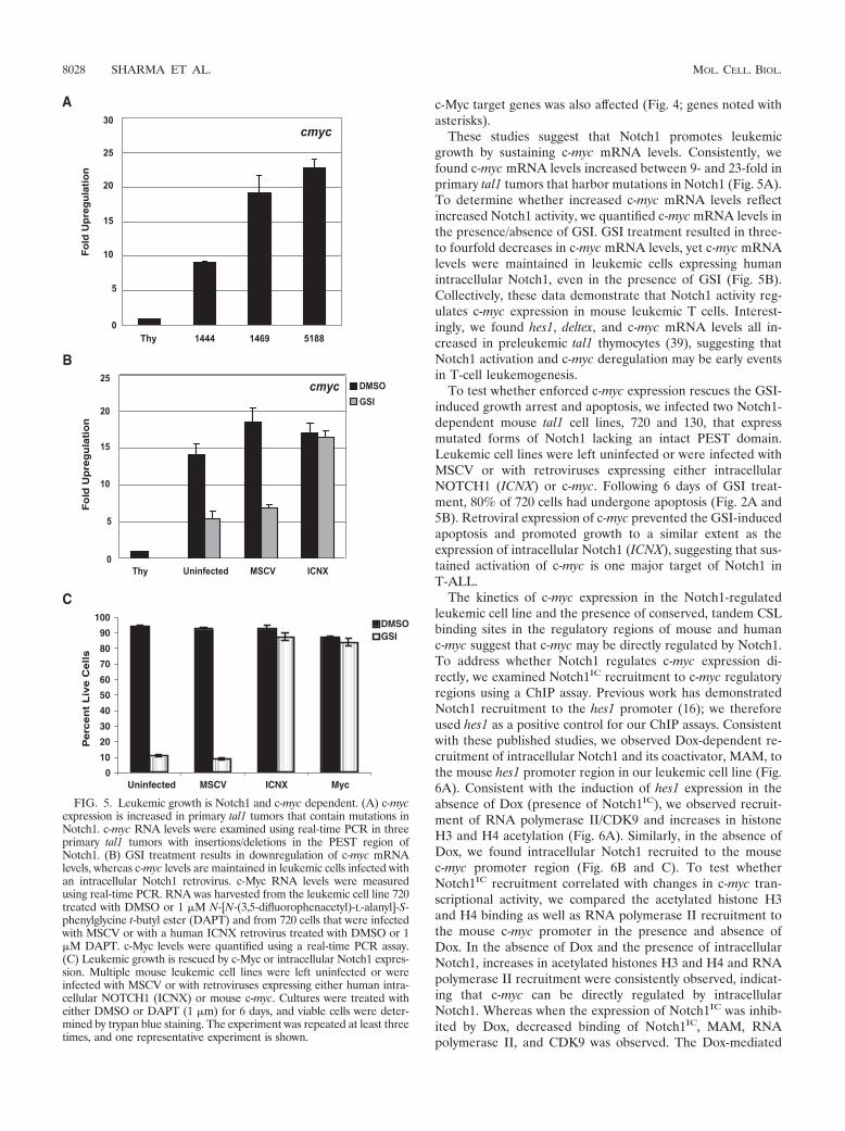
These studies suggest that Notch1 promotes leukemicgrowth by sustaining c-myc mRNA levels. Consistently, wefound c-myc mRNA levels increased between 9- and 23-fold inprimary tal1 tumors that harbor mutations in Notch1 (Fig. 5A).To determine whether increased c-myc mRNA levels reflectincreased Notch1 activity, we quantified c-myc mRNA levels inthe presence/absence of GSI. GSI treatment resulted in three-to fourfold decreases in c-myc mRNA levels, yet c-myc mRNAlevels were maintained in leukemic cells expressing humanintracellular Notch1, even in the presence of GSI (Fig. 5B).Collectively, these data demonstrate that Notch1 activity reg-ulates c-myc expression in mouse leukemic T cells. Interest-ingly, we found hes1, deltex, and c-myc mRNA levels all in-creased in preleukemic tal1 thymocytes (39), suggesting thatNotch1 activation and c-myc deregulation may be early eventsin T-cell leukemogenesis.
To test whether enforced c-myc expression rescues the GSI-induced growth arrest and apoptosis, we infected two Notch1-dependent mouse tal1 cell lines, 720 and 130, that expressmutated forms of Notch1 lacking an intact PEST domain.Leukemic cell lines were left uninfected or were infected withMSCV or with retroviruses expressing either intracellularNOTCH1 (ICNX) or c-myc. Following 6 days of GSI treat-ment, 80% of 720 cells had undergone apoptosis (Fig. 2A and5B). Retroviral expression of c-myc prevented the GSI-inducedapoptosis and promoted growth to a similar extent as theexpression of intracellular Notch1 (ICNX), suggesting that sus-tained activation of c-myc is one major target of Notch1 inT-ALL.
The kinetics of c-myc expression in the Notch1-regulatedleukemic cell line and the presence of conserved, tandem CSLbinding sites in the regulatory regions of mouse and humanc-myc suggest that c-myc may be directly regulated by Notch1.To address whether Notch1 regulates c-myc expression di-rectly, we examined Notch1IC recruitment to c-myc regulatoryregions using a ChIP assay. Previous work has demonstratedNotch1 recruitment to the hes1 promoter (16); we thereforeused hes1 as a positive control for our ChIP assays. Consistentwith these published studies, we observed Dox-dependent re-cruitment of intracellular Notch1 and its coactivator, MAM, tothe mouse hes1 promoter region in our leukemic cell line (Fig.6A). Consistent with the induction of hes1 expression in theabsence of Dox (presence of Notch1IC), we observed recruit-ment of RNA polymerase II/CDK9 and increases in histoneH3 and H4 acetylation (Fig. 6A). Similarly, in the absence ofDox, we found intracellular Notch1 recruited to the mousec-myc promoter region (Fig. 6B and C). To test whetherNotch1IC recruitment correlated with changes in c-myc tran-scriptional activity, we compared the acetylated histone H3and H4 binding as well as RNA polymerase II recruitment tothe mouse c-myc promoter in the presence and absence ofDox. In the absence of Dox and the presence of intracellularNotch1, increases in acetylated histones H3 and H4 and RNApolymerase II recruitment were consistently observed, indicat-ing that c-myc can be directly regulated by intracellularNotch1. Whereas when the expression of Notch1IC was inhib-ited by Dox, decreased binding of Notch1IC, MAM, RNApolymerase II, and CDK9 was observed. The Dox-mediated
FIG. 5. Leukemic growth is Notch1 and c-myc dependent. (A) c-mycexpression is increased in primary tal1 tumors that contain mutations inNotch1. c-myc RNA levels were examined using real-time PCR in threeprimary tal1 tumors with insertions/deletions in the PEST region ofNotch1. (B) GSI treatment results in downregulation of c-myc mRNAlevels, whereas c-myc levels are maintained in leukemic cells infected withan intracellular Notch1 retrovirus. c-Myc RNA levels were measuredusing real-time PCR. RNA was harvested from the leukemic cell line 720treated with DMSO or 1 �M N-[N-(3,5-difluorophenacetyl)-L-alanyl]-S-phenylglycine t-butyl ester (DAPT) and from 720 cells that were infectedwith MSCV or with a human ICNX retrovirus treated with DMSO or 1�M DAPT. c-Myc levels were quantified using a real-time PCR assay.(C) Leukemic growth is rescued by c-Myc or intracellular Notch1 expres-sion. Multiple mouse leukemic cell lines were left uninfected or wereinfected with MSCV or with retroviruses expressing either human intra-cellular NOTCH1 (ICNX) or mouse c-myc. Cultures were treated witheither DMSO or DAPT (1 �m) for 6 days, and viable cells were deter-mined by trypan blue staining. The experiment was repeated at least threetimes, and one representative experiment is shown.
8028 SHARMA ET AL. MOL. CELL. BIOL.

suppression of Notch1IC expression was also accompanied bydecreases in histone H3 and H4 acetylation at the c-myc reg-ulatory regions (Fig. 6B and C). None of these interactionswere observed in the absence of specific antisera, and none ofthese proteins bound mouse c-myc in a 3� intergenic regiondownstream of the polyadenylation site (data not shown). Insum, the Dox-induced growth arrest and apoptosis were coin-cident with the depletion of the Notch1IC/MAM transcrip-tional complex from c-myc regulatory regions and with de-
creases in c-myc expression. Considered together, these studiesstrongly suggest that Notch1 contributes to T-cell leukemogen-esis by directly regulating the expression of c-myc.
DISCUSSION
Retroviral insertional mutagenesis revealed that the T-celloncogene tal1 cooperates with activated Notch1 to induce T-ALL in mice. The notch1 proviral integrations occurred incluster region I, which contains the LIN-12/Notch repeat(LNR) and epidermal growth factor-like repeat (EGFR) mo-tifs. Conversely, no integrations were detected in cluster regionII, located within the 3� regulatory regions that contain thePEST domain. Previous RIM screens of both E2A-Pbx1 andMMTVD/myc transgenic mice detected MoMLV integrationsin the 3� region of notch1, and these were shown to cooperatewith the initiating oncogene to cause disease (13, 17). It isunclear why spontaneous tal1 tumors appear to preferentiallytruncate the PEST domain (32), whereas analysis of MoMLV-infected tal1 tumors detected integrations in the midregion ofnotch1 and no 3� integrations were detected. One possibility isthat too few tumors were examined in the RIM screen (n � 25)and that with larger screens, 3� integrations in notch1 wouldalso be observed. Nonetheless, this screen revealed that in-creases in Notch1 signaling contribute to mouse T-ALL andprompted us to investigate the status of the Notch1 receptor inspontaneous Tal1 mouse tumors. That analysis revealed thatmouse tal1 tumors harbor truncated versions of intracellularNotch1 that delete the PEST regulatory region (32).
To understand how Notch1 mutations contribute to T-cellleukemia, we developed a mouse leukemic line that expressesNotch1IC in a Dox-dependent manner. The conditional Notch1IC
T-ALL line provides multiple advantages over other experi-mental systems to examine the function of Notch1 in leuke-mogenesis. Although GSI are commonly used to inhibit thecleavage and activation of Notch receptors (8), they do notspecifically inhibit Notch1 but also inhibit cleavage of a num-ber of other transmembrane proteins (7, 18, 25, 29). Moreover,the response of human and mouse leukemic cell lines to GSI isnot uniform: sensitive cell lines undergo variable amounts ofcell cycle arrest/apoptosis, and effects on cell growth occuranywhere from 3 to 7 days following GSI treatment (32, 43). Incontrast, doxycycline specifically inhibits Notch1IC expressionand allows temporal comparison of the recruitment of intra-cellular Notch1 and its coactivators to target gene loci.
Gene expression profiling of this Dox-regulated Notch1IC
T-ALL line demonstrated upregulation of numerous knownNotch1IC target genes, including nrarp, nr2f2, notch3, hes1,cd25, adam19 (or �-meltrin), deltex, il-10, irf-4, egr1, and pre-T�(1, 9, 20). Ingenuity Systems software analysis revealed thec-Myc pathway as the central pathway (other than Notch1)induced in the absence of Dox or presence of active Notch1signaling. c-Myc and 35 known c-Myc target genes (cad, odc1,nol1, bcat1, srm, and others denoted with an asterisk in Fig. 4)were induced/repressed in the absence of Dox or presence ofintracellular Notch1. We demonstrate that Notch1IC mediatesleukemic growth by inducing c-myc expression since retroviralexpression of c-myc rescues the growth arrest/apoptosis in-duced upon GSI treatment of multiple mouse tal1 leukemiclines. In our RIM screen, proviral insertions were also ob-
FIG. 6. Dox-dependent recruitment of Notch1IC and MAM to thehes1 and c-myc promoters. (A) ChIP assay for CSL, Notch1IC, MAM,RNA polymerase II (Pol II), CDK9, and acetylated histones H3 andH4. Briefly, the Dox-dependent 3404 leukemic cell line was grown inRPMI medium with 10% fetal bovine serum at 37°C and treated withdoxycycline (2 �g/ml) for 48 h (�Dox) or left untreated (�Dox).Immunoprecipitations (IPs) were carried out using the respectivemonoclonal or polyclonal antibodies or without antibody (No-ab) as acontrol. Immunoprecipitated and input DNAs were amplified by PCRusing primers specific for the mouse hes1 promoter (A) or the mousec-myc promoter (B and C). PCR-amplified products were analyzed on2% agarose gels and stained with ethidium bromide.
VOL. 26, 2006 Notch1 REGULATES c-myc IN LEUKEMIA 8029

served in the c-myc locus (6/25 tumors), suggesting that acertain threshold level of c-myc expression is essential for in-duction/maintenance of T-ALL (achieved by either Notch1activation or direct insertion near c-myc).
Preleukemic studies of mouse models of T-ALL have dem-onstrated that T-cell oncogenes like tal1 and/or lmo1/2 arrestthymocyte development (5, 19, 31), and thus activation ofNotch1 and potentially c-myc may occur to rescue develop-mentally arrested, preleukemic thymocytes. Consistent withthis model, we detected evidence of Notch1 activation andincreased c-myc levels in preleukemic tal1 thymocytes (39),suggesting that Notch1 mutations are early events in T-cellleukemogenesis and may reflect a response to differentiationarrest. Evidence of early Notch1 activation has also been ob-served in SCL/LMO1 transgenic mice (26). Although c-myclevels clearly correlate with Notch1 activity in preleukemic orleukemic stages of disease and retroviral c-myc expression pre-vents GSI-induced cell cycle arrest (Fig. 5C), it was unclearwhether c-myc activation was a direct or indirect effect ofNotch1 activation. The kinetics of c-myc activation (as comparedto the direct Notch1 target, hes1) suggests that Notch1IC/MAMcomplex may directly stimulate c-myc transcription. Consistentwith the direct model, the promoter of the mouse c-myc genecontains three CSL binding sites (Fig. 6B and C). One of thethree CSL binding sites found in the promoter region is alsoconserved in the human c-MYC promoter region, suggestingthat this may also be Notch1 regulated in human T-ALL.Moreover, increases in hes1 or c-myc mRNA levels were ac-companied by an enrichment of Notch1IC, Mastermind, andRNA polymerase II/CDK9 at the promoter regions containingthe CSL binding sites. The increase in c-myc transcriptionalactivity was also associated with increases in histone acetyla-tion. These Dox-dependent changes were specific to the pro-moter region of c-myc that contained conserved CSL bindingsites and when 3� regions of c-myc were interrogated by ChIP,no evidence of Notch1IC or MAM binding was observed.
c-myc has also been recently implicated as a Notch1 targetgene in mouse mammary tumorigenesis (22). Mouse mammarytumor virus (MMTV) LTR-driven expression of Notch1IC in-duces mammary tumors in female mice, and an absence ofc-myc delays tumor incidence and penetrance. Mammary tu-mors express increased c-myc mRNA levels, and a Notch1/Cbf1 complex was shown to bind the c-myc promoter (22).Thus, our study and that of Klinakis et al. reveal c-myc as adirect Notch1 target gene in diverse transformed cell types(i.e., mouse leukemic T cells and mammary tumor cells). Al-though Klinakis et al. provide genetic evidence that linksNotch1 to c-myc, Notch1IC-induced mammary tumors doform, albeit with increased latency, in the absence of c-myc(22). Thus, it remains unclear whether Notch1 target genesother than c-myc also contribute to mammary tumorigenesis orwhether Myc family members compensate for an absence ofc-myc in the tumors that do form in the MMTV-Notch1IC
“null” Myc mice. For potential therapeutic reasons, it will beimportant to test whether mammary tumor growth, like T-cellleukemic growth, remains Notch1 dependent.
In our study, we provide functional data that demonstratethat Notch1 supports leukemic growth by maintaining c-mycmRNA levels. Consistently, primary tumors isolated from tal1transgenic mice with increased endogenous Notch1 activity
due to deletions in the PEST region (32) exhibit 9- to 23-foldincreases in c-myc mRNA levels (Fig. 5A). Notch1 inhibitionresults in three- to fourfold decreases in c-myc mRNA levelsand induction of G1 arrest and apoptosis (Fig. 2 and 5). Thus,this work identifies c-myc as both a direct and critical target ofNotch1 in T-cell leukemogenesis.
Although c-Myc promotes entry into the cell cycle by in-creasing E2F1 levels, increased c-Myc and/or E2F1 levels havebeen shown to activate the Arf-p53 oncogene checkpoint (11,46). Consistently, E�-myc transgenic mice exhibit apoptosisand acquire Ink4a/Arf deletions (46). INK4A/ARF losses arealso frequent in human T-ALL and in mouse tal1 tumors (14,39). Thus, Notch1 mutation may induce and sustain elevatedc-myc levels and thereby provide selective pressure for INK4A/ARF loss.
A percentage of mouse T-ALL lines exhibit evidence ofNotch1 activation but appear resistant to GSI, suggesting thatadditional members of the Notch1 signaling pathway may bemutated in these tumors. For example, inactivating mutationsin the negative regulators Numb, Itch, or Fbw7/Sel10 or in thekinases such as CycC:CDK8 (16) which trigger Notch1 ubiq-uitination could lead to increased Notch1 activation and GSIresistance. Similarly, mutations in Notch1 coactivators Master-mind-like 1, 2, and 3 may also contribute to T-ALL. Althoughthese in vitro studies suggest that GSI may have efficacy in 50%of human T-ALL patients that harbor mutations in the Notch1receptor (43), the identification of GSI-resistant mouse andhuman T-ALL lines and the implication that Notch1 activationcontributes to mammary tumorigenesis (22) urge the develop-ment of additional Notch1 pathway therapeutics perhapsaimed at displacing the intracellular Notch1/MAM complexfrom the CSL repressor or potentially directly targeting c-Myc.
ACKNOWLEDGMENTS
We thank Gerry Weinmaster for rat notch1 cluster I and II cDNAprobes, Jon Aster for the human NOTCH1 ICNX retroviral construct,and Warren Pear for the anti-Notch1 antibody. We thank MerckResearch Laboratories, Inc., for supplying the GSI (MRK-003) used insome of the in vitro studies.
V.M.S. is a Lymphoma Research Foundation Fellow, and A.J.C. andM.A.K. are Scholars of the Leukemia and Lymphoma Society ofAmerica. This work was supported by grants from the ACS and NCI toA.J.C. and by CA-096899 from the NCI to M.K.
REFERENCES
1. Adler, S. H., E. Chiffoleau, L. Xu, N. M. Dalton, J. M. Burg, A. D. Wells,M. S. Wolfe, L. A. Turka, and W. S. Pear. 2003. Notch signaling augments Tcell responsiveness by enhancing CD25 expression. J. Immunol. 171:2896–2903.
2. Bain, G., E. C. Maandag, D. J. Izon, D. Amsen, A. M. Kruisbeek, B. C.Weintraub, I. Krop, M. S. Schlissel, A. J. Feeney, and M. van Roon. 1994.E2A proteins are required for proper B cell development and initiation ofimmunoglobulin gene rearrangements. Cell 79:885–892.
3. Beverly, L. J., and A. J. Capobianco. 2003. Perturbation of Ikaros isoformselection by MLV integration is a cooperative event in Notch(IC)-induced Tcell leukemogenesis. Cancer Cell 3:551–564.
4. Beverly, L. J., D. W. Felsher, and A. J. Capobianco. 2005. Suppression of p53by Notch in lymphomagenesis: implications for initiation and regression.Cancer Res. 65:7159–7168.
5. Chervinsky, D. S., X.-F. Zhao, D. H. Lam, M. Ellsworth, K. W. Gross, andP. D. Aplan. 1999. Disordered T-cell development and T-cell malignancies inSCL LMO1 double-transgenic mice: parallels with E2A-deficient mice. Mol.Cell. Biol. 19:5025–5035.
6. Cortes, M., E. Wong, J. Koipally, and K. Georgopoulos. 1999. Control oflymphocyte development by the Ikaros gene family. Curr. Opin. Immunol.11:167–171.
7. Cowan, J. W., X. Wang, R. Guan, K. He, J. Jiang, G. Baumann, R. A. Black,
8030 SHARMA ET AL. MOL. CELL. BIOL.

M. S. Wolfe, and S. J. Frank. 2005. Growth hormone receptor is a target forpresenilin-dependent gamma-secretase cleavage. J. Biol. Chem. 280:19331–19342.
8. Deftos, M. L., Y. W. He, E. W. Ojala, and M. J. Bevan. 1998. Correlatingnotch signaling with thymocyte maturation. Immunity 9:777–786.
9. Deftos, M. L., E. Huang, E. W. Ojala, K. A. Forbush, and M. J. Bevan. 2000.Notch1 signaling promotes the maturation of CD4 and CD8 SP thymocytes.Immunity 13:73–84.
10. De Strooper, B., W. Annaert, P. Cupers, P. Saftig, K. Craessaerts, J. S.Mumm, E. H. Schroeter, V. Schrijvers, M. S. Wolfe, W. J. Ray, A. Goate, andR. Kopan. 1999. A presenilin-1-dependent gamma-secretase-like proteasemediates release of Notch intracellular domain. Nature 398:518–522.
11. Dimri, G. P., K. Itahana, M. Acosta, and J. Campisi. 2000. Regulation of asenescence checkpoint response by the E2F1 transcription factor andp14ARF tumor suppressor. Mol. Cell. Biol. 20:273–285.
12. Dumortier, A., R. Jeannet, P. Kirstetter, E. Kleinmann, M. Sellars, N. R. dosSantos, C. Thibault, J. Barths, J. Ghysdael, J. A. Punt, P. Kastner, and S.Chan. 2006. Notch activation is an early and critical event during T-cellleukemogenesis in Ikaros-deficient mice. Mol. Cell. Biol. 26:209–220.
13. Feldman, B. J., T. Hampton, and M. L. Cleary. 2000. A carboxy-terminaldeletion mutant of Notch1 accelerates lymphoid oncogenesis in E2A-PBX1transgenic mice. Blood 96:1906–1913.
14. Ferrando, A. A., D. S. Neuberg, J. Staunton, M. L. Loh, C. Huard, S. C.Raimondi, F. G. Behm, C. H. Pui, J. R. Downing, D. G. Gilliland, E. S.Lander, T. R. Golub, and A. T. Look. 2002. Gene expression signaturesdefine novel oncogenic pathways in T cell acute lymphoblastic leukemia.Cancer Cell 1:75–87.
15. Fryer, C. J., E. Lamar, I. Turbachova, C. Kintner, and K. A. Jones. 2002.Mastermind mediates chromatin-specific transcription and turnover of theNotch enhancer complex. Genes Dev. 16:1397–1411.
16. Fryer, C. J., J. B. White, and K. A. Jones. 2004. Mastermind recruits CycC:CDK8 to phosphorylate the Notch ICD and coordinate activation withturnover. Mol. Cell 16:509–520.
17. Girard, L., Z. Hanna, N. Beaulieu, C. D. Hoemann, C. Simard, C. A. Kozak,and P. Jolicoeur. 1996. Frequent provirus insertional mutagenesis of Notch1in thymomas of MMTVD/myc transgenic mice suggests a collaboration ofc-myc and Notch1 for oncogenesis. Genes Dev. 10:1930–1944.
18. Haas, I. G., M. Frank, N. Veron, and R. Kemler. 2005. Presenilin-dependentprocessing and nuclear function of gamma-protocadherins. J. Biol. Chem.280:9313–9319.
19. Herblot, S., A. M. Steff, P. Hugo, P. D. Aplan, and T. Hoang. 2000. SCL andLMO1 alter thymocyte differentiation: inhibition of E2A-HEB function andpre-T alpha chain expression. Nat. Immunol. 1:138–144.
20. Huang, Y. H., D. Li, A. Winoto, and E. A. Robey. 2004. Distinct transcrip-tional programs in thymocytes responding to T cell receptor, Notch, andpositive selection signals. Proc. Natl. Acad. Sci. USA 101:4936–4941.
21. Kelliher, M. A., D. C. Seldin, and P. Leder. 1996. Tal-1 induces T cell acutelymphoblastic leukemia accelerated by casein kinase IIalpha. EMBO J. 15:5160–5166.
22. Klinakis, A., M. Szabolcs, K. Politi, H. Kiaris, S. Artavanis-Tsakonas, and A.Efstratiadis. 2006. Myc is a Notch1 transcriptional target and a requisite forNotch1-induced mammary tumorigenesis in mice. Proc. Natl. Acad. Sci.USA 103:9262–9267.
23. Krebs, L. T., M. L. Deftos, M. J. Bevan, and T. Gridley. 2001. The Nrarpgene encodes an ankyrin-repeat protein that is transcriptionally regulated bythe notch signaling pathway. Dev. Biol. 238:110–119.
24. Kurooka, H., and T. Honjo. 2000. Functional interaction between the mousenotch1 intracellular region and histone acetyltransferases PCAF and GCN5.J. Biol. Chem. 275:17211–17220.
25. Lammich, S., M. Okochi, M. Takeda, C. Kaether, A. Capell, A. K. Zimmer,D. Edbauer, J. Walter, H. Steiner, and C. Haass. 2002. Presenilin-dependentintramembrane proteolysis of CD44 leads to the liberation of its intracellulardomain and the secretion of an Abeta-like peptide. J. Biol. Chem. 277:44754–44759.
26. Lin, Y. W., R. A. Nichols, J. J. Letterio, and P. D. Aplan. 2006. Notch1mutations are important for leukemic transformation in murine models ofprecursor-T leukemia/lymphoma. Blood 107:2540–2543.
27. Matsuno, K., R. J. Diederich, M. J. Go, C. M. Blaumueller, and S. Artavanis-
Tsakonas. 1995. Deltex acts as a positive regulator of Notch signalingthrough interactions with the Notch ankyrin repeats. Development 121:2633–2644.
28. McGill, M. A., and C. J. McGlade. 2003. Mammalian numb proteins pro-mote Notch1 receptor ubiquitination and degradation of the Notch1 intra-cellular domain. J. Biol. Chem. 278:23196–23203.
29. Ni, C. Y., M. P. Murphy, T. E. Golde, and G. Carpenter. 2001. Gamma-secretase cleavage and nuclear localization of ErbB-4 receptor tyrosinekinase. Science 294:2179–2181.
30. Noseda, M., L. Chang, G. McLean, J. E. Grim, B. E. Clurman, L. L. Smith,and A. Karsan. 2004. Notch activation induces endothelial cell cycle arrestand participates in contact inhibition: role of p21Cip1 repression. Mol. Cell.Biol. 24:8813–8822.
31. O’Neil, J., M. Billa, S. Oikemus, and M. Kelliher. 2001. The DNA bindingactivity of TAL-1 is not required to induce leukemia/lymphoma in mice.Oncogene 20:3897–3905.
32. O’Neil, J., J. Calvo, K. McKenna, V. Krishnamoorthy, J. C. Aster, C. H.Bassing, F. W. Alt, M. Kelliher, and A. T. Look. 2006. Activating Notch1mutations in mouse models of T-ALL. Blood 107:781–785.
33. O’Neil, J., J. Shank, N. Cusson, C. Murre, and M. Kelliher. 2004. TAL1/SCLinduces leukemia by inhibiting the transcriptional activity of E47/HEB. Can-cer Cell 5:587–596.
34. Oswald, F., B. Tauber, T. Dobner, S. Bourteele, U. Kostezka, G. Adler, S.Liptay, and R. M. Schmid. 2001. p300 acts as a transcriptional coactivator formammalian Notch-1. Mol. Cell. Biol. 21:7761–7774.
35. Qiu, L., C. Joazeiro, N. Fang, H. Y. Wang, C. Elly, Y. Altman, D. Fang, T.Hunter, and Y. C. Liu. 2000. Recognition and ubiquitination of Notch byItch, a hect-type E3 ubiquitin ligase. J. Biol. Chem. 275:35734–35737.
36. Reschly, E. J., C. Spaulding, T. Vilimas, W. V. Graham, R. L. Brumbaugh,I. Aifantis, W. S. Pear, and B. L. Kee. 2006. Notch1 promotes survival ofE2A-deficient T cell lymphomas through pre-T cell receptor-dependent and-independent mechanisms. Blood 107:4115–4121. (First published 31 Janu-ary 2006; doi:10.1182/blood-2005-09-3551.)
37. Ronchini, C., and A. J. Capobianco. 2001. Induction of cyclin D1 transcrip-tion and CDK2 activity by Notchic: implication for cell cycle disruption intransformation by Notchic. Mol. Cell. Biol. 21:5925–5934.
38. Satoh, Y., I. Matsumura, H. Tanaka, S. Ezoe, H. Sugahara, M. Mizuki, H.Shibayama, E. Ishiko, J. Ishiko, K. Nakajima, and Y. Kanakura. 2004. Rolesfor c-Myc in self-renewal of hematopoietic stem cells. J. Biol. Chem. 279:24986–24993.
39. Shank-Calvo, J. A., K. Draheim, M. Bhasin, and M. A. Kelliher. 2006.p16Ink4a or p19Arf loss contributes to Tal1-induced leukemogenesis inmice. Oncogene 25:3023–3031.
40. Sun, L., M. L. Crotty, M. Sensel, H. Sather, C. Navara, J. Nachman, P. G.Steinherz, P. S. Gaynon, N. Seibel, C. Mao, A. Vassilev, G. H. Reaman, andF. M. Uckun. 1999. Expression of dominant-negative Ikaros isoforms inT-cell acute lymphoblastic leukemia. Clin. Cancer Res. 5:2112–2120.
41. Tetzlaff, M. T., W. Yu, M. Li, P. Zhang, M. Finegold, K. Mahon, J. W.Harper, R. J. Schwartz, and S. J. Elledge. 2004. Defective cardiovasculardevelopment and elevated cyclin E and Notch proteins in mice lacking theFbw7 F-box protein. Proc. Natl. Acad. Sci. USA 101:3338–3345.
42. Wallberg, A. E., K. Pedersen, U. Lendahl, and R. G. Roeder. 2002. p300 andPCAF act cooperatively to mediate transcriptional activation from chroma-tin templates by Notch intracellular domains in vitro. Mol. Cell. Biol. 22:7812–7819.
43. Weng, A. P., A. A. Ferrando, W. Lee, J. P. T. Morris, L. B. Silverman, C.Sanchez-Irizarry, S. C. Blacklow, A. T. Look, and J. C. Aster. 2004. Activat-ing mutations of NOTCH1 in human T cell acute lymphoblastic leukemia.Science 306:269–271.
44. Weng, A. P., and A. Lau. 2005. Notch signaling in T-cell acute lymphoblasticleukemia. Fut. Oncol. 1:511–519.
45. Yun, T. J., and M. J. Bevan. 2003. Notch-regulated ankyrin-repeat proteininhibits Notch1 signaling: multiple Notch1 signaling pathways involved in Tcell development. J. Immunol. 170:5834–5841.
46. Zindy, F., C. M. Eischen, D. H. Randle, T. Kamijo, J. L. Cleveland, C. J.Sherr, and M. F. Roussel. 1998. Myc signaling via the ARF tumor suppressorregulates p53-dependent apoptosis and immortalization. Genes Dev. 12:2424–2433.
VOL. 26, 2006 Notch1 REGULATES c-myc IN LEUKEMIA 8031
Copyright © 2022 FDOKUMEN