Hydration Dynamics in a Partially Denatured Ensemble of the Globular Protein Human alpha-Lactalbumin...
11
Hydration Dynamics in a Partially Denatured Ensemble of the Globular Protein Human a-Lactalbumin Investigated with Molecular Dynamics Simulations Neelanjana Sengupta, Simon Jaud, and Douglas J. Tobias Department of Chemistry, University of California, Irvine, California ABSTRACT Atomistic molecular dynamics simulations are used to probe changes in the nature and subnanosecond dynamical behavior of solvation waters that accompany partial denaturation of the globular protein, human a-lactalbumin. A simulated ensemble of subcompact conformers, similar to the molten globule state of human a-lactalbumin, demonstrates a marginal increase in the amount of surface solvation relative to the native state. This increase is accompanied by subtle but distinct enhancement in surface water dynamics, less favorable protein-water interactions, and a marginal decrease in the anomalous behavior of solvation water dynamics. The extent of solvent influx is not proportional to the increased surface area, and the partially denatured conformers are less uniformly solvated compared to their native counterpart. The observed solvation in partially denatured conformers is lesser in extent compared to earlier experimental estimates in molten globule states, and is consistent with more recent descriptions based on nuclear magnetic relaxation dispersion studies. INTRODUCTION Solvation plays a vital role in protein structure, dynamics, and function. The wide variety of studies that have elicited the dependence of such properties on protein solvation in- clude light scattering and fluorescence experiments (1–5), small-angle x-ray and neutron scattering spectroscopy (6– 12), nuclear spin echo (13,14), solid-state NMR (15), nuclear magnetic relaxation dispersion (16–20), Fourier transform infrared (FTIR) measurements (21), and terahertz spectros- copy (22–25). In addition, molecular dynamics simulations have been used extensively to characterize the dynamics of protein solvation (26–30) and its association with protein conformation, function, and dynamics (10,11,31–35). It is important to note that solvent effects are understood to play a fundamental role in protein folding and unfolding (36–40). For example, a simulation study of the SH3 protein identified the existence of an ensemble of ‘‘near-native’’ structures, the transition to the native state being facilitated by expulsion of water molecules from the core of the protein (40). A coarse- grained simulation of protein-solvent systems has been used to suggest that loss in conformational entropy in the folding protein is compensated by gain in translational entropy of solvent particles (41). It has been seen that protein folding and unfolding kinetics can be modulated by solvent viscosity (42,43). It is now well accepted that most nascent protein polymers encounter at least a few metastable states en route to the natively folded functional form, and developing a knowledge of the intermediates is vital for developing an understanding of the folding process (44,45). The transition pathways connecting the metastable states may not be unique, and the folding ensemble is comprised of a large number of energet- ically equivalent substates. The loss in conformational entropy as the protein progresses toward the folded form implies that the ensemble ‘‘size’’, or heterogeneity, should decrease along the folding pathway. In view of the fundamental relationship between protein solvation and dynamics, it is important to understand the dynamical and energetic changes in the protein solvating layer that accompany the folding or unfolding process (38,39). Recent studies with fluorescent probes attached to the protein surfaces and dynamic light- scattering experiments have elucidated differences in solvation times between the fully denatured, ‘‘premolten’’ globule, molten globule, and native states (3,4). Molecular dynamics simulations have been used to point out clear differences in the secondary structure dependence of solvation dynamics between the native and a hypothetical molten globule state of the protein HP-36 (46). Simulations have also been used to correlate partial unfolding of specific secondary structure elements with mobility of the solvation layer and the protein- water hydrogen bonding kinetics (47). Site-specific mutations and solvation correlation functions constructed from femto- second-resolved fluorescence transients show site-dependent enhancement of water dynamics in the molten globule state compared to the native state of myoglobin (5). Despite recent developments, there remains a lack of un- derstanding of the changes in some general, but presumably important, aspects of protein solvation, such as the extent, anomalous behavior, and energetic interactions as a protein proceeds along its folding pathway. In this study, we attempt to understand the dynamical changes in protein solvation accompanying partial denaturation of the globular protein human a-lactalbumin (HaLA), which occur on timescales up to a few hundred picoseconds. We use molecular dynamics doi: 10.1529/biophysj.108.136531 Submitted May 2, 2008, and accepted for publication July 28, 2008. Address reprint requests to Douglas J. Tobias, Dept. of Chemistry, University of California, Irvine, CA 92697. E-mail: [email protected]. Editor: Arthur G. Palmer 3rd. Ó 2008 by the Biophysical Society 0006-3495/08/12/5257/11 $2.00 Biophysical Journal Volume 95 December 2008 5257–5267 5257
Transcript of Hydration Dynamics in a Partially Denatured Ensemble of the Globular Protein Human alpha-Lactalbumin...
Hydration Dynamics in a Partially Denatured Ensemble of the GlobularProtein Human a-Lactalbumin Investigated with MolecularDynamics Simulations
Neelanjana Sengupta, Simon Jaud, and Douglas J. TobiasDepartment of Chemistry, University of California, Irvine, California
ABSTRACT Atomistic molecular dynamics simulations are used to probe changes in the nature and subnanoseconddynamical behavior of solvation waters that accompany partial denaturation of the globular protein, human a-lactalbumin. Asimulated ensemble of subcompact conformers, similar to the molten globule state of human a-lactalbumin, demonstrates amarginal increase in the amount of surface solvation relative to the native state. This increase is accompanied by subtle butdistinct enhancement in surface water dynamics, less favorable protein-water interactions, and a marginal decrease in theanomalous behavior of solvation water dynamics. The extent of solvent influx is not proportional to the increased surface area,and the partially denatured conformers are less uniformly solvated compared to their native counterpart. The observed solvationin partially denatured conformers is lesser in extent compared to earlier experimental estimates in molten globule states, and isconsistent with more recent descriptions based on nuclear magnetic relaxation dispersion studies.
INTRODUCTION
Solvation plays a vital role in protein structure, dynamics,
and function. The wide variety of studies that have elicited
the dependence of such properties on protein solvation in-
clude light scattering and fluorescence experiments (1–5),
small-angle x-ray and neutron scattering spectroscopy (6–
12), nuclear spin echo (13,14), solid-state NMR (15), nuclear
magnetic relaxation dispersion (16–20), Fourier transform
infrared (FTIR) measurements (21), and terahertz spectros-
copy (22–25). In addition, molecular dynamics simulations
have been used extensively to characterize the dynamics of
protein solvation (26–30) and its association with protein
conformation, function, and dynamics (10,11,31–35). It is
important to note that solvent effects are understood to play a
fundamental role in protein folding and unfolding (36–40).
For example, a simulation study of the SH3 protein identified
the existence of an ensemble of ‘‘near-native’’ structures, the
transition to the native state being facilitated by expulsion of
water molecules from the core of the protein (40). A coarse-
grained simulation of protein-solvent systems has been used
to suggest that loss in conformational entropy in the folding
protein is compensated by gain in translational entropy of
solvent particles (41). It has been seen that protein folding
and unfolding kinetics can be modulated by solvent viscosity
(42,43).
It is now well accepted that most nascent protein polymers
encounter at least a few metastable states en route to the
natively folded functional form, and developing a knowledge
of the intermediates is vital for developing an understanding
of the folding process (44,45). The transition pathways
connecting the metastable states may not be unique, and the
folding ensemble is comprised of a large number of energet-
ically equivalent substates. The loss in conformational entropy
as the protein progresses toward the folded form implies
that the ensemble ‘‘size’’, or heterogeneity, should decrease
along the folding pathway. In view of the fundamental
relationship between protein solvation and dynamics, it is
important to understand the dynamical and energetic changes
in the protein solvating layer that accompany the folding or
unfolding process (38,39). Recent studies with fluorescent
probes attached to the protein surfaces and dynamic light-
scattering experiments have elucidated differences in solvation
times between the fully denatured, ‘‘premolten’’ globule,
molten globule, and native states (3,4). Molecular dynamics
simulations have been used to point out clear differences in
the secondary structure dependence of solvation dynamics
between the native and a hypothetical molten globule state
of the protein HP-36 (46). Simulations have also been used
to correlate partial unfolding of specific secondary structure
elements with mobility of the solvation layer and the protein-
water hydrogen bonding kinetics (47). Site-specific mutations
and solvation correlation functions constructed from femto-
second-resolved fluorescence transients show site-dependent
enhancement of water dynamics in the molten globule state
compared to the native state of myoglobin (5).
Despite recent developments, there remains a lack of un-
derstanding of the changes in some general, but presumably
important, aspects of protein solvation, such as the extent,
anomalous behavior, and energetic interactions as a protein
proceeds along its folding pathway. In this study, we attempt
to understand the dynamical changes in protein solvation
accompanying partial denaturation of the globular protein
human a-lactalbumin (HaLA), which occur on timescales up
to a few hundred picoseconds. We use molecular dynamics
doi: 10.1529/biophysj.108.136531
Submitted May 2, 2008, and accepted for publication July 28, 2008.
Address reprint requests to Douglas J. Tobias, Dept. of Chemistry,
University of California, Irvine, CA 92697. E-mail: [email protected].
Editor: Arthur G. Palmer 3rd.
� 2008 by the Biophysical Society
0006-3495/08/12/5257/11 $2.00
Biophysical Journal Volume 95 December 2008 5257–5267 5257
(MD) simulations to create an ‘‘ensemble’’ of partially de-
natured conformers that are conformationally stable over
those timescales. In this respect, and in the extent of their loss
of compactness relative to the native (N) state, the members
of our ensemble resemble the subcompact molten globule
(MG) state of HaLA. The metastable MG states are thought
to be the equilibrium analogs of early folding intermediates in
globular proteins (44,48–50), and it has even been recog-
nized that they have pathologically important roles (51,52).
Compared with the native state, we find consistent changes in
the dynamical behavior of the solvation waters in our par-
tially denatured ensemble. These changes are such that the
well-known slowdown of protein surface waters relative to
bulk (11,16,17,27,28,30,53–55) is decreased or, in other
words, there is a shift toward bulklike behavior. This shift is
subtle but distinct, and is observed for both translational and
rotational dynamics of the solvating waters, and found to be
widely distributed over the ensemble. Increased dynamics is
also accompanied by marginally greater solvation in almost
all ensemble members. The average increase in the solvation
number (Ns) is, however, much less than the quantities re-
ported in earlier experimental studies of MG states (13,56,57).
We have used the dynamical second-rank rotational correla-
tion function calculated from our simulations in combination
with the surface solvation number to obtain the NMRD hy-
dration parameter Nsrs (18). The average value of this quantity
for the partially denatured ensemble is very close to the value
for the native-state simulation. This finding is in agreement
with the observed constancy of Nsrs, within error limits, dur-
ing the N/MG transition for a variety of proteins (18).
When comparing our results to experimental data, it is
important to keep in mind the disparity in conformational
sampling: experiments reporting the average behavior of
1015–1018 molecules often fail to describe the behavior of an
individual species, whereas the longest atomistic MD simu-
lations will not encompass all conformers present in an ex-
perimental sample (38). Extensive sampling is required to
characterize properties of metastable intermediates that ex-
change on long timescales and form ensembles with a high
degree of heterogeneity. The consistent changes observed in
our ensemble of partially denatured conformers, whose level
of compactness is similar to the MG state of HaLA, ought to
be suggestive of phenomena occurring in actual MG samples.
The quantities measured experimentally are likely to be
closer to the ‘‘ensemble average’’ than to quantities obtained
from any single simulation. A previously reported protocol
for creating large ensembles of conformers structurally sim-
ilar to the experimental MG state of HaLA involved dena-
turation by application of biasing forces in a continuum
solvent (58). In this study, we focus on characterizing the
changes in dynamical behavior of the solvation molecules
brought about by partial protein denaturation in implicit
solvent, by creating a modest-sized ensemble that is subse-
quently investigated using simulations in explicit solvent. A
similar thermal denaturation protocol has been implemented
in a study of solvation in the native state and a representative
MG conformer of another globular protein (46). Our con-
formers, with radii of gyration stable over the timescale of
interest, may reasonably be assumed to have solvation water
dynamical properties akin to those of MG conformers with a
similar loss in compaction.
METHODS
Simulation protocol
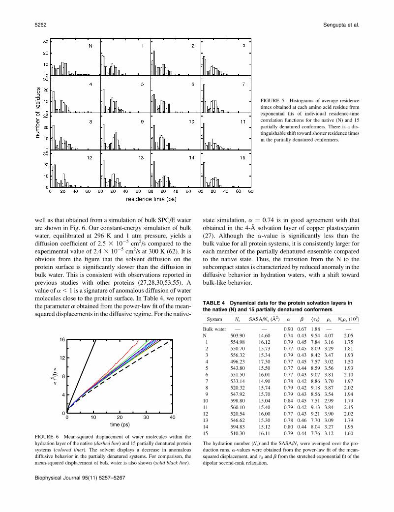
The 1.7-A resolution x-ray crystal structure of HaLA, obtained from the
Protein Data Bank, entry 1A4V (59), was the starting point from which
partially denatured conformers were created (59). For computational effi-
ciency, a thermal unfolding process was simulated with the native protein
structure placed in an implicit solvent environment. Implicit solvent models
represent the solvent as a dielectric continuum, in contrast to including the
solvent molecules explicitly. The generalized Born solvent area (GB/SA)
implicit solvent model was used (60), and the simulation was carried out with
the Maestro simulation program (Schrodinger, Portland, OR). A cutoff of
20 A was set for electrostatic calculations, 8 A for van der Waals interactions,
and 4 A for hydrogen bonding, and the dielectric constant was set to 78. Ini-
tially, the protein-solvent system was heated over a period of 1 ns, with the
temperature increased gradually from 50 to 300 K. This did not cause any
significant unfolding in the structure, and the radius of gyration (Rg) of the
system remained close to the native value of 14.35 A (59). The temperature was
then set to 400 K, and the system responded with a jump in the Rg. At this
temperature, the maximum Rg of 18.45 A was reached in 50 ps, soon after
which the system contracted, with Rg values settling in the vicinity of 16 A. The
unfolding behavior of the protein in implicit solvent for a period of 2 ns is
shown in Fig. 1. There is a slow conformational change between 0.5 and 2 ns,
as seen from the gradual rise in Rg during that time span. However, the con-
formations correspond to the experimentally measured Rg range of 16–19 A (1).
Ten roughly equispaced conformational snapshots from the simulation
trajectory were subjected to independent simulation runs of 200 ps in implicit
solvent. The conformations at the start and end of these individual simula-
tions were then selected to represent 20 putative members of a partially
denatured ensemble. This method ensures the conformational heterogeneity
of the ensemble while maintaining the experimentally determined extent of
denaturation. To model conformers similar to the low-pH ‘‘A’’ state of the
HaLA molten globule (61), the glutamate, aspartate, and histidine side
chains and the C-terminus were protonated, the Ca21 ion was removed, and
16 Cl� counterions were added. Fifteen of these partially denatured con-
formers (randomly selected) were transferred to a preequilibrated box of
water measuring ;57 A on each side. The Rg of the selected systems at this
FIGURE 1 The unfolding behavior of the compact native HaLA in
implicit solvent at 400 K.
5258 Sengupta et al.
Biophysical Journal 95(11) 5257–5267
stage have been listed in Table 1. The SPC/E water model (62) was em-
ployed, as it has been shown to accurately describe dynamical aspects of
protein hydration (53). From that point, the explicitly solvated systems were
simulated using the NAMD simulation program (63), with the CHARMM22
all-atom force field for the protein (64). After 50 ps of conjugate gradient
energy minimization, each solvated protein system was individually simu-
lated at a pressure of 1 atm and a temperature of 296 K. Constant temperature
was maintained using Langevin dynamics with a collision frequency of
1 ps�1. The Nose-Hoover Langevin piston algorithm (65,66), as im-
plemented in the NAMD package, was used to maintain constant pressure.
The lengths of bonds involving hydrogen atoms were held fixed with the
SHAKE algorithm (67). A simulation time step of 1 fs and periodic boundary
conditions were employed. Electrostatic interactions were calculated with
particle mesh Ewald (68). The real-space electrostatic and van der Waals
cutoff distances were set at 11 A, with smooth truncation starting at 10 A. The
Rg of each system took between 3.5 and 6.5 ns to settle within the 16–19 A
range, after which the systems were equilibrated at constant volume for 500
ps. The dynamical correlation functions investigated in this study largely
decay within the first 100 ps. Thus, our production runs consisted of 400 ps of
constant-energy MD, with coordinates saved every 0.1 ps. For comparison, a
simulation of the explicitly solvated native state of HaLA was performed
within the same protocol. The Rg of the native and the 15 partially denatured
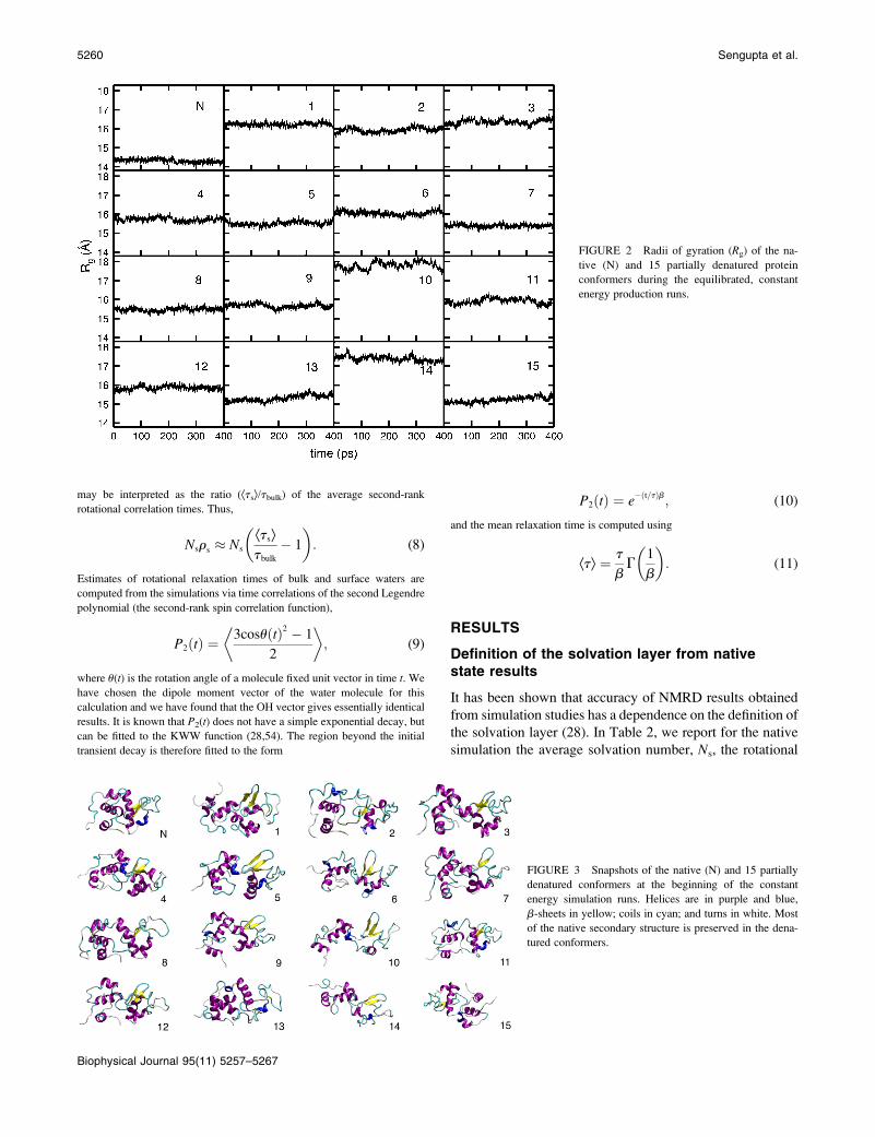
states during the production runs remained stable, as seen in Fig. 2. The
backbone root mean-squared deviations (RMSDs) of the conformers relative
to the native crystal structure are found to be stable during the production
runs (data not shown). The mean values of the Rg and the backbone Ca
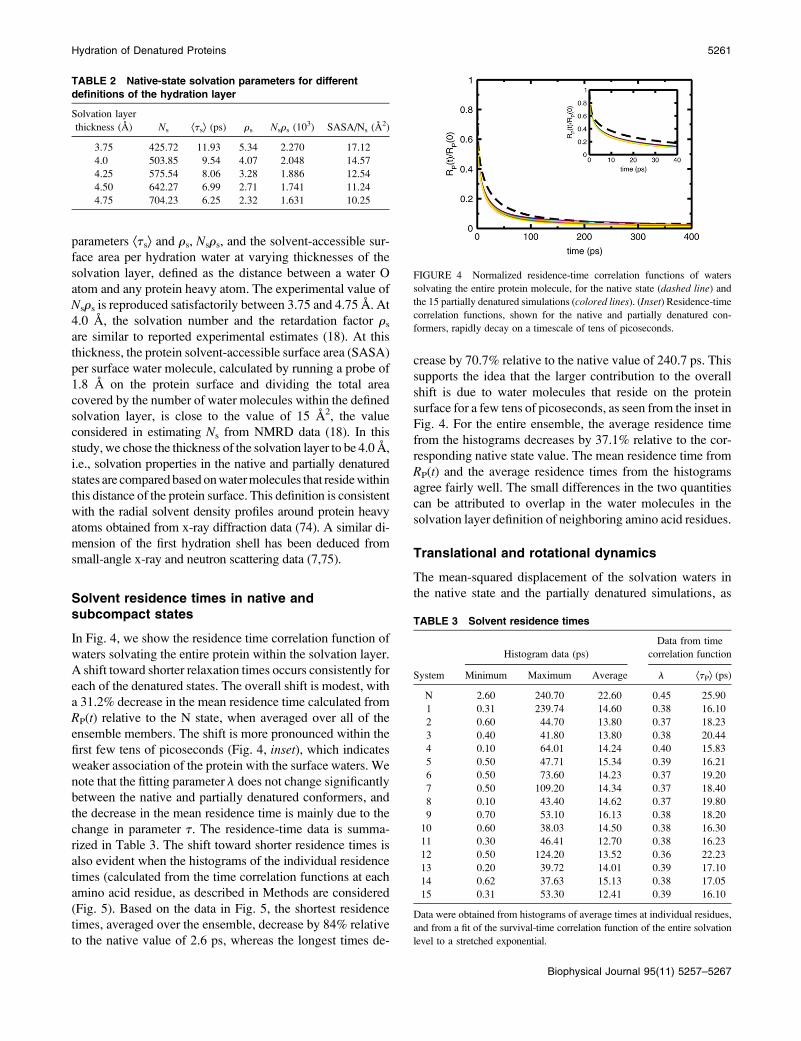
RMSD are given in Table 1. Fig. 3 shows snapshots of the native and de-
natured states at the beginning of the production runs created with the Visual
Molecular Dynamics program (69).
Analysis methodology
Solvent residence times and mean-squared displacement
The residence time correlation function from which the average survival time
is calculated, is obtained from the equispaced production run configurations as
RAðtÞ ¼1
NðtÞ+t0+Nw
j¼1
pA;jðt0; t0 1 tÞ: (1)
Here, the binary function pA,j(t0,t0 1 t) assumes a value of 1 if the jth water
molecule remains within the solvation layer of site A continuously for time t,
starting from an arbitrary time origin t0, and 0 otherwise. Nw is the total
number of water molecules in the system. The correlation function is
calculated with a moving time origin t0, and N(t) is the number of segments
corresponding to each time span t. Each of the 123 amino acid residues of
HaLA has been treated as a ‘‘solvation site’’. The mean residence times are
obtained from exponential fits of the normalized correlation functions, RA(t)/RA(0). The histograms of the mean residence times for each system are used
to obtain the arithmetic minimum, maximum, and average residence time.
The average residence time obtained in this manner can be compared to the
mean residence time of waters solvating the entire protein system, calculated
from the function RP(t), which has the same functional form as RA(t) but is
constructed as an average over all sites. We fit the normalized residence
correlation function obtained for the entire protein to the stretched exponen-
tial, known as the Kohlrausch-Williams-Watts (KWW) function,
RPðtÞRPð0Þ
¼ e�ðt=tÞl
: (2)
The integral of the stretched exponential function gives the mean solvent
residence time around the protein,
ÆtPæ ¼ t
lG
1
l
� �: (3)
The diffusion of water molecules in the protein solvation layer is known to be
described by non-Brownian kinetics (27). In this ‘‘dispersive diffusion’’
regime, the mean-squared displacement (MSD) of the water oxygen atoms
within the solvation layer is fitted to the power-law form
Ær2ðtÞæ } ta: (4)
The coefficient a, compared to the value obtained for bulk water, gives a
direct measure of the anomalous nature of surface solvation.
Rotational dynamics and 17O NMRD parameters
We highlight the salient features of quadrupolar spin relaxation theory as
required by this study. For greater appreciation of the theoretical framework
and applications of the nuclear magnetic relaxation dispersion (NMRD)
technique, the reader is referred to a comprehensive series of articles by Halle
et al. (16,17,70–73). From the theoretical foundations of quadrupolar spin
relaxation theory, the 17O NMRD longitudinal relaxation rate profile depends
on the resonance frequency, v0, as follows (17,70):
R1ðv0Þ ¼ Rbulk 1 a 1 btcF1ðv0tbÞ: (5)
The term btcF1(v0tb) is the frequency-dependent dispersion step describing
the slow rotation of the protein molecule and its associated integral waters,
and Rbulk is the frequency-independent relaxation rate of bulk water. Here,
a describes the relaxation parameters of water molecules near the protein
surface, which can be expressed as
a ¼ Ns
NT
� �ðÆRsæ� RbulkÞ; (6)
where Ns and NT are, respectively, the number of surface water molecules
and the total number of water molecules, and ÆRsæ is the average surface
relaxation rate. The quantity obtained from the experimental relaxation
profiles is
Nsrs ¼aNT
Rbulk
¼ Ns
ÆRsæRbulk
� 1
� �: (7)
Since the 17O quadrupolar coupling constant is expected to be the same for
water molecules next to the protein surface and in bulk water (16), ÆRsæ/Rbulk
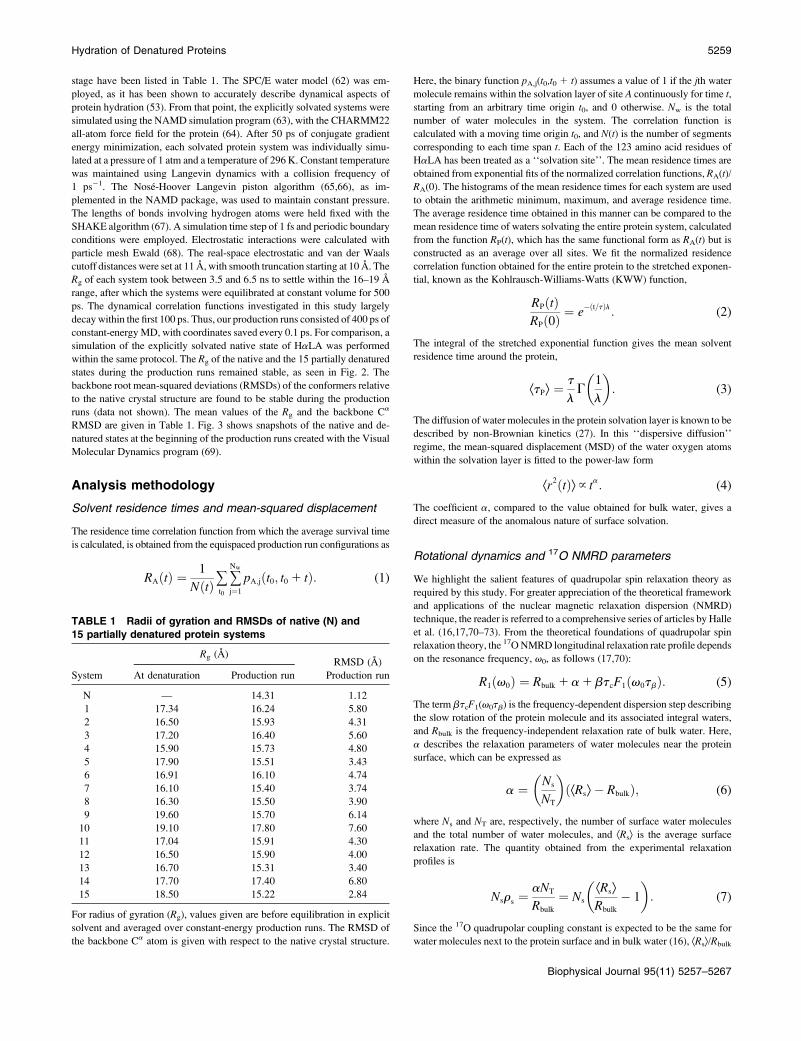
TABLE 1 Radii of gyration and RMSDs of native (N) and
15 partially denatured protein systems
Rg (A)RMSD (A)
System At denaturation Production run Production run
N — 14.31 1.12
1 17.34 16.24 5.80
2 16.50 15.93 4.31
3 17.20 16.40 5.60
4 15.90 15.73 4.80
5 17.90 15.51 3.43
6 16.91 16.10 4.74
7 16.10 15.40 3.74
8 16.30 15.50 3.90
9 19.60 15.70 6.14
10 19.10 17.80 7.60
11 17.04 15.91 4.30
12 16.50 15.90 4.00
13 16.70 15.31 3.40
14 17.70 17.40 6.80
15 18.50 15.22 2.84
For radius of gyration (Rg), values given are before equilibration in explicit
solvent and averaged over constant-energy production runs. The RMSD of
the backbone Ca atom is given with respect to the native crystal structure.
Hydration of Denatured Proteins 5259
Biophysical Journal 95(11) 5257–5267
may be interpreted as the ratio (Ætsæ/tbulk) of the average second-rank
rotational correlation times. Thus,
Nsrs � Ns
Ætsætbulk
� 1
� �: (8)
Estimates of rotational relaxation times of bulk and surface waters are
computed from the simulations via time correlations of the second Legendre
polynomial (the second-rank spin correlation function),
P2ðtÞ ¼�
3cosuðtÞ2 � 1
2
�; (9)
where u(t) is the rotation angle of a molecule fixed unit vector in time t. We
have chosen the dipole moment vector of the water molecule for this
calculation and we have found that the OH vector gives essentially identical
results. It is known that P2(t) does not have a simple exponential decay, but
can be fitted to the KWW function (28,54). The region beyond the initial
transient decay is therefore fitted to the form
P2ðtÞ ¼ e�ðt=tÞb
; (10)
and the mean relaxation time is computed using
Ætæ ¼ t
bG
1
b
� �: (11)
RESULTS
Definition of the solvation layer from nativestate results
It has been shown that accuracy of NMRD results obtained
from simulation studies has a dependence on the definition of
the solvation layer (28). In Table 2, we report for the native
simulation the average solvation number, Ns, the rotational
FIGURE 2 Radii of gyration (Rg) of the na-
tive (N) and 15 partially denatured protein
conformers during the equilibrated, constant
energy production runs.
FIGURE 3 Snapshots of the native (N) and 15 partially
denatured conformers at the beginning of the constant
energy simulation runs. Helices are in purple and blue,
b-sheets in yellow; coils in cyan; and turns in white. Most
of the native secondary structure is preserved in the dena-
tured conformers.
5260 Sengupta et al.
Biophysical Journal 95(11) 5257–5267
parameters Ætsæ and rs, Nsrs, and the solvent-accessible sur-
face area per hydration water at varying thicknesses of the
solvation layer, defined as the distance between a water O
atom and any protein heavy atom. The experimental value of
Nsrs is reproduced satisfactorily between 3.75 and 4.75 A. At
4.0 A, the solvation number and the retardation factor rs
are similar to reported experimental estimates (18). At this
thickness, the protein solvent-accessible surface area (SASA)
per surface water molecule, calculated by running a probe of
1.8 A on the protein surface and dividing the total area
covered by the number of water molecules within the defined
solvation layer, is close to the value of 15 A2, the value
considered in estimating Ns from NMRD data (18). In this
study, we chose the thickness of the solvation layer to be 4.0 A,
i.e., solvation properties in the native and partially denatured
states are compared based on water molecules that reside within
this distance of the protein surface. This definition is consistent
with the radial solvent density profiles around protein heavy
atoms obtained from x-ray diffraction data (74). A similar di-
mension of the first hydration shell has been deduced from
small-angle x-ray and neutron scattering data (7,75).
Solvent residence times in native andsubcompact states
In Fig. 4, we show the residence time correlation function of
waters solvating the entire protein within the solvation layer.
A shift toward shorter relaxation times occurs consistently for
each of the denatured states. The overall shift is modest, with
a 31.2% decrease in the mean residence time calculated from
RP(t) relative to the N state, when averaged over all of the
ensemble members. The shift is more pronounced within the
first few tens of picoseconds (Fig. 4, inset), which indicates
weaker association of the protein with the surface waters. We
note that the fitting parameter l does not change significantly
between the native and partially denatured conformers, and
the decrease in the mean residence time is mainly due to the
change in parameter t. The residence-time data is summa-
rized in Table 3. The shift toward shorter residence times is
also evident when the histograms of the individual residence
times (calculated from the time correlation functions at each
amino acid residue, as described in Methods are considered
(Fig. 5). Based on the data in Fig. 5, the shortest residence
times, averaged over the ensemble, decrease by 84% relative
to the native value of 2.6 ps, whereas the longest times de-
crease by 70.7% relative to the native value of 240.7 ps. This
supports the idea that the larger contribution to the overall
shift is due to water molecules that reside on the protein
surface for a few tens of picoseconds, as seen from the inset in
Fig. 4. For the entire ensemble, the average residence time
from the histograms decreases by 37.1% relative to the cor-
responding native state value. The mean residence time from
RP(t) and the average residence times from the histograms
agree fairly well. The small differences in the two quantities
can be attributed to overlap in the water molecules in the
solvation layer definition of neighboring amino acid residues.
Translational and rotational dynamics
The mean-squared displacement of the solvation waters in
the native state and the partially denatured simulations, as
TABLE 2 Native-state solvation parameters for different
definitions of the hydration layer
Solvation layer
thickness (A) Ns Ætsæ (ps) rs Nsrs (103) SASA/Ns (A2)
3.75 425.72 11.93 5.34 2.270 17.12
4.0 503.85 9.54 4.07 2.048 14.57
4.25 575.54 8.06 3.28 1.886 12.54
4.50 642.27 6.99 2.71 1.741 11.24
4.75 704.23 6.25 2.32 1.631 10.25
FIGURE 4 Normalized residence-time correlation functions of waters
solvating the entire protein molecule, for the native state (dashed line) and
the 15 partially denatured simulations (colored lines). (Inset) Residence-time
correlation functions, shown for the native and partially denatured con-
formers, rapidly decay on a timescale of tens of picoseconds.
TABLE 3 Solvent residence times
Histogram data (ps)
Data from time
correlation function
System Minimum Maximum Average l ÆtPæ (ps)
N 2.60 240.70 22.60 0.45 25.90
1 0.31 239.74 14.60 0.38 16.10
2 0.60 44.70 13.80 0.37 18.23
3 0.40 41.80 13.80 0.38 20.44
4 0.10 64.01 14.24 0.40 15.83
5 0.50 47.71 15.34 0.39 16.21
6 0.50 73.60 14.23 0.37 19.20
7 0.50 109.20 14.34 0.37 18.40
8 0.10 43.40 14.62 0.37 19.80
9 0.70 53.10 16.13 0.38 18.20
10 0.60 38.03 14.50 0.38 16.30
11 0.30 46.41 12.70 0.38 16.23
12 0.50 124.20 13.52 0.36 22.23
13 0.20 39.72 14.01 0.39 17.10
14 0.62 37.63 15.13 0.38 17.05
15 0.31 53.30 12.41 0.39 16.10
Data were obtained from histograms of average times at individual residues,
and from a fit of the survival-time correlation function of the entire solvation
level to a stretched exponential.
Hydration of Denatured Proteins 5261
Biophysical Journal 95(11) 5257–5267
well as that obtained from a simulation of bulk SPC/E water
are shown in Fig. 6. Our constant-energy simulation of bulk
water, equilibrated at 296 K and 1 atm pressure, yields a
diffusion coefficient of 2.5 3 10�5 cm2/s compared to the
experimental value of 2.4 3 10�5 cm2/s at 300 K (62). It is
obvious from the figure that the solvent diffusion on the
protein surface is significantly slower than the diffusion in
bulk water. This is consistent with observations reported in
previous studies with other proteins (27,28,30,53,55). A
value of a , 1 is a signature of anomalous diffusion of water
molecules close to the protein surface. In Table 4, we report
the parameter a obtained from the power-law fit of the mean-
squared displacements in the diffusive regime. For the native-
state simulation, a ¼ 0.74 is in good agreement with that
obtained in the 4-A solvation layer of copper plastocyanin
(27). Although the a-value is significantly less than the
bulk value for all protein systems, it is consistently larger for
each member of the partially denatured ensemble compared
to the native state. Thus, the transition from the N to the
subcompact states is characterized by reduced anomaly in the
diffusive behavior in hydration waters, with a shift toward
bulk-like behavior.
FIGURE 5 Histograms of average residence
times obtained at each amino acid residue from
exponential fits of individual residence-time
correlation functions for the native (N) and 15
partially denatured conformers. There is a dis-
tinguishable shift toward shorter residence times
in the partially denatured conformers.
FIGURE 6 Mean-squared displacement of water molecules within the
hydration layer of the native (dashed line) and 15 partially denatured protein
systems (colored lines). The solvent displays a decrease in anomalous
diffusive behavior in the partially denatured systems. For comparison, the
mean-squared displacement of bulk water is also shown (solid black line).
TABLE 4 Dynamical data for the protein solvation layers in
the native (N) and 15 partially denatured conformers
System Ns SASA/Ns (A2) a b ÆtSæ rs Nsrs (103)
Bulk water — — 0.90 0.67 1.88 — —
N 503.90 14.60 0.74 0.43 9.54 4.07 2.05
1 554.98 16.12 0.79 0.45 7.84 3.16 1.75
2 550.70 15.73 0.77 0.45 8.09 3.29 1.81
3 556.32 15.34 0.79 0.43 8.42 3.47 1.93
4 496.23 17.30 0.77 0.45 7.57 3.02 1.50
5 543.80 15.50 0.77 0.44 8.59 3.56 1.93
6 551.50 16.01 0.77 0.43 9.07 3.81 2.10
7 533.14 14.90 0.78 0.42 8.86 3.70 1.97
8 520.32 15.74 0.79 0.42 9.18 3.87 2.02
9 547.92 15.70 0.79 0.43 8.56 3.54 1.94
10 598.80 15.04 0.84 0.45 7.51 2.99 1.79
11 560.10 15.40 0.79 0.42 9.13 3.84 2.15
12 520.54 16.00 0.77 0.43 9.21 3.90 2.02
13 546.62 15.30 0.78 0.46 7.70 3.09 1.79
14 594.83 15.12 0.80 0.44 8.04 3.27 1.95
15 510.30 16.11 0.79 0.44 7.76 3.12 1.60
The hydration number (Ns) and the SASA/Ns were averaged over the pro-
duction runs. a-values were obtained from the power-law fit of the mean-
squared displacement, and tS and b from the stretched exponential fit of the
dipolar second-rank relaxation.
5262 Sengupta et al.
Biophysical Journal 95(11) 5257–5267
Fig. 7 shows the time evolution of the second-rank rota-
tional correlation functions of hydration waters in the native
and partially denatured states, as well as for bulk water.
Consistent with the decreased residence times and enhanced
translational dynamics discussed above, the rotational cor-
relation function decays faster for waters solvating the par-
tially denatured proteins relative to those solvating the native
state conformer. In Table 4, we also report the parameters
obtained from a fit of P2(t) to the stretched exponential form
after the initial transient decay. The mean rotational time is
noticeably shorter for most of the partially denatured con-
formers. Similar to the behavior of the residence-time cor-
relation functions, the stretching parameter, b, does not
change significantly between the native and partially dena-
tured forms, and the decrease in the mean rotational corre-
lation time is mainly due to a change in t. Considering the
entire denatured ensemble, the mean rotational time is found
to decrease by 12.3% relative to the native state value.
As described earlier in Methods, the mean rotational time
of a protein from P2(t) can be combined with the corre-
sponding value in bulk to obtain the quantity rs (Ætsæ/tbulk �1). The quantity extracted from the high-frequency region of
NMRD relaxation profiles is the parameter Nsrs. In Table 4,
we report the values of Nsrs obtained from our native and
partially denatured simulations of HaLA. The native state
value of Nsrs (2.05 3 103) is quite close to the quantity
measured experimentally (1.83 6 0.16 3 103) for the ho-
mologous bovine a-lactalbumin (BaLA) (18). We mention
here that, to our knowledge, no significant difference has
previously been found in the measured Nsrs between the
native and the partially denatured molten globule state for a
variety of conformationally different proteins (18). The Nsrs
of our partially denatured ensemble lies in the range (1.88 6
0.18 3 103), which is very close to the experimental value of
(2.0 6 0.2 3 103) obtained for the MG state of BaLA.
DISCUSSION AND CONCLUSIONS
In this report, we have highlighted changes in a few aspects of
protein solvation that occur when a globular protein moves
from its natively folded to a partially denatured form, or vice
versa. We have chosen an ensemble of conformers that are
subcompact relative to the native state. As most of the native
secondary structural elements are preserved (Fig. 3), and
owing to conformational stability in the subnanosecond time
regime evidenced by their stable Rg values, which are similar
to experimental values, the partially denatured ensemble may
be considered a reasonable representation of the MG state
of HaLA for the purpose of studying surface hydration dy-
namics. Based on our results, we can infer that solvent
dynamics in such metastable intermediates along the protein
folding pathway should be faster compared to the native
state. The reduction in the mean residence and mean rota-
tional relaxation times are due to changes in the values of
the characteristic times obtained from fits of the correlation
functions to stretched exponentials, and the stretching pa-
rameter from the fit of these functions to the KWW form is
not affected significantly. The native state is also marked by
greater anomaly in the diffusion of its solvation waters
compared to the denatured ensemble. It should be remarked
here that anomalous diffusion of solvation waters is ascribed
to topological disorder of the protein surface, or to temporal
disorder arising from heterogeneity of the protein energy
landscape (30). In this case, the changes in the parameter
a obtained from fits of the mean-squared displacement
to the power law form, along with the relative insensitivity
of the stretched exponential parameters, denote a complexity
in the two types of contribution. Although a relative decrease
in surface roughness in the partially denatured conformers
could lead to less anomaly of the surface waters, the fact that
the native and partially unfolded forms occupy different re-
gions of the folding landscape could also play a role. Further
studies would be required to fully discern the relative con-
tributions of either factor to the observed phenomenon.
The dynamical differences from the native state, observed
consistently for each partially denatured conformer, suggest
that the protein-water interactions in the two states may
be different. To verify this, we have calculated the total
energy of interaction (electrostatic and van der Waals) of
every solvation water molecule with the protein, using the
NAMDEnergy plugin of the Visual Molecular Dynamics
program (69) (Table 5). In Fig. 8, we show histograms of
these water-protein interaction energies from equispaced
snapshots of the production runs, normalized such that the
integrals of the histograms equal Ns in each system. The
figures indicate that the interactions are more favorable in
the native state compared to the partially denatured states.
The native state histogram peaks at �3.5 kcal mol�1,
whereas the corresponding value for the denatured ensemble
is approximately�2.0 kcal mol�1. The mean protein-solvent
interaction energy is �8.23 kcal mol�1 for the native state,
FIGURE 7 Second-rank rotational correlation function for hydration water
dipoles in the native (dashed line) and partially denatured protein systems
(colored lines). The decay of the rotational correlation functions in the
partially denatured states is characterized by shorter rotational times. The
corresponding correlation function for bulk water is also shown (solid black
line). (Inset) Log-log plot of the correlation functions.
Hydration of Denatured Proteins 5263
Biophysical Journal 95(11) 5257–5267
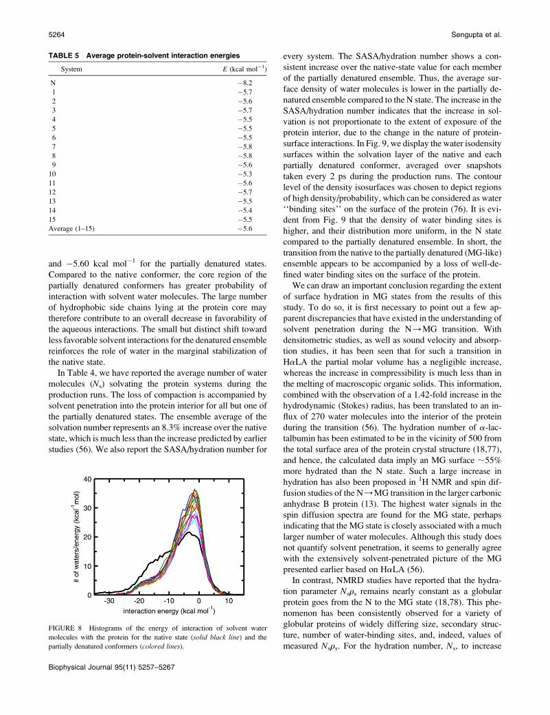
and �5.60 kcal mol�1 for the partially denatured states.
Compared to the native conformer, the core region of the
partially denatured conformers has greater probability of
interaction with solvent water molecules. The large number
of hydrophobic side chains lying at the protein core may
therefore contribute to an overall decrease in favorability of
the aqueous interactions. The small but distinct shift toward
less favorable solvent interactions for the denatured ensemble
reinforces the role of water in the marginal stabilization of
the native state.
In Table 4, we have reported the average number of water
molecules (Ns) solvating the protein systems during the
production runs. The loss of compaction is accompanied by
solvent penetration into the protein interior for all but one of
the partially denatured states. The ensemble average of the
solvation number represents an 8.3% increase over the native
state, which is much less than the increase predicted by earlier
studies (56). We also report the SASA/hydration number for
every system. The SASA/hydration number shows a con-
sistent increase over the native-state value for each member
of the partially denatured ensemble. Thus, the average sur-
face density of water molecules is lower in the partially de-
natured ensemble compared to the N state. The increase in the
SASA/hydration number indicates that the increase in sol-
vation is not proportionate to the extent of exposure of the
protein interior, due to the change in the nature of protein-
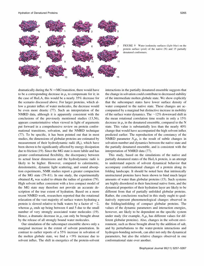
surface interactions. In Fig. 9, we display the water isodensity
surfaces within the solvation layer of the native and each
partially denatured conformer, averaged over snapshots
taken every 2 ps during the production runs. The contour
level of the density isosurfaces was chosen to depict regions
of high density/probability, which can be considered as water
‘‘binding sites’’ on the surface of the protein (76). It is evi-
dent from Fig. 9 that the density of water binding sites is
higher, and their distribution more uniform, in the N state
compared to the partially denatured ensemble. In short, the
transition from the native to the partially denatured (MG-like)
ensemble appears to be accompanied by a loss of well-de-
fined water binding sites on the surface of the protein.
We can draw an important conclusion regarding the extent
of surface hydration in MG states from the results of this
study. To do so, it is first necessary to point out a few ap-
parent discrepancies that have existed in the understanding of
solvent penetration during the N/MG transition. With
densitometric studies, as well as sound velocity and absorp-
tion studies, it has been seen that for such a transition in
HaLA the partial molar volume has a negligible increase,
whereas the increase in compressibility is much less than in
the melting of macroscopic organic solids. This information,
combined with the observation of a 1.42-fold increase in the
hydrodynamic (Stokes) radius, has been translated to an in-
flux of 270 water molecules into the interior of the protein
during the transition (56). The hydration number of a-lac-
talbumin has been estimated to be in the vicinity of 500 from
the total surface area of the protein crystal structure (18,77),
and hence, the calculated data imply an MG surface ;55%
more hydrated than the N state. Such a large increase in
hydration has also been proposed in 1H NMR and spin dif-
fusion studies of the N/MG transition in the larger carbonic
anhydrase B protein (13). The highest water signals in the
spin diffusion spectra are found for the MG state, perhaps
indicating that the MG state is closely associated with a much
larger number of water molecules. Although this study does
not quantify solvent penetration, it seems to generally agree
with the extensively solvent-penetrated picture of the MG
presented earlier based on HaLA (56).
In contrast, NMRD studies have reported that the hydra-
tion parameter Nsrs remains nearly constant as a globular
protein goes from the N to the MG state (18,78). This phe-
nomenon has been consistently observed for a variety of
globular proteins of widely differing size, secondary struc-
ture, number of water-binding sites, and, indeed, values of
measured Nsrs. For the hydration number, Ns, to increase
FIGURE 8 Histograms of the energy of interaction of solvent water
molecules with the protein for the native state (solid black line) and the
partially denatured conformers (colored lines).
TABLE 5 Average protein-solvent interaction energies
System E (kcal mol�1)
N �8.2
1 �5.7
2 �5.6
3 �5.7
4 �5.5
5 �5.5
6 �5.5
7 �5.8
8 �5.8
9 �5.6
10 �5.3
11 �5.6
12 �5.7
13 �5.5
14 �5.4
15 �5.5
Average (1–15) �5.6
5264 Sengupta et al.
Biophysical Journal 95(11) 5257–5267
dramatically during the N/MG transition, there would have
to be a corresponding decrease in rs to compensate for it; in
the case of HaLA, this would be a nearly 35% decrease for
the scenario discussed above. For larger proteins, which al-
low a greater influx of water molecules, the decrease would
be even more drastic (77). Such an interpretation of the
NMRD data, although it is apparently consistent with the
conclusions of the previously mentioned studies (13,56),
appears counterintuitive when viewed in light of arguments
put forward in a comprehensive review on protein confor-
mational transitions, solvation, and the NMRD technique
(77). To be specific, it has been pointed out that in most
studies, the dimensions of globular proteins are estimated by
measurement of their hydrodynamic radii (Rh), which have
been shown to be significantly affected by energy dissipation
due to friction (19). Since the MG state is more labile and has
greater conformational flexibility, the discrepancy between
its actual linear dimensions and the hydrodynamic radii is
likely to be higher. However, compared to calorimetric,
densitometric, dynamic light scattering, and sound absorp-
tion experiments, NMR studies report a greater compaction
of the MG state (79–81). In one study, the experimentally
obtained Rh was scaled to obtain the radius of gyration (79).
High solvent influx consistent with a less compact model of
the MG state may therefore not provide an accurate de-
scription of the true extent of hydration. Based on a more
recent NMRD work, researchers reported that the rotational
relaxation of the vast majority of surface waters hydrating a
protein is slowed relative to bulk waters by a factor of ;2,
whereas rs ends up being between 4 and 5 due to the small
number of very strongly associated water molecules (19).
Hence, a dramatic decrease in rs can only be brought about
by the release of all strongly bound water molecules.
Our simulation of the subcompact ensemble shows only a
marginal increase in the extent of solvent penetration. In
contrast to earlier reports of a 55% increase in solvation of
the molten globule state, we find a ,9% increase due to
solvent influx. The shift in energetics of the protein-solvent
interactions in the partially denatured ensemble suggests that
the change in solvation could contribute to decreased stability
of the intermediate molten globule state. We show explicitly
that the subcompact states have lower surface density of
water compared to the native state. These changes are ac-
companied by a marginal but distinctive increase in mobility
of the surface water dynamics. The ;12% downward shift in
the mean rotational correlation time results in only a 15%
decrease in rs in the denatured ensemble, compared to the N
state. This value is substantially less than the nearly 40%
change that would have accompanied the high solvent influx
predicted earlier. The reproduction of the constancy of the
NMRD parameter Nsrs is the result of subtle changes in
solvation number and dynamics between the native state and
the partially denatured ensemble, and is consistent with the
interpretation of NMRD data (77).
This study, based on the simulations of the native and
partially denatured states of the HaLA protein, is an attempt
to understand aspects of solvent dynamical behavior that
accompany conformational changes of a protein along its
folding landscape. It should be noted here that intrinsically
unstructured proteins have been shown to bind much larger
amounts of water than globular proteins (15). Such systems
are highly disordered in their functional native form, and the
dynamical properties of their hydration layer are likely to be
different from that of partially unfolded globular proteins.
Rather, the conclusions reached in this study ought to qual-
itatively represent phenomenological changes observed in
the folding/unfolding of compact globular proteins. The
values of the dynamic parameters and interaction energies,
however, are likely to be dependent on the specific system
under study (for example, Nsrs has different values for dif-
ferent globular proteins). Also, changes in the solvent envi-
ronment, such as those brought about by the addition of salt,
and by perturbations to the water-protein interactions and
hydrogen-bonding network, can alter not only the dynamical
parameters, but also the relative changes observed in one
conformational state over another.
FIGURE 9 Water isodensity surfaces (light blue) on the
protein surface (pink) of the native (N) and 15 partially
denatured conformers.
Hydration of Denatured Proteins 5265
Biophysical Journal 95(11) 5257–5267
This work was supported by the National Science Foundation (Grants
CHE-0417158 and CHE-0750175). We are grateful to Pinaki Sinha for
assistance with data analysis.
REFERENCES
1. Gast, K., D. Zirwer, H. Welfle, V. E. Bychkova, and O. B. Ptitsyn.1986. Quasielastic light scattering from human a-lactalbumin: com-parison of molecular dimensions in native and ‘‘molten globule’’states. Int. J. Biol. Macromol. 8:231–236.
2. Pal, S. K., J. Peon, B. Bagchi, and A. H. Zewail. 2002. Biologicalwater: femtosecond dynamics of macromolecular hydration. J. Phys.Chem. B. 106:12376–12395.
3. Sen, P., S. Mukherjee, P. Dutta, A. Halder, D. Mandal, R. Banerjee, S.Roy, and K. Bhattacharyya. 2003. Solvation dynamics in the moltenglobule state of a protein. J. Phys. Chem. B. 107:14563–14568.
4. Samaddar, S., A. K. Mandal, S. K. Mondal, K. Sahu, K. Bhattacharyya,and S. Roy. 2006. Solvation dynamics of a protein in the pre moltenglobule state. J. Phys. Chem. B. 110:21210–21215.
5. Zhang, L., L. Wang, Y.-T. Kao, W. Qiu, Y. Yang, O. Okobiah, and D.Zhong. 2007. Mapping hydration dynamics around a protein surface.Proc. Natl. Acad. Sci. USA. 104:18461–18466.
6. Ferrand, M., A. J. Dianoux, W. Petry, and G. Zaccai. 1993. Thermalmotions and function of bacteriorhodopsin in purple membranes:effects of temperature and hydration studied by neutron scattering.Proc. Natl. Acad. Sci. USA. 90:9668–9672.
7. Svergun, D. I., S. Richard, M. H. J. Koch, Z. Sayers, S. Kuprin, and G.Zaccai. 1998. Protein hydration in solution: experimental observation byx-ray and neutron scattering. Proc. Natl. Acad. Sci. USA. 95:2267–2272.
8. Teeter, M. M., A. Yamano, B. Stec, and U. Mohanty. 2001. On thenature of a glassy state of matter in a hydrated protein: relation toprotein function. Proc. Natl. Acad. Sci. USA. 98:11242–11247.
9. Doster, W., and M. Settles. 2005. Protein-water displacement distri-butions. Biochim. Biophys. Acta. 1749:173–186.
10. Wood, K., M. Plazanet, F. Gabel, B. Kessler, D. Oesterhelt, D. J.Tobias, G. Zaccai, and M. Weik. 2007. Coupling of protein andhydration-water dynamics in biological membranes. Proc. Natl. Acad.Sci. USA. 104:18049–18054.
11. Wood, K., A. Frolich, A. Paciaroni, M. Moulin, M. Hartlein, G. Zaccai,D. J. Tobias, and M. Weik. 2008. Coincidence of dynamical transitions ina soluble protein and its hydration water: direct measurements by neutronscattering and MD simulations. J. Am. Chem. Soc. 130:4586–4587.
12. Stanley, C., S. Krueger, V. A. Parsegian, and D. C. Rau. 2008. Proteinstructure and hydration probed by SANS and osmotic stress. Biophys.J. 94:2777–2789.
13. Kutyshenko, V. P., and M. Cortijo. 2000. Water-protein interactions inthe molten-globule state of carbonic anhydrase b: an NMR spin-diffusionstudy. Protein Sci. 9:1540–1547.
14. Krzystyniak, M., G. Shen, J. H. Golbeck, and M. L. Antonkine. 2008.Investigation of water bound to photosystem I with multiquantumfiltered 17O nuclear magnetic resonance. J. Chem. Phys. 128:014503–014515.
15. Bokor, M., V. Csizmok, D. Kovacs, P. Banki, P. Friedrich, P. Tompa,and K. Tompa. 2005. NMR relaxation studies on the hydrate layer ofintrinsically unstructured proteins. Biophys. J. 88:2030–2037.
16. Denisov, V. P., and B. Halle. 1995. Protein hydration dynamics inaqueous solution: a comparison of bovine pancreatic trypsin inhibitorand ubiquitin by oxygen-17 spin relaxation dispersion. J. Mol. Biol.245:682–697.
17. Denisov, V. P., and B. Halle. 1996. Protein hydration dynamics inaqueous solution. Faraday Disc. 103:227–244.
18. Denisov, V. P., B. H. Jonsson, and B. Halle. 1999. Hydration ofdenatured and molten globule proteins. Nat. Struct. Biol. 6:253–260.
19. Halle, B., and M. Davidovic. 2003. Biomolecular hydration: fromwater dynamics to hydrodynamics. Proc. Natl. Acad. Sci. USA. 100:12135–12140.
20. Modig, K., E. Liepinsh, G. Otting, and B. Halle. 2004. Dynamics ofprotein and peptide hydration. J. Am. Chem. Soc. 126:102–114.
21. Giuffrida, S., G. Cottone, and L. Cordone. 2006. Role of solvent onprotein-matrix coupling in MbCO embedded in water-saccharide sys-tems: a Fourier transform infrared spectroscopy study. Biophys. J.91:968–980.
22. Knab, J., J.-Y. Chen, and A. Markelz. 2006. Hydration dependence ofconformational dielectric relaxation of lysozyme. Biophys. J. 90:2576–2581.
23. Xu, J., K. W. Plaxco, and S. J. Allen. 2006. Probing the collectivevibrational dynamics of a protein in liquid water by terahertz absorp-tion spectroscopy. Protein Sci. 15:1175–1181.
24. Ebbinghaus, S., S. J. Kim, M. Heyden, X. Yu, U. Heugen, M.Gruebele, D. M. Leitner, and M. Havenith. 2007. An extended dynam-ical hydration shell around proteins. Proc. Natl. Acad. Sci. USA. 104:20749–20752.
25. Ebbinghaus, S., S. J. Kim, M. Heyden, X. Yu, M. Gruebele, D. M.Leitner, and M. Havenith. 2008. Protein sequence- and pH-dependenthydration probed by terahertz spectroscopy. J. Am. Chem. Soc. 130:2374–2375.
26. Lounnas, V., and B. M. Pettitt. 1994. Distribution function implieddynamics versus residence times and correlations: solvation shells ofmyoglobin. Proteins. 18:148–160.
27. Rocchi, C., A. R. Bizzarri, and S. Cannistraro. 1998. Water dynamicalanomalies evidenced by molecular-dynamics simulations at the sol-vent-protein interface. Phys. Rev. E. 57:3315–3325.
28. Marchi, M., F. Sterpone, and M. Ceccarelli. 2002. Water rotationalrelaxation and diffusion in hydrated lysozyme. J. Am. Chem. Soc.124:6787–6791.
29. Tarek, M., and D. J. Tobias. 1999. Environmental dependence of thedynamics of protein hydration water. J. Am. Chem. Soc. 121:9740–9741.
30. Pizzitutti, F., M. Marchi, F. Sterpone, and P. J. Rossky. 2007. How pro-tein surfaces induce anomalous dynamics of hydration water. J. Phys.Chem. B. 111:7584–7590.
31. Rhee, Y. M., E. J. Sorin, G. Jayachandran, E. Lindahl, and V. S. Pande.2004. Simulations of the role of water in the protein-folding mecha-nism. Proc. Natl. Acad. Sci. USA. 101:6456–6461.
32. Barth, P., T. Alber, and P. B. Harbury. 2007. Accurate, conformation-dependent predictions of solvent effects on protein ionization con-stants. Proc. Natl. Acad. Sci. USA. 104:4898–4903.
33. Vitkup, D., D. Ringel, G. A. Petsko, and M. Karplus. 2000. Solventmobility and the protein ‘‘glass’’ transition. Nat. Struct. Biol. 7:34–38.
34. Tarek, M., G. J. Martyna, and D. J. Tobias. 2000. Amplitudes andfrequencies of protein dynamics: analysis of discrepancies betweenneutron scattering and molecular dynamics simulations. J. Am. Chem.Soc. 122:10450–10451.
35. Heberle, J., J. Fitter, H. J. Sass, and G. Buldt. 2000. Bacteriorhodopsin:the functional details of a molecular machine are being resolved. Bio-phys. Chem. 85:229–248.
36. Mattos, C. 2002. Protein-water interactions in a dynamic world. TrendsBiochem. Sci. 27:203–208.
37. Papoian, G. A., J. Ulander, M. P. Eastwood, Z. Luthey-Schulten, andP. G. Wolynes. 2004. Water in protein structure prediction. Proc. Natl.Acad. Sci. USA. 101:3352–3357.
38. Daggett, V. 2006. Protein folding-simulation. Chem. Rev. 106:1898–1916.
39. Levy, Y., and J. N. Onuchic. 2006. Water mediation in protein foldingand molecular recognition. Annu. Rev. Biophys. Biomol. Struct. 35:389–415.
40. Cheung, M. S., A. E. Garcia, and J. N. Onuchic. 2002. Protein foldingmediated by solvation: water expulsion and formation of the hydro-phobic core occur after the structural collapse. Proc. Natl. Acad. Sci.USA. 99:685–690.
41. Harano, Y., and M. Kinoshita. 2004. Large gain in translationalentropy of water is a major driving force in protein folding. Chem.Phys. Lett. 399:342–348.
5266 Sengupta et al.
Biophysical Journal 95(11) 5257–5267
42. Jas, G. S., W. A. Eaton, and J. Hofrichter. 2001. Effect of viscosity onthe kinetics of a-helix and b-hairpin formation. J. Phys. Chem. B. 105:261–272.
43. Pradeep, L., and J. B. Udgaonkar. 2007. Diffusional barrier in theunfolding of a small protein. J. Mol. Biol. 366:1016–1028.
44. Dyson, H. J., and P. E. Wright. 1998. Equilibrium NMR studiesof unfolded and partially folded proteins. Nat. Struct. Mol. Biol. 5:499–503.
45. Cheung, M. S., L. L. Chavez, and J. N. Onuchic. 2004. The energylandscape for protein folding and possible connections to function.Polymer. 45:547–555.
46. Bandyopadhyay, S., S. Chakraborty, and B. Bagchi. 2006. Explorationof the secondary structure specific differential solvation dynamicsbetween the native and molten globule states of the protein HP-36.J. Phys. Chem. B. 110:20629–20634.
47. Chakraborty, S., and S. Bandyopadhyay. 2008. Dynamics of water inthe hydration layer of a partially unfolded structure of the proteinHP-36. J. Phys. Chem. B. 112:6500–6507.
48. Kuwajima, K. 1989. The molten globule state as a clue for under-standing the folding and cooperativity of globular-protein structure.Proteins. 6:87–103.
49. Ptitsyn, O. B. 1995. Molten globule and protein folding. Adv. ProteinChem. 47:83–229.
50. Wolynes, P. G., J. N. Onuchic, and D. Thirumalai. 1995. Navigatingthe folding routes. Science. 267:1619–1620.
51. Sutovsky, H., and E. Gazit. 2004. The von Hippel-Lindau tumorsuppressor protein is a molten globule under native conditions: implica-tions for its physiological activities. J. Biol. Chem. 279:17190–17196.
52. Mahley, R. W., and Y. Huang. 2006. Apolipoprotein (apo) E4 andAlzheimer’s disease: unique conformational and biophysical propertiesof apoE4 can modulate neuropathology. Acta. Neurolog. Scand. 114:8–14.
53. Tarek, M., and D. J. Tobias. 2000. The dynamics of protein hydrationwater: a quantitative comparison of molecular dynamics simulationsand neutron-scattering experiments. Biophys. J. 79:3244–3257.
54. Abseher, R., H. Schreiber, and O. Steinhauser. 1996. The influence of aprotein on water dynamics in its vicinity investigated by moleculardynamics simulation. Proteins. 25:366–378.
55. Settles, M., and W. Doster. 1996. Anomalous diffusion of adsorbedwater: a neutron scattering study of hydrated myoglobin. FaradayDisc. 103:269–279.
56. Kharakoz, D. P., and V. E. Bychkova. 1997. Molten globule of humana-lactalbumin: hydration, density, and compressibility of the interior.Biochemistry. 36:1882–1890.
57. Fink, A. L. 1995. Compact intermediate states in protein folding. Annu.Rev. Biophys. Biomol. Struct. 24:495–522.
58. Paci, E., L. J. Smith, C. M. Dobson, and M. Karplus. 2001. Explorationof partially unfolded states of human a-lactalbumin by moleculardynamics simulation. J. Mol. Biol. 306:329–347.
59. Acharya, K. R., J. Ren, D. I. Stuart, D. C. Phillips, and R. E. Fenna.1991. Crystal structure of human a-lactalbumin at 1.7 A resolution.J. Mol. Biol. 221:571–581.
60. Qiu, D., P. S. Shenkin, F. P. Hollinger, and W. C. Still. 1997. The GB/SAcontinuum model for solvation. A fast analytical method for the calcu-lation of approximate Born radii. J. Phys. Chem. A. 101:3005–3014.
61. Kuwajima, K., Y. Hiraoka, M. Ikeguchi, and S. Sugai. 1985. Comparisonof the transient folding intermediates in lysozyme and a-lactalbumin.Biochemistry. 24:874–881.
62. Berendsen, H. J. C., J. R. Grigera, and T. P. Straatsma. 1987. Themissing term in effective pair potentials. J. Phys. Chem. 91:6269–6271.
63. Kale, L., R. Skeel, M. Bhandarkar, R. Brunner, A. Gursoy, N. Krawetz,J. Phillips, A. Shinozaki, K. Varadarajan, and K. Schulten. 1999.NAMD2: greater scalability for parallel molecular dynamics. J. Com-put. Phys. 151:283–312.
64. Brooks B. R., R. E. Bruccoleri, B. D. Olafson, D. J. States, S.Swaminathan, and M. Karplus. 1983. CHARMM: a program formacromolecular energy, minimization, and dynamics calculations.J. Comput. Chem. 4:187–217.
65. Martyna, G. J., D. J. Tobias, and M. L. Klein. 1994. Constant pressuremolecular dynamics algorithms. J. Chem. Phys. 101:4177–4189.
66. Feller S. E., Y. Zhang, R. W. Pastor, and B. R. Brooks. 1995. Constantpressure molecular dynamics simulation: the Langevin piston method.J. Chem. Phys. 103:4613–4621.
67. Martyna, G. J., M. E. Tuckerman, D. J. Tobias, and M. L. Klein. 1996.Explicit reversible integrators for extended systems dynamics. Mol.Phys. 87:1117–1157.
68. Essmann, U., L. Perera, M. L. Berkowitz, T. Darden, H. Lee, and L. G.Pedersen. 1995. A smooth particle mesh Ewald method. J. Chem. Phys.103:8577–8593.
69. Humphrey, W., A. Dalke, and K. Schulten. 1996. VMD: visual mole-cular dynamics. J. Mol. Graph. 14:33–38.
70. Halle, B., and H. Wennerstroem. 1981. Nearly exponential quadrupolarrelaxation. A perturbation treatment. J. Magn. Reson. 44:89–100.
71. Denisov, V. P., K. Venu, J. Peters, H. D. Horlein, and B. Halle. 1997.Orientational disorder and entropy of water in protein cavities. J. Phys.Chem. B. 101:9380–9389.
72. Denisov, V. P., G. Carlstrom, K. Venu, and B. Halle. 1997. Kinetics ofDNA hydration. J. Mol. Biol. 268:118–136.
73. Denisov, V. P., J. L. Schlessman, B. E. Garcia-Moreno, and B. Halle.2004. Stabilization of internal charges in a protein: water penetration orconformational change? Biophys. J. 87:3982–3994.
74. Burling, F. T., W. I. Weis, K. M. Flaherty, and A. T. Brunger. 1996.Direct observation of protein solvation and discrete disorder withexperimental crystallographic phases. Science. 271:72–77.
75. Merzel, F., and J. C. Smith. 2002. Is the first hydration shell oflysozyme of higher density than bulk water? Proc. Natl. Acad. Sci.USA. 99:5378–5383.
76. Makarov, V., B. M. Pettitt, and M. Feig. 2002. Solvation and hydrationof proteins and nucleic acids: a theoretical view of simulation andexperiment. Acc. Chem. Res. 35:376–384.
77. Halle, B., V. P. Denisov, K. Modig, and M. Davidoc. 2005. Proteinconformational transitions as seen from the solvent: magnetic relaxa-tion dispersion studies of water, co-solvent, and denaturant interactionswith nonnative proteins. In Protein Folding Handbook. J. Buchner andT. Kiefhaber, editors. Wiley-VCH, Weinheim, Germany. 201–242.
78. Shortle, D. 1999. Protein folding as seen from water’s perspective. Nat.Struct. Biol. 6:203–205.
79. Redfield, C., B. A. Schulman, M. A. Milhollen, P. S. Kim, and C. M.Dobson. 1999. a-Lactalbumin forms a compact molten globule in theabsence of disulfide bonds. Nat. Struct. Biol. 6:948–952.
80. Balbach, J. 2000. Compaction during protein folding studied by real-time NMR diffusion experiments. J. Am. Chem. Soc. 122:5887–5888.
81. Chakraborty, S., V. Ittah, P. Bai, L. Luo, E. Haas, and Z. Y. Peng.2001. Structure and dynamics of the a-lactalbumin molten globule:fluorescence studies using proteins containing a single tryptophanresidue. Biochemistry. 40:7228–7238.
Hydration of Denatured Proteins 5267
Biophysical Journal 95(11) 5257–5267