Semaphorin 3F forms an anti-angiogenic barrier in outer retina
NATURE MEDICINE • VOLUME 8 • NUMBER 3 • MARCH 2002 225
ARTICLES
Kaposi sarcoma (KS) is an angio-proliferative disease character-ized by angiogenesis, endothelial spindle-cell growth (KS cells),inflammatory-cell infiltration and edema1,2. KS is associatedwith human herpesvirus 8 (HHV8) infection, and is particu-larly frequent and aggressive in HIV-1/HHV8 co-infected indi-viduals (AIDS-KS)1–3. KS development is associated with HHV8reactivation and spread to blood and tissues. Reactivation, inturn, is induced by Th-1 type cytokines (interferon-γ, inter-leukin-1β and tumor necrosis factor-α) that are increased in KSpatients or in at-risk individuals1,2,4. Inflammatory cytokines(ICs) also activate vessels promoting the tissue extravasation ofinfected lympho-monocytes and spindle-cell progenitors1,2. Inearly-developing lesions, HHV8 load is low or undetectablewhereas it is high in late-nodular lesions, which mostly showlatent infection1,2,5. This finding suggests that HHV8 may havea key role in KS progression to a frank tumor as latently ex-pressed viral genes may induce cell transformation2, a featureobserved in some late-nodular AIDS-KS lesions6. Increase of ICscan also induce production of angiogenic factors and othermolecules that initiate lesion formation, as observed in HIV-1-infected individuals that develop KS or show KS progressionafter systemic administration of interferon-γ and interleukin-2or tumor necrosis factor-α7. Other studies confirm that highHHV8 burden and expression of ICs and angiogenic factors arefound in all forms of KS (refs. 1,2,7).
Basic fibroblast growth factor (bFGF) and vascular endothe-lial growth factor (VEGF) are the most highly expressed angio-genic factors8–10. They are also increased in sera of KS patientsor in at-risk individuals11,12, and are expressed in early lesions
by KS cells, endothelial cells and infiltrating cells in responseto ICs (refs. 1,7,8,10,13,14). bFGF promotes in autocrine fash-ion the growth of KS cells and synergizes with VEGF to mediatethe development of angioproliferative KS-like lesions inducedby the inoculation of KS cells in nude mice8,10,13,15–17. Such KS-like lesions are composed of mouse cells and are induced by thefactors produced by KS cells, and they are highly vascularized,closely resemble early KS and regress as KS regresses in hu-mans1,7. Conversely, inoculation with bFGF in nude mice andits in vivo induction with ICs reproduces the vascular lesionspromoted in mice by the inoculation of KS cells and thosefound in human KS (refs. 8,14). Finally, bFGF synergizes withthe HIV-1 Tat protein released by infected cells and increasesthe frequency and aggressiveness of KS in HIV-1-infected indi-viduals8,18,19. Thus, bFGF and VEGF are crucial to the develop-ment and progression of all forms of KS as well as to the growthof solid tumors20.
Recent reports have described a reduced incidence21 or theregression22,23 of KS in HIV-1-infected patients treated withhighly active antiretroviral therapy (HAART) that includes atleast one HIV-protease inhibitor (PI) such as indinavir orsaquinavir24. PIs have also been shown to directly affect cellmetabolism, interfere with host or fungal proteases and blockT-cell activation and dendritic-cell function25,32, even thoughthey were designed to selectively interfere with the catalyticsite of HIV protease24. Given that proteases are also essentialfor angiogenic and inflammatory processes and for tumorgrowth, we reasoned that at least part of the lower incidenceand regression of KS observed in PI-treated patients resulted
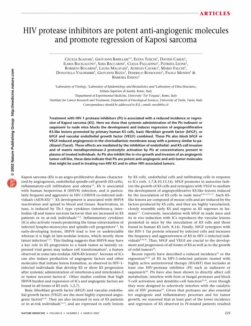
HIV protease inhibitors are potent anti-angiogenic moleculesand promote regression of Kaposi sarcoma
CECILIA SGADARI1, GIOVANNI BARILLARI1,4, ELENA TOSCHI1, DAVIDE CARLEI1, ILARIA BACIGALUPO1, SARA BACCARINI1, CLELIA PALLADINO1, PATRIZIA LEONE1, ROBERTO BUGARINI2, LAURA MALAVASI1, AURELIO CAFARO1, MARIO FALCHI3,
DONATELLA VALDEMBRI5, GIOVANNI REZZA2, FEDERICO BUSSOLINO5, PAOLO MONINI1 &BARBARA ENSOLI1
1Laboratory of Virology, 2Laboratory of Epidemiology and Biostatistics and 3Laboratory of Ultra Structures,Istituto Superiore di Sanità, Rome, Italy
4Department of Experimental Medicine, University ‘Tor Vergata’, Rome, Italy5Institute for Cancer Research and Treatment, Department of Oncological Sciences, University of Turin, Turin, Italy
Correspondence should be addressed to B.E.; email: [email protected]
Treatment with HIV-1 protease inhibitors (PI) is associated with a reduced incidence or regres-sion of Kaposi sarcoma (KS). Here we show that systemic administration of the PIs indinavir orsaquinavir to nude mice blocks the development and induces regression of angioproliferativeKS-like lesions promoted by primary human KS cells, basic fibroblast growth factor (bFGF), orbFGF and vascular endothelial growth factor (VEGF) combined. These PIs also block bFGF orVEGF-induced angiogenesis in the chorioallantoic membrane assay with a potency similar to pa-clitaxel (Taxol). These effects are mediated by the inhibition of endothelial- and KS-cell invasionand of matrix metalloproteinase-2 proteolytic activation by PIs at concentrations present inplasma of treated individuals. As PIs also inhibit the in vivo growth and invasion of an angiogenictumor-cell line, these data indicate that PIs are potent anti-angiogenic and anti-tumor moleculesthat might be used in treating non-HIV KS and in other HIV-associated tumors.
©20
02 N
atu
re P
ub
lish
ing
Gro
up
h
ttp
://m
edic
ine.
nat
ure
.co
m
226 NATURE MEDICINE • VOLUME 8 • NUMBER 3 • MARCH 2002
ARTICLES
from direct anti-KS and/or anti-angiogenic effect(s) of thesedrugs.
To test this hypothesis, we used in vitro and in vivo models ofangiogenesis, KS-lesion formation and tumor growth that aredevoid of any viral agents and T cells (that is, nude mice andchorioallantoic membrane (CAM) assays), and are establishedpreclinical models to evaluate the efficacy of anti-KS and anti-angiogenic therapies8,15,33,34. Here we show that PIs have directanti-angiogenic, anti-KS and anti-tumor activity at concentra-tions present in plasma of treated individuals.
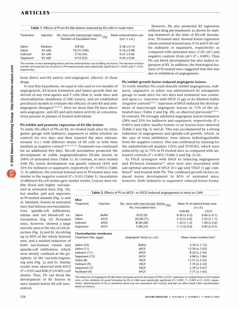
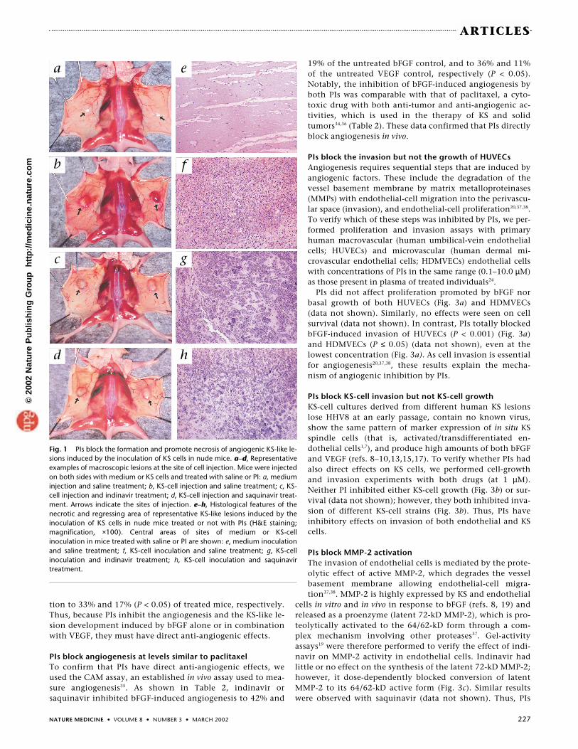
PIs inhibit and promote regression of KS-like lesionsTo study the effect of PIs on KS, we treated nude mice by intra-gastric gavage with indinavir, saquinavir or saline solution (ascontrol) for two days and then injected the mice subcuta-neously (s.c.) with different strains of KS cells or with theirmedium as negative control5,8,10,14,15,34. Treatment was continuedfor an additional five days. KS-cell inoculation promoted thedevelopment of visible angioproliferative KS-like lesions in100% of untreated mice (Table 1). In contrast, in mice treatedwith PIs, lesion development was greatly reduced (43% and25% for indinavir or saquinavir, respectively (P < 0.001)) (Table1). In addition, the external lesional area in PI-treated mice wassimilar to the negative control (P < 0.01) (Table 1). Inoculationof different KS-cell strains gave similar results. Lesions were vis-ibly florid and highly vascular-ized in untreated mice (Fig. 1b),but smaller, pail and regressivein PI-treated animals (Fig. 1c andd). Similarly, lesions in untreatedmice had intense neovasculariza-tion, spindle-cell infiltration,edema and red blood-cell ex-travasation (Fig. 1f). PI-treatedmice, however, showed a largenecrotic area at the site of cell in-jection (Fig. 1g and h), involvingup to 85% of the whole lesionalarea, and a marked reduction ofboth neo-formed vessels andspindle-cell infiltration, whichwere mostly confined at the pe-riphery of the necrotic/regress-ing area (Fig. 1g and h). Similarresults were observed with KS12(P = 0.05) and KS8 (P ≤ 0.001) cellstrains. Thus, PIs can block thedevelopment of KS lesions inmice treated before KS-cell inoc-ulation.
However, PIs also promoted KS regressionwithout drug pre-treatment, as shown by start-ing treatment at the time of KS-cell inocula-tion. PI-treated mice showed lesion regression(mean external lesional area: 0.54 and 0.50 cm2
for indinavir or saquinavir, respectively) ascompared with untreated mice (1.02 cm2) andnegative controls (0.66 cm2) (P < 0.001). ThusPIs can block development but also induce re-gression of KS. In addition, the histological fea-tures of PI-treated mice suggested that this wasdue to inhibition of angiogenesis.
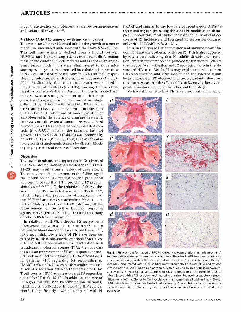
PIs inhibit growth factor–induced angiogenic lesionsTo verify whether PIs could directly inhibit angiogenesis, indi-navir, saquinavir or saline was administered by intragastricgavage to nude mice for two days and treatment was contin-ued upon s.c. injection with bFGF (1 µg) or its dilution buffer(negative control)8,10,14. Injection of bFGF induced the develop-ment of macroscopic angiogenic lesions in 71% of the un-treated mice (Table 2 and Fig. 2b), as observed previously8,10,14.In contrast, PIs strongly inhibited angiogenic lesion formation(28% and 25% for indinavir and saquinavir, respectively (P <0.001)) and either smaller lesions or no lesions were observed(Table 2 and Fig. 2c and d). This was accompanied by a strongreduction of angiogenesis and spindle-cell growth, which, inthe case of total inhibition, were almost indistinguishablefrom the negative control. This was confirmed by staining forthe endothelial-cell markers CD31 and FVIII-RA, which werereduced by up to 75% in PI-treated mice as compared with un-treated controls (P < 0.001) (Table 2 and Fig. 2e–h).
As VEGF synergizes with bFGF in inducing angiogenesisand KS-lesion formation10, mice were also inoculated withsub-optimal amounts of bFGF (0.1 µg) and VEGF (1 µg) com-bined10 and treated with PIs. The combined growth factors in-duced lesion development in 83% of untreated mice;however, both indinavir or saquinavir reduced lesion forma-
Table 1 Effects of PI on KS-like lesions induced by KS cells in nude mice
Treatment Injection No. mice with macroscopic lesions (%)
Mean external lesion sizeNumber of inoculated mice (cm2 ± s.d.)
Saline Medium 0/8 (0) 0.58 ± 0.13Saline KS cells 14/14 (100) 0.76 ± 0.08Indinavir KS cells 7/16 (43) 0.57 ± 0.06Saquinavir KS cells 4/16 (25) 0.56 ± 0.06
The number of mice developing lesions and the external lesion size at killing are shown. The decrease of lesionnumber and external size of lesions in PI-treated animals were statistically significant (P < 0.001 and P < 0.01,respectively).
Table 2 Effects of PI on bFGF- or VEGF-induced angiogenesis in mice or CAM
MiceTreatment Injection No. mice with macroscopic lesions (%) Mean % of stained lesion area
No. inoculated mice (± s.d.)FVIII-RA CD31
Saline Buffer 0/22 (0) 0.20 (± 0.2) 0.06 (± 0.1)Saline bFGF 20/28 (71) 4.32 (± 2.0) 3.45 (± 1.7)Indinavir bFGF 8/28 (28) 1.32 (± 1.2) 1.00 (± 0.6)Saquinavir bFGF 7/28 (25) 1.13 (± 0.6) 0.83 (± 0.3)
Chorioallantoic membraneTreatment (No. eggs) Angiogenic factor (± s.d.) Mean vessel number/mm2
Saline (23) Buffer 4.39 (± 1.12)Saline (11) bFGF 13.50 (± 3.03)Indinavir (11) bFGF 8.22 (± 2.54)Saquinavir (13) bFGF 6.08 (± 1.84)Saline (8) VEGF 13.51 (± 2.42)Indinavir (9) VEGF 7.70 (± 2.52)Saquinavir (7) VEGF 5.39 (± 0.67)Paclitaxel (4) bFGF 7.71 (± 1.63)
The reduction of angiogenic KS-like lesion formation and the decrease of FVIII or CD31 expression in residual lesions of PI-treatedmice, or the inhibition of vessel formation by PIs in CAM were statistically significant (P < 0.001, P < 0.001 or P < 0.05, respec-tively). Administration of PIs or paclitaxel alone was not associated with toxicity and did not affect basal CAM vascularization(data not shown).
©20
02 N
atu
re P
ub
lish
ing
Gro
up
h
ttp
://m
edic
ine.
nat
ure
.co
m
NATURE MEDICINE • VOLUME 8 • NUMBER 3 • MARCH 2002 227
ARTICLES
tion to 33% and 17% (P < 0.05) of treated mice, respectively.Thus, because PIs inhibit the angiogenesis and the KS-like le-sion development induced by bFGF alone or in combinationwith VEGF, they must have direct anti-angiogenic effects.
PIs block angiogenesis at levels similar to paclitaxelTo confirm that PIs have direct anti-angiogenic effects, weused the CAM assay, an established in vivo assay used to mea-sure angiogenesis35. As shown in Table 2, indinavir orsaquinavir inhibited bFGF-induced angiogenesis to 42% and
19% of the untreated bFGF control, and to 36% and 11%of the untreated VEGF control, respectively (P < 0.05).Notably, the inhibition of bFGF-induced angiogenesis byboth PIs was comparable with that of paclitaxel, a cyto-toxic drug with both anti-tumor and anti-angiogenic ac-tivities, which is used in the therapy of KS and solidtumors34,36 (Table 2). These data confirmed that PIs directlyblock angiogenesis in vivo.
PIs block the invasion but not the growth of HUVECsAngiogenesis requires sequential steps that are induced byangiogenic factors. These include the degradation of thevessel basement membrane by matrix metalloproteinases(MMPs) with endothelial-cell migration into the perivascu-lar space (invasion), and endothelial-cell proliferation20,37,38.To verify which of these steps was inhibited by PIs, we per-formed proliferation and invasion assays with primaryhuman macrovascular (human umbilical-vein endothelialcells; HUVECs) and microvascular (human dermal mi-crovascular endothelial cells; HDMVECs) endothelial cellswith concentrations of PIs in the same range (0.1–10.0 µM)as those present in plasma of treated individuals24.
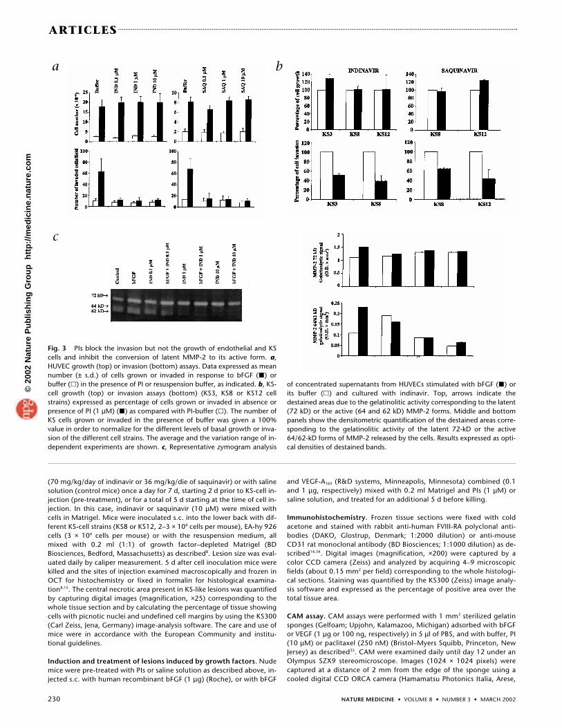
PIs did not affect proliferation promoted by bFGF norbasal growth of both HUVECs (Fig. 3a) and HDMVECs(data not shown). Similarly, no effects were seen on cellsurvival (data not shown). In contrast, PIs totally blockedbFGF-induced invasion of HUVECs (P < 0.001) (Fig. 3a)and HDMVECs (P ≤ 0.05) (data not shown), even at thelowest concentration (Fig. 3a). As cell invasion is essentialfor angiogenesis20,37,38, these results explain the mecha-nism of angiogenic inhibition by PIs.
PIs block KS-cell invasion but not KS-cell growthKS-cell cultures derived from different human KS lesionslose HHV8 at an early passage, contain no known virus,show the same pattern of marker expression of in situ KSspindle cells (that is, activated/transdifferentiated en-dothelial cells1,7), and produce high amounts of both bFGFand VEGF (refs. 8–10,13,15,17). To verify whether PIs hadalso direct effects on KS cells, we performed cell-growthand invasion experiments with both drugs (at 1 µM).Neither PI inhibited either KS-cell growth (Fig. 3b) or sur-vival (data not shown); however, they both inhibited inva-sion of different KS-cell strains (Fig. 3b). Thus, PIs haveinhibitory effects on invasion of both endothelial and KScells.
PIs block MMP-2 activationThe invasion of endothelial cells is mediated by the prote-olytic effect of active MMP-2, which degrades the vesselbasement membrane allowing endothelial-cell migra-tion37,38. MMP-2 is highly expressed by KS and endothelial
cells in vitro and in vivo in response to bFGF (refs. 8, 19) andreleased as a proenzyme (latent 72-kD MMP-2), which is pro-teolytically activated to the 64/62-kD form through a com-plex mechanism involving other proteases37. Gel-activityassays19 were therefore performed to verify the effect of indi-navir on MMP-2 activity in endothelial cells. Indinavir hadlittle or no effect on the synthesis of the latent 72-kD MMP-2;however, it dose-dependently blocked conversion of latentMMP-2 to its 64/62-kD active form (Fig. 3c). Similar resultswere observed with saquinavir (data not shown). Thus, PIs
Fig. 1 PIs block the formation and promote necrosis of angiogenic KS-like le-sions induced by the inoculation of KS cells in nude mice. a–d, Representativeexamples of macroscopic lesions at the site of cell injection. Mice were injectedon both sides with medium or KS cells and treated with saline or PI: a, mediuminjection and saline treatment; b, KS-cell injection and saline treatment; c, KS-cell injection and indinavir treatment; d, KS-cell injection and saquinavir treat-ment. Arrows indicate the sites of injection. e–h, Histological features of thenecrotic and regressing area of representative KS-like lesions induced by theinoculation of KS cells in nude mice treated or not with PIs (H&E staining;magnification, ×100). Central areas of sites of medium or KS-cell inoculation in mice treated with saline or PI are shown: e, medium inoculationand saline treatment; f, KS-cell inoculation and saline treatment; g, KS-cell inoculation and indinavir treatment; h, KS-cell inoculation and saquinavir treatment.
a
b
c
d
e
f
g
h
©20
02 N
atu
re P
ub
lish
ing
Gro
up
h
ttp
://m
edic
ine.
nat
ure
.co
m
228 NATURE MEDICINE • VOLUME 8 • NUMBER 3 • MARCH 2002
ARTICLES
block the activation of proteases that are key for angiogenesisand tumor-cell invasion37,38.
PIs block EA-hy 926 tumor growth and cell invasionTo determine whether PIs could inhibit the growth of a tumormodel, we inoculated nude mice with the EA-hy 926 cell line.This cell line, which is derived from a hybrid betweenHUVECs and human lung adenocarcinoma cells39, retainsmost of the endothelial-cell markers and is used as an angio-genic tumor model40. PIs were administered to nude micestarting two days before tumor-cell inoculation. Tumors arosein 83% of untreated mice but only in 33% and 25%, respec-tively, of mice treated with indinavir or saquinavir (P < 0.05)(Table 3). Similarly, the external tumor area was reduced inmice treated with both PIs (P < 0.05), reaching the size of thenegative controls (Table 3). Residual tumors in treated ani-mals showed a strong reduction of both tumorgrowth and angiogenesis as determined histologi-cally and by staining with anti-FVIII-RA or anti-CD31 antibodies as compared with controls (P <0.001) (Table 3). Inhibition of tumor growth wasalso observed in the absence of drug pre-treatment.In these animals, external tumor size was reducedby more than 50% as compared with untreated con-trols (P < 0.001). Finally, the invasion but notgrowth of EA-hy 926 cells (Table 3) was inhibited byboth PIs (at 1 µM) (P < 0.05). Thus, PIs can inhibit invivo growth of angiogenic tumors by directly block-ing angiogenesis and tumor-cell invasion.
DiscussionThe lower incidence and regression of KS observedin HIV-1-infected individuals treated with PIs (refs.21–23) may result from a variety of drug effects.These may include one or more of the following: 1)the inhibition of HIV replication and productionand release of the HIV-1 Tat protein, a KS progres-sion factor5,8,14,18,40,41; 2) the reduction of the synthe-sis of ICs by HIV-1-infected or activated T cells28,30,42,which triggers the production of angiogenic fac-tors1,2,5,7,10,13,14 and HHV8 reactivation1,2,4; 3) the di-rect inhibitory effects on HHV8 infection; 4) theimprovement of protective immune responsesagainst HHV8 (refs. 1,43,44); and 5) direct blockingeffects on KS-lesion formation.
In relation to HHV8, although KS regression isoften associated with a reduction of HHV8 load inperipheral blood mononuclear cells and tissues1,22,23,no direct inhibitory effects of PIs have been de-tected by us (data not shown) or others45 on HHV8-infected cells before or after virus reactivation withtetradecanoyl phorbol acetate (TPA). Previous dataindicate an improvement of T-cell responses or nat-ural killer–cell activity against HHV8-infected cellsin patients with regressing KS responding toHAART (refs. 1,43). However, other studies indicatea lack of association between the increase of CD4+
T-cell counts, HIV-1 suppression and KS regressionupon HAART (refs. 46,47). In addition, the rate ofKS regression with non PI-combination therapies,which are still efficacious in blocking HIV replica-tion48, is significantly lower as compared with PI
HAART and similar to the low rate of spontaneous AIDS-KSregression in years preceding the use of PI-combination thera-pies49. By contrast, most studies indicate that a significant de-crease of KS incidence and increased KS regression occurredonly with PI HAART (refs. 21–23).
Thus, in addition to HIV suppression and immunoreconstitu-tion, PIs must exert other activities on KS. This is also suggestedby recent data indicating that PIs inhibit dendritic-cell func-tion, antigen presentation and proteosome function25,29, effectsthat reduce T-cell activation and IC production also in the ab-sence of HIV (refs. 30,42). This may explain the reduction ofHHV8 reactivation and virus load22,23 and the lowered serumlevels of bFGF (ref. 12) observed in PI-treated patients. However,this also suggests that the effect of PIs on KS may be largely de-pendent on direct and unknown effects of these drugs.
We have shown here that PIs have direct anti-angiogenic,
Fig. 2 PIs block the formation of bFGF-induced angiogenic lesions in nude mice. a–d,Representative examples of macroscopic lesions at the site of bFGF injection. a, Mice in-jected on both sides with buffer and treated with saline. b, Mice injected on both sideswith bFGF and treated with saline. c, Mice injected on both sides with bFGF and treatedwith indinavir. d, Mice injected on both sides with bFGF and treated with saquinavir, re-spectively. e–h, Representative examples of CD31 expression at the injection sites ofmice injected with bFGF or buffer and treated with saline, indinavir or saquinavir (mag-nification, ×100). e, Site of buffer inoculation in a mouse treated with saline. f, Site ofbFGF inoculation in a mouse treated with saline. g, Site of bFGF inoculation of in amouse treated with indinavir. h, Site of bFGF inoculation of a mouse treated withsaquinavir.
a
b
c
d
e
f
g
h
©20
02 N
atu
re P
ub
lish
ing
Gro
up
h
ttp
://m
edic
ine.
nat
ure
.co
m

NATURE MEDICINE • VOLUME 8 • NUMBER 3 • MARCH 2002 229
ARTICLES
anti-KS and anti-tumor effects. In particular, the in vitro andin vivo models of angiogenesis, KS-lesion formation andtumor growth used here contain neither HIV nor HHV8, andthey are observed in vivo in nude mice or CAM assays and,therefore, in the absence of T cells. This combination of fea-tures supports a direct anti-angiogenic and anti-tumor effectof these drugs, which may contribute to the inhibition of KSseen in treated patients. In fact, by inhibiting endothelial-cellinvasion, PIs block angiogenesis and tumor growth. Notably,PIs block the angiogenesis mediated by both bFGF and VEGF,which are the two key factors for KS-lesion formation.However, PIs did not block the production or release of bFGFor VEGF by KS cells (data not shown), as already suggested bythe lack of effects on KS-cell growth, which requires bFGF inautocrine fashion1,7,15.
bFGF and/or VEGF are also produced by most tumors tosupport their own angiogenesis20. Blocking endothelial-cellinvasion is the basis of current anti-angiogenic and anti-tumor therapies20,37,38. However, PIs also directly inhibit KS-and tumor-cell invasion and therefore exert an additional ef-fect. The inhibitory effect of PIs is similar to that observedwith paclitaxel, a microtuble-binding drug with cytotoxicand anti-angiogenic activity used in the therapy of KS andsolid tumors34,36. PIs also inhibited the growth of EA-hy 926tumor cells in nude mice. Finally, PIs not only blocked thedevelopment of KS or tumors in mice treated with the drugsbefore cell inoculation, but they were also effective whengiven at the time of cell inoculation. This suggests that PIscan both prevent development and induce regression of KS ortumors, in agreement with both the lower incidence and theregression of KS seen with PI HAART.
Endothelial-cell invasion requires the degradation of thebasement membrane by MMP-2 that is produced by endothe-lial cells in response to angiogenic factors8,18,19,37. MMP-2 isalso produced by KS cells and highly expressed in human KSlesions19, and it is required by tumor cells to invade tissues37.Blocking MMP-2 leads to inhibition of endothelial-cell inva-sion and, consequently, angiogenesis37,38. Similarly, treatmentof endothelial cells with PIs was found to reduce the bFGF-in-duced activation of pro-MMP-2 to its active forms. Thus, al-
though other proteases may be involved, inhibition of MMP-2 activity may be one of the mechanisms by which PIs inhibitcell invasion. However, no direct inhibitory effects of PIswere observed on recombinant activated MMP-2 (data notshown). In fact, MMP-2 cleaves after a glycine, whereas theHIV protease is an aspartyl protease, and MMP-2 has no se-quence homology with the HIV protease catalytic site26 tar-geted by PIs (data not shown). Thus, PIs may inhibit one ormore steps leading to MMP-2 activation.
These results indicate that PIs are promising anti-angio-genic and anti-tumor compounds, and, in addition to AIDSpatients with KS or at risk of KS, they should also be evaluatedfor the treatment of other tumors in HIV-infected individualsand for the therapy of KS in seronegative individuals. In par-ticular, PIs may represent a pathogenetic therapy for all formsof KS given that—in addition to directly blocking angiogene-sis—they also reduce T-cell activation and production of ICs(refs. 30,42). Consequently, they inhibit HHV8 reactivationand angiogenic factor production1,2,7,10,13,18,22,23. In addition,combination therapies containing at least one PI should beevaluated for the treatment of late-nodular KS—an aggressivecancer that is often refractory to chemotherapy alone, as sug-gested by the remission of AIDS-KS by associating PIs with pa-clitaxel50. By analogy, PIs should be investigated, alone or incombination with other drugs, for the therapy of other tu-mors in HIV-1-infected individuals.
MethodsCell cultures. HUVECs and HDMVECs (BioWhittaker, Verviers, Belgium)(passages 3–7), primary AIDS-KS spindle-cell cultures (KS3, KS8 and KS12,passages 6–13) and the EA-hy 926 cell line, a HUVEC–adenocarcinoma im-mortalized cell hybrid, were derived and cultured as described13,14,18,39-41.
Induction and treatment of lesions induced by tumor cells. The sameindinavir or saquinavir formulation (Merck Sharp & Dohme, Haarlem,the Netherlands, and Roche, Hertfordshire, Great Britain, respectively)administered to AIDS patients was dissolved in 0.4 ml saline solution andadministered by intragastric gavage to nude mice (5–6-wk-old femaleBALB/c nu/nu from Charles River, Calco, Italy). No systemic nor organtoxicity was ever observed in PI-treated animals (data not shown). Micewere treated with the same doses of PIs given to HIV-infected patients24
Table 3 Effects of PI on angiogenic tumors induced by EA-hy 926 cells
Mice
Injection Treatment No. mice with macroscopic lesions (%)
External lesion size Mean % stained tissue areaNo. inoculated mice (cm2 ± s.d.) (± s.d.)
FVIII-RA CD31Medium Saline 0/8 (0) 0.58 ± 0.15 0 0EA-hy 926 Saline 10/12 (83.3) 0.71 ± 0.2 2.91 (± 1) 2.30 (± 1.7)EA-hy 926 Indinavir 4/12 (33.3) 0.63 ± 0.17 1.01 (± 1.5) 0.70 (± 0.6)EA-hy 926 Saquinavir 3/12 (25) 0.55 ± 0.14 0.71 (± 0.8) 0.08 (± 0.1)
Cell invasion
Stimulus Treatment Mean invaded cells/field (± variation range)
Buffer Buffer 8.5 (± 0.5)bFGF Buffer 17 (± 4)bFGF Indinavir 13 (± 3)bFGF Saquinavir 9 (± 1)
The decrease of macroscopic angiogenic tumor lesion formation, external lesion size, and FVIII-RA or CD31 expression in PI-treated animals was statistically significant (P < 0.05, P< 0.05 and P < 0.001, respectively) as compared with saline-treated mice. PIs had no effects on EA-hy 926-cell proliferation (data not shown). The block of EA-hy 926-cell invasion(average and variation range of 2 independent experiments each in duplicate) by PIs was statistically significant (P < 0.05).
©20
02 N
atu
re P
ub
lish
ing
Gro
up
h
ttp
://m
edic
ine.
nat
ure
.co
m
230 NATURE MEDICINE • VOLUME 8 • NUMBER 3 • MARCH 2002
ARTICLES
(70 mg/kg/day of indinavir or 36 mg/kg/die of saquinavir) or with salinesolution (control mice) once a day for 7 d, starting 2 d prior to KS-cell in-jection (pre-treatment), or for a total of 5 d starting at the time of cell in-jection. In this case, indinavir or saquinavir (10 µM) were mixed withcells in Matrigel. Mice were inoculated s.c. into the lower back with dif-ferent KS-cell strains (KS8 or KS12, 2–3 × 10⁶ cells per mouse), EA-hy 926cells (3 × 10⁶ cells per mouse) or with the resuspension medium, allmixed with 0.2 ml (1:1) of growth factor–depleted Matrigel (BDBiosciences, Bedford, Massachusetts) as described8. Lesion size was eval-uated daily by caliper measurement. 5 d after cell inoculation mice werekilled and the sites of injection examined macroscopically and frozen inOCT for histochemistry or fixed in formalin for histological examina-tion8,15. The central necrotic area present in KS-like lesions was quantifiedby capturing digital images (magnification, ×25) corresponding to thewhole tissue section and by calculating the percentage of tissue showingcells with picnotic nuclei and undefined cell margins by using the KS300(Carl Zeiss, Jena, Germany) image-analysis software. The care and use ofmice were in accordance with the European Community and institu-tional guidelines.
Induction and treatment of lesions induced by growth factors. Nudemice were pre-treated with PIs or saline solution as described above, in-jected s.c. with human recombinant bFGF (1 µg) (Roche), or with bFGF
and VEGF-A165 (R&D systems, Minneapolis, Minnesota) combined (0.1and 1 µg, respectively) mixed with 0.2 ml Matrigel and PIs (1 µM) orsaline solution, and treated for an additional 5 d before killing.
Immunohistochemistry. Frozen tissue sections were fixed with coldacetone and stained with rabbit anti-human FVIII-RA polyclonal anti-bodies (DAKO, Glostrup, Denmark; 1:2000 dilution) or anti-mouseCD31 rat monoclonal antibody (BD Biosciences; 1:1000 dilution) as de-scribed14,34. Digital images (magnification, ×200) were captured by acolor CCD camera (Zeiss) and analyzed by acquiring 4–9 microscopicfields (about 0.15 mm2 per field) corresponding to the whole histologi-cal sections. Staining was quantified by the KS300 (Zeiss) image analy-sis software and expressed as the percentage of positive area over thetotal tissue area.
CAM assay. CAM assays were performed with 1 mm3 sterilized gelatinsponges (Gelfoam; Upjohn, Kalamazoo, Michigan) adsorbed with bFGFor VEGF (1 µg or 100 ng, respectively) in 5 µl of PBS, and with buffer, PI(10 µM) or paclitaxel (250 nM) (Bristol–Myers Squibb, Princeton, NewJersey) as described35. CAM were examined daily until day 12 under anOlympus SZX9 stereomicroscope. Images (1024 × 1024 pixels) werecaptured at a distance of 2 mm from the edge of the sponge using acooled digital CCD ORCA camera (Hamamatsu Photonics Italia, Arese,
Fig. 3 PIs block the invasion but not the growth of endothelial and KScells and inhibit the conversion of latent MMP-2 to its active form. a,HUVEC growth (top) or invasion (bottom) assays. Data expressed as meannumber (± s.d.) of cells grown or invaded in response to bFGF (�) orbuffer (�) in the presence of PI or resuspension buffer, as indicated. b, KS-cell growth (top) or invasion assays (bottom) (KS3, KS8 or KS12 cellstrains) expressed as percentage of cells grown or invaded in absence orpresence of PI (1 µM) (�) as compared with PI-buffer (�). The number ofKS cells grown or invaded in the presence of buffer was given a 100%value in order to normalize for the different levels of basal growth or inva-sion of the different cell strains. The average and the variation range of in-dependent experiments are shown. c, Representative zymogram analysis
a b
c
of concentrated supernatants from HUVECs stimulated with bFGF (�) orits buffer (�) and cultured with indinavir. Top, arrows indicate thedestained areas due to the gelatinolitic activity corresponding to the latent(72 kD) or the active (64 and 62 kD) MMP-2 forms. Middle and bottompanels show the densitometric quantification of the destained areas corre-sponding to the gelatinolitic activity of the latent 72-kD or the active64/62-kD forms of MMP-2 released by the cells. Results expressed as opti-cal densities of destained bands.
©20
02 N
atu
re P
ub
lish
ing
Gro
up
h
ttp
://m
edic
ine.
nat
ure
.co
m
NATURE MEDICINE • VOLUME 8 • NUMBER 3 • MARCH 2002 231
ARTICLES
Italy). Vessel numbers were quantified with the ImageProPlus 4.0 imag-ing software (Media Cybernetics, Silver Spring, Maryland) in 3 ran-domly selected areas per egg (1 mm3).
Cell-proliferation assay. HUVECs, HDMVECs or KS cells were seeded intriplicate or duplicate wells in gelatin-coated 12-well plates (1.5 × 10⁴, 2× 10⁴ or 1 × 10³ cells per well, respectively) in the presence of PI (0.1–10µM) or dilution buffer (PBS). Assays were performed by the cell-count-ing method as described15,18. For all the in vitro studies, PIs as endo-toxin-free pure powder (gift of Merck Sharp & Dohme and Roche) wereresuspended in distilled water.
Cell-invasion assay. HUVECs, HDMVECs, KS cells or EA-hy 926 cellswere cultured for 5–6 d with PI or buffer (control). Invasion assays wereperformed with the Boyden chamber as described18 in the presence ofPI or buffer. bFGF (20 or 50 ng/ml) was used as chemoattractant. Thenumber of invaded cells was evaluated as described18.
Gelatinolytic activity assay. HUVECs were cultured for 24 h in 10% FBSRPMI 1640 with PI (0.1–10 µM) or buffer with or without bFGF (100ng/ml). Cells were incubated overnight in serum-free medium contain-ing PIs and cell supernatants analyzed by gel zymography as de-scribed19. Densitometry of destained areas was quantified with anImaging Densitometer GS-700 connected to a Macintosh Performacomputer with Multi-Analyst software (Bio-Rad).
Statistical methods. Statistical significance of PI effects were deter-mined by the standard test for proportions (macroscopic lesions), thegeneralized equation estimate (size of lesion), the one-way analysis ofvariance (ANOVA) and the Student–Neuman–Keuls test (CAM assays),and Student’s t-test or log linear model (in vitro assays). Statisticalanalysis was performed using the STATA 7 software (STATA, CollegeStation, Texas) or the STATISTICA software version 4.5 (StatSoft, Tulsa,Oklahoma).
AcknowledgmentsWe thank G. Moracci, R. Marinelli and C. Ciccolella for technical help; andA. Lippa and A. Carinci for editorial assistance. I.B. was a recipient of a fel-lowship from the Federazione Italiana per la Ricerca sul Cancro (FIRC).This study was supported by grants from the Italian Ministry of Health (IXAIDS project) and the Associazione Italiana per la Ricerca sul Cancro(AIRC) to B.E. and to F.B., and from the Italian Ministry of Education,University and Research (MIUR) to G.B.
Competing interests statementThe authors declare that they have no competing financial interests.
RECEIVED 13 DECEMBER 2001; ACCEPTED 29 JANUARY 2002
1. Ensoli, B., Sturzl, M. & Monini,P. Reactivation and role of HHV-8 in Kaposi’s sarcomainitiation. Adv. Cancer Res. 81, 161–200 (2001).
2. Sturzl, M., Zietz, C., Monini, P. & Ensoli, B. Human herpesvirus-8 and Kaposi’s sar-coma: relationship with the multistep concept of tumorigenesis. Adv. Cancer Res. 81,125–159 (2001).
3. Chang, Y. et al. Identification of herpesvirus-like DNA sequences in AIDS-associatedKaposi’s sarcoma. Science 266, 1865–1869 (1995).
4. Monini, P. et al. Reactivation and persistence of human herpesvirus-8 infection in Bcells and monocytes by Th-1 cytokines increased in Kaposi’s sarcoma. Blood 93,4044–4058 (1999).
5. Fiorelli,V. et al. γ-Interferon produced by CD8+ T cells infiltrating Kaposi’s sarcoma in-duces spindle cells with angiogenic phenotype and synergy with human immunodefi-ciency virus-1 Tat protein: an immune response to human herpesvirus-8 infection?Blood 91, 956–967 (1998).
6. Rabkin, C.S. et al. Monoclonal origin of multicentric Kaposi’s sarcoma lesions. N. Engl.J. Med. 336, 988–993 (1997).
7. Ensoli, B. & Stürzl, M. Kaposi’s sarcoma: a result of the interplay among inflammatorycytokines, angiogenic factors and viral agents. (eds. Sporn, M. & Vilcek, J.T.). CytokineGrowth Factor Rev. 9, 63–83 (1998).
8. Ensoli, B. et al. Synergy between basic fibroblast growth factor and HIV-1 Tat protein ininduction of Kaposi’s sarcoma. Nature 371, 674–680 (1994).
9. Cornali, E. et al. Vascular endothelial growth factor regulates angiogenesis and vascu-lar permeability in Kaposi’s sarcoma. Am. J. Pathol. 149, 1851–1869 (1996).
10. Samaniego, F. et al. Vascular endothelial growth factor and basic fibroblast growth fac-tor present in Kaposi’s sarcoma (KS) are induced by inflammatory cytokines and syner-gize to promote vascular permeability and KS lesion development. Am. J. Pathol. 152,1433–1443 (1998).
11. Ascherl, G. et al. Infection with human immunodeficiency virus-1 increases expressionof vascular endothelial-cell growth factor in T cells: implications for acquired immun-odeficiency syndrome-associated vasculopathy. Blood 93, 4232–4241 (1999).
12. Ascherl, G. et al. Serum concentrations of fibroblast growth factor 2 are increased inHIV type 1-infected patients and inversely related to survival probability. AIDS Res.Hum. Retroviruses 17, 1035–1039 (2001).
13. Samaniego, F., Markham, P.D., Gendelman, R., Gallo, R.C. & Ensoli, B. Inflammatorycytokines induce endothelial cells to produce and release basic fibroblast growth factorand to promote Kaposi’s sarcoma-like lesions in nude mice. J. Immunol. 158,1887–1894 (1997).
14. Barillari, G. et al. Inflammatory cytokines synergize with the HIV-1 Tat protein to pro-mote angiogenesis and Kaposi’s sarcoma via induction of basic fibroblast growth fac-tor and the αvβ3 integrin. J. Immunol. 163, 1929–1935 (1999).
15. Ensoli, B. et al. Block of AIDS-Kaposi’s sarcoma (KS) cell growth, angiogenesis, and le-sion formation in nude mice by antisense oligonucleotide targeting basic fibroblastgrowth factor. A novel strategy for the therapy of KS. J. Clin. Invest. 94, 1736–1746(1994).
16. Masood, R. et al. Vascular endothelial growth factor/vascular permeability factor is anautocrine growth factor for AIDS-Kaposi sarcoma. Proc. Natl. Acad. Sci. USA 94,979–984 (1997).
17. Nakamura, S., Murakami-Mori, K., Rao, N., Weich, H.A. & Rajeev, B. Vascular en-dothelial growth factor is a potent angiogenic factor in AIDS-associated Kaposi’s sar-coma-derived spindle cells. J. Immunol. 158, 4992–5001 (1997).
18. Barillari, G. et al. The Tat protein of human immunodeficiency virus type-1 promotesvascular cell growth and locomotion by engaging the α5β1 and αvβ3 integrins and bymobilizing sequestered basic fibroblast growth factor. Blood 94, 663–672 (1999).
19. Toschi, E. et al. Activation of matrix-metalloproteinase-2 and membrane-type-1-ma-trix-metalloproteinase in endothelial cells and induction of vascular permeability in vivoby human immunodeficiency virus-1 tat protein and basic fibroblast growth factor.Mol. Biol. Cell 12, 2934–2946 (2001).
20. Carmeliet, P. & Jain, R.K. Angiogenesis in cancer and other diseases. Nature 407,249–257 (2000).
21. International Collaboration on HIV and Cancer. Highly active antiretroviral therapyand incidence of cancer in human immunodeficiency virus-infected adults. J. Natl.Cancer Inst. 92, 1823–1830 (2000).
22. Lebbe, C. et al. Clinical and biological impact of antiretroviral therapy with protease in-hibitors on HIV-related Kaposi’s sarcoma. AIDS 12, F45–F49 (1998).
23. Cattelan, A.M. et al. Regression of AIDS-related Kaposi’s sarcoma following antiretrovi-ral therapy with protease inhibitors: biological correlates of clinical outcome. Eur. J.Cancer 35, 1809–1815 (1999).
24. Deeks, S.G., Smith,M., Holodniy, M. & Kahn, J.O. HIV-1 protease inhibitors. A reviewfor clinicians. JAMA 277, 145–153 (1997).
25. Andre, P. et al. An inhibitor of HIV-1 protease modulates proteasome activity, antigenpresentation, and T cell responses. Proc. Natl. Acad. Sci. USA 95, 13120–13124 (1998).
26. Carr, A., Samaras, K., Chisholm, D.J. & Cooper, D.A. Pathogenesis of HIV-1-proteaseinhibitor-associated peripheral lipodystrophy, hyperlipidaemia, and insulin resistance.Lancet 351, 1881–1883 (1998).
27. Cassone, A. et al. In vitro and in vivo anticandidal activity of human immunodeficiencyvirus protease inhibitors. J. Infect. Dis. 180, 448–453 (1999).
28. Weichold, F.F. et al. HIV-1 protease inhibitor ritonavir modulates susceptibility to apop-tosis of uninfected T cells. J. Hum. Virol. 2, 261–269 (1999).
29. Gruber, A., Wheat, J.C., Kuhen, K.L., Looney, D.J. & Wong-Staal, F. Differential effectsof HIV-1 protease inhibitors on dendritic cell immunophenotype and function. J. Biol.Chem. 276, 47840–47843 (2001).
30. Tovo, P.A. Highly active antiretroviral therapy inhibits cytokine production in HIV-un-infected subjects. AIDS 14, 743–744 (2000).
31. Chavan, S., Kodoth, S., Pahwa, R. & Pahwa, S. The HIV protease inhibitor Indinavir in-hibits cell-cycle progression in vitro in lymphocytes of HIV-infected and uninfected in-dividuals. Blood 98, 383–389 (2001).
32. Liang, J.S. et al. HIV protease inhibitors protect apolipoprotein B from degradation bythe proteasome: A potential mechanism for protease inhibitor-induced hyperlipi-demia. Nature Med. 7, 1327–1331 (2001).
33. Koivunen, E. et al. Tumor targeting with a selective gelatinase inhibitor. NatureBiotechnol. 17, 768–774 (1999).
34. Sgadari, C. et al. Mechanism of paclitaxel activity in Kaposi’s sarcoma. J. Immunol. 165,509–517 (2000).
35. Ribatti, D., Vacca, A., Roncali, L. & Dammacco, F. The chick embryo chorioallantoicmembrane as a model for in vivo research on angiogenesis. Int. J. Dev. Biol. 40,1189–1197 (1996).
36. Belotti, D. et al. The microtubule-affecting drug paclitaxel has antiangiogenic activity.Clin. Cancer Res. 2, 1843–1849 (1996).
37. Stetler-Stevenson, W.G. Matrix metalloproteinases in angiogenesis: a moving targetfor therapeutic intervention. J. Clin. Invest 103, 1237–1241 (1999).
38. Hidalgo, M. & Eckhardt, S.G. Development of matrix metalloproteinase inhibitors incancer therapy. J. Natl. Cancer Inst. 93, 178–193 (2001).
39. Edgell, C.J., McDonald, C.C. & Graham, J.B. Permanent cell line expressing human fac-tor VIII-related antigen established by hybridization. Proc. Natl. Acad. Sci. USA 80,3734–3737 (1983).
40. Albini, A. et al. The angiogenesis induced by HIV-1 tat protein is mediated by the Flk-1/KDR receptor on vascular endothelial cells. Nature Med. 2, 1371–1375 (1996).
41. Ensoli, B., Barillari, G., Salahuddin, S.Z., Gallo, R.C. & Wong-Staal, F. Tat protein of
©20
02 N
atu
re P
ub
lish
ing
Gro
up
h
ttp
://m
edic
ine.
nat
ure
.co
m
232 NATURE MEDICINE • VOLUME 8 • NUMBER 3 • MARCH 2002
ARTICLES
HIV-1 stimulates growth of cells derived from Kaposi’s sarcoma lesions of AIDS pa-tients. Nature 345, 84–86 (1990).
42. Ledru, E. et al. Alteration of tumor necrosis factor-α T-cell homeostasis following po-tent antiretroviral therapy: contribution to the development of human immunodefi-ciency virus-associated lipodystrophy syndrome. Blood 95, 3191–3198 (2000).
43. Osman, M. et al. Identification of human herpesvirus 8-specific cytotoxic T-cell re-sponses. J. Virol. 73, 6136–6140 (1999).
44. Wang, Q.J. et al. CD8+ cytotoxic T lymphocyte responses to lytic proteins of humanherpes virus 8 in human immunodeficiency virus type 1-infected and -uninfected indi-viduals. J. Infect. Dis. 182, 928–932 (2000).
45. Kedes, D.H., & Ganem, D. Sensitivity of Kaposi’s sarcoma-associated herpesvirus repli-cation to antiviral drugs. Implications for potential therapy. J. Clin. Invest 99,2082–2086 (1997).
46. Bower, M. et al. Highly active anti-retroviral therapy (HAART) prolongs time to treat-ment failure in Kaposi’s sarcoma. AIDS 13, 2105–2111 (1999).
47. Torre-Cisneros, J. et al. Patterns of lymphotropic herpesvirus viraemia in HIV-infectedpatients with Kaposi’s sarcoma treated with highly active antiretroviral therapy and li-posomal daunorubicin. AIDS 14, 2215–2217 (2000).
48. Cooper, D.A. et al. Zidovudine in persons with asymptomatic HIV infection and CD4+
cell counts greater than 400 per cubic millimeter. The European-AustralianCollaborative Group. N. Engl. J. Med. 329, 297–303 (1993).
49. Real, F.X. & Krown, S.E. Spontaneous regression of Kaposi’s sarcoma in patients withAIDS. N. Engl. J. Med. 313, 1659 (1985).
50. Vaccher, E., di Gennaro, G., Nasti, G., Juzbasic, S. & Tirelli, U. HAART is effective asanti-Kaposi’s sarcoma therapy only after remission has been induced by chemother-apy. J. Acquir. Immune. Defic. Syndr. 22, 407–408 (1999).
©20
02 N
atu
re P
ub
lish
ing
Gro
up
h
ttp
://m
edic
ine.
nat
ure
.co
m
Copyright © 2022 FDOKUMEN