Effective Dynamics of Interacting Fermions from Semiclassical ...
Upload
khangminh22Category
view
1download
0
RESEARCH ARTICLE
Highly Selective Anti-Cancer Activity ofCholesterol-Interacting Agents Methyl-β-Cyclodextrin and Ostreolysin A/PleurotolysinB Protein Complex on Urothelial Cancer CellsNataša Resnik1, Urška Repnik2, Mateja Erdani Kreft1, Kristina Sepčić3, Peter Maček3,Boris Turk2, Peter Veranič1*
1 Institute of Cell Biology, Faculty of Medicine, University of Ljubljana, Vrazov trg 2, Ljubljana, Slovenia,2 Department of Biochemistry and Molecular and Structural Biology, Jožef Stefan Institute, Jamova 39,Ljubljana, Slovenia, 3 Department of Biology, Biotechnical Faculty, University of Ljubljana, Večna pot 111,Ljubljana, Slovenia
AbstractCholesterol content can vary distinctly between normal and cancer cells, with elevated lev-
els in cancer cells. Here, we investigated cholesterol sequestration with methyl-β-cyclodex-
trin (MCD), and pore-formation with the ostreolysin A/pleurotolysin B (OlyA/PlyB) protein
complex that binds to cholesterol/sphingomyelin-rich membrane domains. We evaluated
the effects on viability of T24 invasive and RT4 noninvasive human urothelial cancer cells
and normal porcine urothelial (NPU) cells. Cholesterol content strongly correlated with
cancerous transformation, as highest in the T24 high-grade invasive urothelial cancer cells,
and lowest in NPU cells. MCD treatment induced prominent cell death of T24 cells, whereas
OlyA/PlyB treatment resulted in greatly decreased viability of the RT4 low-grade noninva-
sive carcinoma cells. Biochemical and transmission electron microscopy analyses revealed
that MCD and OlyA/PlyB induce necrotic cell death in these cancer cells, while viability of
NPU cells was not significantly affected by either treatment. We conclude that MCD is more
toxic for T24 high-grade invasive urothelial cancer cells, and OlyA/PlyB for RT4 low-grade
noninvasive urothelial cancer cells, and neither is toxic for NPU cells. The cholesterol and
cholesterol/sphingomyelin-rich membrane domains in urothelial cancer cells thus constitute
a selective therapeutic target for elimination of urothelial cancer cells.
IntroductionIn most eukaryotic cells, the plasma-membrane cholesterol content represents as much as 90%of total cell cholesterol [1,2]. Cholesterol is a crucial membrane component, and it affectsmembrane structure and function, including membrane fluidity and membrane protein activ-ity [3,4,5]. Together with sphingomyelin, cholesterol accumulates in membrane domains that
PLOSONE | DOI:10.1371/journal.pone.0137878 September 11, 2015 1 / 19
OPEN ACCESS
Citation: Resnik N, Repnik U, Kreft ME, Sepčić K,Maček P, Turk B, et al. (2015) Highly Selective Anti-Cancer Activity of Cholesterol-Interacting AgentsMethyl-β-Cyclodextrin and Ostreolysin A/Pleurotolysin B Protein Complex on Urothelial CancerCells. PLoS ONE 10(9): e0137878. doi:10.1371/journal.pone.0137878
Editor: Marie-Claude Potier, Institut du Cerveau et dela Moelle, FRANCE
Received: December 2, 2014
Accepted: August 24, 2015
Published: September 11, 2015
Copyright: © 2015 Resnik et al. This is an openaccess article distributed under the terms of theCreative Commons Attribution License, which permitsunrestricted use, distribution, and reproduction in anymedium, provided the original author and source arecredited.
Data Availability Statement: All relevant data arewithin the paper.
Funding: This work was funded by SlovenianResearch Agency (http://www.arrs.gov.si/en/agencija), grants P3-0108, P1-0207, P1-0140, andJ1-4305. The funders had no role in study design,data collection and analysis, decision to publish, orpreparation of the manuscript.
Competing Interests: The authors have declaredthat no competing interests exist.

are known as membrane rafts. The specific lipid and protein compositions of these cholesterol/sphingomyelin-rich membrane domains are implicated in many key signaling pathways associ-ated with cell growth, migration and apoptosis [6,7].
Cholesterol metabolism is strictly regulated, to maintain the appropriate cholesterol contentin healthy cells. Clinical and experimental evidence suggests that perturbations in cholesterolmetabolism can have important roles in cancerogenesis and tumor development (reviewed in[8,9]). Such perturbations have been demonstrated in several malignancies [10,11,12], and cho-lesterol metabolites can promote or suppress cancers [13]. Increased cholesterol levels havebeen observed in noninvasive [10,14,15] and invasive [16] cancer cells, and these cholesterolincreases can modulate membrane-raft dynamics and raft-related coordination of various sig-naling pathways in cancer cells [17].
The cholesterol content of cancer cells is usually increased through up-regulation of3-hydroxy-3-methylglutaryl (HMG)-CoA reductase, a regulatory enzyme in cholesterol syn-thesis, which leads to coalescence of membrane rafts, and can stimulate cancerogenic pathways[18]. Omega-3 polyunsaturated fatty acids suppress HMG-CoA reductase and have antican-cerogenic properties through the induction of cell necrosis or apoptosis [19,20]. Other well-known anticancerogenic lipids that interfere with membrane-raft functions through choles-terol homeostasis are the alkylphospholipids, such as edelfosine [21]. George andWu sug-gested that the significance of the membrane-raft composition and integrity for cell viabilityand proliferation is cell-type specific, due to fine tuning of the signaling pathways that can leadto cell death or cell survival [22]. Therefore, cholesterol-enriched membrane domains arepotential targets for raft-disturbing agents, to affect cell proliferation and viability. Cytotoxiceffects of such agents can lead to at least three forms of cell death: apoptosis, autophagic celldeath, and necrosis [18,23,24]. Cholesterol depletion with methyl-β-cyclodextrin (MCD),which sequesters plasma-membrane cholesterol, renders melanoma cells susceptible to apopto-sis [25] and triggers apoptosis in breast and prostate cancer cell lines, which have abundantmembrane rafts [18].
In artificial and biological membranes, cholesterol/ sphingomyelin-rich membrane domainscan be labeled with the ostreolysin A/ pleurotolysin B protein complex (OlyA/PlyB) both veryselectively and with high affinity [26,27]. OlyA and PlyB are produced by the mushroom Pleur-otus ostreatus, and they assemble into a pore-forming complex where each of the proteins has aparticular role. OlyA serves as the membrane-binding component, and it binds exclusively tomembrane domains that are enriched in sphingomyelin and sterols [26,27,28,29]. This bindingcan recruit PlyB to the membrane surface, leading to the formation of the 13-fold transmem-brane pore, whereby each subunit is comprised of a PlyB molecule positioned on a membrane-bound OlyA dimer [26,30].
The interaction of OlyA with cholesterol/ sphingomyelin membranes is highly cooperativewith respect to membrane cholesterol levels above an ~30 mol% threshold [28]. Once OlyA isrecruited to these membranes, and if the concentration of OlyA/PlyB is high enough, PlyB haspore-forming activity [31]. The pre-treatment of cells with either MCD or sphingomyelinasedramatically abolishes the binding of OlyA [27] (and of OlyA/PlyB [3,32]) to the membranesof different cells.
To the best of our knowledge, there have been no reports on the role of membrane choles-terol as a potential target for the treatment of bladder cancer. In the present study, we in-vestigated whether urothelial cells at different stages of cancerous transformation, and alsonontransformed normal porcine urothelial (NPU) cells, differ in their sensitivities to choles-terol-interacting agents according to the differences in their cholesterol levels. To verify theinfluence of potential interspecies variability of cholesterol content, we measured the choles-terol content also in invasive and noninvasive mouse urothelial cell lines. We compared the
Anti-Cancer Activity of MCD and OlyA/PlyB on Urothelial Cancer Cells
PLOS ONE | DOI:10.1371/journal.pone.0137878 September 11, 2015 2 / 19

sensitivity to MCD and OlyA/PlyB of T24 human urothelial cancer cells (as a high-grade inva-sive urothelial carcinoma model), RT4 human urothelial cancer cells (as a low-grade noninva-sive papillary carcinoma model), and NPU cells, which morphologically and physiologicallyclosely resemble normal human urothelium [33,34,35]. We took advantage of the uniquemechanism of OlyA/PlyB protein complex to allow, on the one hand, efficient immunolabelingof cholesterol/ sphingomyelin-enriched membrane domains [3,32,36], and on the other hand,controlled pore-formation in these domains [28,29,32]. Our study demonstrates that these T24and RT4 human urothelial cancer cells have markedly greater sensitivity for both cholesterolsequestration by MCD and pore-formation by OlyA/PlyB, compared to the nontransformedNPU cells. Moreover, these data offer a new and highly selective approach for treatment of life-threatening urothelial metastatic cells and the most-frequent noninvasive bladder cancer cells.
Materials and Methods
Cell culturesThe human urinary bladder T24 and RT4 cancer cell lines (ATTC, Manassas, VA, USA) andmouse urinary bladder, NUC-1 and g/G cells (a kind gift from Prof de Boer, Leiden UniversityMedical Center, The Netherlands [37]) were cultured in advanced Dulbecco's modified Eagle'smedium (A-DMEM)/F12 (1:1), 5% fetal calf serum (FCS), 100 U/ml penicillin, and 100 μg/mlstreptomycin (control medium for cancer urothelial cells) at 37°C, in a humidified atmospherewith 5% CO2.
Secondary cultures of NPU cells from the fifth to twelfth passages were prepared asdescribed previously [33,38,39]. The NPU cells were cultured in MCDB153 (Sigma-Aldrich,Taufkirchen, Germany)/A-DMEM (1:1), 2.5% FCS, 0.1 mM phosphoethanolamine (Sigma),0.5 μg/ml hydrocortisone, 5 μg/ml insulin (Sigma), 4 mM glutamax, 100 U/ml penicillin, and100 μg/ml streptomycin (control medium for normal urothelial cells). The culture media andsupplements were purchased from Invitrogen (Vienna, Austria), unless otherwise stated.
Methyl-β-cyclodextrin and OlyA/PlyB treatmentsMethyl-β-cyclodextrin (MCD) was dissolved in control medium (for cancer or normal urothe-lial cells) using cholesterol-free FCS (Thermo Scientific Hyclone, Logan, UT, USA) instead ofFCS with cholesterol.
A mixture of OlyA/PlyB (molar ratio 9:1) was prepared from fresh fruiting bodies of P.ostreatus as described previously [26,40]. Prior to the cell treatment, the OlyA/PlyB was dilutedin cholesterol-free control medium (for cancer or normal urothelial cells). For membrane-raftlabeling, a nonlytic concentration of OlyA/PlyB (2.5 μg/ml) was applied to the urothelial cellsfor 1 h and 3 h at 37°C. For studies of cytotoxity, OlyA/PlyB was used at 30 μg/ml for 1 h and 3h at 37°C.
Total cell cholesterol contentThe T24, RT4, NUC-1 and g/G cells were seeded at 1 ×10⁴ cells/cm2, and the NPU cells at1 ×105 cells/cm2, and were grown in control medium to 100% confluence. The T24, RT4 andNPU cells were also treated with 7 mMMCD for 6 h, and 250-μl cell suspensions were pre-pared. The lipids were extracted from 200 μl of these cell suspensions, following the method ofBligh and Dyer [41]. These lipid extracts were dried with N2, and the lipids were dissolved in50 μl isopropyl alcohol. Quantification of the free cholesterol in these lipid extracts was basedon the use of cholesterol oxidase and the coupling of the hydrogen peroxide produced with4-hydroxybenzoic acid and 4-aminopyridine with the peroxidase reaction. The quinoneimine
Anti-Cancer Activity of MCD and OlyA/PlyB on Urothelial Cancer Cells
PLOS ONE | DOI:10.1371/journal.pone.0137878 September 11, 2015 3 / 19

dye that was formed as a result of the peroxidase was quantified at 560 nm (Konelab cholesterolkits, Thermo Fisher Scientific, Waltham, USA). Total protein concentrations were determinedfrom 50 μl cell suspension aliquots using the Bradford assay [42]. The total cell cholesterol con-tent is given as μg cholesterol/mg cell protein.
OlyA and HMG-CoA reductase immunolabelings and fluorescenceintensity measurementsThe T24 and RT4 cells were cultured on coverslips and the NPU cells on 0.4-μm porous mem-branes (BD Falcon, Pharmingen, San Diego, CA, USA). For OlyA immunolabelling, cells wereincubated with 2.5 μg/ml OlyA/PlyB for 30 min at 37°C. The cells were then washed with phos-phate-buffered saline (PBS), and fixed in 4% formaldehyde (FA) for 10 min at 4°C. After 30min of blocking with 2% bovine serum albumin (BSA) at 37°C, the cells were incubated withrabbit polyclonal anti- OlyA antibodies (1:2500) for 1 h at 37°C, then washed with PBS andincubated with Alexa Fluor 488-conjugated anti-rabbit secondary antibodies (1:600, MolecularProbes, Eugene, OR, USA) for 30 min at 37°C. For HMG-CoA reductase immunolabelling,cells were fixed in 4% FA for 10 min at 4°C, and put in blockade of 2% BSA at 37°C for 30 min.Then cells were incubated with rabbit anti- HMG-CoA reductase polyclonal antibodies (1:200,Merck Millipore 09–356) for 1 h at 37°C, washed with PBS and incubated with Alexa Fluor488-conjugated anti-rabbit secondary antibodies for 30 min at 37°C.
The cells were mounted in Vectashield containing 4,6-diamidino-2-phenylindole (DAPI;Vector Laboratories, Burlingamme, CA, USA) and analyzed under fluorescent microscopy(AxioImager Z1) using an oil-immersion objective (63× oil/NA 1.40), and with an ApoTomedevice for optical section generation. The images were acquired using the AxioVision program(Carl Zeiss, Germany).
We analyzed 13–20 images per culture condition and measured the mean green fluores-cence intensity per field of view (AxioVision program). The images were taken at the sameexposure time (469 ms) for each cell culture condition.
Immunoblotting analysisAfter the MCD treatment, the cells were lysed with 2 mM EDTA, 150 mMNaCl, 100 mM Tris,1% Triton X-100, 1 mM aprotinin, 1 mM phenylmethanesulfonylfluoride, and 1 mMNa3VO4,for 30 min at 4°C. Equal amounts of protein (20 μg) were resolved using SDS-PAGE and trans-ferred to nitrocellulose membranes, which were then probed with rabbit anti-LC3 polyclonalantibodies (1:1000; MBL; PM036), rabbit anti-PARP polyclonal antibodies (1:1000; Roche LifeScience; 11835238001), and rabbit anti-β-actin polyclonal antibodies (1:1000; Sigma; A2066).Horse-radish-peroxidase-conjugated anti-rabbit polyclonal secondary antibodies (1:1000;Sigma) were detected using the enhanced chemiluminescence technique (Pierce ECLWesternblotting substrate, Thermo Scientific).
Measurement of caspase activityThe caspase activity was determined by measuring cleavage of acetyl-Asp-Glu-Val-Asp-7-amino-4-trifluoromethylcoumarin (Ac-DEVD-AFC; Bachem, Bubendorf, Switzerland) innontreated (control) and MCD-treated T24, RT4, and NPU cells as described previously [43].For the positive control of caspase activation, RT4 cells were exposed to UV irradiation for 30 sto induce apoptosis, and then incubated for additional 6 h. The DEVD-ase activity was mea-sured using a microplate reader (Safire; Tecan, Mannedorf, Switzerland). The initial ratesof the reactions were calculated and are presented as relative fluorescent units per seconds(RFU/s).
Anti-Cancer Activity of MCD and OlyA/PlyB on Urothelial Cancer Cells
PLOS ONE | DOI:10.1371/journal.pone.0137878 September 11, 2015 4 / 19

Flow cytometryThe treated and nontreated urothelial cells were cultured in 24-well plates (1 ×10⁵ cells/well)and grown to confluence for the MCD and OlyA/PlyB treatments. The harvested cells wereincubated with the annexin V–PE reagent (BD Biosciences, Pharmingen, San Diego, CA, USA)and with 0.2 μg/ml propidium iodide (PI; Sigma).The cells were analyzed for annexin V–PEand PI fluorescence with a FACSCalibur flow cytometer and CellQuest software (both BectonDickinson San Jose, CA, USA). We discriminated the fractions of viable (annexin V negativeand PI negative), early apoptotic (annexin V positive, PI negative), and dead (PI positive) cellpopulations. Three independent experiments were performed, with each carried out induplicate.
Transmission electron microscopyThe cells treated with MCD and OlyA/PlyB and the untreated cells were fixed with 2.5% glutar-aldehyde in cacodylate buffer for 3 h at 4°C. After washing, the cells were incubated with 1%OsO4 and 3% K4Fe(CN)6, prepared in cacodylate buffer, for 1 h at room temperature. Then0.3% thiocarbohydrozide in cacodylate buffer was added for 5 min at room temperature. Afterwashing, 1% OsO4 was added for 20 min at room temperature. The cells were then dehydratedand embedded in Epon (Serva, Heidelberg, Germany). Ultrathin sections were stained withuranyl acetate and lead citrate (both from Merck, Darmstadt, Germany). These sections wereexamined under transmission electron microscopy (TEM) using a Phillips CM100 electronmicroscope.
StatisticsThe data are presented as means ±standard error of two or three independent experiments,each performed in duplicate or triplicate, and were evaluated using Students’ t-tests, and one-way ANOVA or Kruskal-Wallis one way analysis of variance. Where pairwise multiple com-parisons were needed, the Holm-Sidak method or the Dunn's method were used (Sigma Plotsoftware, Systat Software Inc, CA, USA). P-values<0.05 were considered to be statistically sig-nificant. The Pearson's correlation coefficient between cholesterol content and HMG-CoAreductase protein expression was calculated (Microsoft Office Excel).
Results
Cholesterol content is higher in invasive urothelial cancer cells incomparison to noninvasive urothelial cells of human and mouse originFor the human urothelial cells, analysis of the cholesterol contents revealed the highest levels inT24 invasive urothelial cancer cells, which were significantly lower in RT4 noninvasive urothe-lial cancer cells, and lowest in NPU cells (28.0 ±0.51; 22.9 ±1.3; 16.6 ±1.9 μg cholesterol/mg cellprotein, respectively; Fig 1A). There was a positive correlation between cholesterol content andHMG CoA reductase protein expression, calculated as Pearson's correlation coefficient beingr = 0.945 measured from the mean intensity of the immunofluorescence (Fig 1B). This immu-nofluorescence was the highest in T24 cells and lowest in NPU cells.
As these nontransformed NPU cells are of porcine origin, to evaluate any interspecies differ-ences in cholesterol content, we analyzed cholesterol content of invasive and noninvasiveurothelial cells of mouse origin. Here, we tested NUC-1 and g/G urothelial cells, that reflect thedistinct phases in urothelial cancerogenesis in mice [37]. NUC-1 cells are tumorigenic andinvasive, and as such, they provide a close comparison to T24 human cells, and g/G mouseurothelial cells are noninvasive, and are thus comparable to RT4 human cells. The cholesterol
Anti-Cancer Activity of MCD and OlyA/PlyB on Urothelial Cancer Cells
PLOS ONE | DOI:10.1371/journal.pone.0137878 September 11, 2015 5 / 19
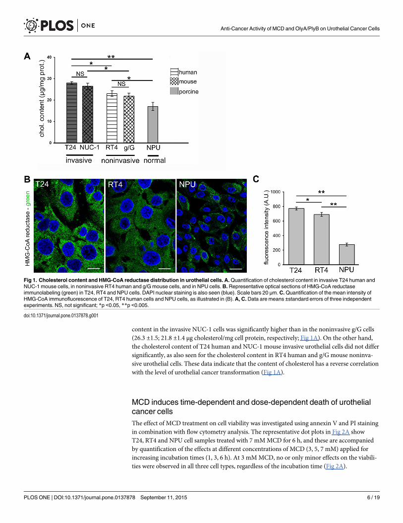
content in the invasive NUC-1 cells was significantly higher than in the noninvasive g/G cells(26.3 ±1.5; 21.8 ±1.4 μg cholesterol/mg cell protein, respectively; Fig 1A). On the other hand,the cholesterol content of T24 human and NUC-1 mouse invasive urothelial cells did not differsignificantly, as also seen for the cholesterol content in RT4 human and g/G mouse noninva-sive urothelial cells. These data indicate that the content of cholesterol has a reverse correlationwith the level of urothelial cancer transformation (Fig 1A).
MCD induces time-dependent and dose-dependent death of urothelialcancer cellsThe effect of MCD treatment on cell viability was investigated using annexin V and PI stainingin combination with flow cytometry analysis. The representative dot plots in Fig 2A showT24, RT4 and NPU cell samples treated with 7 mMMCD for 6 h, and these are accompaniedby quantification of the effects at different concentrations of MCD (3, 5, 7 mM) applied forincreasing incubation times (1, 3, 6 h). At 3 mMMCD, no or only minor effects on the viabili-ties were observed in all three cell types, regardless of the incubation time (Fig 2A).
Fig 1. Cholesterol content and HMG-CoA reductase distribution in urothelial cells. A.Quantification of cholesterol content in invasive T24 human andNUC-1 mouse cells, in noninvasive RT4 human and g/G mouse cells, and in NPU cells.B. Representative optical sections of HMG-CoA reductaseimmunolabeling (green) in T24, RT4 and NPU cells. DAPI nuclear staining is also seen (blue). Scale bars 20 μm. C.Quantification of the mean intensity ofHMG-CoA immunofluorescence of T24, RT4 human cells and NPU cells, as illustrated in (B).A, C. Data are means ±standard errors of three independentexperiments. NS, not significant; *p <0.05, **p <0.005.
doi:10.1371/journal.pone.0137878.g001
Anti-Cancer Activity of MCD and OlyA/PlyB on Urothelial Cancer Cells
PLOS ONE | DOI:10.1371/journal.pone.0137878 September 11, 2015 6 / 19

Fig 2. Cell viability and cholesterol content of urothelial cells after MCD treatments. A. Left: Representative dot plots from flow cytometry analysis ofT24, RT4 and NPU cells grown in control media and after 7 mMMCD treatment for 6 h. Annexin V–PE and PI were used to discriminate between viable
Anti-Cancer Activity of MCD and OlyA/PlyB on Urothelial Cancer Cells
PLOS ONE | DOI:10.1371/journal.pone.0137878 September 11, 2015 7 / 19

In T24 cells, which were the most sensitive to MCD of these three cell types, cell viabilitywas significantly reduced after 5 mM and 7 mMMCD treatment for 3 h, to 92% and 89%,respectively. Six hours after the treatment viability was further reduced to 77% at 5 mMMCDand to only 47% at 7 mMMCD (Fig 2A). In RT4 cells, the only significant reduction in viabilityover a range of MCD concentrations and incubation times was after 6 h with 7 mMMCD (to82%; Fig 2A). An even smaller effect was observed for NPU cells, where only the treatmentwith 7 mMMCD for 6 h showed a minor reduction in cell viability (to 92%; Fig 2A). Whencompared to T24 and RT4 cells, the effect of 6-h treatment with 7 mMMCD on cell viabilitywas statistically lower in NPU cells (Fig 2A). In addition, we could observe that dying, PI posi-tive cells were annexin V negative or positive, whereas a population of single annexin V posi-tive cells was never prominent, which suggests that the cell death is necrotic rather thanapoptotic (Fig 2A).
Interestingly, after 7 mMMCD treatment for 6 h, the total cholesterol content of T24, RT4,and NPU cells was reduced in comparison to the respective untreated cells, by 86%, 57%, and29%, respectively (Fig 2B).
The analysis of cell viability thus showed that the extent of cytotoxicity of MCD increasedwith the concentration and duration of the treatment. To further define these different sensitiv-ities to MCD treatment of these three types of urothelial cells, cell morphology was analyzedafter 7 mMMCD treatment, where significantly decreases in cell viability were seen. T24 cellstreated with 7 mMMCD for 3 h, and RT4 cells treated with 7 mMMCD for 6 h, becamerounded and were weakly attached (Fig 3). T24 cells detached after 7 mMMCD treatment for6 h (Fig 3). The extent of cell rounding was most pronounced in T24 cells, and less pronouncedin RT4 and NPU cells, where only minor morphological effects were observed. These data cor-related well with the cell-death analysis.
In all cases, with the PI-positive staining indicating that cell viability was reduced due tonecrosis, ultrastructural analysis by TEM provided further evidence of necrotic cell death (Fig4). The plasma membranes of T24 and RT4 cells after 7 mMMCD treatment for 6 h were rup-tured and the cytoplasm was released, although the nuclear envelope remained intact (Fig 4).However, NPU cells showed no characteristics of necrosis after this MCD treatment (Fig 4).
To more specifically investigate involvement of apoptosis after MCD treatment, the activa-tion of caspases was analyzed. By monitoring the cleavage of the fluorogenic Ac-DEVD-AFCsubstrate no significant caspase activation was observed upon 6 h of incubation with 3 mM, 5mM or 7 mMMCD either in T24, RT4 or NPU cells. (Fig 5A). We were also unable to detectcaspase activation in cells incubated with 5 and 7mMMCD for 6 hours by immunoblotting ofthe PARP p85 fragment (Fig 5B), which is produced by activated caspases [44]. For compari-son, UV radiation caused the cleavage of both the DEVD peptide and the PARP protein in T24and RT4 cells. The lack of caspase activation combined with the absence of annexin V signaland ultrastructural changes characteristic of necrosis collectively suggested that the MCD treat-ment of urothelial cells triggered necrotic rather than apoptotic cell death.
Western blotting of T24, RT4 and NPU cells after treatment with 7 mMMCD for 6 h didnot reveal any conversion of LC3-I to LC3-II (Fig 5C). This indicated that MCD did not induceautophagy. Moreover, observation of T24, RT4 and NPU cells by TEM did not reveal any typi-cal autophagic structures, such as double-membrane vesicles or autophagosomes (Fig 4).
(double negative), early apoptotic (single annexin V positive) and dead (PI positive) cells. Right: Quantification of the cell viability from the flow cytometryanalysis of T24, RT4 and NPU cells after 3 mM, 5 mM, and 7 mMMCD treatments for 1 h, 3 h and 6 h. B.Quantification of cell cholesterol depletion in T24,RT4 and NPU cells after 7 mMMCD treatment for 6 h. Data are means ±standard errors of duplicate measurements from three independent experiments. *p<0.05, **p <0.005, ***p <0.001.
doi:10.1371/journal.pone.0137878.g002
Anti-Cancer Activity of MCD and OlyA/PlyB on Urothelial Cancer Cells
PLOS ONE | DOI:10.1371/journal.pone.0137878 September 11, 2015 8 / 19

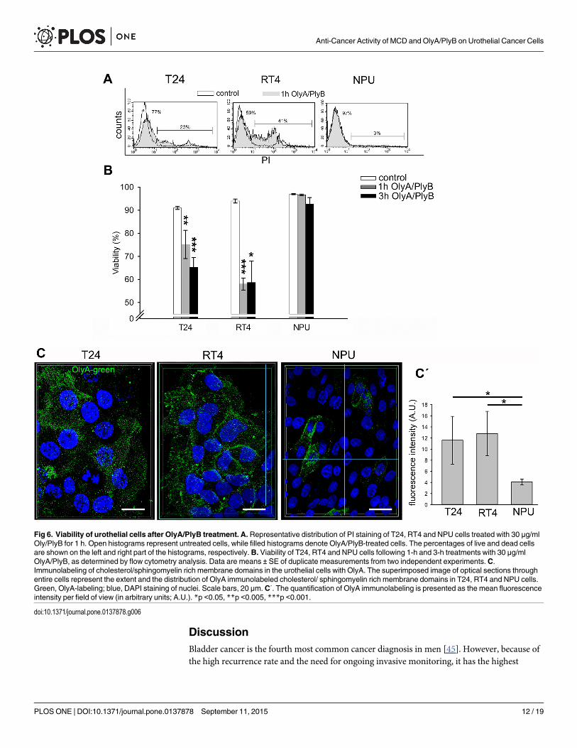
OlyA/PlyB-treatment induces necrosis of urothelial cancer cellsOlyA/PlyB was used for two different applications. A nonlytic concentration of OlyA/PlyB(2.5 μg/ml) was used to label cholesterol/ sphingomyelin-rich domains of the plasma mem-branes (Fig 6C), and a lytic concentration (30 μg/ml) was applied to permeabilize plasma mem-branes. The most prominent OlyA immunolabeling (antibodies were raised against OlyArespectively) was seen for RT4 cells and revealed by quantification of OlyA fluorescence (Fig6C'). This OlyA labeling was more uniform over the plasma membrane in RT4 cells. In con-trast, the OlyA distribution in T24 cells was less even, and its distribution was discontinuous.Labeling of NPU cells showed fewer OlyA-positive cells that significantly differs from T24 andRT4 cells (Fig 6C).
Cell viability after 1-h and 3-h treatments with 30 μg/ml OlyA/PlyB was analyzed by flowcytometry. The cell viabilities after 30 μg/ml OlyA/PlyB treatments for 1 h and 3 h were deter-mined as above, by flow cytometry analysis. As the annexin-V labeling was negative in all of
Fig 3. Morphological changes of urothelial cells after treatment with 7 mMMCD. T24, RT4 and NPU cells (as indicated) were untreated (control) ortreated with 7 mMMCD for 3 h and 6 h, and then examined under an inverted microscope. Cell rounding was cell-type dependent (T24 > RT4 > NPU) andtime dependent (control < 3 h < 6 h). T24 and RT4 cells changed shape from flat and polygonal (control) to spherical (T24 cells, 3 h, 6 h treatment; RT4 cells,6 h treatment; arrowheads). Scale bar, 20 μm.
doi:10.1371/journal.pone.0137878.g003
Anti-Cancer Activity of MCD and OlyA/PlyB on Urothelial Cancer Cells
PLOS ONE | DOI:10.1371/journal.pone.0137878 September 11, 2015 9 / 19

the cells, the fractions of the PI-positive (i.e., dead) and PI-negative cells were determined asindicated by the representative histograms shown in Fig 6A. After 30 μg/ml OlyA/PlyB treat-ment for 1 h, the cell viability of T24 cells was significantly decreased to 75% (p<0.005),and after 3-h, to 65% (p<0.001). Similarly, the cell viability of RT4 cells decreased to 58%(p<0.001) after 1 h, although this did not change further for the 3-h treatment. In contrast,
Fig 4. Ultrastructure of urothelial cells after treatment with 7 mMMCD. T24, RT4 and NPU cells wereuntreated (control) or treated with 7 mMMCD for 6 h, and then processed for TEM. Necrotic changes wereseen in individual T24 cells (star) and RT4 cells, including plasma-membrane disruption (arrowheads) andrelease of cell contents. NPU cells remained intact after this MCD treatment, with glycocalyx still present(open arrowheads). n, nucleus. Scale bars, 1 μm.
doi:10.1371/journal.pone.0137878.g004
Anti-Cancer Activity of MCD and OlyA/PlyB on Urothelial Cancer Cells
PLOS ONE | DOI:10.1371/journal.pone.0137878 September 11, 2015 10 / 19

the cell viability of NPU cells was not significantly affected by the 30 μg/ml OlyA/PlyB treat-ments for either 1 h or 3 h (Fig 6B). Across all of these three cell types tested, the differences inthe cell viabilities between the 30 μg/ml OlyA/PlyB treatments for 1 h and 3 h did not reach sig-nificance. The results of immunolabeling of cholesterol/sphingomyelin rich membranedomains with OlyA were in accordance with the analysis of cell viability.
Necrotic cell death of T24 and RT4 cells after 30 μg/ml OlyA/PlyB treatment for 1 h wasindicated by the PI-positive cells (Fig 6A) and their characteristic ultrastructural changes (Fig7). For these cells, the most prominent changes were seen as increased plasma-membrane per-meability and leakage of cytoplasm, along with organelle swelling (Fig 7). The ultrastructure ofNPU cells was not affected by 30 μg/ml OlyA/PlyB treatment for 1 h, with these control andtreated cells showing similar morphology and with intact plasma membrane (Fig 7).
Fig 5. No apoptosis or autophagy of urothelial cells after MCD treatments for 6 h. T24, RT4 and NPUcells were treated with 3 mM (A), 5 mM (A) or 7 mM (A-C) MCD for 6 h. A. Caspase activity measurements byAc-DEVD-AFC cleavage did not show any increase in caspase activity. The positive controls (UV 30s)showed apoptosis-related caspase activities induced in T24 and RT4 cells at 6 h. B. Immunoblotting analysisdid not show cleavage of full-length PARP (120 kDa) into an 85-kDa fragment.C. Immunoblotting analysisdid not show conversion of LC3-I (18 kDa) to LC3-II (16 kDa). Actin was used as the loading control. Data aremeans ±standard error of triplicate measurements.
doi:10.1371/journal.pone.0137878.g005
Anti-Cancer Activity of MCD and OlyA/PlyB on Urothelial Cancer Cells
PLOS ONE | DOI:10.1371/journal.pone.0137878 September 11, 2015 11 / 19
DiscussionBladder cancer is the fourth most common cancer diagnosis in men [45]. However, because ofthe high recurrence rate and the need for ongoing invasive monitoring, it has the highest
Fig 6. Viability of urothelial cells after OlyA/PlyB treatment. A. Representative distribution of PI staining of T24, RT4 and NPU cells treated with 30 μg/mlOly/PlyB for 1 h. Open histograms represent untreated cells, while filled histograms denote OlyA/PlyB-treated cells. The percentages of live and dead cellsare shown on the left and right part of the histograms, respectively. B. Viability of T24, RT4 and NPU cells following 1-h and 3-h treatments with 30 μg/mlOlyA/PlyB, as determined by flow cytometry analysis. Data are means ± SE of duplicate measurements from two independent experiments. C.Immunolabeling of cholesterol/sphingomyelin rich membrane domains in the urothelial cells with OlyA. The superimposed image of optical sections throughentire cells represent the extent and the distribution of OlyA immunolabeled cholesterol/ sphingomyelin rich membrane domains in T24, RT4 and NPU cells.Green, OlyA-labeling; blue, DAPI staining of nuclei. Scale bars, 20 μm. C΄. The quantification of OlyA immunolabeling is presented as the mean fluorescenceintensity per field of view (in arbitrary units; A.U.). *p <0.05, **p <0.005, ***p <0.001.
doi:10.1371/journal.pone.0137878.g006
Anti-Cancer Activity of MCD and OlyA/PlyB on Urothelial Cancer Cells
PLOS ONE | DOI:10.1371/journal.pone.0137878 September 11, 2015 12 / 19

lifetime treatment costs per patient of all cancers [46]. Indeed, up to 70% of patients have localrecurrences after intravesical chemotherapy or immunotherapy [47]. It is likely that the lowcure rate is due to micrometastatic disease that can be induced at the time of transurethralresection or cystectomy, which results in relapse of the urothelial tumors or in metastases inthe lymph nodes, bones, lung, skin, and liver [48]. Moreover, mortality is mainly caused by
Fig 7. Ultrastructure of urothelial cells after treatment with 30 μg/ml OlyA/PlyB. T24, RT4 and NPU cellswere treated with 30 μg/ml OlyA/PlyB for 1 h and then processed for TEM. RT4 and T24 cells show loss ofmembrane integrity (arrowheads), release of cytoplasm, and organelle swelling (stars), which indicate cellnecrosis. The ultrastructure of treated NPU cells resembles that of untreated NPU cells, with abundantglycocalyx (open arrowheads). n, nucleus. Scale bars, 1 μm.
doi:10.1371/journal.pone.0137878.g007
Anti-Cancer Activity of MCD and OlyA/PlyB on Urothelial Cancer Cells
PLOS ONE | DOI:10.1371/journal.pone.0137878 September 11, 2015 13 / 19

invasive, metastatic urothelial carcinomas that become resistant to chemotherapy. For this rea-son, new anticancer therapies involving different approaches are being investigated to treatbladder cancer.
In the present study, the characteristic differences in the cholesterol content between NPUcells and cancerous urothelial cells was initially established and then further used to defineselective targeting in the treatment of these cancer cells. The lipid composition of the cell mem-branes has been shown to change early in carcinogenesis, and it is severely affected when nor-mal cells are transformed into malignant cells in breast cancer [49], which suggests thatmembrane lipid composition has an important role in cancer progression. Specifically, thecomposition and dynamics of membrane rafts and their involvement in cell signaling appear tobe pivotal in cancer development [17].
In this respect, however, no similar data are available for bladder cancer. Thus, two humanurothelial cell lines with different levels of cancer transformation, as T24 and RT4 cells, wereanalyzed for cytotoxicity along with NPU cells in the present study. Since normal humanurothelium is difficult-to-obtain tissue, we used normal porcine urothelial cell culture, whichshows identical differentiation markers as well as cell biological and histological similarities tohuman urothelium [35,50].
To this end, we show here that cholesterol content increases in correlation with the transfor-mation grade, from nontransformed NPU cells, over RT4 low-grade human papillary cancercells to T24 high-grade invasive human urothelial cancer cells,. Among the possible mecha-nisms that provoke up-regulation of cellular cholesterol synthesis there is increased activity ofHMG-CoA reductase. Our quantification of HMG-CoA reductase protein expression clearlycorresponded to the analyses of the cholesterol concentrations in these cells. We additionallyshow that the same correlation is evident for similar cancer transformation in mouse cells.Cholesterol content in invasive NUC-1 mouse cells was higher than in non-tumorigenic g/Gmouse cells, providing strong evidence that elevated cholesterol content is a characteristic ofhigh-grade urothelial cancer. It is likely that the increase in cholesterol content in T24 andNUC-1 cells originates from high cholesterol import and up-regulated cholesterol synthesis, ashas been shown in other malignancies [51,52,53]. These increased cholesterol levels mightserve for biogenesis of new membranes during the accelerated proliferation of these cancercells.
Our aim was to determine whether MCD and OlyA/PlyB affect cell viability of T24 and RT4urothelial cancer cells compared to NPU cells, and whether this is related to different choles-terol contents of urothelial cells. Presented data show that MCD treatment reduces viability ofthese urothelial cancer cells in time-dependent and dose-dependent manners. There was signif-icant reduction in cell viability of T24 cells, which is in agreement with their highest content ofcholesterol and which would mean that cholesterol is more available to MCD in this cell type.DEVD-ase activity and ultrastructure analyses revealed that MCD treatment of T24 and RT4urothelial cancer cells induced necrosis rather than apoptosis, thereby excluding the classicalapoptotic pathway. This is consistent with previous studies suggesting that necrotic cell deathcan be the consequence of alterations to the membrane fluidity and/or breakdown of cell mem-brane integrity [6,54]. This is also in agreement with previous reports for breast cancer, wherelipid composition was shown to be of major importance in cancer progression [49]. The cho-lesterol-depletion in the present study clearly shows that the availability of cholesterol andtheir sensitivity to MCD treatment is increased in high-grade urothelial cells. On the otherhand, NPU cells, which had the lowest cholesterol content, were the most resistant to the MCDextraction. This can be interpreted in terms of the chemical activity or availability of cholesterolin the membranes, as related to its cellular distribution and homeostasis. It has been shownrecently that the availability of cholesterol for both MCD and cholesterol oxidase increases
Anti-Cancer Activity of MCD and OlyA/PlyB on Urothelial Cancer Cells
PLOS ONE | DOI:10.1371/journal.pone.0137878 September 11, 2015 14 / 19

above a cholesterol content threshold that is specific for each cholesterol/ phospholipid mixtureand for red blood cell membranes [2]. One possible explanation for NPU resistance in compar-ison to cancer cells could be that in differentiated urothelial cells apical plasma membranescontain special proteins uroplakins which arrange urothelial plaques [55] as crystal structuresand give urothelial plasma membranes rigidity and inaccessibility to MCD. Thus, our data sug-gest that the level of cellular cholesterol can be considered as an indicator of the malignancy ofurothelial cells and the efficiency of its depletion with MCD can potentially be used for selectiveremoval of invasive urothelial cancer cells.
Lytic OlyA/PlyB treatment caused the greatest decrease in viability of RT4 cells, and less ofT24 cells, while it had no effect on the viability of nontransformed NPU cells. This correlateswell with the extent of OlyA localization and quantification to cholesterol/ sphingomyelin-richmembrane domains in these cells, as determined with OlyA immunolabeling. These data indi-cate diversity in the abundance of the cholesterol/ sphingomyelin membrane domains amongthese urothelial cells at different stages of cancer transformation, and in comparison with thenontransformed NPU cells. Indeed, it was shown that determination of the membrane lipidprofile can distinguish between different phases of cancer development [56].
Analyses of cell viability and ultrastructure revealed that OlyA/PlyB-induced cell death isnot apoptotic, but necrotic, similar to the MCD treatment. Necrosis after OlyA/PlyB treatmentwas shown here to be the prevalent cell death mechanism for RT4 cells, and was most likelycaused by membrane perforation. It has been reported previously that the interactions of OlyA,as well as with the OlyA/PlyB protein mixture, with cholesterol/ sphingomyelin membranes ishighly cooperative with respect to membrane cholesterol concentrations above a ~30 mol%threshold [28,29,32].
Our data thus indicate that cholesterol-rich membrane domains provide a new target fortreatment of bladder cancer. In vivo, either MCD or OlyA/PlyB might be applied transureter-ally into the bladder cavity as supportive reagents for the elimination of micrometastases (i.e.,considering T24 cells as a model) or against recurring papilloma cells (i.e., considering RT4cells as a model). As shown in our model system, NPU cells should not be particularly harmedby this treatment, as neither MCD nor OlyA/PlyB affected the viability of NPU cells. Of course,these analyses of the cholesterol content and the responses to MCD and OlyA/PlyB will needto be confirmed also with a model of well-differentiated human urothelial cells. Unfortunately,such in-vitromodel is very difficult to obtain, because of the limited sources of healthy urothe-lial tissue and the need for constant renewal of the cultured cells, which can otherwise notachieve highly differentiated levels.
From our previous analyses, we can conclude that the potential minor damage caused byapplication of MCD and OlyA/PlyB would not be detrimental to normal urothelial tissue,because of the high regenerative capacity of these cells under in-vivo conditions [57]. What ismore, MCD or OlyA/PlyB-mediated release of the intracellular contents from necrotic cancercells might represent an immunomodulatory event, and might initiate a targeted and efficientimmune response, as in Bacillus Calmette–Guérin-treated patients with bladder carcinoma[24].
The experimental data from the present study on MCD and OlyA/PlyB demonstrate thatboth of these cholesterol-disturbing agents can selectively decrease the viability of urothelialcancer cells. However, although urothelial cancer cell lines are invaluable in studies of cancer-cell behavior, such in-vitro studies neglect the important control of cell growth and differentia-tion of the in-vivo tissue environment. To confirm the therapeutic potential of MCD andOlyA/PlyB, both ofagents need to be evaluated in biomimetic in-vitromodels or in animalmodels with orthotopic bladder tumors.
Anti-Cancer Activity of MCD and OlyA/PlyB on Urothelial Cancer Cells
PLOS ONE | DOI:10.1371/journal.pone.0137878 September 11, 2015 15 / 19

ConclusionsTo summarize, we have demonstrated that in these urothelial cancer cells, both the increasedcontent of cholesterol and the increased levels of cholesterol/ sphingomyelin-rich membranedomains constitute therapeutic targets for selective induction of cell death. MCD was moretoxic to the metastatic T24 high-grade urothelial cancer cells, and OlyA/PlyB to the RT4 low-grade urothelial cancer cells. Both MCD and OlyA/PlyB leave the nontransformed NPU cellslargely intact. Therefore, this study is aimed to stimulate further research into the area of lipid-related targeting that might provide new locally applied therapies that can be used for elimina-tion of urothelial cancer cells, with the prospect of restricting tumorigenesis and urothelialtumor recurrence.
AcknowledgmentsThis study was funded by the Slovenian Research Agency (http://www.arrs.gov.si/en/agencija),grants P3-0108, P1-0207, P1-0140 and JI-4305. The authors thank Dr. Christopher Berrie forcritical reading and scientific editing of the manuscript. We thank Sanja Čabraja, Nada PavlicaDubarič, Linda Štrus, and Sabina Železnik for technical assistance.
Author ContributionsConceived and designed the experiments: NR PV. Performed the experiments: NR URMEK.Analyzed the data: NR URMEK. Contributed reagents/materials/analysis tools: KS BT PV.Wrote the paper: NR PV UR. Revised and drafted the work: NR PV URMEK KS BT PM.
References1. Yeagle PL (1991) Modulation of membrane function by cholesterol. Biochimie 73: 1303–1310. PMID:
1664240
2. Lange Y, Tabei SM, Ye J, Steck TL (2013) Stability and stoichiometry of bilayer phospholipid-choles-terol complexes: relationship to cellular sterol distribution and homeostasis. Biochemistry 52: 6950–6959. doi: 10.1021/bi400862q PMID: 24000774
3. Resnik N, Sepčić K, Plemenitas A, Windoffer R, Leube R, Veranič P(2011) Desmosome assembly andcell-cell adhesion are membrane raft-dependent processes. The Journal of biological chemistry 286:1499–1507. doi: 10.1074/jbc.M110.189464 PMID: 21071449
4. Harikumar KG, Potter RM, Patil A, Echeveste V, Miller LJ, Henke BR et al. (2013) Membrane choles-terol affects stimulus-activity coupling in type 1, but not type 2, CCK receptors: use of cell lines with ele-vated cholesterol. Lipids 48: 231–244. doi: 10.1007/s11745-012-3744-4 PMID: 23306829
5. Schwan C, Nolke T, Kruppke AS, Schubert DM, Lang AE, Aktories K(2011) Cholesterol- and sphingoli-pid-rich microdomains are essential for microtubule-based membrane protrusions induced by Clostrid-ium difficile transferase (CDT). The Journal of biological chemistry 286: 29356–29365. doi: 10.1074/jbc.M111.261925 PMID: 21705797
6. Sandal S, Tuneva J, Yilmaz B, Carpenter DO (2009) Effects of cholesterol and docosahexaenoic acidon cell viability and (Ca(2+))(i) levels in acutely isolated mouse thymocytes. Cell biochemistry and func-tion 27: 155–161. doi: 10.1002/cbf.1549 PMID: 19274771
7. Simons K, Toomre D (2000) Lipid rafts and signal transduction. Nat Rev Mol Cell Biol 1: 31–39. PMID:11413487
8. Cruz PM, Mo H, McConathy WJ, Sabnis N, Lacko AG (2013) The role of cholesterol metabolism andcholesterol transport in carcinogenesis: a review of scientific findings, relevant to future cancer thera-peutics. Frontiers in pharmacology 4: 119. doi: 10.3389/fphar.2013.00119 PMID: 24093019
9. Silvente-Poirot S, Poirot M (2012) Cholesterol metabolism and cancer: the good, the bad and the ugly.Current opinion in pharmacology 12: 673–676. doi: 10.1016/j.coph.2012.10.004 PMID: 23103112
10. Li HY, Appelbaum FR, Willman CL, Zager RA, Banker DE (2003) Cholesterol-modulating agents killacute myeloid leukemia cells and sensitize them to therapeutics by blocking adaptive cholesterolresponses. Blood 101: 3628–3634. PMID: 12506040
Anti-Cancer Activity of MCD and OlyA/PlyB on Urothelial Cancer Cells
PLOS ONE | DOI:10.1371/journal.pone.0137878 September 11, 2015 16 / 19

11. Antalis CJ, Uchida A, Buhman KK, Siddiqui RA (2011) Migration of MDA-MB-231 breast cancer cellsdepends on the availability of exogenous lipids and cholesterol esterification. Clinical & experimentalmetastasis 28: 733–741.
12. Bennis A, Mehadji BA, Soulami S, Tahiri A, Chraibi N (1993) Cardiovascular manifestations of heredi-tary dysplasias of connective tissue. Annales de cardiologie et d'angeiologie 42: 173–181. PMID:8517593
13. Silvente-Poirot S, Poirot M (2014) Cancer. Cholesterol and cancer, in the balance. Science 343: 1445–1446. doi: 10.1126/science.1252787 PMID: 24675946
14. Freeman MR, Solomon KR (2004) Cholesterol and prostate cancer. J Cell Biochem 91: 54–69. PMID:14689582
15. Kolanjiappan K, Ramachandran CR, Manoharan S (2003) Biochemical changes in tumor tissues oforal cancer patients. Clin Biochem 36: 61–65. PMID: 12554062
16. Schroeder F, Gardiner JM (1984) Membrane lipids and enzymes of cultured high- and low-metastaticB16 melanoma variants. Cancer research 44: 3262–3269. PMID: 6146400
17. Patra SK (2008) Dissecting lipid raft facilitated cell signaling pathways in cancer. Biochim Biophys Acta1785: 182–206. doi: 10.1016/j.bbcan.2007.11.002 PMID: 18166162
18. Li YC, Park MJ, Ye SK, Kim CW, Kim YN (2006) Elevated levels of cholesterol-rich lipid rafts in cancercells are correlated with apoptosis sensitivity induced by cholesterol-depleting agents. Am J Pathol168: 1107–1118; quiz 1404–1105. PMID: 16565487
19. Funahashi H, Satake M, Hasan S, Sawai H, Newman RA, Reber HA et al. (2008) Opposing effects ofn-6 and n-3 polyunsaturated fatty acids on pancreatic cancer growth. Pancreas 36: 353–362. doi: 10.1097/MPA.0b013e31815ccc44 PMID: 18437081
20. Sauer LA, Blask DE, Dauchy RT (2007) Dietary factors and growth and metabolism in experimentaltumors. J Nutr Biochem 18: 637–649. PMID: 17418560
21. Marco C, Rios-Marco P, Jimenez-Lopez JM, Segovia JL, Carrasco MP (2014) Antitumoral Alkylpho-spholipids Alter Cell Lipid Metabolism. Anti-cancer agents in medicinal chemistry.
22. George KS, Wu S (2012) Lipid raft: A floating island of death or survival. Toxicology and applied phar-macology 259: 311–319. doi: 10.1016/j.taap.2012.01.007 PMID: 22289360
23. Cheng J, Ohsaki Y, Tauchi-Sato K, Fujita A, Fujimoto T (2006) Cholesterol depletion induces autop-hagy. Biochemical and biophysical research communications 351: 246–252. PMID: 17056010
24. SeeWA, Zhang G, Chen F, Cao Y, Langenstroer P, Sandlow J (2009) Bacille-Calmette Guerin inducescaspase-independent cell death in urothelial carcinoma cells together with release of the necrosis-associated chemokine high molecular group box protein 1. BJU international 103: 1714–1720. doi: 10.1111/j.1464-410X.2008.08274.x PMID: 19154459
25. Fedida-Metula S, Elhyany S, Tsory S, Segal S, Hershfinkel M, Sekler I et al. (2008) Targeting lipid raftsinhibits protein kinase B by disrupting calcium homeostasis and attenuates malignant properties of mel-anoma cells. Carcinogenesis 29: 1546–1554. doi: 10.1093/carcin/bgn146 PMID: 18579561
26. Ota K, Leonardi A, Mikelj M, Skočaj M, Wohlschlager T, Kunzler M et al. (2013) Membrane cholesteroland sphingomyelin, and ostreolysin A are obligatory for pore-formation by a MACPF/CDC-like pore-forming protein, pleurotolysin B. Biochimie.
27. Skočaj M, Resnik N, Grundner M, Ota K, Rojko N, Hodnik V et al. (2014) Tracking Cholesterol/Sphingo-myelin-Rich Membrane Domains with the Ostreolysin A-mCherry Protein. PloS one 9: e92783. doi: 10.1371/journal.pone.0092783 PMID: 24664106
28. Sepčić K, Berne S, Rebolj K, Batista U, Plemenitaš A, Šentjurc M et al. (2004) Ostreolysin, a pore-form-ing protein from the oyster mushroom, interacts specifically with membrane cholesterol-rich lipiddomains. FEBS Lett 575: 81–85. PMID: 15388337
29. Rebolj K, Ulrih NP, Maček P, Sepčić K (2006) Steroid structural requirements for interaction of ostreoly-sin, a lipid-raft binding cytolysin, with lipid monolayers and bilayers. Biochim Biophys Acta 1758: 1662–1670. PMID: 16857161
30. Aggarwal G, Dhawan S, Harikumar SL (2013) Formulation, in vitro, and in vivo evaluation of matrix-type transdermal patches containing olanzapine. Pharmaceutical development and technology 18:916–925. doi: 10.3109/10837450.2011.609993 PMID: 21913873
31. Schlumberger S, Kristan KC, Ota K, Frangež R, Molgomicron J, Sepčić K et al. (2014) Permeabilitycharacteristics of cell-membrane pores induced by ostreolysin A/pleurotolysin B, binary pore-formingproteins from the oyster mushroom. FEBS Lett 588: 35–40. doi: 10.1016/j.febslet.2013.10.038 PMID:24211835
32. Chowdhury HH, Rebolj K, Kreft M, Zorec R, Maček P, Sepčić K. (2008) Lysophospholipids preventbinding of a cytolytic protein ostreolysin to cholesterol-enriched membrane domains. Toxicon 51:1345–1356. doi: 10.1016/j.toxicon.2008.03.010 PMID: 18455213
Anti-Cancer Activity of MCD and OlyA/PlyB on Urothelial Cancer Cells
PLOS ONE | DOI:10.1371/journal.pone.0137878 September 11, 2015 17 / 19

33. Kreft ME, Di Giandomenico D, Beznoussenko GV, Resnik N, Mironov AA, Jezernik K (2010) Golgiapparatus fragmentation as a mechanism responsible for uniform delivery of uroplakins to the apicalplasmamembrane of uroepithelial cells. Biology of the cell / under the auspices of the European CellBiology Organization 102: 593–607.
34. Visnjar T, Kreft ME (2015) The complete functional recovery of chitosan-treated biomimetic hyper-plastic and normoplastic urothelial models. Histochemistry and cell biology 143: 95–107. doi: 10.1007/s00418-014-1265-3 PMID: 25161121
35. Zupančič D, Romih R (2013) Heterogeneity of uroplakin localization in human normal urothelium, papil-loma and papillary carcinoma. Radiology and oncology 47: 338–345. doi: 10.2478/raon-2013-0052PMID: 24294178
36. Lokar M, Kabaso D, Resnik N, Sepčić K, Kralj-Iglič V, Veranič P et al. (2012) The role of cholesterol-sphingomyelin membrane nanodomains in the stability of intercellular membrane nanotubes. Interna-tional journal of nanomedicine 7: 1891–1902. doi: 10.2147/IJN.S28723 PMID: 22605937
37. De Boer WI, Rebel JM, Foekens JA, Vermey M, Van der Kwast TH (1993) Characterization of mouseurothelial cell lines in different phases of transitional-cell carcinogenesis. International journal of cancerJournal international du cancer 54: 1022–1027. PMID: 7687588
38. Visnjar T, Kreft ME (2013) Air-liquid and liquid-liquid interfaces influence the formation of the urothelialpermeability barrier in vitro. In vitro cellular & developmental biology Animal 49: 196–204.
39. Jerman UD, Veranič P, Kreft ME (2014) Amniotic membrane scaffolds enable the development of tis-sue-engineered urothelium with molecular and ultrastructural properties comparable to that of nativeurothelium. Tissue engineering Part C, Methods 20: 317–327. doi: 10.1089/ten.TEC.2013.0298 PMID:23947657
40. Berne S, Križaj I, Pohleven F, Turk T, Maček P, Sepčić K (2002) Pleurotus and Agrocybe hemolysins,new proteins hypothetically involved in fungal fruiting. Biochim Biophys Acta 1570: 153–159. PMID:12020804
41. Bligh EG, Dyer WJ (1959) A rapid method of total lipid extraction and purification. Can J Biochem Phy-siol 37: 911–917. PMID: 13671378
42. Bradford MM (1976) A rapid and sensitive method for the quantitation of microgram quantities of proteinutilizing the principle of protein-dye binding. Anal Biochem 72: 248–254. PMID: 942051
43. Rozman-Pungerčar J, Kopitar-Jerala N, Bogyo M, Turk D, Vasiljeva O, Stefe I et al. (2003) Inhibition ofpapain-like cysteine proteases and legumain by caspase-specific inhibitors: when reaction mechanismis more important than specificity. Cell death and differentiation 10: 881–888. PMID: 12867995
44. Lazebnik YA, Kaufmann SH, Desnoyers S, Poirier GG, EarnshawWC (1994) Cleavage of poly(ADP-ribose) polymerase by a proteinase with properties like ICE. Nature 371: 346–347. PMID: 8090205
45. Siegel R, Naishadham D, Jemal A (2012) Cancer statistics, 2012. CA: a cancer journal for clinicians62: 10–29.
46. Hong YM, Loughlin KR (2008) Economic impact of tumor markers in bladder cancer surveillance. Urol-ogy 71: 131–135. doi: 10.1016/j.urology.2007.08.014 PMID: 18242381
47. Lindgren D, Gudjonsson S, Jee KJ, Liedberg F, Aits S, Andersson A et al. (2008) Recurrent and multi-ple bladder tumors show conserved expression profiles. BMC Cancer 8: 183. doi: 10.1186/1471-2407-8-183 PMID: 18590527
48. Kaufman DS (2006) Challenges in the treatment of bladder cancer. Annals of oncology: official journalof the European Society for Medical Oncology / ESMO 17 Suppl 5: v106–112.
49. Hilvo M, Denkert C, Lehtinen L, Muller B, Brockmoller S, Seppanen-Laakso T et al. (2011) Novel thera-nostic opportunities offered by characterization of altered membrane lipid metabolism in breast cancerprogression. Cancer research 71: 3236–3245. doi: 10.1158/0008-5472.CAN-10-3894 PMID:21415164
50. Lasič E, Višnjar T, Kreft ME (2015) Properties of the Urothelium that Establish the Blood-Urine Barrierand Their Implications for Drug Delivery. Reviews of physiology, biochemistry and pharmacology.
51. May GL, Wright LC, Dyne M, MackinnonWB, Fox RM, Mountford CE (1988) Plasmamembrane lipidcomposition of vinblastine sensitive and resistant human leukaemic lymphoblasts. Int J Cancer 42:728–733. PMID: 3263327
52. Rudling M, Gafvels M, Parini P, Gahrton G, Angelin B (1998) Lipoprotein receptors in acute myeloge-nous leukemia: failure to detect increased low-density lipoprotein (LDL) receptor numbers in cell mem-branes despite increased cellular LDL degradation. Am J Pathol 153: 1923–1935. PMID: 9846982
53. Vitols S, Norgren S, Juliusson G, Tatidis L, Luthman H (1994) Multilevel regulation of low-density lipo-protein receptor and 3-hydroxy-3-methylglutaryl coenzyme A reductase gene expression in normal andleukemic cells. Blood 84: 2689–2698. PMID: 7919382
Anti-Cancer Activity of MCD and OlyA/PlyB on Urothelial Cancer Cells
PLOS ONE | DOI:10.1371/journal.pone.0137878 September 11, 2015 18 / 19

54. Van Blitterswijk WJ, De Veer G, Krol JH, Emmelot P (1982) Comparative lipid analysis of purifiedplasmamembranes and shed extracellular membrane vesicles from normal murine thymocytes andleukemic GRSL cells. Biochim Biophys Acta 688: 495–504. PMID: 7104337
55. Kreft ME, Robenek H (2012) Freeze-fracture replica immunolabelling reveals urothelial plaques in cul-tured urothelial cells. PloS one 7: e38509. doi: 10.1371/journal.pone.0038509 PMID: 22768045
56. Merchant TE, Diamantis PM, Lauwers G, Haida T, Kasimos JN, Guillem J et al. (1995) Characterizationof malignant colon tumors with 31P nuclear magnetic resonance phospholipid and phosphatic metabo-lite profiles. Cancer 76: 1715–1723. PMID: 8625039
57. Veranič P, Erman A, Kerec-Kos M, Bogataj M, Mrhar A, Jezernik K(2009) Rapid differentiation of super-ficial urothelial cells after chitosan-induced desquamation. Histochemistry and cell biology 131: 129–139. doi: 10.1007/s00418-008-0492-x PMID: 18797916
Anti-Cancer Activity of MCD and OlyA/PlyB on Urothelial Cancer Cells
PLOS ONE | DOI:10.1371/journal.pone.0137878 September 11, 2015 19 / 19
Copyright © 2022 FDOKUMEN