The origin of human chromosome 2 analyzed by comparative chromosome mapping with a DNA microlibrary
Upload
espe-aquitaineCategory
view
0download
0
Genomics 70, 273–285 (2000)doi:10.1006/geno.2000.6389, available online at http://www.idealibrary.com on
High-Resolution Integrated Map Encompassing the Breast CancerLoss of Heterozygosity Region on Human Chromosome 16q22.1
Eirik Frengen,*,1 Philippe Rocca-Serra,†,1 Sergey Shaposhnikov,* Laurence Taine,‡Jim Thorsen,* Christophe Bepoldin,† Martin Krekling,* Delfine Lafon,† Kaja Klykken Aas,*
Azza Abd El Moneim,‡ Henning Johansen,* Michel Longy,† Hans Prydz,*,2 andFrancoise Dorion-Bonnet†
*The Biotechnology Centre of Oslo, University of Oslo, Oslo, Norway; †Laboratoire de Genetique Moleculaire (EA515), InstitutBergonie, Bordeaux, France; and ‡Laboratoire de Genetique, Centre Hospitalier Universitaire Pellegrin, Bordeaux, France
Received June 12, 2000; accepted September 15, 2000
Loss of heterozygosity (LOH) on the long arm ofhuman chromosome 16 is a common genetic alterationobserved in both invasive ductal and invasive lobularbreast carcinomas. We have generated a high-resolu-tion integrated map encompassing the smallest regionof LOH overlap within chromosome 16q22.1 (SRO2).Southern hybridization experiments using more than140 probes resulted in the assembly of 152 bacteriallarge-insert clones into a 2.8-Mb contig covering SRO2.The structure of the contig was verified by long-rangemapping using total human genomic DNA, and thecontig orientation was determined by fluorescence insitu hybridization. A total of 68 transcripts have beenidentified in the map. One of the genes residing withinSRO2 is the E-cadherin gene, CDH1, which has previ-ously been shown to be mutated in lobular breast car-cinomas, resulting in loss of E-cadherin expression. Inmost cases of ductal carcinoma, which is the majormammary cancer type, E-cadherin is normally ex-pressed, suggesting that other genes within 16q22.1are involved in the development of this tumor subtype.The high-resolution map presented in this study pro-vides a valuable resource for identification of tumorsuppressor genes expected to be involved in the etiol-ogy of breast carcinomas. © 2000 Academic Press
INTRODUCTION
Little is known about the molecular events leading tobreast cancer onset, and no clear mechanisms havethus far been proposed. One of the mechanisms sus-pected of being involved in tumorigenesis or progres-
Sequence data from this article have been deposited with theEMBL/GenBank Data Libraries under Accession Nos. AZ081512–AZ081595.
1 These authors should be considered joint first authors.2 To whom correspondence should be addressed at Biotechnology
Centre of Oslo, University of Oslo, P.O. Box 1125, Blindern, N-0349Oslo, Norway. Telephone: 147-22-84-05-32. Fax: 147-22-84-05-01.
E-mail: [email protected].273
sion is loss of tumor suppressor gene function, whichnormally acts as a negative regulator of cell prolifera-tion. It is generally thought that tumor suppressorgenes are recessive, requiring mutation or loss of bothalleles for functional inactivation. Occurrence of loss ofheterozygosity (LOH) in sporadic cancer may thereforeattest to the presence of tumor suppressor genes in-volved in the development or progression of tumors.
Recurrent genetic alterations such as translocations,amplifications, or loss of chromosomal regions are thor-oughly documented in breast tumors (reviewed by Dri-ouch and Lidereau, 1999). More specifically, cytoge-netic studies have revealed an unbalancedtranslocation, t(1;16), leading to trisomy 1q and mono-somy 16q (Dutrillaux et al., 1990). This rearrangementhas in some cases been observed as the sole chromo-somal abnormality (Pandis et al., 1992), suggestingthat it is an early event on the path to tumorigenesis.In addition, nonrandom allelic imbalance affecting thelong arm of chromosome 16 has been detected in morethan 50% of the primary breast tumors studied (Lars-son et al., 1990; Cleton-Jansen et al., 1994; Tsuda et al.,1994; Doggett et al., 1996). Furthermore, the presenceof such alterations in preinvasive lesions suggests thatthese are crucial and early events leading to breastcancer onset (Radford et al., 1995; Vos et al., 1999).Allelic imbalance on chromosome 16q has also beenshown in hepatocarcinoma (Tsuda et al., 1990), pros-tate carcinoma (Latil et al., 1997), Wilms’ tumors (Mawet al., 1992), and ovarian carcinoma (Sato et al., 1991),suggesting the presence of tumor suppressor genes onthis chromosome arm.
Refined LOH mapping in primary breast tumor sam-ples has demonstrated at least two different regionsdisplaying allelic imbalance on chromosome 16q:SRO1, located within 16q24.3, and SRO2, locatedwithin 16q22.1. A physical map covering SRO1 haspreviously been constructed (Whitmore et al., 1998).
The map information in the region within chromosome0888-7543/00 $35.00Copyright © 2000 by Academic Press
All rights of reproduction in any form reserved.

D
11r(etstlitmf
-mb1vsp
tsPdsc
wpsTcg[apwtba
ltN
tbp1wa
sPaelczRsmta
Dwptmim
274 FRENGEN ET AL.
16q22.1, however, is incomplete (Doggett et al., 1995;eloukas et al., 1998). The SRO2 has previously been
shown to be bracketed by the markers D16S397 andD16S496 (Cleton-Jansen et al., 1994; Tsuda et al.,994; Dorion-Bonnet et al., 1995; Skirnisdottir et al.,995; Doggett et al., 1996). Here we present a high-esolution map consisting of altogether 152 P1-derivedPAC) and bacterial artificial (BAC) chromosomes cov-ring 2.8 Mb including the entire SRO2. We have iden-ified 68 transcripts within the map by CpG islandubcloning and screening with expressed sequenceags (ESTs). The E-cadherin gene (CHD1), which isocated within SRO2, has previously been shown to benactivated in lobular breast carcinomas, a rarer sub-ype of breast carcinoma (Berx et al., 1995b). Further-ore, CDH1 germline mutations have been detected in
amilial gastric carcinomas (Guilford et al., 1999). So-matic mutations have been reported in diffuse gastriccancers (Berx et al., 1998) and in invasive lobular car-cinomas, resulting in loss of expression or abnormalcellular distribution of E-cadherin (Berx et al., 1995b).However, most invasive ductal carcinomas, the majortype of human mammary cancers, show no loss of E-cadherin expression and no CDH1 mutation (Kashi-waba et al., 1995; Berx et al., 1995b, 1996). Theseresults suggest that other genes are involved in thedevelopment of this breast tumor subtype. The presenthigh-resolution map provides an excellent resource foridentification of the tumor suppressor genes expectedto be involved in development of breast carcinomas.
MATERIALS AND METHODS
PAC library screening. P1-derived artificial chromosome (PAC;Ioannou et al., 1994) clones were obtained from the RPCI-1, -5, and6 libraries, providing a combined 13-fold representation of the hu-an genome (http://www.chori.org/bacpac). The RPCI-1 and -5 li-
raries were constructed in the vector pCYPAC2 (Osoegawa et al.,999), and the RPCI-6 library was constructed using the pPAC4ector (Frengen et al., 2000). In addition, some BAC clones wereelected from the RPCI-11 library, which was constructed using theBACe3.6 vector (Frengen et al., 1999).Microsatellite markers defining SRO2 (Doggett et al., 1996) and
wo additional sequence-tagged sites (STSs) were used in the initialcreening of the RPCI-1 library. This screening was performed byCR using the “Down to the Well” kit (Genome Systems). High-ensity colony membranes containing the RPCI-1 library werecreened with the marker D16S124 and probes from the LCAT geneluster (Frengen et al., 1995, 1997).High-density membranes containing the RPCI-5 and -6 librariesere further screened using pools of radiolabeled probes. A variety ofrobes were used, including overgos and end fragments derived fromelected PACs and insert fragments from cDNA probes (Fig. 1 andable 1). Probes from the CDH1 gene, the CA7 gene, the LCATluster, and the marker D16S124 were also used (described in Fren-en et al., 1995). The overgos were prepared and filled in witha-32P]dCTP as described by McPherson (1999), and the subclonesnd cDNA probes were labeled with [a-32P]dCTP using the randomriming protocol (Feinberg and Vogelstein, 1984). Hybridizationsere carried out according to Church and Gilbert (1984). Hybridiza-
ions of repetitive sequences in the genomic probes were suppressedy adding sonicated human placenta DNA as described by Sealey et
l. (1985).Contig construction. PAC DNA was isolated by a modified alka-ine extraction protocol (Osoegawa et al., 1999). To enable mapping ofhe PAC clones, the ends of selected PAC inserts were cloned asotI/SacI fragments into the vector pNEB193N (Frengen et al.,
1997). Fragments flanking the NotI sites within the PAC insertswere also obtained in these experiments.
For mapping, DNA from the PACs was digested with NotI andseparated by pulsed-field gel electrophoresis (PFGE) in a Bio-RadCHEF Mapper. A minimal set of PAC clones was further selected fordetailed mapping using eight CpG cutters (AscI, BsiWI, BssHII,EagI, MluI, NotI, NruI, and SacII; New England Biolabs) and all thedouble combinations of these enzymes. The PAC DNA was blottedfrom the pulsed-field gels onto nylon membranes and sequentiallyhybridized with Sp6 and T7 oligonucleotides to detect PAC endfragments. Oligonucleotides were labeled with [g-32P]ATP by usinghe T4 polynucleotide kinase (New England Biolabs). The Southernlots were further hybridized with all the PAC subclones and overgorobes listed in Table 1 as well as all IMAGE clones listed in Tablesand 2 and Fig. 1. Probe removal prior to subsequent hybridizationsas performed by soaking the membranes in boiling 0.5% SDS andllowing the solution to cool to room temperature.
Genomic Southern hybridization. A long-range map was con-tructed by hybridizing probes evenly distributed throughout theAC contig to total human genomic DNA. Human DNA was isolatednd embedded into agarose according to standard protocols (South-rn et al., 1987) at a final concentration of 1 3 107 peripheral bloodeukocytes per milliliter. DNA was digested with the eight CpGutters listed above and all the double combinations of these en-ymes, separated by PFGE, and blotted onto nylon membranes.adiolabeled probes were hybridized to these membranes as de-cribed above. The same DNA preparation was used in all experi-ents, and all fragments were compared on autoradiograms from
he same Southern blot membrane, leading to sizes relative to onenother.
PCR-based EST screening. Markers residing within the interval16S3031–D16S3139 were selected from GeneMap’99 (http://ww.ncbi.nlm.nih.gov/genemap99) and the radiation hybrid mapsrovided by Stanford Human Genome Center and Whitehead Insti-ute/MIT Center for Genome Research. Figure 1 and Table 3 sum-arize the markers that were analyzed. After every library screen-
ng round, PAC DNAs were pooled and typed by PCR for thearkers. PCRs were performed in a 25-ml total volume using 1 U of
Taq polymerase per reaction (Promega). IMAGE clones correspond-ing to ESTs shown by PCR to map to the contigs were obtained fromHGMP-RC (Sanger Centre, Cambridge, UK). The identities of theIMAGE clones were verified by sequencing.
Sequencing. PAC subclones and IMAGE clones were sequencedeither on an ALF sequencing apparatus (Apbiotech) or on an ABI 377automated sequencer (Perkin–Elmer, ABI) according to the manu-facturer’s recommendations. PAC ends were directly sequenced onan ABI 377 automated sequencer using Big Dye terminator kits.This sequencing was performed using 0.5–1 mg QIAtip 100 purifiedPAC DNA (Qiagen) as template, 4 pmol of either a 23-mer Sp6 (GGCCGT CGA CAT TTA GGT GAC AC) or a 24-mer T7 (CCG CTA ATACGA CTC ACT ATA GGG) primer, and specific thermocycling con-ditions (http://www.chori.org/bacpac), including 100 cycle runs on aHybaid touchdown apparatus. Sequences were analyzed using theRepeatMasker program (http://ftp.genome.washington.edu/cgi-bin/RepeatMasker) and the BLASTN program (Altschul et al., 1990) fordatabase searches.
Karyotyping and fluorescence in situ hybridization (FISH). High-resolution prometaphase chromosomes were prepared from PHA-stimulated peripheral blood lymphocytes by conventional methods.Qiagen-purified PAC DNA was labeled using the Dig-Nick Transla-tion Mix (Boehringer Mannheim Inc.) with digoxigenin-11–dUTP orthe Bionick Labeling System (Life Technologies Inc.) with biotin-14–dATP. Briefly, 200 ng of the resulting digoxigeninylated and biotin-ylated DNA was preannealed with 5 mg Cot 1 DNA (Gibco BRL) and
5 mg sonicated salmon sperm DNA (Sigma) in a total volume of 10 ml
FIG
.1.
An
arra
ysh
owin
gth
eP
CR
-bas
edS
TS
scre
enin
gre
sult
son
sele
cted
PA
Ccl
ones
span
nin
gth
eS
RO
2.A
plu
ssi
gn(1
)in
dica
tes
aP
AC
posi
tive
for
the
mar
ker.
Col
um
ns
show
the
nu
mbe
rof
mar
kers
anch
ored
toa
give
nP
AC
.Th
em
arke
rssh
own
ingr
ayco
rres
pon
dto
the
CA
nm
arke
rsde
fin
ing
the
SR
O2,
wh
ich
wer
eu
sed
for
the
init
ials
cree
nin
gof
the
RP
CI-
1li
brar
y.T
he
IMA
GE
clon
en
um
bers
are
indi
cate
dfo
ral
lth
ecD
NA
su
sed
ash
ybri
diza
tion
prob
esin
the
con
stru
ctio
nof
the
PA
Cco
nti
g.C
lon
esfr
omth
eR
PC
I-1
and
RP
CI-
6li
brar
ies
are
prefi
xed
wit
h1
and
6,re
spec
tive
ly,
and
clon
esfr
omth
eR
PC
I-5
libr
ary
are
not
assi
gned
apr
efix.
Gen
Ban
kac
cess
ion
nu
mbe
rsar
esh
own
inpa
ren
thes
es.
Not
eth
atth
em
arke
rsD
16S
3086
and
D16
S30
1,an
dal
soth
eD
16S
421
and
the
D16
S31
8m
arke
rs,
are
iden
tica
l.N
ote
that
pool
edD
NA
from
the
min
imal
tili
ng
path
cove
rin
gth
eL
CA
Tge
ne
clu
ster
was
use
dfo
rth
eS
TS
scre
enin
g.S
peci
fic
prim
ers
wer
eu
sed
toan
alyz
eth
elo
cati
onof
the
TR
AD
Dge
ne
(TR
AD
DF
wd,
CA
GG
CA
AG
AT
TG
AT
TC
CT
GT
TT
;an
dT
RA
DD
Rev
,CT
GA
AA
CT
TC
CA
CT
TG
GC
CT
AT
)an
dth
eC
BF
Bge
ne
(CB
FB
Fw
d,G
GC
AA
AA
GC
AA
TC
TG
GT
AG
C;
and
CB
FB
Rev
,C
CA
TG
CT
GC
TT
CT
GT
CT
GT
C).
*Pro
bes
use
dfo
rth
esc
reen
ing
ofth
eR
PC
I-5
and
-6li
brar
ies.
1 AP
CR
-am
plifi
edfr
agm
ent
from
SL
C9A
5,ex
on3,
and
the
CT
CF
cDN
A(U
2543
5)w
ere
use
das
prob
es.
275DETAILED PHYSICAL MAP OF THE LOH REGION WITHIN 16q22.1

276 FRENGEN ET AL.
TABLE 1
PAC Subclone and End Sequences
Probes (GSS#) Genbank Accession No. Comments (sequence identities observed)a IMAGE clones
71C24T7b AZ081563 T7 end88A20Sp6 AZ081558 Sp6 end88A20T7 AZ081557 T7 end155B22Sp6 AZ081589 Sp6 end155B22T7 AZ081590 T7 end, contains sequences similar to human amyloid
precursor protein-binding protein 1 mRNA (U50939)p42D11#15 AZ081553 The DNCLI2 gene (AF035812) 824203p42D11#5 AZ081554 The DNCLI2 gene (AF035812) 824203p42D11Sp6b AZ081555 Sp6 end1042E2Sp6 AZ081513 Sp6 end1042E2T7 AZ081512 T7 endp943M1Sp6 AZ081514 Sp6 endp943M1Sp6_u AZ081515 The CDH16 gene (AF016272) 1493060p943M1#5 AZ081516 The RRAD gene (U46165)p132O4Sp6 AZ081534 Sp6 endp133B6T7b AZ081567 T7 endp133B6#4 AZ081535 The TRADD gene (L41690)p133B6#13 AZ081565 The E2F4 gene (U15641) 195924p133B6#11 AZ081564 The E2F4 gene (U15641) 195924p133B6#3 AZ081566 The HSF4 gene (D87673) 231788p133B6Sp6 AZ081591 Sp6 endp100O24T7 AZ081536 T7 end, the HSF4 gene (D87673)p100O24#22 AZ081569 The HSF4 gene (D87673) 231788p100O24#23 AZ081570p100O24Sp6b AZ081537 Sp6 endp85K16T7b AZ081538 T7 endp85K16#5 AZ081539 The HSD11B2 gene, 59-end (U27317) 70843p85K16#1 AZ081540 The HSD11B2 gene (U27317) 70843p85K16Sp6 AZ081541 Sp6 endp155L14Sp6 AZ081542 Sp6 end (similar to 1743719)p155L14#5U AZ081543 The CTCF gene (U25435)p155L14#6U AZ081585 970750p155L14#13R AZ081545 878147p155L14#13U AZ081544 (Similar to 632106)p155L14T7 AZ081586 T7 endp102E11T7 AZ081526 T7 endp102E11#9 AZ081527 159926p102E11#25 AZ081580 1750586p102E11#29 AZ081579 1290033p102E11#35 AZ081528 1020497p102E11Sp6 AZ081581 Sp6 endp213H19T7 AZ081582 T7 endp160E12Sp6b AZ081529 Sp6 endp73L13Sp6b AZ081546 Sp6 end, the CTG-B43a mRNA (L10378.1) 648818p179N7Sp6b AZ081530 Sp6 endp138C8Sp6 AZ081583 Sp6 end (similar to EST188148)p138C8#kurt AZ081531 The MDDX28 gene (Hs.155049) 345808p138C8#2 AZ081584 The MDDX28 gene (Hs.155049) 345808p138C8T7b AZ081532 T7 end (similar to tyrosine kinase brk) EST181446c
p64I21Sp6 AZ081533 Sp6 endp1032E14#4b AZ081517 1715697p1032E14#27 AZ081518p1032E14#25 AZ081577 The SLC7A6 gene (NM_003983) 264517p1032E14#3 AZ081519 The SLC7A6 gene (NM_003983) 264517p1032E14Sp6 AZ081578 Sp6 endp1011A13T7 AZ081520 T7 endp203L6T7 AZ081547 T7 endp203L6Sp6b AZ081548 Sp6 endp1050C9Sp6 AZ081521 Sp6 endp1050C9#19 AZ081522 Contains sequences identical to XrepBE mRNA (X13896)p1050C9T7 AZ081523 T7 endp1013D13T7 AZ081562 T7 endp100O11T7 AZ081549 T7 end
100O11Sp6 AZ081550 Sp6 end
pnSt
277DETAILED PHYSICAL MAP OF THE LOH REGION WITHIN 16q22.1
hybridization mix (50% formamide, 2 3 SSC, 10% dextran sulfate)for 30 min at 37°C. Hybridization to the previously denatured chro-mosomes was performed overnight at 37°C. Posthybridization wash-ing was carried out at 72°C in 23 SSC (5 min). Probe detection was
erformed with mouse antidigoxin-conjugated fluorescein isothiocya-ate (FITC; Sigma) and extravidin-conjugated cyanin 3 (Cy3;igma). Slides were mounted in antifade solution (Vectashield, Vec-or Laboratories) containing 49,6-diamidino-2-phenylindole (DAPI)
as counterstain. Slides were analyzed on a Zeiss Axioskop micro-scope equipped with a Pinkel filter set for visualization of FITC, Cy3,and DAPI fluorescence. Digital images were recorded with a Photo-metrics cooled CCD camera. Pseudo-coloring and merging of imageswere performed with Mac Probe 3.3 software (PSI, League City, TX).Metaphases were karyotyped by direct identification of chromosomeson the digital image of the DAPI and Vectashield double stain ac-cording to the reverse-band standard karyotype (Korenberg et al.,1992). Symmetrical labeling of sister chromatids on both chromo-some homologues was evaluated on more than 20 metaphasespreads.
RESULTS
Assembly of a Contig Spanning the SRO2
The SRO2 within human chromosome 16q22.1 haspreviously been refined using an extended set of ge-netic markers on a large panel of tumor samples (re-viewed by Dogget et al., 1996). Markers defining theSRO2 (indicated in Fig. 1) were used for PCR screeningof a PAC library, RPCI-1, anchoring the markers togenomic clones. The markers D16S347, D16S3019,D16S3086, and D16S496 did not identify PAC clones inthe RPCI-1 library, probably due to the low redun-dancy of this library. The RPCI-1 library was alsoscreened by hybridization with the marker D16S124and probes from the LCAT gene cluster (Frengen et al.,1995, 1997). The RPCI-1 PACs identified served asnucleation points in the subsequent chromosome walk-ing process, which was performed by screening of theRPCI-5 and -6 libraries (10-fold redundancy). The firstscreening was performed using the RPCI-1 PACs as
TABLE 1
Probes (GSS#) Genbank Accession No. Com
77A18T7 AZ081551 T7 end77A18Sp6 AZ081552 Sp6 endp30L20T7 AZ081593 T7 endp30L20#6 AZ081572 The CDH1p30L20Sp6b AZ081594 Sp6 endp86F1T7 AZ081595 T7 endp86F1Sp6 AZ081573 Sp6 end1136M7T7b AZ081587 T7 end1136M7Sp6 AZ081588 Sp6 endp17B17T7 AZ081556 T7 endp237i1T7 AZ081576 T7 endp237I1 #2 AZ081575p237I1#1b AZ081574 The SNT2Bp1116A20Sp6 AZ081524 Sp6 endp1116A20T7 AZ081525 T7 end
Note. PAC end sequences are given as clone name and sequence pa GenBank accession numbers are given in parentheses.b These probes were also used for the screening of the RPCI-5 andc An EST clone obtained from ATCC.
probes. The subsequent screening steps were carried
out using 15 probes derived from PACs located at theends of the contigs (see Table 1) and 8 cDNA probesfrom closely linked genes (see Fig. 1) and the CDH1and CA7 probes (Frengen et al., 1995).
The PAC clones were initially organized by hybrid-izing the screening probes to Southern membranescontaining NotI-digested DNA from all the clones iden-tified in the screening. PAC insert ends and fragmentsflanking internal NotI sites were cloned from selectedPACs providing additional probes for the contig map-ping. The resulting subclones and additional PAC endsequences obtained by direct sequencing are listed inTable 1.
Two partially sequenced BAC clones, RPCI-11-297D21 (GenBank Accession No. AC009061) and 11-258J3 (Genbank Accession No. AC012040), were iden-tified by using sequences from PAC sublones indatabase searches. These BACs were included in themap construction. In addition, two completely se-quenced BAC clones, A-116A10 (GenBank AccessionNo. AC004638) and A-67A1 (GenBank Accession No.AC004531), are included in Fig. 1 based on sequenceidentity to probes from the contig (updated informationon genomic sequence from this chromosome region isavailable at http://www.ncbi.nlm.nih.gov/genome/seq/HsHome.shtml).
Based on the initial mapping, a minimal set of PACswas selected for detailed mapping. This minimal setwas digested with eight CpG cutters and all the doublecombinations of these enzymes. The digested PACsamples were separated by PFGE, and the DNA wastransferred to Southern blot membranes. Southernmembranes containing all clones digested with NotIand the single- and double-digested minimal set ofPACs were hybridized with Sp6 and T7 oligonucleo-tides and all the PAC subclones and cDNA probes
ontinued
nts (sequence identities observed)a IMAGE clones
e, exon 16
33530ene (U40572) 364823
er (100o24T7). Subclones are prefixed with p.
libraries.
—C
me
gen
2 g
rim
-6
described under Materials and Methods. A contig map

278 FRENGEN ET AL.

tcahdmtt
T
flco
279DETAILED PHYSICAL MAP OF THE LOH REGION WITHIN 16q22.1
based on the assembly of all the hybridization results isshown in Fig. 2E. All overlaps between PAC/BACclones are supported by a minimum of two sharedhybridization markers. The 2.8-Mb contig consists of152 genomic clones covering the region defined by themarkers within SRO2.
A Long-Range Restriction Map of SRO2
The construction of the contig was primarily per-formed using probes derived from the end of the start-ing PACs in repeated library screenings. If the regionmapped contains large duplicated sequence elements,this chromosome walking could potentially lead to theextension of the map into unlinked chromosomal re-gions. To verify the organization of the PAC map, wehave therefore performed long-range mapping using 14cDNA probes (indicated with an asterisk in Figs. 2Band 2D). Examples of common fragments obtained ingenomic Southern hybridization experiments areshown in Fig. 3. A long-range restriction map based onthe hybridization with all the probes used is presentedin Fig. 2A, which is aligned to the contig based on theposition of the probes in the PAC map. The apparentmolecular size of DNA fragments detected by PFGEincreases with increasing DNA concentration, and theeffect is greater for the smallest fragments on each gel(Doggett et al., 1992). The larger sizes observed whenhe fragments detected in human genomic DNA areompared with fragments in purified PAC DNA (Fig. 2)re in accordance with the greater complexity of theuman DNA. However, both the linear order and theistances between the probes used are fully in agree-ent with the positions of the corresponding genes in
he contig, demonstrating that the high-resolution in-egrated map faithfully represents the SRO2 region.
he Centromere–Telomere Orientation of the SRO2Contig
To validate further the structure of the contig map,uorescence in situ hybridization experiments werearried out using PAC clones corresponding to the endsf the initial contigs (see Fig. 2). Hybridization of each
FIG. 2. An integrated physical and partial transcriptional mapfragments observed after hybridization to human genomic DNA are pprobes used in the genomic hybridizations originated from the geneprobe are indicated with black dashed arrows. (B) The genes are shoarrows. (C) Map of polymorphic markers. The borders of the possiblnot possible to resolve D16S3141 and D16S3067, which are located wdetected within the map. The limits of the possible positions of the pIMAGE Consortium numbers for the cDNA probes are given in Fig. 1bars, which are shown to scale. Clones from the RPCI-1, RPCI-6, andfrom the RPCI-5 library are not assigned a prefix. The restriction frclones are indicated with blue vertical dashed lines. Clones positivvertical dashed lines. The addresses of the PAC clones mapped bAccession No. AC004638) and A-67A1 (GenBank Accession No. AC00indicated with dashed lines. Filled circles show the T7 end of the PAC(M), and NotI (N) have been completely mapped in the contig. Clustthe presence of CpG islands (shown in red). Selected sites for the en
map highlighting putative CpG islands.PAC to human metaphase spreads revealed symmet-rical labeling of both sister chromatids in the q22.1region on both homologues of chromosome 16. The con-tig orientation was obtained using two-color FISH onhigh-resolution prometaphase chromosomes. Double-labeling using the probe pairs 6-131G17/1-102E11,1-102E11/6-203L6, and 6-77A18/1-17B17 (Fig. 4) re-vealed the order centromere 3 6-131G17 31-102E11 3 6-203L6 3 6-77A18 3 1-17B17 3 telo-mere. This order is in agreement with the contig map(Fig. 2).
The Order of the Microsatellite Markers DefiningSRO2
The microsatellite markers defining the SRO2 weremapped in the contig by PCR experiments using DNAfrom single PACs as template (Fig. 1). In addition,some microsatellite markers were mapped by hybrid-ization using radiolabeled primers. Based on these ex-periments, all the polymorphic markers used to definethe SRO2 have been located in the map (Fig. 2C).
Gene Identification
Two complementary methods have been used toidentify genes in the contig map: EST screening, andmapping and subcloning of CpG islands.
Since clusters of rare-cutter restriction sites are verystrong evidence for the presence of CpG islands (Bick-more, 1992; Larsen et al., 1992), 40 putative CpG is-lands have been indicated in the map based on long-range mapping and detailed PAC mapping (Fig. 2E).The CpG islands indicated are likely to signal thepresence of genes. Fragments from putative CpG is-lands were subcloned and sequenced. Databasesearches using sequences from the PAC subclones re-vealed the sequence identities summarized in Table 1.
In parallel, markers from the GB3 and GB4 radia-tion hybrid maps were selected for PCR typing. Themarkers assigned to the contig by this method areshown in Fig. 1.
Partial sequencing of all the IMAGE clones identi-fied both from PAC subclones and from EST screening
ering the LOH region within human chromosome 16q22.1. (A) Theented as open boxes, with the fragment sizes given in kilobases. Thend the ESTs marked with asterisks. Fragments detected with eachas red lines with the size and transcriptional direction indicated bysitions of the STS markers are indicated with gray shading. It wasin the same 20-kb BssHII fragment in the PACs. (D) EST markers
es based on the hybridization results are depicted as red boxes. Thed Tables 1 and 2. (E) The PAC and BAC clones are indicated as blue
CI-11 libraries are prefixed with 1, 6, and 11, respectively, and clonesents detected with the PAC subclones in the respective PAC/BAC
r cDNA probes by Southern blot analysis are indicated with blackISH are labeled in yellow. Two BAC clones, A-116A10 (GenBank1), are included in the map based on sequence identity to the probesC clones. Sites for the restriction enzymes AscI (A), BsiWI (Bi), MluIof rare-cutter restriction sites are indicated in the map, suggestinges BssHII (B), EagI (E), NruI (Nr), and SacII (S) are shown in the
covress awne po
ithrob, anRPagm
e foy F453/BAerszym

ate
280 FRENGEN ET AL.
revealed identity to the genes summarized in Fig. 1and Table 2. All the cDNA clones listed were alsohybridized to PAC Southern membranes to verify thecontig structure and to assign a position to each of the
FIG. 3. Autoradiograms of Southern blots of two pulsed-field gelNOL3, HSD11B2 (59-end, p85K16#5), HSD11B2, IMAGE Clone 6488DNA was digested and separated by PFGE in 1.0% agarose gels at 200pulse time from 0.47 s to 1 min 34 s (A) or a 28-h run using a linearwith the following enzymes: A, AscI; Bi, BsiWI; M, MluI; N, NotI; Nrlow range and the yeast chromosome PFG markers (NEB) are indic
transcripts in the map (Figs. 2B and 2D).
Most of the genes identified have previously beenmapped to the region. Exceptions are the TMSB4Xgene (Hs.75968) and the PP15 gene (Hs.151734), whichwe assigned to the SRO2 contig by PCR screening (Fig.
fter hybridization with the following probes: IMAGE Clone 115086,SLC7A6, IMAGE Clone 37648, CDH1, and SNTB2. Human genomicunder the following conditions: a 20-h run using a linearly increasingncreasing pulse time from 18 s to 3 min and 24 s (B). Digestion wasruI; and A 1 Bi, AscI 1 BsiWI (double digest). The fragments in thed. The fragments detected are shown in Fig. 2A.
s a18,V
ly i, N
1). These genes are assigned to both chromosomes 16

tat
281DETAILED PHYSICAL MAP OF THE LOH REGION WITHIN 16q22.1
and X in GeneMap’99 (http://www.ncbi.nlm.nih.gov/genemap99). These observations may suggest the pres-
FIG. 4. Two-color fluorescence in situ hybridization on humanchromosomes. High-resolution prometaphase chromosomes hybrid-ized with the PAC clones 6-131G17 (red) and 1-102E11 (green) (a),and 1-102E11 (green) and 6-203L6 (red) (b). Interphase nuclei werehybridized with the PAC clones 1-17B17 (green), 6-77A18 (red), and1-102E11 (green), demonstrating the orientation centromere 31-102E113 6-77A183 1-17B17 (c). The distances in the contig areapproximately 1 Mb between 6-131G17 and 1-102E11, 0.6 Mb be-tween 1-102E11 and 6-203L6, and 0.3 Mb between 6-77A18 and1-17B17. Arrows indicate the chromosome 16 telomere and centro-mere.
ence of pseudogenes or that there is more than one
closely related gene for these genes in the human ge-nome.
No products were detected in PCRs with the markerssummarized in Table 3, using PAC DNA as templates.PCR fragments of the expected size were detected incontrol reactions with total human DNA, suggestingthat these markers are located outside the region cov-ered by the contig. This agrees with data presented inGeneMap’99, which indicates that the CDH5, CDH8,and CDH11 genes are located centromeric to the con-tig, whereas the HPR and DIA4 genes are expected tobe located telomeric to the SRO2 contig.
The transcriptional directions for 20 genes are indi-cated in the present map (Fig. 2B). Alignments of se-quences from the PAC subclones to cDNA entries in thedatabases revealed the orientation of the followinggenes in the map: DNCLI2, CDH16, RRAD, TRADD,HSF4, HSD11B2, CTCF, MDDX28, SLC7A6, CDH1,and SNT2B2 (Table 1). Database searches with sub-clone sequences also identified two completely se-quenced BAC clones. These sequence resources re-solved the gene structures for 4 genes in the contig: theAPP-BP1 and CA7 genes on BAC clone A-116A10 (Gen-Bank Accession No. AC004638) and the NFATC4 geneand the MDDX28 gene on BAC clone A-67A1 (Gen-Bank Accession No. AC004531). In addition, thegenomic organization of 10 genes in the map has pre-viously been described: HSF4 (Tanabe et al., 1999),SLC9A5 (Baird et al., 1999), HSD11B2 (Agarwal et al.,1995), CDH3 (Bussemakers et al., 1994), and CDH1(Berx et al., 1995a), and the 5 genes in the LCATgene cluster: PSKH-1, CTRL, PSMB10, LCAT, andSLC12A4 (Larsen et al., 1993; Frengen et al., 1995). Inotal, 29 known genes (Fig. 2B) and 39 ESTs (Figs. 1nd 2D) were identified in the physical map coveringhe SRO2.
DISCUSSION
The genomic interval between D16S397 andD16S496 has been identified as a possible site for tu-mor suppressor genes based on the high frequency ofallele loss in sporadic breast tumors. This work reportsthe generation of an integrated physical map of thedeletion region. A 2.8-Mb contig was established byhybridizing more than 140 probes to restriction di-gested PAC DNA on Southern membranes, resulting inapproximately one sequenced landmark every 20 kb.Any two overlapping clones in the contig hybridizewith a minimum of two common markers. The 152continuously overlapping PAC/BAC clones provide anaverage 6.7-fold redundant coverage of the SRO2. Thestructure of the SRO2 contig was verified by hybridiz-ing probes evenly distributed in the map to total hu-man genomic DNA in PFGE experiments. The align-ment of the resulting long-range map to the contig mapdemonstrates that the contig faithfully represents theregion within human chromosome 16q22.1. Further-
more, the orientation of the contig in relation to the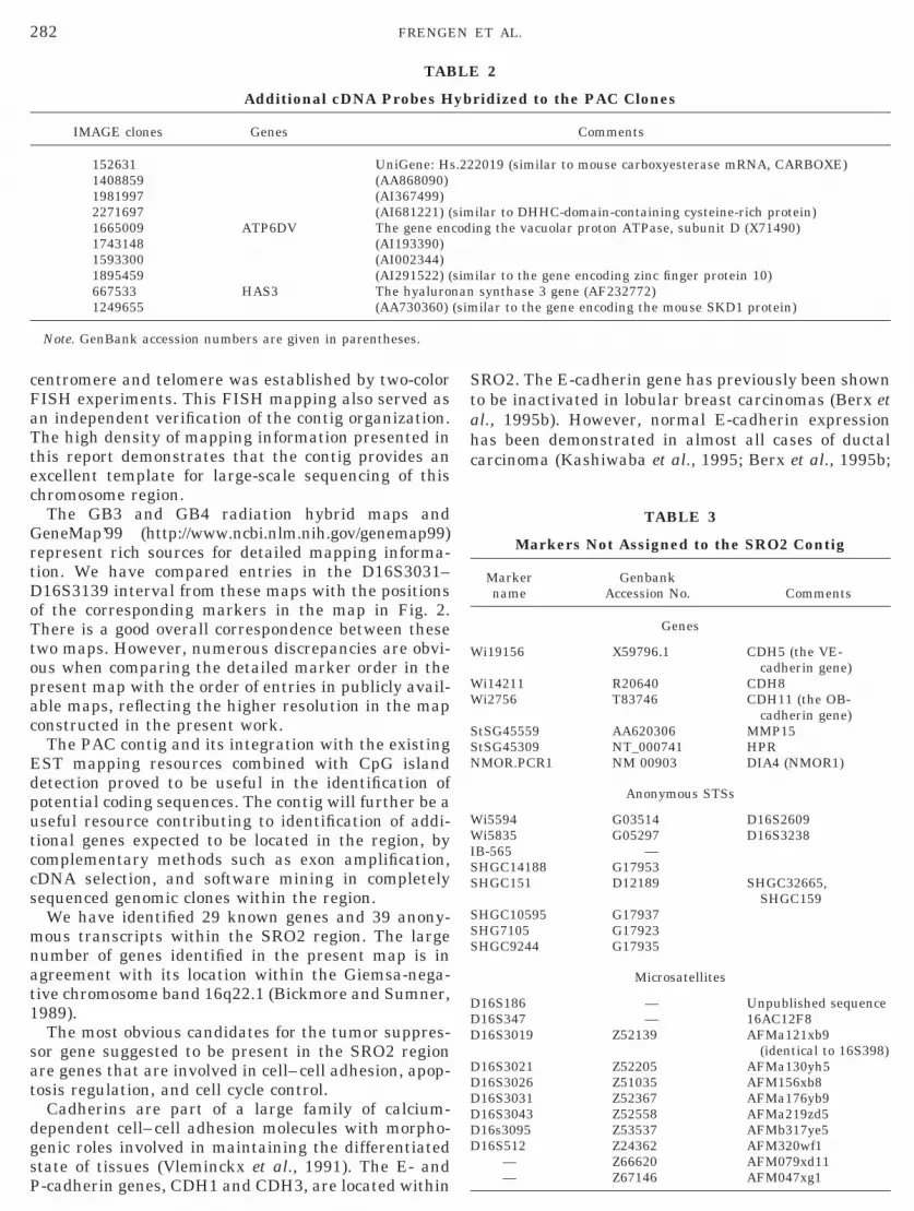
282 FRENGEN ET AL.
centromere and telomere was established by two-colorFISH experiments. This FISH mapping also served asan independent verification of the contig organization.The high density of mapping information presented inthis report demonstrates that the contig provides anexcellent template for large-scale sequencing of thischromosome region.
The GB3 and GB4 radiation hybrid maps andGeneMap’99 (http://www.ncbi.nlm.nih.gov/genemap99)represent rich sources for detailed mapping informa-tion. We have compared entries in the D16S3031–D16S3139 interval from these maps with the positionsof the corresponding markers in the map in Fig. 2.There is a good overall correspondence between thesetwo maps. However, numerous discrepancies are obvi-ous when comparing the detailed marker order in thepresent map with the order of entries in publicly avail-able maps, reflecting the higher resolution in the mapconstructed in the present work.
The PAC contig and its integration with the existingEST mapping resources combined with CpG islanddetection proved to be useful in the identification ofpotential coding sequences. The contig will further be auseful resource contributing to identification of addi-tional genes expected to be located in the region, bycomplementary methods such as exon amplification,cDNA selection, and software mining in completelysequenced genomic clones within the region.
We have identified 29 known genes and 39 anony-mous transcripts within the SRO2 region. The largenumber of genes identified in the present map is inagreement with its location within the Giemsa-nega-tive chromosome band 16q22.1 (Bickmore and Sumner,1989).
The most obvious candidates for the tumor suppres-sor gene suggested to be present in the SRO2 regionare genes that are involved in cell–cell adhesion, apop-tosis regulation, and cell cycle control.
Cadherins are part of a large family of calcium-dependent cell–cell adhesion molecules with morpho-genic roles involved in maintaining the differentiatedstate of tissues (Vleminckx et al., 1991). The E- and
TAB
Additional cDNA Probes H
IMAGE clones Genes
152631 UniGene: Hs1408859 (AA868090)1981997 (AI367499)2271697 (AI681221) (1665009 ATP6DV The gene en1743148 (AI193390)1593300 (AI002344)1895459 (AI291522) (667533 HAS3 The hyaluro1249655 (AA730360)
Note. GenBank accession numbers are given in parentheses.
P-cadherin genes, CDH1 and CDH3, are located within
SRO2. The E-cadherin gene has previously been shownto be inactivated in lobular breast carcinomas (Berx etal., 1995b). However, normal E-cadherin expressionhas been demonstrated in almost all cases of ductalcarcinoma (Kashiwaba et al., 1995; Berx et al., 1995b;
TABLE 3
Markers Not Assigned to the SRO2 Contig
Markername
GenbankAccession No. Comments
Genes
Wi19156 X59796.1 CDH5 (the VE-cadherin gene)
Wi14211 R20640 CDH8Wi2756 T83746 CDH11 (the OB-
cadherin gene)StSG45559 AA620306 MMP15StSG45309 NT_000741 HPRNMOR.PCR1 NM 00903 DIA4 (NMOR1)
Anonymous STSs
Wi5594 G03514 D16S2609Wi5835 G05297 D16S3238IB-565 —SHGC14188 G17953SHGC151 D12189 SHGC32665,
SHGC159SHGC10595 G17937SHG7105 G17923SHGC9244 G17935
Microsatellites
D16S186 — Unpublished sequenceD16S347 — 16AC12F8D16S3019 Z52139 AFMa121xb9
(identical to 16S398)D16S3021 Z52205 AFMa130yh5D16S3026 Z51035 AFM156xb8D16S3031 Z52367 AFMa176yb9D16S3043 Z52558 AFMa219zd5D16s3095 Z53537 AFMb317ye5D16S512 Z24362 AFM320wf1
— Z66620 AFM079xd11
2
ridized to the PAC Clones
Comments
2019 (similar to mouse carboxyesterase mRNA, CARBOXE)
ilar to DHHC-domain-containing cysteine-rich protein)ing the vacuolar proton ATPase, subunit D (X71490)
ilar to the gene encoding zinc finger protein 10)synthase 3 gene (AF232772)ilar to the gene encoding the mouse SKD1 protein)
LE
yb
.22
simcod
simnan(sim
— Z67146 AFM047xg1

283DETAILED PHYSICAL MAP OF THE LOH REGION WITHIN 16q22.1
1996), suggesting that the E-cadherin gene is not in-volved in the development of invasive ductal carcinomas.
The P-cadherin gene is expressed in myoepithelialcells in the ducts of the mammary glands and in thecap cells of the terminal end buds (Rasbridge et al.,1993). Moreover, the mammary glands of P-cadherinknock-out mice do not have a ductal morphology, andthe most obvious phenotypic effect in the P-cadherin2/2
mice was precocious mammary gland development andtendency to mammary hyperplasia (Radice et al.,1997). This indicates that P-cadherin plays a criticalrole in maintaining the growth of the ductal structuresof the breast (Radice et al., 1997), and its gene shouldtherefore be considered a strong candidate for the tu-mor suppressor gene expected to be involved in thespecific etiology of ductal breast carcinomas.
Another cadherin gene, CDH16 (encoding Ksp-cad-herin), has also been assigned to the SRO2. In adults,Ksp-cadherin expression is restricted to the basolat-eral membrane of tubular epithelial kidney cells (Iga-rashi et al., 1999), and due to its tissue-specific expres-sion, CDH16 is not likely to be a candidate geneinvolved in breast cancer development. However, al-lelic imbalance within 16q22.1 has also been detectedin Wilms’ tumors, which includes a heterogeneous typeof kidney tumor (Newsham et al., 1995; Mertens,1997). It is therefore tempting to speculate that theKsp-cadherin gene might act as a tumor suppressorgene in Wilms’ tumors.
Genes involved in cell cycle control could be thegenomic targets of the recurrent alterations observedwithin 16q22.1. Genes such as E2F4 and CTCF maytherefore be considered candidate tumor suppressorgenes (Le Cam et al., 1999; Filippova et al., 1998;Delgado et al., 1999).
The high-resolution SRO2 map presented in thisstudy will be a useful resource contributing to geneidentification and studies of gene expression patternsin the region. A minimal-tiling path contig of the dele-tion interval consists of PAC clones from the RPCI-6library. Clones from this library have the potential ofbeing transferred to mammalian cells as stably repli-cating episomes (Frengen et al., 2000). The delivery ofclones into mammalian cells as episomes offers a solu-tion to avoid chromosomal integration and associatedtranscriptional shutoff and also eliminating the posi-tion effect on the expression of the genes under study.Thus, the clones from this library may be used infunctional screening experiments in which the genescarried on the inserts complement a mutant pheno-type. This feature might facilitate the use of a func-tional strategy by introducing PAC clones into tumorcells for the identification, verification, and character-ization of the tumor suppressor gene expected to bepresent within human chromosome 16q22.1.
Additional studies are under way to obtain the com-plete sequences of selected cDNA clones and to inves-tigate tumor mutations and biological function. The
order of the microsatellite markers revealed in thepresent work will be used to reassess allelic imbalancedata to redefine the SRO2, which would facilitate afocused search for genes in a subregion of the contig,reducing the task of evaluating candidates for the16q22.1 tumor suppressor gene.
ACKNOWLEDGMENTS
We are thankful to Professor Lobanenkov for providing the CTCFcDNA probe. The expert assistance of Jorun Solheim, Line Jensen,and Line Vaagland Jakobsen is gratefully acknowledged. We alsothank Dr. Philippe Gorry for fruitful discussions. The present workwas supported by The Norwegian Cancer Society, The ResearchCouncil of Norway, The Jahre Foundation (E.F. and H.P.),l’Association pour la Recherche contre le Cancer, et les ComitesDepartementaux de la Ligue Nationale contre le Cancer du Lot-et-Garonne,de la Gironde et de la Dordogne, and the European Molec-ular Biology Organization (Grant STF 8970).
REFERENCES
Agarwal, A. K., Rogerson, F. M., Mune, T., and White, P. C. (1995).Gene structure and chromosomal localization of the humanHSD11K gene encoding the kidney (type 2) isozyme of 11 b-hy-droxysteroid dehydrogenase. Genomics 29: 195–199.
Altschul, S. F., Gish, W., Miller, W., Myers, E. W., and Lipman, D. J.(1990). Basic local alignment search tool. J. Mol. Biol. 215: 403–410.
Baird, N. R., Orlowski, J., Szab, E. Z., Zaun, H. C., Schultheis, P. J.,Menon, A. G., and Shull, G. E. (1999). Molecular cloning, genomicorganization, and functional expression of Na1/H1 exchanger iso-form 5 (NHE5) from human brain. J. Biol. Chem. 274: 4377–4382.
Berx, G., Staes, K., van Hengel, J., Molemans, F., Bussemakers,M. J., van Bokhoven, A., and van Roy, F. (1995a). Cloning andcharacterization of the human invasion suppressor gene E-cad-herin (CDH1). Genomics 26: 281–289.
Berx, G., Cleton-Jansen, A. M., Nollet, F., De Leeuw, W. J., van deVijver, M., Cornelisse, C., and van Roy, F. (1995b). E-cadherin is atumour/invasion suppressor gene mutated in human lobularbreast cancers. EMBO J. 14: 6107–6115.
Berx, G., Cleton-Jansen, A. M., Strumane, K., de Leeuw, W. J.,Nollet, F., van Roy, F., and Cornelisse, C. (1996). E-cadherin isinactivated in a majority of invasive human lobular breast cancersby truncation mutations throughout its extracellular domain. On-cogene 13: 1919–1925.
Berx, G., Becker, K. F., Hofler, H., and van Roy, F. (1998). Mutationsof the human E-cadherin (CDH1) gene. Hum. Mutat. 12: 226–237.
Bickmore, W. (1992). Analysis of genomic DNAs by pulsed-field gelelectrophoresis. In “Techniques for the Analysis of Complex Ge-nomes” (R. Anand, Ed.), pp. 19–38, Academic Press, London.
Bickmore, W. A., and Sumner, A. T. (1989). Mammalian chromosomebanding—An expression of genome organization. Trends Genet. 5:144–148.
Bussemakers, M. J., van Bokhoven, A., Voller, M., Smit, F. P., andSchalken, J. A. (1994). The genes for the calcium-dependent celladhesion molecules P- and E-cadherin are tandemly arranged inthe human genome. Biochem. Biophys. Res. Commun. 203: 1291–1294.
Church, G. M., and Gilbert, W. (1984). Genomic sequencing. Proc.Natl. Acad. Sci. USA 81: 1991–1995.
Cleton-Jansen, A. M., Moerland, E. W., Kuipers-Dijkshoorn, N. J.,Callen, D. F., Sutherland, G. R., Hansen, B., Devilee, P., andCornelisse, C. J. (1994). At least two different regions are involvedin allelic imbalance on chromosome arm 16q in breast cancer.
Genes Chromosomes Cancer 9: 101–107.
D
D
F
F
G
I
I
K
K
L
L
L
L
L
M
M
M
N
O
P
R
R
R
284 FRENGEN ET AL.
Delgado, M. D., Chernukhin, I. V., Bigas, A., Klenova, E. M., andLeon, J. (1999). Differential expression and phosphorylation ofCTCF, a c-myc transcriptional regulator, during differentiation ofhuman myeloid cells. FEBS Lett. 444: 5–10.
Deloukas, P., Schuler, G. D., Gyapay, G., Beasley, E. M., Soderlund,C., Rodriguez-Tome, P., Hui, L., Matise, T. C., McKusick, K. B.,Beckmann, J. S., Bentolila, S., Bihoreau, M.-T, Birren, B. B.,Browne, J., Butler, A., Castle, A. B., Chiannilkulchai, N., Clee, C.,Day, P. J. R., Dehejia, A., Dibling, T., Drouot, N., Duprat, S.,Fizames, C., et al. (1998). A physical map of 30,000 human genes.Science 282: 744–746.oggett, N. A., Smith, C. L., and Cantor, C. R. (1992). The effect ofDNA concentration on mobility in pulsed field gel electrophoresis.Nucleic Acids Res. 20: 859–864.oggett, N. A., Goodwin, L. A., Tesmer, J. G., Meincke, L. J., Bruce,D. C., Clark, L. M., Altherr, M. R., Ford, A. A., Chi, H. C., andMarrone, B. L. (1995). An integrated physical map of humanchromosome 16. Nature 377: 335–365.
Doggett, N. A., Breuning, M. H., and Callen, D. F. (1996). Report ofthe Fourth International Workshop on Human Chromosome 16Mapping 1995. Cytogenet. Cell. Genet. 72: 271–293.
Dorion-Bonnet, F., Mautalen, S., Hostein, I., and Longy, M. (1995).Allelic imbalance study of 16q in human primary breast carcino-mas using microsatellite markers. Genes Chromosomes Cancer 14:171–181.
Driouch, K., and Lidereau, R. (1999). New genomic markers in theprogression of breast cancer. Women Cancer 1: 58–83.
Dutrillaux, B., Gerbault-Seureau, M., and Zafrani, B. (1990). Char-acterization of chromosomal anomalies in human breast cancer. Acomparison of 30 paradiploid cases with few chromosome changes.Cancer Genet. Cytogenet. 49: 203–217.
Feinberg, A. P., and Vogelstein, B. (1984). A technique for radiola-beling DNA restriction endonuclease fragments to high specificactivity. [Addendum] Anal. Biochem. 137: 266–267.
Filippova, G. N., Lindblom, A., Meincke, L. J., Klenova, E. M.,Neiman, P. E., Collins, S. J., Doggett, N. A., and Lobanenkov, V. V.(1998). A widely expressed transcription factor with multiple DNAsequence specificity, CTCF, is localized at chromosome segment16q22.1 within one of the smallest regions of overlap for commondeletions in breast and prostate cancers. Genes Chromosomes Can-cer 22: 26–36.
Frengen, E., Brede, G., Larsen, F., Skretting, G., and Prydz, H.(1995). Physical linkage of the gene cluster containing the LCATgene to the DNA marker D16S124 at human chromosome region16q22.1. Cytogenet. Cell. Genet. 68: 194–196.
Frengen, E., Thomsen, P. D., Brede, G., Solheim, J., de Jong, P. J.,and Prydz, H. (1997). The gene cluster containing the LCAT geneis conserved between human and pig. Cytogenet. Cell. Genet. 76:53–57.
rengen, E., Weichenhan, D., Zhao, B., Osoegawa, K., van Geel, M.,and de Jong, P. J. (1999). A modular, positive selection bacterialartificial chromosome vector with multiple cloning sites. Genomics58: 250–253.
rengen, E., Zhao, B., Howe, S., Weichenhan, D., Osoegawa, K.,Gjernes, E., Jessee, J., Prydz, H., Huxley, C., and de Jong, P. J.(2000). Modular bacterial artificial chromosome vectors for trans-fer of large inserts into mammalian cells. Genomics 68: 118–126.uilford, P. J., Hopkins, J. B., Grady, W. M., Markowitz, S. D.,Willis, J., Lynch, H., Rajput, A., Wiesner, G. L., Lindor, N. M.,Burgart, L. J., Toro, T. T., Lee, D., Limacher, J. M., Shaw, D. W.,Findlay, M. P., and Reeve, A. E. (1999). E-cadherin germlinemutations define an inherited cancer syndrome dominated by dif-fuse gastric cancer. Hum. Mutat. 14: 249–255.
garashi, P., Shashikant, C. S., Thomson, R. B., Whyte, D. A., Liu-Chen, S., Ruddle, F. H., and Aronson, P. S. (1999). Ksp-cadheringene promoter. II. Kidney-specific activity in transgenic mice.
Am. J. Physiol. 277: F599–610.oannou, P. A., Amemiya, C. T., Garnes, J., Kroisel, P. M., Shizuya,H., Chen, C., Batzer, M. A., and de Jong, P. J. (1994). A newbacteriophage P1-derived vector for the propagation of large hu-man DNA fragments. Nat. Genet. 6: 84–89.ashiwaba, M., Tamura, G., Suzuki, Y., Maesawa, C., Ogasawara,S., Sakata, K., and Satodate, R. (1995). Epithelial-cadherin gene isnot mutated in ductal carcinomas of the breast. Jpn. J. Cancer Res.86: 1054–1059.orenberg, J. R., Yang-Feng, T., Schreck, R., and Chen, X. N. (1992).Using fluorescence in situ hybridization (FISH) in genome map-ping. Trends Biotechnol. 10: 27–32.
arsen, F., Gundersen, G., and Prydz, H. (1992). Choice of enzymesfor mapping based on CpG islands in the human genome. Genet.Anal. Tech. Appl. 9: 80–85.
arsen, F., Solheim, J., Kristensen, T., Kolsto, A. B., and Prydz, H.(1993). A tight cluster of five unrelated human genes on chromo-some 16q22.1. Hum. Mol. Genet. 2: 1589–1595.
arsson, C., Bystrom, C., Skoog, L., Rotstein, S., and Nordenskjold,M. (1990). Genomic alterations in human breast carcinomas.Genes Chromosomes Cancer 2: 191–197.
atil, A., Cussenot, O., Fournier, G., Driouch, K., and Lidereau, R.(1997). Loss of heterozygosity at chromosome 16q in prostate ad-enocarcinoma: Identification of three independent regions. CancerRes. 57: 1058–1062.
e Cam, L., Polanowska, J., Fabbrizio, E., Olivier, M., Philips, A., NgEaton, E., Classon, M., Geng, Y., and Sardet, C. (1999). Timing ofcyclin E gene expression depends on the regulated association of abipartite repressor element with a novel E2F complex. EMBO J.18: 1878–1890.aw, M. A., Grundy, P. E., Millow, L. J., Eccles, M. R., Dunn, R. S.,Smith, P. J., Feinberg, A. P., Law, D. J., Paterson, M. C., andTelzerow, P. E. (1992). A third Wilms’ tumor locus on chromosome16q. Cancer Res. 52: 3094–3098.cPherson J. D. (1999). Probing with overgos. In “Genome Analysis:A Genome Laboratory Manual” (B. Birren, E. D. Green, P. Hieter,S. Klapholz, R. M. Myers, H. Riethman, and J. Roskams, Eds.), pp.207–213, Cold Spring Harbor Laboratory Press, Cold Spring Har-bor, NY.ertens, F. (1997). Chromosomal imbalance maps of malignant solidtumors: A cytogenetic survey of 3185 neoplasms. Cancer Res. 57:2765–2780.ewsham, I., Kindler-Rohrborn, A., Daub, D., and Cavenee, W.(1995). A constitutional BWS-related t(11;16) chromosome trans-location occurring in the same region of chromosome 16 implicatedin Wilms’ tumors. Genes Chromosomes Cancer 12: 1–7.soegawa, K., de Jong, P. J., Frengen, E., and Ioannou, P. A. (1999).Construction of bacterial artificial chromosome (BAC/PAC) librar-ies. In “Current Protocols in Human Genetics” (N. C. Dracopoli,J. L. Haines, B. R. Korf, D. T. Moir, C. C. Morton, C. E. Seidman,J. G. Seidman, and D. R. Smith, Eds.), pp. 5.15.1–5.15.33. Wiley,New York.
andis, N., Heim, S., Bardi, G., Idvall, I., Mandahl, N., and Mitel-man, F. (1992). Whole-arm t(1;16) and i(1q) as sole anomaliesidentify gain of 1q as a primary chromosomal abnormality inbreast cancer. Genes Chromosomes Cancer 5: 235–238.
adford, D. M., Fair, K. L., Phillips, N. J., Ritter, J. H., Steinbrueck,T., Holt, M. S., and Donis-Keller, H. (1995). Allelotyping of ductalcarcinoma in situ of the breast: Deletion of loci on 8p, 13q, 16q, 17pand 17q. Cancer Res. 55: 3399–3405.
adice, G. L., Ferreira-Cornwell, M. C., Robinson, S. D., Rayburn,H., Chodosh, L. A., Takeichi, M., and Hynes, R. O. (1997). Preco-cious mammary gland development in P-cadherin-deficient mice.J. Cell Biol. 139: 1025–1032.asbridge, S. A., Gillett, C. E., Sampson, S. A., Walsh, F. S., andMillis, R. R. (1993). Epithelial (E-) and placental (P-) cadherin celladhesion molecule expression in breast carcinoma. J. Pathol. 169:
245–250.
S
S
S
285DETAILED PHYSICAL MAP OF THE LOH REGION WITHIN 16q22.1
Sato, T., Saito, H., Morita, R., Koi, S., Lee, J. H., and Nakamura, Y.(1991). Allelotype of human ovarian cancer. Cancer Res. 51: 5118–5122.
ealey, P. G., Whittaker, P. A., and Southern, E. M. (1985). Removalof repeated sequences from hybridisation probes. Nucleic AcidsRes. 13: 1905–1922.
kirnisdottir, S., Eiriksdottir, G., Baldursson, T., Barkardottir,R. B., Egilsson, V., and Ingvarrson, S. (1995). High frequency ofallelic imbalance at chromosome region 16q22–23 in human breastcancer: Correlation with high PgR and low S phase. Int. J. Cancer64: 112–116.
outhern, E. M., Anand, R., Brown, W. R. A., and Fletcher,D. S. (1987). A model for the separation of large DNA moleculesby crossed field gel electrophoresis. Nucleic Acids Res. 15: 5925–5943.
Tanabe, M., Sasai, N., Nagata, K, Liu, X. D., Thiele, D. J., and Nakai,A. (1999). The mammalian gen generates both an activator and arepressor of heat shock genes by alternative splicing. J. Biol.Chem. 274: 27845–27856.
Tsuda, H., Zhang, W. D., Shimosato, Y., Yokota, J., Terada, M.,
Sugimura, T., Miyamura, T., and Hirohashi, S. (1990). Allele losson chromosome 16 associated with progression of human hepato-cellular carcinoma. Proc. Natl. Acad. Sci. USA 87: 6791–6794.
Tsuda, H., Callen, D. F., Fukutomi, T., Nakamura, Y., and Hiro-hashi, S. (1994). Allele loss on chromosome 16q24.2–qter occursfrequently in breast cancers irrespectively of differences in pheno-type and extent of spread. Cancer Res. 54: 513–517.
Vleminckx, K., Vakaet, L., Jr., Mareel, M., Fiers, W., and van Roy, F.(1991). Genetic manipulation of E-cadherin expression by epithelialtumor cells reveals an invasion suppressor role. Cell 66: 107–119.
Vos, C. B., ter Haar, N. T., Rosenberg, C., Peterse, J. L., Cleton-Jansen, A. M., Cornelisse, C. J., and Van de Vijver, M. J. (1999).Genetic alterations on chromosome 16 and 17 are importantfeatures of ductal carcinoma in situ of the breast and are asso-ciated with histologic type. Br. J. Cancer 81: 1410 –1418.
Whitmore, S. A., Crawford, J., Apostolou, S., Eyre, H., Baker, E.,Lower, K. M., Settasatian, C., Goldup, S., Seshadri, R., Gibson,R. A., Mathew, C. G., Cleton-Jansen, A. M., Savoia, A., Pronk,J. C., Auerbach, A. D., Doggett, N. A., Sutherland, G. R., andCallen, D. F. (1998). Construction of a high-resolution physicaland transcription map of chromosome 16q24.3: A region of fre-quent loss of heterozygosity in sporadic breast cancer. Genomics
50: 1– 8.Copyright © 2022 FDOKUMEN