Cardiovascular abnormalities inFolr1 knockout mice and folate rescue
Hindawi Publishing CorporationOxidative Medicine and Cellular LongevityVolume 2013, Article ID 340731, 17 pageshttp://dx.doi.org/10.1155/2013/340731
Research ArticleHepatic Gene Expression Profiling in Nrf2 Knockout Mice afterLong-Term High-Fat Diet-Induced Obesity
Dionysios V. Chartoumpekis,1,2 Panos G. Ziros,1 Apostolos Zaravinos,3,4
Ralitsa P. Iskrenova,1 Agathoklis I. Psyrogiannis,1 Venetsana E. Kyriazopoulou,1
Gerasimos P. Sykiotis,1 and Ioannis G. Habeos1
1 Division of Endocrinology, Department of Internal Medicine, Medical School, University of Patras, 26504 Patras, Greece2 Department of Pharmacology and Chemical Biology, School of Medicine, University of Pittsburgh, Pittsburgh, PA 15261, USA3 Laboratory of Virology, Medical School, University of Crete, 71110 Heraklion, Greece4Department of Biological Sciences, Molecular Medicine Research Center and Laboratory of Molecular and Medical Genetics,University of Cyprus, 1678 Nicosia, Cyprus
Correspondence should be addressed to Ioannis G. Habeos; [email protected]
Received 9 January 2013; Revised 5 March 2013; Accepted 9 March 2013
Academic Editor: Jingbo Pi
Copyright © 2013 Dionysios V. Chartoumpekis et al. This is an open access article distributed under the Creative CommonsAttribution License, which permits unrestricted use, distribution, and reproduction in any medium, provided the original work isproperly cited.
Introduction. The transcription factor NFE2-related factor 2 (Nrf2) is a central regulator of antioxidant and detoxification geneexpression in response to electrophilic or oxidative stress. Nrf2 has recently been shown to cross-talk with metabolic pathways, andits gene deletion protected mice from high-fat-diet-(HFD-) induced obesity and insulin resistance. This study aimed to identifypotential Nrf2-regulated genes of metabolic interest by comparing gene expression profiles of livers of wild-type (WT) versus Nrf2knockout (Nrf2-KO) mice after a long-term HFD. Methods. WT and Nrf2-KO mice were fed an HFD for 180 days; total RNAwas prepared from liver and used for microarray analysis and quantitative real-time RT-PCR (qRT-PCR). Results. The microarrayanalysis identified 601 genes that were differentially expressed between WT and Nrf2-KO mice after long-term HFD. Selectedgenes, including ones known to be involved in metabolic regulation, were prioritized for verification by qRT-PCR: Cyp7a1 andFabp5 were significantly overexpressed in Nrf2-KOmice; in contrast, Car, Cyp2b10, Lipocalin 13, Aquaporin 8, Cbr3,Me1, andNqo1were significantly underexpressed in Nrf2-KOmice. Conclusion. Transcriptome profiling after HFD-induced obesity confirms thatNrf2 is implicated in liver metabolic gene networks. The specific genes identified here may provide insights into Nrf2-dependentmechanisms of metabolic regulation.
1. Introduction
Obesity, type 2 diabetes, and the metabolic syndrome aremultifactorial diseases [1] that are considered an epidemicin westernized societies [2]; by increasing the risk of cardio-vascular events, cancer, and other diseases, they have detri-mental effects on life expectancy and quality [3, 4]. Althoughknowledge on the pathophysiology of obesity and diabetesis expanding, the identification of new molecular pathwaysinvolved in these disorders is necessary to better understandtheir pathogenesis and to identify potential drug targets.A transcription factor that has recently been implicated in
obesity and metabolic dysregulation is Nrf2 (NFE2-relatedfactor 2), encoded byNFE2L2 (nuclear erythroid factor 2-like2) [5].
Nrf2 is a transcription factor of the “cap n’collar” familythat has a central role in maintaining cellular homeostasis inresponse to oxidative and electrophilic stress [6–9]. Underbasal conditions Nrf2 is localized mainly in the cytoplasmwhere it binds to the Kelch-like ECH-associating protein(Keap1) and is thereby targeted for ubiquitination and pro-teasomal degradation. Upon exposure to oxidative and elec-trophilic stress, Nrf2 escapes Keap1-mediated degradationand accumulates in the nucleus where it binds to cis elements

2 Oxidative Medicine and Cellular Longevity
in the regulatory domains (antioxidant response elements,AREs) of antioxidant and detoxification genes, inducing theirexpression [10].
Nrf2 has been described to have a protective functionagainst a number of pathologies that are caused or aggra-vated by oxidative stress such as cancer, pulmonary disease,and neurodegenerative or inflammatory conditions [11, 12].Recently, a role of Nrf2 in obesity has also been discovered.Using mainly the Nrf2 knockout (Nrf2-KO) mice as a model,it has been shown by our group and by others that deletion ofNfe2l2 protected mice from diet-induced obesity and insulinresistance [13–16]. In these studies, a variety of diet types hasbeen used: high-fat diet with 60 kcal% fat [13, 16], high-fatdiet with 41 kcal% fat [14], and high-fat western diet with39.7 kcal% fat [15]. The exact mechanisms underlying theprotective effect of Nrf2 deletion in high-fat diet-inducedobesity remain to be elucidated. However, there is evidencethat the cross-talk ofNrf2with othermetabolic factors such asperoxisome proliferator-activated receptor gamma (PPAR𝛾)[14] or fibroblast growth factor 21 (FGF21) [13] may, atleast partially, explain this phenotype. In a recent study, wedescribed the phenotypic comparison of WT versus Nrf2-KOmice under high-fat diet (HFD, 60 kcal% fat) or a controldiet (standard diet, St.D.) for 180 days [13]. Briefly, underSt.D. no difference was observed in body weight gain, glucosetolerance, or insulin tolerance between the two genotypes.While under HFD, both genotypes initially gained weight atabout the same rate, the Nrf2-KO mice reached a plateauearlier than WT, and after about 90 days on HFD weighedsignificantly lower thanWT (about 15% lower). Already after30 days on HFD, the Nrf2-KO mice were significantly moreglucose tolerant than WT, and after 180 days they werealso significantly more insulin sensitive (as evidenced byintraperitoneal (i.p.) glucose tolerance test and i.p. insulintolerance test) [13].
Gene expression profiling studies in Nrf2-KOmice undermetabolic stress have not yet been reported.Thepresent studyused microarray analysis to investigate hepatic genes andgene networks that are regulated directly or indirectly byNrf2in mice on a long-term (180 days) HFD regimen.
2. Materials and Methods
2.1. Mice. All animal procedures were approved by theinstitutional review board of the University of Patras Med-ical School and were in accordance with E.C. Directive86/609/EEC. C57BL6J 𝑁𝑟𝑓2+/− mice, originally developedby Professor M. Yamamoto, were obtained from RIKENBRC (Tsukuba, Japan). Wild type (WT) and Nrf2-KO micewere generated by mating 𝑁𝑟𝑓2+/− male and female mice;the offspring were genotyped as previously described [17].Male WT and Nrf2-KO mice (9-10 weeks old) were fedad libitum an St.D. (10 kcal% fat) or an HFD (60 kcal% fat,Research Diets, New Brunswick, NJ) for 180 days (𝑛 = 8 foreach group). Mice were housed in the animal facility of theUniversity of Patras Medical School in temperature-, light-,and humidity-controlled rooms with a 12 h light/dark cycle.
2.2. Liver RNA Isolation. Liver was excised from mice andwas submerged immediately in RNA later solution (Ambion,Foster City, CA). Total RNA was isolated from liver samplesfrom WT or Nrf2-KO mice using the TRIzol reagent (LifeTechnologies, Carlsbad, CA), following the manufacturer’sinstructions, and further purified using the RNeasy mini-kit (Qiagen, Hilden, Germany). RNA yield and quality weredeterminedwith aNanoDrop 1000 Spectrophotometer (Nan-oDrop Technologies, Montchanin, DE) and a 2100 Bioana-lyzer (Agilent Technologies, SantaClara, CA). Formicroarrayanalysis, pooled RNA from either WT or Nrf2-KO mice wasused. For qRT-PCR purposes, RNA from individual liversamples was used to generate cDNA.
2.3. Microarray Experimentation and Analysis. The microar-ray experiments were performed using total pooled RNAfrom liver samples of 8 WT or 8 Nrf2-KO mice fed an HFDfor 180 days. Four technical replicates were used for eachgenotype. Microarray experiments were carried out as one-color hybridizations on murine 4plex arrays from Agilent.The Agilent Whole Mouse Genome Microarray 4x44K slideswere used (8 slides in total), each slide including 39430probes. The labelling reaction of total RNA was performedusing the Low InputQuickAmpLabelling Kit (Agilent) using100 ng of total RNA as starting material, according to themanufacturer’s instructions. cRNA synthesis was regardedsuccessful provided that ≥1.65 𝜇g of cRNA with a Cy3-incorporation rate ≥8.0 pmol/𝜇g cRNA were synthesized.Fragmentation and hybridization of cRNA was performedas follows: 1.65 𝜇g of Cy3-labelled cRNA were fragmentedaccording to the manufacturer’s instructions, and 1.425 𝜇gof fragmented cRNA were hybridized. The hybridizationwas performed at 65∘C for 17 h in an Agilent hybridizationoven. Agilent arrays were then washed, scanned, and pro-cessed according to the supplier’s protocol. After scanningat 5 𝜇m resolution with a DNA microarray laser scanner(Agilent), features were extracted with image analysis toolversion A.8.3.1 using default protocols and settings (Agilent).Primary data analysis was performed using Agilent’s FeatureExtraction Software (version 10.7.3.1). ATLAS Biolabs (Berlin,Germany) performed labelling and hybridization of samplesas well as generation of the primary data.
The raw microarray data were initially background cor-rected, normalized using quantile normalization, and furtherlog2 transformed. Significantly up- or downregulated geneswere identified using Significance Analysis of Microarrays(SAM) in the software platform MeV 4.8 (TM4 MicroarraySoftware Suite) [18, 19]. SAM assigns a score to each geneon the basis of a change in gene expression relative to thestandard deviation of repeated measurements. For geneswith scores greater than an adjustable threshold, SAM usespermutations of the repeated measurements to estimate thepercentage of genes identified by chance (the false discoveryrate, FDR). Analysis parameters (Delta) were set to result inFDR ≤ 1% (a stringent criterion).
2.4. GEO Accession Numbers. Microarray data discussedin this publication are MIAME compliant and have been

Oxidative Medicine and Cellular Longevity 3
deposited in NCBI gene expression omnibus with the fol-lowing accession number: GSE33575 (GSM830131 throughGSM830138).
2.5. Gene Ontology (GO) and Enrichment Analysis. Geneontology (GO) analysis is helpful for the deduction of con-clusions from microarray data. GO is a database withcurated annotations for known genes, that is, gene bio-logical processes, molecular functions, and cellular compo-nents. GO analysis was performed, using the Genesis 1.7.2software and the WebGestalt toolkit (http://bioinfo.vander-bilt.edu/webgestalt/), as previously reported [20, 21]. Thehypergeometric test with Bonferroni correction was used forenrichment evaluation analysis.The𝑅 function adjPwas usedin order to adjust the nominal 𝑃 values of the large numberof categories at the same time. The significance level for theadjusted 𝑃 value was set at 0.01, and the minimum number ofgenes for a category was set at 2.
2.6. Ingenuity Pathway Analysis. Differentially expressedgenes (DEGs) were investigated for network interrelation byingenuity pathway analysis (IPA) software (Ingenuity Sys-tems, Redwood City, CA). IPA scans the set of input genes toidentify networks by using the ingenuity pathway knowledgebase for interactions between identified “focus genes.” In thisstudy, the liver DEGs between WT and Nrf2-KO mice andhypothetical interacting genes (stored in the knowledge basein IPA software) were used to generate a set of networkswith amaximumnetwork size of 35 genes/proteins. Networksare displayed graphically as genes/gene products (nodes) andthe biological relationships between the nodes (edges). Alledges are from canonical information stored in the ingenuitypathways knowledge base. In addition, IPA computes a scorefor each network according to the fit of the user’s set ofsignificant genes. The score indicates the likelihood that thefocus genes in a network from ingenuity’s knowledge base arefound together due to random chance. A score of 3, as thecutoff for identifying gene networks, indicates that there isonly a 1/1000 chance that the focus genes shown in a networkare due to random chance; therefore, a score ≥3 indicates a99.9% confidence level.
2.7. Quantitative Real-Time PCR. Total RNA from individualliver samples was used for cDNA synthesis after a DNAsedigestion step (Turbo DNase, Life Technologies) so as toprevent genomic DNA contamination. cDNA was synthe-sized using the superscript first-strand synthesis system(Life Technologies), and quantitative real-time PCRs wereperformed in triplicate 20𝜇L reaction volumes on a StepOne-Plus Instrument (Applied Biosystems, Foster City, CA)using Fast SYBR Green Master Mix (Applied Biosystems).Relative mRNA levels were calculated by the comparativethreshold cycle method using TBP (TATA-binding protein)as the housekeeping gene. PCR efficiency was determinedfrom a standard curve, and the Pfaffl method was usedto calculate fold changes [22]. The correct size of thePCR products was confirmed by electrophoresis on a 2.5%agarose gel stained with ethidium bromide. Purity of the
amplified products was assessed by melting curve analysisusing the StepOne Software version 2.1 (Applied Biosystems).The primers used for Cyp7a1 (cytochrome P450, family7, subfamily A, polypeptide 1), Fabp5 (fatty acid bindingprotein 5), Car (constitutive androstane receptor), Cyp2b10(cytochrome P450, family 2, subfamily B, polypeptide 10),Lipocalin 13, Aquaporin 8, Cbr3 (carbonyl reductase 3),Me1 (malic enzyme 1), and Nqo1 (NADPH dehydrogenasequinone 1) were obtained from the PrimerBank (Center forComputational and Integrative Biology, Harvard MedicalSchool, Massachusetts, USA) [23–25]. All primer sequencesare shown in Table S1 (see Supplementary Material availableonline at http://dx.doi.org/10.1155/2013/340731).
2.8. Statistical Analysis. Inmicroarrays, normality of the datadistribution was checked by the Kolmogorov-Smirnov test.Differences in gene expression levels between WT and Nrf2-KO mice in liver were explored using SAM and the t-test.Numerical values were expressed as the mean ± standarddeviation (SD). Statistical significance was set at the 95%confidence level (𝑃 < 0.05), and the fold change cutoff wasset at 2. For statistical analysis of qRT-PCR data, one-wayANOVA followed by Tukey’s test was used; qRT-PCR datawere expressed as the mean ± SD. The number of biologicalor technical replicates used is described in the correspondingresults. Statistical significance was set at the 95% confidencelevel (𝑃 < 0.05). The statistical package GraphPad Prism wasused for calculations (GraphPad Software, La Jolla, CA).
3. Results
3.1. Differentially Expressed Liver Genes between Nrf2-KOand WT Mice Fed an HFD for 180 Days. SAM analysis,based on strict statistical criteria (fold change >2; medianFDR < 0.01; 90th percentile FDR = 0.32), identified 601liver differentially expressed genes (DEGs) between Nrf2-KO and WT mice after 180 days on HFD. Of these genes,428 were significantly overexpressed (Table S2) and 173were significantly underexpressed (Table S3) in Nrf2-KOversus WT mice. The 601 DEGs were clustered using a two-dimensional hierarchical clustering with Euclidean distance.Figure S1 depicts the heatmap of the genes that were over-(Figure S1A) or underexpressed (Figure S1B) in Nrf2-KOversus WT mice.
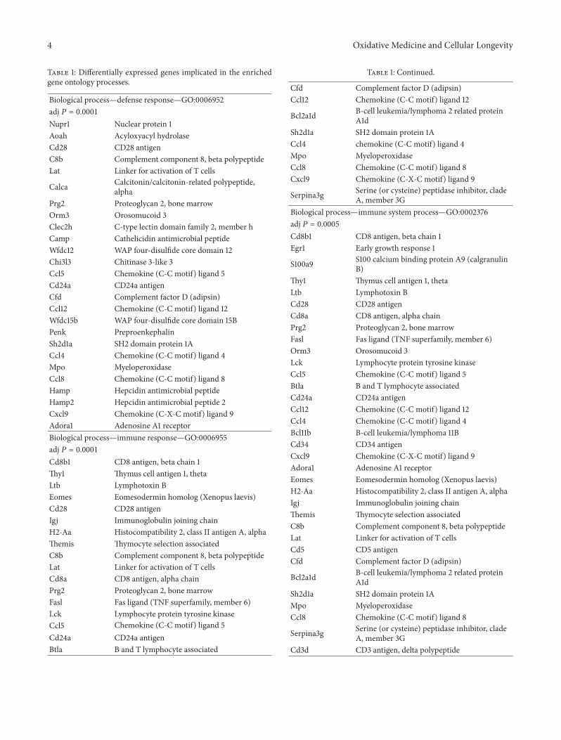
3.2. Gene Ontology (GO) Analysis of Differentially ExpressedGenes. To obtain insights into the functions of the 601DEGs, gene ontology (GO) analysis was performed. Themain processes that these genes are involved in are cate-gorized as follows: (1) immune response; (2) inflammatoryresponse; (3) carbohydrate and pattern binding; (4) G proteinand chemokine receptor binding; (5) glutathione transferaseactivity; (6) peptidase inhibitor activity; (7) cell surface,plasmamembrane, and extracellular region genes; and (8) ionhomeostasis. Table 1 lists the differentially expressed genesimplicated in each of the aforementioned functions.
4 Oxidative Medicine and Cellular Longevity
Table 1: Differentially expressed genes implicated in the enrichedgene ontology processes.
Biological process—defense response—GO:0006952adj 𝑃 = 0.0001Nupr1 Nuclear protein 1Aoah Acyloxyacyl hydrolaseCd28 CD28 antigenC8b Complement component 8, beta polypeptideLat Linker for activation of T cells
Calca Calcitonin/calcitonin-related polypeptide,alpha
Prg2 Proteoglycan 2, bone marrowOrm3 Orosomucoid 3Clec2h C-type lectin domain family 2, member hCamp Cathelicidin antimicrobial peptideWfdc12 WAP four-disulfide core domain 12Chi3l3 Chitinase 3-like 3Ccl5 Chemokine (C-C motif) ligand 5Cd24a CD24a antigenCfd Complement factor D (adipsin)Ccl12 Chemokine (C-C motif) ligand 12Wfdc15b WAP four-disulfide core domain 15BPenk PreproenkephalinSh2d1a SH2 domain protein 1ACcl4 Chemokine (C-C motif) ligand 4Mpo MyeloperoxidaseCcl8 Chemokine (C-C motif) ligand 8Hamp Hepcidin antimicrobial peptideHamp2 Hepcidin antimicrobial peptide 2Cxcl9 Chemokine (C-X-C motif) ligand 9Adora1 Adenosine A1 receptorBiological process—immune response—GO:0006955adj 𝑃 = 0.0001Cd8b1 CD8 antigen, beta chain 1Thy1 Thymus cell antigen 1, thetaLtb Lymphotoxin BEomes Eomesodermin homolog (Xenopus laevis)Cd28 CD28 antigenIgj Immunoglobulin joining chainH2-Aa Histocompatibility 2, class II antigen A, alphaThemis Thymocyte selection associatedC8b Complement component 8, beta polypeptideLat Linker for activation of T cellsCd8a CD8 antigen, alpha chainPrg2 Proteoglycan 2, bone marrowFasl Fas ligand (TNF superfamily, member 6)Lck Lymphocyte protein tyrosine kinaseCcl5 Chemokine (C-C motif) ligand 5Cd24a CD24a antigenBtla B and T lymphocyte associated
Table 1: Continued.
Cfd Complement factor D (adipsin)Ccl12 Chemokine (C-C motif) ligand 12
Bcl2a1d B-cell leukemia/lymphoma 2 related proteinA1d
Sh2d1a SH2 domain protein 1ACcl4 chemokine (C-C motif) ligand 4Mpo MyeloperoxidaseCcl8 Chemokine (C-C motif) ligand 8Cxcl9 Chemokine (C-X-C motif) ligand 9
Serpina3g Serine (or cysteine) peptidase inhibitor, cladeA, member 3G
Biological process—immune system process—GO:0002376adj 𝑃 = 0.0005Cd8b1 CD8 antigen, beta chain 1Egr1 Early growth response 1
S100a9 S100 calcium binding protein A9 (calgranulinB)
Thy1 Thymus cell antigen 1, thetaLtb Lymphotoxin BCd28 CD28 antigenCd8a CD8 antigen, alpha chainPrg2 Proteoglycan 2, bone marrowFasl Fas ligand (TNF superfamily, member 6)Orm3 Orosomucoid 3Lck Lymphocyte protein tyrosine kinaseCcl5 Chemokine (C-C motif) ligand 5Btla B and T lymphocyte associatedCd24a CD24a antigenCcl12 Chemokine (C-C motif) ligand 12Ccl4 Chemokine (C-C motif) ligand 4Bcl11b B-cell leukemia/lymphoma 11BCd34 CD34 antigenCxcl9 Chemokine (C-X-C motif) ligand 9Adora1 Adenosine A1 receptorEomes Eomesodermin homolog (Xenopus laevis)H2-Aa Histocompatibility 2, class II antigen A, alphaIgj Immunoglobulin joining chainThemis Thymocyte selection associatedC8b Complement component 8, beta polypeptideLat Linker for activation of T cellsCd5 CD5 antigenCfd Complement factor D (adipsin)
Bcl2a1d B-cell leukemia/lymphoma 2 related proteinA1d
Sh2d1a SH2 domain protein 1AMpo MyeloperoxidaseCcl8 Chemokine (C-C motif) ligand 8
Serpina3g Serine (or cysteine) peptidase inhibitor, cladeA, member 3G
Cd3d CD3 antigen, delta polypeptide

Oxidative Medicine and Cellular Longevity 5
Table 1: Continued.
Biological process—inflammatory response—GO:0006954adj 𝑃 = 0.0010Nupr1 Nuclear protein 1Aoah Acyloxyacyl hydrolaseCd28 CD28 antigenC8b Complement component 8, beta polypeptideLat Linker for activation of T cells
Calca Calcitonin/calcitonin-related polypeptide,alpha
Orm3 Orosomucoid 3Chi3l3 Chitinase 3-like 3Ccl5 Chemokine (C-C motif) ligand 5Cd24a CD24a antigenCfd Complement factor D (adipsin)Ccl12 Chemokine (C-C motif) ligand 12Ccl4 Chemokine (C-C motif) ligand 4Ccl8 Chemokine (C-C motif) ligand 8Cxcl9 Chemokine (C-X-C motif) ligand 9Adora1 Adenosine A1 receptorBiological process—cellular di-, tri-valent inorganic cationhomeostasis—GO:0030005adj 𝑃 = 0.0035Lck Lymphocyte protein tyrosine kinaseCd24a CD24a antigenLtf LactotransferrinGpr12 G-protein coupled receptor 12Mt2 Metallothionein 2Hamp Hepcidin antimicrobial peptideHamp2 Hepcidin antimicrobial peptide 2
Mfi2 Antigen p97 (melanoma associated) identifiedby monoclonal antibodies 133.2 and 96.5
Calca Calcitonin/calcitonin-related polypeptide,alpha
Slc39a5 Solute carrier family 39 (metal iontransporter), member 5
Biological process—cellular cation homeostasis—GO:0030003adj 𝑃 = 0.0054Lck Lymphocyte protein tyrosine kinaseCd24a CD24a antigenLtf LactotransferrinGpr12 G-protein coupled receptor 12Mt2 Metallothionein 2Hamp Hepcidin antimicrobial peptideHamp2 Hepcidin antimicrobial peptide 2
Mfi2 Antigen p97 (melanoma associated) identifiedby monoclonal antibodies 133.2 and 96.5
Calca Calcitonin/calcitonin-related polypeptide,alpha
Slc39a5 Solute carrier family 39 (metal iontransporter), member 5
Table 1: Continued.
Biological process—response to wounding—GO:0009611adj 𝑃 = 0.0054
Slc1a3 Solute carrier family 1 (glial high affinityglutamate transporter), member 3
Nupr1 Nuclear protein 1Aoah Acyloxyacyl hydrolaseCd28 CD28 antigenC8b Complement component 8, beta polypeptideLat Linker for activation of T cells
Calca Calcitonin/calcitonin-related polypeptide,alpha
Tff1 Trefoil factor 1Orm3 Orosomucoid 3Chi3l3 Chitinase 3-like 3Ccl5 Chemokine (C-C motif) ligand 5Cd24a CD24a antigenCfd Complement factor D (adipsin)Ccl12 Chemokine (C-C motif) ligand 12Ccl4 Chemokine (C-C motif) ligand 4Ccl8 Chemokine (C-C motif) ligand 8Cxcl9 Chemokine (C-X-C motif) ligand 9Adora1 Adenosine A1 receptorBiological process—di-, tri-valent inorganic cationhomeostasis—GO:0055066adj 𝑃 = 0.0054Lck Lymphocyte protein tyrosine kinaseCd24a CD24a antigenLtf LactotransferrinGpr12 G-protein coupled receptor 12Mt2 Metallothionein 2Hamp Hepcidin antimicrobial peptideHamp2 Hepcidin antimicrobial peptide 2
Mfi2 Antigen p97 (melanoma associated) identifiedby monoclonal antibodies 133.2 and 96.5
Calca Calcitonin/calcitonin-related polypeptide,alpha
Slc39a5 Solute carrier family 39 (metal iontransporter), member 5
Biological process—T cell activation—GO:0042110adj 𝑃 = 0.0067Lck Lymphocyte protein tyrosine kinaseEgr1 Early growth response 1Cd5 CD5 antigenBtla B and T lymphocyte associatedCd24a CD24a antigen
Bcl2a1d B-cell leukemia/lymphoma 2 related proteinA1d
Cd28 CD28 antigenH2-Aa Histocompatibility 2, class II antigen A, alphaThemis Thymocyte selection associated

6 Oxidative Medicine and Cellular Longevity
Table 1: Continued.
Bcl11b B-cell leukemia/lymphoma 11BCd3d CD3 antigen, delta polypeptideCd8a CD8 antigen, alpha chainBiological process—defense response to fungus—GO:0050832adj 𝑃 = 0.0067Hamp Hepcidin antimicrobial peptideHamp2 Hepcidin antimicrobial peptide 2Mpo MyeloperoxidaseBiological process—T cell differentiation—GO:0030217adj 𝑃 = 0.0067Lck Lymphocyte protein tyrosine kinaseEgr1 Early growth response 1
Bcl2a1d B-cell leukemia/lymphoma 2 related proteinA1d
Cd28 CD28 antigenH2-Aa Histocompatibility 2, class II antigen A, alphaThemis Thymocyte selection associatedBcl11b B-cell leukemia/lymphoma 11BCd3d CD3 antigen, delta polypeptideCd8a CD8 antigen, alpha chainMolecular function—carbohydrate binding—GO:0030246adj 𝑃 = 0.0029Vcan Versican
Klrg1 Killer cell lectin-like receptor subfamily G,member 1
Ltf LactotransferrinGpnmb Glycoprotein (transmembrane) nmbReg1 Regenerating islet-derived 1Lgals2 Lectin, galactose-binding, soluble 2Sftpd Surfactant associated protein DPrg2 Proteoglycan 2, bone marrowClec2h C-type lectin domain family 2, member hChi3l3 Chitinase 3-like 3Cd24a CD24a antigenNcam1 Neural cell adhesion molecule 1
Abp1 Amiloride binding protein 1 (amine oxidase,Copper-containing)
Mpo Myeloperoxidase
Klrd1 Killer cell lectin-like receptor, subfamily D,member 1
Crispld2 Cysteine-rich secretory protein LCCL domaincontaining 2
Ccl8 Chemokine (C-C motif) ligand 8Molecular function—peptidase inhibitor activity—GO:0030414adj 𝑃 = 0.0029Wfdc12 WAP four-disulfide core domain 12
Serpina9Serine (or cysteine) peptidase inhibitor, cladeA (alpha-1 antiproteinase, antitrypsin),member 9
Table 1: Continued.
Serpinb1a Serine (or cysteine) peptidase inhibitor, cladeB, member 1a
Bcl2a1d B-cell leukemia/lymphoma 2 related proteinA1d
Wfdc2 WAP four-disulfide core domain 2Wfdc15b WAP four-disulfide core domain 15BCst7 Cystatin F (leukocystatin)Spink4 Serine peptidase inhibitor, Kazal type 4
Serpina3g Serine (or cysteine) peptidase inhibitor, cladeA, member 3G
Spink3 Serine peptidase inhibitor, Kazal type 3
Serpina12Serine (or cysteine) peptidase inhibitor, cladeA (alpha-1 antiproteinase, antitrypsin),member 12
Molecular function—glutathione transferaseactivity—GO:0004364adj 𝑃 = 0.0032Gstt3 Glutathione S-transferase, theta 3Gstm1 Glutathione S-transferase, mu 1Gstm4 Glutathione S-transferase, mu 4Gsta2 Glutathione S-transferase, alpha 2 (Yc2)Gstm3 Glutathione S-transferase, mu 3Molecular function—endopeptidase inhibitoractivity—GO:0004866adj 𝑃 = 0.0032Wfdc12 WAP four-disulfide core domain 12
Serpina9Serine (or cysteine) peptidase inhibitor, cladeA (alpha-1 antiproteinase, antitrypsin),member 9
Serpinb1a Serine (or cysteine) peptidase inhibitor, cladeB, member 1a
Bcl2a1d B-cell leukemia/lymphoma 2 related proteinA1d
Wfdc2 WAP four-disulfide core domain 2Cst7 Cystatin F (leukocystatin)Spink4 Serine peptidase inhibitor, Kazal type 4
Serpina3g Serine (or cysteine) peptidase inhibitor, cladeA, member 3G
Spink3 Serine peptidase inhibitor, Kazal type 3
Serpina12Serine (or cysteine) peptidase inhibitor, cladeA (alpha-1 antiproteinase, antitrypsin),member 12
Molecular function—G-protein-coupled receptorbinding—GO:0001664adj 𝑃 = 0.0033Ccl8 Chemokine (C-C motif) ligand 8Ccl5 Chemokine (C-C motif) ligand 5Cxcl9 Chemokine (C-X-C motif) ligand 9Adrb3 Adrenergic receptor, beta 3Ccl12 Chemokine (C-C motif) ligand 12
Calca Calcitonin/calcitonin-related polypeptide,alpha

Oxidative Medicine and Cellular Longevity 7
Table 1: Continued.
Ccl4 Chemokine (C-C motif) ligand 4Molecular function—serine-type endopeptidase inhibitoractivity—GO:0004867adj 𝑃 = 0.0065Wfdc12 WAP four-disulfide core domain 12
Serpina9Serine (or cysteine) peptidase inhibitor, cladeA (alpha-1 antiproteinase, antitrypsin),member 9
Serpinb1a Serine (or cysteine) peptidase inhibitor, cladeB, member 1a
Serpina3g Serine (or cysteine) peptidase inhibitor, cladeA, member 3G
Spink4 Serine peptidase inhibitor, Kazal type 4Spink3 Serine peptidase inhibitor, Kazal type 3
Serpina12Serine (or cysteine) peptidase inhibitor, cladeA (alpha-1 antiproteinase, antitrypsin),member 12
Wfdc2 WAP four-disulfide core domain 2Molecular function—chemokine activity—GO:0008009adj 𝑃 = 0.0097Ccl8 Chemokine (C-C motif) ligand 8Ccl5 Chemokine (C-C motif) ligand 5Cxcl9 Chemokine (C-X-C motif) ligand 9Ccl12 Chemokine (C-C motif) ligand 12Ccl4 Chemokine (C-C motif) ligand 4Molecular function—chemokine receptorbinding—GO:0042379adj 𝑃 = 0.0097Ccl8 Chemokine (C-C motif) ligand 8Ccl5 Chemokine (C-C motif) ligand 5Cxcl9 Chemokine (C-X-C motif) ligand 9Ccl12 Chemokine (C-C motif) ligand 12Ccl4 Chemokine (C-C motif) ligand 4Molecular function—pattern binding—GO:0001871adj 𝑃 = 0.0097Vcan VersicanChi3l3 Chitinase 3-like 3Ncam1 Neural cell adhesion molecule 1Ltf LactotransferrinGpnmb Glycoprotein (transmembrane) nmb
Abp1 Amiloride binding protein 1 (amine oxidase,copper-containing)
Crispld2 Cysteine-rich secretory protein LCCL domaincontaining 2
Mpo MyeloperoxidaseCcl8 Chemokine (C-C motif) ligand 8Molecular function—polysaccharide binding—GO:0030247adj 𝑃 = 0.0097Vcan VersicanChi3l3 Chitinase 3-like 3Ncam1 Neural cell adhesion molecule 1
Table 1: Continued.
Ltf LactotransferrinGpnmb Glycoprotein (transmembrane) nmb
Abp1 Amiloride binding protein 1 (amine oxidase,copper-containing)
Crispld2 Cysteine-rich secretory protein LCCL domaincontaining 2
Mpo MyeloperoxidaseCcl8 Chemokine (C-C motif) ligand 8Cellular component—extracellular region—GO:0005576adj 𝑃 = 5.75𝑒 − 09Col5a2 Collagen, type V, alpha 2Vcan Versican
Slc1a3 Solute carrier family 1 (glial high affinityglutamate transporter), member 3
Dmbt1 Deleted in malignant brain tumors 1Ltb Lymphotoxin BItgbl1 Integrin, beta-like 1Mgp Matrix Gla proteinCst7 Cystatin F (leukocystatin)Fgl2 Fibrinogen-like protein 2Gzma Granzyme AProm1 Prominin 1
Ltbp2 Latent transforming growth factor betabinding protein 2
Afp Alpha fetoprotein
Calca Calcitonin/calcitonin-related polypeptide,alpha
Tff1 Trefoil factor 1Fasl Fas ligand (TNF superfamily, member 6)Camp Cathelicidin antimicrobial peptideOrm3 Orosomucoid 3Ccl5 Chemokine (C-C motif) ligand 5Ccl12 Chemokine (C-C motif) ligand 12
Abp1 Amiloride binding protein 1 (amine oxidase,copper-containing)
Penk PreproenkephalinWfdc15b WAP four-disulfide core domain 15BCcl4 Chemokine (C-C motif) ligand 4
Adipoq Adiponectin, C1Q and collagen domaincontaining
Hamp Hepcidin antimicrobial peptideCxcl9 Chemokine (C-X-C motif) ligand 9Il17rb Interleukin 17 receptor BMmp8 Matrix metallopeptidase 8
Svep1 Sushi, von Willebrand factor type A, EGF andpentraxin domain containing 1
Chi3l1 Chitinase 3-like 1Gcg Glucagon
Serpina9Serine (or cysteine) peptidase inhibitor, cladeA (alpha-1 antiproteinase, antitrypsin),member 9

8 Oxidative Medicine and Cellular Longevity
Table 1: Continued.
Oosp1 Oocyte secreted protein 1Ltf Lactotransferrin
Sema3g Sema domain, immunoglobulin domain (Ig),short basic domain, secreted, (semaphorin) 3G
Wfdc2 WAP four-disulfide core domain 2C8b Complement component 8, beta polypeptideLcn13 Lipocalin 13Esm1 Endothelial cell-specific molecule 1Dkk4 Dickkopf homolog 4 (Xenopus laevis)Gm128 Predicted gene 128Sftpd Surfactant associated protein DMuc4 Mucin 4Chi3l3 Chitinase 3-like 3Wfdc12 WAP four-disulfide core domain 12Tff2 Trefoil factor 2 (spasmolytic protein 1)Tff3 Trefoil factor 3, intestinalCpxm2 Carboxypeptidase X 2 (M14 family)Cfd Complement factor D (adipsin)Nts Neurotensin
Crispld2 Cysteine-rich secretory protein LCCL domaincontaining 2
Cd2 CD2 antigenMmp7 Matrix metallopeptidase 7Ccl8 Chemokine (C-C motif) ligand 8Cgref1 Cell growth regulator with EF hand domain 1Hamp2 Hepcidin antimicrobial peptide 2Gpc1 Glypican 1Dpt Dermatopontin
Mfi2 Antigen p97 (melanoma associated) identifiedby monoclonal antibodies 133.2 and 96.5
Spink4 Serine peptidase inhibitor, Kazal type 4Spink3 Serine peptidase inhibitor, Kazal type 3
Serpina12Serine (or cysteine) peptidase inhibitor, cladeA (alpha-1 antiproteinase, antitrypsin),member 12
Fam3b Family with sequence similarity 3, member BCellular component—plasma membrane—GO:0005886adj 𝑃 = 1.09𝑒 − 07Cd8b1 CD8 antigen, beta chain 1Thy1 Thymus cell antigen 1, thetaAdrb3 Adrenergic receptor, beta 3Sirpb1 Signal-regulatory protein beta 1Cd28 CD28 antigen
Treh Trehalase (brush-border membraneglycoprotein)
Cd8a CD8 antigen, alpha chain
Slc39a5 Solute carrier family 39 (metal iontransporter), member 5
Klrc1 Killer cell lectin-like receptor subfamily C,member 1
Clec2h C-type lectin domain family 2, member h
Table 1: Continued.
Gpr68 G protein-coupled receptor 68Gpr171 G protein-coupled receptor 171Btla B and T lymphocyte associatedCd24a CD24a antigenKrt19 Keratin 19Lair1 Leukocyte-associated Ig-like receptor 1Gldn GliomedinGpr110 G protein-coupled receptor 110Plxdc2 Plexin domain containing 2Adora1 Adenosine A1 receptorMuc3 Mucin 3, intestinalDsg1c Desmoglein 1 gammaIl17rb Interleukin 17 receptor BMas1 MAS1 oncogene
Abcc3 ATP-binding cassette, sub-family C(CFTR/MRP), member 3
Lor LoricrinGbp5 Guanylate binding protein 5H2-Aa Histocompatibility 2, class II antigen A, alphaEspn Espin
Glycam1 Glycosylation dependent cell adhesionmolecule 1
Cd3g CD3 antigen, gamma polypeptideLat Linker for activation of T cells
Ildr1 Immunoglobulin-like domain containingreceptor 1
Cxcr6 Chemokine (C-X-C motif) receptor 6Plek2 Pleckstrin 2Gpr65 G-protein coupled receptor 65Aqp8 Aquaporin 8
Kcnn4Potassium intermediate/small conductancecalcium-activated channel, subfamily N,member 4
Slco1a1 Solute carrier organic anion transporterfamily, member 1a1
Pdcd1 Programmed cell death 1Mapt Microtubule-associated protein tauCd5 CD5 antigenClstn3 Calsyntenin 3Gpr12 G-protein coupled receptor 12Cd2 CD2 antigenGbp1 Guanylate binding protein 1
Vsig2 V-set and immunoglobulin domaincontaining 2
Gpc1 Glypican 1Gna14 Guanine nucleotide binding protein, alpha 14
Slc1a3 Solute carrier family 1 (glial high affinityglutamate transporter), member 3
Itgbl1 Integrin, beta-like 1Ltb Lymphotoxin BArhgap10 Rho GTPase activating protein 10

Oxidative Medicine and Cellular Longevity 9
Table 1: Continued.
Gbp2 Guanylate binding protein 2Prom1 Prominin 1Car9 Carbonic anhydrase 9
Pip5k1a Phosphatidylinositol-4-phosphate 5-kinase,type 1 alpha
Rab25 RAB25, member RAS oncogene familyFasl Fas ligand (TNF superfamily, member 6)Tjp3 Tight junction protein 3Lck Lymphocyte protein tyrosine kinaseGpr174 G protein-coupled receptor 174
Slc16a13 Solute carrier family 16 (monocarboxylic acidtransporters), member 13
Gnat2 Guanine nucleotide binding protein, alphatransducing 2
Ncam1 Neural cell adhesion molecule 1Igsf9 Immunoglobulin superfamily, member 9
Klrd1 Killer cell lectin-like receptor, subfamily D,member 1
Art2b ADP-ribosyltransferase 2bDsg1b Desmoglein 1 betaCd34 CD34 antigenOscp1 Organic solute carrier partner 1Itk IL2-inducible T-cell kinaseGpr128 G protein-coupled receptor 128Rasgrp2 RAS, guanyl releasing protein 2Slc26a3 Solute carrier family 26, member 3Cdh1 Cadherin 1Il2rb Interleukin 2 receptor, beta chainGpnmb Glycoprotein (transmembrane) nmb
Ms4a4b Membrane-spanning 4-domains, subfamily A,member 4B
C8b Complement component 8, beta polypeptideGrin3b Glutamate receptor, ionotropic, NMDA3BClic6 Chloride intracellular channel 6Grm8 Glutamate receptor, metabotropic 8
Mfi2 Antigen p97 (melanoma associated) identifiedby monoclonal antibodies 133.2 and 96.5
Cd3d CD3 antigen, delta polypeptideSlc30a10 Solute carrier family 30, member 10Pcdh20 Protocadherin 20Cellular component—cell surface—GO:0009986adj 𝑃 = 4.72𝑒 − 07Cd8b1 CD8 antigen, beta chain 1
Slc1a3 Solute carrier family 1 (glial high affinityglutamate transporter), member 3
Thy1 Thymus cell antigen 1, thetaIl2rb Interleukin 2 receptor, beta chainCd28 CD28 antigenH2-Aa Histocompatibility 2, class II antigen A, alphaProm1 Prominin 1H2-M2 Histocompatibility 2, M region locus 2
Table 1: Continued.
Cd8a CD8 antigen, alpha chainFasl Fas ligand (TNF superfamily, member 6)Pdcd1 Programmed cell death 1
Klrc1 Killer cell lectin-like receptor subfamily C,member 1
Cd24a CD24a antigenBtla B and T lymphocyte associatedCd5 CD5 antigenNcam1 Neural cell adhesion molecule 1
Klrd1 Killer cell lectin-like receptor, subfamily D,member 1
Cd2 CD2 antigenCd34 CD34 antigenIl17rb Interleukin 17 receptor BCellular component—external side of plasmamembrane—GO:0009897adj 𝑃 = 1.43𝑒 − 06Cd8b1 CD8 antigen, beta chain 1Cd5 CD5 antigenBtla B and T lymphocyte associatedCd24a CD24a antigenThy1 Thymus cell antigen 1, thetaNcam1 Neural cell adhesion molecule 1Il2rb Interleukin 2 receptor, beta chainCd28 CD28 antigenH2-Aa Histocompatibility 2, class II antigen A, alphaCd2 CD2 antigen
Klrd1 Killer cell lectin-like receptor, subfamily D,member 1
Cd34 CD34 antigenCd8a CD8 antigen, alpha chainFasl Fas ligand (TNF superfamily, member 6)Pdcd1 Programmed cell death 1
Klrc1 Killer cell lectin-like receptor subfamily C,member 1
Cellular component—plasma membrane part—GO:0044459adj 𝑃 = 1.08𝑒 − 05Cd8b1 CD8 antigen, beta chain 1Thy1 Thymus cell antigen 1, thetaItgbl1 Integrin, beta-like 1Cd28 CD28 antigenProm1 Prominin 1Cd8a CD8 antigen, alpha chainFasl Fas ligand (TNF superfamily, member 6)
Slc39a5 Solute carrier family 39 (metal iontransporter), member 5
Tjp3 Tight junction protein 3
Klrc1 Killer cell lectin-like receptor subfamily C,member 1
Clec2h C-type lectin domain family 2, member h

10 Oxidative Medicine and Cellular Longevity
Table 1: Continued.
Gnat2 Guanine nucleotide binding protein, alphatransducing 2
Cd24a CD24a antigenBtla B and T lymphocyte associatedNcam1 Neural cell adhesion molecule 1Igsf9 Immunoglobulin superfamily, member 9Art2b ADP-ribosyltransferase 2b
Klrd1 Killer cell lectin-like receptor, subfamily D,member 1
Dsg1b Desmoglein 1 betaCd34 CD34 antigenMuc3 Mucin 3, intestinalDsg1c Desmoglein 1 gammaIl17rb Interleukin 17 receptor BRasgrp2 RAS, guanyl releasing protein 2Slc26a3 Solute carrier family 26, member 3
Abcc3 ATP-binding cassette, sub-family C(CFTR/MRP), member 3
Lor LoricrinCdh1 Cadherin 1Il2rb Interleukin 2 receptor, beta chainGpnmb Glycoprotein (transmembrane) nmbH2-Aa Histocompatibility 2, class II antigen A, alphaEspn EspinCd3g CD3 antigen, gamma polypeptide
Ms4a4b Membrane-spanning 4-domains, subfamily A,member 4B
C8b Complement component 8, beta polypeptideLat Linker for activation of T cellsGrin3b Glutamate receptor, ionotropic, NMDA3BAqp8 Aquaporin 8
Kcnn4Potassium intermediate/small conductancecalcium-activated channel, subfamily N,member 4
Slco1a1 Solute carrier organic anion transporterfamily, member 1a1
Pdcd1 Programmed cell death 1Cd5 CD5 antigenGrm8 Glutamate receptor, metabotropic 8Cd2 CD2 antigen
Vsig2 V-set and immunoglobulin domaincontaining 2
Cd3d CD3 antigen, delta polypeptideGna14 Guanine nucleotide binding protein, alpha 14Cellular component—extracellular region part—GO:0044421adj 𝑃 = 0.0043Col5a2 Collagen, type V, alpha 2Vcan Versican
Slc1a3 Solute carrier family 1 (glial high affinityglutamate transporter), member 3
Dmbt1 Deleted in malignant brain tumors 1Ltb Lymphotoxin B
Table 1: Continued.
Prom1 Prominin 1Afp Alpha fetoprotein
Calca Calcitonin/calcitonin-related polypeptide,alpha
Sftpd Surfactant associated protein DFasl Fas ligand (TNF superfamily, member 6)Orm3 Orosomucoid 3Ccl5 Chemokine (C-C motif) ligand 5Tff3 Trefoil factor 3, intestinalCpxm2 Carboxypeptidase X 2 (M14 family)Cfd Complement factor D (adipsin)Ccl12 Chemokine (C-C motif) ligand 12Ccl4 Chemokine (C-C motif) ligand 4
Crispld2 Cysteine-rich secretory protein LCCL domaincontaining 2
Adipoq Adiponectin, C1Q and collagen domaincontaining
Mmp7 Matrix metallopeptidase 7Ccl8 Chemokine (C-C motif) ligand 8Gpc1 Glypican 1Dpt DermatopontinCxcl9 Chemokine (C-X-C motif) ligand 9Mmp8 Matrix metallopeptidase 8Fam3b Family with sequence similarity 3, member BCellular component—intrinsic to plasma membrane—GO:0031226adj 𝑃 = 0.0093Slc26a3 Solute carrier family 26, member 3Thy1 Thymus cell antigen 1, thetaItgbl1 Integrin, beta-like 1
Abcc3 ATP-binding cassette, sub-family C(CFTR/MRP), member 3
Gpnmb Glycoprotein (transmembrane) nmb
Ms4a4b Membrane-spanning 4-domains, subfamily A,member 4B
Prom1 Prominin 1C8b Complement component 8, beta polypeptideLat Linker for activation of T cellsGrin3b Glutamate receptor, ionotropic, NMDA3BAqp8 Aquaporin 8
Kcnn4Potassium intermediate/small conductancecalcium-activated channel, subfamily N,member 4
Slco1a1 Solute carrier organic anion transporterfamily, member 1a1
Clec2h C-type lectin domain family 2, member hCd24a CD24a antigenBtla B and T lymphocyte associatedGrm8 Glutamate receptor, metabotropic 8Cd2 CD2 antigen
Vsig2 V-set and immunoglobulin domaincontaining 2
Il17rb Interleukin 17 receptor B

Oxidative Medicine and Cellular Longevity 11
3.3. qRT-PCR Verification of Microarray Results. For vali-dation of the microarray data, a group of genes known tobe directly or indirectly implicated in lipid or carbohydratemetabolismwere selected for quantification with quantitativereal-time RT-PCR (qRT-PCR). These genes were Cyp7a1(cytochrome P450, family 7, subfamily A, polypeptide 1),which is the rate limiting enzyme in bile acid synthesis fromcholesterol; Fabp5 (fatty acid binding protein 5); Car (Nr1I3)(constitutive androstane receptor); Cyp2b10 (cytochromeP450,family 2, subfamily B, polypeptide 10), which is a Cartarget; Lipocalin 13;Aquaporin 8;Cbr3 (carbonyl reductase 3);and Me1 (malic enzyme 1). Nqo1 (NAD(P)H dehydrogenase,quinone 1) was selected as a prototypical Nrf2 target gene.These genes were quantified not only in the liver of WT andNrf2-KO mice under HFD for 180 days, but also in the liverof WT and Nrf2-KO mice under standard diet (St.D.) forthe same time period. The relative gene expression levels aredepicted in Figure 1.Cyp7a1 and Fabp5were found to be over-expressed in the livers of Nrf2-KO mice after HFD feedingcompared to WT mice, while the rest of the genes wereunder-expressed. There was excellent agreement betweenthe microarray and the qRT-PCR data (Pearson correlationcoefficient = 0.919; 𝑃 value < 0.001) (Figure 2).
3.4. Canonical Pathways and Networks Impacted by Nrf2under HFD Feeding. Ingenuity pathway analysis (IPA) wasused to rank gene networks by order of consistency of themicroarray results with relationships confirmed by previ-ously published results. Figure S2 presents a network whichcomprises Nrf2 and was generated by the microarray data.Nrf2 and genes that are under-expressed in the Nrf2-KOmice are shown in green; genes that are over-expressed inthe Nrf2-KO mice are shown in red. It is obvious that allof the under-expressed genes in this network have beendescribed to be directly regulated by Nrf2: carboxylesterase1 g (Ces1g) [26]; glutathione S-transferase mu 5 (Gstm5)[27];glutathione S-transferase alpha 5 (Gsta5) [28]; andNAD(P)Hdehydrogenase quinone 1 (Nqo1) [29]. In contrast, none ofthe genes that are over-expressed in the Nrf2-KO mice isknown to be directly regulated by Nrf2. These genes arecalcitonin-related polypeptide beta (Calcb); collagen typeV alpha 2 (Col5a2); cytochrome P450 family 2 subfamilyC polypeptide 8 (Cyp2c8); growth factor independent 1(Gfi1); H2-M2 histocompatibility 2, M region, locus 2 (H2-m2); interferon gamma (Ifng); solute carrier family 14 (ureatransporter) member 1 (Slc14a1); solute carrier family 26member 3 (Slc26a3); solute carrier family 9 (sodium hydro-gen exchanger)member 3 (Slc9a3); serine peptidase inhibitor,Kazal type 4 (Spink4); sulfotransferase family 1E, estrogenpreferring, member 1 (Su1lte1); and trefoil factor 1 (Tff1).
IPA analysis identified the statistically significant canon-ical pathways in the gene list. A corrected Fischer’s exact test𝑃 value < 0.05 was used as the threshold of significance(Figure 3). The number of genes (n) that were differentiallyexpressed in each canonical pathway is shown below alongwith the 𝑃 value and the ratio. In the “xenobiotic metabolismsignaling” pathway (𝑛 = 7; 𝑃 value = 3.95𝐸 − 06; ratio= 0.027), Cyp2c8 and Sult1e1 were overexpressed, whereas
Gstm5, Ces1g, Gsta5, Nqo1, Nfe2l2 (as expected), and sul-fotransferase family cytosolic 2A dehydroepiandrosterone-preferring member 1 (Sult2a1) were under-expressed. In the“aryl hydrocarbon receptor signaling” pathway (𝑛 = 6; 𝑃value = 5.88𝐸 − 06; ratio = 0.038), Tff1 and Fas ligand (Faslg)were over-expressed, whereasGstm5,Gsta5,Nqo1, andNfe2l2were underexpressed. In the “LPS/IL-1 mediated inhibitionof RXR function” pathway (𝑛 = 5; 𝑃 value = 5.49𝐸 −04; ratio = 0.022), Cyp2c8 and Sult1e1 were over-expressed,whereas Gstm5, Gsta5, and Sult2a1 were underexpressed.In the “metabolism of xenobiotics by cytochrome P450”pathway (𝑛 = 4; 𝑃 value = 5.65𝐸 − 04; ratio = 0.02), Cyp2c8was over-expressed and Cyp2b13/Cyp2b9, Gstm5, and Gsta5were underexpressed. In the “sulfur metabolism” pathway(𝑛 = 2; 𝑃 value = 1.96𝐸 − 03; ratio = 0.034), Sult1e1 was over-expressed, whereas Sult2a1 was underexpressed.
4. Discussion
The findings of our previous study that male Nrf2-KOmice were at least partially protected from HFD-induced(60 kcal% fat) obesity and were more insulin sensitive andmore glucose tolerant compared to their WT counterparts[13] are consistent with previous reports using comparablebut different treatment parameters (40 kcal% fat diet ormodified high-fat-diets) [14–16]. The purpose of this studywas to identify hepatic genes differentially expressed betweenWT and Nrf2-KO mice after long-term (180 days) high-fat-diet-(HFD-) induced obesity. Such information couldprovide insights into the recently appreciated implication ofNrf2 in the development of obesity and metabolic syndrome.To this end, microarray-based transcriptome analysis wasperformed, employing strict statistical criteria.
The microarray-based gene expression analysis in thesemice generated a total of 601 genes that were differentiallyexpressed between the two genotypes: 478 genes were over-expressed in theNrf2-KOmice, and 173were underexpressed.These genes are not only implicated in functions that aretypical of Nrf2 target genes, such as immune response [30],inflammation [31–33], and glutathione transferase activity[34, 35], but some gene groups were also found to be asso-ciated with carbohydrate and pattern binding (interactingselectively and noncovalently with a repeating or polymericstructure, such as a polysaccharide or peptidoglycan); Gprotein and chemokine receptor binding; peptidase inhibitoractivity; cell surface, plasma membrane, and extracellularregion genes; and ion homeostasis. These results furtherreinforce the notion that Nrf2 should not be regarded solelyas a central antioxidant transcription factor, but it may bealso implicated directly or indirectly in various tissue-specifichomeostatic and/or physiological processes [11].
Among a total 601 differentially expressed genes (DEGs),a subset was selected for validation by qRT-PCR quantifi-cation based on their relevance to metabolic pathways. Themetabolic pathways and the respective representative geneschosen were bile acid synthesis from cholesterol (Cyp7a1),free fatty acid binding and transport (Fabp5), glucosemetabolism (Lipocalin 13), glycerol transport (Aquaporin 8),

12 Oxidative Medicine and Cellular Longevity
Fabp5 Car
0
0.5
1
1.5
2
2.5
3
Relat
ive m
RNA
leve
l
0
0.5
1
1.5
Relat
ive m
RNA
leve
l
0
0.5
1
1.5
Relat
ive m
RNA
leve
l
0
0.5
1
1.5
Relat
ive m
RNA
leve
l
0.5
1
1.5
Relat
ive m
RNA
leve
l
0
0.5
1
1.5
Relat
ive m
RNA
leve
l
0
0
0.5
1
1.5
Relat
ive m
RNA
leve
l
0
0.5
1
1.5
0
0.5
1
1.5
2
2.5
3
Relat
ive m
RNA
leve
l
Relat
ive m
RNA
leve
l
Cyp7a1
Cyp2b10
WT,
HFD
Nrf2
-KO
, HFD
WT,
ST.
D.
Nrf2
-KO
, ST.
D.
WT,
HFD
Nrf2
-KO
, HFD
WT,
ST.
D.
Nrf2
-KO
, ST.
D.
WT,
HFD
Nrf2
-KO
, HFD
WT,
ST.
D.
Nrf2
-KO
, ST.
D.
WT,
HFD
Nrf2
-KO
, HFD
WT,
ST.
D.
Nrf2
-KO
, ST.
D.
WT,
HFD
Nrf2
-KO
, HFD
WT,
ST.
D.
Nrf2
-KO
, ST.
D.
WT,
HFD
Nrf2
-KO
, HFD
WT,
ST.
D.
Nrf2
-KO
, ST.
D.
WT,
HFD
Nrf2
-KO
, HFD
WT,
ST.
D.
Nrf2
-KO
, ST.
D.
WT,
HFD
Nrf2
-KO
, HFD
WT,
ST.
D.
Nrf2
-KO
, ST.
D.
WT,
HFD
Nrf2
-KO
, HFD
WT,
ST.
D.
Nrf2
-KO
, ST.
D.
∗∗
∗
∗∗∗∗∗∗
∗∗∗∗
∗∗∗
∗∗∗ ∗∗∗
∗∗∗
∗∗∗
∗∗∗
∗∗∗
Lipocalin 13 Aquaporin 8
Cbr3
∗∗∗
∗∗∗∗∗∗
∗∗∗
∗∗∗∗∗
Me1 Nqo1∗∗∗
∗∗∗
∗∗∗
∗∗∗
∗∗∗ ∗∗∗
∗∗∗
∗∗∗∗∗
∗
0.25
0.75
1.25
1.75
2.25
2.75
0.25
0.75
1.25
1.75
2.25
2.75
0.25
0.75
1.25
0.25
0.75
1.25
0.25
0.75
1.25
0.25
0.75
1.25
0.25
0.75
1.25
0.25
0.75
1.25
Figure 1: RelativemRNA levels based on qRT-PCR analysis.Cyp7a1, Fabp5,Car,Cyp2b10, Lipocalin 13,Aquaporin8,Cbr3,Me1, andNqo1wereselected for quantification with qRT-PCR in WT and Nrf2-KO mice under standard diet (St.D.) or high-fat diet (HFD) for 180 days (𝑛 = 8for each genotype in either diet type). The qRT-PCR was performed in triplicate wells for each sample. Bars show means ± SD. ∗𝑃 < 0.01,∗∗
𝑃 < 0.001, ∗∗∗𝑃 < 0.0001.
fatty acid biosynthesis (Me1), and energy homeostasis (Carand its target gene Cyp2b10).Nqo1mRNA levels were quanti-fied as Nqo1 is considered a prototypical Nrf2 target gene.
The qRT-PCR-based mRNA quantification of specificgenes of metabolic interest that were either over-expressed(Cyp7a1 and Fabp5) or under-expressed (Car, Cyp2b10,Lipocalin 13, Aquaporin 8, Cbr3, and Me1) in Nrf2-KO micecompared to WT under HFD revealed potential candidategenes that may be implicated in the development of thedifferent metabolic phenotype of Nrf2-KO mice. As shown
in Figure 1, the differential expression of some of these geneswas also evident under the St.D. regimen (with the exceptionof Aquaporin 8, Lipocalin 13, and Cbr3), indicating that thesegenes may be regulated by Nrf2 under basal conditions aswell. HFD for 180 days increased the expression of these genes(with the exception ofCyp2b10 in both genotypes and ofNqo1in the Nrf2-KO mice), and the fold difference between thetwo genotypes was generally accentuated. This observationmay indicate that the possible regulation (direct or indirect)of these genes byNrf2 becomes more prominent under stress

Oxidative Medicine and Cellular Longevity 13
MicroarraysqRT-PCR
Fabp
5
Car
Cyp7
a1
Cyp2
b10
Lipo
calin
13
Aqua
porin
8
Cbr3
Me1
Nqo1
−10
−5
0
5
Fold
expr
essio
n
Figure 2: Correlation betweenmicroarray and qRT-PCR data. Ninegenes identified by microarray analysis differentially expressed inthe liver between Nrf2-KO and WT mice after 180 days on HFDwere selected for qRT-PCR quantification. The red bars show thefold difference in the mRNA expression of a gene as calculatedby microarray analysis, and the blue bars show the relevant folddifference as calculated by qRT-PCR analysis. Very good agreementwas observed between the microarrays and qRT-PCR (Pearsoncorrelation coefficient = 0.919; 𝑃 = 0.0004).
conditions (HFD-induced obesity). Given that Nqo1 is aprototypical Nrf2 target, its over-expression afterHFD inWTmice suggests an increase in the transcriptional activity ofNrf2 by HFD, as supported by the fact that Nqo1 inductionwas not observed in the Nrf2-KO mice under HFD. Thepossible functional importance of each of the other validatedDEGs is discussed below.
Cyp7a1 is the rate-limiting enzyme in bile acid synthesisfrom cholesterol. In agreement with previous studies [36],we show that Cyp7a1 mRNA levels increased significantlyin both genotypes after HFD feeding (Figure 1). The Cyp7a1mRNA levels also differed between the two genotypes, withthe Nrf2-KO mice showing higher levels under St.D. (about60% higher) and much higher levels under HFD (about120% higher) compared to WT. In a previous study, a short-term (30 days) HFD did not accentuate the basal differencebetween the two genotypes [36], probably because an HFDfeeding for a shorter period exposed the animals to lowermetabolic and oxidative stress, such that the differencescaused by Nrf2 deletion were not as pronounced. Moreover,given that small heterodimer partner (Shp) represses Cyp7a1expression [37], and Nrf2 induces Shp gene expression [38],a reasonable hypothesis could be that Nrf2 represses Cyp7a1expression through Shp.This repression ofCyp7a1 expressionis abrogated by Nrf2 deletion, leading to increased levelsof Cyp7a1 in Nrf2-KO mice compared to WT, which maypartially protect them from obesity [39]. To clarify thesemolecular mechanisms, future experiments should involveShp and Nrf2 single and double KO mice.
Fabp5 is a member of the fatty acid-binding proteinswhich binds free fatty acids and regulates lipid metabolismand transport; it was first identified as being upregulatedin psoriasis tissue [40]. In this study, Fabp5 was increasedafter HFD feeding in both genotypes (Figure 1), which is inagreement with previous studies that have shown strong up-regulation of Fabp5 by western-type diet or HFD [41, 42].Fabp5 also exhibited higher mRNA levels in Nrf2-KO miceunder either St.D. or HFD; a study using proteomic analysisshowed similar results [43]. The specific physiological sig-nificance of Fabp5 elevation in Nrf2-KO mice remains to beelucidated. Further experimentswithNrf2 over-expression orsilencing and with concurrent measurement of Nrf2 levels inhepatocytes are warranted to elucidate the possible regulationof Fabp5 by Nrf2.
Car (Nr1I3), initially characterized as a sensor of xenobi-otics that regulates responses to toxicants [44], has recentlybeen implicated in the control of energy and metabolism[45]. Car has been ascribed a function as an antiobesityreceptor, because treatment of mice with a Car agonist par-tially prevented HFD-induced obesity in mice, and partiallyreversed obesity in mice that were already obese [46, 47]. Inthe present study, mRNA levels of Car, along with those ofits primary target gene, Cyp2b10 [48], were lower in Nrf2-KOmice compared to WT under either St.D. or HFD (Figure 1).In this case, the lower expression of Car cannot justify theameliorated metabolic phenotype of Nrf2-KO compared toWT after long-term HFD. Nevertheless, the observation thatCar and Cyp2b10 mRNA levels are lower in Nrf2-KO micethan WT is in accordance with previous studies [49, 50].Car mRNA was found to be increased in both genotypesafter the HFD regimen. But Cyp2b10 that is considered aCar target gene does not follow the same trend. This mayindicate a difference in the mRNA turnover of Cyp2b10 thatmay or may not be reflected in the protein levels. Furtherinvestigations, such as cell culture studies with manipulationofNrf2 levels/activity andmeasurement ofCar levels/activity,are necessary to clarify the mechanisms that underlie thepossible regulation of Car by Nrf2.
Lipocalin 13 is a lipocalin family member involved inglucose metabolism, and its deficiency is associated withobesity [51].Herein, lipocalin 13 livermRNA levelswere foundto be lower in Nrf2-KO mice than in WT after long-termHFD; no difference was observed between the two genotypesunder St.D. (Figure 1). As the existing data on the role oflipocalin 13 in obesity are scarce, this differential lipocalin 13mRNA expression between the two genotypes after the HFDfeeding for 180 days warrants further elucidation.
Aquaporin family members are mainly water channels,but some of them have also been found to transport glyceroland to be involved in the development of obesity [52].Aquaporin 8 is expressed in liver [53], and in the presentexperimental model it exhibited lower mRNA expression inthe liver of Nrf2-KO mice compared to WT after HFD. Nodifference was found between the two genotypes under St.D.,but aquaporin 8 was markedly induced in both genotypesafter HFD with its levels being lower in the Nrf2-KO mice(Figure 1). As aquaporins may be implicated in the transportof glycerol (a product of the catabolism of triacylglycerols), a

14 Oxidative Medicine and Cellular Longevity
0
1
2
3
4
5
Ratio
0
0.025
0.05
0.075
0.1
0.125
Ratio
Xeno
biot
ic m
etab
olism
sign
alin
g
LPS/
IL-1
med
iate
d in
hibi
tion
ofRX
R fu
nctio
n
Met
abol
ism o
f Xen
obio
tics b
ycy
toch
rom
e P45
0
Sulfu
r met
abol
ism
Allo
graft
reje
ctio
n sig
nalin
g
NRF
2-m
edia
ted
oxid
ativ
e stre
ssre
spon
se
Gra
ft-ve
rsus
-hos
t dise
ase
signa
ling
Chon
droi
tin su
lfate
bio
synt
hesis
Prim
ary
imm
unod
efici
ency
sig
nalin
g
Cyste
ine m
etab
olism
Ary
l hyd
roca
rbon
rece
ptor
sig
nalin
g
Threshold−lo
g(𝑃
val
ue)
Figure 3: Significantly enriched canonical pathways identified by IPA.The diagram shows significantly overrepresented canonical pathways.Amultiple testing corrected 𝑃 value was calculated using the Benjamini-Hochberg method to control the rate of false discoveries in statisticalhypothesis testing.The ratio value represents the number ofmolecules in a given pathway thatmeet cutoff criteria, divided by the total numberof molecules associated with the respective biological function.
possible indirect regulation of aquaporin 8 byNrf2may be ofmetabolic interest.
Cbr3 catalyzes the reduction of carbonyl compounds(highly reactive lipid aldehydes) to the corresponding alco-hols (inactive compounds) [54]. A recent clinical study [55]showed that a genetic variation in Cbr3 gene in humanscorrelates with type 2 diabetes and this effect can poten-tially be attributed to the catalysis of the conversion ofprostaglandin E2 to prostaglandin F2𝛼. In the present study,Cbr3 liver mRNA levels were about 5 times lower in Nrf2-KO mice after HFD compared to WT. Although WT micetended to have greater Cbr3 mRNA levels under standarddiet, this difference was not statistically significant. Giventhat recent studies have shown that the Cbr3 promotercomprises antioxidant response element (ARE) sequencesthat are recognized byNrf2 to induceCbr3 expression [50, 56,57], it would be interesting to test whether the Nrf2-regulatedCbr3 expression can contribute to the observed phenotype inthe Nrf2-KO mice after HFD feeding.
Me1 is an enzyme that generates NADPH for fatty acidbiosynthesis. In this study, Me1 showed decreased mRNAlevels in Nrf2-KOmice under St.D. or HFD; this is consistentwith previous gene expression profiling studies that describeMe1 as a Nrf2-dependent gene [58–60]. It has been previouslyshown that Nrf2 can redirect glucose and glutamine toanabolic pathways in cancer cells, and Me-1 is implicated inthese pathways [60]; however, these results in cancer cellscannot necessarily be safely extrapolated to nontransformedhepatocytes [61].
A limitation of this study is that microarray analysis wasperformed only in mice under HFD and not also on standard
diet (St.D.). Thus, it is not possible to delineate amongthe 601 DEGs those genes that are differentially expressedirrespective of the treatment versus those that demonstratediet-induced differential expression, except for the subsetof genes that were validated by qRT-PCR, which analyzedgene expression in mice under both St.D. and HFD. Anotherlimitation of this study is that the hepatic gene expression inthis whole body knock-out model may be affected indirectlyby endocrine factors that are secreted from other tissues(e.g., adipose tissue, muscle) that are also deficient in Nrf2.Therefore, some of the differentially expressed genes wedetect in the liver may be affected by extrahepatic factors.The use of a liver-specific knock-out model could resolve thisissue.
5. Conclusions
In conclusion, the current study showed that Nrf2 deletionsignificantly altered the hepatic gene expression profile afterlong-term HFD, yielding a set of 601 DEGs that can be thefocus of further studies on the role of Nrf2 in obesity. Themajority of these DEGs are involved in pathways relevant tothe defense against oxidative and electrophilic stress, alreadyknown to be regulated by Nrf2. However, certain genes suchas Cyp7a1, Fabp5, Car, Cbr3, and Me1 can have specificmetabolic effects and appear to be directly or indirectlyregulated by Nrf2; these genes may be implicated in the lessobese and more insulin sensitive metabolic phenotype ofthe Nrf2-KO mice. Novel mechanistic understanding andtherapeutic interventions for obesity/metabolic syndrome

Oxidative Medicine and Cellular Longevity 15
may arise from the elucidation of the cross-talk of Nrf2 withmetabolic pathways regulated by these genes.
Conflict of Interests
All the authors declare that they have no conflict of interests.
Authors’ Contribution
Dionysios V. Chartoumpekis and Panos G. Ziros contributedequally to this work. Dionysios V. Chartoumpekis, PanosG. Ziros, Gerasimos P. Sykiotis, and Ioannis G. Habeosconceived and designed the experiments. Dionysios V. Char-toumpekis, Panos G. Ziros, Ralista P. Iskrenova, and IoannisG. Habeos performed the experiments. Apostolos Zaravi-nos, Dionysios V. Chartoumpekis, and Ioannis G. Habeosanalyzed data. Apostolos Zaravinos, Agathoklis I. Psyrogian-nis, and Venetsana E. Kyriazopoulou contributed to theseexperiments with the reagents, materials, and analysis tools.Dionysios V. Chartoumpekis, Apostolos Zaravinos, Agathok-lis I. Psyrogiannis, Venetsana E. Kyriazopoulou, Gerasimos P.Sykiotis, and Ioannis G. Habeos wrote the paper.
Acknowledgments
This work was partially supported by a grant from theHellenic Endocrine Society (I. G. Habeos) and by the Uni-versity of Patras internal funding programme “ConstantinCaratheodory” (I. G. Habeos). The addresses 2 and 4 arethe present addresses of Dionysios V. Chartoumpekis andApostolos Zaravinos.
References
[1] S. Haffner and H. Taegtmeyer, “Epidemic obesity and themetabolic syndrome,”Circulation, vol. 108, no. 13, pp. 1541–1545,2003.
[2] P. T. James, R. Leach, E. Kalamara, andM. Shayeghi, “Theworld-wide obesity epidemic,” Obesity Research, vol. 9, supplement 4,pp. 228S–233S, 2001.
[3] E. E. Calle and R. Kaaks, “Overweight, obesity and cancer:epidemiological evidence and proposed mechanisms,” NatureReviews Cancer, vol. 4, no. 8, pp. 579–591, 2004.
[4] R. N. Bergman, S. P. Kim, I. R. Hsu et al., “Abdominalobesity: role in the pathophysiology of metabolic disease andcardiovascular risk,” American Journal of Medicine, vol. 120, no.2, supplement 1, pp. 3–8, 2007.
[5] G. P. Sykiotis, I. G. Habeos, A. V. Samuelson, and D. Bohmann,“The role of the antioxidant and longevity-promoting Nrf2pathway in metabolic regulation,” Current Opinion in ClinicalNutrition and Metabolic Care, vol. 14, no. 1, pp. 41–48, 2011.
[6] M. J. Calkins, D. A. Johnson, J. A. Townsend et al., “TheNrf2/ARE pathway as a potential therapeutic target in neurode-generative disease,”Antioxidants andRedox Signaling, vol. 11, no.3, pp. 497–508, 2009.
[7] M. Kobayashi and M. Yamamoto, “Nrf2-Keap1 regulation ofcellular defense mechanisms against electrophiles and reactiveoxygen species,” Advances in Enzyme Regulation, vol. 46, no. 1,pp. 113–140, 2006.
[8] G. P. Sykiotis and D. Bohmann, “Stress-activated cap’n’collartranscription factors in aging and human disease,” ScienceSignaling, vol. 3, no. 112, p. re3, 2010.
[9] T. W. Kensler, N. Wakabayashi, and S. Biswal, “Cell survivalresponses to environmental stresses via the Keap1-Nrf2-AREpathway,” Annual Review of Pharmacology and Toxicology, vol.47, pp. 89–116, 2007.
[10] W. O. Osburn and T. W. Kensler, “Nrf2 signaling: an adaptiveresponse pathway for protection against environmental toxicinsults,”Mutation Research, vol. 659, no. 1-2, pp. 31–39, 2008.
[11] J. M. Lee, J. Li, D. A. Johnson et al., “Nrf2, a multi-organprotector?” FASEB Journal, vol. 19, no. 9, pp. 1061–1066, 2005.
[12] H. Motohashi and M. Yamamoto, “Nrf2-Keap1 defines a phys-iologically important stress response mechanism,” Trends inMolecular Medicine, vol. 10, no. 11, pp. 549–557, 2004.
[13] D. V. Chartoumpekis, P. G. Ziros, A. I. Psyrogiannis et al., “Nrf2represses FGF21 during long-termhigh-fat diet-induced obesityin mice,” Diabetes, vol. 60, no. 10, pp. 2465–2473, 2011.
[14] J. Pi, L. Leung, P. Xue et al., “Deficiency in the nuclear factorE2-related factor-2 transcription factor results in impairedadipogenesis and protects against diet-induced obesity,” Journalof Biological Chemistry, vol. 285, no. 12, pp. 9292–9300, 2010.
[15] Y. K. Zhang, K. C. Wu, J. Liu, and C. D. Klaassen, “Nrf2deficiency improves glucose tolerance in mice fed a high-fatdiet,” Toxicology and Applied Pharmacology, vol. 264, no. 3, pp.305–314, 2012.
[16] A. K. Meher, P. R. Sharma, V. A. Lira et al., “Nrf2 deficiency inmyeloid cells is not sufficient to protect mice from high-fat diet-induced adipose tissue inflammation and insulin resistance,”Free Radical Biology & Medicine, vol. 52, no. 9, pp. 1708–1715,2012.
[17] K. Itoh, T. Chiba, S. Takahashi et al., “An Nrf2/small Mafheterodimer mediates the induction of phase II detoxifyingenzyme genes through antioxidant response elements,” Bio-chemical and Biophysical Research Communications, vol. 236,no. 2, pp. 313–322, 1997.
[18] A. I. Saeed, N. K. Bhagabati, J. C. Braisted et al., “TM4microarray software suite,”Methods in Enzymology, vol. 411, pp.134–193, 2006.
[19] A. I. Saeed, V. Sharov, J. White et al., “TM4: a free, open-source system for microarray data management and analysis,”BioTechniques, vol. 34, no. 2, pp. 374–378, 2003.
[20] B. Zhang, S. Kirov, and J. Snoddy, “WebGestalt: an integratedsystem for exploring gene sets in various biological contexts,”Nucleic Acids Research, vol. 33, no. 2, pp. W741–W748, 2005.
[21] A. Zaravinos, G. I. Lambrou, I. Boulalas, D. Delakas, and D. A.Spandidos, “Identification of common differentially expressedgenes in urinary bladder cancer,”PLoSONE, vol. 6, no. 4, ArticleID e18135, 2011.
[22] M. W. Pfaffl, “A new mathematical model for relative quantifi-cation in real-time RT-PCR,”Nucleic Acids Research, vol. 29, no.9, article e45, 2001.
[23] A. Spandidos, X.Wang, H.Wang, S. Dragnev, T.Thurber, and B.Seed, “A comprehensive collection of experimentally validatedprimers for Polymerase Chain Reaction quantitation of murinetranscript abundance,” BMC Genomics, vol. 9, article 633, 2008.
[24] A. Spandidos, X. Wang, H. Wang, and B. Seed, “Primer-Bank: a resource of human and mouse PCR primer pairs forgene expression detection and quantification,” Nucleic AcidsResearch, vol. 38, no. 1, pp. D792–D799, 2010.

16 Oxidative Medicine and Cellular Longevity
[25] X. Wang and B. Seed, “A PCR primer bank for quantitativegene expression analysis,”Nucleic Acids Research, vol. 31, no. 24,article e154, 2003.
[26] Y. Zhang, X. Cheng, L. Aleksunes, and C. D. Klaassen,“Transcription factor-mediated regulation of carboxylesteraseenzymes in livers of mice,” Drug Metabolism and Disposition,vol. 40, no. 6, pp. 1191–1197, 2012.
[27] J. D. Hayes, S. A. Chanas, C. J. Henderson et al., “The Nrf2transcription factor contributes both to the basal expression ofglutathione S-transferases in mouse liver and to their induc-tion by the chemopreventive synthetic antioxidants, butylatedhydroxyanisole and ethoxyquin,” Biochemical Society Transac-tions, vol. 28, no. 2, pp. 33–41, 2000.
[28] M. S. Yates, M. K. Kwak, P. A. Egner et al., “Potent protectionagainst aflatoxin-induced tumorigenesis through induction ofNrf2-regulated pathways by the triterpenoid 1-[2-cyano-3-,12-dioxooleana-1, 9(11)-dien-28-oyl]imidazole,” Cancer Research,vol. 66, no. 4, pp. 2488–2494, 2006.
[29] P. Nioi, M. McMahon, K. Itoh, M. Yamamoto, and J. D. Hayes,“Identification of a novel NRF2-regulated antioxidant responseelement (ARE) in the mouse NAD(P)H:quinone oxidoreduc-tase 1 gene: reassessment of the ARE consensus sequence,”Biochemical Journal, vol. 374, no. 2, pp. 337–348, 2003.
[30] R. K. Thimmulappa, H. Lee, T. Rangasamy et al., “Nrf2 is acritical regulator of the innate immune response and survivalduring experimental sepsis,” Journal of Clinical Investigation,vol. 116, no. 4, pp. 984–995, 2006.
[31] K. Itoh, M. Mochizuki, Y. Ishii et al., “Transcription factor Nrf2regulates inflammation by mediating the effect of 15-deoxy-Δ12,14-prostaglandin j(2),” Molecular and Cellular Biology, vol.24, no. 1, pp. 36–45, 2004.
[32] N. G. Innamorato, A. I. Rojo, A. J. Garcıa-Yague, M. Yamamoto,M. L. De Ceballos, and A. Cuadrado, “The transcription factornrf2 is a therapeutic target against brain inflammation,” Journalof Immunology, vol. 181, no. 1, pp. 680–689, 2008.
[33] N. Li and A. E. Nel, “Role of the Nrf2-mediated signalingpathway as a negative regulator of inflammation: implicationsfor the impact of particulate pollutants on asthma,”Antioxidantsand Redox Signaling, vol. 8, no. 1-2, pp. 88–98, 2006.
[34] S. A. Chanas, Q. Jiang, M. McMahon et al., “Loss of the Nrf2transcription factor causes a marked reduction in constitutiveand inducible expression of the glutathione S-transferase Gsta1,Gsta2, Gstm1, Gstm2, Gstm3 and Gstm4 genes in the livers ofmale and female mice,” Biochemical Journal, vol. 365, no. 2, pp.405–416, 2002.
[35] R. K. Thimmulappa, K. H. Mai, S. Srisuma, T. W. Kensler,M. Yamamoto, and S. Biswal, “Identification of Nrf2-regulatedgenes induced by the chemopreventive agent sulforaphane byoligonucleotidemicroarray,”Cancer Research, vol. 62, no. 18, pp.5196–5203, 2002.
[36] Y. Tanaka, L. M. Aleksunes, R. L. Yeager et al., “NF-E2-relatedfactor 2 inhibits lipid accumulation and oxidative stress in micefed a high-fat diet,” Journal of Pharmacology and ExperimentalTherapeutics, vol. 325, no. 2, pp. 655–664, 2008.
[37] Y.K. J. Zhang,G. L.Guo, andC.D.Klaassen, “Diurnal variationsof mouse plasma and hepatic bile acid concentrations as wellas expression of biosynthetic enzymes and transporters,” PLoSONE, vol. 6, no. 2, Article ID e16683, 2011.
[38] J. Huang, I. Tabbi-Anneni, V. Gunda, and L. Wang, “Transcrip-tion factor Nrf2 regulates SHP and lipogenic gene expression inhepatic lipid metabolism,” American Journal of Physiology, vol.299, no. 6, pp. G1211–G1221, 2010.
[39] W. F. Yiu, P. L. Kwan, C. Y.Wong et al., “Attenuation of fatty liverand prevention of hypercholesterolemia by extract of Curcumalonga through regulating the expression of CYP7A1, LDL-receptor, HO-1, and HMG-CoA reductase,” Journal of FoodScience, vol. 76, no. 3, pp. H80–H89, 2011.
[40] G. Slegenthaler, R. Hotz, D. Chatellard-Gruaz, S. Jaconi, and J.H. Saurat, “Characterization and expression of a novel humanfatty acid-binding protein: the epidermal type (E-FABP),” Bio-chemical andBiophysical ResearchCommunications, vol. 190, no.2, pp. 482–487, 1993.
[41] M. Hoekstra, M. Stitzinger, E. J. A. Van Wanrooij et al.,“Microarray analysis indicates an important role for FABP5 andputative novel FABPs on a Western-type diet,” Journal of LipidResearch, vol. 47, no. 10, pp. 2198–2207, 2006.
[42] A. A. Toye, M. E. Dumas, C. Blancher et al., “Subtle metabolicand liver gene transcriptional changes underlie diet-inducedfatty liver susceptibility in insulin-resistant mice,” Diabetologia,vol. 50, no. 9, pp. 1867–1879, 2007.
[43] N. R. Kitteringham, A. Abdullah, J. Walsh et al., “Proteomicanalysis of Nrf2 deficient transgenic mice reveals cellulardefence and lipid metabolism as primary Nrf2-dependentpathways in the liver,” Journal of Proteomics, vol. 73, no. 8, pp.1612–1631, 2010.
[44] P. Wei, J. Zhang, M. Egan-Haftey, S. Liang, and D. D. Moore,“The nuclear receptor CARmediates specific xenobiotic induc-tion of drug metabolism,” Nature, vol. 407, no. 6806, pp. 920–923, 2000.
[45] T. Wada, J. Gao, and W. Xie, “PXR and CAR in energymetabolism,” Trends in Endocrinology and Metabolism, vol. 20,no. 6, pp. 273–279, 2009.
[46] J. Gao, J. He, Y. Zhai, T. Wada, and W. Xie, “The constitutiveandrostane receptor is an anti-obesity nuclear receptor thatimproves in sulin sensitivity,” Journal of Biological Chemistry,vol. 284, no. 38, pp. 25984–25992, 2009.
[47] B. Dong, P. K. Saha, W. Huang et al., “Activation of nuclearreceptor CAR ameliorates diabetes and fatty liver disease,”Proceedings of the National Academy of Sciences of the UnitedStates of America, vol. 106, no. 44, pp. 18831–18836, 2009.
[48] P. Honkakoski, I. Zelko, T. Sueyoshi, and M. Negishi, “Thenuclear orphan receptor CAR-retinoid X receptor heterodimeractivates the phenobarbital-responsive enhancer module of theCYP2B gene,”Molecular and Cellular Biology, vol. 18, no. 10, pp.5652–5658, 1998.
[49] A. Anwar-Mohamed, O. S. Degenhardt, M. A. M. El Gendy,J. M. Seubert, S. R. Kleeberger, and A. O. S. El-Kadi, “Theeffect of Nrf2 knockout on the constitutive expression of drugmetabolizing enzymes and transporters in C57Bl/6 mice livers,”Toxicology in Vitro, vol. 25, no. 4, pp. 785–795, 2011.
[50] K. C. Wu, J. Y. Cui, and C. D. Klaassen, “Effect of graded Nrf2activation on phase-I and -II drug metabolizing enzymes andtransporters in mouse liver,” PLoS One, vol. 7, no. 7, Article IDe39006, 2012.
[51] K.W.Cho, Y. Zhou, L. Sheng, and L. Rui, “Lipocalin-13 regulatesglucose metabolism by both insulin-dependent and insulin-independent mechanisms,”Molecular and Cellular Biology, vol.31, no. 3, pp. 450–457, 2011.
[52] E. M. Wintour and B. A. Henry, “Glycerol transport: anadditional target for obesity therapy?” Trends in Endocrinologyand Metabolism, vol. 17, no. 3, pp. 77–78, 2006.
[53] F. Garcıa, A. Kierbel, M. C. Larocca et al., “The water channelaquaporin-8 is mainly intracellular in rat hepatocytes, and

Oxidative Medicine and Cellular Longevity 17
its plasma membrane insertion is stimulated by cyclic AMP,”Journal of Biological Chemistry, vol. 276, no. 15, pp. 12147–12152,2001.
[54] E. S. Pilka, F. H. Niesen, W. H. Lee et al., “Structural basis forsubstrate specificity in humanmonomeric carbonyl reductases,”PLoS ONE, vol. 4, no. 10, article e7113, 2009.
[55] Y. C. Chang, P. H. Liu, Y. C. Tsai et al., “Genetic variation in thecarbonyl reductase 3 gene confers risk of type 2 diabetes andinsulin resistance: a potential regulator of adipogenesis,” Journalof Molecular Medicine, vol. 90, no. 7, pp. 847–858, 2012.
[56] Q. Cheng, J. L. Kalabus, J. Zhang, and J. G. Blanco, “A conservedantioxidant response element (ARE) in the promoter of humancarbonyl reductase 3 (CBR3) mediates induction by the masterredox switch Nrf2,” Biochemical Pharmacology, vol. 83, no. 1, pp.139–148, 2012.
[57] B. Ebert, M. Kisiela, P. Malatkova, Y. El-Hawari, and E. Maser,“Regulation of human carbonyl reductase 3 (CBR3; SDR21C2)expression by Nrf2 in cultured cancer cells,” Biochemistry, vol.49, no. 39, pp. 8499–8511, 2010.
[58] R. Hu, C. Xu, G. Shen et al., “Gene expression profiles inducedby cancer chemopreventive isothiocyanate sulforaphane in theliver of C57BL/6J mice and C57BL/6J/Nrf2 (-/-) mice,” CancerLetters, vol. 243, no. 2, pp. 170–192, 2006.
[59] R. Hu, C. Xu, G. Shen et al., “Identification of Nrf2-regulatedgenes induced by chemopreventive isothiocyanate PEITC byoligonucleotide microarray,” Life Sciences, vol. 79, no. 20, pp.1944–1955, 2006.
[60] Y. Mitsuishi, K. Taguchi, Y. Kawatani et al., “Nrf2 redirectsglucose and glutamine into anabolic pathways in metabolicreprogramming,” Cancer Cell, vol. 22, no. 1, pp. 66–79, 2012.
[61] M. A. Lazar and M. J. Birnbaum, “Physiology. De-meaning ofmetabolism,” Science, vol. 336, no. 6089, pp. 1651–1652, 2012.
Copyright © 2022 FDOKUMEN