Hepatic hemangioendothelioma: Clinical experience and management strategy
9
Hepatic Hemangioendothelioma: Clinical Experience and Management Strategy By John A. Daller, Javier Bueno, Jorge Gutierrez, lgor Dvorchik, Richard B. Towbin, Paul S. Dickman, George Mazariegos, and Jorge Reyes Pittsburgh, Pennsylvania Purpose:This study sought to define management strategies based on clinical experience in treating infantile hepatic hemangioendothelioma. Methods:A retrospective analysis of patients with hemangio- endothelioma presenting to a tertiary liver transplantation center between 1989 and 1997 was performed. ResukThirteen patients (median age, 14 days) with heman- gioendothelioma were identified. Congestive heart failure (P< .03) and abdominal mass (P < .081) were predictive of 5-month mortality rates. Ultrasonography and computerized axial tomography were the diagnostic modalities most com- monly used. Treatment strategies consisted of medical man- agement (steroids and a-interferon) and interventional mo- dalities (hepatic artery ligation or embolization, resectional surgery, or orthotopic liver transplantation). Patients who underwent resectional surgery, with or without orthotopic liver transplantation, had a lower 5-month mortality rate (P < .02) and a greater Z-year survival rate (P < .003) than did those who underwent hepatic artery ligation or embolization. Early morbidity and mortality tended to be a consequence of the primary lesion, whereas late morbidity and mortality were reflective of the treatment modality used. Conclusions: In cases of failed medical management, resec- tional therapy should be used when possible. If partial hepatectomy is not technically achievable, hepatic artery embolization should be used either as definitive therapy or as a temporizing measure until orthotopic liver transplantation is possible. J Pediatr Surg 34:98-106. Copyright o 7999 by W.B. Saunders Company. INDEX WORDS: Infantile hepatic hemangioendothelioma, orthotopic liver transplantation, steroids, a-interferon. I NFANTILE HEPATIC hemangioendothelioma is a rare benign vascular tumor that presents most com- monly before the age of 6 months.’ Because these vascular lesions can act as arteriovenous fistulas, they can produce life-threatening high output congestive heart failure with respiratory compromise and a resultant mortality rate as high as 9070.’ Hemangioendotheliomas also have been associated with Kasabach-Merritt syn- drome,3 anemia,” intraperitoneal hemorrhage secondary to rupture,5 consumptive coagulopathy,6 obstructive jaun- dice,’ and vascular malformations involving the brain, skin, gut, and other organs.8-‘0 Although children with asymptomatic lesions may experience spontaneous regression within a year,” symp- From the Thomas E. Starzl Transplant Institute, University oj Pittsburgh and Children k Hospital of Pittsburgh Transplantation SurgeT. and the Departments of Pediatric Radiology and Pathology, Children k Hospital of Pittsburgh, Pittsburgh. PA. Presented at the 29th Annual Meeting of the American Pediatric Surgical Association, Hilton Head. South Carolina, May 10-13, 1998. Supported in part by research grants from the Veterans Administra- tion and Project Grant No. DK-29961 from the National Institutes of Health, Bethesda, MD. Address correspondence to Jorge Reyes. MD. Children $ Hospital oj Pittsburgh, 3705 Fifh Ave. Pittsburgh. PA 15213. Copyright o 1999 by WB. Saunders Company 0022-3468/99/340/-0020$03.00/O 98 tomatic lesions require aggressive management because the disease may be rapidly fatal once symptoms com- mence.(j A variety of therapeutic options, including medical and interventional modalities, have been sug- gested in the management of these lesions. Medical therapy includes corticosteroids’* and radiation therapy I3 to reduce arteriovenous shunting as well as digoxin and diuretics for symptomatic management of congestive heart failure.‘4,‘5 a-Interferon recently has been used to induce regression of these lesions purportedly by inhibit- ing angiogenic growth. I5 Interventional modalities in- clude embolization or ligation of the hepatic artery,l.16 resectional surgery,” and orthotopic liver transplanta- tion.iB-20 The purpose of this report is to describe our experience with infantile hemangioendothelioma and to present an algorithm for management of this potentially aggressive lesion in light of our experience and the current literature. MATERIALS AND METHODS Children’s Hospital of Pittsburgh is a tertiary liver transplantation center. A retrospective analysis of patients with hepatic tumors between 1989 and 1997 was performed. A total of 14 patients were identified with the diagnosis of hemangioendothelioma from medical record discharge codes and databases kept by the departments of radiology, critical care medicine, and pathology. Charts were independently reviewed by two authors and the following information collected: age at presentation. sex. symptoms, diagnostic tests performed, distribution of JournalofPediafricSurgery, Vol34,No 1 (January),1999:pp98-106
-
Upload
independent -
Category
Documents
-
view
2 -
download
0
Transcript of Hepatic hemangioendothelioma: Clinical experience and management strategy

Hepatic Hemangioendothelioma: Clinical Experience and Management Strategy
By John A. Daller, Javier Bueno, Jorge Gutierrez, lgor Dvorchik, Richard B. Towbin, Paul S. Dickman, George Mazariegos, and Jorge Reyes
Pittsburgh, Pennsylvania
Purpose:This study sought to define management strategies based on clinical experience in treating infantile hepatic hemangioendothelioma.
Methods:A retrospective analysis of patients with hemangio- endothelioma presenting to a tertiary liver transplantation center between 1989 and 1997 was performed.
ResukThirteen patients (median age, 14 days) with heman- gioendothelioma were identified. Congestive heart failure (P< .03) and abdominal mass (P < .081) were predictive of 5-month mortality rates. Ultrasonography and computerized axial tomography were the diagnostic modalities most com- monly used. Treatment strategies consisted of medical man- agement (steroids and a-interferon) and interventional mo- dalities (hepatic artery ligation or embolization, resectional surgery, or orthotopic liver transplantation). Patients who underwent resectional surgery, with or without orthotopic liver transplantation, had a lower 5-month mortality rate
(P < .02) and a greater Z-year survival rate (P < .003) than did those who underwent hepatic artery ligation or embolization. Early morbidity and mortality tended to be a consequence of the primary lesion, whereas late morbidity and mortality were reflective of the treatment modality used.
Conclusions: In cases of failed medical management, resec- tional therapy should be used when possible. If partial hepatectomy is not technically achievable, hepatic artery embolization should be used either as definitive therapy or as a temporizing measure until orthotopic liver transplantation is possible. J Pediatr Surg 34:98-106. Copyright o 7999 by W.B. Saunders Company.
INDEX WORDS: Infantile hepatic hemangioendothelioma, orthotopic liver transplantation, steroids, a-interferon.
I NFANTILE HEPATIC hemangioendothelioma is a rare benign vascular tumor that presents most com-
monly before the age of 6 months.’ Because these vascular lesions can act as arteriovenous fistulas, they can produce life-threatening high output congestive heart failure with respiratory compromise and a resultant mortality rate as high as 9070.’ Hemangioendotheliomas also have been associated with Kasabach-Merritt syn- drome,3 anemia,” intraperitoneal hemorrhage secondary to rupture,5 consumptive coagulopathy,6 obstructive jaun- dice,’ and vascular malformations involving the brain, skin, gut, and other organs.8-‘0
Although children with asymptomatic lesions may experience spontaneous regression within a year,” symp-
From the Thomas E. Starzl Transplant Institute, University oj Pittsburgh and Children k Hospital of Pittsburgh Transplantation SurgeT. and the Departments of Pediatric Radiology and Pathology, Children k Hospital of Pittsburgh, Pittsburgh. PA.
Presented at the 29th Annual Meeting of the American Pediatric Surgical Association, Hilton Head. South Carolina, May 10-13, 1998.
Supported in part by research grants from the Veterans Administra- tion and Project Grant No. DK-29961 from the National Institutes of Health, Bethesda, MD.
Address correspondence to Jorge Reyes. MD. Children $ Hospital oj Pittsburgh, 3705 Fifh Ave. Pittsburgh. PA 15213.
Copyright o 1999 by WB. Saunders Company 0022-3468/99/340/-0020$03.00/O
98
tomatic lesions require aggressive management because the disease may be rapidly fatal once symptoms com- mence.(j A variety of therapeutic options, including medical and interventional modalities, have been sug- gested in the management of these lesions. Medical therapy includes corticosteroids’* and radiation therapy I3 to reduce arteriovenous shunting as well as digoxin and diuretics for symptomatic management of congestive heart failure.‘4,‘5 a-Interferon recently has been used to induce regression of these lesions purportedly by inhibit- ing angiogenic growth. I5 Interventional modalities in- clude embolization or ligation of the hepatic artery,l.16 resectional surgery,” and orthotopic liver transplanta- tion.iB-20
The purpose of this report is to describe our experience with infantile hemangioendothelioma and to present an algorithm for management of this potentially aggressive lesion in light of our experience and the current literature.
MATERIALS AND METHODS
Children’s Hospital of Pittsburgh is a tertiary liver transplantation center. A retrospective analysis of patients with hepatic tumors between 1989 and 1997 was performed. A total of 14 patients were identified with the diagnosis of hemangioendothelioma from medical record discharge codes and databases kept by the departments of radiology, critical care medicine, and pathology. Charts were independently reviewed by two authors and the following information collected: age at presentation. sex. symptoms, diagnostic tests performed, distribution of
JournalofPediafricSurgery, Vol34,No 1 (January),1999:pp98-106

HEPATIC HEMANGIOENDOTHELIOMA 99
disease, liver function tests. coagulation parameters. hematocrit level. white blood cell count. albumin. medical therapeutics. interventional modalities. complications of therapy, and outcome.
Statistical analysis was performed by the SPSS statistical program using a two-sided Fisher’s Exact x? test. Statistical significance was demonstrated by P values less than or equal to .05. Because of the uneven distribution of survival experience, two outcomes of interest were identified: (I ) death within 150 days and (2) long-term survival rate (greater than 2 years). The influence of various clinical variables on these two events was then evaluated.
RESULTS
A total of fourteen children (nine girls and five boys) with the admitting diagnosis of hemangioendothelioma were identified during the study period. Patients were referred either after not responding to conventional medical management (n = 8) or being deemed to have extensive disease requiring aggressive interventional therapies such as resection or transplantation. One girl was found to have metastatic angiosarcoma after ortho- topic liver transplantation and was excluded from further analysis. Of the remaining 13 patients, the mean age at presentation was 3 11 days (median, 14 days). including two children with unusual presentations at 3.4 and 6.8 years of age. Excluding these two children, the mean age at presentation was 29 days. Two patients were lost to follow-up after 5 months.
Hepatomegaly. abdominal mass, skin lesions, and congestive heart failure were the most common features found (Table 1). The mothers of two children used fertility drugs. Survival analysis based on symptomatol- ogy showed that of five patients who did not undergo transplantation and manifesting more than five of the symptoms noted in the table, only one survived more than 300 days. The two patients who underwent transplanta- tion with more than five symptoms have achieved survival of 21/, years or more. Two of five patients with fewer than five symptoms have survived over 2% years, one is alive and well at 81/z months after diagnosis, one was lost to follow-up after 5 months, and one died at 1 month of hepatic failure after hepatic artery emboliza- tion. The presence of congestive heart failure (P < .03)
Table 1. Presenting Symptoms
No. (%)
Hepatomegaly 10 (83)
Abdominal mass 8 (66) Skin hemangioma 8 (66) Congestive heart failure 7 (58) Splenomegaly 6 (50) Jaundice 6 (50) Kassabach-Merrit syndrol me 4 (33) Ascites 3 (25) Gastrointestinal bleeding 3 (25) Anemia 3 (251 Feeding abnormality 3 (25) Hepatic bruit 2 (16)
and abdominal mass (P < ,081, not significant) were predictive of death by 5 months after presentation. Congestive heart failure continued to portend a worse outcome 2 years after diagnosis (P < .09, not signifi- cant). Ten patients had at least one abnormal liver function test finding; these were equally divided between canalicular enzymes and transaminases. Three patients had thrombocytopenia, and one had an abnormal pro- thrombin time at the time of initial evaluation. None presented with coagulopathy, although a bleeding diathe- sis as a complication of therapy developed in one child after attempted medical management and hepatic artery embolization. Of seven patients who had albumin mea- sured, three had levels above 3 mg/dL: these children all survived.
A variety of diagnostic examinations were used in patient management. Ultrasonography and computerized axial tomography were used in most cases, whereas arteriography was reserved for those patients requiring interventional modalities. Echocardiography was used as clinically necessary for congestive heart failure and other modalities such as magnetic resonance imaging and nuclear medicine scans were used occasionally. These studies showed bilobar disease in eight patients, left lobe disease in four patients, and right lobe disease in one individual. Those with single-lobe disease tended to have solitary lesions, whereas those with bilobar disease had multinodular lesions. Characteristic early and late comput- erized axial tomography images are shown in Fig 1.
Pathological examination confirmed the diagnosis of hemangioendothelioma in 7 of 13 patients. One patient had angiosarcoma. In the remaining six cases, the diagno- sis of hemangioendothelioma was based on characteristic clinical and radiographic features of the disease. One patient’s lesion (Table 2, #2) demonstrated features of vascular malformation and mesenchymal hamartoma in addition to areas typical of hemangioendothelioma. Fig- ure 2 demonstrates the microscopic features of thin- walled vascular channels lined by flattened endothelial cells found in the unilobar lesion of patient 1 (Table 2).
Treatment options were divided into medical and interventional modalities. Medical management was based on high-dose steroids (1 to 4 mg/kg/d prednisone) for periods ranging up to 2 months and adjunctive o-inter- feron ( 1 to 3 mU mm2 ddr ) in selected patients. Digoxin and diuretics were given for symptomatic congestive heart failure. Subsequently, interventional modalities were used that included hepatic artery ligation (n = 2) or embolization (n = 6), conventional resectional surgery (n = 5), and orthotopic liver transplantation (n = 3). Resectional surgery was performed primarily in those patients with unilobar disease. Orthotopic liver transplan- tation was performed for severe bilobar disease in one patient and for the development of Budd-Chiari syn-

DALLER ET AL
Fig 2. Unilobar hemangioendothelioma found in patient 1. The lesion is composed of thin-welled vascular channels lined by flat- tened endothelial cells. (H&E, original magnification x511.
Fig 1. Early phase postcontrast abdominal CT scan shows diffuse hypervascular nodules involving the entire liver. Note the enlarged celiac axis (large arrowheads) and main hepatic artery (small arrow- heads).
drome in two patients after primary resection. Emboliza- tion was also used in cases of bilobar disease after arteriographic evaluation of the lesion’s vascular supply. Figure 3 demonstrates the arteriographic appearance of
hemangioendothelioma both before and after hepatic artery embolization. Complications of therapy included pulmonary disease (pneumonia, ARDS, and atelectasis), infection, multiple organ failure, and posttransplant lymph- oproliferative disease. Early complications tended to be related to the primary disease, whereas later complica- tions tended to be a reflection of the treatment modalities used.
Patients who underwent resectional surgery with or without orthotopic liver transplantation enjoyed a signifi- cantly greater 5-month survival rate (P < .02) than did those patients who underwent either hepatic artery liga- tion or embolization as well as a 2-year survival rate greater than other forms of therapy (P < ,003). Eight of the original 14 patients have died, including the patient with angiosarcoma. Of those patients with unilobar disease, four of five have survived; of the remaining eight patients with bilobar disease, only two have survived. As expected, patients with bilobar disease exhibited a signifi-
Table 2. Management and Outcome of Patients With Hepatic Hemangioendothelioma
Patient No. Disease Extent Treatment Outcome Cause of Death Survival (d)
1 Left lobe Lobectomy Alive *
2 Bilobar St, IFN, Dig, Dt, HAE Dead CHF 75 3 Bilobar St, IFN, Dig, Dt, HAE Dead CHF 29
4 Left lobe Segmentectomy Alive *
5 Bilobar St, IFN, Dig, Dt, HAE Alive 315 6 Bilobar St, IFN, Dt, HAE Dead Sepsis 94 7 Bilobar St, IFN, Dig, Dt, HAE Alive 259 8 Right lobe Dt, lobectomy, OLTX Alive 6,936
9 Left lobe Segmentectomy Alive 1,906 10 Bilobar St, IFN, HAE Dead MOF 15
11 Bilobar St, OLTX Dead IVH 2,203 12 Left lobe Segmentectomy, OLTX Dead PTLD 940 13 Bilobar St, Dig, Dt, Chm, XRT, HAL Dead Sepsis 141
Abbreviations: St, steroids; IFN, a-interferon; Dig, digoxin; Dt, diuretic; Chm, chemotherapy, cytoxan; XRT, radiation, 466 rads x 4; HAE, hepatic
arteryembolization; HAL, hepatic artery ligation; OLTX, orthotopic livertransplantation; CHF, congestive heartfailure; MOF, multiple organ failure; IVH, intraventricular hemorrhage; PTLD, posttransplant lymphoproliferative disease.
*Lost to follow-up after 5 months.

HEPATIC HEMANGIOENDOTHELIOMA
Fig 3. Selective hepatic artery injec- tion before (A) and after embolization (B). (A) A 3F catheter is positioned in the proximal right hepatic artery. There is dense, irregular vascular blush in- volving all segments of the right he- patic lobe. Early sinusoidal filling was noted (not shown). (Bl Three O.&cm Hilal coils (arrowheads) were placed in the right hepatic artery. There is nor- mal reduction of intrahepatic hyper- vascularity.
cantly greater short-term mortality rate (less than 5 months) than did those patients with unilobar disease (P < .05). Although two of the three patients who underwent transplantation have died, their survival ranged from 2.5 to 19 years, longer than those patients treated with conventional means (Table 2).
Causes of death included sepsis (n = 2), congestive heart failure (n = 2) intraventricular hemorrhage, post- transplant lymphoproliferative disease, and multiple or- gan failure. As expected, short-term deaths were related to the disease process itself, whereas the longer survivors (namely those who received liver transplantation) suc- cumbed to complications of the transplant.
DISCUSSION
Infantile hepatic hemangioendothelioma, although rare. is the most common vascular tumor of the liver in childhood, exhibiting a 1.5 to 2: 1 female predomi- nance.Z’JZ Usually, 85% of lesions manifest within the first 3 months of life.’ Pathologically, two patterns are described in infantile hepatic hemangioendothelioma. The more common pattern, type 1. consists of dilated vascular channels lined by plump endothelial cells that are cytologically benign: in the type 2 pattern, irregularly branched vascular channels are lined by tufted pleomor- phic endothelial cellsZ3 with papillary structures budding into the vessels. Lesions with a type 2 pattern may resemble angiosarcoma and may not be distinguishable from sarcoma on a small-needle biopsy. The hemangioen- dothelioma lacks mitotic activity, giant cell formation, solid sarcomatous areas. and invasion of sinusoids and hepatic. and portal veins.Zo.‘4 In our cases, most lesions consisted of the type 1 pattern with focal areas of type 2 changes. A 2/;-year-old girl, patient 14 in our series, originally had the diagnosis of hemangioendothelioma and had type 2 features in the lesion when hepatectomy and orthotopic liver transplant were performed. Angiosar- coma was diagnosed in a metastatic bone lesion and further review of the hepatectomy specimen showed features of sarcoma as well. The patient died of metastatic
disease. Vascular tumors presenting in older children should be suspected of harboring angiosarcoma rather than being benign lesions.
This age-related difference in disease behavior is further exemplified in the adult form of hepatic epitheli- oid hemangioendothelioma. which is a multifocal low- grade malignant lesion characterized by an epithelioid appearance with vascular endothelial histogenesis, which usually presents with abdominal pain and hepatomegaly. Because of the unpredictable biological behavior of this tumor, previous studies have suggested the use of ortho- topic liver transplant for the adult lesion, even in the presence of limited extrahepatic disease.Zs,Z6
Once identified, asymptomatic lesions can be man- gaged conservatively because spontaneous regression will likely occur. l.‘i The presence of hepatomegaly, conges- tive heart failure, and cutaneous hemangiomas’.‘3.‘7.‘x is diagnostic of hepatic hemangioendothelioma.‘9 Conges- tive heart failure and consumptive coagulopathy with thrombocytopenia (Kassabach-Merritt syndrome) are the symptoms that most often herald a fatal course1~‘~6 and it is the consensus that aggressive therapy is necessary because these symptoms can progress rapidly. Because lesions may regress with time, management of symptoms is also essential to improve survival.” Various studies have reported a 12% to 40% survival rate if symptoms alone are treated while awaiting tumor regression, whereas survival rate is improved to 70% if specific therapy such as steroids or resection is undertaken.30 Although patient age at symptom onset did not affect survival in our series as suggested in other studies.h our data demonstrate a fatal outcome in six of seven patients with more than five symptoms, whereas four of five patients with fewer than five symptoms survived. The finding that two of the patients were offspring of mothers who had used fertility drugs is interesting, because oral contraceptives have been associated with the adult form of epithelioid heman- gioendothelioma.3r.3’ Oral contraceptives also have been reported to damage hepatic vasculature leading to a Budd-Chiari-like syndrome.33-35
102 DALLER ET AL
Digitalis and diuretics have been advocated for treat- ment of congestive heart failure,‘“J5 whereas coagulopa- thy and anemia are likewise corrected.7,“6 Steroids have been used to stimulate regression of hemangioendothelio- mas with some success.2,37-39 Of patients treated with steroids alone, 30% will not respond to therapy, 30% will achieve resolution of the lesion, and 40% will have a modified although inevitably unaltered course. The re- sponse is often noted within 1 or 2 weeks,38 with approximately 5 months needed for complete resolu- tion.40 Because too rapid steroid withdrawal frequently results in relapse of symptoms, it appears that the remission is secondary to therapy rather than the natural course of the lesion.37 Although the precise mechanism of action is unclear, it is postulated that the steroids cause vasoconstriction of the rapidly proliferating immature endothelial cells lining the vascular channels of the lesion3’ Recent studies suggest the use of o-inter- feron’“P’ and y-interferon,43 antiangiogenic agents that inhibit endothelial cell migration and proliferation, may also be helpful in causing early regression of hemangioen- dothelioma resistant to corticosteroids.15,42s44 Finally, ra- diotherapyi3%45-5i and chemotherapy with cyclophospha- mide45,52J3 or Adriamycin5’ have been used with variable degrees of success. In our series, whereas eight patients received some type of medical therapy, eventually they all required invasive intervention because of treatment failure. As a tertiary referral center, our referral pattern likely did not include those patients who responded to medical therapy alone and required no further interven- tion. Therefore, our experience reflects the fact that our patients tended to present with more severe disease and represent a worse case scenario.
Although suspicious masses should not be biopsied because of the potential for significant hemorrhage, there are several diagnostic modalities that may point to the diagnosis of hepatic hemangioendothelioma. Sonographi- tally, a complex liver mass, large draining hepatic veins, and a dilated proximal aorta strongly suggest the diagno- sis.1,54,55 Although not frequently used. hepatic scintigra- phy, both static and dynamic, can elucidate the nature, size, and vascularity of the lesion. Other characteristics include abnormal persistent activity in the dynamic phase and focal central defects on static images.56 Computer- ized axial tomography has been shown to demonstrate a characteristic pattern for patients with hemangioendothe- lioma. The lesions appear as well-encapsulated homoge- neous masses with lower attenuation coefficients than that of normal hepatic parenchyma on precontrast im- ages. In the early phase after the administration of intravenous contrast, there is preferential enhancement of the rounded lesion. Reduced size of the aorta distal to the lesion is also common. On delayed images, multinodular lesions become isodense to normal liver, whereas solitary
lesions may show variable degrees of centripetal enhance- ment and persistent central unenhanced regions5’ He- patic arteriography shows enlarged and tortuous feeding arteries, a dilated proximal, and small aorta distal to the hepatic artery along with enlarged, early draining hepatic veins with large vascular lakes are all characteristic of hemangioendothelioma. 58.59 In our series, the diagnosis was made uniformly by computed tomography (CT) scan with confirmation provided by ultrasonography or mag- netic resonance imaging. By using noninvasive imaging, these lesions can be followed up after medical therapy.60
Once medical therapy has failed or if symptoms rapidly are progressive, interventional therapies should be instituted. Arteriography is performed to delineate the vascular supply. The arterial supply may arise from collaterals from the superior mesenteric artery, intercos- tals, adrenals or phrenic arteries, and portal venous conections may be present. Hepatic artery ligation, first reported in 1967,” has been reported by several investiga- tors to stabilize infants by reducing arteriovenous shunt- ing thereby allowing time for tumor regression.6’-63 However, because of variable results, some investigators suggest avoidance of ligation.” Hepatic artery ligation also has more recently been advocated for bilobar multiple hemangioendotheliomas,36 because devascular- ization of the liver is well tolerated if portal venous flow is maintained and sepsis avoided.” In a review of the literature, Becker and Heitler6 reported on 18 patients treated with ligation, 13 of whom were cured, and five of whom died as a result of intractable congestive heart failure (three cases), intracranial hemorrhage (one case), and sepsis (one case).6 Hepatic artery embolization, although less invasive, is an alternative to ligation30.Jo,65-67 and may function to temporize the patient’s condition until transplantation can be performed. Two of six patients in our series who underwent embolization have survived more than 8 months. Moreover, because its effect may only be temporary, subsequent therapy with an antiangiogenic agent such as o-interferon may be benefi- cial.& In cases of extensive collaterization and significant portal contribution, hepatic artery embolization should be avoided because it can result in hepatic necrosis, dissemi- nated intravascular coagulopathy and death.5’,6s,69
Surgical resection may be categorized as either partial or complete with subsequent orthotopic liver transplanta- tion. In cases in which the lesion is confined to one lobe, conventional anatomic resection may be undertaken.*7,70 With surgical advancements, extended lobectomies have been performed with aid of circulatory arrest.” Finally, there have been several reports of orthotopic liver transplantation for cases in which the lesion is extensive and cannot be managed without total hepatectomy.i8-?”
Based on our experience and the current literature, we propose the following algorithm for the treatment of

HEPATIC HEMANGIOENDOTHELIOMA 103
Abdominal Mass
i Ultrasound
+
CT Scan
+
Hemaogloendothelioma \ Bilobar Disease
J Asymptomatic Symptomatic
Observatmn digoxin diuretics resection
Fig 4. Management algorithm for infantile hepatic hemangioendothe- lioma.
Asymptomatic
4 Steroids a-ne3feron
Manageable Symptoms
1 Steroids a-interferon digoxio diuretics Arteriography
4 Emholization Transplantation
Unmanageable Symptoms
4
Aneriography Embolization Transplantation
children with infantile hepatic hemangioendothelioma (Fig 4). Once the diagnosis is suggested either by patient symptomatology or when discovered as an incidental finding, prompt diagnostic evaluation is mandatory. Com- puterized axial tomography should be the initial test of choice. Arteriography should be performed only when contemplating interventional modalities based on CT findings or medical therapy failure. Asymptomatic unilo- bar lesions may be observed for regression. However, both asymptomatic bilobar lesions and symptomatic lesions should be treated aggressively with steroids and or a-interferon. Diuretics and digoxin should be used to treat congestive heart failure. If, despite medical treat- ment. symptoms progress over several weeks or develop
in the case of asymptomatic lesions, interventional modali- ties should be employed immediately. If the lesion is accessible to surgical resection, partial hepatectomy should be performed as definitive therapy. If this fails, embolization should be performed and medical therapy continued either as an attempt at definitive cure or as a temporizing measure while the patient is evaluated and listed for cadaveric orthotopic liver transplantation or until a suitable living-related donor is identified. For cases with significant collaterization, in whom emboliza- tion either cannot be performed or has failed and for extensive symptomatic, bilobar disease, total hepatec- tomy followed by orthotopic liver transplantation should be contemplated as primary therapy.
REFERENCES
I. Dachman AH. Lichtenstein JE. Friedman AC, et al: Infantile hemangioendothelioma of the liver. A radiologic-pathologic-clinical correlation. AJR 130:1091-1096. 1983
2 Jackson C. Greene HL. O’Neill JA. et al: Hepatic hemangioendo- thelioma. .4m J Dis Child 131:73-77, 1977
3. Karabach HA, Merritt KK: Capillary hemangioma with extensive purpura: Report of a case. Am J Dis Child 59: 1063-1070, 1940
1. DelLorimer AA, Simpson EB. Baum RS. et al: Hepatic artery ligation for hepatic hemangiomatoris. N Engl J Med X77:333-337. 1967
5. Hendrick JG: Hemangioma of the liver causing death in a newborn infant. J Pediatrics 32:309-3 IO. 191X
6. Becker JM. Heitler MS: Hepatic hemangioendotheliomas in infancy. Surg Gynecol Obstet 168: 1 X9-200
7. Lindrrhamp 0. Hopnrr F. Klose H. et al: Solitary hepatic hemangioma in a newborn infant complicated by cardiac failure. consumptive coagulopathy. microangiopathic hemolytic anemia and ob\tructivr jaundice: Case report and review of the literature. Eur J Pediatr 174:33-29. 1976
X. Bar-Sever Z. Horev G. Lubin E. et al: A rare coexistence of a multicentric hepatic hemangioendothelioma with a large brain hemangi- oma in a preterm infant. Pediatr Radio1 ‘74:141-14’2. 1994
9. Wollstein M: Malignant hemangioma of the lung with multiple viscera1 foci. Arch Pathol 12562.571, 1931
IO. Stamm C: Beitrage zur lehre gie Gef%sgeschwiilsten. Disserta- tion, Gottingen, I891
11. Nguyen L. Shandling B. Ein S. et al: Hepatic hemangioma in childhood: medical management or surgical management‘? J Pediatr Surg 17:576-579, 1982
12. Goldberg SJ, Fonkalsrud E: Successful treatment of hepatic hemangioendothelioma with corticosteroids. JAMA 1082473-2474. 1969
13. Rotman M. Madhu J, Stowe S, et al: Radiation treatment of pediatric hepatic emangiomatosis and coexisting cardiac failure. N Engl J Med 302852, 1980
14. Srlke AC, Cornell SH: Infantile hepatic hemangioendothelioma. Am J Roentgenol Rad Ther Nucl Med 106:?00-103. 1969
15. Woltering MC, Robben S, Egeler RM: Hepatic hemangioendothe- lioma of infancy: Treatment with interferon 01. J Pediatr Gastroenterol Nutr 2-l:348-35 1. 1997
16. Johnson DH, Vinson AM. Wirth FH, et al: Management of hepatic hemangioendotheliomas of infancy by transarterial emboliza- tion: A report of two cases. Pediatrics 733546.549. 1984

104 DALLER ET AL
17. Matalo NM. Johnson DG: Surgical treatment of hepatic heman- gioma in the newborn. Arch Surg 106:725-727, 1973
18. Egawa H. Berquist W, Garcia-Kennedy, et al: Respiratory distress from benign liver tumors: A report of two unusual cases treated with hepatic transplantation. J Pediatr Gastroenterol Nutr 19: 114. I 17. 1994
19. Achilleos OA, Buist LJ. Kelly DA, et al: Unresectable hepatic tumors in childhood and the role of liver transplantation. J Pediatr Surg 31:1563-1567, 1996
20. Calder CJ, Raafat F, Buckels JAC, et al: Orthotopic liver transplantation for type 2 hepatic infantile haemangioendothelioma. Histopathology 28:271-273. 1996
21. Cornelius AS. Womer RB. Jakacki R: Multiple hemangioendothe- liomas of the liver. Med Pediatr Oncol 17:501-504. 1989
22. Selby DM. Stocker JT. Waclawiw MA: Infantile hemangioendo- thelioma of the liver. Hepatology 20:39-45, 1994
33. Dehner LP, Ishak KG: Vascular tumors of the liver in infants and children: A study of 30 cases and review of the literature. Arch Path01 92:101-Ill, 1971
24. Kelleher MB, Iwatsuki S. Sheahan DG: Epithelioid haemangio- endothelioma of the liver. Clinicopathological correlation of 10 cases treated by orthotopic liver transplantation. Am J Surg Path01 13:999- 1008, 1989
25. Madariaga JR, Marino IR, Karavias DD. et al: Long-term results after liver transplantation for primary hepatic epithelioid hemangioendo- thelioma. Ann Surg Oncol2:483-487, 1995
26. Marino IR, Todo S, Tzakis AG, et al: Treatment of hepatic hemangioendothelioma with liver transplantation. Cancer 622079. 2084. 1988
27. Mclean RH. Moller JH, Warwick WJ. et al: Multinodular hemangiomatosis of the liver in infancy. Pediatrics 49:563-573, 1972
28. Blumenfeld TA, Fleming ID, Johnson WW: Juvenile hemangio- endothelioma of the liver. Cancer 24:853-857. 1969
29. Edmondson HA: Differential diagnosis of tumors and tumor-like lesions of the liver in infancy and childhood. Am J Dis Child 91:168-186. 1956
30. Burrows PE, Rosenberg HC, Chuang HS: Diffuse hepatic hemangiomas: Percutaneous transcatheter embolization with detach- able silicone balloons. Radiology 156:85-88. 1985
3 1. Dean PJ. Haggitt C, O’Hara CJ: Malignant epitheliohemangioen- dothelioma of the liver in young women: relationship to oral contracep- tive use. Am J Surg Path01 9:695-704, 1985
32. Fukayama MF, Nihei Z, Takizawa T. et al: Malignant epithelioid hemangioendothelioma of the liver, spreading through the hepatic veins. Virchows Arch 404:275-287, 1984
33. Alpert LI: Veno-occlusive disease of the liver associated with oral contraceptives: Case report and review of the literature. Hum Pathol 7:709-718. 1976
34. Ecker JA. McKittrick JE, Failing RM: Thrombosis of the hepatic veins. “The Budd-Chiari syndrome”-A possible link between oral contraceptives and thrombosis formation. Am J Gastroenterol 45:429- 443, 1966
35. Nime E Pickren JW, Vana J, et al: The histology of liver turnouts in oral contraceptive users observed during a national survey by the American College of Surgeons Commission on Cancer. Cancer 44: 148 I - 1489. 1979
36. Davenport M. Hansen L, Heaton ND, et al: Hemangioendothe- lioma of the liver in infants. J Pediatr Surg 30:44-48. 1995
37. Rocchini AP, Rosenthal A, Issenberg HJ, et al: Hepatic hemangio- endothelioma: Hemodynamic observations and treatment. Pediatrics 57:131-135. 1976
38. Touloukian RJ: Hemangioendothelioma during infancy: Pathol- ogy, diagnosis and treatment with prednisone. Pediatrics 45:7 l-76, 1970
39. Brown SH, Neerhout RC, Fonkalsrud EW: Prednisone therapy in
the management of large hemangioma in infants and children. Surgery 71:168-173, 1972
40. Enjolras 0, Riche MC, Merland JJ, et al: Management of alarming hemangiomas in infancy: A review of 25 cases. Pediatrics 85:49 I-498, 1990
41. Folkman J: Successful treatment of an angiogenic disease. N EnglJMed320:1211-1212. 1989
42. Ezekowitz RAB, Mulliken JB, Folkman J: Interferon alfa-?a therapy for life threatening hemangiomas in infancy. N Engl J Med 326:1456-1463, 1992
43. Friesel R, Komoriya A, MacKay T: Inhibition of endothelial cell proliferation by gamma interferon. J Cell Biol 104:689-699, 1987
44. Fok TF. Chan MSY. Metreweli C. et al: Hepatic hemangioendo- thelioma presenting with early heart failure in a newborn: Treatment with hepatic artery embolization and interferon. Acta Paediatr 85: 1373. 1375, 1996
45. Al Rashid RA: Cyclophosphamide and radiation therapy in the treatment of hemangioendothelioma with disseminated intravascular clotting. Cancer 27:364-368, 1971
46. Taylor AC. Moore E: Multiple hemangiomas showing certain malignant characteristics in an infant. Am J Cancer 19:31-39. 1933
47. Park WC, Philips R: The role of radiation therapy in the management of hemangiomaa of the liver. JAMA 212: 1496.1498. 1970
48. Pereyra R. Andrassy RJ, Mahour GH: Management of massive hepatic hemangiomas in infants and children: A review of 13 cases. Pediatrics 70:254-258, 1982
49. Corbella F. Arico M. Podesta AF: Infantile hepatic hemangioen- dothelioma treated by radiotherapy. Pediatr Radio1 13:297-300, 1983
50. Haselow R, Nesbit M, Dehner L, et al: Second neoplasm following megavoltage radiation in a pediatric population. Cancer 42:1185-1191. 1978
5 1. Komfalt S, Norgren A. Henrikson H: Hepatic hemangioendothe- lioma treated by irradiation. Z Kinderchir 16:37, 1975
52. Holcomb GW, O’Neill JA, Mahboubi S, et al: Experience with hepatic hemangioendothelioma in infancy and childhood. J Pediatr Surg 23:661-666, 1988
53. Manglani M. Chari G, Sharma U, et al: Successful treatment with cyclophosphamide in a large hepatic hemangioendothelioma. Indian Pediatr 31:875-877. 1994
54. Abramson SJ, Lack EE. Teele RL: Benign vascular tumors of the liver in infants: sonographic appearance. AJR 138:629-632, 1982
55. Smith WL, Franken EA, Mitros WJ: Liver tumors in children. Semin Roentgen01 18: 136-148. 1983
56. Stanley P, Gates GE Eto RS, et al: Hepatic cavernous hemangio- mas and hemangioendotheliomas in infancy. AJR l29:3 17-324, 1977
57. Lucaya J. Enriquez G, Amat L, et al: Computed tomography of infantile hepatic hemangioendothelioma. AJR 144:821-826. 1985
58. Slovis TL, Berdon WE, Haller JO, et al: Hemangiomas of the liver in infants. AJR 123:791-801, 1975
59. Moss AA. Clark RE, Palubinskas AJ. et al: Angiographic appearance of benign and malignant hepatic tumors in infants and children. AJR 113:61-69, 1971
60. Berger TM. Berger MF, Hoffman AD. et al: lmaging diagnosis and follow-up of infantile hepatic haemangioendothelioma: A case report. Eur J Pediatr 153:100-102, 1994
61. Moazam F, Rodgers BM, Talbert JL: Hepatic artery ligation for hepatic hemangiomatosis of infancy. J Pediatr Surg 18:120-123, 1983
62. Verse HB, Smith EI, Luckstead EF. et al: Hepatic hemangioma- tosis of infancy. Am J Dis Child 137:672-673, 1983
63. Shannon K, Buchanan GR. Votteler TP: Multiple hepatic heman- giomas: Failure of corticosteroid therapy and successful hepatic artery ligation. Am J Dis Child 136:275-276, 1982
64. Mays ET, Conti S. Fallahzadeh H, et al: Hepatic artery ligation. Surgery X6:536-541. 1979
HEPATIC HEMANGIOENDOTHELIOMA
65. Kaufman SL. Kumar AAJ. Roland JMA. et al: Transcatheter embolization in the management of congenital anerio-venous malforma- tions. Radiology 137:21-29, 1980
66. Stanley P. Grinnell VS. Stanton RE, et al: Therapeutic emboliza- tion of hepatic hemangioma with polyvinyl alcohol. AJR 131: 1047. 1051, 1983
67. Ffllowh KE, Hot% FA. Markowitz RI. et al: Multiple collaterals to hepatic infantile hemangioendotheliomah and arteriovenous malfor- mations: effect on embolization. Radiology i 8 I :8 13-818, I99 I
68. Burrows PE: Variations in the vascular supply to infantile hepatic hemangioendothelioma,. Radiology 18 I :63 l-632, I99 I
105
69. Burke DR. Verstandig A. Edwards 0. et al: Infantile hemangioen- dothelioma: Angiographic features and factors determining eff%zacy of hepatic artery embolization. Cardiovasc Intervent Radio1 9: 154 157. 1986
70. Young RSK, Zalneraitis EL. Bauman ML: Disseminated nrona- tal hrmangiomatosis: cuccessful hepatic resection with subsequent development of hydrocephalus. J Pediatr Surg 16:752-753. 1981
71. Ranne RD. Ashcraft KW. Holder TM, et al: Hepatic hemangi- oma: Resection using hypothermic circulatory arrest in the newborn. J Pediatr Surg 23:923-926. 1988
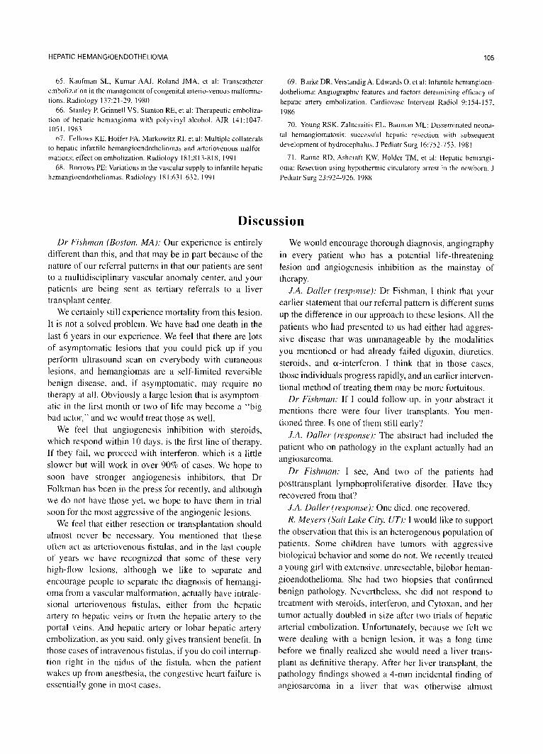
Discussion
Dr Fishman (Boston. MA): Our experience is entirely different than this, and that may be in part because of the nature of our referral patterns in that our patients are sent to a multidisciplinary vascular anomaly center, and your patients are being sent as tertiary referrals to a liver transplant center.
We certainly still experience mortality from this lesion. It is not a solved problem. We have had one death in the last 6 years in our experience. We feel that there are lots of asymptomatic lesions that you could pick up if you perform ultrasound scan on everybody with cutaneous lesions, and hemangiomas are a self-limited reversible benign disease, and, if asymptomatic. may require no therapy at all. Obviously a large lesion that is asymptom- atic in the first month or two of life may become a “big bad actor,” and we would treat those as well.
We feel that angiogenesis inhibition with steroids, which respond within 10 days. is the first line of therapy. If they fail, we proceed with interferon, which is a little slower but will work in over 90% of cases. We hope to soon have stronger angiogenesis inhibitors, that Dr Folkman has been in the press for recently. and although we do not have those yet, we hope to have them in trial soon for the most aggressive of the angiogenic lesions.
We feel that either resection or transplantation should almost never be necessary. You mentioned that these often act as arteriovenous fistulas. and in the last couple of years we have recognized that some of these very high-flow lesions. although we like to separate and encourage people to separate the diagnosis of hemangi- oma from a vascular malformation. actually have intrale- sional arteriovenous fistulas, either from the hepatic artery to hepatic veins or from the hepatic artery to the portal veins. And hepatic artery or lobar hepatic artery embolization, as you said, only gives transient benefit. In those cases of intravenous fistulas, if you do coil interrup- tion right in the nidus of the fistula, when the patient wakes LIP from anesthesia, the congestive heart failure is essentially gone in most cases.
We would encourage thorough diagnosis, angiography in every patient who has a potential life-threatening lesion and angiogenesis inhibition as the mainstay of therapy.
J.A. Driller (r-espotrsr): Dr Fishman, I think that your earlier statement that our referral pattern is different sums up the difference in our approach to these lesions. All the patients who had presented to us had either had aggres- sive disease that was unmanageable by the modalities you mentioned or had already failed digoxin, diuretics, steroids, and a-interferon. 1 think that in those cases, those individuals progress rapidly, and an earlier interven- tional method of treating them may be more fortuitous.
Dr Fishman: If I could follow-up, in your abstract it mentions there were four liver transplants. You men- tioned three. Is one of them still early?
J.A. Duller (response): The abstract had included the patient who on pathology in the explant actually had an angiosarcoma.
Dr Fishmat~: I see, And two of the patients had posttransplant lymphoproliferative disorder. Have they recovered from that?
J.A. Duller (response): One died, one recovered. R. Meyers (Suit Lake City UT): I would like to support
the observation that this is an heterogenous population of patients. Some children have tumors with aggressive biological behavior and some do not. We recently treated a young girl with extensive, unresectable, bilobar heman- gioendothelioma. She had two biopsies that confirmed benign pathology. Nevertheless, she did not respond to treatment with steroids, interferon, and Cytoxan, and her tumor actually doubled in size after two trials of hepatic arterial embolization. Unfortunately, because we felt we were dealing with a benign lesion, it was a long time before we finally realized she would need a liver trans- plant as definitive therapy. After her liver transplant, the pathology findings showed a 4-mm incidental finding of angiosarcoma in a liver that was otherwise almost

106 DALLER ET AL
entirely replaced by a benign-appearing hemangioendo- thelioma. She did well for the first year, but untreatable widely metastatic angiosarcoma has developed since. The malignant transformation of this hemangioendothe- lioma to malignant angiosarcoma might have been pre- ventable if we had recognized the virulence of her “benign disease” earlier and gone to liver transplantation sooner.
J.A. Daller (response): How old is she? R. Meyers: She is 4 years old. J.A. Daller (response): Frequently in the patients who
present at an older age, they tend to have foci of hemangiosarcoma, and, as you suggested, those people
probably should be evaluated earlier on to prevent that complication.
K. Anderson (Los Angeles, CA): Of your asymptomatic patients that you treated with either steroids or interferon, what was your end point of measurement, and did any of those patients go on to need liver transplant?
J.A. Daller (response): Of the people who received liver transplant, one had bilobar disease. Two of them had complications of attempted resection. As far as the management with the steroids and a-interferon, these patients had been referred to us after not responding those modalities. They had received it for periods of time ranging from several weeks up to 6 months.