A Secretory Golgi Bypass Route to the Apical Surface Domain of Epithelial MDCK Cells
Upload
mountsaintvincentCategory
view
3download
0
The Rockefeller University Press, 0021-9525/99/03/1057/11 $2.00The Journal of Cell Biology, Volume 144, Number 5, March 8, 1999 1057–1067http://www.jcb.org 1057
Hensin Remodels the Apical Cytoskeleton and Induces Columnarization of Intercalated Epithelial Cells: Processes that ResembleTerminal Differentiation
S. Vijayakumar, Jiro Takito, Chinami Hikita, and Qais Al-Awqati
Department of Medicine and Department of Physiology, College of Physicians and Surgeons of Columbia University, New York 10032
Abstract.
Intercalated epithelial cells exist in a spec-trum of phenotypes; at one extreme,
b
cells secrete HCO
3
by an apical Cl/HCO
3
exchanger and a basolat-eral H
+
ATPase. When an immortalized
b
cell line is seeded at high density it deposits in its extracellularmatrix (ECM) a new protein, hensin, which can reverse the polarity of several proteins including the Cl/HCO
3
exchanger (an alternately spliced form of band 3) and the proton translocating ATPase. When seeded at low density and allowed to form monolayers these polar-ized epithelial cells maintain the original distribution of these two proteins. Although these cells synthesize and secrete hensin, it is not retained in the ECM, but rather, hensin is present in a large number of intracellular vesi-cles. The apical cytoplasm of low density cells is devoid of actin, villin, and cytokeratin19. Scanning electron mi-croscopy shows that these cells have sparse microvilli, whereas high density cells have exuberant apical sur-face infolding and microvilli. The apical cytoplasm of high density cells contains high levels of actin, cyto-keratin19, and villin. The cell shape of these two pheno-types is different with high density cells being tall with a small cross-sectional area, whereas low density cells are
low and flat. This columnarization and the remodeling of the apical cytoplasm is hensin-dependent; it can be induced by seeding low density cells on filters condi-tioned by high density cells and prevented by an anti-body to hensin. The changes in cell shape and apical cy-toskeleton are reminiscent of the processes that occur in terminal differentiation of the intestine and other ep-ithelia. Hensin is highly expressed in the intestine and prostate (two organs where there is a continuous pro-cess of differentiation). The expression of hensin in the less differentiated crypt cells of the intestine and the basal cells of the prostate is similar to that of low den-sity cells; i.e., abundant intracellular vesicles but no lo-calization in the ECM. On the other hand, as in high density cells hensin is located exclusively in the ECM of the terminally differentiated absorptive villus cells and the prostatic luminal cell. These studies suggest that hensin is a critical new molecule in the terminal differ-entiation of intercalated cell and perhaps other epithe-lial cells.
Key words: epithelial polarity • terminal differentia-tion • intercalated cells • cell shape • hensin
Address correspondence to Qais Al-Awqati, Department of Medicineand Physiology, College of Physicians and Surgeons of Columbia Univer-sity, 630 West 168
th
Street, New York, NY 10032. Tel.: 212-305-3512. Fax:212-305-3475. E-mail: [email protected]
1.
Abbreviations used in this paper:
CUB, complement subcomponentsClr/Cls, Uegf, Bmp1; ECM, extracellular matrix; FAK, focal adhesion ki-nase; kAE1, kidney form of the anion exchanger1; SRCR, scavenger re-ceptor cysteine rich; ZP, zona pellucida.
E
PITHELIA
, the first differentiated cells to develop inthe embryo, exhibit polarized plasma membranedomains and cytoplasm, with the nuclei occupying
the basal half of the cell (Eaton and Simons, 1995; Drubinand Nelson, 1996). Epithelia are attached to each other byseveral types of junctions and rest on an extracellular ma-trix (ECM)
1
that they largely synthesize. The fact that epi-
thelial cells in different organs share all these character-istics, yet are recognizably different from each other,implies that development and differentiation of epitheliaproceeds in at least two steps: conversion of a nonepithe-lial cell to a proto-epithelium, and terminal differentiation.Proto-epithelial cells exhibit the fundamental propertiesof epithelia, but are still able to divide and migrate. Termi-nally differentiated epithelia, while losing motile and pro-liferative behavior, acquire specific features such as: brushborders, specialized organelles (e.g., storage granules),characteristic cell shape (e.g., columnarization), and othertissue specific properties. Terminal differentiation contin-ues to occur in adult animals in the intestine, skin, pros-tate, and other organs. Recent studies in many organismshave shown that interruption of such differentiation pro-grams leads to unbridled growth leading to, or contribut-ing to, the development of cancer.

The Journal of Cell Biology, Volume 144, 1999 1058
We were led to issues of terminal differentiation whilestudying the targeting of ion transport proteins in a kidneyepithelial cell line derived from intercalated cells. Thesecells are specialized for transepithelial acid transport, me-diated by a polarized H
+
ATPase and a Cl
2
/HCO
3
2
ex-changer, which is an alternately spliced form of the red cellband 3 (kAE1, kidney anion exchanger1). In situ, they ex-ist in a spectrum of forms with two extremes: one secretesacid by an apical H
+
ATPase and a basolateral anionexchanger (the
a
-form), whereas the other (
b
) secretesHCO
3
2
by an apical exchanger and a basolateral ATPase(for review see Schuster, 1993; Al-Awqati et al., 1998). H
+
ATPase (
a
-form) is packaged in vesicles that continuouslyrecycle by endocytosis and exocytosis at the apical mem-brane in a process regulated by cell pH and calcium(Gluck et al., 1982; Cannon et al., 1985). Remarkably, the
b
cells had no apical endocytosis, suggesting that the apicalcytoplasm of the two cell types were fundamentally differ-ent, as was clear in transmission electron micrographs ofthese cells.
We (and others) noticed that feeding animals an aciddiet reversed the ion transport properties of the collectingtubule of the kidney from HCO
3
2
secretion to HCO
3
2
ab-sorption (i.e., H
+
secretion). We provided initial physio-logical evidence that this reversal happened because
b
cells converted to
a
cells (Schwartz et al., 1985). To studythe biochemical pathway, we generated an immortalized
b
cell line that had an apical kAE1 and a basolateral H
+
ATP-ase (Edwards et al., 1992). When these clonally derivedcells were seeded at subconfluent density and examined(after they formed an epithelium), they secreted HCO
3
2
to the lumen and had apical kAE1 and basolateral H
+
ATPase (van Adelsberg et al., 1993). Seeding the cells atconfluent densities resulted in their conversion to the fol-lowing: an
a
phenotype, with transepithelial HCO
3
2
ab-sorption; an apical H
+
ATPase; a basolateral kAE1; andvigorous apical endocytosis. The high density cells se-creted a new protein and localized it to their ECM (vanAdelsberg et al., 1994). Using an assay for apical endocy-tosis, we purified this protein from high density ECM. Thepurified protein was able to reverse the phenotype ofthese cells. This new protein, which we termed hensin (forchange in body in Japanese) is expressed in epithelial cellsand the brain. In the kidney it is restricted to the collectingtubule (Takito et al., 1996). We show here that hensin in-duces a reorganization of the apical membrane and its as-sociated cytoskeleton with the expression of villin andcytokeratin19, two proteins that are necessary for the for-mation of microvilli. Further, these changes lead to colum-narization of the intercalated cells. All of these changesare similar to what occurs in terminal differentiation ofother epithelia. Therefore, we suggest that the change inphenotype is a process of terminal differentiation and thathensin is a critical new molecule in this pathway.
Materials and Methods
Cell Culture
Stock cultures of clone C of
b
-intercalated cells established from rabbitkidney cortex were maintained as described (van Adelsberg et al., 1993,1994). The cells were trypsinized and seeded on polycarbonate filters
(pore size, 0.4
m
m; Costar Corp.) at a density of 2
3
10
4
cells/cm
2
(lowdensity), or 10
6
cells/cm
2
(high density), and transferred to 40
8
C to inacti-vate the T antigen. At temperatures between 37
8
and 40
8
C, no positivestaining of the T antigen was observed by immunofluorescence or immu-noblot analysis.
Pulse Labeling and Immunoprecipitation of Hensin
High or low density cells on polycarbonate filters were pulse-labeled with100
m
Ci/ml of [
35
S]methionine added to both apical and basal media (
35
S-protein labeling mix; DuPont-NEN) for 5 min at 40
8
C at various time in-tervals. Cells were lysed in buffer A (1% SDS, 1 mM EDTA, 1% TritonX-100, 10 mM Tris-HCl, pH 8.0) and boiled for 3 min. Insoluble materialswere removed by a brief centrifugation (14,000
g
for 5 min at room tem-perature) and the protein concentration of the supernatants was deter-mined by the Bradford reagent (Bio-Rad Laboratories). An equal amountof protein was taken from each sample, diluted 10-fold with 10 mM Tris-HCl, pH 8.0, and used for immunoprecipitation.
Clone C cells seeded at high or low density were cultured for 5 d and la-beled with
35
S-protein labeling mix added to both apical and basal mediafor 12 h. Apical and basolateral media were collected separately and cen-trifuged at 5,000
g
for 5 min at 4
8
C. The supernatants were mixed with 1/10vol of buffer A and analyzed by immunoprecipitation.
Samples from the pulse labeling experiments and secretion studieswere incubated with 1:500 dilution of guinea pig anti-hensin antiserum at4
8
C for 1 h. Immunoprecipitates were collected by mixing the sampleswith protein A–Sepharose CL-4B (Pharmacia Biotech, Inc.) at 4
8
C for 1 h.The beads were washed with a buffer containing 0.1% SDS, 0.1 mMEDTA, 0.1% Triton X-100, and 10 mM Tris-HCl, pH 8.0. Immunoprecip-itated protein samples were extracted from the beads by boiling them inSDS-PAGE sample buffer for 3 min. Thus, samples extracted were sub-jected to SDS gel electrophoresis in 7.5% gel. Gels were fixed with 10%acetic acid, 10% methanol, soaked in solution (Amplify; Amersham Phar-macia Biotech), dried, and exposed to X-ray film (Kodax X-OMAT) at
2
80
8
C. Films were developed and analyzed using a densitometer (model300A; Molecular Dynamics, Inc.).
Immunoblot Analysis
Intermediate filament fraction was prepared from confluent monolayercultures of both low and high density cells with a modified procedure ofpreviously published protocols (Achtstaeter et al., 1986; Stasiak et al.,1989). In brief, monolayers were lysed with a solution containing 10 mMTris-HCl, pH 7.6, 140 mM NaCl, 5 mM EDTA, 15 mM
b
-mercaptoetha-nol, 1 mM PMSF, and 1% Triton X-100 for 3–5 min at 4
8
C. The lysisbuffer was aspirated and the monolayer was extracted with a high saltbuffer (10 mM Tris-HCl, pH 7.6, 140 mM NaCl, 1.5 M KCl, 5 mM EDTA,0.5% (wt/vol) Triton X-100, 15 mM
b
-mercaptoethanol, and 1 mM PMSF,at 4
8
C for 1 h. The extract was centrifuged for 30 min at 3,000
g
(4
8
C) andthe pellet was washed with buffer (10 mM Tris-HCl, pH 7.6, 140 mMNaCl, and 5 mM EDTA). The final pellet was dissolved in SDS-PAGEbuffer, the sample was electrophoresed in a 10% SDS-PAGE gel, trans-ferred to a nitrocellulose membrane, and probed with anticytokeratin19antibody (MAB1675). These samples were prepared from an equal num-ber of cells.
Immunocytochemistry
The immortalized intercalated cells (clone C) were plated at high or lowdensity and cultured for 1–2 wk at 40
8
C on Transwell filters, depending onthe experiment. The following procedures were performed at room tem-perature: cells were fixed in 4% paraformaldehyde for 10 min, blocked,and permeabilized in a solution of 3% BSA and 0.075% saponin in PBS,pH 7.4, for 1 h. The Transwell filters were incubated in primary antibodiesdiluted 1:100 in the PBS/BSA/saponin solution for 1–2 h. The followingprimary antibodies were used: mouse mAb to E-cadherin (MAB 1996),fodrin (MAB 1622), cytokeratin19 (MAB 1675), villin (MAB 1671) andrat anti-ZO1 antibody (MAB 1520) (all from Chemicon International,Inc.) and anti–
b
-tubulin antibody (Boehringer Mannheim GmbH). Fluo-rescein- or rhodamine-labeled goat anti–mouse IgG and rhodamine-labeled donkey anti–rat IgG (Jackson ImmunoResearch Laboratories)were used as secondary antibodies. For F-actin staining, the filters wereincubated with 300–600 U of rhodamine-phalloidin (R-415) (MolecularProbes, Inc.).
Stained monolayers were mounted on glass slides with 90% glycerol inPBS with 0.1% phenylene diamine and viewed by an Axiovert 100 laser-

Vijayakumar et al.
Hensin Induces Terminal Epithelial Differentiation
1059
scanning confocal microscope (model LSM 410; Carl Zeiss). Excitationwas accomplished with an argon-krypton laser producing lines at 488, 568,or 647 nm. Rhodamine- and Cy3-labeled samples were viewed with the568-nm channel (red), whereas the fluorescein–Hoechst 33342-labeledsamples were viewed with the 488-nm channel (green). In the case of tri-ple labeling with Hoechst/hensin and collagen type IV, the Cy5-conju-gated collagen type IV antigens were visualized with the deep red 645-nmchannel. The images were collected at 1
m
m thickness optical sections andanalyzed by the Zeiss LSM-PC software. The final images were processedwith Adobe Photoshop software.
Immunocytochemistry with Anti–hensin Antibody
Guinea pig anti–hensin antibodies were obtained as described earlier (Ta-kito et al., 1996). A fusion protein containing scavenger receptor cysteinerich (SRCR) domains 5 and 6 of hensin (Takito et al., 1996) was used togenerate these antibodies. The immortalized intercalated cells (clone C)were plated at high or low density and cultured for 1–2 wk at 40
8
C onTranswell filters depending on the experiment. In the studies aimed at de-termining the extracellular accessibility of hensin, the filters were first in-cubated with 10–20
m
g/ml of Hoechst 33342 dye (Molecular Probes,Inc.). After extensive washing with PBS, cells were incubated in 1:50 dilu-tion of guinea pig anti-hensin antibody solution in PBS alone, or with1:100 dilution of the mouse anti–collagen type IV mAb (MAB 1910;Chemicon International, Inc.) overnight at 4
8
C. The filters were washedwith PBS and incubated with rhodamine–Cy3 conjugated anti–guinea pigIgG (Jackson ImmunoResearch Laboratories, Inc.) alone, or with Cy5conjugated anti–mouse IgG (for collagen IV triple label experiment) for1 h at room temperature. The filters were washed, fixed for 30 s–1 minwith ice-cold methanol, washed extensively with PBS, and mounted andanalyzed with confocal microscopy as described before. Control experi-ments were performed to test whether prolonged incubation at 4
8
C re-sulted in cellular permeabilization. When the guinea pig anti-hensin anti-body and anti–
b
-tubulin mAb were incubated together under the sameconditions, we found no specific staining for tubulin, whereby indicatingthat the cells remained impermeable to these molecules.
For studies involving the determination of cellular localization ofhensin, the confluent monolayers of high or low density clone C cells werefirst incubated in Hoechst 33342 at 10–20
m
g/ml for 1 h at room tem-perature, washed with PBS, and fixed with ice-cold methanol for 1 min.After extensive washing with PBS, cells were permeabilized by incubatingwith a solution of PBS/BSA/saponin (as described above) and incubatedfurther with guinea pig anti-hensin antibody, followed by rhodamine–Cy3conjugated anti–guinea pig IgG.
Immunohistochemistry of Rabbit Tissues
Prostate glands and intestinal tissues obtained from male New Zealandwhite rabbits (Hare Marland) were embedded and frozen in tissue-tekOCT compound (Miles Laboratories) until cut into 5-
m
m cryostat sec-tions. The sections were fixed in ice-cold methanol for 8 min, washed ex-tensively with PBS, and blocked with 10% FCS and 1% donkey serum(Jackson ImmunoResearch Laboratories, Inc.) in PBS for 30 min. Thesections were incubated with guinea pig anti–hensin antibody (Takito et al.,1996), diluted 1:100 in PBS at room temperature for 2 h and incubatedwith rhodamine-conjugated donkey anti–mouse IgG. The prostate sec-tions were further incubated with FITC-conjugated monoclonal anti-cytokeratin7 (F-3772; Sigma Chemical Co.) diluted 1:25 in PBS. Finally,sections were extensively washed and mounted for analysis under a confo-cal microscope.
Analysis of the Shape and Height of Clone C Cells
All analyses of cell size and height were performed with Microsoft Win-dows
®
based version of the Zeiss 410 laser scanning microscope software(Carl Zeiss GmbH). The stepper motor and the image analysis softwareuse the internal calibration method for this microscope. All 1-
m
m sectionsof a particular field from a sample of phalloidin-stained low or high den-sity cells were projected on the screen together with maximum overlapand all other default settings of the Zeiss LSM-PC software. For size mea-surements, individual cell boundaries were delineated using a mouse-driven marker (via the Mark Area subfunction of the Area Measure func-tion of this software) and the area obtained in
m
m
2
is recorded. The areaof 10–15 individual cells of each sample were recorded and were analyzedstatistically with the program Corel Quattro Pro.
For measuring the height of the cells, the xz-section of the sequence ofimages from each specific sample is used. The two “posit” (positionmarker) cursors are placed on the same x-coordinate of the xz-section.However, one “posit” cursor is placed on the y-coordinate, correspondingto the first observed apical stain above the nucleus, and the other is placedon the last observed basal stain below the nucleus. The distance measure-ment function of the Zeiss LSM-PC software is invoked to measure theheight. Again, 10–15 individual measurements were recorded for eachsample and were analyzed statistically using Corel Quattro Pro. For allthese measurements, the phalloidin- or phallodin–Hoechst 33342-stainedsamples were used to ensure the uniformity of the measurements.
Scanning Electron Microscopy
Filters were washed in PBS and fixed with 2.5% glutaraldehyde in 100 mMphosphate buffer. The filters were postfixed for 1 h in 1% osmium tetrox-ide/1.5% potassium ferricyanide and dehydrated through a graded etha-nol series: 50, 70, 85, 95, 100, 100, 100% for 10 min at each concentration.Afterwards, cells underwent critical point drying using CO
2
in an OmarSPC-1500 critical point dryer. The filters were cut from their holders andmounted on copper specimen supports, sputter-coated with
z
11 nm ofAu-Pd in a sputtering unit (Pelco 91000; Ted Pella Inc.), and viewed at20 kV in an electron microscope (JEOL 100 CX-II; JEOL USA, Inc.)equipped with an ASID scanning unit and images were recorded on Po-laroid Type 55 positive/negative film.
Antibody Blocking Experiments
Guinea pig anti–hensin antisera (Takito et al., 1996) were diluted 1:100 inthe culture medium. Anti-laminin B2 mAb obtained as hybridoma super-natant D18 from the Developmental Studies Hybridoma Bank (Univer-sity of Iowa, Iowa City, IA) was diluted in the culture medium to a finalconcentration of 30
m
g/ml and mouse anti–human collagen type IV mAbwas diluted to a final concentration of 60
m
g/ml in the culture medium.Confluent monolayers of clone C cells grown at 32
8
C were trypsinized andcentrifuged. The cell pellet was resuspended in the appropriate antibody-containing culture medium and plated at high density (10
6
cells/cm
2
). An-tibody-containing culture medium was replaced daily for 5 d and the cellswere processed for immunocytochemistry as described above.
Experiments on Low Density Cells Grown on High Density Matrix and Various ECM Components
Hensin containing ECM-coated filters were obtained through the follow-ing procedure: (a) High density cells cultured on Transwell filters for 1 wkwere extracted with 1% Triton X-100, 1 mM calcium chloride for 1 h at4
8
C on a rotary shaker. (b) Cell extracts were removed and filters werescraped in this solution with a cell scraper to remove loosely attached ma-terials. (c) Filters were washed thoroughly with the same solution for an-other hour at 4
8
C followed by three washes with the culture medium. (d)The filters were left overnight in the culture medium on a 4
8
C-rotaryshaker and clone cells were seeded on such filters and cultured as usual.Murine laminin-coated Transwell filters, human fibronectin coated Trans-well filters, and Matrigel basement membrane matrix- (from Engelbreth-Holm-Swarm Mouse Sarcoma) coated cell culture inserts were obtainedfrom Becton Dickinson Labware (models: 40441, 40615, 40443, respec-tively). The laminin- and fibronectin-coated filters were rehydrated by in-cubation with culture medium for 30 min at room temperature beforeseeding the cells. In the case of Matrigel-coated filters, the filters werethawed overnight on a level surface at 4
8
C and the Matrigel solution fromone Transwell filter was spread on two Transwell filters (model 3412; Co-star Corp.) before rehydration and seeding. This was necessary as thethick gelatinous layer of matrigel present on the precoated filters (model40443; Becton Dickinson Labware) prevented the immunohistochemicalanalysis of the cells cultured on them.
Results
Hensin Is Localized in the Extracellular Space of High Density but Not Low Density Cells
Initial studies showed that high density ECM contained anew protein (hensin) that induced the reversal of polarity,
The Journal of Cell Biology, Volume 144, 1999 1060
but the low density matrix did not. We generated antibod-ies to a hensin fusion protein containing SRCR domains 6and 7 (Takito et al., 1996) that were used for immunopre-cipitation and immunocytochemistry. Fig. 1 shows the re-sults of immunocytochemistry using confocal microscopy.Low density monolayers incubated with the anti-hensinantibodies, in the absence of detergents, showed no hensin
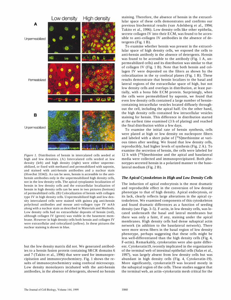
staining. Therefore, the absence of hensin in the extracel-lular space of these cells demonstrates and confirms ourprevious biochemical results (van Adelsberg et al., 1994;Takito et al., 1996). Low density cells like other epithelia,secrete collagen IV into their ECM, was found to be acces-sible to anti–collagen IV antibodies in the absence of de-tergents (Fig. 1 B).
To examine whether hensin was present in the extracel-lular space of high density cells, we exposed the cells toanti-hensin antibody in the absence of detergents. Hensinwas found to be accessible to the antibody (Fig. 1 A, un-permeabilized cells) and its distribution was similar to thatof collagen IV (Fig. 1 B). Note that both hensin and col-lagen IV were deposited on the filters as shown by thecolocalization in the xy confocal planes (Fig. 1 B). Theseresults demonstrate that hensin localizes to the basal andlateral regions of the extracellular space of high, but notlow density cells and overlaps in distribution, at least par-tially, with a bona fide ECM protein. Surprisingly, whenthe cells were permeabilized by saponin, we found thateven low density cells contained a large number of hensin-containing intracellular vesicles located diffusely through-out the cell, including the apical half. On the other hand,the high density cells contained few intracellular vesiclesstaining for hensin. This difference in distribution startedat the earliest time examined (3 h of plating) and reachedthe final distribution within a few days.
To examine the initial rate of hensin synthesis, cellswere plated at high or low density on nucleopore filtersand labeled with a short pulse of [
35
S]methionine at vari-ous times after seeding. We found that low density cells,reproducibly, had higher levels of synthesis (Fig. 2 A). Toexamine the secretion of hensin, the cells were labeled for12 h with [
35
S]methionine and the apical and basolateralmedia were collected and immunoprecipitated. Both phe-notypes secreted hensin in a polarized manner to the baso-lateral medium (Fig. 2 B).
The Apical Cytoskeleton in High and Low Density Cells
The induction of apical endocytosis is the most dramaticand reproducible effect in the conversion of low densityphenotype to that of high density. Apical endocytosis, orits lack, clearly reflects large alterations in the apical cy-toskeleton. We examined components of this cytoskeletonand found dramatic differences as a function of seedingdensity (see Figs. 3–5). F-actin, in low density cells, was lo-cated underneath the basal and lateral membranes butthere was only a faint, if any, staining under the apicalmembranes. High density cells had dense subapical actinnetwork (in addition to the basolateral network). Therewere more stress fibers in the basal region of low densityphenotype, perhaps suggesting that these cells might beless well-differentiated than the high density cells (Fig. 4,F-actin). Remarkably, cytokeratins were also quite differ-ent. Cytokeratin19, recently implicated in the organizationof the terminal web of intestinal epithelial cells (Salas et al.,1997), was largely absent from low density cells but wasabundant in high density cells (Fig. 4, Cytokeratin-19).More significantly, cytokeratin19 was located mostly inthe subapical region of the cells. These studies suggest thatthe terminal web, an actin–cytokeratin mesh critical for the
Figure 1. Distribution of hensin in intercalated cells seeded athigh and low densities. (A) Intercalated cells seeded at lowdensity (left) and high density (right) were either unperme-abilized, or fixed with methanol and permeabilized with saponin,and stained with anti-hensin antibodies and a nuclear stain(Hoechst 33342). As can be seen, hensin is accessible to the anti-hensin antibodies only in the unpermeabilized high density cells,not in the low density cells. The apical cytoplasmic localization ofhensin in low density cells and the extracellular localization ofhensin in high density cells can be seen in two pictures (bottom)of permeabilized cells. (B) Colocalization of hensin with collagentype IV in high density cells. Unpermeabilized high and low den-sity intercalated cells were stained with guinea pig anti-hensinpolyclonal antibodies and mouse anti–collagen type IV mAbalong with a nuclear stain as described in Materials and Methods.Low density cells had no extracellular deposits of hensin (red),although collagen IV (green) was visible in the basement mem-brane. However in high density cells both hensin and collagen IVwere extracellular and colocalized (yellow). In these pictures thenuclear staining is shown in blue.

Vijayakumar et al.
Hensin Induces Terminal Epithelial Differentiation
1061
formation of microvilli, was substantially different in the twophenotypes. When the microfilament fraction of low den-sity cells was extracted and subjected to immunoblot anal-ysis, no cytokeratin19 was found, whereas that of highdensity cells was quite enriched in this protein. This resultsuggests that high density cells can synthesize this protein(Fig. 5).
Next, we examined the distribution of villin, the mostcritical actin-binding protein for microvillar structure (Friedrich et al., 1989), and found that its expression in low
density cells was barely detectable; when seen it was ex-pressed in a faint cytoplasmic pattern. In high density cells,villin was highly expressed and located entirely in the api-cal region of the cell in a manner suggestive of incorpora-tion in microvilli (Figs. 3 and 4, Villin). These studies arereminiscent of the development of the apical cytoskeletonin the intestine; the epithelial cells of the crypt (includingstem cells) have essentially no villin or subapical cytokera-tin but as they migrate up the villus to become terminallydifferentiated, they start expressing villin and cytokeratinsand develop a thick brush border (Louvard et al., 1992).This complex pattern of new gene expression suggest acti-vation of a differentiation pathway.
Although the above observations might suggest that lowdensity cells are undifferentiated, in fact, these cells arebona fide epithelial cells. They show polarized distribu-tions of proteins (van Adelsberg et al., 1993, 1994), lipids(van’t Hof et al., 1997), and polarized secretion of hensin(Fig. 2). They are also capable of steady state transepithe-lial transport of HCO
3
2
, which would not be possible with-
Figure 2. Biochemical characterization of the synthesis and se-cretion of hensin. (A) Low density and high density cells werepulse-labeled with [35S]methionine for 5 min at the indicated timeintervals after seeding and analyzed by immunoprecipitation asdescribed in Materials and Methods. Densitometric analysis ofthe results is shown below. (B) Intercalated cells seeded at low orhigh density, cultured for 5 d, and labeled with [35S]methioninefor 12 h. Hensin was immunoprecipitated from the apical and ba-solateral media. Both low and high density cells secreted hensinin a polarized manner to the basolateral medium.
Figure 3. Distribution of villin in low density and high densityphenotypes. Low and high density phenotype intercalated cellswere stained with anti-villin mAb (red) and the nuclear stainHoechst 33342 (green).
Figure 4. Distribution of F-actin, cytokeratin19, and villin in in-tercalated cells. Intercalated cells seeded at low (left) or highdensities (middle) and at low density on a high density ECM(right). They were analyzed by immunocytochemistry 1 wk afterseeding as described in Materials and Methods. Actin was visual-ized by staining with rhodamine-phalloidin, whereas villin andcytokeratin19 were stained with mAb. In the xz sections, the bot-tom of each image represents the level of the filter. Bar, 25 mm.

The Journal of Cell Biology, Volume 144, 1999 1062
out polarized distribution of transport proteins and suffi-ciently impermeant intercellular junctions (van Adelsberget al., 1993, 1994). Further, low density cells have all of thefollowing: well-formed tight junctions (Fig. 6, ZO-1); ad-herent junctions (Fig. 6, E-Cadherin); a polarized cytoskel-eton, where F-actin was present in lateral and basal sur-faces (Fig. 4, F-actin); and fodrin in the lateral membranes(Fig. 6, Fodrin). There was also no significant differencein the distribution of paxillin and focal adhesion kinase(FAK; not shown) in the two phenotypes.
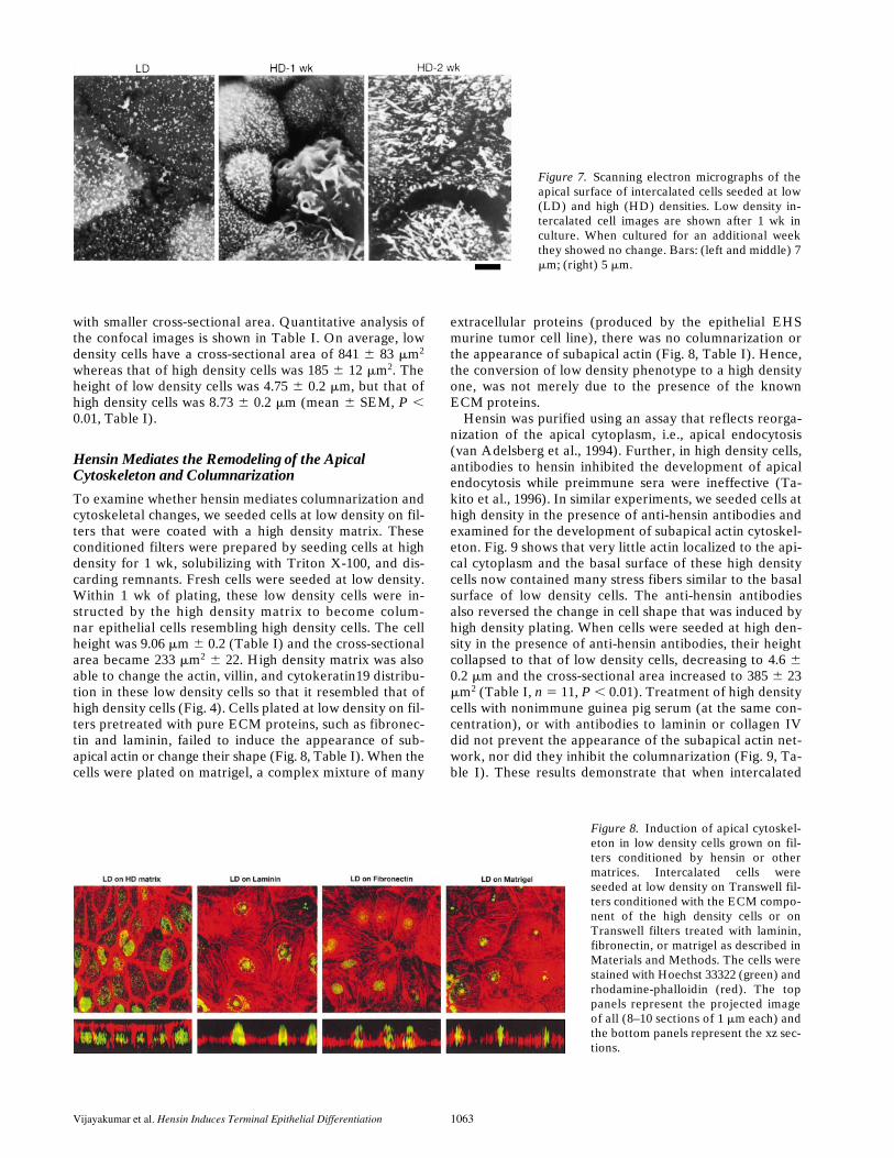
Intercalated cells in situ do not exhibit brush borders,but scanning electron microscopy showed that their apicalcell membranes have impressive specializations:
b
-interca-lated cells have apical microvilli whereas
a
-intercalatedcells have apical folds, termed microplicae (Hagege andRichet, 1970; LeFurgey and Tisher, 1979). The latter arebroad apical ridges that are readily observed in scanningelectron micrographs of mammalian kidney and theirhomologous epithelia in amphibia and reptiles. We per-formed scanning electron microscopy on low and highdensity cells and found that low density cells had few api-cal microvilli scattered over the apical surface that did notchange with length of time in culture. Fig. 7 shows lowdensity cells after 1 wk in culture. After 1 wk in culture athigh density, the number of well-formed microvilli wasmuch higher and they appeared to be taller and thickerthan those in low density cells. In addition, a substantialfraction of the cells had clearly established microplicae(Fig. 7). After 2 wk in culture, all high density cells had mi-croplicae of different dimensions and very large microvilli(Fig. 7). These results demonstrate that the transition oflow to high density phenotypes represents a fundamentalchange in the properties of the apical cytoskeleton and byanalogy with other epithelia, we suggest that it representsa terminal differentiation phenomenon.
High Seeding Density Induces Columnarization
Differentiation of epithelial cells is associated withchanges in cell shape. Indeed, it has been suggested that achange in shape itself might induce terminal differentia-tion (Chen et al., 1997). Protoepithelial cells are flat with alarge apical surface area, whereas terminally differentiatedepithelia are taller and more tightly packed. This process
of columnarization is frequently seen during development(Bard, 1990). Examination of Figs. 3, 4, 6, and 7, showsthat low density cells are thin and have a large cross-sec-tional area whereas high density cells are columnar, tall
Figure 5. Immunoblot analysis ofcytokeratin19 in the microfilamentfraction of low and high density inter-calated cells. Intermediate filamentswere extracted from equal number ofcells of both low and high density phe-notype intercalated cells as describedin Materials and Methods and probedwith a mAb to cytokeratin19.
Figure 6. Distribution of E-cadherin, fodrin, ZO-1, and tubulinin intercalated cells seeded at low and high seeding densities. In-tercalated cells were seeded at the indicated densities and cul-tured for 1 wk. The cells were fixed and stained with mAb toE-cadherin, fodrin, ZO-1, and tubulin. The xz sections of bothphenotypes reveal the characteristic basolateral localizations forE-cadherin and fodrin and the apical tight junction localizationfor ZO-1. The tubulin network is similar in both phenotypes.
Table I. Measurement of Cross-sectional Area and Cell Height
Height Cross-sectional area
m
m
m
m
2
Low density 4.75
6
0.2 841
6
83High density 8.73
6
0.3 185
6
12High density
1
anti-hensin Ab 4.61
6
0.2 385
6
23High density
1
anti–collagen IV Ab 9.06
6
0.2 205
6
18High density
1
anti–laminin Ab 8.61
6
0.3 225
6 15Low on high density matrix 9.06 6 0.2 233 6 22Low density on laminin 5.53 6 0.2 1,867 6 145Low density on fibronectin 5.32 6 0.3 2,234 6 162Low density on matrigel 5.77 6 0.3 2,343 6 143
Results obtained from confocal images of phalloidin stained cells as described in Ma-terials and Methods. For each experiment, 11 cells were analyzed and the results areexpressed as means 6 SEM.
Vijayakumar et al. Hensin Induces Terminal Epithelial Differentiation 1063
with smaller cross-sectional area. Quantitative analysis ofthe confocal images is shown in Table I. On average, lowdensity cells have a cross-sectional area of 841 6 83 mm2
whereas that of high density cells was 185 6 12 mm2. Theheight of low density cells was 4.75 6 0.2 mm, but that ofhigh density cells was 8.73 6 0.2 mm (mean 6 SEM, P ,0.01, Table I).
Hensin Mediates the Remodeling of the Apical Cytoskeleton and Columnarization
To examine whether hensin mediates columnarization andcytoskeletal changes, we seeded cells at low density on fil-ters that were coated with a high density matrix. Theseconditioned filters were prepared by seeding cells at highdensity for 1 wk, solubilizing with Triton X-100, and dis-carding remnants. Fresh cells were seeded at low density.Within 1 wk of plating, these low density cells were in-structed by the high density matrix to become colum-nar epithelial cells resembling high density cells. The cellheight was 9.06 mm 6 0.2 (Table I) and the cross-sectionalarea became 233 mm2 6 22. High density matrix was alsoable to change the actin, villin, and cytokeratin19 distribu-tion in these low density cells so that it resembled that ofhigh density cells (Fig. 4). Cells plated at low density on fil-ters pretreated with pure ECM proteins, such as fibronec-tin and laminin, failed to induce the appearance of sub-apical actin or change their shape (Fig. 8, Table I). When thecells were plated on matrigel, a complex mixture of many
extracellular proteins (produced by the epithelial EHSmurine tumor cell line), there was no columnarization orthe appearance of subapical actin (Fig. 8, Table I). Hence,the conversion of low density phenotype to a high densityone, was not merely due to the presence of the knownECM proteins.
Hensin was purified using an assay that reflects reorga-nization of the apical cytoplasm, i.e., apical endocytosis(van Adelsberg et al., 1994). Further, in high density cells,antibodies to hensin inhibited the development of apicalendocytosis while preimmune sera were ineffective (Ta-kito et al., 1996). In similar experiments, we seeded cells athigh density in the presence of anti-hensin antibodies andexamined for the development of subapical actin cytoskel-eton. Fig. 9 shows that very little actin localized to the api-cal cytoplasm and the basal surface of these high densitycells now contained many stress fibers similar to the basalsurface of low density cells. The anti-hensin antibodiesalso reversed the change in cell shape that was induced byhigh density plating. When cells were seeded at high den-sity in the presence of anti-hensin antibodies, their heightcollapsed to that of low density cells, decreasing to 4.6 60.2 mm and the cross-sectional area increased to 385 6 23mm2 (Table I, n 5 11, P , 0.01). Treatment of high densitycells with nonimmune guinea pig serum (at the same con-centration), or with antibodies to laminin or collagen IVdid not prevent the appearance of the subapical actin net-work, nor did they inhibit the columnarization (Fig. 9, Ta-ble I). These results demonstrate that when intercalated
Figure 7. Scanning electron micrographs of theapical surface of intercalated cells seeded at low(LD) and high (HD) densities. Low density in-tercalated cell images are shown after 1 wk inculture. When cultured for an additional weekthey showed no change. Bars: (left and middle) 7mm; (right) 5 mm.
Figure 8. Induction of apical cytoskel-eton in low density cells grown on fil-ters conditioned by hensin or othermatrices. Intercalated cells wereseeded at low density on Transwell fil-ters conditioned with the ECM compo-nent of the high density cells or onTranswell filters treated with laminin,fibronectin, or matrigel as described inMaterials and Methods. The cells werestained with Hoechst 33322 (green) andrhodamine-phalloidin (red). The toppanels represent the projected imageof all (8–10 sections of 1 mm each) andthe bottom panels represent the xz sec-tions.

The Journal of Cell Biology, Volume 144, 1999 1064
cells are seeded at high density, it is hensin that mediatesthe reorganization of the actin cytoskeleton and colum-narization rather than other ECM proteins.
Distribution of Hensin In Two Differentiating Epithelia
One of the best described terminal differentiation eventsis the intestinal stem cell ascends to the villus tip to be-come the terminally differentiated absorptive enterocyte(Louvard et al., 1992; Simon and Gordon, 1995). Becausethe highest level of expression of hensin is in the intestine(Takito et al., 1996), we examined the distribution of hensinin intestinal epithelia. Remarkably, hensin was present inthe crypt, largely in a diffuse intracellular pattern that wasmostly in the apical half of the cell with no staining of theECM, (Fig. 10 A) similar to the staining pattern of per-meabilized low density cells shown in Fig. 1 A. In the ter-minally differentiated villus cells, hensin was presentexclusively in a basolateral pattern likely to represent ex-tracellular localization, an identical distribution to that inpermeabilized high density cells.
The epithelium of the prostate gland is another tissuethat undergoes terminal differentiation in situ. Basal cellsare sparsely located in the epithelium and they give rise tothe luminal cells, which are terminally differentiated epi-
thelial cells. Basal cells of the rodent prostate gland ex-press cytokeratin7, whereas the luminal cells do not (Hay-ward et al., 1996a,b). As can be seen in Fig. 10 B, therabbit basal cells also express cytokeratin7 (in green);these cells have a large number of vesicles that containhensin (in red). On the other hand, in the luminal cells,hensin surrounds the cells in a pattern entirely similar tothat of high density cells.
DiscussionThere is increasing evidence that a number of polarizedmembrane proteins are targeted in a cell-type specificmanner (for review see Al-Awqati et al., 1998). For in-stance, the human LDL receptor when expressed as atransgene in mice is located in the apical membrane of kid-ney epithelia but is basolateral in the intestine and liver(Pathak et al., 1990). The Na, K ATPase is basolateral inmany epithelia, but is apical in the retinal pigment epithe-lium and choroid plexus (Gundersen et al., 1991). MostGPI-linked proteins are apical but they are basolateral in athyroid cell line (Zurzolo et al., 1993). In the intercalatedcell, we demonstrated that the same cell is capable of re-targeting at least two proteins, the anion exchanger kAE1and the H1 ATPase to opposite membrane domains, buttwo other proteins, a basolateral glucose transporter andan apical lectin-binding protein did not change their polar-ized distribution (van Adelsberg et al., 1994).
These studies demonstrate that there does not appear tobe a single class of mechanisms responsible for targetingpolarized proteins in all cells. Because the protein that ex-hibits plastic polarity (e.g., Na, K ATPase or kAE1) is wellpolarized regardless of whether it is located in the apicalor basolateral domain, one can conclude that each proteincontains at least two potential targeting signals that arerecognized differently by the cell machinery. Another ver-sion of this statement is that there may be a hierarchy ofsignals and that each cell reads these signals depending onits complement of targeting pathways. But remarkably,the cellular machinery that decodes these signals is alsocell type specific. For instance, syntaxin 3, a t-SNARE crit-ically involved in delivery of protein to the apical mem-brane of many epithelia (Low et al., 1996; Galli et al.,1998), is present in the basolateral membrane of the inter-calated cell (Mandon et al., 1997). Furthermore, the path-way that vesicles take is also different: the apical targetingof dipeptidyl peptidaseIV is direct in some cells (MDCK),indirect in others (CaCO2; Hubbard et al., 1991), or ex-hibit both pathways in still others (LLC-PK1; Low et al.,1991). Recent observations showed that apical epithelialproteins when transfected into nonpolarized cells (fibro-blasts) are localized in vesicles or membrane domains thatare different from those that contain transfected basolat-eral proteins (Musch et al., 1996; Yoshimori et al., 1996).In summary, these studies demonstrate that there are mul-tiple targeting pathways in many, even all cells. To gener-ate a tissue-specific phenotype would probably require lo-cal factors that would provide additional information tothe basic cellular targeting machinery or might even in-duce new pathways.
To begin to analyze these local factors, it is worth recall-ing that some of these plastic targeting events occur during
Figure 9. Anti-hensin antibodies prevent the reorganization ofthe actin cytoskeleton in high density cells. High density interca-lated cells were seeded in the presence or absence of polyclonalanti-hensin serum (1:100), anti–laminin mAb (30 mg/ml), andanti–collagen IV mAb (60 mg/ml). The culture media with andwithout the antibodies were replaced daily. Staining withrhodamine-phalloidin was performed 5 d after seeding. A 2-mmthick apical section (left) is shown. A basolateral optical section(right) of the same thickness is shown. Bar, 25 mm.

Vijayakumar et al. Hensin Induces Terminal Epithelial Differentiation 1065
development. The Na, K ATPase is found to be localizedin the apical membrane in some nephrons during embry-onic life and the probability of its basolateral locationincreases as the kidney matures (Avner et al., 1992).A membrane protein in the retinal pigment epitheliumchanges its polarized distribution during postnatal devel-opment (Marmorstein et al., 1996). Perhaps the signal forthis reversal of polarity comes from a developmentallyregulated extracellular instructive molecule. In the retinalpigment epithelium, the N-CAM molecule is apical in situ,but when the epithelium is dissociated from the neural ret-ina and cultured in vitro N-CAM moves to the basolateralsurface (Gundersen et al., 1993). Many developmentalphenomena are mediated by factors that bind tightly toECM proteins including: TGFb family members, wnt’s,and fibroblast growth factors. Further, ECM proteins thatbind to these critical factors are themselves part of the de-velopmental pathway and serve not only to generate gra-dients but also as reservoirs, activators, or inhibitors ofthese factors before they bind to their plasma membranereceptors. Our recent studies had demonstrated that onesuch protein, hensin, when deposited in the ECM of inter-calated cells is sufficient to reverse the polarized distribu-tion of the kidney form of band 3 (kAE1), and the protonATPase (van Adelsberg et al., 1994; Takito et al., 1996).Hence, one hypothesis to emerge from this analysis is thattissue-specific proteins deposited in the ECM during de-velopment might instruct differentiating epithelia to targetpolarized proteins to one or another domain of the cell. Ifthese ECM proteins are not present in that tissue, then thevesicle targeting pathway will come under the influence ofother less dominant factors. To exhibit reversal of polarity,according to this formulation, the protein must containmultiple targeting sequences.
These speculations were solidified by the surprising find-ing of the present paper that hensin appeared to retargetseveral proteins and induce terminal differentiation in the
intercalated cells. Both forms of the intercalated cells arestable when the two monolayers are confluent; all theircells are forming a true epithelial sheet, yet they exhibitdifferent properties. Therefore, hensin must be acting as abinary switch to trigger the conversion of one phenotypeto another, a process that is reminiscent of the induction ofa differentiation program during development. Epithelialcells of the kidney (and many other organs) begin as mes-enchymal cells that are induced to form the elementaryepithelial phenotype, proto-epithelial cell. However, thesecells only acquire a fully differentiated phenotype laterin development. Terminally differentiated epithelia haveseveral characteristics: (a) They contain a rich apical cy-toskeletal network, the terminal web, which is composedof an actin mesh and specific cytokeratins. (b) Their apicalmembrane contains extensive specializations such as cilia,flagella, or brush-border microvilli. (c) They frequentlycontain vesicles or granules located in the apical half of thecell that are capable of fusion with the apical membrane ina manner regulated by cell calcium or other second mes-sengers. (d) Their nuclei are present in the basal half of thecell. (e) They also assume a recognizable shape in simpleepithelia, being columnar or cuboidal.
Remarkably, all of these characteristics were induced byhensin. The cells developed a terminal web of actin andcytokeratin19, their apical membranes formed an exuber-ant microvillar structure, and their shape became colum-nar. They even developed vesicles capable of regulatedexocytosis. We had previously shown that the H1 ATPasein the a-intercalated cell is packaged in vesicles that fuseswith the apical membrane in response to an increase in theambient pCO2 acting to increase cell calcium (Gluck et al.,1982; Cannon et al., 1985). We suggest that hensin is thefirst protein in a new pathway for differentiation in thesecell types. It had been known for some time that the ECMis critical for differentiation of epithelial cells. In fact, re-cent studies showed that fetal intestinal epithelia can start
Figure 10. Distribution of hensin in small intes-tine and prostate gland. (Top) Rabbit small in-testine was fixed, sectioned, and stained withanti-hensin antibodies. Confocal images of intes-tinal crypts (left) and villi (right) are shown.Note the staining pattern in crypts shows diffuseintracellular staining located mostly in the apicalhalf of the cell. In villi, hensin is present in a ba-solateral pattern that surrounds the cell. (Bot-tom) Prostate glands: simultaneous staining withhensin (rhodamine) and cytokeratin7 (fluores-cein). (Left) Red indicates the staining pattern ofhensin in a typical prostate gland section. (Mid-dle) shows the staining pattern of cytokeratin7,an exclusive marker of basal epithelia in rodentprostate in the same section. The right panel isthe combined image.

The Journal of Cell Biology, Volume 144, 1999 1066
to express terminally differentiated proteins such as villinand cytokeratin when cultured on matrigel, a complexECM prepared from cancer cells (Sanderson et al., 1996).Hensin is expressed in a large number of epithelial tissuesand at a high level in the intestine. Its distribution in intes-tinal crypt cells is similar to that of low density intercalatedcells, whereas in villus cell it was similar to the terminallydifferentiated intercalated cell. We are presently investi-gating whether hensin is also involved in the differentia-tion of intestinal epithelia.
Hensin was accessible to its antibodies even when thehigh density cells were not permeabilized, and its distribu-tion colocalized with collagen IV. How does the high den-sity plating cause precipitation in the ECM? The localiza-tion of fibronectin fibrils in the ECM suggests a possiblemechanism. Soluble fibronectin monomers cannot formfibrils unless their receptor, the integrin a6b1 is activated(Wu et al., 1995). Once activated the receptor’s affinity forfibronectin increases, allowing it to bind fibronectin and toform fibrils (Sechler et al., 1996). Perhaps high densityseeding induces a change in the affinity of the hensin re-ceptor that refolds hensin into an insoluble form. Alterna-tively, high density cells might secrete another protein thatwill cause precipitation of hensin in the extracellular space.We are presently investigating these two possibilities.
The role of the ECM in controlling cell shape is well-established in many cell types including epithelia (Ingber etal., 1986). Many studies have identified a pathway medi-ated by binding of integrins to components of the ECMthat leads to activation of a signaling pathway mediatedlargely by activation of FAK. This protein is critical for or-ganization of the actin cytoskeleton and leads to formationof a complex of actin binding proteins such as paxillin,ezrin, and moesin. Cell shape, motility, and ruffle forma-tion are thought to be direct consequences of these events.Most of this information has been obtained in fibroblastsor in blood cells and the role of focal adhesions in epitheliais not as well studied. We found no change in the distribu-tion of paxillin and FAK during the transition from lowdensity to high density phenotypes. However, it was clearthat there were large changes in other cytoskeletal compo-nents. A dramatic reorganization of the apical cytoplasmoccurred that was characterized by all of the following: in-duction of vigorous apical endocytosis; development ofexuberant microvillar formation, the new subapical local-ization of actin, and the possible induction or at least in-creased concentration of villin and cytokeratin19 in thesubapical cytoplasm. The basal actin cytoskeleton was alsodifferent in the two phenotypes. Low density cells hadmany “stress fibers” in the basal region similar to what isseen in nonepithelial cells whereas the actin network inhigh density cells appeared to be more diffuse. How a ba-solateral extracellular molecule induces these changes incell shape and the changes in apical cytoplasm is unknownat present. Recent studies have suggested that mechanicalstress, such as that following binding of ECM proteins totheir receptors have profound effects on cell functions, in-cluding the induction of complex programs of differentia-tion (Chen et al., 1997; Chicurel et al., 1998). The cellularmechanisms by which hensin induces these events awaitsthe identification of its signal transduction pathway andpreliminary evidence suggests that a basolateral integral
membrane glycoprotein acquires tyrosine phosphorylationwithin a short time after seeding these cells at high but notat low density.
The deduced amino acid sequence of hensin shows thatit is composed of three domains, SRCR, (complement sub-components Clr/Cls, Lleg F, Bmp1) CUB, and (Zona Pel-lucida) ZP (Takito et al., 1996 and manuscript submittedfor publication). SRCR domains are cysteine rich domainsof unknown function found in a variety of proteins (Res-nick et al., 1994). The CUB domain was first identified intolloid, a protein that activates decapentaplegic (dpp, theDrosophila TGFb homologue) and mutations in that do-main prevent dpp activation (Bork and Beckmann, 1993;Childs et al., 1994). The ZP domain, present in ZP spermreceptor proteins, bears some similarity to another TGFb-binding protein, the type III receptor (Bork and Sander,1992). Although these results suggest that hensin mightbind to TGFb or one of its many mammalian homologues,there is no evidence for this at present. Three other cDNAshave been reported that are composed of SRCR, CUB,and ZP domains. These include: CRP-ductin, a cDNA over-expressed in mouse intestinal crypt cells (Cheng et al.,1996); ebnerin, a partial cDNA overexpressed in rat vonEbner’s gland (Li and Snyder, 1995); and DMBT1, a largehuman sequence that resides in chromosome 10q25-26 isdeleted in a substantial fraction of several malignant braintumors such as gliomas and glioblastomas (Mollenhauer etal., 1997). This region has also been implicated in other ep-ithelial cancers such as prostate cancer. Because these se-quences were first identified in four different species andthe presence of large number of proteins with one or moreof these domains in each species, it was not possible to de-cide by simple sequence comparisons whether they repre-sented a new gene family, or whether they were alter-nately spliced versions of the same gene. We cloned themouse and rabbit genomic sequences of hensin and partialsequences of these two led to the conclusion that all foursequences are derived from the same gene (Takito, J., L.Yan, C. Hikita, S. Vijayakumar, D. Warburtar, and Q. Al-Awqati, manuscript submitted for publication). Hensinappears to be involved in terminal differentiation and it iswell-known that interruption in terminal differentiationpathways often leads to cancer. These occurrences raisethe intriguing possibility that hensin is a tumor suppressor.
We are grateful to Lee Cohen-Gould for the performance of scanningelectron microscopy and Theresa Swayne for expert assistance in confocalmicroscopy.
This work was supported by National Institutes of Health (NIH) grantsDK-20999 and DK-39532. The Columbia Confocal Facility is supportedby NIH grants RR10506 and CA13696.
Received for publication 23 July 1998 and in revised form 25 January1999.
References
Achtstaetter, T., M. Hatzfield, R.A. Quinlan, D.C. Parmelee, and W.W.Franke. 1986. Separation of cytokeratin polypeptides by gel electrophoreticand chromatographic techniques and their identification by immunoblotting.Methods Enzymol. 134:355–371.
Al-Awqati, Q., S. Vijayakumar, C. Hikita, J. Chen, and J. Takito. 1998. Pheno-typic plasticity in the intercalated cell: The hensin pathway. Am. J. Physiol.275:F183–F190.
Avner, E.D., W.E. Sweeney, Jr., and W.J. Nelson. 1992. Abnormal sodiumpump distribution during renal tubulogenesis in congenital murine polycys-
Vijayakumar et al. Hensin Induces Terminal Epithelial Differentiation 1067
tic kidney disease. Proc. Natl. Acad. Sci. USA. 89:7447–7451.Bard, J. The epithelial repertoire. In Morphogenesis: The Cellular and Molecu-
lar Processes of Development Anatomy. Cambridge University Press, NewYork. 1992. 181–237.
Bork, P., and C. Sander. 1992. A large domain common to sperm receptors(Zp2 and Zp3) and TGF-beta type III receptor. FEBS (Fed. Eur. Biochem.Soc.) Lett. 300:237–240.
Bork, P., and G. Beckmann. 1993. The CUB domain. A widespread module indevelopmentally regulated proteins. J. Mol. Biol. 231:539–545.
Cannon, C., J. van Adelsberg, S. Kelly, and Q. Al-Awqati. 1985. Carbon-diox-ide-induced exocytotic insertions of H1 pumps in turtle-bladder luminalmembrane: role of cell pH and calcium. Nature. 314:443–446.
Chen, C.S., M. Mrkisch, S. Huang, G.M. Whitesides, and D.E. Ingber. 1997.Geometric control of cell life and death. Science. 276:1425–1428.
Chen, J., S. Vijayakumar, X. Li, and Q. Al-Awqati. 1998. Kanadaptin is a pro-tein that interacts with the kidney but not the erythroid form of band 3. J.Biol. Chem. 273:1038–1043.
Cheng, H., M. Bjerknes, and H. Chen. 1996. CRP-ductin: a gene expressed inintestinal crypts and in pancreatic and hepatic ducts. Anat. Rec. 244:327–343.
Chicurel, M.E., C.S. Chen, and D.E. Ingber. 1998. Cellular control lies in thebalance of forces. Curr. Opin. Cell Biol. 10:232–239.
Childs, S.R., and M.B. O’Connor. 1994. Two domains of the tolloid proteincontribute to its unusual genetic interaction with decapentaplegic. Dev. Biol.162:209–220.
Drubin, D.G., and W.J. Nelson. 1996. Origins of cell polarity. Cell. 84:335–344.Eaton, S., and K. Simons. 1995. Apical, basal, and lateral cues for epithelial po-
larization. Cell. 82:5–8.Edwards, J.C., J. van Adelsberg, M. Rater, D. Herzlinger, J. Lebowitz, and Q.
Al-Awqati. 1992. Conditional immortalization of bicarbonate-secreting in-tercalated cells from rabbit. Am. J. Physiol. 263:C521–C529.
Friederich, E., C. Huet, M. Arpin, and D. Louvard. 1989. Villin induces mi-crovilli growth and actin redistribution in transfected fibroblasts. Cell. 59:461–475.
Galli, T., A. Zahraoui, V.V. Vaidyanathan, G. Raposo, J.M. Tian, M. Karin, H.Niemann, and D. Louvard. 1998. A novel tetanus neurotoxin-insensitive ves-icle-associated membrane protein in SNARE complexes of the apicalplasma membrane of epithelial cells. Mol. Biol. Cell. 9:1437–1448.
Gluck, S., C. Cannon, and Q. Al-Awqati. 1982. Exocytosis regulates urinaryacidification in turtle bladder by rapid insertion of H1 pumps into the lumi-nal membrane. Proc. Natl. Acad. Sci. USA. 79:4327–4331.
Gundersen, D., J. Orlowski, and E. Rodriguez-Boulan. 1991. Apical polarity ofNa, K-ATPase in retinal pigment epithelium is linked to a reversal of theankyrin-fodrin submembrane cytoskeleton. J. Cell Biol. 112:863–872.
Gundersen, D., S.K. Powell, and E. Rodriguez-Boulan. 1993. Apical polariza-tion of N-CAM in retinal pigment epithelium is dependent on contact withthe neural retina. J. Cell Biol. 121:335–343.
Hagege, J., and G. Richet. 1970. Etude par microscopie electronique de la sur-face apicale des cellules de tube contourné distal du rein de rat. C. R. Se-ances. Acad. Sci. D. 271:331–332.
Hayward, S.W., L.S. Baskin, P.C. Haughney, A.R. Cunha, B.A. Foster, R. Da-hiya, G.S. Prins, and G.R. Cunha. 1996a. Epithelial development in the ratventral prostate, anterior prostate and seminal vesicle. Acta Anat. 155:81–93.
Hayward, S.W., J.R. Brody, and G.R. Cunha. 1996b. An edgewise look at basalepithelial cells: Three-dimensional views of the rat prostate, mammary glandand salivary gland. Differentiation. 60:219–227.
Hubbard, A.L. 1991. Targeting of membrane and secretory proteins to the api-cal domain in epithelial cells. Semin. Cell Biol. 2:365–374.
Ingber, D.E., J.A. Madri, and J.D. Jamieson. 1986. Basement membrane as aspatial organizer of polarized epithelia. Exogenous basement membrane re-orients pancreatic epithelial tumor cells in vitro. Am. J. Pathol. 122:129–139.
LeFurgey, A., and C.C. Tisher. 1979. Morphology of the rabbit collecting duct.Am. J. Anat. 155:111–124.
Li, X.J., and S.H. Snyder. 1995. Molecular cloning of Ebnerin, a von Ebner’sgland protein associated with taste buds. J. Biol. Chem. 270:17674–17679.
Louvard, D., M. Kedinger, and H.P. Hauri. 1992. The differentiating intestinalepithelial cell: Establishment and maintenance of functions through interac-
tions between cellular structures. Ann. Rev. Cell Biol. 8:157–195.Low, S.H., S.H. Wong, B.L. Tang, and W.J. Hong. 1991. Involvement of both
vectorial and transcytotic pathways in the preferential apical cell surface lo-calization of rat dipeptidyl peptidase IV in transfected LLC-PK1 cells. J.Biol. Chem. 266:19710–19716.
Low, S.H., S.J. Chapin, T. Weimbs, L.G. Komuves, M.K. Bennett, and K.E.Mostov. 1996. Differential localization of syntaxin isoforms in polarized Ma-din-Darby canine kidney cells. Mol. Biol. Cell. 7:2007–2018.
Mandon, B., S. Nielsen, B.K. Kishore, and M.A. Knepper. 1997. Expression ofsyntaxins in rat kidney. Am. J. Physiol. 273:F718–F730.
Marmorstein, A.D., V.L. Bonilha, S. Chiflet, J.M. Neill, and E. Rodriguez-Bou-lan. 1996. The polarity of the plasma membrane protein RET-PE2 in retinalpigment epithelium is developmentally regulated. J. Cell. Sci. 109:3025–3034.
Mollenhauer, J., S. Wiemann, W. Schurlen, B. Korn, Y. Hayashi, K.K. Wilgen-bus, A.V. Deimling, and A. Poustka. 1997. DMBT1, a new member of theSRCR superfamily, on chromosome 10q25.3–26.1 is deleted in malignantbrain tumours. Nat. Genet. 17:32–39.
Musch, A., H. Xu, D. Shields, and E. Rodriguez-Boulan. 1996. Transport of ve-sicular stomatitis virus G protein to the cell surface is signal mediated in po-larized and nonpolarized cells. J. Cell Biol. 133:543–558.
Pathak, R.K., M. Yokodoe, R.E. Hammer, S.L. Hoffman, M.S. Brown, J.L.Goldstein, and R.G. Anderson. 1990. Tissue-specific sorting of the humanLDL receptor in polarized epithelia of transgenic mice. J. Cell Biol. 111:347–359.
Resnick, D., A. Pearson, and M. Krieger. 1994. The SRCR superfamily: a fam-ily reminiscent of the Ig superfamily. Trends Biochem. Sci. 19:5–8.1994.
Salas, P.J., M.L. Rodriguez, A.L. Viciana, D.E. Vega-Salas, and H.P. Hauri.1997. The apical submembrane cytoskeleton participates in the organizationof the apical pole in epithelial cells. J. Cell Biol. 137:359–375.
Sanderson, I.R., R.M. Ezzell, M. Kedinger, M. Erlanger, Z.X. Xu, E. Pringault,S. Leon-Robine, D. Louvard, and W.A. Walker. 1996. Human fetal enetero-cytes in vitro: Modulation of the phenotype by extracellular matrix. Proc.Natl. Acad. Sci. USA. 93:7717–7722.
Schuster, V.L. 1993. Function and regulation of collecting duct intercalatedcells. Annu. Rev. Physiol. 55:267–288.
Schwartz, G.J., J. Barasch, and Q. Al-Awqati. 1985. Plasticity of functional epi-thelial polarity. Nature. 318:368–371.
Sechler, J.L., Y. Takada, and J.E. Schwarzbauer. 1996. Altered rate of fibronec-tin matrix assembly by deletion of the first type III repeats. J. Cell Biol. 134:573–583.
Simon, T.C., and J.I. Gordon. 1995. Intestinal epithelial cell differentiation: newinsights from mice, flies, and nematodes. Curr. Opin. Genet. Dev. 5:577–586.
Stasiak, C.P., P.E. Purkis, I.M. Leigh, and E.B. Lane. 1989. Keratin 19: pre-dicted amino acid sequence and broad tissue distribution suggest it evolvedfrom keratinocyte keratins. J. Invest. Dermatol. 92:707–716.
Takito, J., C. Hikita, and Q. Al-Awqati. 1996. Hensin, a new collecting ductprotein involved in the in vitro plasticity of intercalated cell polarity. J. Clin.Invest. 98:2325–2331.
van Adelsberg, J.S., J.C. Edwards, and Q. Al-Awqati. 1993. The apical Cl/HCO3
2 exchanger of b intercalated cells. J. Biol. Chem. 268:11283–11289.van Adelsberg, J., J.C. Edwards, J. Takito, B. Kiss, and Q. Al-Awqati. 1994. An
induced extracellular matrix protein reverses the polarity of band 3 in inter-calated epithelial cells. Cell. 76:1053–1061.
van’t Hof, W., A. Malik, S. Vijayakumar, J.Z. Qiao, J. van Adelsberg, and Q.Al-Awqati. 1997. The effect of apical and basolateral lipids on the functionof the band 3 anion exchange protein. J. Cell Biol. 139:941–949.
Wu, C., V.M. Keivens, T.E. O’Toole, J.A. McDonald, and M.H. Ginsberg.1995. Integrin activation and cytoskeletal interaction are essential for the as-sembly of a fibronectin matrix. Cell. 83:715–724.
Yoshimori, T., P. Keller, M.G. Roth, and K. Simons. 1996. Different biosyn-thetic transport routes to the plasma membrane in BHK and CHO cells. J.Cell Biol. 133:247–256.
Zurzolo, C., M.P. Lisanti, I.P. Caras, L. Nitsch, and E. Rodriguez-Boulan. 1993.Glycophosphatidylinositol-anchored proteins are preferentially targeted tothe basolateral surface in Fischer rat thyroid epithelial cells. J. Cell Biol. 121:1031–1039.

The Journal of Cell Biology, Volume 144, 1999 1068
Copyright © 2022 FDOKUMEN


![Rivka Gersht, 1999, The Aspiration of Roman Women to Resemble their Empresses: Case Study of Several Roman Sculptures Discovered in Israel, MOTAR 4 [Hebrew with English abstract]](https://static.fdokumen.com/doc/165x107/6332028f83bb92fe98043362/rivka-gersht-1999-the-aspiration-of-roman-women-to-resemble-their-empresses-case.jpg)