Unfolding Various Concepts of Junctional Epithelium - Thieme ...
Upload
independentCategory
view
0download
0
Epidermal Growth Factor Protects the Apical JunctionalComplexes from Hydrogen peroxide in Bile Duct Epithelium
Srikar R. Guntaka1, Geetha Samak1, Ankur Seth1, Nicholas F. LaRusso2, and RadhakrishnaRao1
1 Department of Physiology, University of Tennessee Health Science Center, Memphis, TN, USA2 Department of Medicine, Mayo Clinic, Rochester, MN, USA
AbstractThe tight junctions of bile duct epithelium form a barrier between the toxic bile and liverparenchyma. Disruption of tight junctions appears to play a crucial role in the pathogenesis ofvarious liver diseases. In this study, we investigated the disruptive effect of hydrogen peroxide andthe protective effect of epidermal growth factor (EGF) on the tight junctions and adherensjunctions in the bile duct epithelium. Oxidative stress in NRC-1 and Mz-ChA-1 cell monolayerswas induced by administration of hydrogen peroxide. Barrier function was evaluated by measuringelectrical resistance and inulin permeability. Integrity of tight junctions, adherens junctions andthe actin cytoskeleton was determined by imunofluorescence microscopy. Role of signalingmolecules was determined by evaluating the effect of specific inhibitors. Hydrogen peroxidecaused a rapid disruption of tight junctions and adherens junctions leading to barrier dysfunctionwithout altering the cell viability. Hydrogen peroxiderapidly increased the levels of p-MLC(myosin light chain) and c-Src(pY418). ML-7 and PP2 (MLCK and Src kinase inhibitors)attenuated hydrogen peroxide-induced barrier dysfunction, tight junction disruption andreorganization of actin cytoskeleton. Pretreatment of cell monolayers with EGF amelioratedhydrogen peroxide-induced tight junction disruption and barrier dysfunction. The protective ffectof EGF was abrogated by ET-18-OCH3 and the Ro-32-0432 (PLCγ and PKC inhibitors).Hydrogen peroxide increased tyrosine-phosphorylation of ZO-1, claudin-3, E-cadherin and β-catenin, and pretreatment of cells with EGF attenuated tyrosine-phosphorylation of these proteins.These results demonstrate that hydrogen peroxide disrupts tight junctions, adherens junctions andthe actin cytoskeleton by an MLCK and Src kinase-dependent mechanism in the bile ductepithelium. EGF prevents hydrogen peroxide-induced tight junction disruption by a PLCγ andPKC-dependent mechanism.
KeywordsAdherens junctions; cholangiocyte; EGF; oxidative stress; protein kinase; tight junction
The luminal surface of intra and extra-hepatic bile ducts is lined by a monolayer ofcholangiocytes (epithelial cells). This cholangiocyte monolayer forms a barrier to thediffusion of the various injurious agents from the bile duct lumen into the hepaticparenchyma (1). The tight junctions, a multi-protein complex, along the apical end of thecell provide the barrier function of different epithelia. Disruption of bile duct epithelial
Users may view, print, copy, download and text and data- mine the content in such documents, for the purposes of academic research,subject always to the full Conditions of use: http://www.nature.com/authors/editorial_policies/license.html#terms
Address for correspondence: R.K. Rao, Ph.D., Professor, Department of Physiology, University of Tennessee, 894 Union Avenue,Memphis, TN 38163, Phone: (901) 448-3235, Fax: (901) 448-7126, [email protected].
NIH Public AccessAuthor ManuscriptLab Invest. Author manuscript; available in PMC 2012 March 1.
Published in final edited form as:Lab Invest. 2011 September ; 91(9): 1396–1409. doi:10.1038/labinvest.2011.73.
NIH
-PA Author Manuscript
NIH
-PA Author Manuscript
NIH
-PA Author Manuscript
integrity is implicated in the pathogenesis of liver diseases that affect biliary tree, such asprimary sclerosing cholangitis (PSC) and primary biliary cirrhosis (PBC) (2–5). Disruptionof tight junction integrity would allow regurgitation of bile acids or other noxious agentsinto the hepatic parenchyma from the bile duct lumen (6). As of now, most of the knowledgewe have on the structure and regulation of tight junctions has been adapted from what isknown about the tight junctions of intestinal and renal epithelia. Very little is known aboutthe structure and regulation of tight junctions in the bile duct epithelium.
The tight junctions are composed of many proteins, including transmembrane proteins suchas occludin, claudins and junctional adhesion molecules, and intracellular scaffold proteinssuch as zona occludens (ZO-1, ZO-2, ZO-3). The scaffold proteins and other associated tightjunction proteins interact with the perijunctional actomyosin ring. The integrity ofactomyosin ring and MLCK (myosin light chain kinase) activity play a crucial role in theregulation of tight junction integrity. Additionally, numerous signaling proteins such as c-Src, phosphatidylinositol 3-Kinase, ERK, PKCζ, PKCη, PP1 and PP2A directly interactwith tight junction proteins, indicating their potential role in the regulation of tight junctionintegrity (7). The pharmacologic and molecular evidence support fact that tight junctionsand paracellular permeability are indeed regulated by signaling molecules such asintracellular calcium, cyclic AMP, GTPase switch protein, protein kinases and proteinphosphatases in the intestinal and renal epithelia (7,8). However, the role of such signalingmechanisms in the regulation of bile duct epithelium is poorly understood. Adherensjunctions are prominent junctional complexes in the intestinal and other epithelia. E-cadherin and catenins are the predominant proteins that are involved in the assembly ofadherens junctions. But, there is no much information available regarding the adherensjunctions in bile duct epithelium. One study, reported nearly two decades ago, described thepresence of an adherens junction-like structure in the bile duct epithelium (9).
A significant body of evidence indicates that oxidative stress plays an important role in thepathogenesis of liver diseases. Compromised antioxidant defense mechanism and elevatedoxidative stress was detected in the liver of patients with PSC and PBC (10–12). Serumoxidative stress level was shown to be elevated in patients with cholecystic bile duct injury(13). Hepatic oxidative stress was also detected in experimental cholecystic injury (14–16).Superoxide dismutase gene therapy attenuated the experimental cholestasis-induced liverfibrosis (17). The mechanism of oxidative stress-induced liver injury is poorly understood.Very little information is available regarding the oxidative stress effect on bile ductepithelial function. One study demonstrated that nitric oxide-mediated inhibition of DNArepair potentiates DNA damage in cholangiocytes (18). However, there is no informationavailable regarding the effect of hydrogen peroxide or oxidative stress on the bile ductepithelial functions.
In the present study, we investigated the effect of hydrogen peroxide-induced oxidativestress on tight junction and adherens junction integrity and the mechanism associated with itin cholangiocyte monolayers. We further evaluated the EGF (epidermal growth factor)-mediated amelioration of tight junction disruption. EGF is a well-established gastrointestinalmucosal protective factor. However, its effect on bile duct function is unknown. NRC-1 cellmonolayers were mainly used for this study. The effect of hydrogen peroxide and EGF onbarrier function was confirmed in Mz-ChA-1 cell monolayers.
MATERIALS AND METHODSChemicals
Cell culture supplies were obtained from Invitrogen (San Jose, CA), and Transwell insertsand other cell culture plastic wares were purchased from Costar (Cambridge, MA). Rat-tail
Guntaka et al. Page 2
Lab Invest. Author manuscript; available in PMC 2012 March 1.
NIH
-PA Author Manuscript
NIH
-PA Author Manuscript
NIH
-PA Author Manuscript
collagen (Type I), ML-7, PP2, genistein and FITC-inulin were obtained from Sigmachemical company (St Louis MO). Ro-32-0432 and ET-18-OCH3 were purchased fromEMD Biochemicals (Gibbstown, NJ). Other fine chemicals and lab supplies were purchasedfrom Fisher Scientific (Tustin, CA) or Sigma chemical company.
AntibodiesMouse monoclonal anti-occludin antibody and rabbit polyclonal anti-occludin, anti-ZO-1,anti-claudin-3 and anti-β-catenin antibodies were purchased from Zymed laboratories (SouthSan Francisco, CA). Biotin-conjugated anti-phospho-tyrosine (p-Tyr), anti-E-cadherin, andanti-actin antibodies were purchased from BD Transduction laboratories (Lexington, KY).Mouse monoclonal anti-Src, anti-p-MLC and anti-c-Src(pY418) antibodies were purchasedfrom Upstate Biotechnology Inc. (Lake Placid, NY). AlexaFlour-488-conjugated anti-mouseIgG was obtained from Molecular Probes (Eugene, OR). Cy3-conjugated anti-rabbit IgGwas purchased from Sigma Immunochemicals (St. Louis, MO).
Cell cultureNRC-1 cells (normal rat cholangiocytes) were cultured in DMEM-F12 supplemented with10% fetal bovine serum, vitamin mix, chemically defined lipid mix, insulin-transferrin-selenium mix (ITS), non essential amino acids and antibiotics (penicillin and streptomycin)as described before (5). These cells were originally derived from intra hepaticcholangiocytes. Cells were cultured on plates or Transwell inserts (12 mm or 24 mmdiameter) coated with rat-tail type I collagen. All experiments were performed on 5–6 dayspost seeding in confluent monolayers. Mz-ChA-1 cells were grown in CMRL Medium-1066containing 10% fetal bovine serum, L-glutamine and antibiotics (penicillin andstreptomycin). Cells were passaged at 90% confluence and seeded on to Transwell inserts(12 mm). Experiments were conducted on 4–5 days post seeding.
Hydrogen peroxide treatmentCell monolayers were preincubated in DMEM for one hour with varying concentrations ofhydrogen peroxide (100–500 μM) at both the apical and basal chambers and placed in 37°Cincubator. At varying times after hydrogen peroxide the paracellular permeability wasevaluated by measuring transepithelial electrical resistance (TER) and unidirectional flux ofFITC-inulin. Inhibitors such as PP2 (3 μM) and ML-7 (10 μM) were administered 60 minprior to hydrogen peroxide administration. EGF (30 nM) was added 10 min prior tohydrogen peroxide. ET-18-OCH3 (15 μM) or Ro-32-0432 (1 μM) was administered 50 minbefore EGF administration. Control monolayers received the inhibitor in the absence of EGFand hydrogen peroxide.
Cell viability assayCell viability was assessed by measuring lactate dehydrogenase (LDH) activity in theincubation medium three hours after incubation with or without 500 μM hydrogen peroxide.LDH activity measured using a kit form Sigma Chemical Company (St Louis, MO). Themetabolic activity of cells was evaluated by using WST-1 cell viability assay kit fromClontech (Mountain View, CA) according to vendor’s instructions. This assay involvescleavage of stale tetrazolium salt of WST-1 to a soluble formazan, which is measured bycolorimetric method. The conversion of WST-1 occurs at cell surface by metabolicallyactive cells, largely dependent on glycolytic production of NADH. Therefore, colorproduced is directly proportional to the number of metabolically active cells. A loss of cellviability results in reduced rate of WST-1 conversion and low color development.
Guntaka et al. Page 3
Lab Invest. Author manuscript; available in PMC 2012 March 1.
NIH
-PA Author Manuscript
NIH
-PA Author Manuscript
NIH
-PA Author Manuscript

Measurement of transepithelial electrical resistance (TER)TER was measured as described before (5) using a Millicell-ERS electrical resistancesystem (Millipore, Bedford, MA). The TER recorded in empty Transwell inserts (usually50–80 Ohms•cm2) was subtracted from all values.
Unidirectional Flux of InulinInulin permeability was measured by incubating cell monolayers in the presence of 0.5 μg/ml FITC-inulin in the apical chamber. At varying times, 100 μl aliquot of basal medium waswithdrawn and fluorescence was measured in a micro plate fluorescence reader (FLx-800,Bio TEK Instruments, Winooski, VT). Flux of FITC-inulin into the basal well, wascalculated as the percentage of total fluorescence administered into the apical well per hourper cm2 surface area.
Immunofluorescence MicroscopyUnder various experimental conditions, cell monolayers were fixed in ice-coldacetone:methanol (1:1, v/v) for 5 min. The fixed cells were rehydrated in PBS (Dulbecco’ssaline containing 1.2 mM CaCl2 and 1 mM MgCl2) and permeabilized with 0.2% Triton-X100 in PBS. Cell monolayers were blocked with 4% nonfat milk in TBST (20 mM Tris,pH 8.0, containing 150 mM NaCl and 0.5% Tween 20). Cells were then stained with amixture of mouse monoclonal anti-occludin and rabbit polyclonal anti-ZO-1 antibodies ormouse monoclonal anti-E-cadherin and rabbit polyclonal anti-β-catenin antibodies. Amixture of AlexaFlour-488-conjugated anti-mouse IgG and Cy3-conjugated anti-rabbit IgGantibodies was used as secondary antibodies. Actin cytoskeleton was stained by incubationof paraformaldehyde-fixed cells with AlexaFluor 488-conjugated phalloidin. Cells weremounted and images collected using a Zeiss LSM 5 PASCAL laser scanning confocalmicroscope and the LSM 5 PASCAL software (Release 3.2) as a series of images from 1.0μm XY sections. Images were stacked by using the Image J software and processed byAdobe Photoshop (Adobe Systems, San Jose, CA).
Analysis of tyrosine phosphorylated proteinsProteins were extracted under denatured conditions using lysis buffer D (50 mM Tris buffer,pH 8.0, containing 0.3% SDS, 2 mM vanadate, 10 mM sodium fluoride and proteaseinhibitors as described above) and heated at 100°C for 10 min. Biotin-conjugated anti-p-Tyrwas used to immunoprecipitate p-Tyr. Immunocomplexes were precipitated withstreptavidin-agarose and immunoblotted for different proteins as described below.
Immunoblot analysisProteins were separated by SDS-PAGE and transferred to PVDF membranes. Blots wereprobed for occludin, ZO-1, c-Src, p-MLC, c-Src(pY418), E-cadherin, β-catenin andclaudin-3. HRP-conjugated anti-mouse IgG or anti-rabbit IgG antibodies were used assecondary antibodies. The blots were developed using the enhanced chemiluminescencemethod (Amersham, Arlington Heights, IL).
StatisticsComparison between two groups was made by the Student’s t tests for grouped data. Thesignificance in all tests was derived at 95% or greater confidence level.
Guntaka et al. Page 4
Lab Invest. Author manuscript; available in PMC 2012 March 1.
NIH
-PA Author Manuscript
NIH
-PA Author Manuscript
NIH
-PA Author Manuscript

RESULTSHydrogen peroxide induces barrier dysfunction in cholangiocyte monolayers
To determine the influence of oxidative stress on the barrier function of bile duct epithelium,NRC-1 cell monolayers were exposed to hydrogen peroxide and the barrier function wasevaluated by measuring TER and inulin permeability. Hydrogen peroxide reduced TER andincreased inulin permeability in a time (Fig. 1A & 1B) and dose (Fig. 1C & 1D)-dependentmanner, indicating that hydrogen peroxide increases the paracellular permeability. Todetermine the effect of hydrogen peroxide on cell viability we evaluated its effect on LDHrelease and cell metabolic capacity as assessed by WST-1 assay. Under our experimentalconditions, hydrogen peroxide did not increase LDH release (Fig. 1E) or decrease WST-1activity (Fig. 1F).
Hydrogen peroxide disrupts tight junctions and adherens junctionsAbove data suggest that hydrogen peroxide increases paracellular permeability in NRC-1cell monolayers without causing any loss of cell viability. Paracellular permeability inepithelial monolayers is restricted due to the presence of intact tight junctions. Althoughadherens junctions do not provide a physical barrier, they are known to indirectly regulatethe integrity of tight junctions. Therefore, we examined the effect of hydrogen peroxide onthe integrity of tight junctions and adherens junctions by immunofluorescence analysis ofsub cellular localization of occludin, ZO-1, E-cadherin and β-catenin. Occludin and ZO-1were co-localized at the intercellular junctions in the control cell monolayers, indicating thepresence of intact tight junctions. hydrogen peroxide treatment induced a redistribution ofoccludin and ZO-1 from the intercellular junctions into the intracellular compartments in atime-dependent manner (Fig. 2). E-cadherin and β-catenin were co-localized at theintercellular junctions in control cell monolayers, and hydrogen peroxide induced aredistribution of both E-cadherin and β-catenin from the intercellular junctions in a time-dependent manner (Fig. 3).
MLCK activity mediates hydrogen peroxide-induced tight junction disruption and barrierdysfunction
MLCK is well known to play a role in tight junction regulation in the intestinal (19) andlung (20) epithelial monolayers. Studies were therefore designed to determine the effect ofhydrogen peroxide on MLCK activation and the effect of ML-7, an MLCK-selectiveinhibitor, on tight junction disruption. Pretreatment of cell monolayers with ML-7significantly attenuated hydrogen peroxide-induced decrease in TER (Fig. 4B) and increasein inulin permeability (Fig. 4C). ML-7 also attenuated hydrogen peroxide-inducedredistribution of occludin and ZO-1 from the intercellular junctions into the intracellularcompartments (Fig. 4D). ML-7 by itself did not influence TER, inulin flux or junctionaldistribution of occludin and ZO-1.
Hydrogen peroxide-induced disruption of tight junctions is mediated by Src kinase activityOur previous studies demonstrated that tyrosine kinases, especially c-Src, play an importantrole in tight junction regulation in the intestinal epithelium. We therefore, evaluated the roleof tyrosine kinase activity in hydrogen peroxide-induced tight junction disruption in NRC-1cell monolayers. Hydrogen peroxide rapidly increased the level of c-Src(pY418) (Fig. 5A),indicating an activation of c-Src in NRC-1 cell monolayers. Pretreatment of cell monolayerswith genistein (a broad range tyrosine kinase inhibitor) or PP2 (a Src kinase inhibitor)significantly attenuated hydrogen peroxide-induced decrease in TER (Fig. 5B) and increasein inulin permeability (Fig. 5C). PP2 also attenuated hydrogen peroxide-inducedredistribution of occludin and ZO-1 from the intercellular junctions (Fig. 5D). Genistein or
Guntaka et al. Page 5
Lab Invest. Author manuscript; available in PMC 2012 March 1.
NIH
-PA Author Manuscript
NIH
-PA Author Manuscript
NIH
-PA Author Manuscript

PP2 under the concentrations used, did not influence TER, inulin permeability or junctionaldistribution of occludin and ZO-1.
hydrogen peroxide-induced activation of MLCK and c-Src are independent of each otherTo investigate the interdependency of MLCK activation and c-Src activation we determinedthe effect of ML-7 and PP2 on MLCK and c-Src activation. Pretreatment of cell monolayerswith ML-7 attenuated hydrogen peroxide-induced increase in p-MLC levels (Fig. 6), but p-MLC levels were unaffected by PP2. Similarly, hydrogen peroxide-induced increase in thelevels of c-Src(pY418) was attenuated by PP2, but not by ML-7 (Fig. 6). These resultsindicate independent activation of MLCK and c-Src by hydrogen peroxide.
Hydrogen peroxide induces reorganization of actin cytoskeleton by an MLCK and Srckinase-dependent mechanism
The tight junction protein complex is known to intimately associate with the actomyosinring, and disruption of actin cytoskeleton leads to disruption of tight junctions (21–23). Inthe control cell monolayers, actin is organized distinctly at the apical, middle and basal parts(Fig. 7). At the apical part, actin stains appear as uniformly distributed punctuates. In themiddle part of the cell, the F-actin filaments appear to be organized as a ring at theperijunctional region. Network of weak stress fibers were seen at the basal part of the cell.Treatment with hydrogen peroxide resulted in a dramatic disorganization of F-actinarrangements at all three levels of the cell (Fig. 7). Pretreatment of cell monolayers withML-7 or PP2 preserved the normal organization of the actin cytoskeleton in hydrogenperoxide-treated cells.
EGF ameliorates hydrogen peroxide-induced tight junction disruption in NRC-1 cellmonolayers
EGF, a gastrointestinal mucosal protective factor, has been extensively investigated for itsmucosal protective role in the gastrointestinal mucosa (24). But, there is no informationavailable on the potential protective role of EGF in the bile ducts. We investigated the effectof EGF on hydrogen peroxide-induced tight junction disruption in NRC-1 cell monolayers.Pretreatment of cell monolayers with EGF for 10 minutes prior to hydrogen peroxideadministration significantly attenuated hydrogen peroxide-induced decrease in TER (Fig.8A) and increase in inulin permeability (Fig. 8B). EGF also attenuated hydrogen peroxide-induced redistribution of occludin and ZO-1 from the intercellular junctions (Fig. 8C). EGFby itself did not cause significant change in TER, inulin flux or junctional distribution ofoccludin and ZO-1. The effect of EGF on hydrogen peroxide-induced tight junctions wassignificantly attenuated by pretreatment of cell monolayers with 0.3 μM AG1478, aselective inhibitor of EGF receptor tyrosine kinase (data not shown).
EGF attenuates hydrogen peroxide-induced c-Src activation and tyrosine phosphorylationof tight junction and adherens junction proteins
Previous studies showed that the integrity of tight junctions and adherens junctions isregulated by Src-mediated tyrosine-phosphorylation of tight junction and adherens junctionproteins (25,26). The present study shows that EGF pretreatment attenuates hydrogenperoxide-induced increase in the level of c-Src(pY418) in NRC-1 cell monolayers (Fig. 9A).Hydrogen peroxide treatment increased the levels of tyrosine-phosphorylated ZO-1, E-cadherin, β-catenin and claudin-3 (Fig. 9B). EGF pretreatment reduced Tyr-phosphorylationof these proteins in hydrogen peroxide-treated cells. Tyr-phosphorylated occludin levelswere high in both untreated and hydrogen peroxide-treated cell monolayers (Fig. 9B). EGFtreatment reduced the levels of tyrosine-phosphorylated occludin in both untreated andhydrogen peroxide-treated cell monolayers.
Guntaka et al. Page 6
Lab Invest. Author manuscript; available in PMC 2012 March 1.
NIH
-PA Author Manuscript
NIH
-PA Author Manuscript
NIH
-PA Author Manuscript
PLCγ and PKC activities are involved in EGF-mediated protection of tight junctionsOne of the major intracellular signaling pathways activated by EGF is PLCγ-mediated PKCactivation as shown in Caco-2 cell monolayers (27). In the present study, we investigated therole of PLCγ and PKC in EGF-mediated tight junction protection in NRC-1 cellmonolayers. Pretreatment of cell monolayers with ET-18-OCH3, a PLCγ-selective inhibitorsignificantly attenuated EGF-mediated prevention of hydrogen peroxide-induced decrease inTER (Fig. 10A) and increase in inulin permeability (Fig. 10B). ET-18-OCH3 also preventedEGF-mediated attenuation of hydrogen peroxide-induced redistribution of occludin andZO-1 from the intercellular junctions (Fig. 10C). Similarly, Ro-32-0432, a PKC-selectiveinhibitor, prevented EGF-mediated attenuation of hydrogen peroxide-induced decrease inTER, increase in inulin permeability and redistribution of occludin and ZO-1 from theintercellular junctions. ET-18-OCH3 or Ro-32-0432 by themselves produced no significantinfluence on TER, inulin flux or junctional distribution of occludin and ZO-1 in control andhydrogen peroxide-treated cell monolayers.
EGF prevents hydrogen peroxide-induced barrier dysfunction in Mz-ChA-1 cellmonolayers
Mz-ChA-1 cell is a human bile duct epithelial cell line that forms a polarized cholangiocytemonolayer when grown in culture. The barrier function of this cholangiocyte monolayer isweak compared to that of NRC-1 cell monolayers. However, treatment of Mz-ChA-1 cellmonolayers with hydrogen peroxide significantly decreased TER (Fig. 11A) and increasedinulin permeability (Fig. 11B). This effect of hydrogen peroxide was attenuated bypretreatment of cell monolayers with ML-7 or PP2. The hydrogen peroxide-induced effecton TER and inulin flux was attenuated by the pretreatment of cells with EGF. This effect ofEGF was prevented by ET-18-OCH3 or Ro-32-0432 (Fig. 11).
DISCUSSIONAlthough evidence indicates that oxidative stress is associated with the bile duct-associatedpathology of liver (10,28,29) the mechanism of oxidative stress-induced bile duct injury isunclear. In the present study, using a primary cell line of rat bile duct epithelium and ahuman cholangiocarcinoma cell line, we show that oxidative stress induced by hydrogenperoxide results in compromised epithelial barrier function. The barrier function of bile ductepithelium is vital to the liver health as it prevents the diffusion of toxic bile componentsinto liver parenchyma, thus protecting the hepatocytes and other cells from bile acid-inducedinjury. Disruption of barrier function and bile acid-induced hepatocyte injury plays a crucialrole in the pathogenesis of liver disease. Furthermore, the factors that prevent oxidativestress-induced barrier function of bile duct epithelium is crucial to our understanding of theregulation of bile duct epithelial barrier function. The present study shows that EGF, agastrointestinal mucosal protective factor, prevents hydrogen peroxide-induced tightjunction disruption and barrier dysfunction.
Decrease in TER and increase in inulin permeability without altering cell viability show thathydrogen peroxide causes increase in paracellular permeability in the bile duct epithelium.Redistribution of tight junction proteins, occludin and ZO-1, from the intercellular junctionsinto the intracellular compartments indicated that hydrogen peroxide-induced barrierdysfunction was caused by the disruption of tight junctions. Occludin and ZO-1 are the mostwidely characterized tight junction proteins. Organization of these proteins involves aninteraction of ZO-1 with the intracellular, C-terminal domain of occludin (30).Redistribution of these proteins from the intercellular junctions into the intracellularcompartment in hydrogen peroxide-treated cholangiocyte monolayer is a clear indication ofdisrupted tight junctions. The hydrogen peroxide concentration used in this study is similar
Guntaka et al. Page 7
Lab Invest. Author manuscript; available in PMC 2012 March 1.
NIH
-PA Author Manuscript
NIH
-PA Author Manuscript
NIH
-PA Author Manuscript

to those (400–1000 micromolar) used in previous experiments in other laboratories (31,32).Accurate measurement of hydrogen peroxide concentration in vivo is difficult due to highreactivity of this molecule and spatial differences in hydrogen peroxide at sub cellularlevels. Evidence indicates that very high level of hydrogen peroxide is detected inneutrophil-nonphagocytosable surface contact points, which is not accessible to catalase(33).
Co-localization of E-cadherin and β-catenin at the intercellular junctions in NRC-1 cellmonolayers indicated that cholangiocytes do form adherens junctions. Very little is knownabout the adherens junctions in bile duct epithelium. Distribution of E-cadherin and β-catenin at the junctions of NRC1 cell monolayers appeared diffuse unlike much morediscrete organization of these proteins in other epithelial cells (34). This is consistent withthe flat and wide morphologic appearance of cholangiocytes. Incubation with hydrogenperoxide induced a redistribution of both E-cadherin and β-catenin from the intercellularjunctions indicating that hydrogen peroxide also disrupts adherens junctions. Previousstudies have shown that disruption of adherens junctions leads to disruption of tightjunctions (35). Therefore, it is likely that adherens junction disruption plays a role infacilitating tight junction disruption in bile duct epithelium.
Activation of MLCK is known to cause disruption of tight junctions and increasedparacellular permeability in the intestinal and lung epithelia (20,36). MLCK activation inbile duct epithelium and its influence on tight junction permeability is unknown. A rapidincrease in the levels of p-MLC indicates that hydrogen peroxide activates MLCK.Attenuation of hydrogen peroxide-induced tight junction disruption and barrier dysfunctionby ML-7 indicated that MLCK is involved in hydrogen peroxide-induced tight junctiondisruption in NRC-1 cell monolayers. This is the first report of MLCK activation in a bileduct epithelium and its influence on the integrity of tight junctions and barrier function.
Our previous studies showed that c-Src plays a crucial role in regulation of tight junctionintegrity in the intestinal epithelium (37). The present study shows that Src kinase activity isalso involved in hydrogen peroxide-induced tight junction disruption in NRC-1 cellmonolayers. Rapid increase in the level of c-Src(pY418) indicated that hydrogen peroxideactivates c-Src in NRC-1 cells, and attenuation of tight junction disruption and barrierdysfunction by PP2 demonstrates that Src kinase activity is involved in the mechanism ofhydrogen peroxide-induced tight junction disruption and barrier dysfunction. The presentstudy also indicates that hydrogen peroxide-induced activations of MLCK and c-Src areindependent of each other. Hydrogen peroxide-induced increase in p-MLC was attenuatedby ML-7, but unaffected by PP2. On the other hand, hydrogen peroxide-induced increase inc-Src(pY418) was attenuated by PP2, but not by ML-7. Therefore, hydrogen peroxideactivates multiple signaling pathways with multiple targets that in concert may affect theintegrity of tight junctions.
Although actin cytoskeleton seems to play an important role in the regulation ofcholangiocyte functions (38,39), very little is known about the organization of actincytoskeleton in bile duct epithelium. The present study shows that actin cytoskeleton inNRC-1 cell monolayers is organized into apical microvillar bundles, middle cortical networkand basal network of stress fibers. Interestingly, hydrogen peroxide treatment resulted in adramatic loss of actin organizations at all three levels. Attenuation of hydrogen peroxide-induced actin reorganization by ML-7 and PP2 indicated the roles of both MLCK and Srckinase activities in disruption of actin cytoskeleton. It is not clear how these activities areinvolved in the disruption of actin cytoskeleton. Modulation of actomyosin ring structureand tyrosine-phosphorylation of actin binding proteins are likely mechanisms.
Guntaka et al. Page 8
Lab Invest. Author manuscript; available in PMC 2012 March 1.
NIH
-PA Author Manuscript
NIH
-PA Author Manuscript
NIH
-PA Author Manuscript

It is well established that EGF protects the gastrointestinal mucosa from a variety of insults(24). EGF was shown to play a role in liver regeneration and hepatocellular carcinoma as apotent mitogen (40-42). EGF effect in bile duct epithelium however is unknown. Thepresent study, for the first time, demonstrates that EGF plays a protective role in normalcholangiocytes. EGF attenuated hydrogen peroxide-induced disruption of tight junctions andbarrier dysfunction. EGF is predominantly produced in salivary glands and kidney and isreleased into the gastrointestinal lumen and renal tubules. A significant body of evidenceindicates that EGF is also released into circulation and liver is the principal organ that clearsthe plasma EGF including its biliary secretion EGF (43). Therefore, EGF receptor activationin the bile duct epithelium is likely to influence the bile duct function under physiologic andpathophysiologic conditions.
The present study shows that one of the mechanisms by which EGF protects tight junctionsin NRC-1 cell monolayers involves suppression of hydrogen peroxide-induced c-Srcactivation. Our previous studies showed that c-Src-induced Tyr-phosphorylation of occludinon specific tyrosine residues results in loss of its interaction with ZO-1 and weakening oftight junction integrity (25). Similarly, c-Src-induced Tyr-phosphorylation of β-cateninresult in loss of adherens junction integrity (26). The potential role of Src kinase activity inhydrogen peroxide-induced tight junction disruption raised the question whether H2O2increased tyrosine-phosphorylation of tight junction and adherens junction proteins inNRC-1 cell monolayers. Results indicate that hydrogen peroxide treatment increased thelevels of tyrosine-phosphorylated E-cadherin and β-catenin, suggesting a potential loss ofinteraction between E-cadherin and β-catenin in hydrogen peroxide-treated cell monolayers.
Occludin was found to be highly tyrosine-phosphorylated in both untreated and hydrogenperoxide-treated cell monolayers. But, Tyr-phosphorylation of ZO-1 and claudin-3 wasincreased by hydrogen peroxide. Although ZO-1 was previously shown to undergo tyrosine-phosphorylation during the tight junction disruption in Caco-2 cell monolayers, the functionof ZO-1 phosphorylation in the mechanism of tight junction disruption is unclear. EGFtreatment reduced the levels of tyrosine-phosphorylated E-cadherin, β-catenin, ZO-1 andclaudin-3, which is likely one of the mechanisms associated with EGF-mediated protectionof tight junctions from hydrogen peroxide in NRC1 cell monolayers. Although high levelsof tyrosine-phosphorylated occludin were equally high in both untreated and hydrogenperoxide-treated cell monolayers, EGF treatment caused a reduction of occludinphosphorylation in both untreated and hydrogen peroxide-treated cell monolayers. Ingeneral, tyrosine-phosphorylation of tight junction and adherens junction proteins is high inhydrogen peroxide-treated cell monolayers, while EGF attenuates such protein tyrosine-phosphorylation.
The mechanism of EGF-mediated protection of epithelial integrity is known to involveactivation of several intracellular signaling pathways. One such mechanism is activation ofPLCγand PLCγ-mediated activation of PKC. Our recent study showed that EGF rapidlyactivates PLCγand PKC in Caco-2 cell monolayers (27). Attenuation of EGF-mediatedprotection of tight junctions from hydrogen peroxide by ET-18-OCH3 and Ro-32-0432indicated that PLCγand PKC activities are involved in EGF-mediated protection of tightjunctions in NRC-1 cell monolayers.
We further examined the effect of hydrogen peroxide and EGF on barrier function in Mz-ChA-1 cell monolayers. Mz-ChA-1 cell is a human cholangiocarcinoma cell line that growsin culture to form a differentiated monolayer of cholangiocytes. Although this cellmonolayer is relatively leaky compared to NRC-1 cell monolayers, hydrogen peroxidetreatment significantly disrupted the barrier function (decrease in TER and increase in inulinpermeability). MLCK and Src kinase inhibitors attenuated this effect of hydrogen peroxide.
Guntaka et al. Page 9
Lab Invest. Author manuscript; available in PMC 2012 March 1.
NIH
-PA Author Manuscript
NIH
-PA Author Manuscript
NIH
-PA Author Manuscript

EGF prevented hydrogen peroxide-induced barrier dysfunction, which was attenuated byPLCγand PKC inhibitors. These results demonstrate that the phenomenon of hydrogenperoxide and EGF effects on barrier function is likely to exist in all types of bile ductepithelia, and not confined to a single cell line.
In conclusion, oxidative stress induced by hydrogen peroxide disrupts tight junctions andadherens junctions, and induces barrier dysfunction in bile duct epithelium by an MLCK andSrc kinase-dependent mechanism. Furthermore, EGF ameliorates hydrogen peroxide-induced tight junction disruption in bile duct epithelium by a PLCγand PKC-dependentmechanism.
AcknowledgmentsSource of support:
This study was supported mainly by a grant from Mussette and Allen Morgan Foundation for Primary SclerosingCholangitis and partially by National Institute of Health grants R01-DK55532 and R01-AA12307.
Abbreviations
AG1478 4-(-3-chloroanilino)-6,7-dimethoxyquinozoline
EGF epidermal growth factor
ET-18-OCH3 1-O-octadecyl-2-O-methyl-rac-glycero-3-phosphorylcholine
HRP horse radish peroxidase
LDH lactate dehydrogenase
ML-7 (5-iodonaphthalene-1-sulfonyl) homopiperazine
MLCK myosin light chain kinase
NRC-1 normal rat cholangiocyte-1 cell line
PBC primary biliary cirrhosis
PBS phosphate buffered saline
PKC protein kinase C
PLC phospholipase C
p-MLC phospho-myosin light chain
PMSF phenylmethyl sulfonyl fluoride
PP2 4-amino-5[chlorophyll]-7-[t-butyl]pyrazolo[3-4-d]pyrimidine
PSC primary sclerosing cholangitis
Ro-32-0432 2-(8-[(dimethylamino)methyl]-6,7,8,9-tetrahydropyridol[1,2-a]indol-3-yl}-3-(1-methylinfol-3-yl)maleimide
SDS sodium dodecyl sulfate
TER transepithelial electrical resistance
ZO-1 zona occludens-1
Guntaka et al. Page 10
Lab Invest. Author manuscript; available in PMC 2012 March 1.
NIH
-PA Author Manuscript
NIH
-PA Author Manuscript
NIH
-PA Author Manuscript

References1. Luedde T, Heinrichsdorff J, de Lorenzi R, De Vos R, Roskams T, Pasparakis M. IKK1 and IKK2
cooperate to maintain bile duct integrity in the liver. Proc Natl Acad Sci U S A. 2008; 105(28):9733–9738. [PubMed: 18606991]
2. Hadj-Rabia S, Baala L, Vabres P, Hamel-Teillac D, Jacquemin E, Fabre M, et al. Claudin-1 genemutations in neonatal sclerosing cholangitis associated with ichthyosis: a tight junction disease.Gastroenterology. 2004; 127(5):1386–1390. [PubMed: 15521008]
3. Hanada S, Harada M, Koga H, Kawaguchi T, Taniguchi E, Kumashiro R, et al. Tumor necrosisfactor-alpha and interferon-gamma directly impair epithelial barrier function in cultured mousecholangiocytes. Liver Int. 2003; 23(1):3–11. [PubMed: 12640721]
4. Nagtzaam IF, van Geel M, Driessen A, Steijlen PM, van Steensel MA. Bile duct paucity is part ofthe neonatal ichthyosis-sclerosing cholangitis phenotype. Br J Dermatol. 163(1):205–207. [PubMed:20645982]
5. Sheth P, Delos Santos N, Seth A, LaRusso NF, Rao RK. Lipopolysaccharide disrupts tight junctionsin cholangiocyte monolayers by a c-Src-, TLR4-, and LBP-dependent mechanism. Am J PhysiolGastrointest Liver Physiol. 2007; 293(1):G308–318. [PubMed: 17446308]
6. Fickert P, Fuchsbichler A, Wagner M, Zollner G, Kaser A, Tilg H, et al. Regurgitation of bile acidsfrom leaky bile ducts causes sclerosing cholangitis in Mdr2 (Abcb4) knockout mice.Gastroenterology. 2004; 127(1):261–274. [PubMed: 15236191]
7. Rao R. Oxidative stress-induced disruption of epithelial and endothelial tight junctions. FrontBiosci. 2008; 13:7210–7226. [PubMed: 18508729]
8. Rao R. Occludin phosphorylation in regulation of epithelial tight junctions. Ann N Y Acad Sci.2009; 1165:62–68. [PubMed: 19538289]
9. Joly P, Gilbert D, Thomine E, Delpech A, Verdier S, Lauret P, et al. Immunofluorescence andimmunoelectron microscopy analyses of a human monoclonal anti-epithelial cell surface antibodythat recognizes a 185-kD polypeptide: a component of the paraneoplastic pemphigus antigencomplex? J Invest Dermatol. 1993; 101(3):339–345. [PubMed: 8370971]
10. Cecere A, Tancredi L, Gattoni A. Primary sclerosing cholangitis. Panminerva Med. 2002; 44(4):313–323. [PubMed: 12434113]
11. Shackel NA, McGuinness PH, Abbott CA, Gorrell MD, McCaughan GW. Identification of novelmolecules and pathogenic pathways in primary biliary cirrhosis: cDNA array analysis ofintrahepatic differential gene expression. Gut. 2001; 49(4):565–576. [PubMed: 11559656]
12. Salem TA, El-Refaei MF, Badra GA. Study of antioxidant enzymes level and phagocytic activityin chronic liver disease patients. Egypt J Immunol. 2003; 10(1):37–45. [PubMed: 15719621]
13. Miranda-Diaz AG, Hermosillo-Sandoval JM, Ortiz GG, Lizardi-Garcia D, Cardona-Munoz EG,Pacheco-Moises F. Serum oxidative stress is increased in patients with post cholecystectomy bileduct injury. Rev Esp Enferm Dig. 102(6):352–356. [PubMed: 20575594]
14. Lissidini G, Piccinni G, Portincasa P, Grattagliano I, Gurrado A, Testini M. Surgically-inducedbile duct injury is followed by early hepatic oxidative stress. A preliminary experimental study inrats. Hepatogastroenterology. 2009; 56(91–92):602–605. [PubMed: 19621663]
15. Tiao MM, Lin TK, Wang PW, Chen JB, Liou CW. The role of mitochondria in cholestatic liverinjury. Chang Gung Med J. 2009; 32(4):346–353. [PubMed: 19664341]
16. Portincasa P, Grattagliano I, Testini M, Caruso ML, Wang DQ, Moschetta A, et al. Parallelintestinal and liver injury during early cholestasis in the rat: modulation by bile salts andantioxidants. Free Radic Biol Med. 2007; 42(9):1381–1391. [PubMed: 17395011]
17. Zhong Z, Froh M, Wheeler MD, Smutney O, Lehmann TG, Thurman RG. Viral gene delivery ofsuperoxide dismutase attenuates experimental cholestasis-induced liver fibrosis in the rat. GeneTher. 2002; 9(3):183–191. [PubMed: 11859421]
18. Jaiswal M, LaRusso NF, Shapiro RA, Billiar TR, Gores GJ. Nitric oxide-mediated inhibition ofDNA repair potentiates oxidative DNA damage in cholangiocytes. Gastroenterology. 2001;120(1):190–199. [PubMed: 11208728]
Guntaka et al. Page 11
Lab Invest. Author manuscript; available in PMC 2012 March 1.
NIH
-PA Author Manuscript
NIH
-PA Author Manuscript
NIH
-PA Author Manuscript

19. Clayburgh DR, Barrett TA, Tang Y, Meddings JB, Van Eldik LJ, Watterson DM, et al. Epithelialmyosin light chain kinase-dependent barrier dysfunction mediates T cell activation-induceddiarrhea in vivo. J Clin Invest. 2005; 115(10):2702–2715. [PubMed: 16184195]
20. Dull RO, Dinavahi R, Schwartz L, Humphries DE, Berry D, Sasisekharan R, et al. Lungendothelial heparan sulfates mediate cationic peptide-induced barrier dysfunction: a new role forthe glycocalyx. Am J Physiol Lung Cell Mol Physiol. 2003; 285(5):L986–995. [PubMed:12754183]
21. Madara JL, Stafford J, Barenberg D, Carlson S. Functional coupling of tight junctions andmicrofilaments in T84 monolayers. Am J Physiol. 1988; 254(3 Pt 1):G416–423. [PubMed:3279816]
22. Meza I, Ibarra G, Sabanero M, Martinez-Palomo A, Cereijido M. Occluding junctions andcytoskeletal components in a cultured transporting epithelium. J Cell Biol. 1980; 87(3 Pt 1):746–754. [PubMed: 7193213]
23. Stevenson BR, Begg DA. Concentration-dependent effects of cytochalasin D on tight junctions andactin filaments in MDCK epithelial cells. J Cell Sci. 1994; 107 ( Pt 3):367–375. [PubMed:8006058]
24. Rao RK. Biologically active peptides in the gastrointestinal lumen. Life Sci. 1991; 48(18):1685–1704. [PubMed: 2020253]
25. Elias BC, Suzuki T, Seth A, Giorgianni F, Kale G, Shen L, et al. Phosphorylation of Tyr-398 andTyr-402 in occludin prevents its interaction with ZO-1 and destabilizes its assembly at the tightjunctions. J Biol Chem. 2009; 284(3):1559–1569. [PubMed: 19017651]
26. Sheth P, Seth A, Atkinson KJ, Gheyi T, Kale G, Giorgianni F, et al. Acetaldehyde dissociates thePTP1B-E-cadherin-beta-catenin complex in Caco-2 cell monolayers by a phosphorylation-dependent mechanism. Biochem J. 2007; 402(2):291–300. [PubMed: 17087658]
27. Suzuki T, Seth A, Rao R. Role of phospholipase Cgamma-induced activation of protein kinaseCepsilon (PKCepsilon) and PKCbetaI in epidermal growth factor-mediated protection of tightjunctions from acetaldehyde in Caco-2 cell monolayers. J Biol Chem. 2008; 283(6):3574–3583.[PubMed: 17991733]
28. Assimakopoulos SF, Grintzalis K, Thomopoulos KC, Papapostolou I, Georgiou CD, Gogos C, etal. Plasma superoxide radical in jaundiced patients and role of xanthine oxidase. Am J Med Sci.2008; 336(3):230–236. [PubMed: 18794617]
29. Harada K, Nakanuma Y. Molecular mechanisms of cholangiopathy in primary biliary cirrhosis.Med Mol Morphol. 2006; 39(2):55–61. [PubMed: 16821141]
30. Furuse M, Itoh M, Hirase T, Nagafuchi A, Yonemura S, Tsukita S. Direct association of occludinwith ZO-1 and its possible involvement in the localization of occludin at tight junctions. J CellBiol. 1994; 127(6 Pt 1):1617–1626. [PubMed: 7798316]
31. Sabaretnam T, Harris MJ, Kockx M, Witting PK, Le Couteur DG, Kritharides L. Effects ofhydrogen peroxide and apolipoprotein E isoforms on apolipoprotein E trafficking in HepG2 cells.Clin Exp Pharmacol Physiol. 2009; 36(12):e96–102. [PubMed: 19793104]
32. Sato H, Takeo T, Liu Q, Nakano K, Osanai T, Suga S, et al. Hydrogen peroxide mobilizes Ca2+through two distinct mechanisms in rat hepatocytes. Acta Pharmacol Sin. 2009; 30(1):78–89.[PubMed: 19079290]
33. Vissers MC, Day WA, Winterbourn CC. Neutrophils adherent to a nonphagocytosable surface(glomerular basement membrane) produce oxidants only at the site of attachment. Blood. 1985;66(1):161–166. [PubMed: 2988666]
34. Rao RK, Basuroy S, Rao VU, Karnaky KJ Jr, Gupta A. Tyrosine phosphorylation and dissociationof occludin-ZO-1 and E-cadherin-beta-catenin complexes from the cytoskeleton by oxidativestress. Biochem J. 2002; 368(Pt 2):471–481. [PubMed: 12169098]
35. Seth A, Sheth P, Elias BC, Rao R. Protein phosphatases 2A and 1 interact with occludin andnegatively regulate the assembly of tight junctions in the CACO-2 cell monolayer. J Biol Chem.2007; 282(15):11487–11498. [PubMed: 17298946]
36. Clayburgh DR, Rosen S, Witkowski ED, Wang F, Blair S, Dudek S, et al. A differentiation-dependent splice variant of myosin light chain kinase, MLCK1, regulates epithelial tight junctionpermeability. J Biol Chem. 2004; 279(53):55506–55513. [PubMed: 15507455]
Guntaka et al. Page 12
Lab Invest. Author manuscript; available in PMC 2012 March 1.
NIH
-PA Author Manuscript
NIH
-PA Author Manuscript
NIH
-PA Author Manuscript
37. Basuroy S, Sheth P, Kuppuswamy D, Balasubramanian S, Ray RM, Rao RK. Expression of kinase-inactive c-Src delays oxidative stress-induced disassembly and accelerates calcium-mediatedreassembly of tight junctions in the Caco-2 cell monolayer. J Biol Chem. 2003; 278(14):11916–11924. [PubMed: 12547828]
38. Doctor RB, Dahl R, Fouassier L, Kilic G, Fitz JG. Cholangiocytes exhibit dynamic, actin-dependent apical membrane turnover. Am J Physiol Cell Physiol. 2002; 282(5):C1042–1052.[PubMed: 11940520]
39. Doctor RB, Fouassier L. Emerging roles of the actin cytoskeleton in cholangiocyte function anddisease. Semin Liver Dis. 2002; 22(3):263–276. [PubMed: 12360420]
40. Berasain C, Castillo J, Perugorria MJ, Latasa MU, Prieto J, Avila MA. Inflammation and livercancer: new molecular links. Ann N Y Acad Sci. 2009; 1155:206–221. [PubMed: 19250206]
41. Furuse J. Growth factors as therapeutic targets in HCC. Crit Rev Oncol Hematol. 2008; 67(1):8–15. [PubMed: 18434184]
42. Michalopoulos GK. Liver regeneration: molecular mechanisms of growth control. FASEB J. 1990;4(2):176–187. [PubMed: 2404819]
43. Marti U, Burwen SJ, Jones AL. Biological effects of epidermal growth factor, with emphasis onthe gastrointestinal tract and liver: an update. Hepatology. 1989; 9(1):126–138. [PubMed:2642290]
Guntaka et al. Page 13
Lab Invest. Author manuscript; available in PMC 2012 March 1.
NIH
-PA Author Manuscript
NIH
-PA Author Manuscript
NIH
-PA Author Manuscript

Figure 1. hydrogen peroxide disrupts barrier function in NRC-1 cell monolayersNRC-1cell monolayers were incubated with (μ) or without (○) varying concentrations ofhydrogenperoxide (C & D) for varying times (A & B). TER (A & C) and inulin permeability(B & D)were measured. The incubation medium was assayed for LDH activity (E) and thecells weresubjected to WST-1 viability test (F). Values are mean ± sem (n = 6). Asterisksindicate thevalues that are significantly (p<0.05) different from corresponding controlvalues.
Guntaka et al. Page 14
Lab Invest. Author manuscript; available in PMC 2012 March 1.
NIH
-PA Author Manuscript
NIH
-PA Author Manuscript
NIH
-PA Author Manuscript
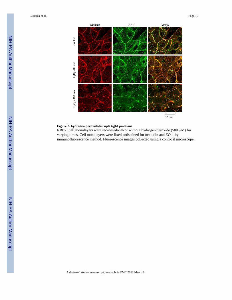
Figure 2. hydrogen peroxidedisrupts tight junctionsNRC-1 cell monolayers were incubatedwith or without hydrogen peroxide (500 μM) forvarying times. Cell monolayers were fixed andstained for occludin and ZO-1 byimmunofluorescence method. Fluorescence images collected using a confocal microscope.
Guntaka et al. Page 15
Lab Invest. Author manuscript; available in PMC 2012 March 1.
NIH
-PA Author Manuscript
NIH
-PA Author Manuscript
NIH
-PA Author Manuscript

Figure 3. hydrogen peroxide disrupts adherens junctionsNRC-1 cell monolayers were incubated with or without hydrogen peroxide (500 μM) forvarying times. Cell monolayers were fixed and stained for E-cadherin and β-catenin byimmunofluorescence method. Fluorescence images collected using a confocal microscope.
Guntaka et al. Page 16
Lab Invest. Author manuscript; available in PMC 2012 March 1.
NIH
-PA Author Manuscript
NIH
-PA Author Manuscript
NIH
-PA Author Manuscript

Figure 4. MLCK activity mediates hydrogen peroxide-induced tight junction disruptionNRC-1 cell monolayers were incubated with (gray) or without ML-7 (white and black bars)followed by incubation with (black and gray bars) or without (white bars) hydrogenperoxidefor 120 min. Protein extracts were immunoblotted for p-MLC and β-actin (A). TER(B) and inulin flux (C) were measured. Values are mean ± sem (n = 6). Asterisks indicatethe values that are significantly (p<0.05) different from corresponding control values.Symbol, #, indicates the value that is significantly (p<0.05) different from cells treated withhydrogen peroxide in the absence of ML-7. Cell monolayers were fixed and stained foroccludin and ZO-1 (D). Fluorescence images collected using a confocal microscope.
Guntaka et al. Page 17
Lab Invest. Author manuscript; available in PMC 2012 March 1.
NIH
-PA Author Manuscript
NIH
-PA Author Manuscript
NIH
-PA Author Manuscript

Figure 5. Src kinase activity mediates hydrogen peroxide-induced tight junction disruptionNRC-1 cell monolayers were incubated without (white and black bars) or with genistein(dark gray bars) or PP2 (light gray bars) followed by incubation with (black bars) or without(white bars) hydrogen peroxide for 120 min. Protein extracts were immunoblotted for c-Src(pY418) and β-actin (A). TER (B) and inulin flux (C) were measured. Values are mean ±sem (n = 6). Asterisks indicate the values that are significantly (p<0.05) different fromcorresponding control values. Symbol, #, indicates the values that are significantly (p<0.05)different from values for cells treated with hydrogen peroxide in the absence of PP2. Cellmonolayers were fixed and stained for occludin and ZO-1 (D). Fluorescence imagescollected using a confocal microscope.
Guntaka et al. Page 18
Lab Invest. Author manuscript; available in PMC 2012 March 1.
NIH
-PA Author Manuscript
NIH
-PA Author Manuscript
NIH
-PA Author Manuscript

Figure 6. Hydrogen peroxide-induced activation of MLCK and c-Src are independent of eachotherNRC-1 cell monolayers were incubated with or without ML-7 or PP2 for 30 min prior toincubation with hydrogen peroxide (500 μM) for varying times. Protein extracts wereimmunoblotted for p-MLC, c-Src(pY418) and βactin.
Guntaka et al. Page 19
Lab Invest. Author manuscript; available in PMC 2012 March 1.
NIH
-PA Author Manuscript
NIH
-PA Author Manuscript
NIH
-PA Author Manuscript
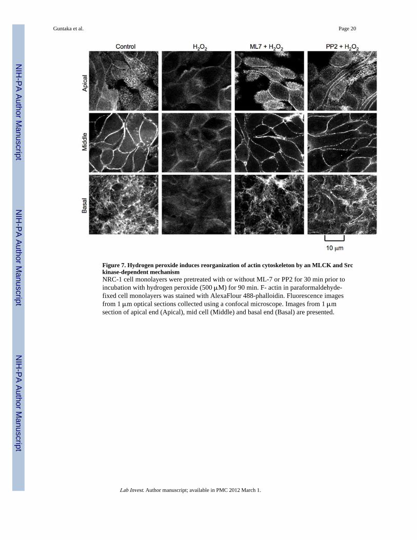
Figure 7. Hydrogen peroxide induces reorganization of actin cytoskeleton by an MLCK and Srckinase-dependent mechanismNRC-1 cell monolayers were pretreated with or without ML-7 or PP2 for 30 min prior toincubation with hydrogen peroxide (500 μM) for 90 min. F- actin in paraformaldehyde-fixed cell monolayers was stained with AlexaFlour 488-phalloidin. Fluorescence imagesfrom 1 μm optical sections collected using a confocal microscope. Images from 1 μmsection of apical end (Apical), mid cell (Middle) and basal end (Basal) are presented.
Guntaka et al. Page 20
Lab Invest. Author manuscript; available in PMC 2012 March 1.
NIH
-PA Author Manuscript
NIH
-PA Author Manuscript
NIH
-PA Author Manuscript
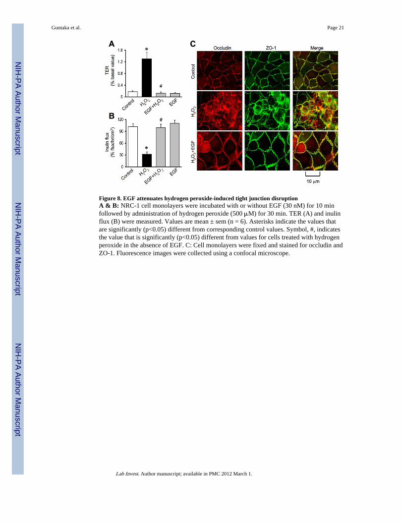
Figure 8. EGF attenuates hydrogen peroxide-induced tight junction disruptionA & B: NRC-1 cell monolayers were incubated with or without EGF (30 nM) for 10 minfollowed by administration of hydrogen peroxide (500 μM) for 30 min. TER (A) and inulinflux (B) were measured. Values are mean ± sem (n = 6). Asterisks indicate the values thatare significantly (p<0.05) different from corresponding control values. Symbol, #, indicatesthe value that is significantly (p<0.05) different from values for cells treated with hydrogenperoxide in the absence of EGF. C: Cell monolayers were fixed and stained for occludin andZO-1. Fluorescence images were collected using a confocal microscope.
Guntaka et al. Page 21
Lab Invest. Author manuscript; available in PMC 2012 March 1.
NIH
-PA Author Manuscript
NIH
-PA Author Manuscript
NIH
-PA Author Manuscript

Figure 9. EGF attenuates hydrogen peroxide-induced Tyr-phosphorylation of tight junction andadherens junction proteinsA: NRC-1 cell monolayers were incubated with or without EGF (30 nM) for 10 minfollowed by administration of hydrogen peroxide (500 μM) for 30 min. Protein extractswere immunoblotted for c-Src(pY418) and c-Src. B: Cell monolayers pretreated with orwithout EGF were incubated with or without hydrogen peroxide (500 μM) for 90 min.Phospho-tyrosine from the denatured protein extracts was immunoprecipitated andimmunoblotted for tight junction and adherens junction proteins.
Guntaka et al. Page 22
Lab Invest. Author manuscript; available in PMC 2012 March 1.
NIH
-PA Author Manuscript
NIH
-PA Author Manuscript
NIH
-PA Author Manuscript

Figure 10. PLCγand PKC activities mediate EGF-mediated protection of tight junction fromhydrogen peroxideA & B: NRC-1 cell monolayers were incubated with or without ET- 18-OCH3 (15 μM) orRo-32-0432 (1μM) for 60 min followed by incubation with or without hydrogen peroxidefor 120 min. TER (A) and inulin flux (B) were measured before (white bars) and after(Black bars) hydrogen peroxide treatment. Values are mean ± sem (n = 6). Asterisks indicatethe values that are significantly (p<0.05) different from corresponding values for hydrogenperoxide group. Symbol, #, indicates the values that are significantly (p<0.05) different fromvalues for cells treated with hydrogen peroxide and EGF in the absence of ET-18- OCH3 orRo-32-0432. C: Cell monolayers were fixed and stained for occludin and ZO-1.Fluorescence images collected using a confocal microscope.
Guntaka et al. Page 23
Lab Invest. Author manuscript; available in PMC 2012 March 1.
NIH
-PA Author Manuscript
NIH
-PA Author Manuscript
NIH
-PA Author Manuscript
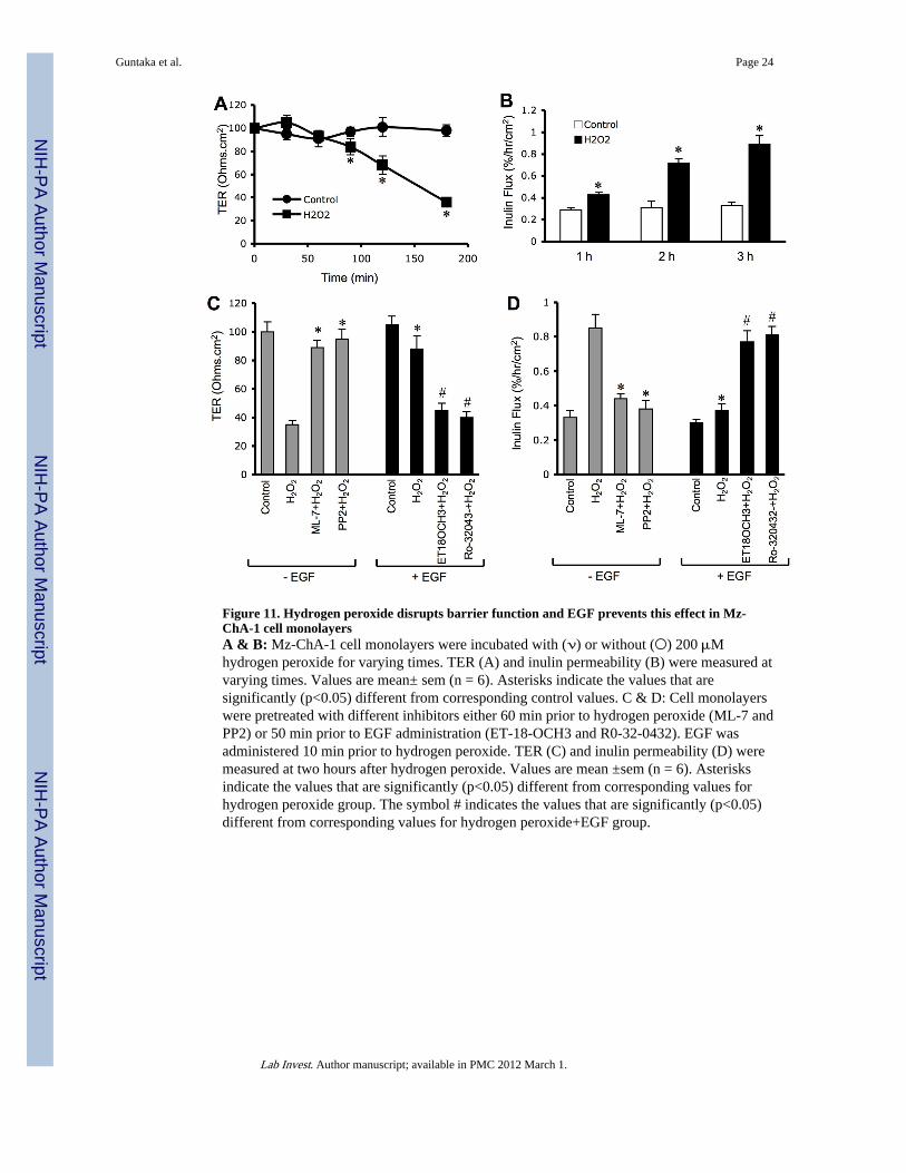
Figure 11. Hydrogen peroxide disrupts barrier function and EGF prevents this effect in Mz-ChA-1 cell monolayersA & B: Mz-ChA-1 cell monolayers were incubated with (ν) or without (○) 200 μMhydrogen peroxide for varying times. TER (A) and inulin permeability (B) were measured atvarying times. Values are mean± sem (n = 6). Asterisks indicate the values that aresignificantly (p<0.05) different from corresponding control values. C & D: Cell monolayerswere pretreated with different inhibitors either 60 min prior to hydrogen peroxide (ML-7 andPP2) or 50 min prior to EGF administration (ET-18-OCH3 and R0-32-0432). EGF wasadministered 10 min prior to hydrogen peroxide. TER (C) and inulin permeability (D) weremeasured at two hours after hydrogen peroxide. Values are mean ±sem (n = 6). Asterisksindicate the values that are significantly (p<0.05) different from corresponding values forhydrogen peroxide group. The symbol # indicates the values that are significantly (p<0.05)different from corresponding values for hydrogen peroxide+EGF group.
Guntaka et al. Page 24
Lab Invest. Author manuscript; available in PMC 2012 March 1.
NIH
-PA Author Manuscript
NIH
-PA Author Manuscript
NIH
-PA Author Manuscript
Copyright © 2022 FDOKUMEN