Junctional and allele-specific residues are critical for ... - Nature
10
ARTICLE Received 20 Apr 2015 | Accepted 30 Jul 2015 | Published 15 Sep 2015 Junctional and allele-specific residues are critical for MERS-CoV neutralization by an exceptionally potent germline-like antibody Tianlei Ying 1 , Ponraj Prabakaran 2,w , Lanying Du 3 , Wei Shi 4 , Yang Feng 2 , Yanping Wang 2 , Lingshu Wang 4 , Wei Li 1,2 , Shibo Jiang 1,3 , Dimiter S. Dimitrov 2 & Tongqing Zhou 4 The MERS-CoV is an emerging virus, which already infected more than 1,300 humans with high (B36%) mortality. Here, we show that m336, an exceptionally potent human anti- MERS-CoV antibody, is almost germline with only one somatic mutation in the heavy chain. The structure of Fab m336 in complex with the MERS-CoV receptor-binding domain reveals that its IGHV1-69-derived heavy chain provides more than 85% binding surface and that its epitope almost completely overlaps with the receptor-binding site. Analysis of antibodies from 69 healthy humans suggests an important role of the V(D)J recombination-generated junctional and allele-specific residues for achieving high affinity of binding at such low levels of somatic hypermutation. Our results also have important implications for development of vaccine immunogens based on the newly identified m336 epitope as well as for elucidation of mechanisms of neutralization by m336-like antibodies and their elicitation in vivo. DOI: 10.1038/ncomms9223 OPEN 1 Key Laboratory of Medical Molecular Virology of Ministries of Education and Health, Shanghai Medical College, Fudan University, Shanghai 200032, China. 2 Protein Interactions Section, Cancer and Inflammation Program, Center for Cancer Research, National Cancer Institute, National Institutes of Health, Frederick, Maryland 21702, USA. 3 Lindsley F. Kimball Research Institute, New York Blood Center, New York, New York 10065, USA. 4 Vaccine Research Center, National Institute of Allergy and Infectious Diseases, National Institutes of Health, Bethesda, Maryland 20892, USA. w Present address: Sanofi Pasteur Biologics, Cambridge, Massachusetts 02139, USA. Correspondence and requests for materials should be addressed to T.Y. (email: [email protected]) or to T.Z. (email: [email protected]). NATURE COMMUNICATIONS | 6:8223 | DOI: 10.1038/ncomms9223 | www.nature.com/naturecommunications 1 & 2015 Macmillan Publishers Limited. All rights reserved.
-
Upload
khangminh22 -
Category
Documents
-
view
0 -
download
0
Transcript of Junctional and allele-specific residues are critical for ... - Nature

ARTICLE
Received 20 Apr 2015 | Accepted 30 Jul 2015 | Published 15 Sep 2015
Junctional and allele-specific residues are criticalfor MERS-CoV neutralization by an exceptionallypotent germline-like antibodyTianlei Ying1, Ponraj Prabakaran2,w, Lanying Du3, Wei Shi4, Yang Feng2, Yanping Wang2, Lingshu Wang4,
Wei Li1,2, Shibo Jiang1,3, Dimiter S. Dimitrov2 & Tongqing Zhou4
The MERS-CoV is an emerging virus, which already infected more than 1,300 humans with
high (B36%) mortality. Here, we show that m336, an exceptionally potent human anti-
MERS-CoV antibody, is almost germline with only one somatic mutation in the heavy chain.
The structure of Fab m336 in complex with the MERS-CoV receptor-binding domain reveals
that its IGHV1-69-derived heavy chain provides more than 85% binding surface and that its
epitope almost completely overlaps with the receptor-binding site. Analysis of antibodies
from 69 healthy humans suggests an important role of the V(D)J recombination-generated
junctional and allele-specific residues for achieving high affinity of binding at such low levels
of somatic hypermutation. Our results also have important implications for development of
vaccine immunogens based on the newly identified m336 epitope as well as for elucidation of
mechanisms of neutralization by m336-like antibodies and their elicitation in vivo.
DOI: 10.1038/ncomms9223 OPEN
1 Key Laboratory of Medical Molecular Virology of Ministries of Education and Health, Shanghai Medical College, Fudan University, Shanghai 200032, China.2 Protein Interactions Section, Cancer and Inflammation Program, Center for Cancer Research, National Cancer Institute, National Institutes of Health,Frederick, Maryland 21702, USA. 3 Lindsley F. Kimball Research Institute, New York Blood Center, New York, New York 10065, USA. 4Vaccine ResearchCenter, National Institute of Allergy and Infectious Diseases, National Institutes of Health, Bethesda, Maryland 20892, USA. w Present address: Sanofi PasteurBiologics, Cambridge, Massachusetts 02139, USA. Correspondence and requests for materials should be addressed to T.Y. (email: [email protected]) or toT.Z. (email: [email protected]).
NATURE COMMUNICATIONS | 6:8223 | DOI: 10.1038/ncomms9223 |www.nature.com/naturecommunications 1
& 2015 Macmillan Publishers Limited. All rights reserved.

The recent discovery of neutralizing monoclonal antibodies(mAbs) with exceptional potency and breadth againsthuman immunodeficiency virus-1 (HIV-1) and influenza
has renewed the interest in designing vaccines to elicit similarantibodies in vivo1–4. However, the vaccine development has beenimpeded by the fact that these broadly neutralizing antibodies(bnAbs) are highly divergent from their putative germlinepredecessors, and that the designed germline antibodies showedlittle or no measurable binding to viral antigens5–7. Interestingly,neutralizing antibodies against some emerging viruses includingsevere acute respiratory syndrome coronavirus (SARS-CoV),Nipah and Hendra viruses, which cause acute infections, could beelicited within relatively short period of time after infection, andsome of these antibodies were very close to their putativegermline predecessors8–10. Therefore, the structural characteri-zation of the interactions between these germline-like antibodiesand the viruses may represent a unique opportunity tounderstand the initial elicitation mechanism of the bnAbs andmay eventually help the design of effective vaccineimmunogens4,7,11–14.
The Middle East respiratory syndrome coronavirus(MERS-CoV) is a novel, highly pathogenic human coronavirusfirst recognized in September 2012 in Saudi Arabia in a patientwho exhibited SARS-like respiratory syndrome and died fromsevere pneumonia and renal failure15,16. Since September 2012,MERS cases have been reported in more than 20 countries15,16,and 1,356 laboratory-confirmed cases have been reported to theWorld Health Organization as of 26 June 2015, including at least484 related deaths (http://www.who.int/csr/don/26-june-2015-mers-korea/en/). Of note, MERS-CoV is phylogenetically distinct
from any human coronavirus including SARS-CoV, but morerelated to the bat coronaviruses HKU4 and HKU5 (refs 17,18).We and others have found that MERS-CoV most likely originatedin bats and has adapted to use human receptor dipeptidylpeptidase-4 (DPP4) to gain entry into host cells19,20. Dromedarycamels in the Middle East and Africa are considered to be oneof the intermediate transmitters of MERS-CoV from bats tohumans21–24. Indeed, it has been found that coronaviruses areable to rapidly and stably adapt to new host species25. Thesefindings, along with the fact that MERS-CoV emerged only adecade after the appearance of the first highly pathogenic humancoronavirus SARS-CoV and has a higher mortality rate(B36 versus B10%), suggest that coronaviruses could be acontinuous and long-term threat to human health.
Therefore, we and others have developed human neutralizingmAbs26–29 against the virus for use as candidate therapeutics andtemplates for vaccine immunogens as well as tools to understandimmune responses against this newly emerged virus. The mostpotent mAb, m336, bound to MERS-CoV receptor-bindingdomain (RBD) with subnanomolar avidity, and inhibitedinfection of pseudotyped and live MERS-CoV with aninhibitory concentration required for 50% neutralization (IC50)of 0.005 and 0.07 mgml� 1, respectively26.
In this study, we analysed the immunogenetic origin of m336and solved its crystal structure in complex with the MERS-CoVRBD. The antibody precisely targets the receptor-binding site(RBS) on MERS-CoV, and a relatively significant number (three)of junctional residues in the heavy-chain complementarity-determining region 3 (HCDR3) and two allele-specific IGHV1-69*06 germline-encoded residues are critical for the RBD binding.Germline mAbs typically exhibit excellent drugability properties,especially lower immunogenicity11, and therefore m336-likemAbs may not only be effective templates for development ofvaccine immunogens but also could have great potential forprophylaxis and therapy of MERS-CoV infection in humans.
ResultsCrystal structure of MERS-CoV RBD in complex with Fab m336.To elucidate the molecular mechanisms of MERS-CoV neutrali-zation by m336 and identify possible templates for developmentof vaccine immunogens, we determined the crystal structure of itscomplex with the virus RBD. We robotically screened crystal-lization conditions for an endoglycosidase H-treated MERS-CoVRBD complexed with the Fab m336. Crystals of diffraction qualitywere obtained from a condition containing 20% mono-Methylpolyethylene glycol 2000 (PEG2000 MME). A 2.65-Å resolutiondata set in space group P212121 was collected at 22-ID beamline atthe Advanced Photon Source with 20% glycerol as cryoprotectant,and structure solution by molecular replacement with theDDP4-bound MERS-CoV RBD and Fab revealed two Fab m336-MERS-CoV RBD complexes occupying an asymmetric unit.Iterative refinement and model building resulted in a structurewith Rwork/Rfree of 19.7%/25.1% (Table 1; Supplementary Fig. 1a).
Fab m336 targets the MERS-CoV RBS. Fab m336 interacts withthe RBS on the MERS-CoV RBD (Fig. 1a). The interaction surfaceburies a total area of about 1,700Å2, with 860Å2 contributed bym336 and 820Å2 by MERS-CoV RBD. Even though both theheavy-chain variable domain (VH) and the light-chain variabledomain (VL) of m336 are involved in the interaction, the VH
provides 485% binding surface. Within the VH, HCDR1,HCDR2 and framework region 3 (FR3) provide B58% ofthe total MERS-CoV RBD-binding surface while HCDR3(of the V(D)J recombination product) provides B29% (Fig. 1b;Supplementary Table 1). With HCDR3 and LCDR3 on one side
Table 1 | Data collection and structure refinement statistics.
m336-MERS-CoV RBD
Data collectionWavelength (Å) 1.0000Space group P 21 21 21Unit cella, b, c (Å) 47.8, 146.9, 200.5
Resolution (Å)* 50–2.65 (2.74–2.65)Rmerge (%) 11.0 (41.0)Rpim (%) 5.8 (23.4)I/sI 14.0 (2.0)Completeness (%) 97.3 (90.3)Redundancy 3.9 (3.2)
RefinementResolution (Å) 45.45–2.65 (2.74–2.65)Unique reflections 41,125 (3,734)Rwork/Rfree (%) 19.7 (27.3)/25.1 (35.8)Number of atomsProtein 9,745Ligands 68Water 236
Wilson B-factor (Å2) 49.1B-factors (Å2)Protein 63.0Ligands 68.3Solvent 46.3
r.m.s.d.Bond lengths (Å) 0.002Bond angles (o) 0.95
RamachandranFavored (%) 95.0Outliers (%) 0.2Clashscore 4.0
*Statistics for the highest-resolution shell are shown in parentheses.
ARTICLE NATURE COMMUNICATIONS | DOI: 10.1038/ncomms9223
2 NATURE COMMUNICATIONS | 6:8223 | DOI: 10.1038/ncomms9223 | www.nature.com/naturecommunications
& 2015 Macmillan Publishers Limited. All rights reserved.

and HCDR1 and HCDR2 on the other side, the antigen-bindingregion of m336 forms a claw-shaped surface. The claw grips theRBS with HCDR1 and HCDR2 penetrating into the concavesurface formed by b5–b8 and HCDR3 and LCDR3 interactingwith the outer edge of b7 and its entrance loop (Fig. 1c). It is ofnote that a non-canonical disulphide bond forms between Cys98and Cys100c (the Kabat numbering definition) to stabilize theHCDR3 into a ‘twisted’ loop conformation (Fig. 1d). Similar tothe receptor DPP4, m336 interacts with both patches 1 and 2 ofthe RBS (Fig. 1d).
Similarity in the RBD recognition by m336 and DPP4. Them336 epitope is composed of residues from the MERS-CoV RBDRBS, including strands b5–b8 and the loop leading to b7 (Fig. 2a),which overlaps extensively with the DPP4-binding site. Pairwisecomparison of ligand-binding surface areas of RBS residues eitherin the m336 epitope or in the DDP4-binding site30,31 showedsignificant correlation (P¼ 0.025), indicating shared usage ofresidues for binding m336 and receptor DPP4. For RBS residuesthat interact with both m336 and DPP4, their pairwise surfaceareas buried by m336 and DPP4 showed even stronger
correlation (P¼ 0.016; Fig. 2b). Indeed, B90% of the m336epitope surface overlaps with the DPP4-binding site on RBS(Fig. 2c; Supplementary Fig. 1). Interestingly, Trp535RBD, whichinteracts with glycans in DPP4, was engaged by m336 HCDR3residue Thr100a and LCDR3 residues Asn92 and Ser93.Importantly, in addition to precise targeting of the RBS, criticalchemical interactions such as the salt bridges between Asp539RBDand Lys267DPP4 were mimicked by HCDR3 Arg100e of m336(Fig. 1d; Supplementary Table 2). The RBD-binding surfaces onboth m336 and DPP4 showed basic electrostatic potentials(Fig. 2d).
Superposition of the RBD in its m336-bound form with that inthe DPP4-bound form30 resulted in Ca root mean squareddeviation (r.m.s.d.) of 0.82Å, indicating that binding of m336 didnot induce significant conformational changes in MERS-CoVRBD. It is interesting to note that the similarity in conformationat the antibody epitope was even higher than the rest ofthe MERS-CoV RBD (Ca r.m.s.d.¼ 0.50Å). Such superpositionof the MERS-CoV RBD also brought m336 and DPP4 intooverlapping positions, with 450% of the Fab m336 encompassedby the volume of DPP4 (Fig. 2e; Supplementary Fig. 2), indicating
HCDR1
Heavy chain Light chain
HCDR2HCDR3LCDR3
Receptor-bindingsubdomain
m336
c
ba
d
HCDR1
HCDR2
HCDR3
LCDR3
β5β6
β7β8
90°
D539
W535E536
R100e
Y94
S93
Y540
V95
R542
W553 F506
D510
S31
F54
Patch 1 interactions Patch 2 interactions
C98
C100c
90°
RBD
Heavy chain1 22 36 52a | | | || --------------FR1-------------_CDR1------FR2-----_______CDR2________QVQLVQSGAEVKKPGSSVKVSCKASGGTFSSYAISWVRQAPGQGLEWMGGIIPIFGTASYAQKFQG
67 82abc 92 100abcdefgh103 113 | |||| | ||||||||| | | --------------FR3-------------________ CDR3________----FR4---- RVTITADKSTSTAYMELSSLRSEDTAVYYCARVGYCSSTSCNRGAFDIWGQGTMVTVSS
Light chain 1 23 35 50 | | | | ----------FR1---------- ___CDR1____ -------FR2-----__CDR2_ DIQLTQSPSSLSASVGDRVTITCRASQGIRNDLGWYQQKPGKAPKLLIYAASSLQS
58 65 80 89 98 107 | | | | | | ----------------FR3------------- ___CDR3 ___---FR4--- GVPSRFSGSGSGTDFTLTISSLQPEDFATYYCQQLNSYPLTFGGGTKVEIK
HCDR1
HCDR2HCDR3LCDR3
W535
β5β6
β7β8
MERS-CoV
Figure 1 | Crystal structures of Fab m336 in complex with MERS-CoV RBD. (a) Complex structure in cartoon representation. Heavy and light chains
of m336 are coloured light green and light blue, respectively. The receptor-binding subdomain on MERS-CoV RBD is coloured orange with Trp535 shown
in shticks representation. (b) Sequences of m336 heavy chain and light chain with residues that interact with the MERS-CoV RBD highlighted with
orange dots. Antibody residues are numbered according to the Kabat nomenclature. (c) Interaction between CDRs of m336 and the MERS-CoV RBD.
m336 CDRs are shown in cartoon representation and the MERS-CoV RBD is depicted in semitransparent surface and cartoon. (d) Detailed interactions
between m336 and patches 1 and 2 of the MERS-CoV RBD. Key residues are highlighted in stick representation. Hydrogen bonds and salt bridges are
displayed with dotted lines connecting interacting atoms. The non-canonical disulphide bond between HCDR3 residues Cys98 and Cys100c is in yellow
sticks. Both panels (c,d) are shown as different 90� views from panel (a).
NATURE COMMUNICATIONS | DOI: 10.1038/ncomms9223 ARTICLE
NATURE COMMUNICATIONS | 6:8223 | DOI: 10.1038/ncomms9223 |www.nature.com/naturecommunications 3
& 2015 Macmillan Publishers Limited. All rights reserved.
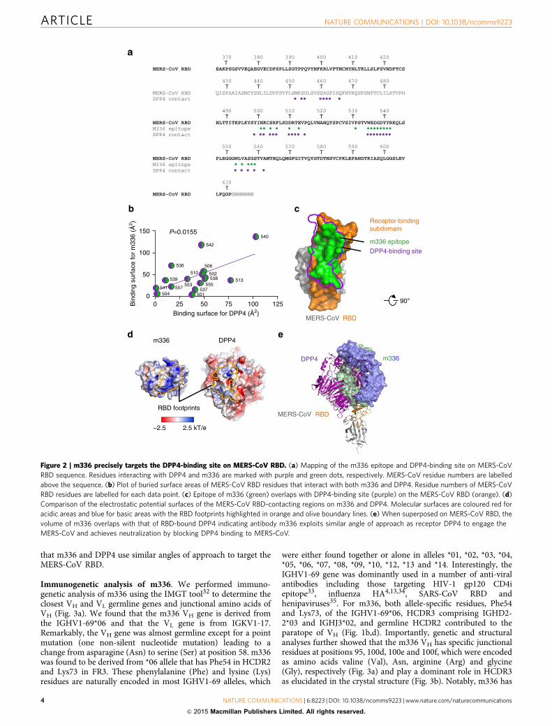
that m336 and DPP4 use similar angles of approach to target theMERS-CoV RBD.
Immunogenetic analysis of m336. We performed immuno-genetic analysis of m336 using the IMGT tool32 to determine theclosest VH and VL germline genes and junctional amino acids ofVH (Fig. 3a). We found that the m336 VH gene is derived fromthe IGHV1-69*06 and that the VL gene is from IGKV1-17.Remarkably, the VH gene was almost germline except for a pointmutation (one non-silent nucleotide mutation) leading to achange from asparagine (Asn) to serine (Ser) at position 58. m336was found to be derived from *06 allele that has Phe54 in HCDR2and Lys73 in FR3. These phenylalanine (Phe) and lysine (Lys)residues are naturally encoded in most IGHV1-69 alleles, which
were either found together or alone in alleles *01, *02, *03, *04,*05, *06, *07, *08, *09, *10, *12, *13 and *14. Interestingly, theIGHV1-69 gene was dominantly used in a number of anti-viralantibodies including those targeting HIV-1 gp120 CD4iepitope33, influenza HA4,13,34, SARS-CoV RBD andhenipaviruses35. For m336, both allele-specific residues, Phe54and Lys73, of the IGHV1-69*06, HCDR3 comprising IGHD2-2*03 and IGHJ3*02, and germline HCDR2 contributed to theparatope of VH (Fig. 1b,d). Importantly, genetic and structuralanalyses further showed that the m336 VH has specific junctionalresidues at positions 95, 100d, 100e and 100f, which were encodedas amino acids valine (Val), Asn, arginine (Arg) and glycine(Gly), respectively (Fig. 3a) and play a dominant role in HCDR3as elucidated in the crystal structure (Fig. 3b). Notably, m336 has
0 25 50 75 100 1250
50
100
150
501
502
504
506
510
513
536
537
538539
540
541
542
553 555557
DPP4-binding site
MERS-CoV RBD
m336
c
a
b
e
P=0.0155
90°
Binding surface for DPP4 (Å2)
Bin
ding
sur
face
for
m33
6 (Å
2 )
m336 epitope
MERS-CoV RBD
DPP4
Receptor-bindingsubdomain
dm336 DPP4
RBD footprints
–2.5 2.5 kT/e
370 380 390 400 410 420 T T T T T
MERS-CoV RBD EAKPSGSVVEQAEGVECDFSPLLSGTPPQVYNFKRLVFTNCNYNLTKLLSLFSVNDFTCS
430 440 450 460 470 480 T T T T T T
MERS-CoV RBD QISPAAIASNCYSSLILDYFSYPLSMKSDLSVSSAGPISQFNYKQSFSNPTCLILATVPH DPP4 contact
490 500 510 520 530 540 T
T
T T T T TMERS-CoV RBD NLTTITKPLKYSYINKCSRFLSDDRTEVPQLVNANQYSPCVSIVPSTVWEDGDYYRKQLS M336 epitope DPP4 contact
550 560 570 580 590 600 T T T T T T
MERS-CoV RBD PLEGGGWLVASGSTVAMTEQLQMGFGITVQYGTDTNSVCPKLEFANDTKIASQLGGSLEV M336 epitope DPP4 contact
610 T
MERS-CoV RBD LFQGPGHHHHHH
Figure 2 | m336 precisely targets the DPP4-binding site on MERS-CoV RBD. (a) Mapping of the m336 epitope and DPP4-binding site on MERS-CoV
RBD sequence. Residues interacting with DPP4 and m336 are marked with purple and green dots, respectively. MERS-CoV residue numbers are labelled
above the sequence. (b) Plot of buried surface areas of MERS-CoV RBD residues that interact with both m336 and DPP4. Residue numbers of MERS-CoV
RBD residues are labelled for each data point. (c) Epitope of m336 (green) overlaps with DPP4-binding site (purple) on the MERS-CoV RBD (orange). (d)
Comparison of the electrostatic potential surfaces of the MERS-CoV RBD-contacting regions on m336 and DPP4. Molecular surfaces are coloured red for
acidic areas and blue for basic areas with the RBD footprints highlighted in orange and olive boundary lines. (e) When superposed on MERS-CoV RBD, the
volume of m336 overlaps with that of RBD-bound DPP4 indicating antibody m336 exploits similar angle of approach as receptor DPP4 to engage the
MERS-CoV and achieves neutralization by blocking DPP4 binding to MERS-CoV.
ARTICLE NATURE COMMUNICATIONS | DOI: 10.1038/ncomms9223
4 NATURE COMMUNICATIONS | 6:8223 | DOI: 10.1038/ncomms9223 | www.nature.com/naturecommunications
& 2015 Macmillan Publishers Limited. All rights reserved.

no somatic mutations in the IGHD and IGHJ genes, which can beunambiguously identified by IMGT/V-Quest (SupplementaryTable 3). In contrast, m336 VL, composed of IGKV1-17*01 andIGKJ4*01, was found to have five amino acid residue changes(five non-silent and three silent nucleotide mutations) resultingfrom mutations in the IGKV gene (Fig. 3a; SupplementaryTable 4). However, none of the VL somatic mutations was foundto be directly involved in binding to RBD, but it is possible thatthey could influence VH–VL interactions or result in otherallosteric effects. Further, the detailed structural analysis of m336complex structure revealed that the CDRH3 residues, particularly,the junctional residues Asn100d and Arg100e, are involved inhydrogen bonding with Gly538 and Asp539 of RBD, respectively,which mimicked the conserved DPP4 interactions Gln286 and
Lys267 (Fig. 2a,b; Supplementary Table 2). Also, the junctionalresidue Arg100e formed a salt bridge with Asp539, whichresembled the conserved slat bridge involving Lys267 of DPP4(Fig. 1d,b; Supplementary Table 2). In summary, we show that thegermline-encoded residues, Phe54 in HCDR2 and Lys73 in FR3,which are conserved in many IGHV1-69 alleles, and, importantly,junctional residues of HCDR3 that make conserved interactionsthat of DPP4, could play critical roles in the m336 paratopeinteractions.
These findings suggest that the selection of specific junctionalamino acids with certain V(D)J recombination and allele-specificamino acids in the heavy chain, and subsequent somaticmutations in the light chain could lead to the high affinity ofm336 for its target RBD. However, the adaptability of somatic
a
b
c
MERS-CoV RBD
Val95Gly100f
Arg100e
Asn100d
d
Heavy chain 10 20 30 40 50 70| | | | | a | |
m336-VH QVQLVQSGAEVKKPGSSVKVSCKASGGTFSSYAISWVRQAPGQGLEWMGGIIPIFGTASYAQKFQGRVTIT HV1-69*06 ..........................................................N............
80 90 100 110| abc | |abcdefgh |
m336-VH ADKSTSTAYMELSSLRSEDTAVYYCARVGYCSSTSCNRGAFDIWGQGTMVTVSS HV1-69*06 ...........................---------------------------HD2-2*03 ----------------------------........------------------HJ3*02 ---------------------------------------...............
10 20 30 40 50 70| | | | | | |
m336-VLKV1-17*01 ...M.........................................R.......................E
80 90 100| | |
m336-VLKV1-17*01 ..................L.H....------------ KJ4*01 ------------------------.............
Light chain
m336
GIA8TG
GESY
IJG1L
HK
AV
1A
TE
UR
KV
1–17*01
GQ6EIIG1QKFV24FHL0R4
F42LC
SC4098
GRX6Q
m336
IUQ23
GW8N9
J1LE
P
DN540
GLZ
KIH
71FM
FG
4AV
I0B
UK
IOLJL
GJIIK
JTG8G
IAE2TF2VYW
JOFEJ
F80WF
IXGN8
G7S4X
JEKWS
G0V6M
FRB3EFLZTE
JEE23JE94OAO7R8
DAP65IVVG
A
JRFO
1
IZF
V5
IIPZ
Vm336 ARVGYCSSTSCNRGAFDI3B11 ARVGYCSSTSCHIGAFDI3B12 ARASYCSTTSCASGAFDIFDKX3 ARAQYCSSTSCYWNWFDPIVENF ARVEYCSSTSCYANWFDPD8MYQ ARAGYCSSTSCYSSWFDPDY4B5 ARDGYCSSTSCYFRYFDLI8G0B ALNGYCSSTSCWVLPFDPCBHU6 ARGMYCSSTSCYGPAFDIHO1JR ARGQYCSSTSCYLYAFDIAR2UL ARGRYCSSTSCKDDAFDICDBVV AISAYCSSTSCYPGSIDY * ***:*** :*
95 100 def
DIQLTQSPSSLSASVGDRVTITCRASQGIRNDLGWYQQKPGKAPKLLIYAASSLQSGVPSRFSGSGSGTD
FTLTISSLQPEDFATYYCQQLNSYPLTFGGGTKVEIK
Figure 3 | Immunogenetic analysis showed a germline heavy chain with a unique junctional motif. (a) m336 VH, VL sequence alignments with germline
gene segments are shown. Identical residues are denoted by dots and junction residues at HCDR3 are boxed. Antibody residues are numbered according
to the Kabat nomenclature. (b) Close-up view of interactions between the junctional amino acids in the HCDR3 of m336 with MERS-CoV RBD. (c) Amino
acid sequences of HCDR3s that were found similar to that of m336 from 454 sequencing analysis of IgM libraries derived from 69 healthy human subjects
(denoted by five-letter codes), and from the report by Tang et al.28 (3B11 and 3B12). Red boxes show the junction amino acid residues in HCDR3s.
(d) Germline-rooted circular phylogenetic tree of m336 VL-like antibody sequences (denoted by five-letter codes, m336 in red and KV1-17*01 in green)
found in IgM libraries derived from 69 healthy human subjects and two newborn babies along with the anti-rabies antibody light chain (SC4098, in purple)
from the report by Kramer et al.38.
NATURE COMMUNICATIONS | DOI: 10.1038/ncomms9223 ARTICLE
NATURE COMMUNICATIONS | 6:8223 | DOI: 10.1038/ncomms9223 |www.nature.com/naturecommunications 5
& 2015 Macmillan Publishers Limited. All rights reserved.

mutations might depend on the nature of junctional residues thatoccur far ahead of any somatic mutations and determine thepathway of affinity maturation36.
To further investigate potential immunogenetic mechanismssuch as IGHV1-69 recombination frequency with specific IGHDand IGHJ genes among other germlines and conserved aminoacid residues at allelic and junctional positions, we analysed indetail the deep sequencing data of our antibodyome studiesfrom 69 healthy human donors35. Our analysis revealedthat a significant (12.7%) proportion of expressed VH geneswere productively recombined from the IGHV1-69 gene(Supplementary Fig. 3). Among a total of 9,454 sequences ofIGHV1-69 germline origin, 6,512 sequences had the germline-encoded Phe54 and 3,057 sequences had the germline-encodedLys73 natural allelic residues in the population of 69 individuals,while 1,452 sequences (15.4%) had both residues, Phe54 andLys73, such as found in m336. This suggests a potentialmechanism contributing to individually variable but effectiveelicitation of m336-like neutralizing antibodies in response toMERS-CoV infection. We further explored the VH sequencesfrom our 454 antibody sequencing database to find VH-relatedm336-like antibodies. We found nine m336-related VH sequencesfrom 69 volunteers that shared the same IGHV1-69 germlinefamily with a characteristic disulphide bond at the middle ofHCDR3 (Fig. 3c). The commonalities among the HCDR3sincluded the similar HCDR3 length of 18 amino acidsand the usage of D2-2 segment. Notably, m336 had a uniquejunctional motif as compared with those nine VH sequencesas well as to those two anti-MERS-CoV antibodies (3B11 and3B12) recently reported28 (Fig. 3c). A disulphide bondcharacterized by the IGHD2-2*03 gene was conserved in all theHCDR3s, implying the same IGHD reading frame usage in allthose nine VH sequences. The disulphide-bonded cysteineresidues in the HCDR3 of m336 involved in the direct bindingto MERS-CoV RBD (Fig. 1b,d) suggested a possible functionalrole of this HCDR3 disulphide motif. Compared with the otheranti-MERS-CoV antibodies, 3B11 and 3B12, m336 used adifferent VL derived from IGK1-17, whereas 3B11 and 3B12used IGKV3-20 and IGKV4-1, respectively for their VL
sequences.Although antibody phage display technique has proved to be
an effective method for generating mAbs, the mAbs so obtainedfrom random combinations of VH and VL may not reflect thenatural VH/VL pairing as found in immune repertoire. To assessthe potential compatibility of VH/VL pairing in m336, wesearched through the IMGT/3Dstructure-DB. We found thatthe same m336-specific VH/VL pairing, IGHV1-69/IGK1-17,exists in the dengue cross-reactive hybridoma-derived mAb 2H12and anti-Homo sapiens epidermal growth factor receptor (EGFR)antibody, imgatuzumab, indicating the immunologically relevantcognate VH/VL pairing type for m336.
To examine the role of IGKV1-17 usage in m336 VL, wefurther analysed the IGKV1-17 lineage sequences retrieved fromnaive IgM repertoires of 69 healthy human subjects as well as inneonatal IgM repertoires of two newborn babies37. We found 40unique m336 VL-like sequences with the IGKV1-17 lineage thatshared some pre-existing amino acid mutations with m336 VL
(Supplementary Fig. 4). Phylogenetic tree analysis of the m336VL-like sequences revealed a highly germline-related nature of them336 VL (Fig. 3d). We previously found that B70% of the heavyand light chains had one to five amino acid changes as naturallyoccurring somatic hypermutation in the cord blood IgMrepertoire37. An interesting aspect of our study is that most ofthe m336 VL mutations are also found as pre-existing mutationsassociated with certain IGKV1-17 lineage sequences in thoseIgM repertoires (Supplementary Fig. 4). Further, we identified an
anti-rabies virus antibody SC4098 by BLAST search against theGenBank database, whose VL chain has all the four mutationsidentical to those in m336 VL as compared with the IGKV1-17germline38. This implies a possible functional role of these VL
mutations. Accordingly, these VL mutations, although in a lesserextent, along with the unique junctional residues and alleleencoding residues of VH, might have played a role in thehigh-affinity binding to MERS-CoV.
Taken together, these results suggest that m336 VH uses the keyimmunogenetic elements including naturally occurring Phe54and Lys73-encoded allelic-specific amino acids and specificjunctional amino acid residues while m336 VL relies on somaticmutations for deriving its high affinity in an unprecedentedmanner.
Binding and neutralization activity of germline precursors. Itwas surprising that m336 as an exceptionally potent MERS-CoVneutralizer had only six amino acid changes from its inferredgermline precursor. To determine whether these mutations areessential for the potency of m336, we reverted the residue(s) inthe heavy chain (m336-gH), light chain (m336-gL) or light-chainframework region (m336-gL-FR) to their germline counterparts,respectively (Fig. 4). The binding of m336 and the germlinepredecessors to the recombinant MERS-CoV S1 protein wasevaluated by enzyme-linked immunosorbent assay (ELISA;Fig. 4b,d) and Biacore (Supplementary Fig. 5), and theirneutralization activity against MERS-CoV S1-pseudotypedviruses in Huh-7 cells was determined using methods previouslydescribed26 (Fig. 4c,d). The reversion of the only mutation in theheavy chain (S59N) did not have any effect on the MERS-CoV-binding affinity and neutralizing activity of m336. The m336-gHexhibited potent neutralizing activity with an IC50 of0.002 mgml� 1, which is very similar to that of m336 measuredhere (0.003mgml� 1) or described previously (0.005 mgml� 1).
The m336 light chain contains three somatic mutations in theframework region (Leu4, Leu46 and Asp70) and two in theLCDR3 region (Gln89 and Leu91). None of these residues makesdirect contact with the MERS-CoV RBD. Interestingly, reversionof the three framework somatic mutations of m336 to theirgermline counterparts (m336-gL-FR) not only decreased thebinding to MERS-CoV RBD (Fig. 4b) but also markedly reducedthe neutralization potency (Fig. 4c). The neutralization activity ofm336-gL-FR (IC50¼ 0.47 mgml� 1) was two orders of magnitudelower than that of m336 (IC50¼ 0.003 mgml� 1). Completereversion of somatic mutations in the m336 VL further decreasedRBD-binding activity and MERS-CoV-neutralizing activity.Interestingly, the dissociation rate constants of the four antibodiesare similar but the association rate constants of m336 andm336-gH are much higher than that of m336-gL and m336-gL-FR(Supplementary Fig. 5). The IC50 value of m336-gL was1.33 mgml� 1, threefold higher than that of m336-gL-FR.Consistent with the immunogenetic analysis, these results supportthe importance of naturally occurring VL mutations, anddemonstrate that non-contact residues in the m336 light chainare essential for its binding and potency.
Binding activity of junctional and allele-specific mutants. Tofurther demonstrate the importance of junctional and allele-specific residues in the binding of m336 to RBD, we generateda series of m336 mutations and measured their binding to MERS-CoV S1 protein by ELISA (Fig. 4e,f). m336 was derived fromIGHV1-69*06 allele that has Phe54 in HCDR2 and Lys73 in FR3.In the 14 IGHV1-69 alleles, eight of them have a phenylalanine atposition 54 while the other six alleles (*02, *04, *08, *09, *10 and*11) have a leucine. Interestingly, the m336 F54L mutant was
ARTICLE NATURE COMMUNICATIONS | DOI: 10.1038/ncomms9223
6 NATURE COMMUNICATIONS | 6:8223 | DOI: 10.1038/ncomms9223 | www.nature.com/naturecommunications
& 2015 Macmillan Publishers Limited. All rights reserved.

found to have greatly reduced binding affinity to MERS-CoV S1protein with at least 1,000-fold reduced EC50 as compared withthe wild-type m336 (Fig. 4e). Similarly, half of the 14 alleles havea lysine at position 73 while the other 7 alleles (*01, *03, *05, *07,*11, *12 and *13) have a glutamic acid, and the m336 K73Emutant was found to have at least 10-fold reduced MERS-CoV S1binding affinity as compared with m336. The alanine mutationsof the four junctional amino acids (V95A, N100dA, R100eA andG100fA) also showed severely reduced binding to MERS-CoV S1as compared with the wild-type m336 (Fig. 4f). These results,along with the structural analysis, confirmed that the junctionaland allele-specific amino acids in m336 are critical for itsMERS-CoV RBD binding and virus neutralization.
DiscussionElicitation of highly somatically mutated bnAbs against viruseswith high antigenic diversity, such as HIV, has been challengingwhen traditional vaccine approaches are applied. Thus, analternative vaccination strategy has been proposed to first activatenaive mature B cells expressing unmutated germline B cellreceptors with germline antibody-binding immunogens, followedby immunogen binding to intermediate antibodies to guide theimmune system through the complex maturation pathways,resulting in, finally, the elicitation of those antibodies with specificsequences11.
However, it appears that potent antibodies with broadneutralizing activity against some viruses, in this case the
Germlinerevertants
Mutations RBDbinding
Neutralization(IC50, μg ml–1)
m336 – +++++ 0.003
m336-gHS59N (heavy
chain) +++++ 0.002
m336-gL-FRL4M/L46R/D70E
(light chain framework region)
m336-gLL4M/L46R/D70E/
Q89L/L91H(light chain)
++ 0.47
+ 1.33
Gln89
Leu91
Leu46
Asp70
Leu4
Ser59
RBD
Light chainHeavy chain
c
a b
d
fe
100
1001010.10.010.0010.0
0.2
0.4
Abs
(40
5 nm
)
0.6
0.8
1.0
1.2
1.4
1.6
m336
m336
m336-gH
m336-gH
m336-gL-FR
m336-gL-FR
m336-gL
m336-gLm102.4
80
60
40
20
0
1E–5 1E–5 1E–3 0.01mAb (µg ml–1)
mAb (µg ml–1)
1,000 1,000100 100101 1010.1 0.10.0
1.0
2.0
Abs
(40
5 nm
) 3.0
4.0
0.0
1.0
2.0
Abs
(40
5 nm
) 3.0
4.0F54LK73E
V95AN100dAR100eAG100fAm336 Fab
m336 Fab
0.01 0.010.001 0.001
mAb (nM) mAb (nM)
0.1 1 10
Per
cent
neu
tral
izat
ion
Figure 4 | The binding and neutralization activities. (a) Representation of the amino acids in m336 that are divergent from those in the predicted
germline precursor. None of the five residues in the light chain has any direct contact with the MERS-CoV RBD. (b) Binding of IgG1s m336, m336-gH,
m336-gL-FR, m336-gL and a negative control IgG1 m102.4 to MERS-CoV S1 protein measured by ELISA. (c) Neutralization of viruses pseudotyped with
the MERS-CoV S glycoprotein. (d) A summary of the binding and neutralization results of m336 and the germline revertants. (e) Binding of Fab m336
allele-specific mutants (F54L and K73E) to MERS-CoV S1 protein measured by ELISA. (f) Binding of Fab m336 junctional mutants (V95A, N100dA, R100eA
and G100fA) to MERS-CoV S1 protein measured by ELISA.
NATURE COMMUNICATIONS | DOI: 10.1038/ncomms9223 ARTICLE
NATURE COMMUNICATIONS | 6:8223 | DOI: 10.1038/ncomms9223 |www.nature.com/naturecommunications 7
& 2015 Macmillan Publishers Limited. All rights reserved.

MERS-CoV, can naturally exist with very low level of somatichypermutation. The MERS-CoV RBD is highly conserved withvery few mutations. By analysing the known RBD sequences, wefound that the epitope of m336 does not overlap with any of thepoint mutations (Supplementary Fig. 6). By using genetic andstructural analyses, we discovered that a germline-encodedantibody, m336, could achieve high-affinity recognition andneutralization of the newly emerged MERS-CoV, mostly by usingallele-specific residues, as well as junctional amino acids. It isnoteworthy that even though germline sequences of specificantibodies (for example, HIV-1 bnAbs) can be predicted byreconstructing B cell clonal lineages39, it may not be possible toinfer the exact naturally occurring junctional amino acids, whichwere introduced by V(D)J rearrangement, a critical processpreceding somatic hypermutation. Therefore, the junctionalamino acids of the mature antibodies are usually retained orbioinformatically derived in the design of germline-encodedprecursor antibodies5–7,40. Notably, it has been demonstrated thatadaptability of somatic mutations might depend on the nature ofjunctional amino acids that occur far ahead of any somaticmutations and could determine the maturation pathway ofantibodies36. This finding, coupled with the fact that the maturedantibodies for some viruses, for example, HIV-1, are highlysomatically mutated, suggests that the junctional residues inmatured antibodies may be quite different from their initialgermline counterparts, a phenomenon that could have importantconsequences for the elicitation of bnAbs. Therefore, we proposehere that alternative strategies to identify naturally occurringgermline predecessors of bnAbs should be explored throughhuman antibodyome rather than theoretically reconstructingthem, thereby confounding the rational design of otherwiseeffective vaccine immunogens.
Interestingly, we also found that m336 has unique junctionalamino acids in the HCDR3, as compared with the related VHs
identified by deep sequencing of antibodyomes from 69 healthyhumans, as well as other recently identified anti-MERS-CoVantibodies of IGHV1-69 origin28,35. These junctional amino acidsplay an important role as they are directly involved in theinteraction between m336 and MERS-CoV RBD. Particularly, allthe junctional amino acids in m336 participate in antigenrecognition, and some, for example, Arg100e that forms saltbridges with Asp539 of MERS-CoV RBD, are critical for the high-affinity interaction. Other key immunogenetic elements includingnaturally occurring Phe54 and Lys73-encoded allelic-specificamino acids have also been identified to play a role in the high-affinity binding of m336 to MERS-CoV RBD. Our results suggestthat the direct antigen recognition effects of the junctionalresidues in the HCDR3, which could affect subsequent somatichypermutation and the maturation pathway of an antibody36,should be considered in the design of germline-encodedprecursor antibodies and related vaccine strategies. Theseresults also demonstrate that junctional diversity and specificalleles can potentially contribute to individual differences inhumoral immune responses, and, as such, they can be keycofactors in selecting potent neutralizing antibodies.
The identification of germline-like potent MERS-CoV-neutralizing antibodies implies that elicitation of m336-likeneutralizing antibody response by immunization could berelatively quick and effective. The crystal structure of theMERS-CoV RBD in complex with the m336 Fab also shows thatm336 bound to an area on the RBS of the MERS-CoV RBD thatencompasses both patch 1 and patch 2 residues. With its epitopealmost completely overlapping the binding site of the naturalreceptor DPP4, m336 not only mimicked critical interactionsbetween DDP4 and MERS-CoV RBD30,31 but also exploited anangle of approach for recognition similar to that of DPP4 to
achieve potent neutralization. It is reasonable to speculate that thedevelopment of viral escape mutants to m336 should beunfavourable, since the reduced binding to m336 of the mutantviruses could also result in the reduced binding to the cellularreceptor DPP4. Thus, the m336 epitope on the RBD identifiedhere may aid in the development of highly effective MERS-CoVvaccines and the antibody itself, due to its germline nature, couldhave great potential for prophylaxis and therapy of MERS-CoVinfection in humans.
MethodsProtein expression and purification. MERS-CoV Eng1 RBD with a C-terminalHRV-3c cleavage site and His6x purification tag was expressed in GnTi� /�
cells41. The MERS-CoV RBD protein was purified by nickel-nitrilotriacetic acid(Ni-NTA) affinity chromatography followed by size exclusion chromatographyusing 1� PBS as buffer. The m336-gH mutant was generated using QuikChange IIXL site-directed mutagenesis kit (Stratagene) with the m336 IgG1-expressingplasmid as a template; the m336 F54L, K73E, V95A, N100dA, R100eA and G100fAmutants were generated with the m336 Fab-expressing plasmid as a template. Thelight chains of m336-gL-FR and m336-gL were synthesized by GenScript(Piscataway, NJ) and inserted into the m336 IgG1-expressing plasmid, respectively,to replace the original light chain of m336. The antibodies were expressed andanalysed, and protein purity was estimated as 495% by SDS–polyacrylamide gelelectrophoresis and protein concentration was measured spectrophotometrically(NanoVue, GE Healthcare).
Crystallization and data collection. The antigen-binding fragment of antibodym336 was prepared using Lys-C (Roche) digestion with an IgG/Lys-C ratio of4,000:1 (w/w)2. The RBD protein was then mixed with the m336 Fab in a 1:1.5molar ratio and incubated for 30min at room temperature. The complexes werepurified by size exclusion chromatography (Superdex S200; GE Healthcare) andconcentrated to B8mgml� 1 for crystallization screening.
Initial crystallizations were carried out at 20 �C using a Mosquito crystallizationrobot (TTP Labtech, UK) and commercially available Hampton (HamptonResearch), Precipitant Synergy (Emerald Biosystems) and Wizard (EmeraldBiosystems) crystallization screens. Droplets were allowed to equilibrate at 20 �Cand imaged at scheduled times with Rock Imager (Formulatrix, MA). Roboticcrystal hits were optimized manually using the hanging drop vapor-diffusionmethod and crystals of diffraction quality were obtained by mixing 0.5 ml of proteincomplex and 0.5 ml of reservoir solutions containing 20% mono-Methylpolyethylene glycol 2000 and 100mM HEPES, pH 7.5.
Diffraction data of the m336/RBD crystals were collected under cryogenicconditions with a buffer containing 20% mono-Methyl polyethylene glycol 2000and 100mM HEPES, pH 7.5 and 20% glycerol as cryoprotectant, at beamline ID-22(SER-CAT) at the Advanced Photon Source, Argonne National Laboratory, with1.0000Å radiation. The 2.65 Å resolution data set was processed and scaled withHKL2000 (ref. 42) in P212121 space group.
Structure determination and refinement. The structure of the m336:MERS-CoVRBD complex was solved by molecular replacement using Phaser43 in the CCP4Program Suite44. To place the two copies of m336/MERS-CoV RBD complex in theasymmetric unit, MERS-CoV RBD from PDB ID 4KQZ was used as the initialmodel to locate the MERS-CoV protein. CDR-loop-trimmed variable domain ofFab VRC-PG04 (PDB ID 3SE9) and its constant domain were used separately tolocate corresponding domains of Fab m336 in the structure.
Refinements were carried out with PHENIX45 with a cross validation (Rfree) testset containing 5% of the data. Starting with torsion-angle simulated annealing withslow cooling, iterative manual model building was carried out in COOT46 with mapsgenerated from combinations of positional, individual B-factor and TLS refinementalgorithms. Ordered solvents were added during each macro cycle. Structurevalidations were performed periodically during the model building/refinementprocess with MolProbity47. Final refinement statistics are summarized in Table 1.
ELISA. Binding of antibodies to the MERS-CoV S1 protein (Sino Biological Inc.)was measured by ELISA. Serial fivefold dilutions of antibodies starting from 1 mMwere diluted in PBS and assayed for binding to MERS-CoV S1 protein. Afterwashing, bound antibodies were detected by horseradish peroxidase (HRP)-conjugated goat anti-human IgG Ab (Sigma-Aldrich, cat. no. A0170) or horse-radish peroxidase-conjugated anti-FLAG Ab (Sigma-Aldrich, cat. no. A8592).
Surface plasmon resonance binding. Surface plasmon resonance measurementswere performed using a BIAcore X100 instrument (GE Healthcare). MERS-CoV S1protein (Sino Biological Inc.) diluted in 10mM sodium acetate buffer (pH 5.5) wasimmobilized on a CM5 biosensor chip using a primary amine coupling method.The running buffer was allowed to flow through the cells at a rate of 30 ml min� 1.The analytes consisted of serial dilution of proteins between 500 to 0.05 nM(500, 50, 5, 0.5 and 0.05 nM).
ARTICLE NATURE COMMUNICATIONS | DOI: 10.1038/ncomms9223
8 NATURE COMMUNICATIONS | 6:8223 | DOI: 10.1038/ncomms9223 | www.nature.com/naturecommunications
& 2015 Macmillan Publishers Limited. All rights reserved.

Neutralization assays. For measuring the neutralization activities of m336, m336-gH, m336-gL-FR and m336-gL, Huh-7 cells were infected with MERS-CoV-Spseudoviruses in the presence of serial dilutions of antibodies at indicatedconcentrations. The culture was re-fed with fresh medium 12 h post infection andincubated for an additional 72 h. Cells were washed with PBS and lysed using lysisreagent included in a luciferase kit (Promega). Aliquots of cell lysates weretransferred to 96-well flat-bottom luminometer plates (Costar), followed byaddition of luciferase substrate (Promega). Relative light units were determinedimmediately using the Ultra 384 luminometer (Tecan USA).
Immunogenetic analysis. Antibody cDNA libraries constructed from peripheralblood B cells of 10 and 59 healthy donors48,49 were sequenced using the 454sequencing technology35. Informed consent was obtained from all humanparticipants. Details of quality control and 454 high-throughput sequence analysisof IgM antibody variable domains consisting of all three CDRs along with FRs wereelaborated elsewhere37. Briefly, IMGT/HighV-QUEST50 was used forimmunogenetics and SAS JMP10 (SAS Institute, Cary, NC) was used for statisticalanalyses. The output results from the IMGT/HighV-QUEST analysis were stored ata local PostgreSQL database. Structured Query Language (SQL) was used toretrieve the IGHV1-69 and IGKV1-17 lineage sequences for immunogeneticanalysis. Sequence alignments were made with ClustalW2. Phylogenetic treeanalysis was performed using Archaeopteryx.
References1. Burton, D. R., Poignard, P., Stanfield, R. L. & Wilson, I. A. Broadly neutralizing
antibodies present new prospects to counter highly antigenically diverseviruses. Science 337, 183–186 (2012).
2. Zhou, T. et al. Structural basis for broad and potent neutralization of HIV-1 byantibody VRC01. Science 329, 811–817 (2010).
3. Zhou, T. et al. Multidonor analysis reveals structural elements, geneticdeterminants, and maturation pathway for HIV-1 neutralization byVRC01-class antibodies. Immunity 39, 245–258 (2013).
4. Pappas, L. et al. Rapid development of broadly influenza neutralizingantibodies through redundant mutations. Nature 516, 418–422 (2014).
5. Xiao, X. et al. Germline-like predecessors of broadly neutralizing antibodieslack measurable binding to HIV-1 envelope glycoproteins: implications forevasion of immune responses and design of vaccine immunogens. Biochem.Biophys. Res. Commun. 390, 404–409 (2009).
6. Hoot, S. et al. Recombinant HIV envelope proteins fail to engage germlineversions of anti-CD4bs bNAbs. PLoS Pathog. 9, e1003106 (2013).
7. Jardine, J. et al. Rational HIV immunogen design to target specific germline Bcell receptors. Science 340, 711–716 (2013).
8. Zhu, Z. et al. Potent cross-reactive neutralization of SARS coronavirus isolatesby human monoclonal antibodies. Proc. Natl Acad. Sci. USA 104, 12123–12128(2007).
9. Xu, K. et al. Crystal structure of the Hendra virus attachment G glycoproteinbound to a potent cross-reactive neutralizing human monoclonal antibody.PLoS Pathog. 9, e1003684 (2013).
10. Geisbert, T. W. et al. Therapeutic treatment of Nipah virus infection innonhuman primates with a neutralizing human monoclonal antibody. Sci.Transl. Med. 6, 242ra82 (2014).
11. Dimitrov, D. S. Therapeutic antibodies, vaccines and antibodyomes. MAbs 2,347–356 (2010).
12. Haynes, B. F., Kelsoe, G., Harrison, S. C. & Kepler, T. B. B-cell-lineageimmunogen design in vaccine development with HIV-1 as a case study. Nat.Biotechnol. 30, 423–433 (2012).
13. Lingwood, D. et al. Structural and genetic basis for development of broadlyneutralizing influenza antibodies. Nature 489, 566–570 (2012).
14. McGuire, A. T. et al. HIV antibodies. Antigen modification regulatescompetition of broad and narrow neutralizing HIV antibodies. Science 346,1380–1383 (2014).
15. Zaki, A. M., van Boheemen, S., Bestebroer, T. M., Osterhaus, A. D. & Fouchier,R. A. Isolation of a novel coronavirus from a man with pneumonia in SaudiArabia. N. Engl. J. Med. 367, 1814–1820 (2012).
16. Assiri, A. et al. Hospital outbreak of Middle East respiratory syndromecoronavirus. N. Engl. J. Med. 369, 407–416 (2013).
17. Van Boheemen, S. et al. Genomic characterization of a newly discoveredcoronavirus associated with acute respiratory distress syndrome in humans.mBio 3, e00473-12 (2012).
18. Lau, S. K. et al. Genetic characterization of Betacoronavirus lineage C viruses inbats reveals marked sequence divergence in the spike protein of pipistrellus batcoronavirus HKU5 in Japanese pipistrelle: implications for the origin of thenovel Middle East respiratory syndrome coronavirus. J. Virol. 87, 8638–8650(2013).
19. Yang, Y. et al. Receptor usage and cell entry of bat coronavirus HKU4 provideinsight into bat-to-human transmission of MERS coronavirus. Proc. Natl Acad.Sci. USA 111, 12516–12521 (2014).
20. Raj, V. S. et al. Dipeptidyl peptidase 4 is a functional receptor for the emerginghuman coronavirus-EMC. Nature 495, 251–254 (2013).
21. Hemida, M. G. et al. Middle East respiratory syndrome (MERS) coronavirusseroprevalence in domestic livestock in Saudi Arabia, 2010 to 2013. EuroSurveill. 18, 20659 (2013).
22. Reusken, C. B. et al. Middle East respiratory syndrome coronavirus(MERS-CoV) serology in major livestock species in an affected region inJordan, June to September 2013. Euro Surveill. 18, 20662 (2013).
23. Reusken, C. B. et al.Middle East respiratory syndrome coronavirus neutralisingserum antibodies in dromedary camels: a comparative serological study. LancetInfect. Dis. 13, 859–866 (2013).
24. Barlan, A. et al. Receptor variation and susceptibility to Middle East respiratorysyndrome coronavirus infection. J. Virol. 88, 4953–4961 (2014).
25. Graham, R. L., Donaldson, E. F. & Baric, R. S. A decade after SARS: strategiesfor controlling emerging coronaviruses. Nat. Rev. Microbiol. 11, 836–848 (2013).
26. Ying, T. et al. Exceptionally potent neutralization of Middle East respiratorysyndrome coronavirus by human monoclonal antibodies. J. Virol. 88,7796–7805 (2014).
27. Jiang, L. et al. Potent neutralization of MERS-CoV by human neutralizingmonoclonal antibodies to the viral spike glycoprotein. Sci. Transl. Med. 6,234ra59 (2014).
28. Tang, X. C. et al. Identification of human neutralizing antibodies againstMERS-CoV and their role in virus adaptive evolution. Proc. Natl Acad. Sci. USA111, E2018–E2026 (2014).
29. Ying, T., Li, H., Lu, L., Dimitrov, D. S. & Jiang, S. Development of humanneutralizing monoclonal antibodies for prevention and therapy of MERS-CoVinfections. Microbes Infect. 17, 142–148 (2014).
30. Pridgen, E. M. et al. Transepithelial transport of Fc-targeted nanoparticles bythe neonatal fc receptor for oral delivery. Sci. Transl. Med. 5, 213ra167 (2013).
31. Wang, N. et al. Structure of MERS-CoV spike receptor-binding domaincomplexed with human receptor DPP4. Cell Res. 23, 986–993 (2013).
32. Brochet, X., Lefranc, M. P. & Giudicelli, V. IMGT/V-QUEST: the highlycustomized and integrated system for IG and TR standardized V-J and V-D-Jsequence analysis. Nucleic Acids Res. 36, W503–W508 (2008).
33. Huang, C. C. et al. Structural basis of tyrosine sulfation and VH-gene usage inantibodies that recognize the HIV type 1 coreceptor-binding site on gp120.Proc. Natl Acad. Sci. USA 101, 2706–2711 (2004).
34. Sui, J. et al. Structural and functional bases for broad-spectrum neutralizationof avian and human influenza A viruses. Nat. Struct. Mol. Biol. 16, 265–273(2009).
35. Prabakaran, P. et al. Origin, diversity, and maturation of human antiviralantibodies analyzed by high-throughput sequencing. Front. Microbiol. 3, 277(2012).
36. Furukawa, K., Akasako-Furukawa, A., Shirai, H., Nakamura, H. & Azuma, T.Junctional amino acids determine the maturation pathway of an antibody.Immunity 11, 329–338 (1999).
37. Prabakaran, P. et al. Expressed antibody repertoires in human cord blood cells:454 sequencing and IMGT/HighV-QUEST analysis of germline gene usage,junctional diversity, and somatic mutations. Immunogenetics 64, 337–350(2012).
38. Kramer, R. A. et al. The human antibody repertoire specific for rabies virusglycoprotein as selected from immune libraries. Eur. J. Immunol. 35, 2131–2145(2005).
39. Kepler, T. B. et al. Reconstructing a B-cell clonal lineage. II. Mutation, selection,and affinity maturation. Front. Immunol. 5, 170 (2014).
40. Wu, X. et al. Focused evolution of HIV-1 neutralizing antibodies revealed bystructures and deep sequencing. Science 333, 1593–1602 (2011).
41. Wang, L. et al. Evaluation of candidate vaccine approaches for MERS-CoV.Nat. Commun. 6, 7712 (2015).
42. Otwinowski, Z. & Minor, W. Processing of X-ray diffraction data collected inoscillation mode. Macromol. Crystallogr. A 276, 307–326 (1997).
43. McCoy, A. J. et al. Phaser crystallographic software. J. Appl. Crystallogr. 40,658–674 (2007).
44. Collaborative Computational Project, Number 4. The CCP4 suite: programs forprotein crystallography. Acta Crystallogr. D Biol. Crystallogr. 50, 760–763(1994).
45. Adams, P. D. et al. PHENIX: building new software for automatedcrystallographic structure determination. Acta Crystallogr. D Biol. Crystallogr.58, 1948–1954 (2002).
46. Emsley, P. & Cowtan, K. Coot: model-building tools for molecular graphics.Acta Crystallogr. D Biol. Crystallogr. 60, 2126–2132 (2004).
47. Davis, I. W. et al. MolProbity: all-atom contacts and structure validation forproteins and nucleic acids. Nucleic Acids Res. 35, W375–W383 (2007).
48. Prabakaran, P. et al. Structure of severe acute respiratory syndrome coronavirusreceptor-binding domain complexed with neutralizing antibody. J. Biol. Chem.281, 15829–15836 (2006).
49. Chen, W. et al. Cross-reactive human IgM-derived monoclonal antibodies thatbind to HIV-1 envelope glycoproteins. Viruses 2, 547–565 (2010).
NATURE COMMUNICATIONS | DOI: 10.1038/ncomms9223 ARTICLE
NATURE COMMUNICATIONS | 6:8223 | DOI: 10.1038/ncomms9223 |www.nature.com/naturecommunications 9
& 2015 Macmillan Publishers Limited. All rights reserved.

50. Li, S. et al. IMGT/HighV QUEST paradigm for T cell receptor IMGT clonotypediversity and next generation repertoire immunoprofiling. Nat. Commun. 4,2333 (2013).
AcknowledgementsWe thank members of the Structural Biology Section, Structural Bioinformatics CoreSection, Vaccine Research Center, National Institute of Allergy and Infectious Diseases,National Institutes for Health, for comments and suggestions on the manuscript. Wethank Peter Kwong and Barney Graham for advices on the structural study. Support forthis work was provided by the Intramural Research Programs of the Vaccine ResearchCenter, National Institute of Allergy and Infectious Diseases and the Center for CancerResearch, National Cancer Institute, National Institutes of Health, as well as by theNational Science and Technology Major Project of China (2012ZX10002002). Use ofsector 22 (Southeast Region Collaborative Access Team) at the Advanced Photon Sourcewas supported by the US Department of Energy, Basic Energy Sciences, Office of Science,under contract number W-31-109-Eng-38.
Author contributionsT.Y., T.Z. and D.S.D. conceived, designed and supervised the project. T.Y., L.D., W.S.,Y.F., Y.W., L.W., W.L. and T.Z. did the experiments. T.Z. collected the data and solvedthe structure. P.P. analysed the next-generation sequencing data. T.Y., P.P., S.J., D.S.D.and T.Z. analysed the data and wrote the paper.
Additional informationAccession codes: Atomic coordinates and structure factor amplitudes for them336-MERS-CoV RBD complex have been deposited in the PBD under accessioncode 4XAK.
Supplementary Information accompanies this paper at http://www.nature.com/naturecommunications
Competing financial interests: The authors declare no competing financialinterests.
Reprints and permission information is available online at http://npg.nature.com/reprintsandpermissions/
How to cite this article: Ying, T. et al. Junctional and allele-specific residues are criticalfor MERS-CoV neutralization by an exceptionally potent germline-like antibody. Nat.Commun. 6:8223 doi: 10.1038/ncomms9223 (2015).
This work is licensed under a Creative Commons Attribution 4.0International License. The images or other third party material in this
article are included in the article’s Creative Commons license, unless indicated otherwisein the credit line; if the material is not included under the Creative Commons license,users will need to obtain permission from the license holder to reproduce the material.To view a copy of this license, visit http://creativecommons.org/licenses/by/4.0/
ARTICLE NATURE COMMUNICATIONS | DOI: 10.1038/ncomms9223
10 NATURE COMMUNICATIONS | 6:8223 | DOI: 10.1038/ncomms9223 | www.nature.com/naturecommunications
& 2015 Macmillan Publishers Limited. All rights reserved.