These are sample MCQs to indicate pattern, may or ... - PHCET
Upload
khangminh22Category
view
4download
0
RESEARCH ARTICLE Open Access
HLA-B52 allele in giant cell arteritis mayindicate diffuse large-vessel vasculitisformation: a retrospective studyKazuo Kushimoto1, Masahiro Ayano1,2* , Keisuke Nishimura3, Miki Nakano1, Yasutaka Kimoto4, Hiroki Mitoma1,Nobuyuki Ono1, Yojiro Arinobu1, Koichi Akashi1, Takahiko Horiuchi4 and Hiroaki Niiro5
Abstract
Background: This study aimed to identify new characteristics of elderly onset large-vessel vasculitis (EOLVV) byfocusing on human leucocyte antigen (HLA) genotype, polymyalgia rheumatica (PMR), and affected vascular lesionsobserved on positron emission tomography/computed tomography (PET/CT) imaging.
Methods: We retrospectively studied 65 consecutive Japanese patients with large-vessel vasculitis (LVV) who hadextracranial vasculitis lesions and underwent PET/CT imaging. PET/CT images were assessed using the semi-quantitative PET visual score of each affected vessel, and the PET vascular activity score (PETVAS) and number ofaffected vessels were calculated. Subjects were subsequently grouped based on age at onset, superficial temporalartery (STA) involvement, and presence of PMR and compared each group according to HLA genotype.Unsupervised hierarchical cluster analysis was used to identify the patients with similar characteristics in terms ofaffected vascular lesions detected through PET/CT imaging. The clinical characteristics and PET/CT findings of thepopulation newly identified in this study were examined.
Results: Twenty-seven patients with EOLVV did not meet the American College of Rheumatology 1990 criteria forgiant cell arteritis (GCA) and Takayasu arteritis and were considered as unclassified EOLVV (UEOLVV). Theunsupervised hierarchical cluster analysis revealed that UEOLVV with PMR and large-vessel GCA (LV-GCA) formed acluster of LVV with GCA features (i.e., PMR and/or STA involvement) when restricted to patients who were HLA-B52-positive. Patients who were HLA-B52-positive with LVV and GCA features had similar clinical characteristics andpatterns of affected vessels and presented with diffuse LVV lesions. HLA-B52-positive patients who had LVV withGCA features also presented with higher PETVAS, more affected vessels, and lower rates of biologics usage andrelapse compared to HLA-B52-positive patients with TAK.
© The Author(s). 2021 Open Access This article is licensed under a Creative Commons Attribution 4.0 International License,which permits use, sharing, adaptation, distribution and reproduction in any medium or format, as long as you giveappropriate credit to the original author(s) and the source, provide a link to the Creative Commons licence, and indicate ifchanges were made. The images or other third party material in this article are included in the article's Creative Commonslicence, unless indicated otherwise in a credit line to the material. If material is not included in the article's Creative Commonslicence and your intended use is not permitted by statutory regulation or exceeds the permitted use, you will need to obtainpermission directly from the copyright holder. To view a copy of this licence, visit http://creativecommons.org/licenses/by/4.0/.The Creative Commons Public Domain Dedication waiver (http://creativecommons.org/publicdomain/zero/1.0/) applies to thedata made available in this article, unless otherwise stated in a credit line to the data.
* Correspondence: [email protected] of Medicine and Biosystemic Science, Kyushu UniversityGraduate School of Medical Sciences, 3-1-1 Maidashi, Higashi-ku, Fukuoka812-8582, Japan2Department of Cancer Stem Cell Research, Kyushu University GraduateSchool of Medical Sciences, Fukuoka, JapanFull list of author information is available at the end of the article
Kushimoto et al. Arthritis Research & Therapy (2021) 23:238 https://doi.org/10.1186/s13075-021-02618-4

Conclusions: Patients who had UEOLVV with PMR had similar characteristics to patients with LV-GCA. Patients whowere HLA-B52-positive and had LVV with GCA features presented with diffuse vascular lesions and may comprise acore population of Japanese patients with EOLVV. The findings of HLA-B52 positivity and diffusely affected vesselsin patients with EOLVV can be considered as suspicious findings of LV-GCA.
Keywords: Large-vessel vasculitis, Giant cell arteritis, Takayasu arteritis, Polymyalgia rheumatica, HLA-B52, PET/CT
BackgroundLarge-vessel vasculitis (LVV) can be generally catego-rized into giant cell arteritis (GCA) and Takayasu arter-itis (TAK) based on the 2012 Revised InternationalChapel Hill Consensus Conference Nomenclature ofVasculitides [1]. Both diseases usually differ according toage of onset and distribution of affected vessels. GCAhas been classically known as a disease involvingvasculitis of the cranial region, particularly the temporalarteries (known as cranial GCA), with an age of onsetover 50 years [1, 2]. On the other hand, TAK usuallyoccurs in patients under 50 years; however, elderly onsetTAK has also been reported recently [3, 4]. Furthermore,in recent years, large-vessel GCA (LV-GCA), character-ized by vascular lesions outside the cranial region, hasbeen widely recognized as a subtype of GCA [2]. Giventhe different treatment strategies and long-term vasculardamage of TAK and GCA, differential diagnosis betweenboth diseases remains important [5, 6]. However, distin-guishing LV-GCA without superficial temporal artery(STA) involvement from TAK using conventional classi-fication criteria has been exceedingly difficult [7, 8].The human leucocyte antigen (HLA) genotype, the
presence or absence of polymyalgia rheumatica (PMR),and the distribution of affected vascular lesions are allimportant in distinguishing between both diseases.HLA-B52 and HLA-DR4 are known characteristic allelesthat are associated with TAK and GCA beyond ethnicity,respectively [9, 10]. Ethnic differences in the prevalenceof TAK and GCA are also thought to be related toethnic differences in the distribution of the disease-associated HLA [9, 11, 12]. Therefore, HLA is animportant test to distinguish between TAK and GCA.However, a recent study showed that patients with TAKover 40 years had a low prevalence of HLA-B52 [13],and some patients with LV-GCA who had HLA-B52were also observed in daily practice. From this point ofview, classifying LVV using HLA alone is difficult, and itis very important to re-examine the significance of HLA-B52 in combination with other clinical findings inpatients with LVV, including elderly onset TAK and LV-GCA. In terms of PMR, a close association betweenPMR and GCA is well-established; therefore, PMR com-plications are highly suggestive of GCA [14, 15]. How-ever, according to the current conventional classificationcriteria of GCA, it is difficult to diagnose LV-GCA in
the absence of temporal artery lesions even in thepresence of PMR. Moreover, the relationship betweenPMR and elderly onset LVV (EOLVV) has not yet beenelucidated [7].Imaging is important for diagnosing and evaluating
LVV considering the difficulty of diagnostic biopsies innumerous large vascular lesions, as well as the similarhistopathological findings of TAK and GCA [2].Recently, studies have emphasized the usefulness ofpositron emission tomography/computed tomography(PET/CT) in LVV, which has allowed earlier assessmentof inflammatory findings reflecting disease activity overa wider range of vessels prior to the appearance ofvascular damage [16, 17]. Accordingly, PET/CT has beenused to clarify differences in the distribution of large-vessel lesions in TAK and GCA, subsequently revealingthat GCA may have more extensive vascular lesions thanTAK, especially in the peripheral extremities [18, 19], inaddition to a previous finding that GCA is more proneto STA lesions and axillary artery involvement comparedto TAK [1, 5, 20]. Although previous studies have com-pared PET/CT findings of TAK and GCA, characteristicPET/CT findings of LV-GCA in combination with theHLA genotype have remained unexplored. Since differ-entiating LV-GCA from elderly onset TAK, which can-not be classified using conventional classificationcriteria, is difficult, it is important to investigate the newfeatures of EOLVV by combining multiple criteria, suchas HLA genotype, presence of PMR, and distribution ofaffected vessels.The present study therefore aimed to determine PET/
CT findings that can be used to distinguish LV-GCAfrom TAK and identify new characteristics of LV-GCArelated to the HLA genotype, presence of PMR, and af-fected vascular lesions detected through PET/CTimaging.
MethodsPatients and study criteriaA retrospective study was conducted at Kyushu UniversityHospital and Kurashiki Central Hospital. After differenti-ating diseases that can cause large-vessel inflammation,such as IgG4-related aortitis, inflammatory aorticaneurysm, infectious aortic aneurysm, syphilitic aortitis,and vascular involvement in Behcet’s disease, a total of 72consecutive Japanese patients diagnosed with LVV who
Kushimoto et al. Arthritis Research & Therapy (2021) 23:238 Page 2 of 10

underwent PET/CT imaging at our department betweenApril 2004 and February 2020 were enrolled herein. Afterexcluding seven patients with isolated cranial GCA, 65patients with extracranial LVV lesions were ultimately an-alyzed. LVV with extracranial lesions can be diagnosedbased on any of the following criteria: inflammatory find-ings on PET/CT, vascular wall thickening and damage onCT and magnetic resonance imaging, or postoperativepathological diagnosis. TAK and GCA were classifiedusing the American College of Rheumatology (ACR) 1990classification criteria [3, 7]. GCA without an evidence ofextracranial LVV lesions was defined as isolated cranialGCA based on previous studies [2, 7]. LVV with temporalartery lesions that satisfied the ACR 1990 criteria for GCAwas defined as LV-GCA. LVV in patients over 50 years ofage that showed no STA involvement and did not fulfillboth criteria were considered as unclassified elderly onsetLVV (UEOLVV). Patients who had LVV with STA in-volvement were defined as those who presented any of thefollowing abnormal temporal artery findings upon diagno-sis: tenderness on palpation, decreased pulsation, abnor-mal ultrasound, and positive histological findings. PMRwas defined according to Bird’s classification criteria [21].This study was approved by the ethics committee ofKyushu University Hospital and Kurashiki CentralHospital and was conducted according to the HelsinkiDeclaration.
Clinical and laboratory assessmentAfter retrospectively reviewing patient data, the followinginformation were collected from medical records: age, sex,symptoms, clinical characteristics, laboratory findings,imaging, histological results, medications, and outcomes.Laboratory data, including platelet count, C-reactive protein(CRP), and erythrocyte sedimentation rate (ESR), werecollected upon PET/CT imaging in patients with newly di-agnosed and untreated LVV. HLA genotypes and anti-phospholipid antibody (aPL) during the clinical course werealso determined. The genotyping of HLA-B and HLA-DRallele groups was determined by polymerase chain reaction-reverse sequence specific oligonucleotide method using theLuminex assay system and HLA typing kits. aPL positivitywas confirmed based on any positivity for lupus anticoagu-lants (dilute Russell viper venom time or activated partialthromboplastin time), IgG anti-cardiolipin antibodies(measured using enzyme-linked immunosorbent assay), orIgG anti-cardiolipin-β2 glycoprotein 1 complex antibodies(measured using enzyme-linked immunosorbent assay).As outcomes, additional biologics usage and disease
relapse from their onset to February 2020 (median[interquartile range] years, 6 [3–10] years) were investi-gated. Disease relapse was defined based on Kerr’s defin-ition for TAK or recurrence of symptoms attributed toactive GCA as previously reported [22, 23].
PET/CT imaging18F-fluorodeoxyglucose (FDG) PET/CT images wereobtained using an integrated PET/CT scanner DiscoverySTE (GE Medical Systems, Milwaukee, WI, USA) andBiograph mCT. Prior to FDG administration, patientsfasted for at least 4 h, after which scans were conductedfrom the middle of the thigh to the top of the skull 60min after FDG administration.
PET/CT interpretation and PET vascular activity scoreThis study compared PET/CT findings in patients withnewly diagnosed and untreated LVV to properly evaluateinflammatory findings without treatment effects. One ortwo nuclear medicine specialists interpreted the PET/CTimages and documented the presence of vasculitislesions. Two experienced rheumatologists then blindlyreviewed the patients’ PET/CT scan and visuallydetermined a semi-quantitative PET visual score on eacharterial territory using a pseudo-color spectrum scale(recommended scoring protocol: 0, no FDG uptake; 1,less than liver; 2, equal to liver; 3, greater than liver)[17, 24, 25]. To determine the details regarding inflam-matory vascular distribution in patients with LVV, 15specific extracranial arterial territories (ascending aorta,aortic arch, descending thoracic aorta, abdominal aorta,innominate artery, right/left carotid arteries, right/leftsubclavian arteries, right/left axially arteries, right/leftiliac arteries, and right/left femoral arteries) were evalu-ated. Affected vessels were defined as those with a PETvisual score of ≥ 2. The PET vascular activity score(PETVAS) was calculated based on a previously re-ported protocol [26] and recorded as the sum of thesemi-quantitative PET visual scores of the 15 specificarterial territories, with scores ranging from 0 to 45.
Statistical analysisData were reported as mean and standard deviation, me-dian and interquartile range, or number and percentage.Comparisons between two groups were performed usingthe Wilcoxon rank sum test for continuous variablesand either the chi-square test or Fisher’s exact test forcategorical variables. Multiple group comparisons wereperformed using the Steel–Dwass test. Unsupervisedhierarchical cluster analysis with Ward's method wasperformed using the PET visual score of each of the 15specific extracranial arterial territories. All analyses wereperformed using JMP statistical software (version 15.1.0,SAS Institute, Cary, NC), with P values < 0.05 indicatingstatistical significance.
ResultsClassification flow diagram of this studyA total of 65 patients who had LVV with extracranialLVV lesions were divided into two groups according to
Kushimoto et al. Arthritis Research & Therapy (2021) 23:238 Page 3 of 10
age, with a cutoff of 50 years: 25 patients with young-onset LVV diagnosed as TAK and 40 patients withEOLVV. Among the 40 patients with EOLVV, 13 werediagnosed with LV-GCA, whereas the remaining 27 werecategorized as UEOLVV (Fig. 1).
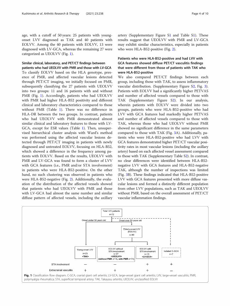
Similar clinical, laboratory, and PET/CT findings betweenpatients who had UEOLVV with PMR and those with LV-GCATo classify EOLVV based on the HLA genotype, pres-ence of PMR, and affected vascular lesions detectedthrough PET/CT imaging, we initially focused on PMR,subsequently classifying the 27 patients with UEOLVVinto two groups: 11 and 16 patients with and withoutPMR (Fig. 1). Accordingly, patients who had UEOLVVwith PMR had higher HLA-B52 positivity and differentclinical and laboratory characteristics compared to thosewithout PMR (Table 1). There was no difference inHLA-DR between the two groups. In contrast, patientswho had UEOLVV with PMR demonstrated almostsimilar clinical and laboratory features to those with LV-GCA, except for ESR values (Table 1). Then, unsuper-vised hierarchical cluster analysis with Ward’s methodwas performed using the affected vascular lesions de-tected through PET/CT imaging in patients with newlydiagnosed and untreated EOLVV, focusing on HLA-B52,which showed a difference in the frequency among pa-tients with EOLVV. Based on the results, UEOLVV withPMR and LV-GCA was found to form a cluster of LVVwith GCA features (i.e., PMR and/or STA involvement)in patients who were HLA-B52-positive. On the otherhand, no such clustering was observed in patients whowere HLA-B52-negative (Fig. 2). Additionally, the evalu-ation of the distribution of the affected vessels showedthat patients who had UEOLVV with PMR and thosewith LV-GCA had almost the same number and similardiffuse pattern of affected vessels, including the axillary
artery (Supplementary Figure S1 and Table S1). Theseresults suggest that UEOLVV with PMR and LV-GCAmay exhibit similar characteristics, especially in patientswho were HLA-B52-positive (Fig. 2).
Patients who were HLA-B52-positive and had LVV withGCA features showed diffuse PET/CT vasculitis findingsthat were different from those of patients with TAK whowere HLA-B52-positiveWe also compared PET/CT findings between eachgroup, including those with TAK, to assess inflammatoryvascular distribution. (Supplementary Figure S2, Fig. 3).Patients with EOLVV had a significantly higher PETVASand number of affected vessels compared to those withTAK (Supplementary Figure S2). In our analysis,wherein patients with EOLVV were divided into twogroups, patients who were HLA-B52-positive who hadLVV with GCA features had markedly higher PETVASand number of affected vessels compared to those withTAK, whereas those who had UEOLVV without PMRshowed no significant difference in the same parameterscompared to those with TAK (Fig. 3A). Additionally, pa-tients who were HLA-B52-positive who had LVV withGCA features demonstrated higher PET/CT vascular posi-tivity rates in most vascular lesions (including the axillaryartery) based on each affected vessel assessment comparedto those with TAK (Supplementary Table S2). In contrast,no clear differences were identified between HLA-B52-negative LVV with GCA features and HLA-B52-negativeTAK, although the number of inspections was limited(Fig. 3B). These findings indicated that HLA-B52-positiveLVV with GCA features presented with more diffuse vas-cular lesions and formed a distinctly different populationfrom other LVV populations, such as TAK and UEOLVVwithout PMR, based on the overall assessment of PET/CTvascular inflammation findings.
Fig. 1 Classification flow diagram. C-GCA, cranial giant cell arteritis; LV-GCA, large-vessel giant cell arteritis; LVV, large-vessel vasculitis; PMR,polymyalgia rheumatica; STA, superficial temporal artery; TAK, Takayasu arteritis; UEOLVV, unclassified EOLVV
Kushimoto et al. Arthritis Research & Therapy (2021) 23:238 Page 4 of 10
HLA-B52-positive patients who had LVV with GCAfeatures exhibited clinical characteristics different fromHLA-B52-positive patients with TAKTo explore the clinical characteristics of HLA-B52-positive patients who had LVV with GCA features, thementioned patients were compared to HLA-B52-positivepatients with TAK (Table 2). Compared to HLA-B52-positive patients with TAK, HLA-B52-positive patientswho had LVV with GCA features demonstrated higheraPL and HLA-DR4 positivity rates. After also comparingthe clinical courses of both diseases, HLA-B52-positivepatients who had LVV with GCA features exhibitedlower rates of biologics usage and relapse compared toHLA-B52-positive patients with TAK. On the otherhand, HLA-B52-positive patients with TAK had a higherrate of biologics usage and relapse compared to HLA-B52-negative ones, whereas no such differences were ob-served in patients who had EOLVV with GCA features(Fig. 4).
DiscussionTo the best of our knowledge, this has been the firstreport to elucidate the differences between EOLVVand TAK by focusing on HLA genotype, PMR, andaffected vascular lesions detected through PET/CTimaging. Accordingly, our results showed thatpatients who had LVV with GCA features (i.e., PMRand/or STA involvement), most of whom had HLA-B52, exhibited a significantly increased number ofextracranial vascular lesions of LVV compared to
those with TAK. The aforementioned findings sug-gest that HLA-B52-positive LVV with GCA features,which is characteristic to diffuse vasculitis, comprisesa core population of patients with EOLVV.The current study showed that the population of
UEOLVV with PMR was very similar to that of LV-GCA(i.e., EOLVV with STA involvement) and distinct fromthat of UEOLVV without PMR. Patients who hadUEOLVV with PMR exhibited similar clinical findings tothose with LV-GCA, especially regarding jaw claudica-tion and axillary artery involvement, which are charac-teristic features of GCA [5, 20]. Additionally, patientswho had UEOLVV with PMR and those with LV-GCAhad an identifiable pattern of widespread vasculitis le-sions in large vessels, unlike patients with TAK,which is consistent with findings presented in severalprevious reports [19, 27]. Our results suggest thatPMR can be as useful a finding as STA involvementfor the diagnosis of GCA. In fact, a recent clinicaltrial utilized PMR as an entry criterion for GCA [23].Therefore, PMR and STA involvement may be valu-able for the diagnosis of GCA.The present study also demonstrated that patients
who had LVV with GCA features demonstrated a higherprevalence of HLA-B52, a disease susceptibility allele ofTAK, compared to patients who had UEOLVV withoutPMR. The findings indicate that HLA-B52 is not aspecific finding in patients with TAK and is associatedwith a population of EOLVV. Among HLA-B52-positivepatients, those who had LVV with GCA features
Table 1 Clinical and laboratory features of patients with EOLVV
Variable UEOLVV withPMR (n = 11)
UEOLVV withoutPMR (n = 16)
p value(vs UEOLVV with PMR)
LV-GCA(n = 13)
p value(vs UEOLVV with PMR)
Age at onset, years, mean ± SD 67.8 ± 8.6 62.9 ± 10.6 0.12 73.5 ± 9.2 0.10
Female, n (%) 9/11 (81.8) 14/16 (87.5) 1 8/13 (61.5) 0.39
Positive temporal artery findings,n (%)
0/11 (0) 0/16 (0) 13/13 (100)
PMR diagnosed, n (%) 11/11 (100) 0/16 (0) 6/13 (46.2)
Jaw claudication, n (%) 3/11 (27.3) 0/12 (0) 0.09 2/11 (18.2) 1
Newly diagnosed and untreatedpatients, n
11/11 10/16 11/13
Laboratory findings
aPL, n (%) 3/7 (42.9) 0/7 (0) 0.19 4/6 (66.7) 0.59
HLA-B52, n (%) 6/7 (85.7) 3/10 (30.0) 0.0498 4/6 (66.7) 0.56
HLA-DR4, n (%) 2/7 (28.6) 3/8 (37.5) 1 2/5 (40) 1
CRP, mg/dl 9.0 (2.6–13.6) 4.4 (1.04–17.4) 0.65 7.5 (4.9–9.9) 0.95
ESR, mm/h 108 (96–116.5) 79.1 (53.8–91.2) < 0.01 89.7 (81.8–102.3) 0.09
Platelet count, × 104 cells/μl 47.8 (42.3–54.4) 29.4 (17.6–32.0) < 0.01 40.3 (29.9–51.1) 0.21
Data are presented as median (IQR) unless otherwise indicated. aPL anti-phospholipid antibody, CRP C-reactive protein, EOLVV elderly onset large-vessel vasculitis,ESR erythrocyte sedimentation rate, HLA human leucocyte antigen, LV-GCA large-vessel GCA, PMR polymyalgia rheumatica, UEOLVV unclassified elderly-onsetlarge-vessel vasculitis
Kushimoto et al. Arthritis Research & Therapy (2021) 23:238 Page 5 of 10

exhibited different clinical characteristics, clinicalcourses, and PET/CT findings compared to those withTAK, indicating that the two diseases are still distinctpopulations even after limiting our analysis to those withHLA-B52. These findings suggest that the role of HLA-B52 in LVV with GCA features may be different from itsrole as a disease susceptibility allele in TAK.HLA-B52-positive patients who had LVV with GCA
features uniformly exhibited the most extensively af-fected vessels based on the number of affected vesselsdetected on PET/CT. The results of this study suggestthat HLA-B52-positive patients with PMR or cranialGCA, which have traditionally been considered over-lapping pathologies [14], are likely to develop LVV le-sions. A recent study on two large cohorts of LVVshowed that the pattern of extensively affected vesselsin LVV was more common in the LV-GCA of theDiagnostic and Classification Criteria for Vasculitiscohort, which included patients with various genetic
backgrounds, compared to LV-GCA of the NorthAmerican cohort [19]. Although more detailed exami-nations of HLA-B52 in these cohorts are needed, wesuspect that such differences could be attributed tothe carrier rate of HLA-B52. In addition, a survey ofHLA in patients with abdominal aortic aneurysms inJapan pointed out an association with MHC class Iand reported that HLA-B52 was more common in pa-tients with abdominal aortic aneurysms who hadaorto-iliac occlusive disease than those who did not[28]. Our results and these findings indicate thatHLA-B52 may be involved in the distribution of af-fected vessels. In this point, the concept of MHC-I-opathy has recently been proposed for diseases, suchas spondyloarthritis and Behcet’s disease. This is aconcept of disease in which clinical features of diseaselocalization are shared by specific MHC class I mole-cules and corresponding tissue antigen structures inthe target tissues at sites of contact between the body
Fig. 2 Dendrogram of the unsupervised hierarchical cluster analysis with Ward’s method in patients with EOLVV. A, B Separatedendrograms of patients who were HLA-B52-positive who had EOLVV and the HLA-B52-negative ones. A The red, purple, and green linesrepresent patients who had UEOLVV without PMR, patients who had UEOLVV with PMR, and patients with LV-GCA, respectively. B Thered line represents patients who had UEOLVV without PMR, and blue line represents patients who had UEOLVV with PMR or patientswith LV-GCA. LVV with GCA features was defined as EOLVV with PMR and/or STA involvement. EOLVV, elderly onset large-vessel vasculitis;GCA, giant cell arteritis; HLA, human leucocyte antigen; LV-GCA, large-vessel GCA; LVV, large-vessel vasculitis; PMR, polymyalgia rheumatica;STA, superficial temporal artery; UEOLVV, unclassified EOLVV
Kushimoto et al. Arthritis Research & Therapy (2021) 23:238 Page 6 of 10

and the external environment (oral mucosa, gut, andskin) or sites of physical stress, such as entheses, in-cluding the mini-entheses in the eye, vessel walls, andvalve regions [29]. Given that this disease conceptmay also be applied to HLA-B52-positive LVV, thedistribution of the corresponding antigen of HLA-B52may play an important role in the formation ofvascular lesions outside the cranial region, suggestingthat when elderly patients are exposed to chronic
inflammatory conditions, such as atherosclerosis,PMR, or GCA, patients who are HLA-B52-positivemay be susceptible to inflammation in the extracra-nial LVV lesions and may be more likely to developvascular lesions. Further investigations on the mecha-nisms underlying the high prevalence of HLA-B52 inpatients with LVV with GCA features and the widerange of affected vascular lesions in HLA-B52-positivepopulation are needed. Our results and findings
Fig. 3 PETVAS and the number of affected vessels in patients with newly diagnosed and untreated LVV. A, B PETVAS and the number of affectedvessels in patients who were HLA-B52-positive (A) or HLA-B52-negative (B) with LVV were compared between the following three groups: TAK,UEOLVV without PMR, and LVV with GCA features. GCA, giant cell arteritis; HLA, human leucocyte antigen; LVV, large-vessel vasculitis; PETVAS, PETvascular activity score; PMR, polymyalgia rheumatica; TAK, Takayasu arteritis; UEOLVV, unclassified elderly onset LVV
Kushimoto et al. Arthritis Research & Therapy (2021) 23:238 Page 7 of 10

indicate that HLA-B52 may be involved in the patho-genesis of diffuse LVV complicated with PMR andcranial GCA.This study showed that patients who were HLA-
B52-positive and had LVV with GCA features exhib-ited lower rates of biologics usage and relapse com-pared to patients who were HLA-B52-positive withTAK, who were more also likely to be refractory totreatment or experience relapse compared to theHLA-B52-negative ones. This was consistent with pre-vious reports [30], whereas patients who were HLA-B52-positive with GCA showed no obvious differencesin clinical course compared to the HLA-B52-negativeones. These results suggest that there is a differencein therapeutic response between patients who wereHLA-B52-positive with TAK and patients who wereHLA-B52-positive and had LVV with GCA features.Unlike TAK, HLA-B52 is possibly not involved intreatment resistance in GCA. Previous studies re-ported that patients with diffuse vasculitis lesionshave shown less vascular damage [19], whereas LV-GCA is more likely to cause vasodilatation and formdissecting lesions, unlike TAK, which tends to formstenotic lesions [6]. These findings indicate that HLA-B52 may be involved not only in the distribution ofaffected vascular lesions but also in the severity ofvascular inflammation and damage, which may resultin differences in treatment response between LV-GCAand TAK.The present study showed that among patients with
UEOLVV, those with PMR exhibited more diffusevascular lesions compared to those without PMR,
which is remarkable in HLA-B52 carriers. PET/CTcan possibly be used to identify the potential presenceof LVV lesions [31]. Reports have shown that imagingcan detect coexisting subclinical LVV lesions in pa-tients diagnosed with PMR [32]. In clinical settings,complicated extracranial LVV lesions are difficult todetermine in patients with PMR based on clinicalfindings alone. As such, patients with PMR, especiallythose with HLA-B52, may need to undergo PET/CTto examine the coexistence of LVV early in thecourse of PMR.The current study has some limitations worth not-
ing. First, this study had a small sample size and waslimited to Japanese patients. Thus, our results needto be verified in a multiracial study with a largersample size. Second, this was a retrospective study,with lacking HLA measurements and a potential forhindsight bias. The treatment course was also stud-ied backward, and treatment selection was left to thediscretion of each attending physician. As such,further prospective studies are needed. Third, themechanism by which HLA-B52 is involved in thepathogenesis of diffuse LVV with GCA features alsoneeds to be investigated in the future. Lastly, long-term outcomes, such as vascular damage were notassessed. Further studies on the distribution of vas-culitis and vascular damage may help predict futurevascular damage in LVV.
ConclusionsThe current study showed that patients who hadEOLVV without STA involvement but with PMR
Table 2 Clinical and laboratory features of HLA-B52-positive patients with LVV
Variable TAK, n = 10 LVV with GCA features, n = 10 p value
Age at onset, years, mean ± SD 29.2 ± 11.9 70.1 ± 9.1 < 0.001
Female, n (%) 7/10 (70.0) 5/10 (50.0) 0.65
Positive temporal artery findings, n (%) 0/10 (0) 4/10 (40.0) 0.09
PMR diagnosed, n (%) 0/10 (0) 8/10 (80.0) < 0.001
Jaw claudication, n (%) 0/9 (0) 1/9 (11.1) 1
Biologics usage, n (%) 8/10 (80) 4/9 (44.4) 0.17
History of relapse, n (%) 7/10 (70) 3/9 (33.3) 0.18
Newly diagnosed and untreated patients, n 5/10 10/10
Laboratory findings
aPL, n (%) 0/4 (0) 4/6 (66.7) 0.08
HLA-B52, n (%) 10/10 (100) 10/10 (100)
HLA-DR4, n (%) 0/9 (0) 4/9 (44.4) 0.08
CRP, mg/dl 4.9 (1.8–12.9) 7.5 (4.3–10.8) 0.50
Platelet count, × 104 cells/μl 47.1 (36.8–53.1) 44.1 (35.0–49.5) 0.67
Data are presented as median (IQR) unless otherwise indicated. aPL anti-phospholipid antibody, CRP C-reactive protein, GCA giant cell arteritis, HLA humanleucocyte antigen, LVV large-vessel vasculitis, PMR polymyalgia rheumatica, TAK Takayasu arteritis
Kushimoto et al. Arthritis Research & Therapy (2021) 23:238 Page 8 of 10

demonstrated similar patient backgrounds and vessel dis-tributions (observed via PET/CT) to patients with LV-GCA who satisfied the 1990 ACR classification criteria.Moreover, HLA-B52-positive patients who had EOLVVwith PMR and/or STA involvement presented with diffusevascular lesions and may comprise a core population forLV-GCA in among Japanese individuals. Thus, the find-ings of HLA-B52-positivity and diffusely affected vessels,as well as those of PMR, in patients with EOLVV can beconsidered as suspicious findings of LV-GCA.
AbbreviationsACR: American College of Rheumatology; aPL: Anti-phospholipid antibody;CRP: C-reactive protein; EOLVV: Elderly onset large-vessel vasculitis;ESR: Erythrocyte sedimentation rate; FDG: Fluorodeoxyglucose; GCA: Giantcell arteritis; HLA: Human leucocyte antigen; LV-GCA: Large-vessel GCA;LVV: Large-vessel vasculitis; PET/CT: Positron emission tomography/computedtomography; PETVAS: PET vascular activity score; PMR: Polymyalgia
rheumatica; STA: Superficial temporal artery; TAK: Takayasu arteritis;UEOLVV: Unclassified elderly onset LVV
Supplementary InformationThe online version contains supplementary material available at https://doi.org/10.1186/s13075-021-02618-4.
Additional file 1.
AcknowledgementsThe authors would like to thank Enago for the English language review.
Authors’ contributionsKK and MA participated in the study conception and design. KK, MA, and KNparticipated in the data acquisition and analysis. KK, MA, KN, MN, YK, HM,NO, YA, KA, TH, and HN contributed to the interpretation of results. KK was amajor contributor in writing the manuscript. All authors read and approvedthe final manuscript.
Fig. 4 Biologics usage and relapse history of patients with LVV. The biologics usage and relapse history of patients with TAK (A) and LVV withGCA features (B) were compared between patients who were HLA-B52-positive and -negative. GCA, giant cell arteritis; HLA, human leucocyteantigen; LVV, large-vessel vasculitis; TAK, Takayasu arteritis. *P < 0.05
Kushimoto et al. Arthritis Research & Therapy (2021) 23:238 Page 9 of 10

FundingNo specific funding was received from any bodies in the public, commercial,or not-for-profit sectors to carry out the work described in this article.
Availability of data and materialsAll data generated or analyzed during this study are included in thispublished article and its supplementary information files.
Declarations
Ethics approval and consent to participateThis study was approved by the ethics committee of Kyushu UniversityHospital (approval number 2020-101) in accordance with the HelsinkiDeclaration. Because this was retrospective research, we disclosed the studyinformation at the site of the related facilities. Obtaining patient consent wasnot required according to the committee’s procedures.
Consent for publicationNot applicable.
Competing interestsThe authors declare that they have no competing interests.
Author details1Department of Medicine and Biosystemic Science, Kyushu UniversityGraduate School of Medical Sciences, 3-1-1 Maidashi, Higashi-ku, Fukuoka812-8582, Japan. 2Department of Cancer Stem Cell Research, KyushuUniversity Graduate School of Medical Sciences, Fukuoka, Japan.3Department of Endocrinology and Rheumatology, Kurashiki Central Hospital,Kurashiki, Japan. 4Department of Internal Medicine, Kyushu University BeppuHospital, Beppu, Japan. 5Department of Medical Education, Kyushu UniversityGraduate School of Medical Sciences, Fukuoka, Japan.
Received: 28 June 2021 Accepted: 28 August 2021
References1. Jennette JC, Falk RJ, Bacon PA, Basu N, Cid MC, Ferrario F, et al. 2012 revised
International Chapel Hill Consensus Conference Nomenclature ofVasculitides. Arthritis Rheum. 2013;65:1–11.
2. Lensen KD, Voskuyl AE, Comans EF, van der Laken CJ, Smulders YM.Extracranial giant cell arteritis: A narrative review. Neth J Med. 2016;74:182–92.
3. Arend WP, Michel BA, Bloch DA, Hunder GG, Calabrese LH, Edworthy SM,et al. The American College of Rheumatology 1990 criteria for theclassification of Takayasu arteritis. Arthritis Rheum. 1990;33:1129–34.
4. Watanabe Y, Miyata T, Tanemoto K. Current clinical features of new patientswith Takayasu arteritis observed from cross-country research in Japan: ageand sex specificity. Circulation. 2015;132:1701–9.
5. Kermani TA. Takayasu arteritis and giant cell arteritis: are they a spectrum ofthe same disease? Int J Rheum Dis. 2019;22(Suppl 1):41–8.
6. Tombetti E, Godi C, Ambrosi A, Doyle F, Jacobs A, Kiprianos AP, et al. Novelangiographic scores for evaluation of large vessel vasculitis. Sci Rep. 2018;8:15979.
7. Hunder GG, Bloch DA, Michel BA, Stevens MB, Arend WP, Calabrese LH,et al. The American College of Rheumatology 1990 criteria for theclassification of giant cell arteritis. Arthritis Rheum. 1990;33:1122–8.
8. Muratore F, Kermani TA, Crowson CS, Green AB, Salvarani C, Matteson EL,et al. Large-vessel giant cell arteritis: a cohort study. Rheumatology (Oxford).2015;54:463–70.
9. Terao C. Revisited HLA and non-HLA genetics of Takayasu arteritis--whereare we? J Hum Genet. 2016;61:27–32.
10. Jacobsen S, Baslund B, Madsen HO, Tvede N, Svejgaard A, Garred P.Mannose-binding lectin variant alleles and HLA-DR4 alleles are associatedwith giant cell arteritis. J Rheumatol. 2002;29:2148–53.
11. Kimura A, Kitamura H, Date Y, Numano F. Comprehensive analysis of HLAgenes in Takayasu arteritis in Japan. Int J Cardiol. 1996;54(Suppl):S61–9.
12. Mackie SL, Taylor JC, Haroon-Rashid L, Martin S, Dasgupta B, Gough A, et al.Association of HLA-DRB1 amino acid residues with giant cell arteritis:genetic association study, meta-analysis and geo-epidemiologicalinvestigation. Arthritis Res Ther. 2015;17:195.
13. Sahin Z, Bıcakcıgil M, Aksu K, Kamali S, Akar S, Onen F, et al. Takayasu’sarteritis is associated with HLA-B* 52, but not with HLA-B* 51, in Turkey.Arthritis Res Ther. 2012;14:R27.
14. Dejaco C, Duftner C, Buttgereit F, Matteson EL, Dasgupta B. The spectrum ofgiant cell arteritis and polymyalgia rheumatica: revisiting the concept of thedisease. Rheumatology (Oxford). 2017;56:506–15.
15. Cantini F, Niccoli L, Storri L, Nannini C, Olivieri I, Padula A, et al. Arepolymyalgia rheumatica and giant cell arteritis the same disease? SeminArthritis Rheum. 2004;33:294–301.
16. Pelletier-Galarneau M, Ruddy TD. PET/CT for diagnosis and management oflarge-vessel vasculitis. Curr Cardiol Rep. 2019;21:34.
17. Walter MA, Melzer RA, Schindler C, Müller-Brand J, Tyndall A, Nitzsche EU.The value of [18F]FDG-PET in the diagnosis of large-vessel vasculitis and theassessment of activity and extent of disease. Eur J Nucl Med Mol Imaging.2005;32:674–81.
18. Prieto-González S, Depetris M, García-Martínez A, Espígol-Frigolé G, Tavera-Bahillo I, Corbera-Bellata M, et al. Positron emission tomography assessmentof large vessel inflammation in patients with newly diagnosed, biopsy-proven giant cell arteritis: a prospective, case-control study. Ann Rheum Dis.2014;73:1388–92.
19. Gribbons KB, Ponte C, Carette S, Craven A, Cuthbertson D, Hoffman GS,et al. Patterns of arterial disease in Takayasu arteritis and giant cell arteritis.Arthritis Care Res (Hoboken). 2020;72:1615–24.
20. Schmidt WA, Seifert A, Gromnica-Ihle E, Krause A, Natusch A. Ultrasound ofproximal upper extremity arteries to increase the diagnostic yield in large-vessel giant cell arteritis. Rheumatology (Oxford). 2008;47:96–101.
21. Bird HA, Esselinckx W, Dixon AS, Mowat AG, Wood PH. An evaluation ofcriteria for polymyalgia rheumatica. Ann Rheum Dis. 1979;38:434–9.
22. Kerr GS, Hallahan CW, Giordano J, Leavitt RY, Fauci AS, Rottem M, et al.Takayasu arteritis. Ann Intern Med. 1994;120:919–29.
23. Unizony SH, Dasgupta B, Fisheleva E, Rowell L, Schett G, Spiera R, et al.Design of the tocilizumab in giant cell arteritis trial. Int J Rheumatol. 2013;2013:912562.
24. Meller J, Strutz F, Siefker U, Scheel A, Sahlmann CO, Lehmann K, et al. Earlydiagnosis and follow-up of aortitis with [(18)F]FDG PET and MRI. Eur J NuclMed Mol Imaging. 2003;30:730–6.
25. Kang F, Han Q, Zhou X, Zheng Z, Wang S, Ma W, et al. Performance of thePET vascular activity score (PETVAS) for qualitative and quantitativeassessment of inflammatory activity in Takayasu’s arteritis patients. Eur JNucl Med Mol Imaging. 2020;47:3107–17.
26. Grayson PC, Alehashemi S, Bagheri AA, Civelek AC, Cupps TR, Kaplan MJ,et al. 18 F-fluorodeoxyglucose-positron emission tomography as an imagingbiomarker in a prospective, longitudinal cohort of patients with large vesselvasculitis. Arthritis Rheumatol. 2018;70:439–49.
27. Power SP, O'Mahony D. Diffuse large vessel giant cell arteritis found by18Fluorodeoxyglucose PET/CT imaging. Lancet. 2019;393:349.
28. Sugimoto T, Sada M, Miyamoto T, Yao H. Genetic analysis on HLA loci inJapanese patients with abdominal aortic aneurysm. Eur J Vasc EndovascSurg. 2003;26:215–8.
29. McGonagle D, Aydin SZ, Gül A, Mahr A, Direskeneli H. ‘MHC-I-opathy’-unified concept for spondyloarthritis and Behçet disease. Nat RevRheumatol. 2015;11:731–40.
30. Ohigashi H, Tamura N, Ebana Y, Harigai M, Maejima Y, Ashikaga T, et al.Effects of immunosuppressive and biological agents on refractory Takayasuarteritis patients unresponsive to glucocorticoid treatment. J Cardiol. 2017;69:774–8.
31. Blockmans D, de Ceuninck L, Vanderschueren S, Knockaert D, Mortelmans L,Bobbaers H. Repetitive 18F-fluorodeoxyglucose positron emissiontomography in giant cell arteritis: a prospective study of 35 patients.Arthritis Rheum. 2006;55:131–7.
32. González-Gay MA, Matteson EL, Castañeda S. Polymyalgia rheumatica.Lancet. 2017;390:1700–12.
Publisher’s NoteSpringer Nature remains neutral with regard to jurisdictional claims inpublished maps and institutional affiliations.
Kushimoto et al. Arthritis Research & Therapy (2021) 23:238 Page 10 of 10
Copyright © 2022 FDOKUMEN