Analysis of genomic breakpoints in p190 and p210 BCR–ABL indicate distinct mechanisms of formation
9
ORIGINAL ARTICLE Analysis of genomic breakpoints in p190 and p210 BCR–ABL indicate distinct mechanisms of formation J Score 1 , MJ Calasanz 2 , O Ottman 3 , F Pane 4,5 , RF Yeh 6 , MA Sobrinho-Simo ˜es 7 , S Kreil 1 , D Ward 1 , C Hidalgo-Curtis 1 , JV Melo 7 , J Wiemels 6 , B Nadel 8 , NCP Cross 1,9 and FH Grand 1,9 1 Wessex Regional Genetics Laboratory, Salisbury and Human Genetics Division, University of Southampton School of Medicine, Southampton, UK; 2 Department of Genetics, School of Science, University of Navarra, Pamplona, Spain; 3 Department of Hematology and Oncology, Goethe University, Frankfurt, Germany; 4 Division of Hematology, University of Naples Federico II, Naples, Italy; 5 CEINGEFBiotecnologie Avanzate, Naples, Italy; 6 Department of Epidemiology and Biostatistics, University of California-San Francisco, San Francisco, CA, USA; 7 Department of Haematology, Hammersmith Hospital, Du Cane Road, Imperial College, London, UK and 8 Centre d’Immunologie de Marseille-Luminy, CNRS-INSERM-Unversite ´ de la Mediterranee, Marseille, France We sought to understand the genesis of the t(9;22) by characterizing genomic breakpoints in chronic myeloid leukemia (CML) and BCR–ABL-positive acute lymphoblastic leukemia (ALL). BCR–ABL breakpoints were identified in p190 ALL (n ¼ 25), p210 ALL (n ¼ 25) and p210 CML (n ¼ 32); reciprocal breakpoints were identified in 54 cases. No evidence for significant clustering and no association with sequence motifs was found except for a breakpoint deficit in repeat regions within BCR for p210 cases. Comparison of reciprocal breakpoints, however, showed differences in the patterns of deletion/insertions between p190 and p210. To explore the possibility that recombinase-activating gene (RAG) activity might be involved in ALL, we performed extra-chromosomal recombination assays for cases with breakpoints close to potential cryptic recombination signal sequence (cRSS) sites. Of 13 ALL cases tested, 1/10 with p190 and 1/3 with p210 precisely recapitulated the forward BCR–ABL breakpoint and 1/10 with p190 precisely recapitulated the reciprocal breakpoint. In contrast, neither of the p210 CMLs tested showed functional cRSSs. Thus, although the t(9;22) does not arise from aberrant variable (V), joining (J) and diversity (D) (V(D)J) recombination, our data suggest that in a subset of ALL cases RAG might create one of the initiating double-strand breaks. Leukemia (2010) 24, 1742–1750; doi:10.1038/leu.2010.174; published online 12 August 2010 Keywords: BCR–ABL; breakpoints; RAG Introduction Chromosomal translocations that produce oncogenic fusion genes are common in hematological malignancies, but the mechanism by which they are formed is incompletely under- stood. Broadly there are three non-mutually exclusive factors that are believed to be relevant to this process: (i) biological selection for specific gene fusions or deregulated gene expres- sion, 1 (ii) susceptibility of particular chromosomal regions to breakage 2 and (iii) the opportunity to recombine due to physical proximity. 3 Analysis to date has indicated that translocations in leukemia result from non-homologous end joining (NHEJ) following two double stranded DNA breaks; homologous recombination does not have a significant role. 4,5 The paradigm for translocations in leukemia is the Philadelphia (Ph) chromosome, the smaller derivative of the t(9;22). 6 The resultant BCR–ABL fusion gene is the defining marker of chronic myeloid leukemia (CML). 6 In CML, the breakpoints in the majority of patients occur within the 5.8 kb major breakpoint cluster region (M-BCR) in the middle of BCR, resulting in a p210 BCR–ABL protein. The Ph chromosome is also found in 30% of adult acute lymphoblastic leukemia (ALL), 3–5% of childhood ALL and 1% of acute myeloid leukemia. 6 In contrast to CML, only about 30–50% of patients with Ph-positive ALL harbor the p210 fusion protein, with the remaining 50–70% being characterized by the smaller p190 fusion protein resulting from breakpoints in the BCR minor breakpoint cluster region (m-BCR) within the 72 kb BCR intron 1. Within ABL, most breaks fall within the 140 kb region between exon 1b and exon 2, however, in CML breaks upstream of exon 1b and between exons 2 and 3 also occur in some cases (Figure 1). Despite this knowledge, the precise mechanism for the formation of the BCR–ABL fusion gene remains largely unknown. One of the reasons for this is that only a small number of genomic fusion junctions have been fully characterized, particularly for p190 for which only 2 break- points have been fully sequenced. 7,8 Nevertheless, studies in CML have noted that BCR–ABL breakpoints are often associated with repeat regions. 5,6,9,10 Also, it has been observed that BCR and ABL tend to be in close proximity compared with control genes during cell division in hematopoietic stem cells, potentially providing the opportunity for aberrant recombination should breakage occur. 3 The mechanisms behind many chromosomal translocations in lymphoid malignancies are clearer and are believed to be the result of the aberrant activity of the machinery for V(D)J or class switch recombination. 11,12 In V(D)J recombination, which takes place early in B-cell differentiation, the recombinase- activating gene (RAG) enzyme complex recognises specific recombination signal sequences (RSSs) flanking the V(D)J segments. RAG makes specific double stranded breaks that are subsequently ligated by NHEJ into a coding joint and a signal joint. The RAG complex can occasionally mistarget recombination to cryptic RSSs and juxtapose proto-oncogenes, such as LMO2 and HOX2, to loci that encode antigen receptors. 13–15 It is also possible for two non-antigen receptor loci to be joined erroneously by illegitimate V(D)J recombina- tion, for example the SIL and SCL genes in some patients with T-ALL. 12,16 In addition, in some B-cell lymphomas associated with Immunoglobulin H translocations, breakpoint regions Received 12 May 2010; revised 16 June 2010; accepted 29 June 2010; published online 12 August 2010 Correspondence: Professor NCP Cross, Wessex Regional Genetics Laboratory, Salisbury NHS Foundation Trust, Salisbury SP2 8BJ, UK. E-mail: [email protected] 9 These authors contributed equally to this work. Leukemia (2010) 24, 1742–1750 & 2010 Macmillan Publishers Limited All rights reserved 0887-6924/10 www.nature.com/leu
Transcript of Analysis of genomic breakpoints in p190 and p210 BCR–ABL indicate distinct mechanisms of formation

ORIGINAL ARTICLE
Analysis of genomic breakpoints in p190 and p210 BCR–ABL indicate distinctmechanisms of formation
J Score1, MJ Calasanz2, O Ottman3, F Pane4,5, RF Yeh6, MA Sobrinho-Simoes7, S Kreil1, D Ward1, C Hidalgo-Curtis1, JV Melo7,J Wiemels6, B Nadel8, NCP Cross1,9 and FH Grand1,9
1Wessex Regional Genetics Laboratory, Salisbury and Human Genetics Division, University of Southampton School of Medicine,Southampton, UK; 2Department of Genetics, School of Science, University of Navarra, Pamplona, Spain; 3Department ofHematology and Oncology, Goethe University, Frankfurt, Germany; 4Division of Hematology, University of Naples Federico II,Naples, Italy; 5CEINGEFBiotecnologie Avanzate, Naples, Italy; 6Department of Epidemiology and Biostatistics, University ofCalifornia-San Francisco, San Francisco, CA, USA; 7Department of Haematology, Hammersmith Hospital, Du Cane Road,Imperial College, London, UK and 8Centre d’Immunologie de Marseille-Luminy, CNRS-INSERM-Unversite de la Mediterranee,Marseille, France
We sought to understand the genesis of the t(9;22) bycharacterizing genomic breakpoints in chronic myeloidleukemia (CML) and BCR–ABL-positive acute lymphoblasticleukemia (ALL). BCR–ABL breakpoints were identified inp190 ALL (n¼ 25), p210 ALL (n¼ 25) and p210 CML (n¼32);reciprocal breakpoints were identified in 54 cases. No evidencefor significant clustering and no association with sequencemotifs was found except for a breakpoint deficit in repeatregions within BCR for p210 cases. Comparison of reciprocalbreakpoints, however, showed differences in the patterns ofdeletion/insertions between p190 and p210. To explore thepossibility that recombinase-activating gene (RAG) activitymight be involved in ALL, we performed extra-chromosomalrecombination assays for cases with breakpoints close topotential cryptic recombination signal sequence (cRSS) sites.Of 13 ALL cases tested, 1/10 with p190 and 1/3 with p210precisely recapitulated the forward BCR–ABL breakpoint and1/10 with p190 precisely recapitulated the reciprocal breakpoint.In contrast, neither of the p210 CMLs tested showed functionalcRSSs. Thus, although the t(9;22) does not arise from aberrantvariable (V), joining (J) and diversity (D) (V(D)J) recombination,our data suggest that in a subset of ALL cases RAG mightcreate one of the initiating double-strand breaks.Leukemia (2010) 24, 1742–1750; doi:10.1038/leu.2010.174;published online 12 August 2010Keywords: BCR–ABL; breakpoints; RAG
Introduction
Chromosomal translocations that produce oncogenic fusiongenes are common in hematological malignancies, but themechanism by which they are formed is incompletely under-stood. Broadly there are three non-mutually exclusive factorsthat are believed to be relevant to this process: (i) biologicalselection for specific gene fusions or deregulated gene expres-sion,1 (ii) susceptibility of particular chromosomal regions tobreakage2 and (iii) the opportunity to recombine due to physicalproximity.3 Analysis to date has indicated that translocations inleukemia result from non-homologous end joining (NHEJ)following two double stranded DNA breaks; homologousrecombination does not have a significant role.4,5
The paradigm for translocations in leukemia is thePhiladelphia (Ph) chromosome, the smaller derivative of thet(9;22).6 The resultant BCR–ABL fusion gene is the definingmarker of chronic myeloid leukemia (CML).6 In CML, thebreakpoints in the majority of patients occur within the 5.8 kbmajor breakpoint cluster region (M-BCR) in the middle of BCR,resulting in a p210 BCR–ABL protein. The Ph chromosome isalso found in 30% of adult acute lymphoblastic leukemia (ALL),3–5% of childhood ALL and 1% of acute myeloid leukemia.6
In contrast to CML, only about 30–50% of patients withPh-positive ALL harbor the p210 fusion protein, with theremaining 50–70% being characterized by the smaller p190fusion protein resulting from breakpoints in the BCR minorbreakpoint cluster region (m-BCR) within the 72 kb BCRintron 1. Within ABL, most breaks fall within the 140 kb regionbetween exon 1b and exon 2, however, in CML breaks upstreamof exon 1b and between exons 2 and 3 also occur in somecases (Figure 1). Despite this knowledge, the precise mechanismfor the formation of the BCR–ABL fusion gene remains largelyunknown. One of the reasons for this is that only asmall number of genomic fusion junctions have been fullycharacterized, particularly for p190 for which only 2 break-points have been fully sequenced.7,8 Nevertheless, studies inCML have noted that BCR–ABL breakpoints are often associatedwith repeat regions.5,6,9,10 Also, it has been observed that BCRand ABL tend to be in close proximity compared with controlgenes during cell division in hematopoietic stem cells,potentially providing the opportunity for aberrant recombinationshould breakage occur.3
The mechanisms behind many chromosomal translocationsin lymphoid malignancies are clearer and are believed tobe the result of the aberrant activity of the machinery for V(D)J orclass switch recombination.11,12 In V(D)J recombination, whichtakes place early in B-cell differentiation, the recombinase-activating gene (RAG) enzyme complex recognises specificrecombination signal sequences (RSSs) flanking the V(D)Jsegments. RAG makes specific double stranded breaks thatare subsequently ligated by NHEJ into a coding joint and asignal joint. The RAG complex can occasionally mistargetrecombination to cryptic RSSs and juxtapose proto-oncogenes,such as LMO2 and HOX2, to loci that encode antigenreceptors.13–15 It is also possible for two non-antigen receptorloci to be joined erroneously by illegitimate V(D)J recombina-tion, for example the SIL and SCL genes in some patients withT-ALL.12,16 In addition, in some B-cell lymphomas associatedwith Immunoglobulin H translocations, breakpoint regions
Received 12 May 2010; revised 16 June 2010; accepted 29 June2010; published online 12 August 2010
Correspondence: Professor NCP Cross, Wessex Regional GeneticsLaboratory, Salisbury NHS Foundation Trust, Salisbury SP2 8BJ, UK.E-mail: [email protected] authors contributed equally to this work.
Leukemia (2010) 24, 1742–1750& 2010 Macmillan Publishers Limited All rights reserved 0887-6924/10
www.nature.com/leu
adopting a non-B form might be mistargeted by RAG even in theabsence of RSSs.17
In CML it is thought that the transforming event takes place ina multipotent stem cell, inferred from the range of cell lineagesthat have been shown to be Ph or BCR–ABL positive.18 InPh-positive ALL, it is debated whether the acquisition of thePh chromosome occurs in a multipotent stem cell,19–21 acommitted lymphoid progenitor22–24 or whether both arepossible.25 It has been suggested that p190 BCR–ABL mightspecifically direct lymphoid development, however, this fusionis seen occasionally in CML,26 and in vivo experiments haveshown that both p210 and p190 can give rise to CML or B-ALLdepending on whether stem cells or committed progenitor cellsare transduced.27 It is possible, therefore, that the rarity of p190in human CML is not a direct consequence of the activity of thisfusion, but rather that BCR intron 1 breaks may be much morefrequent in ALL because they are formed by a lymphoid-specificmechanism.27 Such a mechanism could be the action of RAG,which is only present in lymphoid-restricted cells.
In this study we sought to characterize and compare break-point sequences in p210 CML, p210 ALL and p190 ALL tohelp clarify the mechanism giving rise to the t(9;22) in ALL. Weaimed specifically to test the hypothesis that RAG-mediatedrecombination may have a role in the genesis of p190 BCR–ABL.
Materials and methods
Patient samplesPatients were randomly selected for analysis on the basis ofsample availability (CML, n¼ 32; p210 ALL, n¼ 32; p190 ALL,n¼ 43) from centers in the UK, Spain, Germany and Italy.The presence of e13a2 and/or e14a2 (p210) or e1a2 (p190)BCR–ABL fusion mRNA was determined by reverse transcrip-tase–PCR.28 For samples with limited DNA, multiple displace-ment amplification (GenomiPhi, GE Healthcare, Amersham,UK) was undertaken according to the manufacturer’s instruc-tions. The study was approved by the Internal Review Boardsfrom participating institutions and informed consent wasprovided according to the Declaration of Helsinki.
Detection of forward and reciprocal breakpointsTo sublocalise p190 BCR–ABL genomic breakpoints, fluores-cence in situ hybridization was performed in some cases usingfosmid clones selected from the Ensembl database (http://www.ensembl.org) and obtained from the Sanger Institute(Hinxton, UK). The clones spanned BCR intron 1 and ABL exon1b to a3 (clones E11, E8, G4, F1, D5; Supplementary Figure S1).
Fosmid clones were labelled and hybridized as previouslydescribed29 after validation on normal control samples.
For amplification of p190 and p210 breakpoints by longtemplate PCR (LT-PCR) all primers were designed using Primer3(http://frodo.wi.mit.edu/primer3/), checked to be free of single-nucleotide polymorphisms (http://genome.ucsc.edu/) and testedon normal control DNA with a suitable reverse primer. Toamplify breakpoint bands up to 12 kb in size, the Expand Long-Template LT-PCR system 2 (Roche, Burgess Hill, UK) was usedwith an annealing temperature of 64 1C and an elongation timeof 8 min. Initially, a simplex (standard two primer) PCR protocolwas used to detect both p190 and p210 breakpoints, however, amultiplex PCR strategy was later implemented, whereby everyBCR forward primer was mixed with 5 ABL reverse primers(Supplementary Table S1). All breakpoint junctions amplified byLT-PCR were confirmed by reamplification from genomic DNAand sequencing across the breakpoint using an AppliedBiosystems 3100 (Foster City, CA, USA). The reverse ABLprimers were the same for the p210 and p190 screening, but theforward BCR primers were different. We attempted to identifyreciprocal (ABL-BCR) breakpoints in all cases with characterizedforward (BCR-ABL) breakpoints using ABL forward and BCRreverse primers approximately 2–4 kb either side of where thebreak was estimated to be. If no product was amplified aftermultiple attempts with multiple primer combinations, multiplexligation-dependent probe amplification (MLPA) was used todetect large deletions, as previously described.30 All nucleotidepositions refer to NM_004327 and NM_007313 for BCR andABL, respectively, with the A from the ATG start codonconsidered as position 1.
Statistical analysisThe Kolmogorov–Smirnov test was carried out to test fordifferences between the distributions of breakpoints in thedifferent sub-types of BCR-ABL-positive leukemia. Scan statisticswere used to detect any clustering of breakpoints and theSilverman’s smoothed bootstrap test was then used to provideevidence for the numbers of clusters. The proximity of break-points to either known or unknown sequence motifs within50 bp either side of the breaks were tested for by comparing theobserved breakpoints to 200 randomly generated breakpoints.31
In silico analysis of BCR–ABL forward breakpointsfor cryptic RSSsBefore functional analysis, the EMBOSS Fuzznuc tool (http://bioweb.pasteur.fr/seqanal/interfaces/fuzznuc.html) was used toanalyze all CML and ALL breakpoints to select the most likelycandidates for illegitimate RAG activity. The search wasconfigured using the consensus heptamer (50-CACAGTG-30)and nonamer (50-ACAAAAACC-30) sequences separated by 12or 23 nucleotides. The CAC in the heptamer had to be presentand up to nine mismatches were allowed in the remainingheptamer and nonamer sequences.32,33
Functional analysis of breakpoints to determineillegitimate RAG activityThe recombination plasmids used for the extra-chromosomalrecombination assays have been described previously.34 Briefly,the two fragments to be tested for V(D)J recombination areseparated by a transcription stop sequence (oop). The Ptac
promoter will transcribe the chloramphenicol acetyltransferasegene when transformed into E.coli only when the stop signal has
50 kb
100 kb
ABL
Breakpoint region
a21a1b
Minor bcr
e2e1BCR
Major bcr
e12 e16
Figure 1 Translocation breakpoint clusters in the ABL and BCR genes(to scale).
Analysis of BCR–ABL breakpointsJ Score et al
1743
Leukemia

been previously deleted by recombination in eukaryotic cells.34
Approximately 18 h before transfection, 1� 106 NIH-3T3 cellswere plated onto 100 mm culture plates. For each transfection2.5 mg of each plasmid (RAG-1, RAG-2, TdT and the testplasmid), 500ml Opti-MEM (Invitrogen, Paisley, UK) and 40mlof FuGene HD (Roche, Welwyn Garden City, UK) reagentwere mixed according to the manufacturer’s instructions. TheFuGene: DNA complex was then added to the NIH-3T3 cellsand after 40–48 h the plasmid DNA was extracted, transformedinto E.coli, plated onto chloramphenicol and ampicillin plates.Colonies were plucked, amplified and sequenced.34 For eachexperiment a positive control (RS32)34 and no test plasmidcontrol were included to ensure the assay was working correctly.
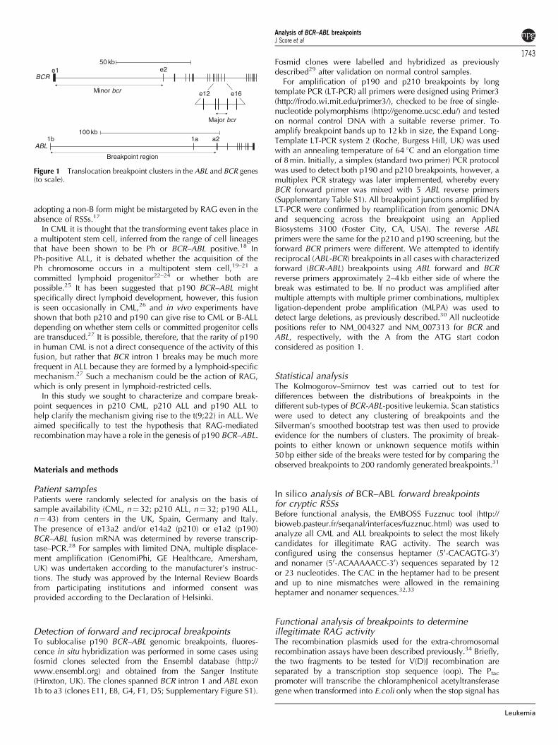
As each BCR–ABL cryptic recombination signal sequence(cRSS) could be used as either a signal or coding joint it wasnecessary to test the selected breakpoints for all possibleputative combinations (Figure 2) by using the authentic RSSsDd2 (from the T-cell receptor alpha gene on chromosome14q11.2). As these authentic sites had been previously testedand contained both 12 and 23 RSSs34 and Dd2 was alreadysupplied in the downstream cassette in both forward and inverseorientations (plasmids RS8 and RS24, respectively),34 wegenerated two additional constructs that were called RS2 andRS3 (Dd2 inverse and forward in the upstream cassette,respectively), giving a total of four test constructs (Figure 2).Each cRSS can potentially be used as either a 12 or 23 RSS andso there were two constructs required for each type of RSS(12, 23, forward or reverse). As many of the cases had more thanone type of putative cRSS at or near the breakpoint a total of 43constructs were made to test all permutations for the 15 casesthat were selected for analysis. Breakpoint junctions (100–300 bp) were amplified by PCR from patient genomic DNA,incorporating appropriate restriction enzyme recognition sites
into the primer sequences to facilitate subcloning. All constructswere resequenced to confirm their identity.
Results
Detection of forward p210 BCR–ABL genomicbreakpointsTwo LT-PCR screens were developed to amplify forward p210BCR–ABL breakpoints: (i) a multiplex assay consisting of eightindividual reactions, each of which contained one (of two)forward BCR primer and five (of 20) reverse ABL primers and(ii) a simplex assay consisting of 40 individual reactions, eachcontaining a single BCR primer and a single ABL reverse primer.Patients were screened initially with the multiplex assay, and ifno product was amplified they were rescreened with the simplexassay. Amplified products were directly sequenced, usually withseveral internal BCR and ABL primers until the breakpoint waslocated and all breakpoints were confirmed by reamplificationwith specific primers from genomic DNA.
A total of 32 CML cases were analyzed that expressed e13a2and/or e14a2 p210 mRNAs, and forward breakpoints weresuccessfully identified in all cases. Of these, six were negativeon the multiplex screen but positive by the simplex screen(Supplementary Table S2). A total of 32 p210 ALLs were alsoanalyzed and breakpoints were successfully amplified from 25(78%): 15 using the multiplex screen and 10 using the simplexscreen (Supplementary Table S3).
Detection of forward p190 BCR–ABL breakpointsIdentification of p190 breakpoints is challenging because of thelarge size of the breakpoint cluster regions in both BCR and ABL.
a
b
c
Figure 2 The four possible ways in which cryptic RSSs may be located at breakpoints. (a) Reverse and forward consensus 12-RSSs (represented byopen triangles) flanking a putative breakpoint. (b) Reverse and forward consensus 23-RSSs (represented by solid triangles) flanking a putativebreakpoint. Each of these can be used by RAG to create either a signal joint (SJ) or coding joint (CJ) depending on the position of the putative RSSrelative to the authentic RSS. (c) The four constructs used for testing BCR or ABL breakpoints in the extra-chromosomal recombination assay.
Analysis of BCR–ABL breakpointsJ Score et al
1744
Leukemia

Initially we pursued a fluorescence in situ hybridization strategyto sublocalize the breakpoints. Five fosmids were isolated (twowithin BCR intron 1 and three within ABL intron 1) that wereused in pairwise combinations to identify the approximatebreakpoint positions in five cases (Supplementary Figure S2).This information was then used to direct LT-PCR screening,resulting in the successful identification of forward breakpointsfor all five cases (#67, #68, #69, #70, #71).
For many cases suitable cytogenetic material was notavailable and so we designed PCR screens on the basis of thestrategy that had been successfully used for p210 cases. Initiallya simplex LT-PCR screen was performed by combining 10 BCRforward spanning intron 1 and 10 ABL reverse primers (all ABLprimers used were the same as for p210 screening). This screenconsisted of 100 individual reactions per case and successfullyamplified the breakpoints in 4 of 13 cases tested. An additional10 ABL primers were designed and a multiplex LT-PCR strategysimilar to p210 screening was implemented. The multiplexamplified one of nine cases that tested negative with thesimplex screen and a further 9 of 25 cases, yielding a total of 19breakpoints from 43 cases. All 19 p190 breakpoints within theBCR fell within the second half of intron 1 despite the fact thatprimers were equally spaced along the entire intron. To focusmore specifically on this region, a further 11 BCR primers weredesigned and, in addition, 5 ABL primers upstream of exon 1bwere also included. Inclusion of these primers in the multiplexassay resulted in the identification of six new breakpoints.In total, therefore, 25/43 (58%) of the p190 breakpoints weresuccessfully amplified (Supplementary Table S4). A summary ofthe breakpoint analysis for all cases is shown on Table 1.
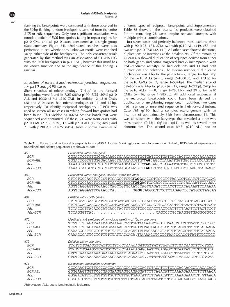
Statistical analysis of forward breakpointsIn addition to the 82 BCR–ABL breakpoints found in this study,an additional 18 p210 CML forward breakpoints (SupplementaryTable S5) that had been identified by the same LT-PCR multiplextechnique at a second center were used for the statisticalanalysis. No significant differences in the distribution of forwardbreakpoints between the different subtypes of leukemia ormRNA fusion were seen (Supplementary Table S6). Similarly nobreakpoint clusters were observed, although some breakpointpreferences were apparent that approached statistical signi-ficance. For p210 ALL, two distinct broad clusters were seen,whereas for p210 CML these were less distinct (Figure 3a). Therewas no clustering of p190 BCR breakpoints although, as notedabove, it is striking that they all fell in the second half of BCRintron 1 (Figure 3b). Within ABL there was a marginal excess ofbreakpoints towards the 30 end of the breakpoint cluster regionfor p190 ALL, whereas for p210 cases there were two broadclusters that were more pronounced when all cases wereconsidered together (Figure 3c).
In addition to clustering, the distributions of the breakpoints inBCR were directly compared with those in ABL for all p210 andp190 cases, however, no significant difference was found
(see Supplementary Figure S3). To check whether the subsetsof breakpoints were associated with known sequence motifs,including functional RSSs, the frequencies of specific sequencemotifs (Supplementary Table S7) within 50 bp of sequence
Table 1 Summary of breakpoint analysis
No. of casesanalyzed
Forward breakpoints identifiednumber (%)
Reciprocal breakpoints identifiednumber (%)
Large deletions detected by MLPANo. found/no. tested
CML 32 32 (100) 21 (66) 5/9p210 ALL 32 25 (78) 12 (48) 5/7P190 ALL 43 25 (58) 21 (84) 1/2
Abbreviations: ALL, acute lymphoblastic leukemia; CML, chronic myeloid leukemia; MLPA, multiplex ligation-dependent probe amplification.
8
6
4
2
0
2
3
4
1
0
40 50
0 50 100
60 70
108.5 109.0 109.5
p210 breakpoints in BCR
p190 breakpoints in BCR
ABL breakpoints
110.0 110.5 111.0
p210p210ALLp210CML
p210
p190
p210ALL
All
p210CML
111.5
# S
ubje
cts
8
10
12
6
4
2
0
# S
ubje
cts
# S
ubje
cts
kb position in BCR sequence NM_004327
kb position in BCR sequence NM_004327
kb position in ABL sequence NM_007313
a
b
c
Figure 3 Evidence for weak clusters in BCR and ABL. Breakpointpositions are shown grouped into 30 intervals (vertical bars) anddensity curves were determined by using the Sheather–Jones solve-the-equation bandwidth method with the peaks indicating clustering.(a) p210 BCR breakpoints (n¼75). (b) p190 BCR breakpoints (n¼25).(c) ABL breakpoints (n¼ 100).
Analysis of BCR–ABL breakpointsJ Score et al
1745
Leukemia
flanking the breakpoints were compared with those observed inthe 50 bp flanking random breakpoints sampled from the entireBCR or ABL sequences. Only one significant association wasfound: a deficit of BCR breakpoints falling in repeat regions forp210 CML and all p210 cases considered as a single group(Supplementary Figure S4). Undirected searches were alsoperformed to see whether any unknown motifs were enriched50 bp either side of the breakpoints. The only consistent resultgenerated by this method was an association of CTGNNTTGwith the BCR breakpoints in p210 ALL, however this motif hasno known function and so the significance of this finding isunclear.
Structure of forward and reciprocal junction sequencesfor p210 and p190 casesShort stretches of microhomology (2–4 bp) at the forwardbreakpoints were found in 7/25 (28%) p190, 5/25 (20%) p210ALL and 10/32 (31%) p210 CML. In addition, 2 p210 CMLs(#8 and #10) cases had microhomologies of 11 and 17 bp,respectively. To identify reciprocal breakpoints, LT-PCR wasused to screen all 82 cases in which forward breakpoints hadbeen found. This yielded 54 (66%) positive bands that weresequenced and confirmed. Of these, 21 were from cases withp210 CML (21/32; 66%), 12 with p210 ALL (12/25; 48%) and21 with p190 ALL (21/25; 84%). Table 2 shows examples of
different types of reciprocal breakpoints and SupplementaryTable S8 shows all the results. No products were obtainedfor the remaining 28 cases despite repeated attempts withmultiple primer combinations.
Just seven cases had perfectly balanced translocations: threewith p190 (#73, #74, #78), two with p210 ALL (#49, #53) andtwo with p210 CML (#2, #10). All other cases showed deletions,duplications or insertions at the breakpoints. Of the remaining47 cases, 8 showed duplication of sequence derived from eitheror both genes (indicating staggered breaks incompatible withRAG-mediated activity), 28 had deletions and 11 had bothduplications and deletions. The median number of duplicatednucleotides was 4 bp for the p190s (n¼ 7, range 3–7 bp), 3 bpfor the p210 ALLs (n¼ 5, range 2–1000 bp) and 173 bp forthe p210 CMLs (n¼ 7, range 1–534 bp). The median size ofdeletions was 4 bp for p190s (n¼ 15, range 1–27 bp), 24 bp forthe p210 ALLs (n¼ 8, range 1–7883 bp) and 29 bp for p210CMLs (n¼ 16, range 1–985 bp). All additional sequences atthe reciprocal breakpoints could have been derived byduplication of neighboring sequences. In addition, two caseshad insertions of unrelated sequence in their forward fusions.Case #65 (p190) had a complex rearrangement with aninsertion of approximately 3 kb from chromosome 11. Thiswas consistent with the karyotype that revealed a three-waytranslocation t(9;22;11)(q34;q11;p11), as well as several otherabnormalities. The second case (#48; p210 ALL) had an
Table 2 Forward and reciprocal breakpoints for six p190 ALL cases. Short regions of homology are shown in bold, BCR-derived sequences areunderlined and deleted sequences are shown as dots
#58 Duplication within one geneBCR GGGACTCGTGTGGGGACAAGCTGAACAGTGTGCTGATCTCTGATCACCACTCAAGCCACAAGTGBCR–ABL GGGACTCGTGTGGGGACAAGCTGAACAGTGTGTTAGCAGCCTAAAATGGTGGCTTTACCAGTTTABL AAAAGTAAGCTGTTGTTACTTTAAGCAAATATTTAGCAGCCTAAAATGGTGGCTTTACCAGTTTABL–BCR AAAAGTAAGCTGTTGTTACTTTAAGCAAATATTTAGATCTCTGATCACCACTCAAGCCACAAGT
#62 Duplication within one gene, deletion within the other
BCR GTTCTGCCACCTGCCCTTTGAGGCTGTGTGGGCACGGTTCCCTCTAGAGCTCCATGTCTAGCAGBCR–ABL GTTCTGCCACCTGCCCTTTGAGGCTGTGTGGGAGTGAGATCTTACCTCTACAGAAATTTAAAAAABL AGGTCAGGAGTTCGAACCCAGCTGTGCAATCTAGTGAGATCTTACCTCTACAGAAATTTAAAAAABL–BCR AGGTCAGGAGTTCGAACCCA. . . . . .TGGGCACGGTTCCCTCTAGAGCTCCATGTCTAGCAG
#64 Deletion within both genes
BCR CTTTGCAGGAAGGATGTGGCTGATGAGACCATCAACCTCAGTCCTGCCAAGGGTGAGGCGGCCCBCR–ABL CTTTGCAGGAAGGATGTGGCTGATGAGACCATGCCCAGTTAGTGATTTTTAAATTGTAGTTCCTABL TCTAGGGTTACAGGCATGAGCTACTGTGCCTGGCCCAGTTAGTGATTTTTAAATTGTAGTTCCTABL–BCR TCTAGGGTTAC. . . . . . . . . . . .. . . . . . . CAGTCCTGCCAAGGGTGAGGCGGCCC
#70 Identical short stretches of homology, deletion of 1bp in one gene
BCR TCGTCTTCAGATAAACAGCAAAACCGTTGGTTAAAAGCTGGTCTAACCCACCTTATTTTGTTGTBCR–ABL TCGTCTTCAGATAAACAGCAAAACCGTTGGTTTTACAAGACTATTTTTAGCCTTTTTTACAAGAABL GAAAGGGATTGCTGTTTTTTATTACCAGATTTTTACAAGACTATTTTTAGCCTTTTTTACAAGAABL–BCR GAAAGGGATTGCTGTTTTTTATTACCAGA.TTAAAAGCTGGTCTAACCCACCTTATTTTGTTGT
#72 Deletion within one gene
BCR TCCTTTTGAAGGTCATTGTTTTCCTAAACAGATGGTTATTTGGACTCTTACAAGTTCTCTGTABCR–ABL TCCTTTTGAAGGTCATTGTTTTCCTAAACAGAGCAATCCCAGGGCTTTAATATCCTTTTTGTAABL GTCTCAAAAAAAAAGAAAAGAAAATTAGAAACTCAATCCCAGGGCTTTAATATCCTTTTTGTAABL–BCR GTCTCAAAAAAAAAGAAAAGAAAATTAGAAACT..TTATTTGGACTCTTACAAGTTCTCTGTA
#74 No deletion, duplication or insertion
BCR GGGGAAGTGGTTCCCGAGGAAGGAGGCAGAGGAGTGTAGATTTTGTAGAGAAGGCTAAGAGAGGBCR ABL GGGGAAGTGGTTCCCGAGGAAGGAGGCAGAGGATCTTCAGATATCTAAAAGAAACTTTGTAACAABL GGGGATGTTTCTGTTGTTGCTCCTTGCTGAGTATCTTCAGATATCTAAAAGAAACTT.GTAACAABL–BCR GGGGATGTTTCTGTTGTTGCTCCTTGCTGAGTAGTGTAGATTTTGTAGAGAAGGCTAAGAGAGG
Abbreviation: ALL, acute lymphoblastic leukemia.
Analysis of BCR–ABL breakpointsJ Score et al
1746
Leukemia
insertion of 57 bp of intronic sequence from the phosphatidyl-serine decarboxylase gene, downstream of BCR. Neither ofthe reciprocal breakpoints was successfully amplified fromthese cases.
Deletions of the 9qþ chromosome at the reciprocal break-point have been described in about 15% of CML cases.30 Todetermine whether the failure to amplify reciprocal breakpointswas owing to the presence of large deletions, we performedMLPA analysis for 18 of the 28 of the reciprocal break-point-negative cases for whom adequate DNA was available.Eleven cases (CML, n¼ 5; p210 ALL, n¼ 5 and p190 ALL, n¼ 1)showed deletion of at least two probes, indicating large dele-tions (Table 1; Supplementary Table S8). Seven cases showed nodeletion or deletion of just one probe (CML, n¼ 4; p210 ALL,n¼ 2 and p190 ALL, n¼ 1). In addition, distances of the forwardbreakpoints from CpG and CAC/GTG sequences were analyzed(Supplementary Table S9), but no obvious differences werefound between the different subtypes of leukemia.
Functional analysis of breakpointsTo determine whether RAG might have a role in the genesis ofthe t(9;22) we performed Fuzznuc analysis to identify candidatecRSS sequences close to the breakpoints. In total, 11/25 (44%)p190 ALL, 11/25 (40%) p210 ALL and 12/32 (38%) p210 CMLforward breakpoints had candidate cRSSs, in which the start ofthe forward or reverse cryptic heptamer fell within 10 bp of thebreakpoint (Supplementary Table S10). Cases with p190 ALL(n¼ 10), p210 ALL (n¼ 3) and p210 CML (n¼ 2) were selectedon the basis of proximity of the cRSS site to the breakpoint, aswell as ensuring a mixture of both ABL and BCR sequences anda mixture of 12 and 23 cRSSs (Supplementary Table S11).
To test whether these candidate cRSS sequences could indeedact as RAG substrates we performed extra-chromosomalrecombination assays using all relevant permutations. PCR andsequencing of the resulting colonies revealed that the high levelof recombination events seen in the control vectors containingtwo pairs of authentic RSSs (RS32 and RS39) were indeedmediated by RAG (Supplementary Table S12). In contrast, themajority of colonies derived from BCR–ABL plasmids showedevidence of break repair (63% of all breakpoint-derivedcolonies) rather than RAG-mediated recombination. Of thecolonies with potential RAG-mediated events, the great majoritywere non-specific with 44/49 BCR–ABL plasmids (24% of allcolonies) using fortuitous RSS sites within the plasmid coreDNA. There were a smaller number of non-breakpoint specificV(D)J recombination events (11% of all colonies; 33/49 of BCR–ABL plasmids) that arose from other fortuitous sites in either BCRor ABL. Altogether, this demonstrates the great sensitivity of ourrecombination substrate assay. Moreover, this indicates that anumber of functional cRSS are present at random in a givensequence, including BCR and ABL breakpoint regions. The factthat such cryptic sites are not usually seen at BCR–ABL junctionsindicates that standard V(D)J-mediated translocation is not acommon event leading to the t(9;22).
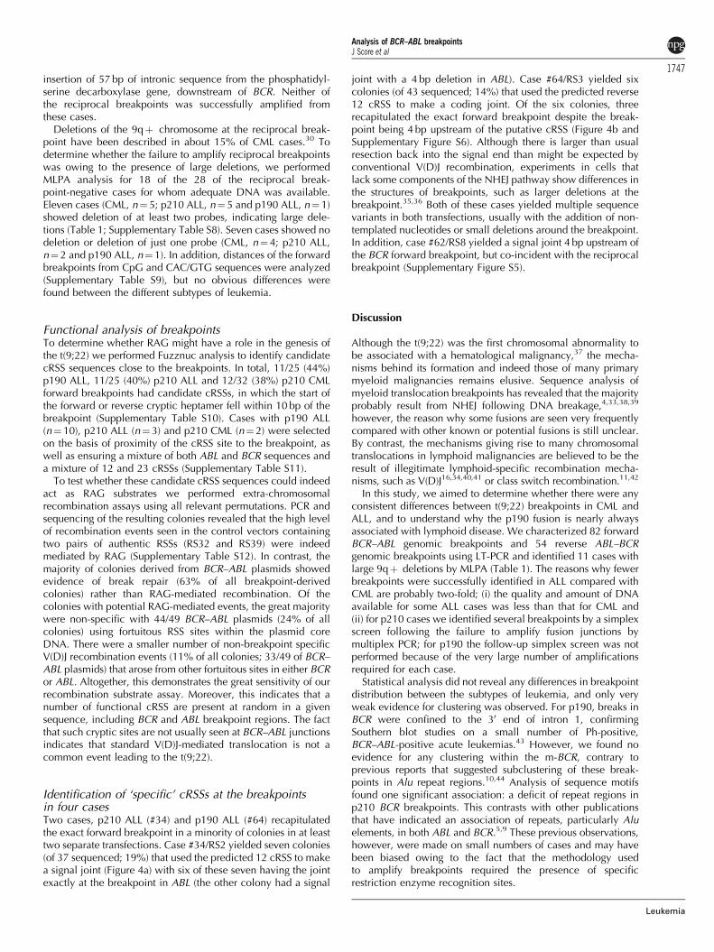
Identification of ‘specific’ cRSSs at the breakpointsin four casesTwo cases, p210 ALL (#34) and p190 ALL (#64) recapitulatedthe exact forward breakpoint in a minority of colonies in at leasttwo separate transfections. Case #34/RS2 yielded seven colonies(of 37 sequenced; 19%) that used the predicted 12 cRSS to makea signal joint (Figure 4a) with six of these seven having the jointexactly at the breakpoint in ABL (the other colony had a signal
joint with a 4 bp deletion in ABL). Case #64/RS3 yielded sixcolonies (of 43 sequenced; 14%) that used the predicted reverse12 cRSS to make a coding joint. Of the six colonies, threerecapitulated the exact forward breakpoint despite the break-point being 4 bp upstream of the putative cRSS (Figure 4b andSupplementary Figure S6). Although there is larger than usualresection back into the signal end than might be expected byconventional V(D)J recombination, experiments in cells thatlack some components of the NHEJ pathway show differences inthe structures of breakpoints, such as larger deletions at thebreakpoint.35,36 Both of these cases yielded multiple sequencevariants in both transfections, usually with the addition of non-templated nucleotides or small deletions around the breakpoint.In addition, case #62/RS8 yielded a signal joint 4 bp upstream ofthe BCR forward breakpoint, but co-incident with the reciprocalbreakpoint (Supplementary Figure S5).
Discussion
Although the t(9;22) was the first chromosomal abnormality tobe associated with a hematological malignancy,37 the mecha-nisms behind its formation and indeed those of many primarymyeloid malignancies remains elusive. Sequence analysis ofmyeloid translocation breakpoints has revealed that the majorityprobably result from NHEJ following DNA breakage,4,33,38,39
however, the reason why some fusions are seen very frequentlycompared with other known or potential fusions is still unclear.By contrast, the mechanisms giving rise to many chromosomaltranslocations in lymphoid malignancies are believed to be theresult of illegitimate lymphoid-specific recombination mecha-nisms, such as V(D)J16,34,40,41 or class switch recombination.11,42
In this study, we aimed to determine whether there were anyconsistent differences between t(9;22) breakpoints in CML andALL, and to understand why the p190 fusion is nearly alwaysassociated with lymphoid disease. We characterized 82 forwardBCR–ABL genomic breakpoints and 54 reverse ABL–BCRgenomic breakpoints using LT-PCR and identified 11 cases withlarge 9qþ deletions by MLPA (Table 1). The reasons why fewerbreakpoints were successfully identified in ALL compared withCML are probably two-fold; (i) the quality and amount of DNAavailable for some ALL cases was less than that for CML and(ii) for p210 cases we identified several breakpoints by a simplexscreen following the failure to amplify fusion junctions bymultiplex PCR; for p190 the follow-up simplex screen was notperformed because of the very large number of amplificationsrequired for each case.
Statistical analysis did not reveal any differences in breakpointdistribution between the subtypes of leukemia, and only veryweak evidence for clustering was observed. For p190, breaks inBCR were confined to the 30 end of intron 1, confirmingSouthern blot studies on a small number of Ph-positive,BCR–ABL-positive acute leukemias.43 However, we found noevidence for any clustering within the m-BCR, contrary toprevious reports that suggested subclustering of these break-points in Alu repeat regions.10,44 Analysis of sequence motifsfound one significant association: a deficit of repeat regions inp210 BCR breakpoints. This contrasts with other publicationsthat have indicated an association of repeats, particularly Aluelements, in both ABL and BCR.5,9 These previous observations,however, were made on small numbers of cases and may havebeen biased owing to the fact that the methodology usedto amplify breakpoints required the presence of specificrestriction enzyme recognition sites.
Analysis of BCR–ABL breakpointsJ Score et al
1747
Leukemia

Amplification of the reciprocal breakpoints was moresuccessful in p190 compared with p210 cases (21/25 versus33/57; P¼ 0.02, Fisher’s exact test). This is consistent withpreviously published data generated by fluorescence in situhybridization and reverse transcriptase-PCR showing that p190cases had a much lower frequency of large 9qþ deletions,45,46
compared with the established figure of around 15% in CML.47
Fine breakpoint mapping revealed that the sizes of deletions and
duplications at the breakpoint junctions were smaller in p190compared with p210 cases. Assuming that any LT-PCR-negative/MLPA-negative cases had deletions that were too large to bedetected by LT-PCR, but too small for MLPA, the median size ofdeletions was 6 bp for p190 and 7883 bp for p210. These datasuggest that p190 and p210 are formed by different mecha-nisms. We did not observe any consistent difference betweenCML and p210 ALL breakpoints, but this is perhaps not
Figure 4 Potential RAG mediated breaks in #34 and #64. (a) Case #34 RS2 ‘specific’ V(D)J recombination found in seven colonies from the cellculture assay. (i) #34 forward breakpoint sequence, with the putative heptamer in blue italics. (ii) Dd2inv (inverted) authentic RSS sequence fromthe T-cell receptor alpha gene on chromosome 14q11.2, showing the reverse 23 RSS and the forward 12 RSS. (iii) Schematic representation of thesignal joint formation, #34 ABL cassette is to scale with all the cryptic RSSs represented by triangles (12 RSSs are white and 12/23 RSSs are green).The authentic RSSs in Dd2inv are represented by red empty or solid tiangles (12 and 23 RSSs, respectively). (iv) Electropherogram from colonysequencing with the breakpoint marked by the vertical black line. (b) #64 RS3 ‘specific’ V(D)J recombination found in six colonies from the cellculture assay. (i) #64 forward and reciprocal breakpoint sequence, with the putative heptamer in blue italics. (ii) Dd2 authentic RSS sequence,showing the reverse 12 RSS and the forward 23 RSS. (iii) Schematic representation of the coding joint formation, #64 ABL cassette is to scale withall the cryptic RSSs represented by triangles (12 RSSs are white, 23 RSSs are black and 12/23 RSSs are green). The authentic RSSs in Dd2 arerepresented by red empty or solid triangles (12 or 23 RSSs, respectively). (iv) Electropherogram from colony sequencing with the breakpointmarked by the vertical black line.
Analysis of BCR–ABL breakpointsJ Score et al
1748
Leukemia
unexpected as an unknown proportion of p210 ALLs may in factbe derived from early transformation of a CML clone. Aspreviously described, we did not find large tracts of homologybetween ABL and BCR at the breakpoint junctions, but smalldeletions and duplications suggestive of NHEJ were frequent.Short stretches of microhomology (2–4 bp) at the forwardbreakpoints were found in 7/25 (28%) p190, 5/25 (20%) p210ALL and 10/32 (31%) p210 CML. In addition, 2 p210 CMLs (#8and #10) cases had microhomologies of 11 and 17 bp,respectively.
As p190 is largely restricted to lymphoid disease weconsidered the possibility that RAG may be involved ingenerating this fusion. The two hallmarks of V(D)J recombina-tion are the addition of non-templated nucleotides andpalindromic sequences, neither of which were observed in ourcases. This does not, however, preclude the possibility that oneor both breaks in ALL may be accomplished by aberrant RAGactivity. We, therefore, searched for potential cRSS signals usingpreviously defined criteria.32,33 Because of high degeneracy,these sequences are expected to occur on average every5000 bp and, therefore, we identified several candidate ALLand CML cases. As it is not possible to predict the functionalityof cRSSs on the basis of sequence alone, we selected arepresentative subset for analysis by the extra-chromosomalrecombination assay.34 Although no strong or recurrent RAGrecognition sites were observed, it is striking that we foundprecise recapitulation of the forward breakpoint in two ALLcases multiple times in multiple transfections. Furthermore, onep190 ALL case was identified, with a cut site at the reciprocalbreakpoint. By contrast specific cRSSs were not identified in thetwo CML cases tested.
Taken together, our data support the hypothesis that p190and p210 BCR–ABL are formed by different mechanisms. Inparticular the presence of functional cryptic RSS sequencesclose to some ALL breakpoints suggests the involvement of RAG,which in turn indicates that these fusions must have arisen in acommitted lymphoid progenitor cell; however, these wereexceptional cases rather than the rule.
Conflict of interest
The authors declare no conflict of interest.
Acknowledgements
This work was funded by Leukaemia and Lymphoma Research(UK); JS was supported by a Gordon Piller PhD Studentship.
Author contributionsThe study was designed by JS, NCPC and FHG. Laboratoryanalysis was performed by JS, MAS-S, SK, CH-C, DW. Dataanalysis was performed by JS, RFY, JW, BN. Samples and datawere provided by MJC, OO, FP, MAS-S, JVM. The initial paperwas written by JS, NCPC and FHG; all authors contributed to thefinal version.
References
1 Deininger MW, Bose S, Gora-Tybor J, Yan XH, Goldman JM, MeloJV. Selective induction of leukemia-associated fusion genes byhigh-dose ionizing radiation. Cancer Res 1998; 58: 421–425.
2 Stanulla M, Wang J, Chervinsky DS, Thandla S, Aplan PD.DNA cleavage within the MLL breakpoint cluster region is aspecific event which occurs as part of higher-order chromatin
fragmentation during the initial stages of apoptosis. MolCell Biol1997; 17: 4070–4079.
3 Neves H, Ramos C, da Silva MG, Parreira A, Parreira L. Thenuclear topography of ABL, BCR, PML, and RARalpha genes:evidence for gene proximity in specific phases of the cell cycleand stages of hematopoietic differentiation. Blood 1999; 93:1197–1207.
4 Reiter A, Saussele S, Grimwade D, Wiemels JL, Segal MR, Lafage-Pochitaloff M et al. Genomic anatomy of the specific reciprocaltranslocation t(15;17) in acute promyelocytic leukemia. GenesChromosomes Cancer 2003; 36: 175–188.
5 Mattarucchi E, Guerini V, Rambaldi A, Campiotti L, Venco A,Pasquali F et al. Microhomologies and interspersed repeatelements at genomic breakpoints in chronic myeloid leukemia.Genes Chromosomes Cancer 2008; 47: 625–632.
6 Melo JV. BCR-ABL gene variants. Baillieres Clin Haematol 1997;10: 203–222.
7 van der Feltz MJ, Shivji MK, Allen PB, Heisterkamp N, Groffen J,Wiedemann LM. Nucleotide sequence of both reciprocal trans-location junction regions in a patient with Ph positive acutelymphoblastic leukaemia, with a breakpoint within the first intronof the BCR gene. Nucleic Acids Res 1989; 17: 1–10.
8 Chen SJ, Chen Z, Font MP, d’Auriol L, Larsen CJ, Berger R.Structural alterations of the BCR and ABL genes in Ph1 positiveacute leukemias with rearrangements in the BCR gene first intron:further evidence implicating Alu sequences in the chromosometranslocation. Nucleic Acids Res 1989; 17: 7631–7642.
9 Jeffs AR, Benjes SM, Smith TL, Sowerby SJ, Morris CM. The BCRgene recombines preferentially with Alu elements in complexBCR-ABL translocations of chronic myeloid leukaemia. Hum MolGenet 1998; 7: 767–776.
10 Chen SJ, Chen Z, d’Auriol L, Le Coniat M, Grausz D, Berger R.Ph1+bcr- acute leukemias: implication of Alu sequences in achromosomal translocation occurring in the new cluster regionwithin the BCR gene. Oncogene 1989; 4: 195–202.
11 Robbiani DF, Bothmer A, Callen E, Reina-San-Martin B, Dorsett Y,Difilippantonio S et al. AID is required for the chromosomal breaksin c-myc that lead to c-myc/IgH translocations. Cell 2008; 135:1028–1038.
12 Tsai AG, Lu H, Raghavan SC, Muschen M, Hsieh CL, Lieber MR.Human chromosomal translocations at CpG sites and a theoreticalbasis for their lineage and stage specificity. Cell 2008; 135:1130–1142.
13 Marculescu R, Vanura K, Montpellier B, Roulland S, Le T, NavarroJM et al. Recombinase, chromosomal translocations and lymphoidneoplasia: targeting mistakes and repair failures. DNA Repair(Amst) 2006; 5: 1246–1258.
14 Roth DB. Restraining the V(D)J recombinase. Nat Rev Immunol2003; 3: 656–666.
15 Brandt VL, Roth DB. Recent insights into the formation ofRAG-induced chromosomal translocations. Adv Exp Med Biol2009; 650: 32–45.
16 Aplan PD, Lombardi DP, Ginsberg AM, Cossman J, Bertness VL,Kirsch IR. Disruption of the human SCL locus by ‘illegitimate’V-(D)-J recombinase activity. Science 1990; 250: 1426–1429.
17 Raghavan SC, Swanson PC, Ma Y, Lieber MR. Double-strand breakformation by the RAG complex at the bcl-2 major breakpointregion and at other non-B DNA structures in vitro. Mol Cell Biol2005; 25: 5904–5919.
18 Fialkow PJ, Denman AM, Jacobson RJ, Lowenthal MN. Chronicmyelocytic leukemia. Origin of some lymphocytes from leukemicstem cells. J Clin Invest 1978; 62: 815–823.
19 Cobaleda C, Gutierrez-Cianca N, Perez-Losada J, Flores T,Garcia-Sanz R, Gonzalez M et al. A primitive hematopoietic cellis the target for the leukemic transformation in human philadelphia-positive acute lymphoblastic leukemia. Blood 2000; 95: 1007–1013.
20 Schenk TM, Keyhani A, Bottcher S, Kliche KO, Goodacre A,Guo JQ et al. Multilineage involvement of Philadelphia chromo-some positive acute lymphoblastic leukemia. Leukemia 1998; 12:666–674.
21 Zaliova M, Fronkova E, Krejcikova K, Muzikova K, Mejstrikova E,Stary J et al. Quantification of fusion transcript reveals a subgroupwith distinct biological properties and predicts relapse in BCR/ABL-positive ALL: implications for residual disease monitoring.Leukemia 2009; 23: 944–951.
Analysis of BCR–ABL breakpointsJ Score et al
1749
Leukemia
22 Bacher U, Haferlach T, Hiddemann W, Schnittger S, Kern W,Schoch C. Additional clonal abnormalities in Philadelphia-positiveALL and CML demonstrate a different cytogenetic pattern atdiagnosis and follow different pathways at progression. CancerGenet Cytogenet 2005; 157: 53–61.
23 Turhan AG, Eaves CJ, Kalousek DK, Eaves AC, Humphries RK.Molecular analysis of clonality and bcr rearrangements inPhiladelphia chromosome-positive acute lymphoblastic leukemia.Blood 1988; 71: 1495–1498.
24 Hotfilder M, Rottgers S, Rosemann A, Schrauder A, Schrappe M,Pieters R et al. Leukemic stem cells in childhood high-risk ALL/t(9;22) and t(4;11) are present in primitive lymphoid-restrictedCD34+. Cancer Res 2005; 65: 1442–1449.
25 Castor A, Nilsson L, Astrand-Grundstrom I, Buitenhuis M, Ramirez C,Anderson K et al. Distinct patterns of hematopoietic stem cellinvolvement in acute lymphoblastic leukemia. Nat Med 2005; 11:630–637.
26 Melo JV, Myint H, Galton DA, Goldman JM. P190BCR-ABLchronic myeloid leukaemia: the missing link with chronic myelo-monocytic leukaemia? Leukemia 1994; 8: 208–211.
27 Li S, Ilaria Jr RL, Million RP, Daley GQ, Van Etten RA. The P190,P210, and P230 forms of the BCR/ABL oncogene induce a similarchronic myeloid leukemia-like syndrome in mice but have differentlymphoid leukemogenic activity. J Exp Med 1999; 189: 1399–1412.
28 Cross NCP, Melo JV, Feng L, Goldman JM. An optimized multiplexpolymerase chain reaction (PCR) for detection of BCR-ABL fusionmRNAs in haematological disorders. Leukemia 1994; 8: 186–189.
29 Baxter EJ, Kulkarni S, Vizmanos JL, Jaju R, Martinelli G, Testoni Net al. Novel translocations that disrupt the platelet-derived growthfactor receptor beta (PDGFRB) gene in BCR-ABL-negative chronicmyeloproliferative disorders. Br J Haematol 2003; 120: 251–256.
30 Kreil S, Pfirrmann M, Haferlach C, Waghorn K, Chase A,Hehlmann R et al. Heterogeneous prognostic impact of derivativechromosome 9 deletions in chronic myelogenous leukemia. Blood2007; 110: 1283–1290.
31 Segal MR, Wiemels JL. Clustering of translocation breakpoints.Journal of the American Statistical Association 2002; 97: 66–76.
32 Gellert M. V(D)J recombination: RAG proteins, repair factors,and regulation. Annu Rev Biochem 2002; 71: 101–132.
33 Wiemels JL, Leonard BC, Wang Y, Segal MR, Hunger SP, Smith MTet al. Site-specific translocation and evidence of postnatal origin ofthe t(1;19) E2A-PBX1 fusion in childhood acute lymphoblasticleukemia. Proc Natl Acad Sci USA 2002; 99: 15101–15106.
34 Marculescu R, Le T, Simon P, Jaeger U, Nadel B. V(D)J-mediatedtranslocations in lymphoid neoplasms: a functional assessment ofgenomic instability by cryptic sites. J Exp Med 2002; 195: 85–98.
35 Weinstock DM, Jasin M. Alternative pathways for the repair ofRAG-induced DNA breaks. Mol Cell Biol 2006; 26: 131–139.
36 Weinstock DM, Brunet E, Jasin M. Formation of NHEJ-derivedreciprocal chromosomal translocations does not require Ku70.Nat Cell Biol 2007; 9: 978–981.
37 Nowell PC, Hungerford DA. Chromosome studies on normal andleukemic human leukocytes. J NatlCancer Inst 1960; 25: 85–109.
38 Reichel M, Gillert E, Nilson I, Siegler G, Greil J, Fey GH et al.Fine structure of translocation breakpoints in leukemic blastswith chromosomal translocation t(4;11): the DNA damage-repairmodel of translocation. Oncogene 1998; 17: 3035–3044.
39 Zhang JG, Goldman JM, Cross NCP. Characterization of genomicBCR-ABL breakpoints in chronic myeloid leukaemia by PCR.BrJ Haematol 1995; 90: 138–146.
40 Tycko B, Sklar J. Chromosomal translocations in lymphoidneoplasia: a reappraisal of the recombinase model. Cancer Cells1990; 2: 1–8.
41 Raghavan SC, Swanson PC, Wu X, Hsieh CL, Lieber MR. A non-B-DNA structure at the Bcl-2 major breakpoint region is cleavedby the RAG complex. Nature 2004; 428: 88–93.
42 Pasqualucci L, Bhagat G, Jankovic M, Compagno M, Smith P,Muramatsu M et al. AID is required for germinal center-derivedlymphomagenesis. Nat Genet 2008; 40: 108–112.
43 Chen SJ, Chen Z, Hillion J, Grausz D, Loiseau P, Flandrin G et al.Ph1-positive, bcr-negative acute leukemias: clustering of break-points on chromosome 22 in the 30 end of the BCR gene firstintron. Blood 1989; 73: 1312–1315.
44 Chen SJ, Chen Z, Font MP, d’Auriol L, Larsen CJ, Berger R.Structural alterations of the BCR and ABL genes in Ph1 positiveacute leukemias with rearrangements in the BCR gene first intron:further evidence implicating Alu sequences in the chromosometranslocation. Nucleic Acids Res 1989; 17: 7631–7642.
45 Melo JV, Gordon DE, Tuszynski A, Dhut S, Young BD, Goldman JM.Expression of the ABL-BCR fusion gene in Philadelphia-positiveacute lymphoblastic leukemia. Blood 1993; 81: 2488–2491.
46 Specchia G, Albano F, Anelli L, Storlazzi CT, Zagaria A, Mancini Met al. Deletions on der(9) chromosome in adult Ph-positiveacute lymphoblastic leukemia occur with a frequency similar tothat observed in chronic myeloid leukemia. Leukemia 2003; 17:528–531.
47 Huntly BJ, Reid AG, Bench AJ, Campbell LJ, Telford N, Shepherd Pet al. Deletions of the derivative chromosome 9 occur at the timeof the Philadelphia translocation and provide a powerful andindependent prognostic indicator in chronic myeloid leukemia.Blood 2001; 98: 1732–1738.
Supplementary Information accompanies the paper on the Leukemia website (http://www.nature.com/leu)
Analysis of BCR–ABL breakpointsJ Score et al
1750
Leukemia