Direct translocation of histone molecules across cell membranes
OENOMICS 1 4 , 657-665 (1992)
A Radiation Hybrid Map of the Proximal Short Arm of the Human X Chromosome Spanning Incontinentia Pigmenti 1
(IP1) Translocation Breakpoints JEROME L. GORSKI,*'t '1 MICHAEL BOEHNKE,~ ERIC L. REYNER,* AND ERIC N. BURRIGHTt
Departments of *Pediatrics and Communicable Diseases, SBiostatistics, and tHuman Genetics, University of Michigan, Ann Arbor, Michigan 48109-0688
Received May 6, 1992; revised July 27, 1992
R a d i a t i o n h y b r i d m a p p i n g w a s u s e d in c o m b i n a t i o n w i t h p h y s i c a l m a p p i n g t e c h n i q u e s to o r d e r a n d es t i - m a t e d i s t a n c e s b e t w e e n 14 loc i in the p r o x i m a l r e g i o n o f t h e s h o r t a r m o f the h u m a n X c h r o m o s o m e . A p a n e l o f r a d i a t i o n h y b r i d s c o n t a i n i n g h u m a n X - c h r o m o s o m a l f r a g m e n t s w a s g e n e r a t e d f r o m a C h i n e s e h a m s t e r - h u - m a n ce l l h y b r i d c o n t a i n i n g an X c h r o m o s o m e as i t s o n l y h u m a n D N A . S i x t y - s e v e n r a d i a t i o n h y b r i d s w e r e s c r e e n e d b y S o u t h e r n h y b r i d i z a t i o n w i t h s e t s o f p r o b e s tha t m a p p e d to t h e r e g i o n X p 1 1 . 4 - X c e n to g e n e r a t e a r a d i a t i o n h y b r i d m a p o f t h e a r e a . A p h y s i c a l m a p o f 14 loc i w a s c o n s t r u c t e d b a s e d on the s e g r e g a t i o n o f the loc i in t h e h y b r i d c l o n e s . U s i n g p u l s e d - f i e l d ge l e l e c t r o p h o - r e s i s ( P F G E ) a n a l y s e s a n d a s o m a t i c ce l l h y b r i d m a p - p i n g p a n e l c o n t a i n i n g n a t u r a l l y o c c u r r i n g X; a u t o s o m e t r a n s l o c a t i o n s , t h e o r d e r o f the 14 loc i w a s v e r i f i e d a n d t h e l oc i n e a r e s t to the X - c h r o m o s o m a l t r a n s l o c a t i o n b r e a k p o i n t s a s s o c i a t e d w i t h the d i s e a s e i n c o n t i n e n t i a p i g m e n t i 1 ( IP1) w e r e ident i f i ed . T h e r a d i a t i o n h y b r i d p a n e l w i l l b e u s e f u l as a m a p p i n g r e s o u r c e for d e t e r - m i n i n g t h e l o c a t i o n , o r d e r , and d i s t a n c e s b e t w e e n o t h e r g e n e s a n d p o l y m o r p h i c loc i in th i s r e g i o n as w e l l as for g e n e r a t i n g a d d i t i o n a l r e g i o n - s p e c i f i c D N A m a r k e r s . © 1992 Academic Press, Inc.
INTRODUCTION
A physical map of a targeted chromosomal region is an essential prerequisite for the positional cloning of hu- man disease-associated genes. The construction and characterization of somatic cell hybrids containing de- fined human chromosomal fragments has been instru- mental in the development of high-resolution physical maps. As part of an effort to clone the incontinentia pigmenti 1 (IP1) locus and improve the resolution of a regional physical map, we sought a strategy to order and
x To whom correspondence and reprint requests should be ad- dressed at Division of Pediatric Genetics, Depar tment of Pediatrics, R m 3570 Medical Science Research Building II, P.O. Box 0688, Uni- versity of Michigan Medical Center, Ann Arbor, MI 48109-0688.
determine the distances between loci previously mapped in the vicinity of the IP1 X-chromosomal translocation breakpoints.
The IP1 locus has been mapped to region Xpll .21 by cytologic (Gilgenkrantz e t al., 1985; Hodgson e t al., 1985; Kajii et al., 1985; Cannizzaro and Hecht, 1987; Crolla e t al., 1989) and molecular (Gorski et al., 1991) analyses. Our previous mapping strategies relied on naturally oc- curring disease-specific translocations; these resources provided a method of mapping DNA markers relative to IP1 translocation breakpoints and assisted in construct- ing a primary physical map of the IP1 locus. This map indicated that the IP1 X-chromosomal translocation breakpoints were separated by at least 1250 kb (Gorski e t al., 1991). Subsequent studies showed that this region was at least twice that size (Gorski e t al., 1992); these results suggested that it would be difficult to determine precisely an order for and distance between the mapped loci solely by PFGE analysis. Therefore, we sought an alternative method for constructing a physical map of the IP1 region.
Radiation hybrid mapping is a powerful somatic cell genetic technique for ordering DNA markers which ex- tend over millions of basepairs of DNA (Cox et al., 1990). In this method, a lethal dose of radiation is used to cleave a human chromosome in a somatic cell hybrid into numerous fragments. Human chromosome frag- ments are subsequently retained nonselectively in a sec- ond rodent cell following cell fusion; the resulting hybrid clones are then screened for the presence or absence of human chromosome-specific loci. The closer two markers are on a chromosome, the less likely they will be separated by radiation fragmentation. By analyzing the cosegregation of the various loci in hybrid clones, a phys- ical map can be constructed (Cox e t al., 1990; Warring- ton e t al., 1991; Ceccherini et al., 1992).
To better define the region flanking the IP1 X-chro- mosomal translocation breakpoints, we used radiation hybrid mapping and molecular mapping techniques to construct a physical map of 14 loci in the proximal re- gion of Xp, including loci mapped to a region between
657 0888-7543/92 $5.00 Copyright © 1992 by Academic Press, Inc.
All rights of reproduction in any form reserved.
6 5 8 GORSKI ET AL.
I P 1 b r e a k p o i n t s , l oc i i m m e d i a t e l y d i s t a l t o t h e I P 1
b r e a k p o i n t s w i t h i n r e g i o n X p l l . 2 1 , a n d a c a n d i d a t e
g e n e f o r X - l i n k e d s i d e r o b l a s t i c a n e m i a ( A L A S 2 ) m a p p e d t o d i s t a l X p 1 1 . 2 1 ( C o t t e r e t a l . , 1992 ; G o r s k i e t
a l . , 1 9 9 2 ) . A p a n e l o f r a d i a t i o n h y b r i d s c o n t a i n i n g f r a g m e n t s o f a
h u m a n X c h r o m o s o m e w a s g e n e r a t e d f r o m a n X - o n l y
s o m a t i c ce l l h y b r i d w e p r e v i o u s l y h a d u s e d t o c o n s t r u c t a
p h y s i c a l m a p o f t h e r e g i o n ( G o r s k i e t a l . , 1991 , 1 9 9 2 ) ;
t h i s p r o v i d e d a m e a n s o f c a l i b r a t i n g t h e r a d i a t i o n h y b r i d
m a p a n d r e l a t i n g p h y s i c a l d i s t a n c e s d e t e r m i n e d b y
P F G E t o k i l o b a s e p a i r s o f D N A . B y a n a l y z i n g t h e s e g r e -
g a t i o n o f t h e 14 m a r k e r s i n t h e h y b r i d c l o n e s , w e w e r e
a b l e t o c o n s t r u c t a p h y s i c a l m a p d e f i n i n g t h e p o s i t i o n ,
o r d e r , a n d d i s t a n c e s b e t w e e n loc i . T h e s e r e s u l t s , i n c o n -
j u n c t i o n w i t h t h e r e s u l t s o f p h y s i c a l m a p p i n g a n a l y s e s ,
p r o v i d e d a m e a n s t o c o n s t r u c t a c o n s e n s u s m a p o f t h e
r e g i o n . T h e r e s u l t a n t m a p p r e d i c t e d c l o s e p h y s i c a l l i n k -
a g e b e t w e e n s e v e r a l l oc i a n d i d e n t i f i e d t h o s e l oc i i n c l o s -
e s t p r o x i m i t y t o t h e I P 1 X - c h r o m o s o m a l t r a n s l o c a t i o n
b r e a k p o i n t s . T h i s m a p p r o v i d e s a s t a r t i n g p o i n t f o r e s -
t a b l i s h i n g c o n t i g s o f Y A C a n d c o s m i d c l o n e s s p a n n i n g
t h e I P 1 b r e a k p o i n t s a n d a m e a n s o f o r d e r i n g a d d i t i o n a l
g e n e s a n d l oc i m a p p e d t o t h i s r e g i o n .
M A T E R I A L S A N D M E T H O D S
Rad ia t ion hybr ids a n d somat ic cell hybrids . Radiation hybrids (RHs) containing portions of a human X chromosome were con- structed by irradiating cell line GM06318, a human-hams te r hybrid containing a single, structurally intact, human X chromosome (Cor- iell Medical Institute, Camden, NJ) and performing a polyethylene glycol-mediated fusion with thymidine kinase deficient ( T K - ) ham- ster cell line a23 (Westerveld et al., 1971). RHs were constructed as described by Cox et al. (1989) with minor modifications; 2 × 10⁷ washed GM06318 cells were resuspended in 10 ml of serum-free DMEM, placed in a 25-cm 2 flask on ice, and exposed to 8000 rads of irradiation using a 1~7Ce source at a rate of 265 rads/min. These cells were fused to 2 × 10⁷ a23 cells as described previously (Gorski et al., 1989). Cells were plated at a density of 106 recipient cells/100-mm tissue culture dish. RHs expressing the Chinese hamster TK gene from cell line GM06318 were selected in medium containing hypoxan- thine, aminopterin, and thymidine (HAT). A total of 67 radiation hy- brid clones were isolated; all were used for the described analyses.
An IP1 somatic cell hybrid mapping panel has been described previ- ously (Gorski et al., 1989, 1991). Briefly, C9-5, B13-3, and C17-1D3 are human-hams te r hybrid cell lines and CLX17-A12 is a human-mouse hybrid cell line constructed from Eps te in-Barr virus-transformed lym- phoblastoid cell lines derived from three unrelated female patients with nonfamilial IP. Hybrids C9-5 and B13-3 contain the translocated X chromosome t(X;9)(p11.21;q34.3) (Gilgenkrantz et al., 1985) and t(X;13)(pll.21;q12.3) (Kajii et al., 1985), respectively; hybrid C17- 1D3 contains the derivative X chromosome, and hybrid CLX17-A12 contains the derivative 17 chromosome t ( X ; 17 ) ( p l l ; p l l ) (Hodgson et al., 1985). Hybrid A63-1A contains the translocation chromosome t (X;20)(20pter-q11. l : :Xq11. l -Xqter) ; hybrid A48-1Fa contains the translocation chromosome t(X;11) (11pter-1 lcen: : Xcen-Xqter) (Mahtani and Willard, 1988). Hybrid GM10501 contains the translo- cation chromosome t (X;17)(17qter -17ql l .2 : :Xpl l .21-Xqter ) (La- freniere et al., 1991) (Coriell Medical Institute).
M u l t i p o i n t m a p p i n g of rad ia t ion hybr id data. RH mapping data were analyzed using the mult ipoint maximum likelihood method of Boehnke et al. (1991). This method assumes tha t X-ray breakage oc- curs randomly along a chromosome and tha t the resulting fragments are independently retained in a RH. In the N-locus case, the likeli-
T A B L E 1
D N A F r a g m e n t s U s e d as P r o b e s
X-chromosomal Probe Locus Fragment regional assignment
ALAS2 1.8-kb EcoRI p11.21 MTHFDL1 2.0-kb EcoRI p11.21 ° 2S/pEa84 DXS706 1.0-kb Sau3A p11.21 a 3S DXS705 0.9-kb Sau3A p11.21 a p58-1 DXS14 1.2-kb HindIII pll.21 a 30C~2.8 DXS370 2.8-kb EcoRI pll.21 a cpX210 DXS422 0.3-kb EcoRI/HindIII pll.21 a RX-66 DXS343 1.2-kb RsaI p11.21 a KZO-48 DXS323 0.8-kb EcoRI/HhaI p11.21 2:30 b DXS741 1.4-kb NotI/HindIII pll.21 b 49A c DXS740 0.8-kb RsaI pll.21 c pHOTC OTC 1.4-kb EcoRI pll.4 pTAK8a DXS146 1.1-kb HindIII /BamHI p11.21 pBamX7 DXZl 2.0-kb HindIII /BamHI cen
Note . Locus identifications, regional assignments, and probe refer- ences are those of Human Gene Mapping 10.5 (Davies et al., 1990) and those previously reported (Lafreniere et al., 1991; Cotter et al., 1992; Gorski et al., 1992).
a Localized to a region between IP1 X-chromosomal translocation breakpoints (Gorski et al., 1991, 1992).
b N o t I l inking clone (Arenstorf et al., 1991); regional assignment determined by mapping data reported in the present work.
c IRS-PCR product amplified from a PFGE fractionated restriction fragment and mapped to region between IP1 breakpoints within Xpl l .21 (Burright and Gorski, 1992).
hood of the RH data is a function of the N-1 breakage probabilities between adjacent loci and one or more fragment retention probabili- ties. This method makes use of data on all loci simultaneously, includ- ing information on partially typed hybrids.
To describe the different fragment retent ion probability models, let rij be the probability of retaining a fragment on which only loci i < i + 1 < • • • < j are present. We considered two such models. In the first, all fragment retention probabilities were assumed to be equal (rii = r for all i ~<j). In the second, we allowed for a centromeric effect by setting rlj = rl and r o = r 2 (1 < i ~<j). Use of this second model was suggested by the decrease in observed locus retention probabilities with increas- ing distance from the centromere (see Results).
For each model, and for a given locus order, breakage and retent ion probabilities were estimated by those values tha t maximized the likeli- hood for the RH mapping data. Orders were compared by their maxi- mum likelihoods, the order with the largest maximum likelihood being best supported by the data. For a given order, fragment retention mod- els were compared by likelihood ratio tests.
Since it was not practical to consider explicitly all 14!/2 (equal re- tention) or 14! (centromeric retention) orders for the 14 loci, we used a stepwise locus ordering algorithm to identify the best locus order (Boehnke et al., 1991). This algorithm builds locus orders by adding one locus at a time. At each stage, it keeps under consideration those partial locus orders no more than K times less likely than the current best partial locus order. We carried out stepwise locus ordering for both the equal and centromeric models for the fourteen loci with K = 101°.
Stepwise locus ordering results in a list of the locus orders with the largest maximum likelihoods. To construct a framework map of loci, we began with those loci whose positions were consistent among all locus orders with maximum likelihoods no more than 1000 times smaller than tha t of the best locus order. We first noted whether these loci could be ordered at 1000:1. To those loci tha t could, we added any other loci whose best position gave a locus order maximum likelihood at least 1000 times greater than their next best position. Given the choice of several loci to include in a framework map, loci present in other genetic and physical maps of this region were given preference.
D N A probes. DNA markers used in this study are listed in Table 1. Plasmid DNA was prepared by s tandard techniques; prior to use as
RADIATION HYBRID MAP OF PROXIMAL Xp 659
probes, DNA fragments were isolated from agarose gels by electrolu- tion (Maniatis et al., 1982).
Standard and pulsed-field Southern hybridizations. For standard hybridizations, DNA was extracted from hybrid cells, digested to com- pletion using EcoRI or HindIII, electrophoresed in 1% agarose in 4x TAE, transferred to Hybond nylon membranes (Amersham), hybrid- ized, and washed as described (Gorski et al., 1991). Radioactive probes were prepared by random oligonucleotide priming (Feinberg and Vo- gelstein, 1983). To score RHs for the presence or absence of DNA markers and control for the quantity of DNA present in each lane, 2 to 3 probes were pooled; each pool included a probe which detected dis- tinct, evolutionarily conserved sequences in hamster DNA. Hybridiza- tions were performed for 16-24 h at 65°C and washed to a final strin- gency of 0.1X SSC, 0.1% SDS at 65°C for at least 15 min.
The methods for the preparation of agarose blocks containing high- molecular-weight DNA, the preparation of yeast and bacteriophage lambda multimers used as size markers, restriction digests, transfers to Hybond membranes, and hybridizations were described (Gorski et al., 1991). PFGE was performed at 14°C in 0.5x TBE buffer (Maniatis et al., 1982) using a contour-clamped homogeneous electrical field (CHEF) gel system (Chu et al., 1986) {CHEF DRII, Bio-Rad). To determine whether two probes hybridized to the same restriction frag- ment, we employed different sets of electrophoresis conditions to shift the fragments into the regions of high resolution on the gel and se- quentially hybridized probes to the same filter. Probes were removed from a filter prior to rehybridization; probe removal was verified by autoradiography.
Yeast artificial chromosomes. Using oligonucleotide primer pairs derived from loci mapped to Xpll.21, the Washington University yeast artificial chromosome (YAC) library was screened to identify yeast colonies containing cloned fragments; pooled YAC clones were screened using a PCR-based assay (Green and Olsen, 1990). To iden- tify individual YAC clones, but circumvent the need for colony hybrid- izations, pooled yeast lysates of arrayed rows and columns were ana- lyzed by PCR (Kwiatkowski et al., 1990). High-molecular-weight yeast DNA was prepared as previously described (Chandrasekharappa et al., 1992). Three YAC clones that contained loci mapped to a region between IP1 X-chromosomal translocation breakpoints within Xpll.21 were isolated; YAC clones B51B4 and B174A9 contained DXS14, and YAC clone B91F3 contained DXS370.
RESULTS
Radia t ion Hybr ids
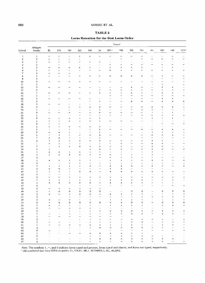
Radia t ion hybr id mapping was used in conjunct ion with physical mapping to determine the order and rela- tive distances between 14 loci in the proximal region of h u m a n Xp. Sixty-seven R H s were isolated. Using a cloned h u m a n L I N E S repetitive D N A sequence as probe (Manuelidis and Biro, 1982), we found tha t 50 RHs (75%) conta ined a human-specif ic 1.9-kb H i n d I I I frag- men t (data not shown); this suggested tha t at least 75% of the RHs conta ined h u m a n X-chromosomal f ragments (Cox et al., 1989). Since most of the RHs appeared to conta in human DNA, all were analyzed; a l though R H 24 was not found to conta in L I N E S sequences, it was found to conta in h u m a n X-chromosomal loci. The locus reten- t ion pa t t e rn for each of the RHs analyzed is summarized in Table 2. Limited quanti t ies of D N A resulted in the incomplete scoring of 2 RHs, R H 49 and R H 53; all o ther RHs were completely scored. The re tent ion frequency of the loci varied dramatical ly: 47 RHs (70%) nonselec- tively retained centromeric D N A (DXZ1) but only 14 RHs (22%) retained the OTC locus; other loci exhibited
retent ion frequencies between these 2 values (Table 4). For each hybrid, the min imum number of obligate breaks required to explain the data ranged from zero to five (Table 2); an obligate break corresponds to a discor- dance in the presence (+) or absence ( - ) of adjacent typed loci (Boehnke et al., 1991).
Mul t ipo in t Ana lys i s o[ Rad ia t ion Hybr id Da ta
We began our mult ipoint analysis by carrying out stepwise locus ordering for bo th the equal and the cen- t romeric re tent ion models. Bo th models gave the same best locus order. The centromeric model fit the data sig- nificantly bet ter t han the equal re tent ion probabil i ty model (x2(1) = 32.88, P < 0.0001), consis tent with our observat ion of decreasing re tent ion as a funct ion of dis- tance from the centromere. Table 3 presents the 13 locus orders with max imum likelihoods no more than 1000 t imes less t han tha t of the best locus order under the centromeric re tent ion probabil i ty model; locus orders are presented only in their mos t likely or ientat ion rela- tive to the centromere. The most likely locus order and the distance est imates between loci under the centro- meric re tent ion probabil i ty model are summarized in Ta- ble 4. To determine whether the data collected for the centromeric locus DXZ1 influenced the order of the other loci, mult ipoint analysis was performed wi thout locus DXZ1 data; the absence of locus DXZ1 did not alter the resulting locus orders. Influential hybrids (Boehnke et al., 1991) and those requiring four or more obligate breaks were rescored. Two scores were changed; however, nei ther change significantly altered the result- ing locus orders or distance estimates.
An interest ing complicat ion arose as we cons t ruc ted a f ramework map from the list of orders in Table 3. F rom these orders, it appeared tha t the loci DXZ1, DXS14, M T H F D L 1 , DXS705, DXS741, ALAS2, DXS146, and OTC should be the basis for a 1000:1 f ramework map. However, when only these loci were analyzed, the rela- tive posit ions of ALAS2, DXS705, and DXS741 could not be determined at tha t level of evidence. Because of the suggestion tha t DXS705 and DXS741 flank the IP1 locus (see Somatic Cell Hybr id Results), we have left all three orders in the near - f ramework maps of these eight loci (Table 5). In these maps, all loci bu t ALAS2, DXS705, and DXS741 are posi t ioned with relative max- imum likelihoods of at least 1000:1. The first near- f ramework map has a max imum likelihood 40 and 58 times greater t han those of the second and the third, respectively, suggesting tha t it is the correct order, al- though not at 1000:1 relative likelihood.
Somat i c Cell Hybr id M a p p i n g P a n e l Resul t s
To assign the 14 loci to dist inct physical intervals within Xp, each probe was hybridized to blots contain- ing IP1 somatic cell hybrid mapping panel DNA. As in- dicated in Table 1, by using this mapping panel, most of these loci have been previously mapped to specific inter- vals within Xp (Gorski et al., 1991, 1992). The results of
660 GORSKI ET AL.
T A B L E 2
L o c u s R e t e n t i o n for t h e B e s t L o c u s O r d e r
L o c u s a
O b l i g a t e
H y b r i d b r e a k s Z 1 3 7 0 7 4 0 4 2 2 3 4 3 14 M L 1 7 0 6 7 0 5 741 A L 3 2 3 1 4 6 O T C
1 1 + + + + + + . . . . . . . .
2 3 + . . . . . . . . + + -- --
3 1 + . . . . . . . . . . . . .
4 0 . . . . . . . . . . . .
5 3 + + + + + + + + + + -- + + --
6 1 + + + + + + + . . . . . . .
7 0 . . . . . . . . . . . . . .
8 1 + + + + + + + + + + + + -- --
9 0 . . . . . . . . . . . . . .
10 0 . . . . . . . . . . . . .
11 0 . . . . . . . . . . . . .
12 3 + . . . . . . + + + + + + --
13 0 . . . . . . . . . . . .
14 1 + + + + + + + + + + + + + --
15 1 + . . . . . . . . . . . . .
16 2 . . . . . . . . + + + + -
17 0 + + + + + + + + + + + + + +
18 0 . . . . . . . . . . . . .
19 3 + + + + + + + + + + + -
2 0 1 + + + + + + + + + + + - - -
21 0 + + + + + + + + + + + + + +
2 2 1 + . . . . . . . . . . .
23 0 + + + + + + + + + + + + + +
2 4 1 + + + + . . . . . . . . .
25 0 . . . . . . . . . . . . .
26 1 + + + + + + . . . . . . .
27 5 + + + + + + - + + + - + + -
2 8 0 + + + ÷ + + + + + + + + + +
29 1 + + + + + + + + + + + + + -
30 1 + + + ÷ + + . . . . . . .
31 4 + + + + + + + + - + + + - +
32 i + + + + + + + + + + + + -
33 2 - + + + + + + + + + + + - -
34 4 + - - - + + - + + + + + + +
35 1 + + + + + + + + + + + - - -
36 1 + + + + + + + + + + + + - -
37 0 . . . . . . . . . .
3 8 4 + + + + + + + + - + + + - +
39 0 . . . . . . . . . . . .
4 0 2 + + + + + + + + + + + + - +
41 3 + + - - - + + + . . . .
42 1 + + + + + + + + + + + +
43 0 . . . . . . . . . . . .
4 4 0 . . . . . . . . . . . . . .
45 1 + + + + + + + + + + + + + -
46 1 + + + + + + + + + + + + + -
4 7 0 . . . . . . . .
4 8 0 . . . . . . .
49 1 4- 0 0 0 0 0 -- -- 0 0 -- 0 0 0
5 0 2 + - - + ÷ + + + + + + + + +
51 0 . . . . . . . . . . . .
52 0 + + + + + + + + + + + + + +
53 0 + 0 0 0 0 0 + + 0 0 + 0 0 0
5 4 1 + + + + + + + + + + + + + -
55 0 . . . . . . . . . . .
5 6 0 + + + + + - - + + + + + + +
57 1 + + + + + + + + + + + + +
58 1 + . . . . . . . . . . .
59 2 + . . . . . . . . . . . +
60 1 + + + + + + + + + + . . . .
61 1 ÷ + + + + + + + + + + + + --
62 2 + + + + + + + + + + - - - +
63 0 . . . . . . . . . . . .
6 4 1 + + + + + + + + + + - - -
65 0 . . . . . . . . . . .
6 6 1 + + + + + + + + + + + + + --
6 7 2 + + + + + + + + + + + + +
Note. T h e s y m b o l s ÷ , - , a n d 0 i n d i c a t e l o c u s t y p e d a n d p r e s e n t , l o c u s t y p e d a n d a b s e n t , a n d l o c u s n o t t y p e d , r e s p e c t i v e l y .
a A l l n u m b e r e d l o c i h a v e D X S a s p r e f i x ; Z 1 , D X Z 1 ; M L 1 , M T H F D L 1 ; A L , A L A S 2 .
RADIATION HYBRID MAP OF PROXIMAL Xp
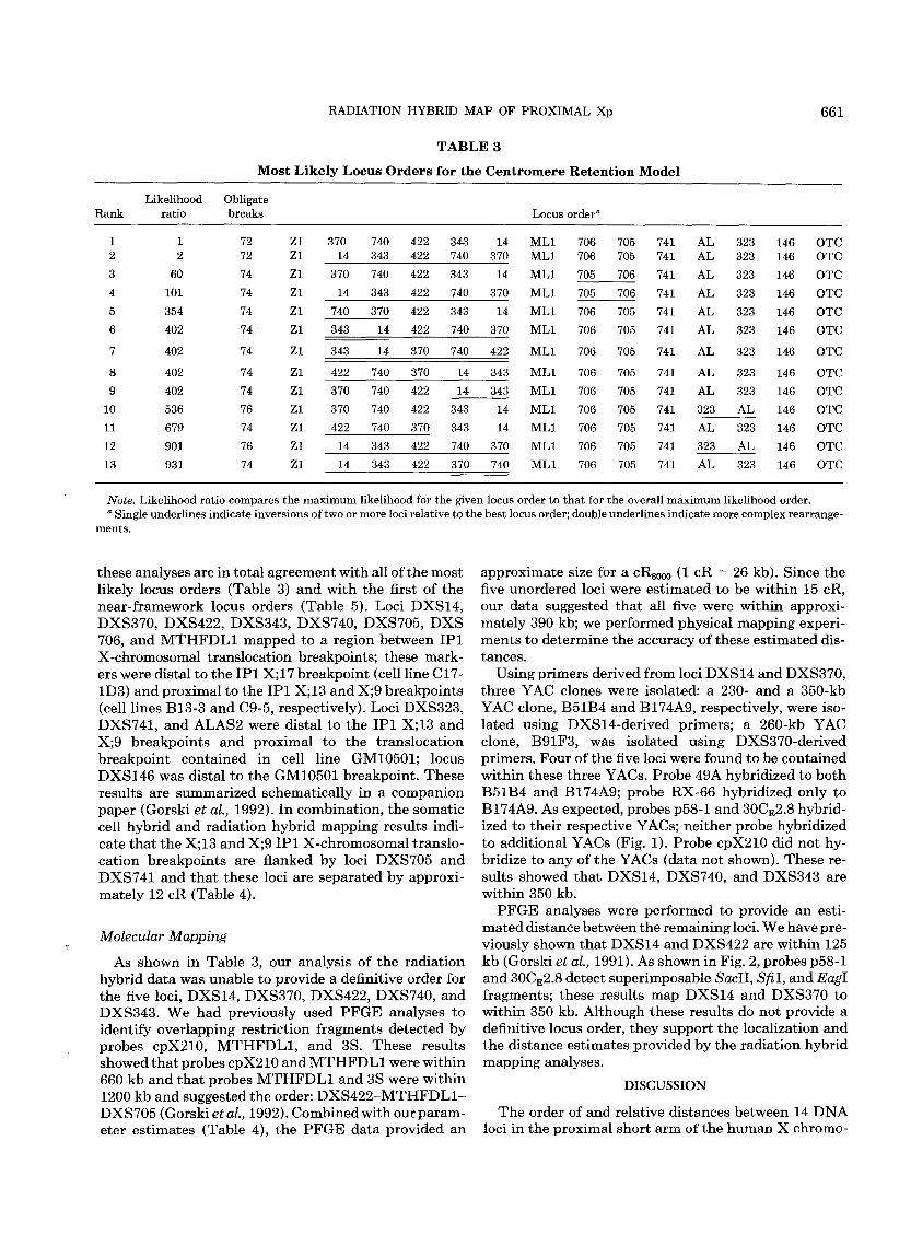
T A B L E 3
M o s t L i k e l y L o c u s O r d e r s for the C e n t r o m e r e R e t e n t i o n M o d e l
661
Likelihood Obligate Rank ratio breaks Locus order ~
1 1 72 Z1 370 740 422 343 14 ML1 706 705 741 AL 323 146 OTC 2 2 72 Zl 14 343 422 740 370 ML1 706 705 741 AL 323 146 OTC 3 60 74 Z1 370 740 422 343 14 ML1 705 706 741 AL 323 146 OTC 4 101 74 Zl 14 343 422 740 370 ML1 705 706 741 AL 323 146 OTC 5 354 74 Z1 740 370 422 343 14 ML1 706 705 741 AL 323 146 OTC 6 402 74 Zl 343 14 422 740 370 ML1 706 705 741 AL 323 146 OTC
7 402 74 Z1 343 14 370 740 422 ML1 706 705 741 AL 323 146 OTC
8 402 74 Zl 422 740 370 14 343 ML1 706 705 741 AL 323 146 OTC 9 402 74 Zl 370 740 422 14 343 ML1 706 705 741 AL 323 146 OTC
10 536 76 Zl 370 740 422 343 14 ML1 706 705 741 323 AL 146 OTC 11 679 74 Z1 422 740 370 343 14 ML1 706 705 741 AL 323 146 OTC 12 901 76 Zl 14 343 422 740 370 ML1 706 705 741 323 AL 146 OTC 13 931 74 Z1 14 343 422 370 740 ML1 706 705 741 AL 323 146 OTC
Note. Likelihood ratio compares the maximum likelihood for the given locus order to that for the overall maximum likelihood order. a Single underlines indicate inversions of two or more loci relative to the best locus order; double underlines indicate more complex rearrange-
ments.
these analyses are in total agreement with all of the most likely locus orders (Table 3) and with the first of the near- f ramework locus orders (Table 5). Loci DXS14, DXS370, DXS422, DXS343, DXS740, DXS705, DXS 706, and M T H F D L 1 mapped to a region between IP1 X-chromosomal translocation breakpoints; these mark- ers were distal to the IP1 X;17 breakpoin t (cell line C17- 1D3) and proximal to the IP1 X;13 and X;9 breakpoints (cell lines B13-3 and C9-5, respectively). Loci DXS323, DXS741, and ALAS2 were distal to the IP1 X;13 and X;9 breakpoints and proximal to the t ranslocat ion breakpoint conta ined in cell line GM10501; locus DXS146 was distal to the GM10501 breakpoint . These results are summarized schematical ly in a companion paper (Gorski et al., 1992). In combinat ion, the somatic cell hybrid and radiat ion hybrid mapping results indi- cate tha t the X;13 and X;9 IP1 X-chromosomal translo- cat ion breakpoints are flanked by loci DXS705 and DXS741 and tha t these loci are separated by approxi- mate ly 12 cR (Table 4).
Molecular Mapping
As shown in Table 3, our analysis of the radiat ion hybrid data was unable to provide a definitive order for the five loci, DXS14, DXS370, DXS422, DXS740, and DXS343. We had previously used P F G E analyses to identify overlapping restr ict ion f ragments detected by probes cpX210, M T H F D L 1 , and 3S. These results showed tha t probes cpX210 and M T H F D L 1 were within 660 kb and tha t probes M T H F D L 1 and 3S were within 1200 kb and suggested the order: D X S 4 2 2 - M T H F D L 1 - DXS705 (Gorski et al., 1992). Combined with our param- eter est imates (Table 4), the P F G E data provided an
approximate size for a cRs000 (1 cR = 26 kb). Since the five unordered loci were es t imated to be within 15 cR, our data suggested tha t all five were within approxi- mately 390 kb; we performed physical mapping experi- ments to determine the accuracy of these est imated dis- tances.
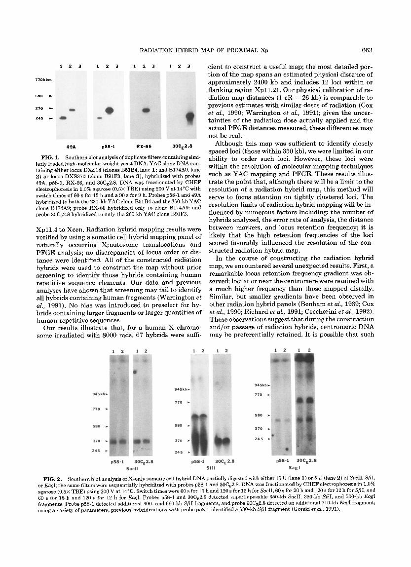
Using primers derived from loci DXS14 and DXS370, three YAC clones were isolated: a 230- and a 350-kb YAC clone, B51B4 and B174A9, respectively, were iso- lated using DXS14-der ived primers; a 260-kb YAC clone, B91F3, was isolated using DXS370-der ived primers. Four of the five loci were found to be conta ined within these three YACs. Probe 49A hybridized to bo th B51B4 and B174A9; probe RX-66 hybridized only to B174A9. As expected, probes p58-1 and 30CE2.8 hybrid- ized to their respective YACs; nei ther probe hybridized to addit ional YACs (Fig. 1). Probe cpX210 did not hy- bridize to any of the YACs (data not shown). These re- sults showed tha t DXS14, DXS740, and DXS343 are within 350 kb.
P F G E analyses were performed to provide an esti- mated distance between the remaining loci. We have pre- viously shown tha t DXS14 and DXS422 are within 125 kb (Gorski et al., 1991). As shown in Fig. 2, probes p58-1 and 30CE2.8 detect superimposable SacII, Sfi I, and EagI fragments; these results map DXS14 and DXS370 to within 350 kb. Al though these results do not provide a definitive locus order, they support the localization and the distance est imates provided by the radiat ion hybrid mapping analyses.
DISCUSSION
The order of and relative distances between 14 D N A loci in the proximal short arm of the h u m a n X chromo-
662 GORSKI E T AL.
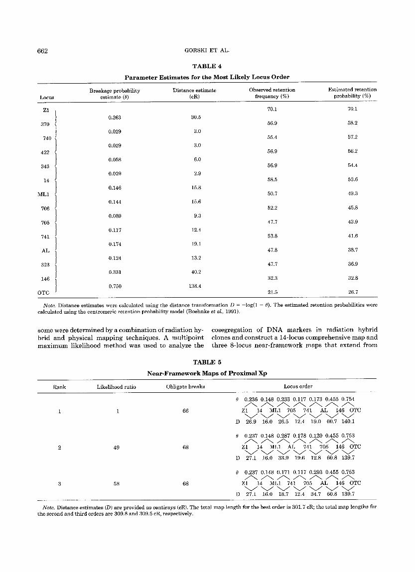
TABLE 4
Parameter Est imates for the M o s t L i k e l y Locus Order
Breakage probabil i ty Dis tance es t imate Observed re ten t ion Es t ima ted re ten t ion Locus es t imate (0) (cR) frequency (%) probabil i ty (%)
70.1 70.1 Z l 0.263 30.5
J 56.9 58.2 370 0.029 3.0
J 55.4 57.2 740 0.029 3.0
J 56.9 56.2 422 0.058 6.0
J 56.9 54.4 343 / 0.029 2.9 J 58.5 53.6 14 1 / 0.146 15.8
ML1 j 50.7 49.3
0.144 15.6 706 52.2 45.8 ]
t 0.089 9.3 J 47.7 43.9 7O5 1 t 0.117 12.4
741 J 53.8 41.6
0.174 19.1 AL 47.8 38.7
t 0.124 13.2 323 ) 47.7 36.9
0.331 40.2 146 32.3 32.8
/ 0.750 138.4 OTC J 21.5 26.7
Note . Distance es t imates were calculated using the dis tance t rans format ion D = - log(1 - 0). The es t imated re ten t ion probabili t ies were calculated using the cent romer ic re ten t ion probabil i ty model (Boehnke et al., 1991).
some were determined by a combination of radiation hy- brid and physical mapping techniques. A multipoint maximum likelihood method was used to analyze the
cosegregation of DNA markers in radiation hybrid clones and construct a 14-locus comprehensive map and three 8-locus near-framework maps that extend from
TABLE 5
Near-Framework Maps of Proximal Xp
Rank Likelihood rat io Obligate b reaks Locus order
0
1 1 66
2 49 68
3 58 68
D
0
D
0
D
0.236 0.148 0.233 0.117 0.173 0.455 0.754
Z1 14 ML1 705 741 AL 146 OTC V V V V V V V 26,9 16.0 26.5 12,4 19.0 60.7 140.1
0.237 0.148 0.287 0.178 0.120 0.455 0.753
Z1 14 ML1 AL 741 705 146 OTC
27.1 16.0 33.9 19.6 12.8 60.8 139.7
0.237 0.148 0.171 0.117 0.293 0.455 0.753
Z1 14 ML1 741 705 AL 146 OTC V V V V V V V 27.1 16.0 18.7 12.4 34.7 60.8 139.7
Note . Distance e s t ima tes (D) are provided as cent i rays (cR). The total m a p l eng th for t he bes t order is 301.7 cR; the total map leng ths for the second and th i rd orders are 309.8 and 309.5 cR, respectively.
RADIATION HYBRID MAP OF PROXIMAL Xp 663
1 2 3
770 k b-,.,-- ....
1 2 3 1 2 3 1 2 3
580 ~- : :i ~i!iiiii!i ilii ̧ / : !:: :~i:: :'~ ~: ~:: !:: ~i!:i:: i!!:: ~ :: 370 ~ ::
245 ~'-
49A pSS"l RX-66 30CE2.8
FIG. 1. Southern blot analysis of duplicate filters containing simi- larly loaded high-molecular-weight yeast DNA; YAC clone DNA con- taining either locus DXS14 (clones B51B4, lane 1; and B174A9, lane 2) or locus DXS370 (clone B91F3, lane 3), hybridized with probes 49A, p58-1, RX-66, and 30CE2.8. DNA was fractionated by CHEF electrophoresis in 1.0% agarose (0.5× TBE) using 200 V at 14°C with switch times of 60 s for 15 h and a 90 s for 9 h. Probes p58-1 and 49A hybridized to both the 230-kb YAC clone B51B4 and the 350-kb YAC clone B174A9; probe RX-66 hybridized only to clone B174A9; and probe 30CE2.8 hybridized to only the 260 kb YAC clone B91F3.
Xp11.4 to Xcen. Radia t ion hybrid mapping results were verified by using a somatic cell hybrid mapping panel of natural ly occurr ing X;au tosome t ranslocat ions and P F G E analysis; no discrepancies of locus order or dis- tance were identified. All of the cons t ruc ted radiat ion hybrids were used to cons t ruc t the map without prior screening to identify those hybrids conta in ing human repetitive sequence elements. Our data and previous analyses have shown tha t screening may fail to identify all hybrids conta in ing h u m a n fragments (Warr ington e t al., 1991). No bias was introduced to preselect for hy- brids conta in ing larger f ragments or larger quanti t ies of h u m a n repetitive sequences.
Our results illustrate that , for a human X chromo- some irradiated with 8000 rads, 67 hybrids were suffi-
cient to cons t ruc t a useful map; the most detailed por- t ion of the map spans an es t imated physical distance of approximately 2400 kb and includes 12 loci within or flanking region Xpl l . 21 . Our physical calibration of ra- diation map distances (1 cR = 26 kb) is comparable to previous est imates with similar doses of radiat ion (Cox e t al., 1990; War r ing ton e t al., 1991); given the uncer- tainties of the radiat ion dose actually applied and the actual P F G E distances measured, these differences may not be real.
Al though this map was sufficient to identify closely spaced loci (those within 350 kb), we were limited in our ability to order such loci. However, these loci were within the resolution of molecular mapping techniques such as YAC mapping and PFGE. These results illus- t rate the point that , a l though there will be a limit to the resolution of a radiat ion hybrid map, this me thod will serve to focus a t ten t ion on t ightly clustered loci. The resolution limits of radiat ion hybrid mapping will be in- fluenced by numerous factors including: the number of hybrids analyzed, the error rate of analysis, the distance between markers, and locus retent ion frequency; it is likely tha t the high retent ion frequencies of the loci scored favorably influenced the resolution of the con- s tructed radiat ion hybrid map.
In the course of const ruct ing the radiat ion hybrid map, we encountered several unexpected results. First, a remarkable locus retent ion frequency gradient was ob- served; loci at or near the cent romere were retained with a much higher frequency than those mapped distally. Similar, but smaller gradients have been observed in other radiat ion hybrid panels (Benham e t al., 1989; Cox e t al., 1990; Richard e t al., 1991; Ceccherini e t al., 1992). These observations suggest tha t during the const ruct ion and /o r passage of radiat ion hybrids, centromeric D N A may be preferential ly retained. I t is possible tha t such
945kb
770
580
370
245
1 2 1 2 1 2
945kb~
770
580
370
245
1 2 1 2 1 2
945kh
770
580
370
245
p58-1 30CE2.8 p58-1 30CE 2.8 p58-1 30CE2.8
$ a c l l S f i l E a g l
FIG. 2. Southern blot analysis of X-only somatic cell hybrid DNA partially digested with either 15 U (lane 1) or 5 U (lane 2) of SacII, SfiI, or EagI; the same filters were sequentially hybridized with probes p58-1 and 30CE2.8. DNA was fractionated by CHEF electrophoresis in 1.0% agarose (0.5× TBE) using 200 V at 14°C. Switch times were 60 s for 15 h and 120 s for 12 h for SacII, 60 s for 20 h and 120 s for 12 h for SfiI, and 60 s for 18 h and 120 s for 12 h for EagI. Probes p58-1 and 30CE2.8 detected superimposable 350-kb SacII, 350-kb SfiI, and 500-kb EagI fragments. Probe p58-1 detected additional 400- and 660-kb SfiI fragments, and probe 30CE2.8 detected an additional 710-kb EagI fragment; using a variety of parameters, previous hybridizations with probe p58-1 identified a 580-kb SfiI fragment (Gorski et al., 1991).
664 GORSKI ET AL.
preferential re tent ion may be secondary to fundamenta l differences between centromeric and non-centromeric DNA; potential differences in susceptibilities to degrada- t ion after f ragmentat ion, integrat ion frequencies, or mi- totic stabilities might influence retent ion frequency. Since centromeric D N A was retained more frequently, it was possible tha t our analyses would be confused by radi- at ion hybrids conta in ing frequent, independent ly segre- gating centromeric fragments. We did not, however, en- counter any ambiguities in the localization of the cen- t romeric DNA, nor did the removal of the centromeric locus data alter our mapping results. I t is possible tha t this potent ia l problem was c i rcumvented by not using randomly selected X-chromosomal loci to score the hy- brids; all loci used to score the hybrids had been previ- ously mapped to an interval in close proximity to the centromere. This suggests t ha t radiat ion hybrid map- ping will be facilitated by using markers previously as- signed to chromosomal intervals.
Second, during the construct ion of the near-frame- work maps, it was unant ic ipa ted tha t the removal of sev- eral loci f rom the mul t ipoint max imum likelihood analy- sis would affect the likelihood of the orders of other non- adjacent loci; this suggests tha t each locus analyzed might potent ial ly alter previous est imates of posit ion and distance. I t remains to be determined with what fre- quency and at what resolution such addit ional breaks and subsequent amended maps might occur. Obviously, undetected data errors may play a role in this observa- tion.
The radiat ion hybrid mapping panel reported here will provide a rapid mapping tool for any identified D N A marker localized to proximal Xp. Once a D N A marker is available, it will be possible to place a D N A sequence relative to mapped markers in several days. The radia- t ion hybrid mapping panel also provides a way of identi- fying groups of loci tha t are close enough to war ran t subsequent analyses with techniques yielding high-reso- lution physical maps such as PFGE, YAC clone analysis (Schlessinger et al., 1991), and multicolor interphase flu- orescent in s i t u hybridizat ion (Trask e t al., 1991).
Most important ly , this map provides a means for identifying markers in close proximity to disease loci and disease-specific X-chromosomal rearrangements . This map has localized a candidate gene for sideroblastic anemia (ALAS2) (Cotter et al., 1992) and a pseudogene ( M T H F D L 1 ) (Italiano et al., 1991) and has identified D N A markers tha t flank the IP1 X-chromosomal t rans- location breakpoints . These results have provided a new focus for our IP1 cloning efforts; wi thout radiat ion hy- brid mapping, there would be no compelling reason to focus on a par t icular pair of loci for subsequent high-re- solution mapping studies. We are current ly screening YAC libraries in an a t t empt to establish a cloned YAC contig spanning the IP1 t ranslocat ion breakpoints. Sev- eral addit ional disease-related loci have been mapped to proximal Xp including: progressive cone dystrophy, Wiskot t Aldridge disease, Norries disease, retinitis pig- mentosa types 2 and 3, congenital s ta t ionary night blindness, and an X-chromosomal t ranslocat ion break-
point associated with synovial sarcoma (Davies et al., 1990); this map and the radiat ion hybrid panel may be of utili ty in mapping these loci.
ACKNOWLEDGMENTS
We thank D. Cox and F. Collins for stimulating conversations and helpful comments; D. Barker, D. Bishop, G. Bruns, K. Davies, T. Kruse, R. Rozen, S. Weissman, and H. Willard, for cloned human DNA probes; H. Willard for somatic cell hybrids A48-1Fa and A63- 1A; and D. Voss for assisting in preparing the manuscript. Cell lines GM06853, GM10501, and GM06318 were obtained from the NIGMS Human Genetic Mutant Cell Repository, Coriell Institute of Medical Research (Camden, NJ). This work was supported, in part, by March of Dimes-Birth Defects Foundation Basic Science Grant 1-91-176 and National Institutes of Health (NIH) Grants HD-23768 and NS- 30771 to J.L.G., NIH Training Grant NIH-5-T32-GM07544-11 to E.N.B., and The University of Michigan Human Genome Center P30- HG00209.
REFERENCES
Arenstorf, H. P., Kandpal, R. P., Baskaran, N., Parimoo, S., Tanaka, Y., Kitajima, S., Yasukochi, Y., and Weissman, S. M. (1991). Con- struction and characterization of a NotI-BsuE linking library from the human X chromosome. Genomics 11: 115-123.
Benham, F., Hart, K., Crolla, J., Bobrow, M., Francavilla, M., and Goodfellow, P. N. (1989). A method for generating hybrids contain- ing nonselected fragments of human chromosomes. Genomics 4: 509-517.
Boehnke, M., Lange, K., and Cox, D. R. (1991). Statistical methods for multipoint radiation hybrid mapping. Am. J. Hum. Genet. 49: 1174-1188.
Burright, E. N., and Gorski, J. L. (1992). Isolation of Alu-PCR prod- ucts from a 660-kb SfiI restriction fragment mapped to a region between incontinentia pigmenti 1 (IP1) X-chromosomal transloca- tion breakpoints. Submitted for publication.
Cannizzaro, L. A., and Hecht, F. (1987). Gene for incontinentia pig- menti maps to band Xpll.21 with an (X;10)(pll;q22) transloca- tion. Clin. Genet. 32: 66-69.
Ceccherini, I., Romeo, G., Lawrence, S., Breuning, M. H., Harris, P. C., Himmelbauer, H., Frischauf, A. M., Sutherland, G. R, Ger- mino, G. G., Reeders, S. T., and Morton, N. E. (1992). Construction of a map of chromosome 16 by using radiation hybrids. Proc. Natl. Acad. Sci. USA 89: 104-108.
Chandrasekharappa, S. C., Marchuk, D. A., and Collins, F. S. (1992). Analysis of yeast artificial chromosome clones. In "Methods in Mo- lecular Biology: Pulsed Gel Electrophoresis Techniques" (L. Ulan- ovsky and M. Burmeister, Eds.), Humana Press, Clifton, NJ, in press.
Chu, G., Vollrath, D., and Davis, R. W. (1986). Separation of large DNA molecules by contour-clamped homogeneous electric fields. Science 234: 1582-1585.
Cotter, P. D., Willard, H. F., Gorski, J. L., and Bishop, D. F. (1992). Assignment of human erythroid delta-aminolevulinate synthase (ALAS2) to a distal subregion of band Xpll.21 by PCR analysis of somatic cell hybrids containing X;autosome translocations. Geno- mics 13: 211-213.
Cox, D. R., Pritchard, C. A., Uglum, E., Casher, D., Kobori, J., and Myers, R. M. (1989). Segregation of the Huntington disease region of human chromosome 4 in a somatic cell hybrid. Genomics 4: 397- 407.
Cox, D. R., Burmeister, M., Price, E. R., Kim, S., and Myers, R. M. (1990). Radiation hybrid mapping: A somatic cell genetic method for constructing high-resolution maps of mammalian chromosomes. Science 250: 245-250.
Crolla, J. A., Gilgenkrantz, S., de Grouchy, J., Kajii, T., and Bobrow,
RADIATION HYBRID MAP OF PROXIMAL Xp 665
M. (1989). Incontinentia pigmenti and X-autosome translocations: Non-isotopic in situ hybridization with an X-centromere-specific probe (pSV2X5) reveals a possible X-centromeric breakpoint in one of five published cases. Hum. Genet. 81: 269-272.
Davies, K. E., Mandel, J. L., Monaco, A. P., Nussbaum, R. L., and Willard, H. F. (1990). Report of the committee on the genetic con- stitution of the X chromosome. Cytogenet. Cell Genet. 55: 254-313.
Feinberg, A., and Vogelstein, B. (1983). A technique for radiolabeling DNA restriction endonuclease fragments to high specific activity. Anal. Biochem. 132: 6-13.
Gilgenkrantz, S., Tridon, P., Pinel-Briquel, N., Beurey, J., and Weber, M. (1985). Translocation (X;9)(p11;q34) in a girl with incontinen- tia pigmenti (IP): Implications for the regional assignment of the IP locus to Xp l l . Ann. Genet. 28: 90-92.
Gorski, J. L., Stein, C. K., and Glover, T. W. (1989). A somatic cell hybrid panel to facilitate identification of DNA sequences in the vicinity of the incontinentia pigmenti locus (IP1). Cytogenet. Cell Genet. 52: 90-92.
Gorski, J. L., Burright, E. N., Harnden, C. E., Stein, C. K., Glover, T. W., and Reyner, E. L. (1991). Localization of DNA sequences to a region within Xp11.21 between incontinentia pigmenti (IP1) X- chromosomal translocation breakpoints. Am. J. Hum. Genet. 48: 53-64.
Gorski, J. L., Burright, E. N., Reyner, E. L., Goodfellow, P. N., and Burgess, D. L. (1992). Isolation of DNA markers from a region be- tween incontinentia pigmenti 1 (IP1) X-chromosomal transloca- tion breakpoints by a comparative PCR analysis of a radiation hy- brid subelone mapping panel. Genomics 14: 649-656.
Green, E. D., and Olson, M. V. (1990). Systematic screening of yeast artificial-chromosome libraries by use of the polymerase chain reac- tion. Proc. Natl. Acad. Sci. USA 87: 1213-1217.
Harnden, D. G., and Klinger, H. P., Eds. (1985). "An International System for Human Cytogenetic Nomenclature," ISCN, published in collaboration with Cytogenet. Cell Genet., Karger, Basel.
Hodgson, S. V., Neville, B., Jones, R. W. A., Fear, C., and Bobrow, M. (1985). Two cases of X; autosome translocation in females with in- continentia pigmenti. Hum. Genet. 71: 231-234.
Italiano, C., John, S. W. M., Hum, D. W., Mackenzie, R. E., and Ro- zen, R. (1991). A pseudogene on the X chromosome for the human trifunctional enzyme MTHFD (methylenetetrahydrofolate dehy- drogenase-methenyltetrahydrofolate cyclohydrolase-formyltetra- hydrofolate synthetase). Genomics 10: 1073-1074.
Kajii, T., Tsukahara, M., Fukushima, Y., Hata, A., Matsuo, K., and Kuroki, Y. (1985). Translocation (X;13)(pll.21; q12.3) in a girl with incontinentia pigmenti and bilateral retinoblastoma. Ann. Genet. 28: 219-223.
Kwiatkowski, T. J., Jr., Zoghbi, H. Y., Ledbetter, S. A., Ellison, K. A., and Chinault, A. C. (1990). Rapid identification of yeast artificial chromosome clones by matrix pooling and crude lysate PCR. Nu- cleic Acids Res. 18: 7191-7192.
Lafreniere, R. G., Brown, C. J., Powers, V. E., Carrel, L., Davies, K. E., Barker, D. F., and Willard, H. F. (1991). Physical mapping of 60 DNA markers in the p21.1-q21.3 region of the human X chromo- some. Genomics 11: 352-363.
Mahtani, M. M., and Willard, H. F. (1988). A primary genetic map of the pericentric region of the human X chromosome. Genomics 2: 294-301.
Maniatis, T., Fritsch, E. F., and Sambrook, J. (1982). "Molecular Cloning: A Laboratory Manual," Cold Spring Harbor Laboratory, Cold Spring Harbor, NY.
Manuelidis, L., and Biro, P. A. (1982). Genomic representation of the HindIII 1.9 kb repeated DNA. Nucleic Acids Res. 10: 3221-3239.
Richard, C. W., III, Withers, D. A., Meeker, T. C., Maurer, S., Evans, G. A., Myers, R. M., and Cox, D. R. (1991). A radiation hybrid map of the proximal long arm of human chromosome 11 containing the multiple endocrine neoplasia type 1 (MEN-I) and bcl-I disease loci. Am. J. Hum. Genet. 49: 1189-1196.
Schlessinger, D., Little, R. D., Freije, D., Abidi, F., Zucchi, I., Porta, G., Pilia, G., Nagaraja, R., Johnson, S. K., Yoon, J-Y., Srivastava, A., Kere, J., Palmieri, G., Ciccodicola, A., Montanaro, V., Romano, G., Casamassimi, A., and D'Urso, M. (1991). Yeast artificial chro- mosome-based genome mapping: Some lessons from Xq24-q28. Ge- nomics 11: 783-793.
Trask, B. J., Massa, H., Kenwrick, S., and Gitschier, J. (1991). Map- ping of human chromosome Xq28 by two-color fluorescence in situ hybridization of DNA sequences to interphase cell nuclei. Am. J. Hum. Genet. 48: 1-15.
Warrington, J. A., Hall, L. V., Hinton, L. M., Miller, J. N., Wasmuth, J. J., and Lovett, M. (1991). Radiation hybrid map of 13 loci on the long arm of chromosome 5. Genomics 11: 701-708.
Westerveld, A., Visser, R. P. L. S., Kahn, P. M., and Bootsma, D. (1971). Loss of human genetic markers in man-Chinese hamster ovary cells. Nature (New Biol.) 234: 20-22.
Copyright © 2022 FDOKUMEN