Phytochemicals of Avocado Residues as Potential ... - MDPI
18
Citation: da Silva, G.G.; Pimenta, L.P.S.; Melo, J.O.F.; Mendonça, H.d.O.P.; Augusti, R.; Takahashi, J.A. Phytochemicals of Avocado Residues as Potential Acetylcholinesterase Inhibitors, Antioxidants, and Neuroprotective Agents. Molecules 2022, 27, 1892. https://doi.org/ 10.3390/molecules27061892 Academic Editor: Simona Rapposelli Received: 28 January 2022 Accepted: 11 March 2022 Published: 15 March 2022 Publisher’s Note: MDPI stays neutral with regard to jurisdictional claims in published maps and institutional affil- iations. Copyright: © 2022 by the authors. Licensee MDPI, Basel, Switzerland. This article is an open access article distributed under the terms and conditions of the Creative Commons Attribution (CC BY) license (https:// creativecommons.org/licenses/by/ 4.0/). molecules Article Phytochemicals of Avocado Residues as Potential Acetylcholinesterase Inhibitors, Antioxidants, and Neuroprotective Agents Geisa Gabriela da Silva 1 ,Lúcia Pinheiro Santos Pimenta 2 ,Júlio Onésio Ferreira Melo 3 , Henrique de Oliveira Prata Mendonça 3 , Rodinei Augusti 2 and Jacqueline Aparecida Takahashi 2, * 1 Department of Food Science, Faculty of Pharmacy, Universidade Federal de Minas Gerais, Av. Antônio Carlos, 6627, Belo Horizonte 31270-901, Brazil; [email protected] 2 Chemistry Department, Universidade Federal de Minas Gerais, Av. Antônio Carlos, 6627, Belo Horizonte 31270-901, Brazil; [email protected] (L.P.S.P.); [email protected] (R.A.) 3 Exact and Biological Sciences Department, Campus Sete Lagoas, Universidade Federal de São João del-Rei, Sete Lagoas 36307-352, Brazil; [email protected] (J.O.F.M.); [email protected] (H.d.O.P.M.) * Correspondence: [email protected]; Tel.: +55-31-34095754 Abstract: Avocado (Persea americana) is a widely consumed fruit and a rich source of nutrients and phytochemicals. Its industrial processing generates peels and seeds which represent 30% of the fruit. Environmental issues related to these wastes are rapidly increasing and likely to double, according to expected avocado production. Therefore, this work aimed to evaluate the potential of hexane and ethanolic peel (PEL-H, PEL-ET) and seed (SED-H, SED-ET) extracts from avocado as sources of neuroprotective compounds. Minerals, total phenol (TPC), total flavonoid (TF), and lipid contents were determined by absorption spectroscopy and gas chromatography. In addition, phytochemicals were putatively identified by paper spray mass spectrometry (PSMS). The extracts were good sources of Ca, Mg, Fe, Zn, ω-6 linoleic acid, and flavonoids. Moreover, fifty-five metabolites were detected in the extracts, consisting mainly of phenolic acids, flavonoids, and alkaloids. The in vitro antioxidant capacity (FRAP and DPPH), acetylcholinesterase inhibition, and in vivo neuroprotective capacity were evaluated. PEL-ET was the best acetylcholinesterase inhibitor, with no significant difference (p > 0.05) compared to the control eserine, and it showed neither preventive nor regenerative effect in the neuroprotection assay. SED-ET demonstrated a significant protective effect compared to the control, suggesting neuroprotection against rotenone-induced neurological damage. Keywords: avocado biomass; Alzheimer’s disease; neuroprotective effect; avocado seed; avocado peel; paper spray mass spectrometry 1. Introduction Originated from Central America, the avocado is the fruit of the Persea americana Mill species from the Lauraceae family. It is rich in minerals, vitamins, proteins, fibers, and unsaturated lipids, the latter known to prevent cardiovascular diseases [1,2]. Moreover, avocado seeds display significant antioxidant, anti-inflammatory, and anti-cancer effects which have been associated with elevated percentages of hydrocarbons, sterols, and unsat- urated fatty acids. Pulp has the same effects, although at a lower intensity [3]. Finally, the peel is rich in phenolic compounds and minerals, and has significant antioxidant activity, comparable to the seeds and far superior to the pulp [4,5]. In 2020, Mexico was the largest avocado producer, accounting for 2393 thousand tons per year, followed by the Dominican Republic, Peru, Colombia, Indonesia, and Brazil [6]. Since the inedible parts of avocado, mainly constituting peels and seeds, are discarded by the industry, restaurants, or at home, with the growth of avocado production, the amount of residues is also increasing, causing environmental problems [7]. These parts can reach up to 30% of the Molecules 2022, 27, 1892. https://doi.org/10.3390/molecules27061892 https://www.mdpi.com/journal/molecules
-
Upload
khangminh22 -
Category
Documents
-
view
2 -
download
0
Transcript of Phytochemicals of Avocado Residues as Potential ... - MDPI
�����������������
Citation: da Silva, G.G.; Pimenta,
L.P.S.; Melo, J.O.F.; Mendonça,
H.d.O.P.; Augusti, R.; Takahashi, J.A.
Phytochemicals of Avocado Residues
as Potential Acetylcholinesterase
Inhibitors, Antioxidants, and
Neuroprotective Agents. Molecules
2022, 27, 1892. https://doi.org/
10.3390/molecules27061892
Academic Editor: Simona Rapposelli
Received: 28 January 2022
Accepted: 11 March 2022
Published: 15 March 2022
Publisher’s Note: MDPI stays neutral
with regard to jurisdictional claims in
published maps and institutional affil-
iations.
Copyright: © 2022 by the authors.
Licensee MDPI, Basel, Switzerland.
This article is an open access article
distributed under the terms and
conditions of the Creative Commons
Attribution (CC BY) license (https://
creativecommons.org/licenses/by/
4.0/).
molecules
Article
Phytochemicals of Avocado Residues as PotentialAcetylcholinesterase Inhibitors, Antioxidants,and Neuroprotective AgentsGeisa Gabriela da Silva 1 , Lúcia Pinheiro Santos Pimenta 2 , Júlio Onésio Ferreira Melo 3 ,Henrique de Oliveira Prata Mendonça 3, Rodinei Augusti 2 and Jacqueline Aparecida Takahashi 2,*
1 Department of Food Science, Faculty of Pharmacy, Universidade Federal de Minas Gerais, Av. Antônio Carlos,6627, Belo Horizonte 31270-901, Brazil; [email protected]
2 Chemistry Department, Universidade Federal de Minas Gerais, Av. Antônio Carlos, 6627,Belo Horizonte 31270-901, Brazil; [email protected] (L.P.S.P.); [email protected] (R.A.)
3 Exact and Biological Sciences Department, Campus Sete Lagoas, Universidade Federal de São João del-Rei,Sete Lagoas 36307-352, Brazil; [email protected] (J.O.F.M.); [email protected] (H.d.O.P.M.)
* Correspondence: [email protected]; Tel.: +55-31-34095754
Abstract: Avocado (Persea americana) is a widely consumed fruit and a rich source of nutrients andphytochemicals. Its industrial processing generates peels and seeds which represent 30% of the fruit.Environmental issues related to these wastes are rapidly increasing and likely to double, accordingto expected avocado production. Therefore, this work aimed to evaluate the potential of hexaneand ethanolic peel (PEL-H, PEL-ET) and seed (SED-H, SED-ET) extracts from avocado as sources ofneuroprotective compounds. Minerals, total phenol (TPC), total flavonoid (TF), and lipid contentswere determined by absorption spectroscopy and gas chromatography. In addition, phytochemicalswere putatively identified by paper spray mass spectrometry (PSMS). The extracts were good sourcesof Ca, Mg, Fe, Zn,ω-6 linoleic acid, and flavonoids. Moreover, fifty-five metabolites were detected inthe extracts, consisting mainly of phenolic acids, flavonoids, and alkaloids. The in vitro antioxidantcapacity (FRAP and DPPH), acetylcholinesterase inhibition, and in vivo neuroprotective capacitywere evaluated. PEL-ET was the best acetylcholinesterase inhibitor, with no significant difference(p > 0.05) compared to the control eserine, and it showed neither preventive nor regenerative effectin the neuroprotection assay. SED-ET demonstrated a significant protective effect compared to thecontrol, suggesting neuroprotection against rotenone-induced neurological damage.
Keywords: avocado biomass; Alzheimer’s disease; neuroprotective effect; avocado seed; avocadopeel; paper spray mass spectrometry
1. Introduction
Originated from Central America, the avocado is the fruit of the Persea americana Millspecies from the Lauraceae family. It is rich in minerals, vitamins, proteins, fibers, andunsaturated lipids, the latter known to prevent cardiovascular diseases [1,2]. Moreover,avocado seeds display significant antioxidant, anti-inflammatory, and anti-cancer effectswhich have been associated with elevated percentages of hydrocarbons, sterols, and unsat-urated fatty acids. Pulp has the same effects, although at a lower intensity [3]. Finally, thepeel is rich in phenolic compounds and minerals, and has significant antioxidant activity,comparable to the seeds and far superior to the pulp [4,5].
In 2020, Mexico was the largest avocado producer, accounting for 2393 thousand tons peryear, followed by the Dominican Republic, Peru, Colombia, Indonesia, and Brazil [6]. Since theinedible parts of avocado, mainly constituting peels and seeds, are discarded by the industry,restaurants, or at home, with the growth of avocado production, the amount of residues isalso increasing, causing environmental problems [7]. These parts can reach up to 30% of the
Molecules 2022, 27, 1892. https://doi.org/10.3390/molecules27061892 https://www.mdpi.com/journal/molecules
Molecules 2022, 27, 1892 2 of 18
fruit. Furthermore, it is predicted that avocado production will double shortly [8]. In thissense, changes in the management of this fruit are necessary to accompany the dynamicsof economic demands, with minimal environmental impacts [8–10]. Because avocado solidwaste retains useful bioactive natural compounds, this residue has been targeted by severalinteresting studies to recover phytochemicals of industrial interest [11–14].
The antioxidant activity of avocado inedible parts can be explored in the developmentof neuroprotective agents against oxidative-related diseases like Alzheimer’s [4,5,11,14].Alzheimer’s disease is one of the most concerning neurodegenerative disorders, affectingover 55 million people, mainly the elderly [15]. This disease can disrupt the synapsesbetween neurons by the action of acetylcholinesterase (AChE), an enzyme that preventsacetylcholine from establishing communication between neurons. There is still no cure forthis pathology, but palliative drugs have been very useful in decreasing symptoms andimprove the quality of life of patients with Alzheimer’s disease. However, these drugsare usually expensive. The monthly cost of galantamine (30 pills) almost reaches USD 200,while drugs for the treatment of other diseases such as diabetes and hypertension cost lessthan USD 20 [16]. Therefore, the development of new drugs to treat neurodegenerativediseases from avocado biowaste could widen access to treatment while generating economicgains for the industry.
In this context, this work aimed to validate the nutritional and functional value ofavocado residues by carrying out chemical and biological assays on seeds and peels of theFortuna variety. The mineral, fatty acid, phenolic, and flavonoid contents were determinedusing spectroscopic and chromatographic methodologies. The phytochemical profilewas determined by Paper Spray Mass Spectrometry (PSMS). Biological assays targetedantioxidant potential, AChE inhibition, and neuroprotective efficacy.
2. Results and Discussion2.1. Residues Weight and Extracts Yields
Avocado fruits were separated into parts and weighted, resulting in 69.9% pulp, 10.2%peel, and 19.9% seed. According to the FAO [17], the world production of avocado isestimated to reach 9.2 tons by 2028. In this scenario, the volume of residues can reachapproximately two tons. The peels were extracted by maceration with hexane and ethanol,providing two extracts, PEL-H (1.41 g/100 g of sample) and PEL-ET (16.94 g/100 g ofsample). The same procedure was repeated with the seeds to furnish hexane (SED-H,0.94 g/100 g of sample) and ethanol (SED-ET, 28.58 g/100 g of sample) extracts. The highyields of ethanol extracts reflect greater extraction power than that of hexane. The substitu-tion of organic solvents with an environmentally favorable solvent, such as ethanol, makesthe recycling of avocado waste important in terms of green chemistry and sustainability.
2.2. Mineral Contents in Avocado Residues
The mineral contents were quantified by atomic absorption spectroscopy, and theresults are presented in Table 1.
Table 1. Mineral content in avocado fresh peels and seeds.
Minerals(mg/100 g of Sample) Peels 1 Seeds 1
Ca 26.78 ± 2.06 41.14 ± 8.50Cu 0.20 ± 0.74 0.48 ± 0.01Fe 0.72 ± 2.06 1.04 ± 7.40Mg 23.87 ± 3.09 31.41 ± 1.82Mn 4.23 ± 10.34 1.80 ± 6.48Zn 0.67 ± 7.02 1.11 ± 0.57
1 Values represent mean standard deviations (n = 5).

Molecules 2022, 27, 1892 3 of 18
According to the results, both avocado peels and seeds are good sources of minerals,especially calcium and magnesium, along with minor amounts of copper, iron, manganese,and zinc. Calcium, together with iron and zinc, are among the most difficult nutrients toobtain using local foods, according to research targeting women and young children fromSoutheast Asia [18]. In the U.S., mineral supplementation is advised during pregnancy [19].Women are likely to be more affected by mineral deficiency, especially during childhood,although in many countries like Brazil, the prevalence of mineral deficiency is underre-ported [20]. Additionally, each ton of an agro-industrial residue containing avocado peelsand seeds would correspond to the FAO/WHO’s daily recommended intake of calcium(1024 mg) for over two thousand individuals for one month, which is very considerablegiven calcium deficiency in some groups. The mineral content of agro-industrial residues isvariable. Peels and seeds of avocado have lower mineral content than cocoa honey, a cocoabyproduct [21], but higher than pea peels [22]. Minerals and other nutrient and bioactivecompounds present in agro-industrial residues can be processed in different ways, such assnack crackers and dry soup [22].
2.3. Fatty Acid Composition
After chemical derivatization to fatty acid methyl esters (FAMEs), the extracts wereevaluated by gas chromatography, allowing identification of the fatty acids presented inTable 2.
Table 2. Fatty acids composition of hexane and ethanolic extracts of avocado peels and seeds.
Fatty Acids Contents (%)
Fatty AcidsPeels Seeds
PEL-H PEL-ET SED-H SED-ET
Miristic 14:0 0.7 1.7 0.7 1.5Palmitic 16:0 42.5 47.9 23.0 22.2
Palmitoleic 16:1 2.7 1.8 2.9 3.2Stearic 18:0 7.0 22.2 4.1 14.7Oleic 18:1 18.2 2.5 17.3 16.2
Linoleic 18:2 4.5 0.7 34.8 27.4Linolenic 18:3 1.0 0.4 3.0 1.6
Total saturated fatty acids 50.2 71.8 27.8 38.4Total unsaturated fatty acids 26.4 5.4 58 48.4
Total 76.6 77.2 85.8 86.8SED-H: seed hexane extract; SED-ET: seed ethanolic extract; PEL-H: peel hexane extract, PEL-ET: peel ethano-lic extract.
Linoleic acid, aω-6 fatty acid, was the component predominant in the seed extracts(27.4 and 34.8%) and palmitic acid, the major component (42.5–47.9%), in the seed extracts(22.2–23%). Oleic acid was distributed in all samples, but its content was lower in the peelethanolic extract. Stearic acid was detected in all samples, but mainly in the ethanol extracts(14.7% in the seeds/22.2% in the peels). The composition of the different extracts revealedthat seeds had a higher content of unsaturated fatty acids, such as oleic and linoleic, andsmall amounts of saturated fatty acids, such as palmitic and stearic.
The human body cannot synthesize linoleic acid, an essential fatty acid; therefore, itmust be acquired through feeding. Linoleic acid plays a fundamental role in fetal neu-rological function and infant growth because it is the precursor of arachidonic acid. Theuptake of this fatty acid decreases the risk of developing heart disease and it is beneficialfor inflammatory processes [23]. Furthermore, palmitic and stearic acids are undesirablein food because saturated fatty acids usually contribute to increased blood cholesterol.However, stearic acid is considered an exception, because a rapid in vivo enzymatic de-hydrogenation reaction catalyzed by stearoyl-CoA ∆9-desaturase converts stearic acid tooleic acid (18:1 ∆9) in the body [24]. The fatty acid composition found in avocado residueextracts suggests that they could be better used in combination with other oils, to balance
Molecules 2022, 27, 1892 4 of 18
the final composition. For instance, a sample of olive oil studied by Orsavova et al. [25]presented 16.4% of linoleic acid and 16.5% of palmitic acid (~1:1 proportion). A 1:1 mixtureof this olive oil and the oil present in the SED-H would increase the proportional quantityof linoleic acid to 26% and the amount of palmitic acid to 19.75%. In the end, such mixedoil, composed of 50% oil recovered from waste biomass, would benefit from an improvedcontent of linoleic acid.
The health claims for avocado fruits are closely related to the high content of fattyacids that are essential for human health [26]. Therefore, the essential fatty acids detectedin the residues already consist of valuable metabolites that could be used as food additivesor might be extracted, adding value to avocado waste biomass, which is a promising andinexpensive alternative source of linoleic acid.
2.4. Determination of the Total Phenolic and Flavonoid Contents
Avocado is a rich source of polyphenols, including hydroxybenzoic acid, caffeoylquinicacid derivatives, and cinnamic acids [5,27]. Flavonoids, such as quercetin, quercetinglycosides, naringenin, catechin, and epigallocatechin, were described in avocado seeds ofthe “Hass” variety [5]. In the current study, the total phenolic content (TPC) was estimatedusing the Folin–Ciocalteu method for each extract and expressed in mg of gallic acidequivalents (GAE)/g of extract. The total flavonoid content (TFC) was determined usingthe aluminum chloride colorimetric method and expressed as miligram of quercetin pergram of extract. Table 3 presents the results of the polyphenol and flavonoid contentsaccording to the previous methodologies mentioned.
Table 3. Concentrations of phenols and flavonoids in different extracts of avocado peels and seeds.
AssaysExtracts
PEL-H PEL-ET SED-H SED-ET
Total phenolic content (TPC) * 26.33 ± 0.48 g 35.40 ± 0.60 d 32.48 ± 2.00 e 32.15 ± 0.39 fe
Total flavonoid content (TFC) ** 1243.78 ± 32.33 j 694.058 ±1.490 l 1199.04 ± 49.39 k 640.72 ± 9.30 l
SED-H: seed hexane extract; SED-ET: seed ethanolic extract; PEL-H: peel hexane extract, PEL-ET: peel ethanolicextract. * (TPC) = expressed as mg gallic acid/g extract; ** (TFC) = expressed in mg quercetin/g of extract. Dataare expressed as the mean ± SD of five replicates. Different letters in the columns indicate statistical differenceaccording to Tukey’s test (p < 0.05).
The TPC values was found in the range of 26.33–35.40 mg of gallic acid/g of extract,and the TFC values ranged from 640.72 to 1199.04 mg of quercetin/g of extract. The peelethanolic extract (PEL-ET) presented the highest content of total phenolic compounds(35.40± 0.599 mg of gallic acid/g of ethanol extract), which was significantly different fromall other extracts. Similarly, the TPC reported for peels from another variety of avocadosusing the same methodology (Folin-Ciocalteau) was 47.9± 2.7 mg of gallic acid/g of ethanolextract [28]. Conversely, peel ethanolic extracts from different varieties of P. Americana,including the Fortuna variety, presented lower phenolic compounds contents [29].
The peel extracts presented significantly higher flavonoid content than the extracts ofthe corresponding seeds. Higher percentages of flavonoid compounds were expected inthe ethanol extracts, given the polar nature of the most representative flavonoids. However,our results showed that the higher percentages were present in the hexane extracts. Thisdiscrepancy can be partially explained by the presence of vitamin E in avocado residues [2].Vitamin E is a phenolic non-polar compound, known to be present in substantial levelsin avocado. Probably, its aliphatic long side chain may have contributed to the results. Inaddition, the percentages were determined by a colorimetric method that, although being awidely used methodology, is not a specific analysis. Flavonoid compounds have also beenidentified in the peels and seeds of the Hass and Fuerte varieties using HPLC-DAD [30].

Molecules 2022, 27, 1892 5 of 18
2.5. Total Antioxidant Capacity and Antioxidant Activity Determined by DPPH RadicalScavenging and Ferric Reducing Power
The results regarding the antioxidant functional properties are shown in Table 4. In thetotal antioxidant activity assay (expressed as mmol of ascorbic acid/g extract) (R2 = 0.9339),the values ranged from 630.23 to 770.01 and all extracts from seeds (SED-H and SED-ET)and peel hexane extracts (PEL-H) showed no significant differences.
Table 4. Total antioxidant capacity, antioxidant activity determined by DPPH radical scavenging andferric reducing power, and acetylcholinesterase inhibition of avocado peels and seeds.
AssaysExtracts
PEL-H PEL-ET SED-H SED-ET
Total antioxidant capacity * 26.33 ± 0.48 g 35.40 ± 0.60 d 32.48 ± 2.00 e 32.15 ± 0.39 fe
DPPH scavenging (%) 7.77 ± 1.44 o 52.20 ± 1.05 m 35.89 ± 1.59 n 37.60 ± 1.67 n
Ferric reducing power (%) ** 4.81 ± 1.37 g 1.11 ± 0.25 i 4.07 ± 1.21 gh 2.38 ± 0.24 hi
AChE inhibition (%) 70.8 ± 9.7 rq 85.6 ± 11.1 pq 65.0 ± 8.9 s 78.0 ± 6.8 qr
SED-H: seed hexane extract; SED-ET: seed ethanolic extract; PEL-H: peel hexane extract, PEL-ET: peel ethanolicextract, DPPH: 2,2-Diphenyl-1-picrylhydrazyl. * (TPC) = expressed as mg gallic acid/g extract; ** (TFC) = ex-pressed in mg quercetin/g of extract. Data are expressed as the mean ± SD of five replicates. Different letters inthe columns indicate statistical differences according to Tukey’s test (p < 0.05). Controls: Ferric reducing power(ascorbic acid): 98.89 ± 9.36%; DPPH capture assay (ascorbic acid): 79.16 ± 0.37%; AChE inhibition (eserine):91.5 ± 1.6%.
Peel and seed hexane extracts presented similar behavior in promoting the reductionof ferricyanide ion to ferrocyanide, forming Prussia’s blue. The reducing power of bothseed and peel ethanolic extracts was lower. The extract capacities of scavenging DPPHradicals were, in general, low. Only the PEL-ET showed moderate activity presentingover 50% of inhibition, with an IC50 92.557 µg/mL, being, therefore, the most promisingextract in this assay. The high antioxidant activity may be explained by the high content ofphenolic compounds, which was observed for peel and seed ethanolic extracts.
2.6. Acetylcholinesterase Inhibition
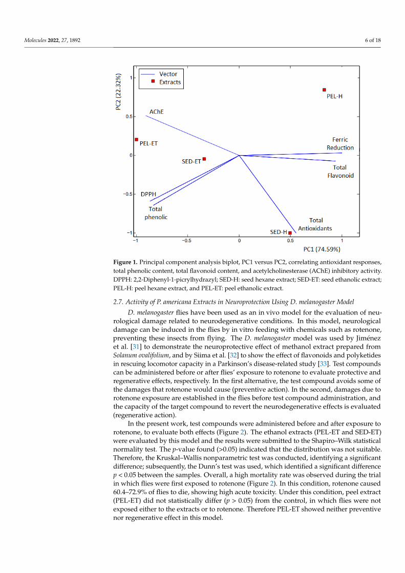
The hexane (SED-H and PEL-H) and ethanolic (SED-ET and PEL-ET) extracts were sub-mitted to acetylcholinesterase inhibition assay. All extracts inhibited acetylcholinesteraseactivity up to 65%, with the ethanolic extracts more active than the hexane ones (Table 4).This range of extract activities is promising when compared with the activity presented bythe control, a pure compound. The extract is a complex mixture and the active constituentsare usually minor constituents in the extracts. In this way, the inhibitory capacity of thepeel ethanolic extract from avocado (85.6 ± 11.1%) over acetylcholinesterase is outstanding,presenting no significant difference (p > 0.05) compared to the activity of the pure standard,eserine (91.5 ± 1.6%). Comparing the whole picture, the extract PEL-ET demonstrated adirect relationship between the amount of total phenolic compounds and AChE inhibition,corroborating the influence of antioxidant phenolic compounds in the acetylcholinesteraseactivity. By contrast, an inverse relationship was perceived for the hexane and ethanol ex-tracts of the seeds. The principal component analysis (PCA) score biplot allowed a generalqualitative visualization of the results obtained in the biological screening (Figure 1).
The PCA biplot shows the close relationship of the ethanolic extracts with the presenceof phenolic compounds, DPPH assay, and acetylcholinesterase inhibition. The peel ethano-lic extract (PEL-ET) demonstrated the closest association with the acetylcholinesteraseenzyme inhibition assay. Concerning the hexane extracts, the seed extract (SED-H) wasmainly related to the total antioxidant activity, while the peel extract (PEL-H) is associatedwith both ferric reduction power and total flavonoid contents. The hexane extracts are moredistant from the AChE assay and present lower activity for acetylcholinesterase inhibition,as observed in the study. Therefore, only the ethanolic extracts were submitted to thein vivo D. melanogaster assay to evaluate the neuroprotective response.
Molecules 2022, 27, 1892 6 of 18Molecules 2022, 27, x FOR PEER REVIEW 6 of 19
Figure 1. Principal component analysis biplot, PC1 versus PC2, correlating antioxidant responses, total phenolic content, total flavonoid content, and acetylcholinesterase (AChE) inhibitory activity. DPPH: 2,2-Diphenyl-1-picrylhydrazyl; SED-H: seed hexane extract; SED-ET: seed ethanolic extract; PEL-H: peel hexane extract, and PEL-ET: peel ethanolic extract.
The PCA biplot shows the close relationship of the ethanolic extracts with the presence of phenolic compounds, DPPH assay, and acetylcholinesterase inhibition. The peel ethanolic extract (PEL-ET) demonstrated the closest association with the acetylcholinesterase enzyme inhibition assay. Concerning the hexane extracts, the seed extract (SED-H) was mainly related to the total antioxidant activity, while the peel extract (PEL-H) is associated with both ferric reduction power and total flavonoid contents. The hexane extracts are more distant from the AChE assay and present lower activity for acetylcholinesterase inhibition, as observed in the study. Therefore, only the ethanolic extracts were submitted to the in vivo D. melanogaster assay to evaluate the neuroprotective response.
2.7. Activity of P. americana Extracts in Neuroprotection Using D. melanogaster Model D. melanogaster flies have been used as an in vivo model for the evaluation of
neurological damage related to neurodegenerative conditions. In this model, neurological damage can be induced in the flies by in vitro feeding with chemicals such as rotenone, preventing these insects from flying. The D. melanogaster model was used by Jiménez et al. [31] to demonstrate the neuroprotective effect of methanol extract prepared from Solanum ovalifolium, and by Siima et al. [32] to show the effect of flavonoids and polyketides in rescuing locomotor capacity in a Parkinson’s disease-related study [33]. Test compounds can be administered before or after flies’ exposure to rotenone to evaluate protective and regenerative effects, respectively. In the first alternative, the test compound avoids some of the damages that rotenone would cause (preventive action). In the second, damages due to rotenone exposure are established in the flies before test compound administration, and the capacity of the target compound to revert the neurodegenerative effects is evaluated (regenerative action).
Figure 1. Principal component analysis biplot, PC1 versus PC2, correlating antioxidant responses,total phenolic content, total flavonoid content, and acetylcholinesterase (AChE) inhibitory activity.DPPH: 2,2-Diphenyl-1-picrylhydrazyl; SED-H: seed hexane extract; SED-ET: seed ethanolic extract;PEL-H: peel hexane extract, and PEL-ET: peel ethanolic extract.
2.7. Activity of P. americana Extracts in Neuroprotection Using D. melanogaster Model
D. melanogaster flies have been used as an in vivo model for the evaluation of neu-rological damage related to neurodegenerative conditions. In this model, neurologicaldamage can be induced in the flies by in vitro feeding with chemicals such as rotenone,preventing these insects from flying. The D. melanogaster model was used by Jiménezet al. [31] to demonstrate the neuroprotective effect of methanol extract prepared fromSolanum ovalifolium, and by Siima et al. [32] to show the effect of flavonoids and polyketidesin rescuing locomotor capacity in a Parkinson’s disease-related study [33]. Test compoundscan be administered before or after flies’ exposure to rotenone to evaluate protective andregenerative effects, respectively. In the first alternative, the test compound avoids some ofthe damages that rotenone would cause (preventive action). In the second, damages due torotenone exposure are established in the flies before test compound administration, andthe capacity of the target compound to revert the neurodegenerative effects is evaluated(regenerative action).
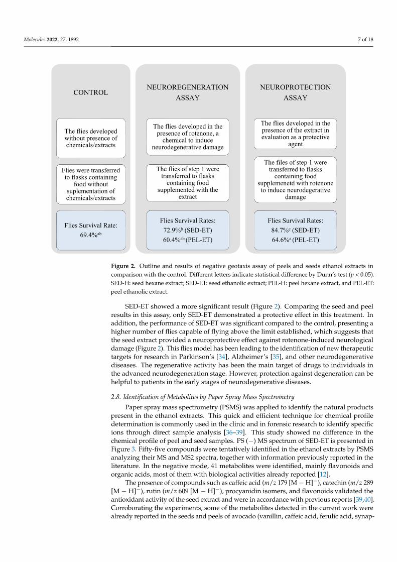
In the present work, test compounds were administered before and after exposure torotenone, to evaluate both effects (Figure 2). The ethanol extracts (PEL-ET and SED-ET)were evaluated by this model and the results were submitted to the Shapiro–Wilk statisticalnormality test. The p-value found (>0.05) indicated that the distribution was not suitable.Therefore, the Kruskal–Wallis nonparametric test was conducted, identifying a significantdifference; subsequently, the Dunn’s test was used, which identified a significant differencep < 0.05 between the samples. Overall, a high mortality rate was observed during the trialin which flies were first exposed to rotenone (Figure 2). In this condition, rotenone caused60.4–72.9% of flies to die, showing high acute toxicity. Under this condition, peel extract(PEL-ET) did not statistically differ (p > 0.05) from the control, in which flies were notexposed either to the extracts or to rotenone. Therefore PEL-ET showed neither preventivenor regenerative effect in this model.
Molecules 2022, 27, 1892 7 of 18
Molecules 2022, 27, x FOR PEER REVIEW 7 of 19
In the present work, test compounds were administered before and after exposure to rotenone, to evaluate both effects (Figure 2). The ethanol extracts (PEL-ET and SED-ET) were evaluated by this model and the results were submitted to the Shapiro–Wilk statistical normality test. The p-value found (>0.05) indicated that the distribution was not suitable. Therefore, the Kruskal–Wallis nonparametric test was conducted, identifying a significant difference; subsequently, the Dunn’s test was used, which identified a significant difference p < 0.05 between the samples. Overall, a high mortality rate was observed during the trial in which flies were first exposed to rotenone (Figure 2). In this condition, rotenone caused 60.4–72.9% of flies to die, showing high acute toxicity. Under this condition, peel extract (PEL-ET) did not statistically differ (p > 0.05) from the control, in which flies were not exposed either to the extracts or to rotenone. Therefore PEL-ET showed neither preventive nor regenerative effect in this model.
Figure 2. Outline and results of negative geotaxis assay of peels and seeds ethanol extracts in comparison with the control. Different letters indicate statistical difference by Dunn’s test (p < 0.05). SED-H: seed hexane extract; SED-ET: seed ethanolic extract; PEL-H: peel hexane extract, and PEL-ET: peel ethanolic extract.
SED-ET showed a more significant result (Figure 2). Comparing the seed and peel results in this assay, only SED-ET demonstrated a protective effect in this treatment. In addition, the performance of SED-ET was significant compared to the control, presenting a higher number of flies capable of flying above the limit established, which suggests that the seed extract provided a neuroprotective effect against rotenone-induced neurological damage (Figure 2). This flies model has been leading to the identification of new therapeutic targets for research in Parkinson’s [34], Alzheimer’s [35], and other neurodegenerative diseases. The regenerative activity has been the main target of drugs to individuals in the advanced neurodegeneration stage. However, protection against degeneration can be helpful to patients in the early stages of neurodegenerative diseases.
CONTROL
The flies developed without presence of chemicals/extracts
Flies were transferred to flasks containing
food without suplementation of chemicals/extracts
Flies Survival Rate:69.4%ab
NEUROREGENERATIONASSAY
The flies developed in the presence of rotenone, a
chemical to induce neurodegenerative damage
The flies of step 1 were transferred to flasks
containing food supplemented with the
extract
Flies Survival Rates:72.9%b (SED-ET)60.4%ab (PEL-ET)
NEUROPROTECTIONASSAY
The flies developed in the presence of the extract in evaluation as a protective
agent
The files of step 1 were transferred to flasks
containing food supplemenetd with rotenone to induce neurodegerative
damage
Flies Survival Rates:84.7%c (SED-ET)64.6%a (PEL-ET)
Figure 2. Outline and results of negative geotaxis assay of peels and seeds ethanol extracts incomparison with the control. Different letters indicate statistical difference by Dunn’s test (p < 0.05).SED-H: seed hexane extract; SED-ET: seed ethanolic extract; PEL-H: peel hexane extract, and PEL-ET:peel ethanolic extract.
SED-ET showed a more significant result (Figure 2). Comparing the seed and peelresults in this assay, only SED-ET demonstrated a protective effect in this treatment. Inaddition, the performance of SED-ET was significant compared to the control, presenting ahigher number of flies capable of flying above the limit established, which suggests thatthe seed extract provided a neuroprotective effect against rotenone-induced neurologicaldamage (Figure 2). This flies model has been leading to the identification of new therapeutictargets for research in Parkinson’s [34], Alzheimer’s [35], and other neurodegenerativediseases. The regenerative activity has been the main target of drugs to individuals inthe advanced neurodegeneration stage. However, protection against degeneration can behelpful to patients in the early stages of neurodegenerative diseases.
2.8. Identification of Metabolites by Paper Spray Mass Spectrometry
Paper spray mass spectrometry (PSMS) was applied to identify the natural productspresent in the ethanol extracts. This quick and efficient technique for chemical profiledetermination is commonly used in the clinic and in forensic research to identify specificions through direct sample analysis [36–39]. This study showed no difference in thechemical profile of peel and seed samples. PS (−) MS spectrum of SED-ET is presented inFigure 3. Fifty-five compounds were tentatively identified in the ethanol extracts by PSMSanalyzing their MS and MS2 spectra, together with information previously reported in theliterature. In the negative mode, 41 metabolites were identified, mainly flavonoids andorganic acids, most of them with biological activities already reported [12].
The presence of compounds such as caffeic acid (m/z 179 [M−H]−), catechin (m/z 289[M − H]−), rutin (m/z 609 [M − H]−), procyanidin isomers, and flavonoids validated theantioxidant activity of the seed extract and were in accordance with previous reports [39,40].Corroborating the experiments, some of the metabolites detected in the current work werealready reported in the seeds and peels of avocado (vanillin, caffeic acid, ferulic acid, synap-

Molecules 2022, 27, 1892 8 of 18
tic acid, kaempferol, catechin, quercetin-3-glucoside, quercetin-O-arabinosyl-glucoside,rutin, dimers A and B, and trimer A of procyanidin) [12]. Based on the antioxidant effect,Segovia et al. [41] described the role of avocado seed extract as an additive to protectoil mixtures from oxidation. The presence of quinic acid derivatives in the extract alsocontributed to validating the antioxidant activity and, therefore, highlighted the beneficialproperties of this residue as food ingredients for industrial use [42]. Table 5 presents thecompounds tentatively identified in the extracts in negative mode.
Molecules 2022, 27, x FOR PEER REVIEW 8 of 19
2.8. Identification of Metabolites by Paper Spray Mass Spectrometry Paper spray mass spectrometry (PSMS) was applied to identify the natural products
present in the ethanol extracts. This quick and efficient technique for chemical profile determination is commonly used in the clinic and in forensic research to identify specific ions through direct sample analysis [36–39]. This study showed no difference in the chemical profile of peel and seed samples. PS (−) MS spectrum of SED-ET is presented in Figure 3. Fifty-five compounds were tentatively identified in the ethanol extracts by PSMS analyzing their MS and MS2 spectra, together with information previously reported in the literature. In the negative mode, 41 metabolites were identified, mainly flavonoids and organic acids, most of them with biological activities already reported [12].
The presence of compounds such as caffeic acid (m/z 179 [M − H]−), catechin (m/z 289 [M − H]−), rutin (m/z 609 [M − H]−), procyanidin isomers, and flavonoids validated the antioxidant activity of the seed extract and were in accordance with previous reports [39,40]. Corroborating the experiments, some of the metabolites detected in the current work were already reported in the seeds and peels of avocado (vanillin, caffeic acid, ferulic acid, synaptic acid, kaempferol, catechin, quercetin-3-glucoside, quercetin-O-arabinosyl-glucoside, rutin, dimers A and B, and trimer A of procyanidin) [12]. Based on the antioxidant effect, Segovia et al. [41] described the role of avocado seed extract as an additive to protect oil mixtures from oxidation. The presence of quinic acid derivatives in the extract also contributed to validating the antioxidant activity and, therefore, highlighted the beneficial properties of this residue as food ingredients for industrial use [42]. Table 5 presents the compounds tentatively identified in the extracts in negative mode.
Figure 3. PS (−) MS full scan of ethanol extracts from seed of avocado.
Table 5. Compounds tentatively identified in avocado seed and peel by (−) PSMS.
m/z Compound Chemical Structure Class MS/MS Extract Reference
151 Vanillin C8H8O3 Aldehyde 136, 107, 93 S, P [43] 179 Caffeic acid C9H8O4 Phenolic acid 151, 135 S [43] 191 Quinic acid C7H12O6 Phenolic acid 93, 111 S, P [43] 197 Syringic acid C9H10O5 Phenolic acid 153, 141,125 S [43] 209 5-Hydroxyferulic acid C10H10O5 Phenolic acid 191, 165, 118 S [44] 223 Sinapic acid C11H12O5 Phenolic acid 179, 151,85 S [43] 269 Apigenin C15H10O5 Flavonoid 252, 223, 197 S, P [43] 285 Kaempferol C15H10O6 Flavonoid 255, 224, 213 S, P [43] 289 Catechin C15H13O6 Flavonoid 274, 245, 217, 199 S, P [44]
100 200 300 400 500 600 700 800 900 1000
m/z
0 10
20
30
40
50
60
70
80
90
100 311325
356
255 297
391 711265 681 199 639 611 405 539445 511 739
Rel
ativ
e Ab
unda
nce
Figure 3. PS (−) MS full scan of ethanol extracts from seed of avocado.
Table 5. Compounds tentatively identified in avocado seed and peel by (−) PSMS.
m/z Compound ChemicalStructure Class MS/MS Extract Reference
151 Vanillin C8H8O3 Aldehyde 136, 107, 93 S, P [43]
179 Caffeic acid C9H8O4 Phenolic acid 151, 135 S [43]
191 Quinic acid C7H12O6 Phenolic acid 93, 111 S, P [43]
197 Syringic acid C9H10O5 Phenolic acid 153, 141, 125 S [43]
209 5-Hydroxyferulic acid C10H10O5 Phenolic acid 191, 165, 118 S [44]
223 Sinapic acid C11H12O5 Phenolic acid 179, 151, 85 S [43]
269 Apigenin C15H10O5 Flavonoid 252, 223, 197 S, P [43]
285 Kaempferol C15H10O6 Flavonoid 255, 224, 213 S, P [43]
289 Catechin C15H13O6 Flavonoid 274, 245, 217, 199 S, P [44]
301 Quercetin C15H10O7 Flavonoid 272, 265, 123 S, P [43]
315 Hydroxytyrosol hexoside C14H20O8 Phenolic glycoside 297, 269, 243 S, P [44]
325 p-coumaroyl hexose C15H17O8 Phenolic glycoside 261, 197, 183, 170 S, P [44]
337 3-O-p-coumaroylquinic acid C16H18O8 Phenolic compound 293, 237, 183 S, P [44]
341 Caffeic acid-hexoside C15H18O9 Phenolic glycoside 280, 185, 183, 179 S, P [44]
353 5-O-caffeoylquinic acid C16H18O9 Phenolic compound 309, 211, 191, 183 S, P [43]
417 Kaempferol-O-pentoside C20H18O10 Flavonoid glycoside 404, 344, 289 S, P [44]
419 Cyanidin 3-O-pentatoside C20H19O10 Flavonoid glycoside 395, 361, 292, 287 S, P [44]
431 Vitexin C21H20O10 Flavonoid glycoside 362, 351, 311, 196 S [44]
433 Peonidin 3-O-pentoside C21H21O11 Flavonoid glycoside 420, 389, 301, 205 S, P [44]
435 Phloridzin C21H24O10 Flavonoid glycoside 426, 416, 369 S [43]
Molecules 2022, 27, 1892 9 of 18
Table 5. Cont.
m/z Compound ChemicalStructure Class MS/MS Extract Reference
447 Kaempferol-O-hexoside C21H19O11 Flavonoid glycoside 420, 403, 352, 301 S, P [44]
449 Dihydroquercetin-3,5-rhamnoside C21H22O11 Flavonoid glycoside 430, 303, 298, 286 S [44]
451 Cinchonain C24H20O9 Flavonoid 424, 414, 377 S, P [44]
461 Isorhamnetin-O-coumaroyl C22H22O11 Flavonoid 461, 417, 216 S, P [44]
463 Quercetin-3-hexoside C21H20O12 Flavonoid glycoside 464, 384, 316, 300 S [44]
473 Quercetin-3-O-hexoside C21H19O12 Flavonoid glycoside 467, 436, 372 S, P [45]
477 Quercetin glucuronide C21H18O13 Flavonoid 431, 262, 231 S [44]
487 Caffeoyl hexose-deoxyhexoside C22H31O12 Flavonoid 442, 298, 173 S [44]
491 Dimethyl ellagic acid hexoside C22H22O13 Flavonoid 343, 275, 269 S, P [44]
563 Apigenin-C-hexoside-C-pentoside C26H28O14 Flavonoid glycoside 531, 446, 298 S [44]
575 Procyanidin dimer A C30H24O12 Flavonoid 431, 404, 329 S [43]
577 Procyanidin dimer B C30H25O12 Flavonoid 532, 516, 420 S [44]
579 Luteolin7-O-(2′′-O-pentosyl)hexoside C26H28O15 Flavonoid glycoside 560, 542, 514 S, P [44]
593 Catechin dihexoside C27H29O15 Flavonoid glycoside 574, 495, 347 S [44]
595 Quercetin-O-pentatosyl-hexoside C26H28O16 Flavonoid glycoside 562, 558, 497 S, P [44]
609 Rutin C27H30O16 Flavonoid glycoside 573, 564, 208 S, P [44]
625 Quercetin-3,4′-O-diglucoside C27H30O17 Flavonoid glycoside 605, 588, 581 S, P [43]
863 Procyanidin trimer A C45H36O18 Flavonoid 845, 826, 555 S, P [43]
865 Procyanidin trimer B-isomer 1 C45H38O18 Flavonoid 829, 735, 560 S [43]
S: Avocado seed; P: Avocado peel.
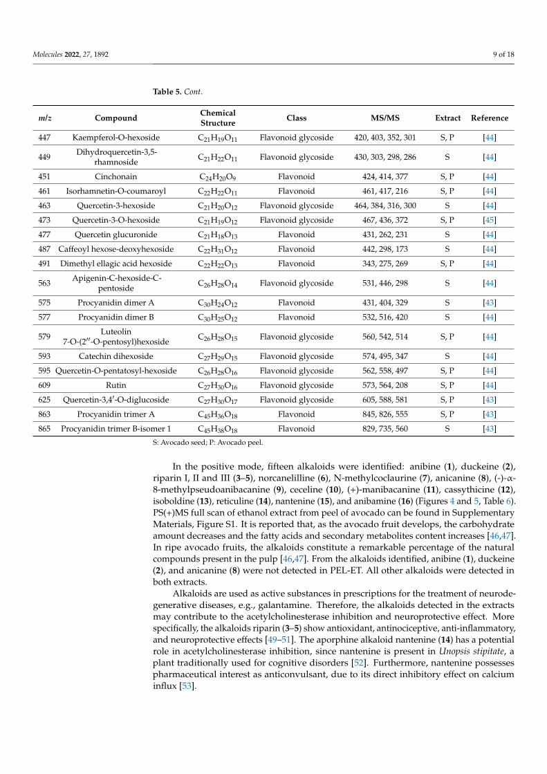
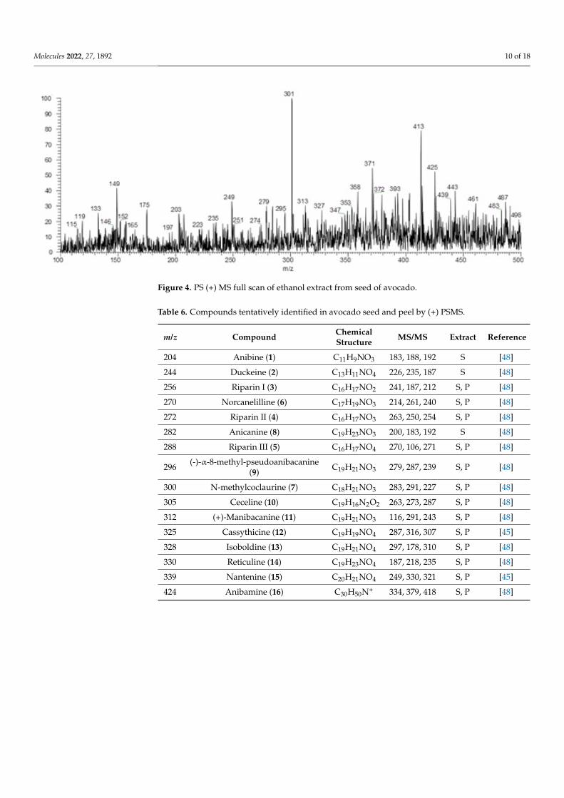
In the positive mode, fifteen alkaloids were identified: anibine (1), duckeine (2),riparin I, II and III (3–5), norcanelilline (6), N-methylcoclaurine (7), anicanine (8), (-)-α-8-methylpseudoanibacanine (9), ceceline (10), (+)-manibacanine (11), cassythicine (12),isoboldine (13), reticuline (14), nantenine (15), and anibamine (16) (Figures 4 and 5, Table 6).PS(+)MS full scan of ethanol extract from peel of avocado can be found in SupplementaryMaterials, Figure S1. It is reported that, as the avocado fruit develops, the carbohydrateamount decreases and the fatty acids and secondary metabolites content increases [46,47].In ripe avocado fruits, the alkaloids constitute a remarkable percentage of the naturalcompounds present in the pulp [46,47]. From the alkaloids identified, anibine (1), duckeine(2), and anicanine (8) were not detected in PEL-ET. All other alkaloids were detected inboth extracts.
Alkaloids are used as active substances in prescriptions for the treatment of neurode-generative diseases, e.g., galantamine. Therefore, the alkaloids detected in the extractsmay contribute to the acetylcholinesterase inhibition and neuroprotective effect. Morespecifically, the alkaloids riparin (3–5) show antioxidant, antinociceptive, anti-inflammatory,and neuroprotective effects [49–51]. The aporphine alkaloid nantenine (14) has a potentialrole in acetylcholinesterase inhibition, since nantenine is present in Unopsis stipitate, aplant traditionally used for cognitive disorders [52]. Furthermore, nantenine possessespharmaceutical interest as anticonvulsant, due to its direct inhibitory effect on calciuminflux [53].
Molecules 2022, 27, 1892 10 of 18Molecules 2022, 27, x FOR PEER REVIEW 10 of 19
Figure 4. PS (+) MS full scan of ethanol extract from seed of avocado.
Table 6. Compounds tentatively identified in avocado seed and peel by (+) PSMS.
m/z Compound Chemical Structure
MS/MS Extract Reference
204 Anibine (1) C11H9NO3 183, 188, 192 S [48] 244 Duckeine (2) C13H11NO4 226, 235, 187 S [48] 256 Riparin I (3) C16H17NO2 241, 187, 212 S, P [48] 270 Norcanelilline (6) C17H19NO3 214, 261, 240 S, P [48] 272 Riparin II (4) C16H17NO3 263, 250, 254 S, P [48] 282 Anicanine (8) C19H23NO3 200, 183, 192 S [48] 288 Riparin III (5) C16H17NO4 270, 106, 271 S, P [48]
296 (-)-α-8-methyl-
pseudoanibacanine (9)
C19H21NO3 279, 287, 239 S, P [48]
300 N-methylcoclaurine
(7) C18H21NO3 283, 291, 227 S, P [48]
305 Ceceline (10) C19H16N2O2 263, 273, 287 S, P [48] 312 (+)-Manibacanine (11) C19H21NO3 116, 291, 243 S, P [48] 325 Cassythicine (12) C19H19NO4 287, 316, 307 S, P [45] 328 Isoboldine (13) C19H21NO4 297, 178, 310 S, P [48] 330 Reticuline (14) C19H23NO4 187, 218, 235 S, P [48] 339 Nantenine (15) C20H21NO4 249, 330, 321 S, P [45] 424 Anibamine (16) C30H50N+ 334, 379, 418 S, P [48]
Alkaloids are used as active substances in prescriptions for the treatment of neurodegenerative diseases, e.g., galantamine. Therefore, the alkaloids detected in the extracts may contribute to the acetylcholinesterase inhibition and neuroprotective effect. More specifically, the alkaloids riparin (3–5) show antioxidant, antinociceptive, anti-inflammatory, and neuroprotective effects [49–51]. The aporphine alkaloid nantenine (14) has a potential role in acetylcholinesterase inhibition, since nantenine is present in Unopsis stipitate, a plant traditionally used for cognitive disorders [52]. Furthermore, nantenine possesses pharmaceutical interest as anticonvulsant, due to its direct inhibitory effect on calcium influx [53].
Figure 4. PS (+) MS full scan of ethanol extract from seed of avocado.
Table 6. Compounds tentatively identified in avocado seed and peel by (+) PSMS.
m/z Compound ChemicalStructure MS/MS Extract Reference
204 Anibine (1) C11H9NO3 183, 188, 192 S [48]
244 Duckeine (2) C13H11NO4 226, 235, 187 S [48]
256 Riparin I (3) C16H17NO2 241, 187, 212 S, P [48]
270 Norcanelilline (6) C17H19NO3 214, 261, 240 S, P [48]
272 Riparin II (4) C16H17NO3 263, 250, 254 S, P [48]
282 Anicanine (8) C19H23NO3 200, 183, 192 S [48]
288 Riparin III (5) C16H17NO4 270, 106, 271 S, P [48]
296 (-)-α-8-methyl-pseudoanibacanine(9) C19H21NO3 279, 287, 239 S, P [48]
300 N-methylcoclaurine (7) C18H21NO3 283, 291, 227 S, P [48]
305 Ceceline (10) C19H16N2O2 263, 273, 287 S, P [48]
312 (+)-Manibacanine (11) C19H21NO3 116, 291, 243 S, P [48]
325 Cassythicine (12) C19H19NO4 287, 316, 307 S, P [45]
328 Isoboldine (13) C19H21NO4 297, 178, 310 S, P [48]
330 Reticuline (14) C19H23NO4 187, 218, 235 S, P [48]
339 Nantenine (15) C20H21NO4 249, 330, 321 S, P [45]
424 Anibamine (16) C30H50N+ 334, 379, 418 S, P [48]

Molecules 2022, 27, 1892 11 of 18Molecules 2022, 27, x FOR PEER REVIEW 11 of 19
Figure 5. Chemical structures, m/z and chemical formulas of alkaloids putatively identified by PSMS in seed and peel of avocado.
N
O
OCH3
O
N
O
HO
OH
OCH3
NHCH3O
O
R1
R2
Anibine (1)
m/z 204 (C11H9NO3)
Duckeine (2)
m/z 244 (C13H11NO4)
R1 = H; R2 = H: Riparin I (3), m/z 256 (C16H17NO2)
R1 = OH; R2 = H: Riparin II (4), m/z 272 (C16H17NO3)
R1= R2 = OH: Riparin III (5), m/z 288 (C16H17NO4)
N
OH
CH3O
HO R
N
OH
H
OH
OCH3
R
N
N
HOH
CH3O
R = H: (-)-Norcanelilline (6)
m/z 270 (C17H19NO3)
R = CH3: N-Methylcoclaurine (7)
m/z 300 (C18H21NO3)
R = H: Anicanine (8), m/z 282
(C19H23NO3)
R = CH3: (-)-Α-8-
methylpseudoanibacanine (9), m/z 296
(C19H21NO3)
Ceceline (10)
m/z 305 (C19H16N2O2)
N
CH3O
CH3O
OH
N O
O
H
OH
OCH3
CH3
NH
CH3O
HO
HO
OCH3
CH3
(+)-Manibacanine (11)
m/z 312 (C19H21NO3)
Cassythicine (12)
m/z 325 (C19H19NO4)
Isoboldine (13)
m/z 328 (C19H21NO4)
N
CH3O
HO CH3
OCH3
OH
N
OO
HCH3O
CH3O
CH3
N+
H
CH3(CH2)7
H
H (CH2)7CH3
H
Reticuline (14)
m/z 330 (C19H23NO4)
Nantenine (15)
m/z 339 (C20H21NO4)
Anibamine (16)
m/z 424 (C30H50N+)
Figure 5. Chemical structures, m/z and chemical formulas of alkaloids putatively identified by PSMSin seed and peel of avocado.

Molecules 2022, 27, 1892 12 of 18
3. Materials and Methods3.1. Materials and Reagents
Ripe avocado (cultivar Persea americana var. Fortuna) fruits were purchased in Ibiritécity (MG, Brazil) in the winter of 2018. P.A. grade hexane (Nox Lab Solutions, Mauá, SP,Brazil) and hydrated ethanol 96% (Emfal, Betim, MG, Brazil) were used. Ascorbic acid(Neon, Suzano, SP, Brazil), gallic acid (Neon, Suzano, SP, Brazil), quercetin (Sigma-Aldrich,St. Louis, MO, USA), and eserine (Sigma-Aldrich, St. Louis, MO, USA) were used aspositive controls in the biological assays. Acetylcholinesterase iodide, acid 5′,5′-dithio-bis-(2-nitrobenzoate), bovine serum albumin, and AChE (Sigma-Aldrich, St. Louis, MO, USA)were used in the AChE inhibition assay. For cultivation of Drosophila melanogaster flies,bacteriological agar (Vetec, Duque de Caxias, SP, Brazil), powdered dry yeast (Fleischmann,Sorocaba, SP, Brazil), and nystatin (oral suspension from Germed, Campinas, SP, Brazil)were used. Rotenone (Sigma-Aldrich, At. Louis, MO, USA) was employed in the negativegeotaxis assay to induce toxicity. The experiments were carried out in quintuplicate unlessotherwise specified.
3.2. Biomass Processing and Extract Preparation
The peels (53.233 g) and seeds (103.431 g) of avocado fruits were separately removedfrom the pulp, sanitized under running water, and dried separately with paper towelsheets. The peels were sliced, the seeds were grated, and 3 g of each was utilized for atomicabsorption spectroscopy analysis. The remaining biomasses (sliced peels and grated seeds)were separately soaked in hexane (600 mL). After 24 h, the hexane fraction was separatedby filtration and another aliquot of hexane (600 mL) was added to the vegetal material.The procedure was repeated three times. The hexane fractions were combined; the solventwas removed in a rotary evaporator (60 ◦C), obtaining the respective hexane extracts ofpeels (PEL-H) and seeds (SED-H). After hexane extraction, the defatted plant material wasfurther extracted with 96% ethanol, following the same procedure previously described, tofurnish the peel (PEL-ET) and seed (SED -ET) ethanolic extracts (Figure 5) [54].
3.3. Mineral Element Analysis by Atomic Absorption
The separated aliquots of fresh peels and seeds (3 g each) were placed in a porcelaincrucible and heated until complete carbonization. Subsequently, the crucibles with thecarbonized plant material were placed in a muffle at 550 ◦C, obtaining the organic matter-free ashes. Nitric acid (2 mL) was added to an aliquot of the ashes (0.05 mg) and the volumewas completed with distilled water to 5 mL. The resulting suspension was analyzed usingAtomic Absorption Spectrometer AA 240 FS (Varian, Australia) to quantify the mineralsCa, Cu, Fe, Mg, Mn, and Zn [4].
3.4. Fatty Acid Gas Chromatography Analysis
Aliquots (10 mg) of the extracts were derivatized to fatty acid methyl esters (FAMEs) [55],solubilized in methanol, and injected into an HP5890 Gas Chromatograph. A Supelcowax-10 analytical column (30 m × 0.2 mm × 0.2 µm) (Supelco) was used for analysis under thefollowing chromatographic conditions (split 1/50): 150 ◦C, 0 min, 10 ◦C /min up to 240 ◦Cand detector at 250 ◦C. Hydrogen was used as carrier gas (4 mL/min) and the injectionvolume was 1 µL. A standard of fatty acid methyl esters (FAME C14-C22) were injected,and their retention times were used to comparatively identify the fatty acids present in theavocado residues.
3.5. Total Content of Phenol and Flavonoids Assay
The determination of total phenolic content was performed spectrophotometricallywith a Folin–Ciocalteau reagent method previously described [56]. Briefly, the ethanolicextract solutions (0.5 mL) were mixed with Folin–Ciocalteau reagent (2.5 mL). The solutionswere incubated for 3 min at room temperature, and 0.3 mL of saturated sodium carbonatesolution was added to each one. After a further incubation period (20 min at 25 ◦C),
Molecules 2022, 27, 1892 13 of 18
absorbance was read at 760 nm using a Biospectro SP-22 Spectrophotometer. The sameprocedure was carried out for gallic acid (standard), which was used for constructing thecalibration curve (10–100 µg/mL; R2 = 0.9291). In the equation of the calibration curveestablished using gallic acid, “y” was replaced by the absorbance and divided by the massof the extract/standard. The results were expressed in mg gallic acid/g extract.
The total flavonoid contents were determined by aluminum chloride spectrophoto-metric method as described by Yang et al. [27]. Briefly, 2.5 mL of the ethanolic extractor standard solutions were mixed with 0.3 mL sodium nitrate solution (5% w/v) withsubsequent addition of aluminum chloride solution (10% w/v) (3.0 mL). The mixture wasvortexed, and 2.0 mL of sodium hydroxide solution (1 M) was added and well vortexed.After 10 min, the absorbances were determined at 510 nm using a Biospectro SP-22 Spec-trophotometer. Quercetin standard solutions (350–600 µg/mL) were used for constructingthe calibration curve (R2 = 0.9887). The result was expressed as mg of quercetin equivalentper g of extract.
3.6. Antioxidant Activity
Ethanol and hexane extracts (SED-ET, PEL-ET, SED-H, and PEL-H) and the standards(ascorbic acid and gallic acid) were dissolved in ethanol (500 µg/mL), except quercetin(1000 µg/mL). All assays were performed in quintuplicate. Microsoft Excel 2013 softwarewas used for data analysis.
3.6.1. Total Antioxidant Capacity
A reagent solution (100 mL) was prepared mixing sulfuric acid (3.22 mL), bibasicphosphate (0.39 g), and ammonium molybdate (0.49 g). An aliquot (0.3 mL) of ethanolicextract solutions (500 µg/mL) was mixed with 3.0 mL of the reagent solution. The resultingtest samples were incubated in an oven at 95 ◦C for 90 min. After the samples hadcooled to room temperature, the absorbances were read at 695 nm on a Biospectro SP22 Spectrophotometer. The same procedure was carried out with ascorbic acid (positivecontrol) and a negative control was carried out with the solvent. The results were expressedin mmol of ascorbic acid per gram of extract (adapted from [57]).
3.6.2. Ferric Reducing Power Assay
An aliquot (1.0 mL) of the ethanolic extract solutions was mixed with 2.5 mL ofphosphate buffer (pH 6.6, 0.2 M) and 2.5 mL of potassium ferricyanide aqueous solution(1% w/v). The mixture was homogenized and incubated in a laboratory water bath at50 ◦C for 20 min. Subsequently, 2.5 mL of trichloroacetic acid solution (10% w/v) wasadded and the mixture was centrifuged (300 rpm, 10 min). An aliquot (2.5 mL) of thesuperior layer was mixed with 2.5 mL distilled water, and 0.5 mL ferric chloride solution(0.1% w/v) was added. The same procedure was conducted with the positive control. Theabsorbances were read at 700 nm on a Biospectro SP-22 Spectrofotometer. The results wereconverted into a percentage of ferric reducing power in relation to the positive controlascorbic acid [57].
3.6.3. Free Radical Capture Assay (DPPH)
Solutions of extracts (100 µL) were added to 96-well plates in quadruplicate, followedby the addition of 2,2-Diphenyl-1-picrylhydrazyl (DPPH) solution (175 µL) prepared inethanol (0.1 mM). The materials were incubated for 30 min in the dark, after which readingwas performed at 490 nm in a microplate reader. Solutions of known concentration ofascorbic acid, the positive control, were used to construct a graph of percent inhibitionversus concentration (µg/mL) [58].
3.7. Acetylcholinesterase Inhibition Assay
The extracts were solubilized in dimethylsulfoxide (DMSO) (10 mg/mL) and analiquot of each one (25 µL) was mixed with aqueous solutions of acetylcholine iodide
Molecules 2022, 27, 1892 14 of 18
(15 mM) (25 µL) and 125 µL of a solution containing 5,5′-dithio-bis (2-nitrobenzoic acid)(3 mM), NaCl solution (0.1 M) and MgCl2·6H2O (20 mM) prepared in Tris/HCl buffer(50 mM, pH 8.0, 50 µL) in wells of 96-well plates. The first set of wavelength readings wasthen performed at 405 nm every 1 min (8 readings). Subsequently, a solution of acetyl-cholinesterase was prepared (0.22 U/mL) in buffer and 0.1% (w/v) of bovine serum albuminand added to the wells (25 µL each). Eserine (10 mg/mL) was used as positive control.Next, new readings were performed at the same wavelength every 1 min (10 readings) [20].The following equation was used to calculate the percentage of inhibition (%I), where Ab:Negative control (DMSO) and Aa: Sample absorbance.
% I =(Ab− Aa)× 100
Ab
3.8. In Vivo Neuroprotection Activity Using Drosophila melanogaster Model
A sterile mixture containing bananas (400 g), bacteriological agar (4 g), commercialyeast (3.5 g), nystatin (2.5 mL), and propionic acid (0.6 mL) in distilled water (350 mL) wasprepared and used to grow D. melanogaster flies for the whole experiment. The cultivationswere performed in glass vessels in triplicate. Twelve 6–8-day-old male flies were grown invessels containing (A) rotenone (neurotoxic inductor, 10 mg/mL in ethanol; 0.4 mL) or (B)extracts (5 mg/mL in water; 0.6 mL). Rotenone and extracts were mixed with the nutritivesubstrate. A control flask (C) was utilized. Neither rotenone nor extract were added to thecontrol flasks. After 7 days, the flies that were in vessels A and B were anesthetized withether and interchanged. After 15 days the flies were transferred to falcon tubes that werehorizontally placed on a support. A photographic camera was positioned approximately30 cm away. Then, the support was subjected to three hits against the bench surface, andten photographs of the tubes containing the flies were recorded. Picture number 6 wasused to account for the number of flies that were able to fly above half the tube height afterhitting [31].
3.9. Paper Spray Mass Spectrometry
Pulps were analyzed using a Thermo Fisher LCQ FLEET ion-trap mass spectrometer(Thermo Scientific, San Jose, CA, USA) equipped with an ambient paper spray ionizationsource. Analyses were performed in triplicate in positive and negative ionization modes.For PSMS analyses, a 2 µL sample volume and 40 µL of methanol were added to a triangularchromatographic paper (equilateral, 1.5 cm lengthwise) positioned 10 mm away from themass spectrometer inlet [37–39]. A high voltage was then applied to the paper held by acopper clamp and attached to a three-dimensional moving platform for data acquisition.Instrumental conditions of operation were: PSMS source voltage equal to +4 kV (positivemode) and −3 kV (negative mode); capillary voltage of 40 V; transfer tube temperatureof 275 ◦C; tube lens voltage of 120 V; mass scanning range of 100 to 1000 m/z in positiveand negative modes. Ions were fragmented using collision energies from 15 to 45 eV.Some representative MS can be found in Figures S2–S48. Tentative identification of thecompounds was carried out using a comparison of the m/z ratios of the data obtained fromliterature associated with instrumental readings and subsequent fragmentation employingsequential mass spectrometry.
3.10. Statistical Analysis
GraphPad Prism 5.0 software was used for statistical analysis, applying varianceanalysis (ANOVA) [20]. To evaluate the differences between the means, the Tukey testwith significance level of 5% was used. Kruskal–Wallis (nonparametric) and Dunn’s post-test, with significance of 5%, were utilized to evaluate data from neuroprotection assay.SensoMaker 1.61 software was used for main components analyses.

Molecules 2022, 27, 1892 15 of 18
4. Conclusions
The avocado residues produced during industrial processing retain nutrients andbioactive compounds independent of the extraction solvent. Concerning the minerals,avocado peels and seeds displayed significant levels of Ca, Mg, Mn, and Zn. The seedspresented high essential fatty acids content, such as linoleic, palmitic, and oleic acids. Seedand peel ethanolic extracts showed phenolic compounds correlated to their antioxidantand AChE inhibitory activities. Additionally, the seed ethanolic extract exhibited in vivoneuroprotective effect. In vivo studies are an important step to determine the real potentialof phytochemicals since they overcome many problems inherent to in vitro models. There-fore, the positive in vivo results encourage research of this extract as a potential candidateto develop drugs to treat patients in the early stages of neurodegenerative illnesses suchas Alzheimer’s and Parkinson’s diseases. The PSMS chemical profile of the residues ledto the identification of fifty-five metabolites, including phenolic, hydroxycinnamic acids,flavonoids, and alkaloids, most of them already reported as pharmacologically active com-pounds. Quantification of the phytochemicals will be the object of future work. In addition,toxicity, bioavailability, and suitable formulations should be further investigated beforeusing avocado residues in pharmacotherapy, but the current results pave the way to deeperstudies on the in vivo potential of avocado residues as a bioresource in the development oflow-cost drugs and functional foods with neuroprotective effects.
Supplementary Materials: The following supporting information can be downloaded at: https://www.mdpi.com/article/10.3390/molecules27061892/s1, Section S1: PSMS of the seed and ethano-lic extracts and of the compounds identified, Figure S1: PS(+)MS full scan of ethanol extract frompeel of avocado, Figure S2: Anibine PS(+)MS fragmentation and chemical structure, Figure S3: Duck-eine PS(+)MS fragmentation and chemical structure, Figure S4: Riparin I PS(+)MS fragmentationand chemical structure, Figure S5: Norcanelilline PS(+)MS fragmentation and chemical structure,Figure S6: Riparin II PS(+)MS fragmentation and chemical structure, Figure S7: Anicanine PS(+)MSfragmentation and chemical structure, Figure S8: Riparin III PS(+)MS fragmentation and chemicalstructure, Figure S9: (-)-α-methylpseudoanibacanine PS(+)MS fragmentation and chemical struc-ture, Figure S10: N- methylcoclaurine PS(+)MS fragmentation and chemical structure, Figure S11:Ceceline PS(+)MS fragmentation and chemical structure, Figure S12: (+)-manibacanine PS(+)MSfragmentation and chemical structure, Figure S13: Cassythicine PS(+)MS fragmentation and chemicalstructure, Figure S14: Isoboldine PS(+)MS fragmentation and chemical structure, Figure S15: Reticu-line PS(+)MS fragmentation and chemical structure, Figure S16: Anibamine PS(+)MS fragmentationand chemical structure, Figure S17: PS(−)MS full scan of peel ethanolic extract of avocado, Figure S18:Vanillin PS(−)MS fragmentation and chemical structure, Figure S19: Caffeic acid PS(−)MS fragmenta-tion and chemical structure, Figure S20: Quinic acid PS(−)MS fragmentation and chemical structure,Figure S21: Syrinc acid PS(−)MS fragmentation and chemical structure, Figure S22: 5-hydroxyferulicacid PS(−)MS fragmentation and chemical structure, Figure S23: Sinapic acid PS(−)MS fragmen-tation and chemical structure, Figure S24: Apigenin PS(−)MS fragmentation and chemical struc-ture, Figure S25: Kaempferol PS(−)MS fragmentation and chemical structure, Figure S26: CatechinPS(−)MS fragmentation and chemical structure, Figure S27: Quercetin PS(−)MS fragmentation andchemical structure, Figure S28: Hydroxytyrosol glucoside PS(−)MS fragmentation and chemical struc-ture, Figure S29: p-coumaroyl hexose PS(−)MS fragmentation and chemical structure, Figure S30:3-O-p-coumaroylquinic acid PS(−)MS fragmentation and chemical structure, Figure S31: Caffeicacid hexoside PS(−)MS fragmentation and chemical structure, Figure S32: 5-O-caffeoylquinic acidPS(−)MS fragmentation and chemical structure, Figure S33: Kaempferol-O-pentoside PS(−)MSfragmentation and chemical structure, Figure S34: Cyanidin-3-O-arabinoside PS(−)MS fragmenta-tion and chemical structure, Figure S35: Vitexin PS(−)MS fragmentation and chemical structure,Figure S36: Peonidin-3-O-pentoside PS(−)MS fragmentation and chemical structure, Figure S37:Phloridzin PS(−)MS fragmentation and chemical structure, Figure S38: Kaempferol-O-hexosidePS(−)MS fragmentation and chemical structure, Figure S39: Dihydroquercetin-3,5-rhamnosidePS(−)MS fragmentation and chemical structure, Figure S40: Isorhamnetin-O-coumaroyl PS(−)MSfragmentation and chemical structure, Figure S41: Quercetin-3-glucoside PS(−)MS fragmentationand chemical structure, Figure S42: Quercetin glucuronide PS(−)MS fragmentation and chemi-cal structure, Figure S43: Caffeoyl hexose-deohyhexoside PS(−)MS fragmentation and chemical
Molecules 2022, 27, 1892 16 of 18
structure, Figure S44: Dimethyl ellagic acid hexoside PS(−)MS fragmentation and chemical struc-ture, Figure S45: Apigenin-C-hexoside-C-pentoside PS(−)MS fragmentation and chemical structure,Figure S46: Luteolin-7-O-(2′′-O-pentosyl)-hexoside PS(−)MS fragmentation and chemical structure,Figure S47: Catechin diglucopyranoside PS(−)MS fragmentation and chemical structure, Figure S48:Quercetin-3,4′-O-diglucoside PS(−)MS fragmentation and chemical structure.
Author Contributions: Conceptual idea, methodology design, writing, and editing: J.A.T.; Datacollection, antioxidant assays, data analysis and interpretation, writing, and editing: G.G.d.S. andL.P.S.P.; Paper Spray analysis and data interpretation: J.O.F.M., H.d.O.P.M. and R.A. All authors haveread and agreed to the published version of the manuscript.
Funding: This research was funded by CAPES (Code 001), Fundação de Amparo à Pesquisa doEstado de Minas Gerais (FAPEMIG PPM-00255-18), and Conselho Nacional de DesenvolvimentoCientífico e Tecnológico (CNPq Grant 304922/2018-8).
Institutional Review Board Statement: Not applicable.
Informed Consent Statement: Not applicable.
Acknowledgments: We acknowledge Lucas Xavier da Silva for drawing the graphical abstracts.
Conflicts of Interest: The authors declare no conflict of interest.
Sample Availability: Samples of the compounds are not available.
References1. Ochoa-Zarzosa, A.; Báez-Magaña, M.; Guzmán-Rodríguez, J.J.; Flores-Alvarez, L.J.; Lara-Márquez, M.; Zavala-Guerrero, B.;
Salgado-Garciglia, R.; López-Gómez, R.; López-Meza, J.E. Bioactive Molecules From Native Mexican Avocado Fruit (Perseaamericana var. drymifolia): A Review. Plant Foods Hum. Nutr. 2021, 76, 133–142. [CrossRef] [PubMed]
2. Tesfaye, T.; Ayele, M.; Gibril, M.; Ferede, E.; Limeneh, D.Y.; Kong, F. Beneficiation of avocado processing industry by-product: Areview on future prospect. Curr. Res. Green Sustain. Chem. 2022, 5, 100253. [CrossRef]
3. Alkhalaf, M.I.; Alansari, W.S.; Ibrahim, E.A.; ELhalwagy, M.E.A. Anti-oxidant, anti-inflammatory and anti-cancer activities ofavocado (Persea americana) fruit and seed extract. J. King Saud Univ. Sci. 2019, 31, 1358–1362. [CrossRef]
4. Borges, C.V.; Maraschin, M.; Coelho, D.S.; Leonel, M.; Gomez, H.A.G.; Belin, M.A.F.; Diamante, M.S.; Amorim, E.P.; Gianeti, T.;Castro, G.R.; et al. Nutritional value and antioxidant compounds during the ripening and after domestic cooking of bananas andplantains. Food Res. Int. 2020, 132, 109061. [CrossRef] [PubMed]
5. Araujo, R.G.; Rodríguez-Jasso, R.M.; Ruíz, H.A.; Govea-Salas, M.; Pintado, M.; Aguilar, C.N. Recovery of bioactive componentsfrom avocado peels using microwave-assisted extraction. Food Bioprod. Process. 2021, 127, 152–161. [CrossRef]
6. Baysal, S.S.; Ülkü, M.A. Food Loss and Waste: A Sustainable Supply Chain Perspective. ICGI-Global: Istanbul, Turkey, 2021;ISBN 9789251317549.
7. Yap, K.M.; Sekar, M.; Seow, L.J.; Gan, S.H.; Bonam, S.R.; Mat Rani, N.N.I.; Lum, P.T.; Subramaniyan, V.; Wu, Y.S.; Fuloria,N.K.; et al. Mangifera indica (Mango): A promising medicinal plant for breast cancer therapy and understanding its potentialmechanisms of action. Breast Cancer Targets Ther. 2021, 13, 471–503. [CrossRef]
8. Béné, C.; Oosterveer, P.; Lamotte, L.; Brouwer, I.D.; de Haan, S.; Prager, S.D.; Talsma, E.F.; Khoury, C.K. When food systems meetsustainability—Current narratives and implications for actions. World Dev. 2019, 113, 116–130. [CrossRef]
9. González-Chang, M.; Wratten, S.D.; Shields, M.W.; Costanza, R.; Dainese, M.; Gurr, G.M.; Johnson, J.; Karp, D.S.; Ketelaar, J.W.;Nboyine, J.; et al. Understanding the pathways from biodiversity to agro-ecological outcomes: A new, interactive approach. Agric.Ecosyst. Environ. 2020, 301, 107053. [CrossRef]
10. Tumwesigye, K.S.; O’Brien, E.; Oliveira, J.C.; Crean, A.; Sousa-Gallagher, M.J. Engineered food supplement excipients from bittercassava for minimisation of cassava processing waste in environment. Future Foods 2020, 1–2, 100003. [CrossRef]
11. Ortega-Arellano, H.F.; Jimenez-Del-Rio, M.; Velez-Pardo, C. Neuroprotective Effects of Methanolic Extract of Avocado Perseaamericana (var. Colinred) Peel on Paraquat-Induced Locomotor Impairment, Lipid Peroxidation and Shortage of Life Span inTransgenic knockdown Parkin Drosophila melanogaster. Neurochem. Res. 2019, 44, 1986–1998. [CrossRef]
12. Salazar-López, N.J.; Domínguez-Avila, J.A.; Yahia, E.M.; Belmonte-Herrera, B.H.; Wall-Medrano, A.; Montalvo-González, E.;González-Aguilar, G.A. Avocado fruit and by-products as potential sources of bioactive compounds. Food Res. Int. 2020, 138,109774. [CrossRef] [PubMed]
13. Del Castillo-Llamosas, A.; del Río, P.G.; Pérez-Pérez, A.; Yáñez, R.; Garrote, G.; Gullón, B. Recent advances to recover value-addedcompounds from avocado by-products following a biorefinery approach. Curr. Opin. Green Sustain. Chem. 2021, 28, 100433.[CrossRef]
14. Fuloria, S.; Yusri, M.A.A.; Sekar, M.; Gan, S.H.; Rani, N.N.I.M.; Lum, P.T.; Ravi, S.; Subramaniyan, V.; Azad, A.K.; Jeyabalan, S.;et al. Genistein: A Potential Natural Lead Molecule for New Drug Design and Development for Treating Memory Impairment.Molecules 2022, 27, 265. [CrossRef] [PubMed]

Molecules 2022, 27, 1892 17 of 18
15. World Health Organization (WHO). Global Action Plan on the Public Health Response to Dementia 2017–2025; World HealthOrganization: Geneva, Switzerland, 2017; p. 27.
16. Bernstein, J.J.J.; Holt, G.B.; Bernstein, J. Price dispersion of generic medications. PLoS ONE 2019, 14, e0225280. [CrossRef][PubMed]
17. FAO; IFAD; UNICEF; WHO. The State of Food Security and Nutrition in the World 2020; FAO: Rome, Italy, 2020; ISBN 9789251329016.[CrossRef]
18. Ferguson, E.L.; Watson, L.; Berger, J.; Chea, M.; Chittchang, U.; Fahmida, U.; Khov, K.; Kounnavong, S.; Le, B.M.; Rojroong-wasinkul, N.; et al. Realistic Food-Based Approaches Alone May Not Ensure Dietary Adequacy for Women and Young Childrenin South-East Asia. Matern. Child Health J. 2019, 23, 55–66. [CrossRef]
19. Adams, J.B.; Sorenson, J.C.; Pollard, E.L.; Kirby, J.K.; Audhya, T. Evidence-based recommendations for an optimal prenatalsupplement for women in the U.S., part two: Minerals. Nutrients 2021, 13, 1849. [CrossRef] [PubMed]
20. Lourenção, L.F.d.P.; de Paula, N.C.; Cardoso, M.A.; Santos, P.R.; de Oliveira, I.R.C.; Fonseca, F.L.A.; da Veiga, G.L.; Alves, B.d.C.A.;Graciano, M.M.d.C.; Pereira-Dourado, S.M. Biochemical markers and anthropometric profile of children enrolled in publicdaycare centers. J. Pediatr. 2021, in press. [CrossRef]
21. Guirlanda, C.P.; da Silva, G.G.; Takahashi, J.A. Cocoa honey: Agro-industrial waste or underutilized cocoa by-product? FutureFoods 2021, 4, 100061. [CrossRef]
22. Mousa, M.M.H.; El-Magd, M.A.; Ghamry, H.I.; Alshahrani, M.Y.; El-Wakeil, N.H.M.; Hammad, E.M.; Asker, G.A.H. Pea peels as avalue-added food ingredient for snack crackers and dry soup. Sci. Rep. 2021, 11, 22747. [CrossRef]
23. Marangoni, F.; Agostoni, C.; Borghi, C.; Catapano, A.L.; Cena, H.; Ghiselli, A.; La Vecchia, C.; Lercker, G.; Manzato, E.; Pirillo,A.; et al. Dietary linoleic acid and human health: Focus on cardiovascular and cardiometabolic effects. Atherosclerosis 2020, 292,90–98. [CrossRef]
24. Park, W.J. The Biochemistry and Regulation of Fatty Acid Desaturases in Animals; Elsevier Inc.: Amsterdam, The Netherlands, 2018;ISBN 9780128112304.
25. Orsavova, J.; Misurcova, L.; Vavra Ambrozova, J.; Vicha, R.; Mlcek, J. Fatty acids composition of vegetable oils and its contributionto dietary energy intake and dependence of cardiovascular mortality on dietary intake of fatty acids. Int. J. Mol. Sci. 2015, 16, 2871.[CrossRef]
26. Guillén-Sánchez, J.; Paucar-Menacho, L.M. Oxidative stability and shelf life of avocado oil extracted cold and hot using discardavocado (Persea americana). Sci. Agropecu. 2020, 11, 127–133. [CrossRef]
27. Velderrain-Rodríguez, G.R.; Quero, J.; Osada, J.; Martín-Belloso, O.; Rodríguez-Yoldi, M.J. Phenolic-rich extracts from avocadofruit residues as functional food ingredients with antioxidant and antiproliferative properties. Biomolecules 2021, 11, 997.[CrossRef] [PubMed]
28. Vinha, A.F.; Moreira, J.; Barreira, S.V.P. Physicochemical Parameters, Phytochemical Composition and Antioxidant Activity of theAlgarvian Avocado (Persea americana Mill.). J. Agric. Sci. 2013, 5, 100–109. [CrossRef]
29. Amado, D.A.V.; Helmann, G.A.B.; Detoni, A.M.; de Carvalho, S.L.C.; de Aguiar, C.M.; Martin, C.A.; Tiuman, T.S.; Cottica, S.M.Antioxidant and antibacterial activity and preliminary toxicity analysis of four varieties of avocado (Persea americana Mill.). Braz.J. Food Technol. 2019, 22, 04418. [CrossRef]
30. Tremocoldi, M.A.; Rosalen, P.L.; Franchin, M.; Massarioli, A.P.; Denny, C.; Daiuto, É.R.; Paschoal, J.A.R.; Melo, P.S.; De Alencar,S.M. Exploration of avocado by-products as natural sources of bioactive compounds. PLoS ONE 2018, 13, e0192577. [CrossRef][PubMed]
31. Jimenez, E.V.; Tovar, J.; Mosquera, O.M.; Cardozo, F. Actividad Neuroprotectora de Solanum ovalifolium (SOlanaceae) contra latoxicidad Inducida por rotenona em Drosophila melanogaster. Rev Fac Cienc. Basicas 2017, 13, 27–35. [CrossRef]
32. Siima, A.A.; Stephano, F.; Munissi, J.J.E.; Nyandoro, S.S. Ameliorative effects of flavonoids and polyketides on the rotenoneinduced Drosophila model of Parkinson’s disease. NeuroToxicology 2020, 81, 209–215. [CrossRef]
33. Kumar, S.; Behl, T.; Sehgal, A.; Chigurupati, S.; Singh, S.; Mani, V.; Aldubayan, M.; Alhowail, A.; Kaur, S.; Bhatia, S.; et al.Exploring the focal role of LRRK2 kinase in Parkinson’s disease. Environ. Sci. Pollut. Res. 2022, 1–15. [CrossRef] [PubMed]
34. Vos, M.; Klein, C. UnCover Cellular Pathways Underlying Parkinson’s Disease. Cells 2021, 10, 579. [CrossRef]35. Bolus, H.; Crocker, K.; Boekhoff-Falk, G.; Chtarbanova, S. Modeling neurodegenerative disorders in drosophila melanogaster. Int.
J. Mol. Sci. 2020, 21, 3055. [CrossRef]36. McBride, E.M.; Mach, P.M.; Dhummakupt, E.S.; Dowling, S.; Carmany, D.O.; Demond, P.S.; Rizzo, G.; Manicke, N.E.; Glaros, T.
Paper spray ionization: Applications and perspectives. TrAC Trends Anal. Chem. 2019, 118, 722–730. [CrossRef]37. Silva, V.D.M.; Macedo, M.C.C.; dos Santos, A.N.; Silva, M.R.; Augusti, R.; Lacerda, I.C.A.; Melo, J.O.F.; Fante, C.A. Bioactive
activities and chemical profile characterization using paper spray mass spectrometry of extracts of Eriobotrya japonica Lindl.leaves. Rapid Commun. Mass Spectrom. 2020, 34, e8883. [CrossRef] [PubMed]
38. García, Y.M.; Ramos, A.L.C.C.; de Oliveira Júnior, A.H.; de Paula, A.C.C.F.F.; de Melo, A.C.; Andrino, M.A.; Silva, M.R.; Augusti,R.; de Araújo, R.L.B.; de Lemos, E.E.P.; et al. Physicochemical Characterization and Paper Spray Mass Spectrometry Analysis ofMyrciaria Floribunda (H. West ex Willd.) O. Berg Accessions. Molecules 2021, 26, 7206. [CrossRef] [PubMed]
39. Do Nascimento, C.D.; de Paula, A.C.C.F.F.; de Oliveira Júnior, A.H.; Mendonça, H.d.O.P.; Reina, L.D.C.B.; Augusti, R.; Figueiredo-Ribeiro, R.d.C.L.; Melo, J.O.F. Paper spray mass spectrometry on the analysis of phenolic compounds in rhynchelytrum repens: Atropical grass with hypoglycemic activity. Plants 2021, 10, 1617. [CrossRef] [PubMed]
Molecules 2022, 27, 1892 18 of 18
40. Weremfo, A.; Adulley, F.; Adarkwah-Yiadom, M. Simultaneous Optimization of Microwave-Assisted Extraction of PhenolicCompounds and Antioxidant Activity of Avocado (Persea americana Mill.) Seeds Using Response Surface Methodology. J. Anal.Methods Chem. 2020, 2020, 7541927. [CrossRef] [PubMed]
41. Segovia, F.J.; Hidalgo, G.I.; Villasante, J.; Ramis, X.; Almajano, M.P. Avocado seed: A comparative study of antioxidant contentand capacity in protecting oil models from oxidation. Molecules 2018, 23, 2421. [CrossRef]
42. Mijangos-Ramos, I.F.; Zapata-Estrella, H.E.; Ruiz-Vargas, J.A.; Escalante-Erosa, F.; Gómez-Ojeda, N.; García-Sosa, K.; Cechinel-Filho, V.; Meira-Quintão, N.L.; Peña-Rodríguez, L.M. Bioactive dicaffeoylquinic acid derivatives from the root extract of Caleaurticifolia. Rev. Bras. Farmacogn. 2018, 28, 339–343. [CrossRef]
43. Rosero, J.C.; Cruz, S.; Osorio, C.; Hurtado, N. Analysis of Phenolic Composition of Byproducts (Seeds and Peels) of Avocado(Persea americana Mill.) Cultivated in Colombia. Molecules 2019, 24, 3209. [CrossRef]
44. Castro-López, C.; Bautista-Hernández, I.; González-Hernández, M.D.; Martínez-Ávila, G.C.G.; Rojas, R.; Gutiérrez-Díez, A.;Medina-Herrera, N.; Aguirre-Arzola, V.E. Polyphenolic Profile and Antioxidant Activity of Leaf Purified Hydroalcoholic Extractsfrom Seven Mexican Persea americana Cultivars. Molecules 2019, 24, 173. [CrossRef]
45. Moita, I.S.; Yamaguchi, K.K.L.; Alcântara, J.M.; Silva, Y.C.; Fernandes, N.S.; Nakamura, C.V.; Veiga Junior, V.F. Phytochemical andBiological Studies on Ocotea ceanothifolia (Nees) Mez. Rev. Virtual Quim. 2019, 11, 1267–1276. [CrossRef]
46. Alagbaoso, C.A.; Osakwe, O.S.; Tokunbo, I.I. Changes in proximate and phytochemical compositions of Persea americana mill.(avocado pear) seeds associated with ripening. J. Med. Biomed. Res. 2017, 16, 28–34.
47. Setyawan, H.Y.; Sukardi, S.; Puriwangi, C.A. Phytochemicals properties of avocado seed: A review. IOP Conf. Ser. Earth Environ.Sci. 2021, 733, 012090. [CrossRef]
48. Silva, Y.C.D. Estudo de Marcadores em Espécies de Aniba (Lauraceae) Bioativas da Amazônia. 2018. Available online: https://tede.ufam.edu.br/handle/tede/6494 (accessed on 23 January 2022).
49. Nunes, G.B.L.; Costa, L.M.; Gutierrez, S.J.C.; Satyal, P.; De Freitas, R.M. Behavioral tests and oxidative stress evaluation inmitochondria isolated from the brain and liver of mice treated with riparin A. Life Sci. 2015, 121, 57–64. [CrossRef] [PubMed]
50. Rodrigues de Carvalho, A.M.; Vasconcelos, L.F.; Moura Rocha, N.F.; Vasconcelos Rios, E.R.; Dias, M.L.; Maria de França Fonteles,M.; Gaspar, D.M.; Barbosa Filho, J.M.; Chavez Gutierrez, S.J.; Florenço de Sousa, F.C. Antinociceptive activity of Riparin II fromAniba riparia: Further elucidation of the possible mechanisms. Chem.-Biol. Interact. 2018, 287, 49–56. [CrossRef] [PubMed]
51. Chaves, R.d.C.; Mallmann, A.S.V.; de Oliveira, N.F.; Capibaribe, V.C.C.; da Silva, D.M.A.; Lopes, I.S.; Valentim, J.T.; Barbosa, G.R.;de Carvalho, A.M.R.; de Fonteles, M.M.F.; et al. The neuroprotective effect of Riparin IV on oxidative stress and neuroinflammationrelated to chronic stress-induced cognitive impairment. Horm. Behav. 2020, 122, 104758. [CrossRef] [PubMed]
52. Singh, S.; Pathak, N.; Fatima, E.; Negi, A.S. Plant isoquinoline alkaloids: Advances in the chemistry and biology of berberine. Eur.J. Med. Chem. 2021, 226, 113839. [CrossRef] [PubMed]
53. Kaur, J.; Famta, P.; Famta, M.; Mehta, M.; Satija, S. Potential anti-epileptic phytoconstituents: An updated review. J. Ethnopharmacol.2021, 268, 113565. [CrossRef] [PubMed]
54. Martins, B.d.A.; Sande, D.; Solares, M.D.; Takahashi, J.A. Antioxidant role of morusin and mulberrofuran B in ethanol extract ofMorus alba roots. Nat. Prod. Res. 2021, 35, 5993–5996. [CrossRef]
55. Sande, D.; Colen, G.; dos Santos, G.F.; Ferraz, V.P.; Takahashi, J.A. Production of omega 3, 6, and 9 fatty acids from hydrolysis ofvegetable oils and animal fat with Colletotrichum gloeosporioides lipase. Food Sci. Biotechnol. 2018, 27, 537–545. [CrossRef] [PubMed]
56. Santos, B.O.; Labanca, R.A. Development and Chemical Characterization of Pequi Pericarp Flour. J. Braz. Chem. Soc. 2022, 00,1–11, (in press).
57. Bashmil, Y.M.; Ali, A.; Bk, A.; Dunshea, F.R.; Suleria, H.A.R. Screening and characterization of phenolic compounds fromAustralian grown bananas and their antioxidant capacity. Antioxidants 2021, 10, 1521. [CrossRef] [PubMed]
58. Seyrekoglu, F.; Temiz, H.; Eser, F.; Yildirim, C. Comparison of the antioxidant activities and major constituents of three Hypericumspecies (H. perforatum, H. scabrum and H. origanifolium) from Turkey. S. Afr. J. Bot. 2022, 146, 723–727. [CrossRef]