LIM KINASE 1 AND COFILIN REGUL ATE ACTIN FILAMENT POPULATION REQUIRED FOR DYNAMIN-DEPENDENT APICAL...
44
1 LIM KINASE 1 AND COFILIN REGULATE ACTIN FILAMENT POPULATION REQUIRED FOR DYNAMIN-DEPENDENT APICAL CARRIER FISSION FROM THE TGN Susana B. Salvarezza 1 , Sylvie Deborde 1 , Ryan Schreiner 1 , Fabien Campagne 3 , Michael M. Kessels 5 , Britta Qualmann 5 , Alfredo Caceres 4 , Geri Kreitzer 2 and Enrique Rodriguez-Boulan 1,2,6 1 Margaret Dyson Vision Research Institute, Weill Medical College of Cornell University. 2 Department of Cell and Developmental Biology, Weill Medical College of Cornell University. 3 Department of Physiology and Biophysics, HRH Prince Alwaleed Bin Talal Bin Abdulaziz Alsaud Institute for Computational Biomedicine, Weill Medical College of Cornell University. 4 Instituto de Investigacion Medica Mercedes y Martin Ferreyra, Cordoba, Argentina. 5 Institute for Biochemistry I, Friedrich Schiller University Jena, 07743 Jena, Germany. 6 Corresponding author, e-mail: [email protected] Running Title: LIM Kinase 1 and Cofilin regulate TGN trafficking Keywords: MDCK / LIM Kinase/ Cofilin / Actin / Vesicular Trafficking http://www.molbiolcell.org/content/suppl/2008/11/03/E08-08-0891.DC1.html Supplemental Material can be found at:
-
Upload
sacklerinstitute -
Category
Documents
-
view
3 -
download
0
Transcript of LIM KINASE 1 AND COFILIN REGUL ATE ACTIN FILAMENT POPULATION REQUIRED FOR DYNAMIN-DEPENDENT APICAL...
1
LIM KINASE 1 AND COFILIN REGULATE ACTIN FILAMENT POPULATION
REQUIRED FOR DYNAMIN-DEPENDENT APICAL CARRIER FISSION FROM THE
TGN
Susana B. Salvarezza1, Sylvie Deborde1, Ryan Schreiner1 , Fabien Campagne3, Michael M. Kessels5, Britta
Qualmann5, Alfredo Caceres4, Geri Kreitzer2 and Enrique Rodriguez-Boulan1,2,6
1 Margaret Dyson Vision Research Institute, Weill Medical College of Cornell University. 2 Department of Cell and Developmental Biology, Weill Medical College of Cornell University. 3 Department of Physiology and Biophysics, HRH Prince Alwaleed Bin Talal Bin Abdulaziz Alsaud Institute for Computational Biomedicine, Weill Medical College of Cornell University. 4 Instituto de Investigacion Medica Mercedes y Martin Ferreyra, Cordoba, Argentina. 5 Institute for Biochemistry I, Friedrich Schiller University Jena, 07743 Jena, Germany. 6 Corresponding author, e-mail: [email protected] Running Title: LIM Kinase 1 and Cofilin regulate TGN trafficking
Keywords: MDCK / LIM Kinase/ Cofilin / Actin / Vesicular Trafficking
http://www.molbiolcell.org/content/suppl/2008/11/03/E08-08-0891.DC1.htmlSupplemental Material can be found at:
2
Abstract
The functions of the actin cytoskeleton in post-Golgi trafficking are still poorly understood.
Here we report the role of LIM Kinase 1 (LIMK1) and its substrate cofilin in the trafficking of
apical and basolateral proteins in MDCK cells. Our data indicate that LIMK1 and cofilin
organize a specialized population of actin filaments at the Golgi complex that is selectively
required for the emergence of an apical cargo route to the plasma membrane (PM). Quantitative
pulse-chase live imaging experiments showed that overexpression of kinase-dead LIMK1
(LIMK1-KD), or of LIMK1 siRNA, or of an activated cofilin mutant (cofilin S3A), selectively
slowed down the exit from the trans Golgi network (TGN) of the apical PM marker p75-GFP but
did not interfere with the apical PM marker, GPI-GFP, or the basolateral PM marker, NCAM-
GFP. High resolution live imaging experiments of carrier formation and release by the TGN and
analysis of peri-Golgi actin dynamics using photoactivatable GFP suggest a scenario in which
TGN-localized LIMK1-cofilin regulate a population of actin filaments required for dynamin-
syndapin-cortactin dependent generation and/or fission of precursors to p75 transporters.
3
Introduction
The organization of polarized intracellular trafficking to the PM involves a close cooperation
between cargo proteins, adaptors and cytoskeletal elements that takes place at major sorting
organelles, e.g., the Golgi complex and Recycling Endosomes (Bonifacino and Traub, 2003;
Rodriguez-Boulan et al., 2005). Newly synthesized proteins destined for endosomes, lysosomes,
and the basolateral PM of epithelial cells segregate into nascent post-Golgi or post-endosomal
vesicles via tyrosine, dileucine, or mono-leucine sorting signals, clathrin adaptors and accessory
proteins (Bonifacino and Traub, 2003; Rodriguez-Boulan et al., 2005; Deborde et al., 2008).
By contrast, proteins destined for the apical PM are sorted into nascent post-Golgi transporters
via N- and O-glycans, specialized transmembrane or cytoplasmic domains or GPI-anchors that
interact with lipid domains (rafts), lectins, or microtubule (MT) motors (Scheiffele et al., 1995;
Simons, 1997; Yeaman et al., 1997; Delacour et al., 2005; Rodriguez-Boulan et al., 2005;
Delacour et al., 2006). It is very well established that MT motors of the kinesin or dynein
families play key roles in the generation and transcytoplasmic transport of tubular and vesicular
transporters destined to the apical PM of MDCK cells (Kreitzer et al., 2000; Noda et al., 2001;
Tai et al., 2001; Allan et al., 2002; Musch, 2004; Jaulin et al., 2007). By contrast, our knowledge
of the specific roles of the actin cytoskeleton in intracellular transport routes remains
fragmentary, unlike the situation at the PM, where various mechanisms involving the actin
cytoskeleton in endocytic routes have been well documented (Erickson et al., 1996; Qualmann et
al., 2000; Allan et al., 2002; Luna et al., 2002; Stamnes, 2002; Carreno et al., 2004; Matas et al.,
2004; Bonazzi et al., 2005; Perret et al., 2005; Yarar et al., 2005; Egea et al., 2006; McNiven
and Thompson, 2006).

4
The exit of cargo proteins from the TGN involves their concentration into vesicular or tubular
transporters; various steps in the generation and fission of these transporters might involve the
actin cytoskeleton and associated proteins. Studies in polarized epithelial cells (MDCK) have
shown that the Rho GTPase cdc42, an actin cytoskeleton organizer found at the Golgi (Erickson
et al., 1996; Kroschewski et al., 1999; Musch et al., 2001), actin-depolymerizing drugs (Lazaro-
Dieguez et al., 2007), and Arp 2/3 (Rozelle et al., 2000; Guerriero et al., 2006) affect
differentially the exit of apical and basolateral proteins from the TGN. In none of these cases,
however, the specific roles of the actin cytoskeleton in cargo protein exit have been documented.
The GTPase dynamin, which associates with the actin cytoskeleton to mediate fission of
endocytic vesicles from the PM (Song et al., 2004), also mediates fission from the TGN of
transport carriers for the apical protein p75 neurotrophin receptor (p75) in MDCK cells (Kreitzer
et al., 2000; Bonazzi et al., 2005; Ya-Wen Liu, 2008) . Interestingly, dynamin 2 mediates fission
from the TGN of VSV G protein carriers in a cell-specific manner (Cao et al., 2000; Bonazzi et
al., 2005; Cao et al., 2005). The cargo/cell-specific TGN fission activity of dynamin 2 may
reflect cargo/cell specific requirements for its partners, cortactin and syndapin 2, involved in its
recruitment to the TGN (Cao et al., 2005; Kessels et al., 2006; Yang et al., 2006). Additionally,
these dynamin partners may contribute to the generation of post-Golgi routes through dynamin-
independent functions, e.g., syndapin 2 ’s abilities to promote actin filament assembly via
WASP or to bend membranes via its BAR domain (Kessels et al., 2006). The latter function of
syndapin 2 could be critically needed, e.g., to shape tubular carriers, such as those used by apical
cargo proteins to exit the TGN (Kreitzer et al., 2000, 2003).
Stow and coworkers (Percival et al., 2004) have shown that the Golgi complex displays a
population of short and branched actin filaments. Practically nothing is known about the
5
regulatory mechanisms involved in the organization of these Golgi filaments and about how
these mechanisms contribute to the generation of specific cargo routes exiting the Golgi
complex. A clue to actin filament organization at the Golgi may lie in recent studies on LIM
Kinases 1 and 2, widely expressed down regulators of the actin-severing activity of cofilin that
in neurons are known to operate downstream of cdc42 and PAK1(Bamburg, 1999; Foletta et al.,
2004; Acevedo et al., 2006). Importantly, a study in hypocampal neurons (Rosso et al., 2004)
showed that LIMK1 localizes to the Golgi complex via its LIM domains and to axonal growth
cones via its PDZ domains, and that overexpression of LIMK1 promotes axonal growth and
differentiation. These experiments also showed that overexpression of wild type LIMK1, or of
various mutants of this protein, accelerated or slowed down axonal growth, promoted or
inhibited accumulation of different axonal markers and altered the dynamics of Golgi markers.
Based on these results, Rosso et al suggested that the changes in axonal morphogenesis they
observed might result from regulation of Golgi protein trafficking by LIMK1. However, their
experiments did not directly analyse the kinetics of cargo protein exit from the TGN and the
long transfection times utilized (12h) did not discard other equally likely interpretations of their
data, i.e., that LIMK1 might alter the biosynthesis or degradation of axonal proteins, their
cytoplasmic transport, or their delivery by vesicular fusion to the PM. Furthermore, although
Rosso et al showed that overexpression of LIMK1 or cofilin resulted in changes in actin levels at
the Golgi, they neither carried out a detailed analysis of actin dynamics at the Golgi nor
characterized the actin-based machinery required for cargo protein exit from the Golgi.
Here, we have rigorously tested the hypothesis that LIMK1-cofilin organizes a population
of actin filaments at the Golgi complex that is required for polarized trafficking of cargo proteins
out of this organelle. To this end, we characterized the roles of LIMK1-cofilin in ER-Golgi and
6
post-Golgi trafficking of apical and basolateral cargo proteins in MDCK cells using biochemical
methods and quantitative live imaging protocols that we previously developed to measure the
kinetics of Golgi exit of PM proteins (Kreitzer et al., 2000; Kreitzer et al., 2003). In addition, we
used MCDK cell lines expressing actin coupled to photo-activatable GFP (Patterson and
Lippincott-Schwartz, 2002) to show that activated cofilin increased actin dynamics at the Golgi
region. Other experiments demonstrated that these actin filaments were involved in recruiting
dynamin 2 and suggest that the dynamin-interacting proteins cortactin and syndapin 2 are also
involved in cargo protein exit from the Golgi complex. Taken together, our results conclusively
show that LIMK1-cofilin organize a dynamic population of actin filaments at the Golgi region
that shows remarkable selectivity in promoting the dynamin-mediated fission of carrier vesicles
for the apical marker p75-GFP and a related receptor, NHR2, but not for a different apical
marker, GPI-GFP, or for a basolateral marker, NCAM-GFP. Our experiments constitute the first
demonstration of a key trafficking role for LIMK1, cofilin and actin filaments these proteins
organize at the Golgi complex.

7
Materials and Methods
Cell culture
MDCK cells were cultured in DMEM (Gibco) containing 10% fetal bovine serum at 37°C in 5%
CO2. Cells were seeded onto heat-sterilized, 25-mm round coverslips and grown for 48 h before
microinjection. After injection, cells were maintained at 37°C in 5% CO2 for 1 h 30 min to allow
the expression of injected cDNAs. Newly synthesized protein was accumulated in the TGN
during a 30 min-3 h incubation at 20°C in bicarbonate-free DMEM with 5% FBS, HEPES and
100µg/ ml cycloheximide (Sigma). The synchronized release of p75–GFP, GPI-GFP or NCAM-
GFP after shifting to the temperature permissive for transport out of the Golgi (32 °C), was
monitored by time-lapse fluorescence microscopy in recording medium (Hank’s solution with
5% FBS, HEPES, glucose and 100 µg/ml cycloheximide) (Kreitzer et al., 2000). In other
experiments, exit from Golgi was recorded in presence of and after a 10 min pretreatment with
cytochalasin D (2 µM, Sigma) or Jasplakinolide (100 nM, Calbiochem).
Microinjection
cDNAs were diluted in injection buffer (10 mM HEPES, 140 mM KCl pH 7.4) prior to
intranuclear microinjection using back-loaded glass capillaries and a Narishige micromanipulator
(Narishige, Japan). Cells were co-injected with cDNAs encoding p75–GFP (1 µg/ml), N-CAM-
GFP (10 µg/ml) or GPI-GFP (10 µg/ml) and LIMKs, cofilin, dynamin, syndapin 2 or cortactin
mutants (20 µg/ml).
Live imaging and quantification of fluorescence
Expression, accumulation in the TGN, time-lapse imaging and analysis of TGN exit rates for
GFP-tagged markers in single cells, were performed as described (Kreitzer et al. 2000). p75–
GFP and N-CAM-GFP fluorescent images were collected using a Nikon inverted microscope
8
(TE 300) with fluorescein filter B-2E/C DM 505 (Chroma) and a CCD camera (ORCAII-ER
from Hamamatsu). The acquisition and intensity measurements of the images were performed
using MetaMorph imaging software (Universal Imaging).
For quantitative measurements of cargo protein exit from TGN, cells were imaged using
a 20X lens (plan fluor, NA 0.50) and images were collected at 15-min intervals for 5 h after the
temperature shift (for p75-GFP), at 5-min intervals for 2.5 h (for N-CAM-GFP) and at 10-min
intervals for 3 h (for GPI-GFP). Expression of p75-GFP, N-CAM-GFP or GPI-GFP always
coincided with the expression of the LIMKs, cofilin, dynamin, syndapin 2 or cortactin mutants
when cDNAs were co-injected, as determined by inmunofluorescence with anti-HA, FLAG,
Xpress or myc antibodies. For morphological analysis at the Golgi complex (high magnification
movies), cells were imaged one cell at a time using a 100X lens (plan apochromat, NA 1.4) and
images were collected at 1s intervals for 1-2 min. To image p75-GFP containing post-Golgi
carriers studies we co-expressed LIMK1-KD or cofilin S3A and p75-GFP, or dynamin 2 wild-
type, LIMK1-KD and p75-GFP. Live frames were acquired every two seconds for three minutes,
using a 40X objective to capture the entire cell. We have been successful in this type of triple
microinjection experiments (Supplementary Figure 5). Expression of dynamin 2 and LIMK1-KD
was assessed by immunofluorescence. p75-GFP positive structures were scored as carriers
provided they exhibited the previously described vesicular or tubular shape and carrier velocity
typical or kinesin transport (Kreitzer et al., 2000).
Quantification of p75–GFP at the cell surface
Transfection of the LIMK1-KD or Cofilin S3A plasmid was performed by nucleofection with
Amaxa™ technology using 20 µg of cDNA/4x106 cells. 24 h later, subconfluent cells were
injected with p75–GFP cDNA. Cells were incubated for 1, 5 h at 37 °C to allow expression of
9
p75–GFP and then p75–GFP was accumulated in the Golgi by incubation at 20°C for 3 h. Cells
were shifted to 32°C and fixed 0, 60, 120 and 240 min after release of the Golgi temperature
block in 2% paraformaldehyde at room temperature for 2 min. p75–GFP at the cell surface was
immuno-labeled with a monoclonal antibody that recognizes an extracellular epitope of p75,
followed by Cy3-conjugated anti-mouse antibodies. Images of p75–GFP and anti-p75-injected
cells were acquired using identical acquisition settings and exposure times for all samples in a
single experiment. The integrated fluorescence intensities in GFP and anti-p75 images was
measured and the ratio anti-p75:p75–GFP intensities was calculated for each group of injected
cells. The integrated fluorescence of 15-20 injected cells was measured at each time point.
Finally, expression of LIMK1-KD and Cofilin S3A were assessed by immunofluorescence after
permeabilization with Triton X-100 during 7min at RT.
Golgi localization of Dynamin 2, Syndapin II and Cortactin
MDCK cells permanently expressing ST-mRFP were transfected with LIMK1-KD alone or with
LIMK1-KD + dynamin 2bb-GFP or syndapinII-GFP constructs by nucleofection with Amaxa™
technology using 20 µg of cDNA per 4 x 10⁶ cells. 24h after plating the cells at subconfluency,
cells were fixed in 4% PFA in 100 mM PIPES, 5 mM EGTA, 5 mM MgCl2, pH 6.9 and stained
with the dynamin monoclonal antibody HUDY-1 or the cortactin polyclonal antibody (c-Tyr)
and the antibody against HA tag to visualized LIMK1-KD . The amount of dynamin, syndapinII
and cortactin at the Golgi level under various experimental conditions was measured by digital
microscopy (Metamorph software) and expressed as the integrated dynamin or syndapinII or
cortactin-specific fluorescence intensity at the TGN as a percentage of the total fluorescence in
the cells.
10
siRNA
To generate LIMK1 siRNA we used oligonucleotides (nt) containing a 21 nucleotide sequence
derived from canine LIMK1 transcript (GAACGUGGUGGUGGCUGACUU). As a negative
control, we construct mutated LIMK1 siRNA in which two bases in the 21-nt target recognition
sequence were mutated (GAACGUUGUGGUGGCUGCCUU). To silence LIMK 2 we used a 21
nucleotide sequence derived from canine LIMK2, (UGUUGACAGAGUACAUCGAUU).
pSUPER construct targeted against luciferase was used as a negative control. The
oligonucleotides were purchased from Dharmacon (Chicago,IL). Transient transfection of the
RNAi plasmid was performed by nucleofection with Amaxa™ technology using 3 µg of
oligonucleotides/4x106 cells. Cells were transfected a second time 72 hr latter and analyzed 48 h
after the second transfection.
Measuring actin dynamics at the Golgi region
MDCK cells permanently transfected with actin-paGFP were locally photoactivated at the Golgi
region, identified by the exogenous Golgi-resident protein ST-mRFP, expressed by injection of
its cDNA. For activation, we used a laser-scanning confocal microscope (Leica model TCS SP2)
with a 405 nm laser (20mW) and a HCX PL APO 63X, 1.4 NA oil objective. Photoactivation
was complete in ~1 s. The decay of fluorescence in the photoactivated region over time was
monitored under 488 nm excitation at 0.8 to 1.6 s intervals for 3 min and used to measure the
mobility of actin. At each time point following the transient Golgi localized activation, we also
measured the fluorescence intensity at several regions of interest (ROIs), using LCS software
(Leica). Curves were generated from between 10 and 15 individual cells recorded for each
experimental condition from 2-3 separate experiments. The photobleaching of GFP during the
time of the experiment was negligible. In order to maximize the signal of pa-GFP it was

11
necessary to open the pinhole aperture to 2.75 airy. Noise reduction on fluorescence images was
accomplished by subtraction of the fluorescence of an identically shaped region outside the cell.
cDNAs encoding cofilin S3A and ST-mRFP were co-injected 4 h before the sequential time-
lapse images were acquired. Jasplakinolide (100nM) and Latrunculin B (1µM) were added to the
recording medium ~10 to 20 min before the imaging. DIC/Normarski images were collected to
monitor changes in the cell shape due to the detachment from the coverslip induced by
latrunculin B. Cells with excessive alterations in cell shape caused by the drug treatment were
discarded.
The curves obtained from logarithmic transformation of the data (or from plotting the
fluorescence data on semilogarithmic paper), were utilized to obtain the half times of actin
diffusion in the Golgi area. Actin diffusion coefficients were calculated from the equation D =
W2 / 4 t1/2, where W is the radius of the photoactivated Golgi region (6 µm in our experiments)
and 4 t1/2 is the half-time of diffusion (Kreis et al., 1982).
Results
LIMK and Cofilin selectively regulate the exit of p75-GFP from the TGN
To study the role of LIMK1, LIMK2 and cofilin in protein exit from the TGN, we co-
microinjected nuclei of subconfluent MDCK cells with cDNAs encoding p75-GFP and the
kinase-dead mutants LIMK1-KD or LIMK2-KD, which are known to behave as dominant-
negative mutants (Arber et al., 1998; Sumi et al., 1999), or with cDNAs encoding constitutively
activated cofilin (cofilin S3A). The cells were kept at 37oC for 1h 30 min to allow synthesis of
the proteins and at 20oC for an additional 3h in the presence of cycloheximide, to allow exit of
12
p75-GFP from the ER and accumulation in the TGN. Expression of LIMK and cofilin mutants
did not interfere with the accumulation of p75-GFP at the Golgi complex (Figure. 1A, top panels,
0 min) and did not noticeably alter Golgi morphology. Radioactive pulse-chase experiments
confirmed that the transport of p75-GFP from ER to Golgi complex was not affected by LIMK1-
KD (Supplementary Figure 1).
The second part of our pulse-chase imaging protocol was to change the cells to transport-
permissive temperature, 32oC, in order to record the exit of p75-GFP by time-lapse microscopy,
as previously described (Kreitzer et al., 2000). In control cells, p75-GFP underwent Golgi-to-
plasma membrane transport within 4 to 5 hours. After 315 min, p75-GFP was predominantly
localized at the cell surface and, correspondingly, fluorescence within the Golgi area was
markedly reduced (Figure1 A, control, lower panel, 315 min). By contrast, when we co-injected
a plasmid encoding a kinase dead form of LIMK1, incapable of phosphorylating cofilin and
down-regulating its activity (Rosso et al., 2004), the exit of p75-GFP from the Golgi area was
strongly inhibited (Figure 1A, LIMK1-KD). Parallel experiments showed that LIMK1 but not
LIMK1-KD promoted phosphorylation of cofilin at the Golgi region (data not shown).
Expression of a constitutively active cofilin mutant also strongly inhibited the exit of p75-GFP
(Figure 1A, Cofilin S3A). Expression of kinase-dead LIMK2 also decreased Golgi exit, albeit to
a much smaller extent than LIMK1-KD and Cofilin S3A (Figure 1A, LIMK2-KD).
Quantification of GFP fluorescence at the Golgi region (see Materials and Methods) at various
short times (0-4 h) after transfer to transport-permissive temperature (Figure 1B) allowed us to
directly measure the exit kinetics of p75-GFP from the Golgi complex under various
experimental conditions. LIMK1-KD and Cofilin S3A drastically reduced the Golgi exit kinetics
of p75-GFP; by contrast, LIMK2-KD only induced a moderate, albeit statistically-significant,

13
reduction of p75-GFP exit kinetics. We constructed a biochemical model of p75-GFP transport
(see Supplementary Materials and Methods) that included rate constants k1 (p75-GFP export out
from the Endoplasmic Reticulum to Golgi), k2 (Golgi export of p75-GFP) and k3 (degradation)
(Figure 1C). This model illustrates that the total p75-GFP fluorescence stays constant in cells
treated with LIMK1-KD (0.4% decrease after 200 minutes), whereas it decreases by 8, 6 and 5%
after 200 minutes in Control, Cofilin S3A and LIMK2KD treated cells. This suggests that the
decrease in total fluorescence in the latter three cases is probably due to a biological event, e.g.,
protein degradation after p75-GFP has reached the plasma membrane, and not to photo-
bleaching (which would affect p75-GFP in a similar manner in any condition).
Using this model, we calculated k2 under the various experimental conditions (Figure
1D). For control cells, k2 was 0.029±0.010 min-1. LIMK1-KD; cofilin S3A and LIMK2-KD
overexpression decreased k2, respectively, to 0.003±0.002 min-1 (9.6x reduction), 0.006±0.003
min-1 (5x reduction) and 0.019±0.006 min-1 (1.5x reduction).
In a separate group of experiments, we quantified the delivery of p75-GFP to the cell
surface by measuring the ratio of surface-associated p75 (immunostaining with an ecto-domain
antibody) to total p75 (p75-GFP fluorescence intensity), as described previously (Kreitzer et al.,
2003). These experiments demonstrated a strong inhibition in p75-GFP surface transport caused
by over-expression of LIMK1-KD or cofilin S3A (Figure 1E). Note that 250 min after release of
the 20oC temperature block, 100% of p75-GFP has arrived at the plasma membrane in control
cells whereas, in cells over-expressing LIMK1-KD, ~50% of the total p75-GFP has arrived at the
cell surface and the rest was retained at the Golgi complex (Figure 1B and E). The apparently
stronger inhibition of Golgi exit than of surface delivery of p75-GFP may be the result of the
14
incoming p75-GFP from the ER that accumulates at the Golgi when exit is inhibited by LIMK1
or cofilin S3A.
Taken together, these experiments suggested an important role of LIM kinases in
regulating the kinetics of p75-GFP exit from the TGN of MDCK cells.
LIMK1-KD and CofilinS3A inhibit the exit from the TGN of NHR2-GFP but not of NCAM-
GFP or GPI-YFP
We next examined the effect of LIMK1-KD on exit from TGN of GPI-YFP, a lipid raft-anchored
apical protein, of NCAM-GFP, a basolateral PM protein, and of NHR2-GFP, a PM protein
structurally and functionally related to p75 (Murray et al., 2004). LIMK1-KD overexpression did
not inhibit the exit of NCAM-GFP (Figure 2A) or GPI-YFP (Figure 2B) but strongly inhibited
the exit from the TGN of NHR2-GFP (Figure 2C). Our results demonstrate that the regulatory
roles of LIMK1 and cofilin on post-TGN trafficking are highly specific for a subset of PM
proteins that currently only include p75-GFP and NHR2-GFP.
RNAi suppression of LIMK1 but not LIMK2 inhibits p75-GFP exit from the TGN.
To test directly the involvement of LIMK1 and LIMK2 in the exit of p75-GFP from the TGN,
we used an RNA interference (si RNA) approach. Introduction of LIMK1 or LIMK2 siRNAs
that have been extensively characterized by other authors (Tomiyoshi et al., 2004, Chen, 2006)
resulted in 72% and 95% reduction in the protein levels, respectively, as determined by
inmunoblot analysis 48h after electroporation (Figure 3A). Efficient knock-down of LIMK1 and
LIMK2 required two sequential electroporations, spaced 3 days apart. Knock-down of either
LIMK1 or LIMK2 suppressed cofilin phosphorylation by ~ 74% without affecting the total
15
levels of cofilin protein (Figure 3A). Strikingly, only LIMK1 knock-down induced a significant
delay in the Golgi exit of p75-GFP (Figure 3B); LIMK2 knock-down had no effect (Figure 3C).
The TGN exit rates of p75-GFP in MDCK cells treated with control siRNA (luciferase and
mutated oligonucleotides) were faster than in microinjected parental MDCK cells. This may
reflect different cellular responses to the different transfection protocols utilized in either case
(microinjection in Figure 1 and electroporation in Figure 3).
LIMK1-KD inhibited the exit of p75-GFP from the TGN more drastically than LIMK1
siRNA (compare Figure 1B with Figure 3B). A possible explanation of this phenomenon is that
whereas we could easily identify the population of cells expressing high levels of LIMK1-KD
through the HA tag added to LIMK1, it was difficult to identify a homogeneous population of
cells expressing low levels of LIMK1 in siRNA experiments because appropriately sensitive
antibodies were not available.
Taken together, the results shown in Figure 1, 2 and 3 demonstrate that LIMK1
selectively regulates the exit of an apical route from the Golgi complex in MDCK cells.
LIMK1 but not LIMK2 localizes to the Golgi apparatus
The different effects of LIMK1 and LIMK2 knock-down in post-Golgi transport were
somewhat surprising considering the similarity of the two enzymes. In order to investigate
whether the different trafficking regulatory activities of LIMK1-KD and LIMK2-KD on p75-
GFP transport may be attributed to different sub cellular localizations of LIMK1 and LIMK2, we
studied the localization of endogenous and over-expressed LIMK1 and LIMK2 in MDCK cell
lines expressing the trans-Golgi / TGN resident proteins Sialyltransferase-monomeric red
fluorescent protein (Sialyl-T) ( Figure 3 D) or galactosyl transferase–cyan fluorescent protein
16
(GalT) (Supplementary Figure 2). Both, endogenous LIMK1 (Figure 3D, top) and over-
expressed LIMK1-KD (Supplementary Figure 2, top) colocalized extensively with Sialyl-T or
Gal-T, in agreement with previous observations in neuronal cells (Rosso et al., 2004). Additional
experiments showed that LIMK1 colocalized more precisely with TGN 38 (Figure 3 E, left) than
with the cis-medial Golgi marker Giantin (Figure 3 E, right). Treatment of MDCK cells with
nocodazole resulted in fragmentation of the Golgi complex, with a concomitant dispersion of the
LIMK1 staining, as expected for a Golgi-associated protein (data not shown). In contrast, both
endogenous LIMK2 (Figure 3D, bottom) and over-expressed LIMK2-KD (Supplementary Figure
2, bottom) displayed a homogeneous distribution throughout the cytoplasm. These data strongly
suggest that the more drastic inhibition of p75-GFP TGN exit by LIMK1-KD than by LIMK2-
KD (Figure 1 B) depends on the ability of the former to localize at the Golgi apparatus. We
suggest that the small effect of overexpressed LIMK2-KD on p75-GFP trafficking can be
attributed to inefficient competition with Golgi-localized LIMK1 for their substrate cofilin. By
contrast, the lack of effect of LIMK2 siRNA on p75GFP exit from TGN (Figure 3 C) can be
attributed to the fact that LIMK2 siRNA should not have any effect on LIMK1 localization or
function.
Taken together, our data demonstrate that LIMK1-mediated activation of its downstream
effector cofilin is strongly spatially restricted.
LIMK1-KD and Cofilin S3A expression inhibit tubulation dynamics of p75-GFP and its
segregation from TGN markers
To gain mechanistic understanding of the role of LIMK1 and cofilin in cargo exit from the TGN,
we next analyzed by high resolution live imaging the dynamic behavior of p75-GFP in the TGN
17
after expression of LIMK1-KD or cofilin S3A. We showed previously that p75-GFP exits the
TGN in tubules that rapidly extend and retract under the control of a kinesin motor and
eventually undergo dynamin 2-dependent fission or/and vesiculation; both tubules and vesicles
migrate on linear paths to the plasma membrane, where their fusion can be documented by total
internal reflection fluorescence microscopy (Kreitzer et al., 2000; Kreitzer et al., 2003). Live
imaging analysis in control cells demonstrated the presence of many dynamic tubules containing
p75-GFP that extended and retracted from the Golgi complex at a rate of 0.25-1 µm/s with an
average lifetime of 20 s; many of these tubules underwent vesicular fission from their tips
(Figure 4A left panels and Figure 4B). By contrast, cells expressing LIMK1-KD or cofilin S3A,
displayed swollen and distended Golgi cisternae with long (10–15 µm) tubular processes
displaying much longer lifetimes (≥60 s) than those of control cells. These tubules did not retract
back towards the Golgi (Figure 4A, right panels and Figure 4B) and did not generate vesicular
carriers at their tips. Dual color high resolution imaging (1 frame s-1 for 1 min) of control cells
expressing p75-GFP and ST-mRFP during the first 10 min after release from the 20°C block
demonstrated that p75-GFP incorporated into tubules that were devoid of ST-mRFP (Figure. 4C
left panel, arrows; see supplementary movie 1). In striking contrast, cells over-expressing
LIMK1-KD or cofilin S3A displayed tubules containing either ST-mRFP alone (53% and 11%,
respectively) or ST-mRFP plus p75-GFP (15-16%), with a dramatic reduction in the number of
tubules containing only p75-GFP (Figure 4C, center and right panels; see supplementary movies
2 and 3 and quantification in Figure 4D). Many of these tubules displayed p75-GFP swellings
(Figure 4C, asterisks).
Consistent with these observations, we observed a drastic decrease in the number of post-
Golgi carriers for p75-GFP. When examined 10 min after release of the 20oC block, control cells
18
exhibited numerous post-Golgi carriers for p75-GFP, identified as structures devoid of ST-mRFP
moving towards the cell periphery with speeds of ~0.5 µm/s, (Jaulin et al., 2007). By contrast
cells treated with LIMK1-KD or cofilin S3A displayed a 90% reduction in the number of such
carriers (Figure 4E and supplementary movies 4 and 5, quantification in right bar graph). The
number of cytoplasmic carriers decreased in control cells 60 min after release of the 20°C
temperature block, correlating with arrival of the protein to the cell surface, whereas they
remained constant or slightly increased, respectively, in cells overexpressing LIMK1-KD or
cofilin S3A (Figure 4E).
Taken together, our results demonstrate that decreased LIMK function or increased
cofilin activity lead to a dramatic decrease in the dynamics and fission of tubules that contain
p75-GFP, resulting in a decrease in the number of post-Golgi carriers released into the
cytoplasm.
Cooperation between LIMK1 and dynamin 2 in vesicle fission from the TGN
Previous work by our laboratory (Kreitzer et al., 2000) and by others (Bonazzi et al., 2005) has
shown that GTPase-deficient dynamin blocks exit of P75-GFP from the TGN by preventing
fission of tubular transporters from the TGN, an effect strikingly similar to that observed in cells
expressing LIMK1-KD and cofilin S3A (Figure 4). On the other hand, it has been shown that
recruitment of dynamin to the Golgi is dependent on actin and cortactin (Cao et al., 2005), as
well as on syndapin 2 (Kessels et al., 2006). A unifying interpretation of these data is that the
role of LIMK in the TGN is to promote the assembly of a population of actin filaments that are
required for generation and dynamin-mediated fission of post-Golgi carriers for p75. A similar
mechanism has been postulated for dynamin’s regulation of endocytosis at the level of the
19
plasma membrane (Qualmann, 2000; Conner and Schmid, 2003; Itoh, 2005) and actin might be
required to provide the required tension in dynamin-dependent fission (Merrifield, 2005;
Kaksonen, 2005). Such a scenario raised the possibility that LIMK1 might be acting upstream of
dynamin 2 and that LIMK1-KD overexpression might therefore inhibit the stimulatory effect of
dynamin on p75-GFP exit from the TGN.
Indeed, over-expression of wild type dynamin 2 increased the number of post-Golgi carriers
carrying p75-GFP relative to control cells (Figure 5A), as expected. Importantly, this stimulation
was prevented by co-expression of LIMK1-KD (Figure 5A). The recruitment of dynamin 2 to the
Golgi is partially dependent on its proline-rich domain (PRD). We found that overexpression of a
truncated dynamin 2 that lacked the PRD (Dyn2 ∆PRD), or overexpression of dynamin’s 2 PRD
alone (Dyn2-PRD) (Cao et al., 2005) mimicked the inhibitory effect of LIMK1-KD on the exit
of p75-GFP from the TGN albeit to a much smaller extent (Figure 5B and Figure 1B).
Quantitative analysis of fluorescent images showed that expression of LIMK1-KD decreased
significantly the levels of endogenous dynamin 2 and overexpressed Dynamin 2bb-GFP at the
Golgi (Figure 5C, left panels). The accompanying micrographs (right panels) show an example
of the higher recruitment of Dynamin 2 (bottom) at the Golgi complex (ST-mRFP, middle) of
control cells (asterisks, top) relative to LIMK 1 KD expressing cells. Taken together, our results
are consistent with the possibility that LIMK1 and dynamin 2 cooperate in mediating fission of
p75-GFP carriers from the TGN. Given that the effects of LIMK1-KD or actin depolymerization
by cytochalasin D (see below) on Golgi exit are larger than the effects on dynamin 2 recruitment,
the results suggest that actin plays a structural role in facilitating the function of dynamin, rather
than a primary role in its recruitment. Furthermore, there may be overlapping mechanisms
responsible for dynamin 2 recruitment.
20
As a next step, we focused on two actin regulatory proteins that bind the PRD of dynamin,
syndapin 2 and cortactin. These proteins form Golgi complexes with dynamin 2 via their SH3
domains (Cao et al., 2005; Kessels et al, 2006) and may be expected to regulate Golgi trafficking
via their interactions with the actin cytoskeleton in the vicinity of this organelle (Qualmann and
Kelly, 2000, Cao, 2003; Kessels et al., 2006). Over-expression of LIMK1-KD decreased the
levels of syndapin 2 and Cortactin at the Golgi (Figure 5D). Furthermore, the overexpression of a
truncated form of cortactin lacking 50 amino acids of the SH3 domain (Cort∆SH3) or of the SH3
domain of syndapin (syndapin 2 SH3) strongly inhibited the Golgi exit of p75-GFP (Figure 5E).
Interestingly, the truncated form of cortactin did not inhibit the Golgi exit of N-CAM
(supplementary Figure 3). Together with the observed effects for Dyn2 ∆PRD and Dyn2-PRD
described above, these experiments suggest that syndapin 2 and cortactin cooperate with
dynamin 2 and LIMK1 to promote Golgi exit of p75-GFP. It is likely that, in addition to
promoting dynamin 2 recruitment, syndapin 2 might independently facilitate p75 exit from TGN
by promoting the formation of tubular carriers via its BAR domain. Additional work is necessary
to understand the nature of the linkages between syndapin, cortactin and dynamin that regulate
Golgi trafficking.
TGN-trafficking effects of LIMK1-KD and cofilin S3A on p75-GFP are consistent with
increased local actin filament depolymerization.
All conditions we found that inhibit p75-GFP exit from the TGN, i.e., overexpression of LIMK1-
KD or cofilin S3A, as well as LIMK1 siRNA, are consistent with a local increase in cofilin
activity. Cofilin severs actin filaments and generates new free barbed ends; it is generally
believed that this leads to increased filament turnover (Carlier et al., 1997; Lappalainen and
21
Drubin, 1997; Arber et al., 1998). However, during cell migration, local increases in cofilin
activity may lead to actin filament formation (Ghosh et al., 2004). Hence, we used two
independent approaches to determine whether the trafficking effects caused by increased cofilin
activity at the Golgi level might be mediated by increased actin filament polymerization or
depolymerization.
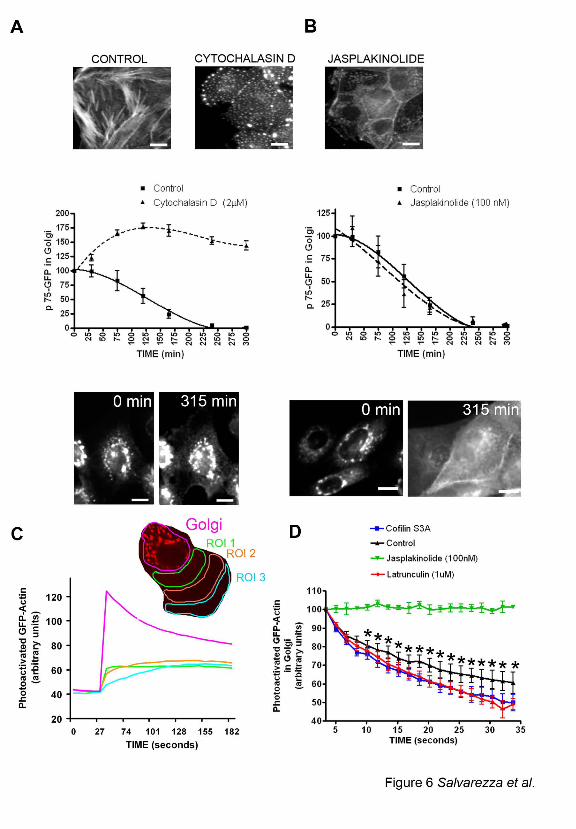
As a first approach, we investigated whether the effect of LIMK1-KD and cofilin S3A
mimicked the effect of stimulators or inhibitors of actin filament disassembly, e.g., cytochalasin
D or Jasplakinolide, respectively. Actin immunofluorescence and phalloidin staining
demonstrated that expression of LIMK1-KD or cofilin S3A, like cytochalasin D, disrupted the
delicate organization of actin filaments in the perinuclear region resulting in the accumulation of
small actin aggregates; Jasplakinolide, instead, induced the formation of large perinuclear actin
aggregates, as previously described (Spector et al., 1999) (Figure 6 A and B, top panels and
Supplementary Figure 4C). Importantly, cytochalasin D (Figure 6A) but not Jasplakinolide
(Figure. 6B) strongly inhibited the exit of p75-GFP from the TGN, confirming our previous
report that Latrunculin B delays the TGN exit of p75-GFP during the first 30 min after release of
the 20oC transport block (Musch et al., 2001). For the experiments reported here, we used
cytochalasin D because it did not promote cell detachment during the 5h required to measure
p75-GFP exit, unlike Latrunculin B. Interestingly, cytochalasin D induced the formation of long
TGN tubules labeled with ST-mRFP (Supplementary Figure 4B) similar to those we observed in
cells overexpressing LIMK1-KD or cofilin S3A (Figure 4 A-C). These results are consistent
with the hypothesis that the effects of LIMK1-KD and Cofilin S3A on post-Golgi trafficking of
p75-GFP are mediated by increased actin filament disassembly.
22
As a second test of whether cofilin S3A promotes or inhibits actin filament disassembly
at the Golgi, we measured local actin dynamics in the Golgi area under various experimental
conditions. To this end, we generated an MDCK cell line that expresses actin linked to photo-
activatable GFP (actin-paGFP) (Patterson and Lippincott-Schwartz, 2002). Actin-paGFP
incorporated into stress fibers and actin filaments at the level of the Golgi that were altered by
treatment with actin-toxic drugs similarly as endogenous actin (Supplementary Figure 4C). We
activated paGFP in the Golgi area -defined by ST-mRFP- with laser illumination at 405 nm
under examination by a confocal microscope and then measured the decay of GFP fluorescence
as an indicator of actin dynamics in that region (see Materials and Methods) (Figure 6C). The
decay of actin-GFP fluorescence in the Golgi area correlated with the progressive appearance of
GFP fluorescence in regions of the cytoplasm away from the Golgi area, which is compatible
with actin diffusion rather than with GFP photo-bleaching (Figure 6C). In control cells, this
decay exhibited a fast component (t1/2= 22s) and a slow component (t1/2 = 54s) which we
presumed corresponded, respectively, to the diffusion of free actin monomer pool and actin
filament turnover in the Golgi area (Figure 6D, black line) (McGrath et al., 1998). Upon
treatment with Jasplakinolide, the diffusion of actin-GFP was dramatically blocked (Figure 6D,
green line). By contrast, treatment with Latrunculin B (Figure 6D, red line) and with cofilin S3A
(Figure 6D, blue line) significantly decreased the half-life of decay of the slow component when
compared to control cells (t1/2 = 34 s and t1/2 = 41 s, respectively, both p<0.001). These results
strongly suggest that increased cofilin activity promotes increased actin filament turnover at the
Golgi level.
Thus, two independent experimental approaches are consistent with a scenario in which
inhibition of LIMK1 activity at the Golgi region and the consequent activation of cofilin, the
23
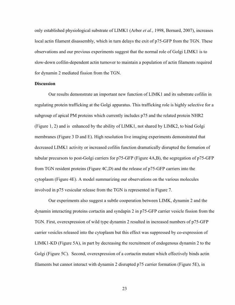
only established physiological substrate of LIMK1 (Arber et al., 1998, Bernard, 2007), increases
local actin filament disassembly, which in turn delays the exit of p75-GFP from the TGN. These
observations and our previous experiments suggest that the normal role of Golgi LIMK1 is to
slow-down cofilin-dependent actin turnover to maintain a population of actin filaments required
for dynamin 2 mediated fission from the TGN.
Discussion
Our results demonstrate an important new function of LIMK1 and its substrate cofilin in
regulating protein trafficking at the Golgi apparatus. This trafficking role is highly selective for a
subgroup of apical PM proteins which currently includes p75 and the related protein NHR2
(Figure 1, 2) and is enhanced by the ability of LIMK1, not shared by LIMK2, to bind Golgi
membranes (Figure 3 D and E). High resolution live imaging experiments demonstrated that
decreased LIMK1 activity or increased cofilin function dramatically disrupted the formation of
tubular precursors to post-Golgi carriers for p75-GFP (Figure 4A,B), the segregation of p75-GFP
from TGN resident proteins (Figure 4C,D) and the release of p75-GFP carriers into the
cytoplasm (Figure 4E). A model summarizing our observations on the various molecules
involved in p75 vesicular release from the TGN is represented in Figure 7.
Our experiments also suggest a subtle cooperation between LIMK, dynamin 2 and the
dynamin interacting proteins cortactin and syndapin 2 in p75-GFP carrier vesicle fission from the
TGN. First, overexpression of wild type dynamin 2 resulted in increased numbers of p75-GFP
carrier vesicles released into the cytoplasm but this effect was suppressed by co-expression of
LIMK1-KD (Figure 5A), in part by decreasing the recruitment of endogenous dynamin 2 to the
Golgi (Figure 5C). Second, overexpression of a cortactin mutant which effectively binds actin
filaments but cannot interact with dynamin 2 disrupted p75 carrier formation (Figure 5E), in
24
agreement with recent observations by (Cao et al., 2005) demonstrating a role of actin and
cortactin in recruiting dynamin 2 to the Golgi. Third, we found that overexpression of syndapin
2’s SH3 domain (which binds dynamin’s PRD), or of dynamin’s PRD , inhibited p75 vesicle
release from the TGN (Figure 5E and B). One possibility to explain these effects is a disruption
of syndapin 2 / dynamin 2 complexes, which support dynamin’s functions and provide functional
coupling of dynamin to actin filament formation (Kessels et al., 2006). That the coupling
between syndapin’s SH3 domain and dynamin 2’s PRD might be required for p75 post-Golgi
carrier formation is also supported by our observation that overexpression of mutant dynamin 2
lacking just the PRD , i.e. with intact GTPase and oligomerization domains, also inhibited p75-
GFP trafficking. Future experiments should test specifically whether syndapin 2’s requirement
for p75 exit from the TGN is solely explained by its participation in the recruitment of dynamin
2 or may also reflect its participation in the bending of TGN membranes via it’s BAR domain to
form tubular carriers characteristic for p75 transport.
The key link between our trafficking observations with LIMK1/cofilin and with dynamin
2/syndapin 2 /cortactin is actin. Experiments with actin toxins suggested that the trafficking
effects of LIMK1-KD or cofilin S3A we observed were due to increased depolymerization of
peri-Golgi actin filaments (Figure 6A, B), in agreement with a recent report (Lazaro-Dieguez et
al., 2007). This was rigorously confirmed by direct measurement of actin dynamics in the Golgi
region using an MDCK cell line that expresses actin-paGFP (Figure 6 C, D). Actin-GFP is a
good probe to study actin dynamics, as it has been used to study the role of actin dynamics in
endocytosis and cell-cell adhesion (Yamada et al., 2005; Okreglak and Drubin, 2007). We found
that the diffusion rate of Golgi actin-paGFP (D ~ 1.7 x 10⁻⁹ cm2.s-1 in control cells) was
significantly enhanced in the presence of either cofilin S3A (D ~2.1x10⁻⁹ cm2.s-1) or latrunculin

25
B (D ~ 2.6x10⁻⁹ cm2.s-1): our control values are comparable to previously reported data obtained
using Fluorescence Photobleaching Recovery (Kreis et al., 1982). Taken together, our
experiments strongly suggest that the normal role of Golgi LIMK1 is to maintain an ideal cofilin
activity level to maintain a population of actin filaments (Percival et al., 2004) required for
dynamin mediated, cortactin/syndapin 2 -supported trafficking of selected PM cargo proteins
out of the TGN.
Cdc42 is known to localize throughout the Golgi stack (Matas et al., 2004), while our
experiments indicate that LIMK1 localizes to the TGN (Figure 3D,E and data not shown).
Furthermore, it has been shown that PAK4, a novel effector for cdc42, localizes to the Golgi
complex (Abo et al., 1998), and stimulates LIMK1’s ability to phosphorylate cofilin (Dan,
2001). Taken together, these observations raise the possibility that cdc42 and LIMK1 cooperate
in post-Golgi transport at the TGN, similarly to the cooperation between cdc42 and N-WASP in
Golgi-ER transport at the cis Golgi (Luna et al., 2002) .
Our results clearly advance the trafficking field in several novel areas beyond the
previous study by Rosso et al (2004). First, we conclusively showed that the trafficking role of
LIMK1 takes place at the Golgi level, by excluding possible effects on protein synthesis or ER-
Golgi transport, and by showing directly that inhibition of LIMK1 function decreases the kinetics
of Golgi exit of PM markers. Second, we showed that the specific trafficking role of LIMK1-
cofilin was on the fission of carrier vesicles from the TGN (Figure 4). Third, we demonstrated a
possible cooperation between LIMK1 and dynamin 2 in this fission process (Figure 5). Fourth,
we further characterized this fission mechanism by demonstrating that syndapin 2 and cortactin
mutants mimic the effect of LIMK1-KD in the Golgi exit of p75-GFP. Fifth, we characterized
the actin dynamics at the Golgi region using actin coupled to photoactivatable GFP. This
26
approach allowed us to conclusively show that LIMK1-cofilin increase the dynamics of actin
depolymerization at the Golgi, thus eliminating the alternative possibility suggested by Condeelis
(Ghosh et al., 2004). Our observations suggest a mechanism to generate the population of actin
filaments at the Golgi complex described by Stow and coworkers (Percival et al., 2004).
In summary, our experiments suggest a model (Figure 7) in which specialized
organizations of actin filaments at the Golgi complex play very specific roles in promoting the
trafficking of groups of PM proteins from the TGN to the PM. It is likely that other specialized
actin organizations will play comparable roles in transport routes out of the other major sorting
compartment of epithelial cells, Common Recycling Endosomes (Cancino et al., 2007; Gravotta
et al., 2007). An important objective for the future is to understand how the selectivity of a
particular organization of the actin cytoskeleton for a particular cargo is established. In
particular, we wish to know how p75 sorting signals, previously defined as the O-glycan cluster
in its ectodomin (Yeaman et al., 1997) upon clustering with galectin 3 (Delacour et al., 2007) are
coupled to the actin regulatory mechanisms required for vesicle fission. Another major
challenge is to identify the mechanisms that couple the actin-dependent fission mechanisms
described here with our recent observation that kinesin 5B is essential for generation and
transport of p75 carrier vesicles from Golgi to PM (Jaulin et al., 2007). In this regard, it is
interesting to mention a recent report that postulates a role for LIMK1 in coordinating MT and
actin cytoskeleton function (Gorovoy et al., 2005).

27
FIGURE LEGENDS
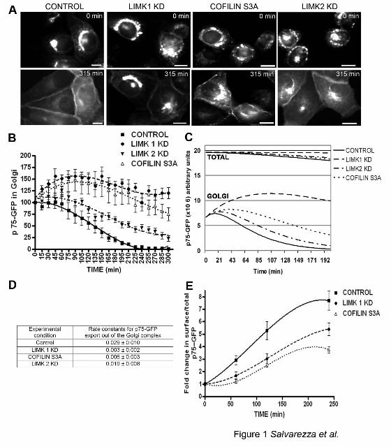
Figure 1: LIMK1-KD and CofilinS3A block Golgi exit and surface delivery of p75-GFP.
A) p75-GFP expressed by nuclear injection of its cDNA in subconfluent MDCK cells,
accumulated in the TGN after a 20oC temperature block; its exit from TGN and transport to the
plasma membrane were assessed after switch to transport-permissive temperature (32oC). Note
surface arrival of p75-GFP after 315 min in control cells. By contrast, in cells co-injected with
cDNAs encoding LIMK1-KD, cofilinS3A, or LIMK2-KD, p75-GFP was retained in the Golgi
region. Scale bars = 20 µm. B) p75-GFP fluorescence at the TGN was quantified for each
condition after shift to 32°C (15-20 cells per experimental condition, experiments were repeated
2-3 times) and expressed as percent of TGN fluorescence at t=0. Differences between control
and LIMK or cofilin samples were statistically significant (p<0.0001). C) Fluorescence data
were fitted to a mathematical model (see text). D) Rate constants for TGN exit, obtained from
this model, are expressed as mean ± SD. E) Arrival of p75-GFP at the cell surface, measured as
the ratio surface immunofluorescence/ total GFP, was inhibited by LIMK1-KD and cofilin S3A
expression (p<0.0001).
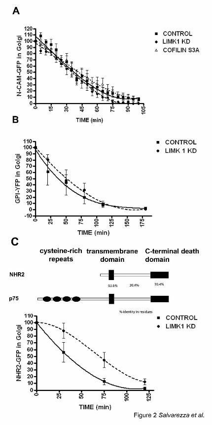
Figure 2: LIMK1-KD does not block exit from TGN of NCAM-GFP or GPI-YFP.
The exit of NCAM-GFP (A), GPI-YFP (B) or NHR2-GFP, a protein related to p75 (C) from the
TGN after shift to 32°C was recorded as described in Materials and Methods (2-3 experiments,
15-20 cells per experimental condition). Note that expression of LIMK1-KD does not interfere
with the exit of NCAM-GFP or GPI-YFP from the TGN, but does interfere with the exit of
NHR2-GFP from the TGN. Expression of cofilin S3A does not affect the exit of NCAM-GFP.
28
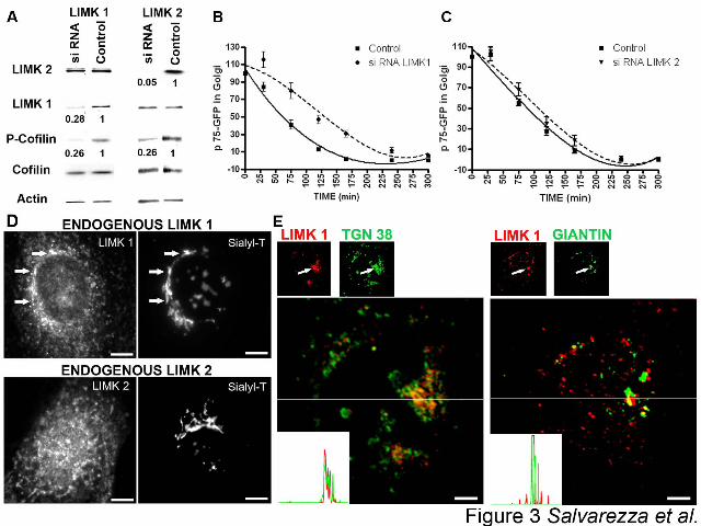
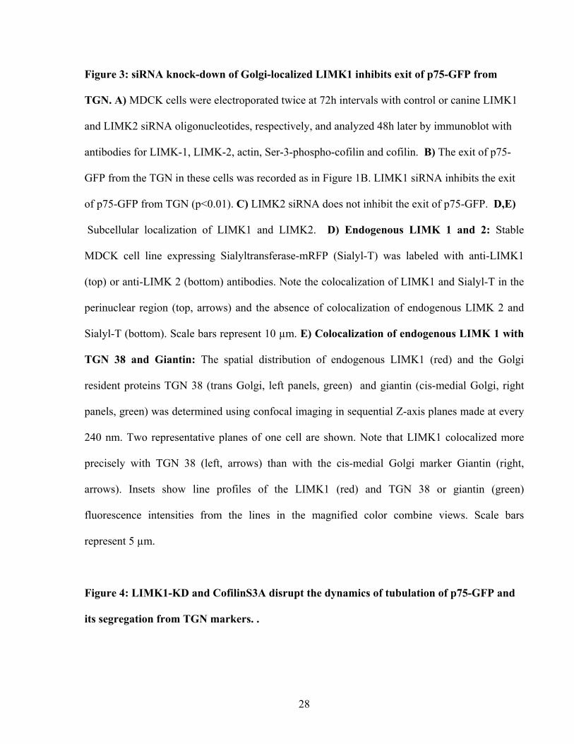
Figure 3: siRNA knock-down of Golgi-localized LIMK1 inhibits exit of p75-GFP from
TGN. A) MDCK cells were electroporated twice at 72h intervals with control or canine LIMK1
and LIMK2 siRNA oligonucleotides, respectively, and analyzed 48h later by immunoblot with
antibodies for LIMK-1, LIMK-2, actin, Ser-3-phospho-cofilin and cofilin. B) The exit of p75-
GFP from the TGN in these cells was recorded as in Figure 1B. LIMK1 siRNA inhibits the exit
of p75-GFP from TGN (p<0.01). C) LIMK2 siRNA does not inhibit the exit of p75-GFP. D,E)
Subcellular localization of LIMK1 and LIMK2. D) Endogenous LIMK 1 and 2: Stable
MDCK cell line expressing Sialyltransferase-mRFP (Sialyl-T) was labeled with anti-LIMK1
(top) or anti-LIMK 2 (bottom) antibodies. Note the colocalization of LIMK1 and Sialyl-T in the
perinuclear region (top, arrows) and the absence of colocalization of endogenous LIMK 2 and
Sialyl-T (bottom). Scale bars represent 10 µm. E) Colocalization of endogenous LIMK 1 with
TGN 38 and Giantin: The spatial distribution of endogenous LIMK1 (red) and the Golgi
resident proteins TGN 38 (trans Golgi, left panels, green) and giantin (cis-medial Golgi, right
panels, green) was determined using confocal imaging in sequential Z-axis planes made at every
240 nm. Two representative planes of one cell are shown. Note that LIMK1 colocalized more
precisely with TGN 38 (left, arrows) than with the cis-medial Golgi marker Giantin (right,
arrows). Insets show line profiles of the LIMK1 (red) and TGN 38 or giantin (green)
fluorescence intensities from the lines in the magnified color combine views. Scale bars
represent 5 µm.
Figure 4: LIMK1-KD and CofilinS3A disrupt the dynamics of tubulation of p75-GFP and
its segregation from TGN markers. .

29
A) A cDNA encoding p75-GFP was injected alone or together with a cDNA encoding for Cofilin
S3A into the nuclei of subconfluent MDCK cells. The formation of tubular precursors to post-
Golgi transporters was analyzed 10 min after release of the 20°C block by high resolution
microscopy (time-lapse images acquired at ~1s intervals for 1 min). Control cells (left panels)
show extension and retraction of a p75-GFP tubule within <1 min (arrow on tubule tip). Cofilin
S3A (right panels) induced long static tubules with no extension-retraction behavior. Scale bars =
5 µm. B) Lengths and life-times of p75-GFP tubules in control, Cofilin S3A or LIMK1-KD
expressing cells. C) Dual color high resolution images of cells expressing p75-GFP,LIMK1-KD
or Cofilin S3A and Sialyltransferase (ST-mRFP) were acquired as described in A. Control: Note
the exclusion of ST-mRFP from tubules containing p75-GFP (arrows, base marked by
arrowhead). See also supplementary movie 1. Note that in cells expressing LIMK1-KD or
Cofilin S3A, emerging tubules (arrows) contain both p75-GFP (green) and ST-mRFP (red). The
green and red images in LIMK1-KD were shifted by 5 pixels. See supplementary movies 2 and
3. Asterisk (*) marks swellings containing both proteins. Scale bars = 5 µm. D) Quantification of
tubules that contain p75-GFP, p75-GFP+ST-mRFP or ST-mRFP alone under different
experimental conditions (n=10 cells per condition, 3 experiments). Asterisks (*) mark
statistically significant differences compared to control. E) Release of post-Golgi carriers
(PGCs). MDCK cells expressing p75GFP were imaged (1-2s intervals for 3 min) 10 minutes
after release from 20°C. In control cells (left panel) P75-GFP is seen in the perinuclear region
and in tubular and spherical PGCs accumulated within the cytoplasm (arrows). PGCs and their
characteristic tracks can be seen in supplementary movie 4. Upon expression of LIMK 1-KD or
Cofilin S3A, the number of mobile p75 PGCs is drastically reduced, as seen in supplementary
movie 5. Bar graphs: Quantification of PGCs demonstrates a statistically significant difference (p

30
< 0.01) between control and LIMK1 -KD or Cofilin S3A injected-cells (6 cells per condition, 3
separate experiments).
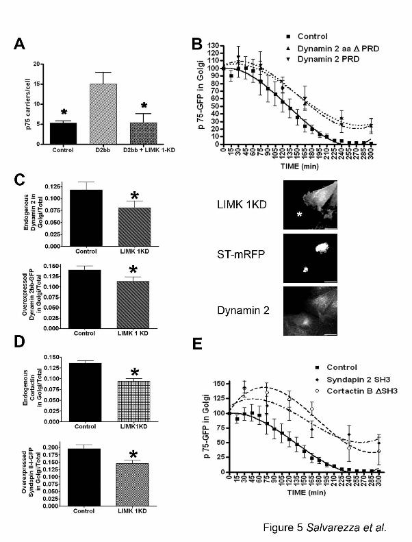
Figure 5: LIMK 1 activity induces recruitment of dynamin 2, which promotes fission of
p75-GFP transporters likely assisted by cortactin and syndapin 2.
A) The release of post Golgi transporters was measured as in Figure 4E in cells expressing
dynamin 2 + LIMK1-KD, 120 min after release from 20°C block (6 cells per condition, 3
experiments). Asterisk indicates statistically significant differences (p<0.01). B) Overexpression
of dynamin 2 proline rich domain or dynamin 2 lacking its domain (Dynamin 2 ∆PRD) inhibited
release of p75-GFP from the TGN. (p<0.05) C) Recruitment of endogenous and overexpressed
dynamin 2bb to the Golgi was inhibited by overexpression of LIMK1-KD (40 cells per
condition, 2 experiments), (p<0.05). Images (right panels) show an example of the higher
recruitment of
Dynamin 2 (botton) at the Golgi (ST-mRFP, middle) of control cells (asterisks, top) compared
with LIMK 1 KD expressing cells. Scale bars = 20 µm. D) Recruitment of endogenous cortactin
and overexpressed syndapin 2 to the Golgi was inhibited by expression of LIMK1-KD (35 cells
per condition, 2 experiments), (p<0.05). E) Overexpression of Syndapin 2 SH3 or cortactin
∆SH3 inhibited exit of p75-GFP from the TGN (p<0.05).
Figure 6: Exit of p75-GFP from TGN is blocked by increased actin filament
depolymerization. A,B) Phallodin staining of control, Cytochalasin D (2 µM) and
Jasplakinolide (100nM) treated cells after a time lapse record. Scale bars represent 20 µm.
Cytochalasin D (A) but not Jasplakinolide (B) inhibits exit of P75-GFP from the TGN and

31
prevents its redistribution to the plasma membrane (p<0.0001, 10 cells in two separate
experiments were quantified for each condition). Representative p75-GFP fluorescence images
are shown at the bottom of each graph. Scale bars = 10 µm. C, D) MDCK cell line expressing
actin coupled to photoactivatable GFP (actin-paGFP) was photoactivated at the Golgi region,
defined by the presence of ST-mRFP (red). GFP fluorescence at the Golgi region and at more
distant regions of interest in the cytoplasm (ROI 1-3) was subsequently recorded. Note in C that
GFP fluorescence decays at the Golgi area but rises at other ROIs, indicating diffusion of actin
monomers. D) Curves represent decay of actin-paGFP fluorescence in Golgi area (5-12 cells per
condition). Note biphasic decay in control cells. Fluorescence decay is blocked by Jasplakinolide
but is faster than in control cells upon Cofilin S3A or Latrunculin B presence. Data are
represented as mean + SEM, Asterisks (*) indicate statistically significant differences (p < 0.001)
between control and Latrunculin B as well as between control and Cofilin S3A effects.
Figure 7: Model. LIMK1 and Cofilin regulate a peri-TGN actin filament network required
for the dynamin-dependent exit of p75-GFP.
(1) A population of actin filaments, anchored to the TGN by an ARF-dependent mechanism
(Percival et al., 2004; Cao et al., 2005) and regulated by cofilin and Golgi anchored LIMK1,
which inactivates cofilin locally (this paper and (Rosso et al., 2004)) facilitates the initial
assembly of transporters for p75-GFP in cooperation with a plus-end directed kinesin (Jaulin et
al., 2007). Note that Golgi markers are excluded from these transporters. (2) p75GFP-containing
tubules extend along MT drawn by a kinesin motor; syndapin 2, via its BAR-domain, binds to
the membrane curvature of the Golgi (Kessels et al., 2006) and facilitates membrane remodelling
necessary for tubulation. (3) p75-GFP tubules undergo fission from the TGN or into smaller
32
transport vesicles; this process depends on actin filaments (regulated by LIMK 1-cofilin), which
recruit cortactin, as well as syndapin 2 . These two molecules recruit dynamin 2 via their SH3
domains and dynamin PRD (Cao et al., 2005; Kessels et al., 2006). (4) After dynamin-mediated
fission, tubular and vesicular carriers for p75-GFP are transported by a plus-end kinesin along
MT to the plasma membrane. (5) Inactivation of LIMK1 or overexpression of constitutively
activated cofilin inhibits the segregation of p75 from TGN markers. (6) This treatment also
inhibits dynamin-mediated fission of p75 transporters from TGN as well as the recruitment of
Dynamin 2, SyndapinII and cortactin to the Golgi.
Acknowledgements
We thank Dr Diego Gravotta for invaluable help with the pulse chase experiments, Dr Ami
Deora for help with the design of oligonucleotides used to knock down LIM kinases, Dr Larry
Leung for preparation of a cell line expressing galactosyl transferase and Dr. Mark McNiven for
generously providing us with cortactin antibodies and constructs. Our special thanks go to Isha
Lucia Thorne for lending us her mother, Dr Aparna Lakkaraju, to help us with the artistic design
of Figure 7. Dr Rodriguez-Boulan was supported by NIH Grants GM 34107 and EY 08538, by
the Research to Prevent Blindness Foundation and by the Dyson Foundation. Michael M Kessels
was supported by the Deutsche Forschungsgemeinschaft (DFG) and Britta Qualmann was
supported by the DFG and the Kultusministerium of the Land Sachsen-Anhalt.

33
References
Abo, A., Qu, J., Cammarano, M.S., Dan, C., Fritsch, A., Baud, V., Belisle, B., and Minden, A. (1998). PAK4, a novel effector for Cdc42Hs, is implicated in the reorganization of the actin cytoskeleton and in the formation of filopodia. Embo J 17, 6527-6540. Acevedo, K., Moussi, N., Li, R., Soo, P., and Bernard, O. (2006). LIM kinase 2 is widely expressed in all tissues. J Histochem Cytochem 54, 487-501. Allan, V.J., Thompson, H.M., and McNiven, M.A. (2002). Motoring around the Golgi. Nat Cell Biol 4, E236-242. Arber, S., Barbayannis, F.A., Hanser, H., Schneider, C., Stanyon, C.A., Bernard, O., and Caroni, P. (1998). Regulation of actin dynamics through phosphorylation of cofilin by LIM-kinase. Nature 393, 805-809. Bamburg, J.R. (1999). Proteins of the ADF/cofilin family: essential regulators of actin dynamics. Annu Rev Cell Dev Biol 15, 185-230. Bernard, O. (2007). Lim kinases, regulators of actin dynamics. Int J Biochem Cell Biol 39, 1071-1076. Bonazzi, M., Spano, S., Turacchio, G., Cericola, C., Valente, C., Colanzi, A., Kweon, H.S., Hsu, V.W., Polishchuck, E.V., Polishchuck, R.S., Sallese, M., Pulvirenti, T., Corda, D., and Luini, A. (2005). CtBP3/BARS drives membrane fission in dynamin-independent transport pathways. Nat Cell Biol 7, 570-580. Bonifacino, J.S., and Traub, L.M. (2003). Signals for sorting of transmembrane proteins to endosomes and lysosomes. Annu Rev Biochem 72, 395-447. Cancino, J., Torrealba, C., Soza, A., Yuseff, I., Gravotta, D., Henklein, P., Rodriguez-Boulan, E., and Gonzalez, A. (2007). Antibody to AP1B adaptor blocks Biosynthetic and Recycling Routes of Basolateral Proteins at Recycling Endosomes. Mol. Biol. Cell in press. Cao, H., Thompson, H.M., Krueger, E.W., and McNiven, M.A. (2000). Disruption of Golgi structure and function in mammalian cells expressing a mutant dynamin. J Cell Sci 113 (Pt 11), 1993-2002. Cao, H., Orth, J.D., Chen, J., Weller, S.G., Heuser, J.E., and McNiven, M.A. (2003). Cortactin is a component of clathrin-coated pits and participates in receptor-mediated endocytosis. Mol Cell Biol 23, 2162-2170. Cao, H., Weller, S., Orth, J.D., Chen, J., Huang, B., Chen, J.L., Stamnes, M., and McNiven, M.A. (2005). Actin and Arf1-dependent recruitment of a cortactin-dynamin complex to the Golgi regulates post-Golgi transport. Nat Cell Biol 7, 483-492. Carlier, M.F., Laurent, V., Santolini, J., Melki, R., Didry, D., Xia, G.X., Hong, Y., Chua, N.H., and Pantaloni, D. (1997). Actin depolymerizing factor (ADF/cofilin) enhances the rate of filament turnover: implication in actin-based motility. J Cell Biol 136, 1307-1322. Carreno, S., Engqvist-Goldstein, A.E., Zhang, C.X., McDonald, K.L., and Drubin, D.G. (2004). Actin dynamics coupled to clathrin-coated vesicle formation at the trans-Golgi network. J Cell Biol 165, 781-788. Chen, X., and Macara, I.G. (2006). Par-3 mediates the inhibition of LIM kinase 2 to regulate cofilin phosphorylation and tight junction assembly. J Cell Biol 172, 671-678. Conner, S.D., and Schmid, S.L. (2003). Regulated portals of entry into the cell. Nature 422, 37-44.
34
Dan, C., Kelly, A., Bernard, O., and Minden, A. (2001). Cytoskeletal changes regulated by the PAK4 serine/threonine kinase are mediated by LIM kinase 1 and cofilin. J Biol Chem 276, 32115-32121. Deborde, S., Perret, E., Gravotta, D., Deora, A., Salvarezza, S., Schreiner, R., and Rodriguez-Boulan, E. (2008). Clathrin is a key regulator of basolateral polarity. Nature 452, 719-723. Delacour, D., Cramm-Behrens, C.I., Drobecq, H., Le Bivic, A., Naim, H.Y., and Jacob, R. (2006). Requirement for galectin-3 in apical protein sorting. Curr Biol 16, 408-414. Delacour, D., Gouyer, V., Zanetta, J.P., Drobecq, H., Leteurtre, E., Grard, G., Moreau-Hannedouche, O., Maes, E., Pons, A., Andre, S., Le Bivic, A., Gabius, H.J., Manninen, A., Simons, K., and Huet, G. (2005). Galectin-4 and sulfatides in apical membrane trafficking in enterocyte-like cells. J Cell Biol 169, 491-501. Delacour, D., Greb, C., Koch, A., Salomonsson, E., Leffler, H., Le Bivic, A., and Jacob, R. (2007). Apical sorting by galectin-3-dependent glycoprotein clustering. Traffic 8, 379-388. Egea, G., Lazaro-Dieguez, F., and Vilella, M. (2006). Actin dynamics at the Golgi complex in mammalian cells. Curr Opin Cell Biol 18, 168-178. Erickson, J.W., Zhang, C., Kahn, R.A., Evans, T., and Cerione, R.A. (1996). Mammalian Cdc42 is a brefeldin A-sensitive component of the Golgi apparatus. J Biol Chem 271, 26850-26854. Foletta, V.C., Moussi, N., Sarmiere, P.D., Bamburg, J.R., and Bernard, O. (2004). LIM kinase 1, a key regulator of actin dynamics, is widely expressed in embryonic and adult tissues. Exp Cell Res 294, 392-405. Ghosh, M., Song, X., Mouneimne, G., Sidani, M., Lawrence, D.S., and Condeelis, J.S. (2004). Cofilin promotes actin polymerization and defines the direction of cell motility. Science 304, 743-746. Gorovoy, M., Niu, J., Bernard, O., Profirovic, J., Minshall, R., Neamu, R., and Voyno-Yasenetskaya, T. (2005). LIM kinase 1 coordinates microtubule stability and actin polymerization in human endothelial cells. J Biol Chem 280, 26533-26542. Gravotta, D., Deora, A., Perret, E., Oyanadel, C., Soza, A., Schreiner, R., Gonzalez, A., and Rodriguez-Boulan, E. (2007). AP1B sorts basolateral proteins in recycling and biosynthetic routes of MDCK cells. Proc Natl Acad Sci U S A 104, 1564-1569. Guerriero, C.J., Weixel, K.M., Bruns, J.R., and Weisz, O.A. (2006). Phosphatidylinositol 5-kinase stimulates apical biosynthetic delivery via an Arp2/3-dependent mechanism. J Biol Chem 281, 15376-15384. Itoh, T., Erdmann, K.S., Roux, A., Habermann, B., Werner, H., and De Camilli, P. (2005). Dynamin and the actin cytoskeleton cooperatively regulate plasma membrane invagination by BAR and F-BAR proteins. Dev Cell 9, 791-804. Jaulin, F., Xue, X., Rodriguez-Boulan, E., and Kreitzer, G. (2007). Polarization-Dependent Selective Transport to the Apical Membrane by KIF5B in MDCK Cells. Dev Cell 13, 511-522. Kaksonen, M., Toret, C.P., and Drubin, D.G. (2005). A modular design for the clathrin- and actin-mediated endocytosis machinery. Cell 123, 305-320. Kessels, M.M., Dong, J., Leibig, W., Westermann, P., and Qualmann, B. (2006). Complexes of syndapin II with dynamin II promote vesicle formation at the trans-Golgi network. J Cell Sci 119, 1504-1516. Kreis, T.E., Geiger, B., and Schlessinger, J. (1982). Mobility of microinjected rhodamine actin within living chicken gizzard cells determined by fluorescence photobleaching recovery. Cell 29, 835-845.
35
Kreitzer, G., Marmorstein, A., Okamoto, P., Vallee, R., and Rodriguez-Boulan, E. (2000). Kinesin and dynamin are required for post-Golgi transport of a plasma-membrane protein. Nat Cell Biol 2, 125-127. Kreitzer, G., Schmoranzer, J., Low, S.H., Li, X., Gan, Y., Weimbs, T., Simon, S.M., and Rodriguez-Boulan, E. (2003). Three-dimensional analysis of post-Golgi carrier exocytosis in epithelial cells. Nat Cell Biol 5, 126-136. Kroschewski, R., Hall, A., and Mellman, I. (1999). Cdc42 controls secretory and endocytic transport to the basolateral plasma membrane of MDCK cells. Nat Cell Biol 1, 8-13. Lappalainen, P., and Drubin, D.G. (1997). Cofilin promotes rapid actin filament turnover in vivo. Nature 388, 78-82. Lazaro-Dieguez, F., Colonna, C., Cortegano, M., Calvo, M., Martinez, S.E., and Egea, G. (2007). Variable actin dynamics requirement for the exit of different cargo from the trans-Golgi network. FEBS Lett 581, 3875-3881. Luna, A., Matas, O.B., Martinez-Menarguez, J.A., Mato, E., Duran, J.M., Ballesta, J., Way, M., and Egea, G. (2002). Regulation of protein transport from the Golgi complex to the endoplasmic reticulum by CDC42 and N-WASP. Mol Biol Cell 13, 866-879. Matas, O.B., Martinez-Menarguez, J.A., and Egea, G. (2004). Association of Cdc42/N-WASP/Arp2/3 signaling pathway with Golgi membranes. Traffic 5, 838-846. McGrath, J.L., Hartwig, J.H., Tardy, Y., and Dewey, C.F., Jr. (1998). Measuring actin dynamics in endothelial cells. Microsc Res Tech 43, 385-394. McNiven, M.A., and Thompson, H.M. (2006). Vesicle formation at the plasma membrane and trans-Golgi network: the same but different. Science 313, 1591-1594. Meberg, P.J., and Bamburg, J.R. (2000). Increase in neurite outgrowth mediated by overexpression of actin depolymerizing factor. J Neurosci 20, 2459-2469. Mendes, P. (1993). GEPASI: a software package for modelling the dynamics, steady states and control of biochemical and other systems. Comput Appl Biosci 9, 563-571. Merrifield, C.J., Perrais, D., and Zenisek, D. (2005). Coupling between clathrin-coated-pit invagination, cortactin recruitment, and membrane scission observed in live cells. Cell 121, 593-606. Murray, S.S., Perez, P., Lee, R., Hempstead, B.L., and Chao, M.V. (2004). A novel p75 neurotrophin receptor-related protein, NRH2, regulates nerve growth factor binding to the TrkA receptor. J Neurosci 24, 2742-2749. Musch, A. (2004). Microtubule organization and function in epithelial cells. Traffic 5, 1-9. Musch, A., Cohen, D., Kreitzer, G., and Rodriguez-Boulan, E. (2001). cdc42 regulates the exit of apical and basolateral proteins from the trans-Golgi network. Embo J 20, 2171-2179. Noda, Y., Okada, Y., Saito, N., Setou, M., Xu, Y., Zhang, Z., and Hirokawa, N. (2001). KIFC3, a microtubule minus end-directed motor for the apical transport of annexin XIIIb-associated Triton-insoluble membranes. J Cell Biol 155, 77-88. Okreglak, V., and Drubin, D.G. (2007). Cofilin recruitment and function during actin-mediated endocytosis dictated by actin nucleotide state. J Cell Biol 178, 1251-1264. Patterson, G.H., and Lippincott-Schwartz, J. (2002). A photoactivatable GFP for selective photolabeling of proteins and cells. Science 297, 1873-1877. Percival, J.M., Hughes, J.A., Brown, D.L., Schevzov, G., Heimann, K., Vrhovski, B., Bryce, N., Stow, J.L., and Gunning, P.W. (2004). Targeting of a tropomyosin isoform to short microfilaments associated with the Golgi complex. Mol Biol Cell 15, 268-280.
36
Perret, E., Lakkaraju, A., Deborde, S., Schreiner, R., and Rodriguez-Boulan, E. (2005). Evolving endosomes: how many varieties and why? Curr Opin Cell Biol 17, 423-434. Qualmann, B., and Kelly, R.B. (2000). Syndapin isoforms participate in receptor-mediated endocytosis and actin organization. J Cell Biol 148, 1047-1062. Qualmann, B., Kessels, M.M., and Kelly, R.B. (2000). Molecular links between endocytosis and the actin cytoskeleton. J Cell Biol 150, F111-116. Rodriguez-Boulan, E., Kreitzer, G., and Musch, A. (2005). Organization of vesicular trafficking in epithelia. Nat Rev Mol Cell Biol 6, 233-247. Rosso, S., Bollati, F., Bisbal, M., Peretti, D., Sumi, T., Nakamura, T., Quiroga, S., Ferreira, A., and Caceres, A. (2004). LIMK1 regulates Golgi dynamics, traffic of Golgi-derived vesicles, and process extension in primary cultured neurons. Mol Biol Cell 15, 3433-3449. Rozelle, A.L., Machesky, L.M., Yamamoto, M., Driessens, M.H., Insall, R.H., Roth, M.G., Luby-Phelps, K., Marriott, G., Hall, A., and Yin, H.L. (2000). Phosphatidylinositol 4,5-bisphosphate induces actin-based movement of raft-enriched vesicles through WASP-Arp2/3. Curr Biol 10, 311-320. Scheiffele, P., Peranen, J., and Simons, K. (1995). N-glycans as apical sorting signals in epithelial cells. Nature 378, 96-98. Simons, K., and E. Ikonen. (1997). Functional rafts in cell membranes. Nature 387, 569-572. Song, B.D., Leonard, M., and Schmid, S.L. (2004). Dynamin GTPase domain mutants that differentially affect GTP binding, GTP hydrolysis, and clathrin-mediated endocytosis. J Biol Chem 279, 40431-40436. Spector, I., Braet, F., Shochet, N.R., and Bubb, M.R. (1999). New anti-actin drugs in the study of the organization and function of the actin cytoskeleton. Microsc Res Tech 47, 18-37. Stamnes, M. (2002). Regulating the actin cytoskeleton during vesicular transport. Curr Opin Cell Biol 14, 428-433. Sumi, T., Matsumoto, K., Takai, Y., and Nakamura, T. (1999). Cofilin phosphorylation and actin cytoskeletal dynamics regulated by rho- and Cdc42-activated LIM-kinase 2. J Cell Biol 147, 1519-1532. Tai, A.W., Chuang, J.Z., and Sung, C.H. (2001). Cytoplasmic dynein regulation by subunit heterogeneity and its role in apical transport. J Cell Biol 153, 1499-1509. Tomiyoshi, G., Horita, Y., Nishita, M., Ohashi, K., and Mizuno, K. (2004). Caspase-mediated cleavage and activation of LIM-kinase 1 and its role in apoptotic membrane blebbing. Genes Cells 9, 591-600. Warnock, D.E., Terlecky, L.J., and Schmid, S.L. (1995). Dynamin GTPase is stimulated by crosslinking through the C-terminal proline-rich domain. Embo J 14, 1322-1328. Ya-Wen Liu, M.C.S., Thomas Schroeter, Vasyl Lukiyanchuk, Sandra L. Schmid. (2008). Isoform and splice-variant specific functions of dynamin-2 revealed by analysis of conditional knock-out cells. Mol Biol Cell, in Press. Yamada, S., Pokutta, S., Drees, F., Weis, W.I., and Nelson, W.J. (2005). Deconstructing the cadherin-catenin-actin complex. Cell 123, 889-901. Yang, J.S., Zhang, L., Lee, S.Y., Gad, H., Luini, A., and Hsu, V.W. (2006). Key components of the fission machinery are interchangeable. Nat Cell Biol 8, 1376-1382. Yarar, D., Waterman-Storer, C.M., and Schmid, S.L. (2005). A dynamic actin cytoskeleton functions at multiple stages of clathrin-mediated endocytosis. Mol Biol Cell 16, 964-975.
37
Yeaman, C., Le Gall, A.H., Baldwin, A.N., Monlauzeur, L., Le Bivic, A., and Rodriguez-Boulan, E. (1997). The O-glycosylated stalk domain is required for apical sorting of neurotrophin receptors in polarized MDCK cells. J Cell Biol 139, 929-940.