Tuba, a novel protein containing bin/amphiphysin/Rvs and Dbl homology domains, links dynamin to...
13
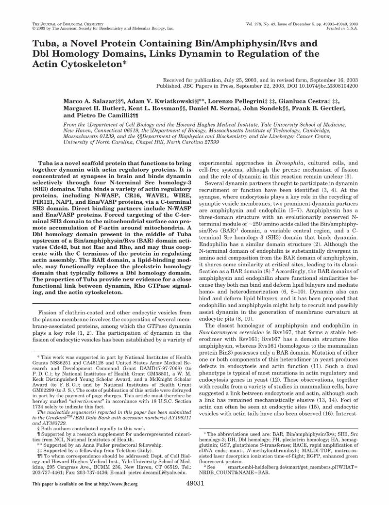
Tuba, a Novel Protein Containing Bin/Amphiphysin/Rvs and Dbl Homology Domains, Links Dynamin to Regulation of the Actin Cytoskeleton* Received for publication, July 25, 2003, and in revised form, September 16, 2003 Published, JBC Papers in Press, September 22, 2003, DOI 10.1074/jbc.M308104200 Marco A. Salazar‡§¶, Adam V. Kwiatkowski§**, Lorenzo Pellegrini‡ ‡‡, Gianluca Cestra‡ ‡‡, Margaret H. Butler‡, Kent L. Rossman§§, Daniel M. Serna, John Sondek§§, Frank B. Gertler, and Pietro De Camilli‡¶¶ From the ‡Department of Cell Biology and the Howard Hughes Medical Institute, Yale University School of Medicine, New Haven, Connecticut 06519, the Department of Biology, Massachusetts Institute of Technology, Cambridge, Massachusetts 01239, and the §§Department of Biophysics and Biochemistry and the Lineberger Cancer Center, University of North Carolina, Chapel Hill, North Carolina 27599 Tuba is a novel scaffold protein that functions to bring together dynamin with actin regulatory proteins. It is concentrated at synapses in brain and binds dynamin selectively through four N-terminal Src homology-3 (SH3) domains. Tuba binds a variety of actin regulatory proteins, including N-WASP, CR16, WAVE1, WIRE, PIR121, NAP1, and Ena/VASP proteins, via a C-terminal SH3 domain. Direct binding partners include N-WASP and Ena/VASP proteins. Forced targeting of the C-ter- minal SH3 domain to the mitochondrial surface can pro- mote accumulation of F-actin around mitochondria. A Dbl homology domain present in the middle of Tuba upstream of a Bin/amphiphysin/Rvs (BAR) domain acti- vates Cdc42, but not Rac and Rho, and may thus coop- erate with the C terminus of the protein in regulating actin assembly. The BAR domain, a lipid-binding mod- ule, may functionally replace the pleckstrin homology domain that typically follows a Dbl homology domain. The properties of Tuba provide new evidence for a close functional link between dynamin, Rho GTPase signal- ing, and the actin cytoskeleton. Fission of clathrin-coated and other endocytic vesicles from the plasma membrane involves the cooperation of several mem- brane-associated proteins, among which the GTPase dynamin plays a key role (1, 2). The participation of dynamin in the fission of endocytic vesicles has been established by a variety of experimental approaches in Drosophila, cultured cells, and cell-free systems, although the precise mechanism of fission and the role of dynamin in this reaction remain unclear (3). Several dynamin partners thought to participate in dynamin recruitment or function have been identified (3, 4). At the synapse, where endocytosis plays a key role in the recycling of synaptic vesicle membranes, two prominent dynamin partners are amphiphysin and endophilin (5–7). Amphiphysin has a three-domain structure with an evolutionarily conserved N- terminal module of 250 amino acids called the Bin/amphiphy- sin/Rvs (BAR) 1 domain, a variable central region, and a C- terminal Src homology-3 (SH3) domain that binds dynamin. Endophilin has a similar domain structure (2). Although the N-terminal domain of endophilin is substantially divergent in amino acid composition from the BAR domain of amphiphysin, it shares some similarity at critical sites, leading to its classi- fication as a BAR domain (8). 2 Accordingly, the BAR domains of amphiphysin and endophilin share functional similarities be- cause they both can bind and deform lipid bilayers and mediate homo- and heterodimerization (6, 8 –10). Dynamin also can bind and deform lipid bilayers, and it has been proposed that endophilin and amphiphysin might help to recruit and possibly assist dynamin in the generation of membrane curvature at endocytic pits (8, 10). The closest homologue of amphiphysin and endophilin in Saccharomyces cerevisiae is Rvs167, that forms a stable het- erodimer with Rsv161; Rvs167 has a domain structure like amphiphysin, whereas Rvs161 (homologous to the mammalian protein Bin3) possesses only a BAR domain. Mutation of either one or both components of this heterodimer in yeast produces defects in endocytosis and actin function (11). Such a dual phenotype is typical of most mutations in actin regulatory and endocytosis genes in yeast (12). These observations, together with results from a variety of studies in mammalian cells, have suggested a link between endocytosis and actin, although such a link has remained mechanistically elusive (13, 14). Foci of actin can often be seen at endocytic sites (15), and endocytic vesicles with actin tails have also been observed (16). Interest- * This work was supported in part by National Institutes of Health Grants NS36251 and CA46128 and United States Army Medical Re- search and Development Command Grant DAMD17-97-7068) (to P. D. C.); by National Institutes of Health Grant GM58801, a W. M. Keck Distinguished Young Scholar Award, and a McKnight Scholar Award (to F. B. G.); and by National Institutes of Health Grant GM62299 (to J. S.). The costs of publication of this article were defrayed in part by the payment of page charges. This article must therefore be hereby marked “advertisement” in accordance with 18 U.S.C. Section 1734 solely to indicate this fact. The nucleotide sequence(s) reported in this paper has been submitted to the GenBank TM /EBI Data Bank with accession number(s) AY196211 and AY383729. § Both authors contributed equally to this work. ¶ Supported by a research supplement for underrepresented minori- ties from NCI, National Institutes of Health. ** Supported by an Anna Fuller predoctoral fellowship. ‡‡ Supported by a fellowship from Telethon (Italy). ¶¶ To whom correspondence should be addressed: Dept. of Cell Biol- ogy and Howard Hughes Medical Inst., Yale University School of Med- icine, 295 Congress Ave., BCMM 236, New Haven, CT 06519. Tel.: 203-737-4461; Fax: 203-737-4436; E-mail: [email protected]. 1 The abbreviations used are: BAR, Bin/amphiphysin/Rvs; SH3, Src homology-3; DH, Dbl homology; PH, pleckstrin homology; HA, hemag- glutinin; GST, glutathione S-transferase; RACE, rapid amplification of cDNA ends; mant-, N-methylanthraniloyl-; MALDI-TOF, matrix-as- sisted laser desorption ionization time-of-flight; EGFP, enhanced green fluorescent protein. 2 See smart.embl-heidelberg.de/smart/get_members.pl?WHAT NRDB_COUNT&NAMEBAR. THE JOURNAL OF BIOLOGICAL CHEMISTRY Vol. 278, No. 49, Issue of December 5, pp. 49031–49043, 2003 © 2003 by The American Society for Biochemistry and Molecular Biology, Inc. Printed in U.S.A. This paper is available on line at http://www.jbc.org 49031
-
Upload
independent -
Category
Documents
-
view
1 -
download
0
Transcript of Tuba, a novel protein containing bin/amphiphysin/Rvs and Dbl homology domains, links dynamin to...
Tuba, a Novel Protein Containing Bin/Amphiphysin/Rvs andDbl Homology Domains, Links Dynamin to Regulation of theActin Cytoskeleton*
Received for publication, July 25, 2003, and in revised form, September 16, 2003Published, JBC Papers in Press, September 22, 2003, DOI 10.1074/jbc.M308104200
Marco A. Salazar‡§¶, Adam V. Kwiatkowski§�**, Lorenzo Pellegrini‡ ‡‡, Gianluca Cestra‡ ‡‡,Margaret H. Butler‡, Kent L. Rossman§§, Daniel M. Serna�, John Sondek§§, Frank B. Gertler�,and Pietro De Camilli‡¶¶
From the ‡Department of Cell Biology and the Howard Hughes Medical Institute, Yale University School of Medicine,New Haven, Connecticut 06519, the �Department of Biology, Massachusetts Institute of Technology, Cambridge,Massachusetts 01239, and the §§Department of Biophysics and Biochemistry and the Lineberger Cancer Center,University of North Carolina, Chapel Hill, North Carolina 27599
Tuba is a novel scaffold protein that functions to bringtogether dynamin with actin regulatory proteins. It isconcentrated at synapses in brain and binds dynaminselectively through four N-terminal Src homology-3(SH3) domains. Tuba binds a variety of actin regulatoryproteins, including N-WASP, CR16, WAVE1, WIRE,PIR121, NAP1, and Ena/VASP proteins, via a C-terminalSH3 domain. Direct binding partners include N-WASPand Ena/VASP proteins. Forced targeting of the C-ter-minal SH3 domain to the mitochondrial surface can pro-mote accumulation of F-actin around mitochondria. ADbl homology domain present in the middle of Tubaupstream of a Bin/amphiphysin/Rvs (BAR) domain acti-vates Cdc42, but not Rac and Rho, and may thus coop-erate with the C terminus of the protein in regulatingactin assembly. The BAR domain, a lipid-binding mod-ule, may functionally replace the pleckstrin homologydomain that typically follows a Dbl homology domain.The properties of Tuba provide new evidence for a closefunctional link between dynamin, Rho GTPase signal-ing, and the actin cytoskeleton.
Fission of clathrin-coated and other endocytic vesicles fromthe plasma membrane involves the cooperation of several mem-brane-associated proteins, among which the GTPase dynaminplays a key role (1, 2). The participation of dynamin in thefission of endocytic vesicles has been established by a variety of
experimental approaches in Drosophila, cultured cells, andcell-free systems, although the precise mechanism of fissionand the role of dynamin in this reaction remain unclear (3).
Several dynamin partners thought to participate in dynaminrecruitment or function have been identified (3, 4). At thesynapse, where endocytosis plays a key role in the recycling ofsynaptic vesicle membranes, two prominent dynamin partnersare amphiphysin and endophilin (5–7). Amphiphysin has athree-domain structure with an evolutionarily conserved N-terminal module of �250 amino acids called the Bin/amphiphy-sin/Rvs (BAR)1 domain, a variable central region, and a C-terminal Src homology-3 (SH3) domain that binds dynamin.Endophilin has a similar domain structure (2). Although theN-terminal domain of endophilin is substantially divergent inamino acid composition from the BAR domain of amphiphysin,it shares some similarity at critical sites, leading to its classi-fication as a BAR domain (8).2 Accordingly, the BAR domains ofamphiphysin and endophilin share functional similarities be-cause they both can bind and deform lipid bilayers and mediatehomo- and heterodimerization (6, 8–10). Dynamin also canbind and deform lipid bilayers, and it has been proposed thatendophilin and amphiphysin might help to recruit and possiblyassist dynamin in the generation of membrane curvature atendocytic pits (8, 10).
The closest homologue of amphiphysin and endophilin inSaccharomyces cerevisiae is Rvs167, that forms a stable het-erodimer with Rsv161; Rvs167 has a domain structure likeamphiphysin, whereas Rvs161 (homologous to the mammalianprotein Bin3) possesses only a BAR domain. Mutation of eitherone or both components of this heterodimer in yeast producesdefects in endocytosis and actin function (11). Such a dualphenotype is typical of most mutations in actin regulatory andendocytosis genes in yeast (12). These observations, togetherwith results from a variety of studies in mammalian cells, havesuggested a link between endocytosis and actin, although sucha link has remained mechanistically elusive (13, 14). Foci ofactin can often be seen at endocytic sites (15), and endocyticvesicles with actin tails have also been observed (16). Interest-
* This work was supported in part by National Institutes of HealthGrants NS36251 and CA46128 and United States Army Medical Re-search and Development Command Grant DAMD17-97-7068) (toP. D. C.); by National Institutes of Health Grant GM58801, a W. M.Keck Distinguished Young Scholar Award, and a McKnight ScholarAward (to F. B. G.); and by National Institutes of Health GrantGM62299 (to J. S.). The costs of publication of this article were defrayedin part by the payment of page charges. This article must therefore behereby marked “advertisement” in accordance with 18 U.S.C. Section1734 solely to indicate this fact.
The nucleotide sequence(s) reported in this paper has been submittedto the GenBankTM/EBI Data Bank with accession number(s) AY196211and AY383729.
§ Both authors contributed equally to this work.¶ Supported by a research supplement for underrepresented minori-
ties from NCI, National Institutes of Health.** Supported by an Anna Fuller predoctoral fellowship.‡‡ Supported by a fellowship from Telethon (Italy).¶¶ To whom correspondence should be addressed: Dept. of Cell Biol-
ogy and Howard Hughes Medical Inst., Yale University School of Med-icine, 295 Congress Ave., BCMM 236, New Haven, CT 06519. Tel.:203-737-4461; Fax: 203-737-4436; E-mail: [email protected].
1 The abbreviations used are: BAR, Bin/amphiphysin/Rvs; SH3, Srchomology-3; DH, Dbl homology; PH, pleckstrin homology; HA, hemag-glutinin; GST, glutathione S-transferase; RACE, rapid amplification ofcDNA ends; mant-, N-methylanthraniloyl-; MALDI-TOF, matrix-as-sisted laser desorption ionization time-of-flight; EGFP, enhanced greenfluorescent protein.
2 See smart.embl-heidelberg.de/smart/get_members.pl?WHAT�NRDB_COUNT&NAME�BAR.
THE JOURNAL OF BIOLOGICAL CHEMISTRY Vol. 278, No. 49, Issue of December 5, pp. 49031–49043, 2003© 2003 by The American Society for Biochemistry and Molecular Biology, Inc. Printed in U.S.A.
This paper is available on line at http://www.jbc.org 49031

ingly, these actin tails contain both dynamin and dynamin-interacting proteins (17, 18).
Several binding partners of dynamin are either physically orfunctionally linked to actin or actin regulatory proteins. Theseinclude, among others, syndapin/pacsin and intersectin/DAP160 (13, 14). Intersectin, in particular, represents a strik-ing example of a multidomain protein that links dynamin toproteins that directly or indirectly control actin polymerization,such as the phosphatidylinositol-4,5-bisphosphate phospha-tase, synaptojanin, N-WASP (neuronal Wiskott-Aldrich syn-drome protein), and the Rho family GTPase Cdc42 (19, 20).N-WASP is a member of the WASP superfamily of proteins thatincludes SCAR/WAVE. All members of this family possess aconserved C terminus that can bind to and activate the Arp2/3complex, a collection of proteins that catalyzes the nucleation ofactin filaments (21). N-WASP exists predominantly in an au-toinhibited state. When activated Cdc42 or the adaptor proteinNck binds to N-WASP, this autoinhibition is relieved. Phos-phatidylinositol 4,5-bisphosphate can further activateN-WASP in combination with Cdc42 and Nck (22–24). Inter-sectin functions to bring together N-WASP and, through theguanyl nucleotide exchange activity of its Dbl homology (DH)domain, activated GTP-bound Cdc42, thus activating Arp2/3and promoting actin nucleation (19). The endocytic proteinsyndapin/pacsin, which, like intersectin, binds both dynaminand N-WASP, can promote N-WASP-dependent actin nucle-ation (25). Thus, intersectin and syndapin function as molecu-lar links between endocytosis and actin assembly.
The Arp2/3 complex plays a key role in generating actin-based protrusive forces that can drive the movement of vesiclesand intracellular pathogens such as Listeria monocytogenes(26). Members of the Ena (Enabled)/VASP (vaodilator-stimu-lated phosphoprotein) protein family (Mena (mammalian En-abled), VASP, and EVL (Ena-VASP-like)) greatly acceleratethe actin-based movement of Listeria (27, 28). Ena/VASP pro-teins are concentrated in the tips of filopodia and lamellipodiaand are known to regulate actin filament geometry in protrud-ing lamellipodia (29, 30). At a molecular level, Ena/VASP pro-teins are thought to antagonize the activity of capping proteins,thereby permitting increased actin filament elongation atbarbed ends and decreasing the density of branched structuresby an unknown mechanism (29, 31).
A search of protein data bases for BAR domains reveals alarge number of BAR domain-containing proteins in a varietyof animal and plant species. In most of these proteins, whichgenerally contain a C-terminal SH3 domain, the BAR domainis located in the N terminus. However, in three homologousvertebrate sequences, the BAR domain is not located at the Nterminus, but is instead found downstream of a DH domain.Most DH modules characterized thus far are located upstreamof a pleckstrin homology (PH) domain, and the lipid-bindingproperties of the PH domain are thought to play a critical rolein the localization and regulation of DH domain activity (32).We have characterized one of these three proteins, which wehave named Tuba. In an independent line of study, we identi-fied the mouse homolog of Tuba in a yeast two-hybrid screenusing the Ena/VASP protein EVL as bait. Collectively, ourstudy demonstrates that Tuba is a large scaffold protein thatbinds dynamin and a variety of actin regulatory proteins andthat activates Cdc42. Our results suggest that Tuba functionsas in important link between dynamin function, Rho GTPasesignaling, and actin dynamics regulated by WASP/WAVE su-perfamily and Ena/VASP proteins.
EXPERIMENTAL PROCEDURES
Antibodies and Reagents—The following antibodies were used in thisstudy: rat anti-hemagglutinin (HA) monoclonal epitope (clone 3F10,
Roche Applied Science), anti-dynamin polyclonal antibody DG1 (ourlaboratory) and monoclonal antibody Hudy-1 (Upstate Biotechnology,Inc.), anti-amphiphysin-1 monoclonal antibody-3 (33) and anti-N-WASP polyclonal antibody (gifts of P. Aspenstrom (Ludvig Institute forCancer Research, Uppsala, Sweden) and M. W. Kirschner (HarvardMedical School)), anti-CR16 polyclonal antibody (gift of H. Y. Ho andM. W. Kirschner), anti-WAVE1 polyclonal antibody (gift of John Scott,Vollum Institute), anti-Mena monoclonal antibody (our laboratory),anti-green fluorescent protein polyclonal antibody (Clontech), anti-ac-tin monoclonal antibody (Sigma), anti-synaptotagmin monoclonal anti-body (gift of Reinhard Jahn, Max-Planck Institute for Biological Chem-istry, Gottingen, Germany), and anti-GM-130 antibody (GrahamWarren, Yale University). Anti-Tuba polyclonal antibodies were gener-ated by injecting rabbits with a glutathione S-transferase (GST) fusionprotein encompassing the final 292 amino acids of human Tuba. Theantibodies were affinity-purified on the antigen coupled to SulfoLinkbeads (Pierce) according to the manufacturer’s instructions. A GSTfusion protein of the PH domain of phospholipase C� was kind gift ofAntonella De Matteis (Consorzio Mario Negri Sud, Italy). A GST fusionprotein of the BAR domain of amphiphysin-1 was described previously(10). The KIAA1010 clone was obtained from the Kazusa Institute.
5�-Rapid Amplification of cDNA Ends (RACE)—To obtain the full-length sequence of Tuba from the KIAA1010 clone, human skeletalmuscle Marathon-Ready cDNAs (Clontech) were utilized for 5�-RACEusing KIAA1010-specific primers and the Advantage 2 PCR enzymesystem (Clontech). Based on this sequence, a full-length clone wasgenerated by PCR using probes corresponding to the 5�- and 3�-ends ofthe Tuba sequence and human brain Marathon-Ready cDNAs (Clon-tech) as a template. Nucleotide sequencing confirmed the sequences ofKIAA1010 and of the N-terminal region of the protein obtained by5�-RACE with the exception of the absence in KIAA1010 of 40 aminoacids in the second half of the BAR domain (see Fig. 1A). Multiple clonesgenerated by PCR in different amplification cycles yielded only se-quences including the 40 amino acids. The nucleotide sequence of hu-man Tuba has been deposited in the GenBankTM/EBI Data Bank underaccession number AY196211.
Yeast Two-hybrid Screen—Full-length EVL was used as bait in theLexA two-hybrid system (Clontech) to probe an embryonic day 19mouse library. Two independent clones of Tuba comprising amino acids1502–1577 and 1092–1577 (see Fig. 1A) were identified as strong in-teractors. Full-length murine Tuba was constructed by ligating thelarger of the two clones with fragments generated by reverse transcrip-tion-PCR using Tuba-specific primers and a mouse cDNA library. Thenucleotide sequence of mouse Tuba has been deposited in the Gen-BankTM/EBI Data Bank under accession number AY383729.
Affinity Chromatography—GST or GST fusion proteins of SH3 do-main-containing regions of Tuba were bound to glutathione beads (Am-ersham Biosciences) and incubated with a Triton X-100-solubilized ratbrain extract. Bound material was recovered by centrifugation, followedby elution with SDS and separation by SDS-PAGE.
For biochemical analysis of the interaction of the SH3-6 domain ofTuba with actin regulatory proteins in non-neuronal cell extracts, D7fibroblastic cells, which lack endogenous expression of all Ena/VASPproteins, were used (34). Uninfected or infected D7 cells were grown toconfluency and extracted in Nonidet P-40 lysis buffer. Cell extractswere clarified by centrifugation and used for affinity chromatographyexperiments as described above. 10–15 �g of GST or GST fusion proteinwas incubated with 1 mg of lysate.
Immunocytochemistry of Brain Tissue—Immunofluorescence of fro-zen rat brain sections was performed by standard procedures on form-aldehyde-perfused brains. Anti-Tuba immunogold labeling was per-formed on lysed synaptosomes embedded in agarose, followed by Eponembedding and thin sectioning as described (35).
Guanine Nucleotide Exchange Assays—Exchange assays using bac-terially expressed and purified Rho GTPases were performed essen-tially as described (36). In particular, 2 �M RhoA(C190S), Rac1(C188S),or Cdc42(C188S) was added to buffer containing 20 mM Tris (pH 7.5),100 mM NaCl, 5 mM MgCl2, 1 mM dithiothreitol, 10% glycerol, and 400nM mant-GTP (Molecular Probes, Inc.) and allowed to equilibrate for 5min before adding the indicated concentrations of His-tagged Tuba DHdomain or 200 nM His-tagged Vav2 DH-PH fragment. Increased fluo-rescence indicative of mant-GTP binding to GTPases was monitoredusing a PerkinElmer LS-55 spectrophotometer (�ex � 360 nm and �em �440 nm, slits � 5/5 nm) thermostatted to 25 °C. Fluorescence was nor-malized to the initial value at the start of the experiment. A fragmentcontaining the DH and BAR domains of Tuba is insoluble upon expressionin Escherichia coli, preventing a comparison of exchange rates betweenthis larger portion of Tuba and the isolated DH domain of Tuba.
Tuba Links Cdc42, Dynamin, and the Actin Cytoskeleton49032
Transferrin Uptake—Chinese hamster ovary cells were transfectedwith pcHA (pcDNA3-based vector)-full-length Tuba or pcHA-Tuba SH3-1,2,3,4 using FuGENE 6 transfection reagent (Roche Applied Science)and incubated overnight. They were then washed with serum-free me-dium and incubated in serum-free medium for 24 h in the presence ofAlexa-transferrin (Molecular Probes, Inc.) during the last 7 min beforea brief wash with phosphate-buffered saline, followed by fixation with4% formaldehyde. Cells were stained by immunofluorescence accordingto standard procedures.
Peptide Binding—Overlapping SPOTs peptides corresponding to theproline-rich regions of mouse N-WASP (amino acids 217–399), SCAR/WAVE1 (amino acids 274–402), EVL (amino acids 157–210), and Mena(amino acids 277–351) were custom-synthesized by Sigma. All peptidesare 12-mers offset by three amino acids, and each spot carries anequivalent amount of peptide (5 nmol) covalently attached to the mem-brane. A negative control peptide was added (no. 118 on the blot) thatcontains a known proline-rich binding site for Ena/VASP proteins thatdoes not bind SH3 domains. The Tuba SH3-6 domain in pGEX-2TK waslabeled by phosphorylating with protein kinase A and [�-32P]ATP. Therelative intensity of each spot was calculated by converting the Phos-phorImager scan into an 8-bit image and measuring the average pixelintensity in a 74-pixel circle centered over each spot in Scion Image.This value was then divided by the background (the average intensityvalue of the 10 lowest spots) for comparison.
Mitochondrial Targeting of the Tuba SH3-6 Domain—The mouseTuba SH3-6 domain was fused in-frame to a modified retroviral expres-sion vector downstream of an ATG start codon and upstream of a fusionbetween DsRed2 and the mitochondrial targeting sequence from theL. monocytogenes protein ActA (excluding Ena/VASP-binding regions)(37). This construct, referred to as SH3-6-mito, was used to transientlytransfect CAD cells. Cells were plated onto poly-D-lysine-coated cover-slips 24 h post-transfection. The medium was replaced with serum-freemedium to induce differentiation 3–4 h after plating. Cells were incu-bated for an additional 24–36 h before fixation with 4% formaldehyde.Previously established procedures were used to visualize cells byimmunofluorescence.
Miscellaneous—We carried out Western and Northern blotting fol-lowing standard procedures. For Northern blotting, human multiple-tissue RNA blots were obtained from Clontech. For multiple-tissueWestern blotting, tissues were isolated and prepared as described pre-viously (38). Far-Western assays and gel overlays were carried outusing standard procedures. Immunoprecipitations were carried out asdescribed previously (38).
RESULTS
Tuba Is a Novel BAR Domain-containing Protein—A BLASTsearch for proteins containing a domain related to the BARdomain of amphiphysin-1 revealed a large number of se-quences. In one (KIAA1010), the putative BAR domain is notlocated at the N terminus of the protein as in most othersequences, but downstream of a DH domain (Fig. 1A). Al-though the overall homology to the amphiphysin-1 BAR do-main is limited (24% identical and 39% similar) (Fig. 1B),similarities are concentrated in regions generally conservedamong the Bin/amphiphysin family. This region in KIAA1010is currently identified as a BAR domain by protein module-recognizing algorithms such as those of the Pfam and SMARTprograms.
We undertook 5�-RACE using human cDNAs from muscleand brain to isolate a full-length protein corresponding toKIAA1010. This protein, which we have named Tuba in linewith the tradition of naming large synaptic proteins after mu-sical instruments (39), comprises 1577 amino acids with apredicted molecular mass of 178 kDa. The corresponding geneis located on human chromosome 10. The domain structure ofTuba (Fig. 1A) includes four N-terminal SH3 domains (referredto as SH3-1, SH3-2, SH3-3, and SH3-4), a predicted coiled-coildomain, a DH domain, the BAR domain, and two additionalC-terminal SH3 domains (SH3-5 and SH3-6). In addition, aproline-rich low complexity region containing putative SH3domain-binding sites is present upstream of the coiled-coilregion (Fig. 1A).
In an independent line of study, a yeast two-hybrid screen
aimed at identifying ligands for the Ena/VASP family proteinEVL from an embryonic mouse library led to the isolation oftwo independent clones highly homologous to the C terminus ofKIAA1010 (Fig. 1A) (data not shown). Reverse transcription-PCR using an embryonic mouse cDNA library was carried outto obtain the full-length gene, which turned out to be the mouseortholog of Tuba. Mouse Tuba is 70% identical and 77% similarto human Tuba, has a similar domain structure, and is encodedby a gene located on mouse chromosome 19.
Searches through genomic and expressed sequence tag databases revealed numerous expressed sequence tags to two genesthat encode proteins homologous to the C-terminal half of Tubain both human and mouse. We have named these proteinsTuba2 and Tuba3 (Fig. 1A). Tuba2 is located on human chro-mosome 4 and mouse chromosome 3, and Tuba3 is located onhuman chromosome 5 and mouse chromosome 8. Tuba2 is 41%identical and 60% similar to Tuba, and Tuba3 is 25% identicaland 41% similar to Tuba. Tuba2 is 31% identical and 48%similar to Tuba3. Recent searches have also revealed whatappears to be an alternately spliced form of Tuba that is similarin structure to Tuba2 and Tuba3 (Fig. 1A).
A putative ortholog of Tuba, GEI-18 (GEX interactor-18),was identified in Caenorhabditis elegans (Fig. 1A) (40). Twoalternate transcripts of GEI-18 are described that comprise theN- and C-terminal halves of the protein. Although not recognizedby Pfam or SMART, the region C-terminal of the DH domain inGEI-18 appears to be very similar to a BAR domain. A compar-ison of the BAR domains of the Tuba family of proteins witheach other and human amphiphysin-1 is shown in Fig. 1B.
Tuba Is Ubiquitous—Northern blot analysis of human tis-sues with a probe corresponding to the C terminus of Tuba(nucleotides 4035–4540) revealed two transcripts of 7.3 and 6kb (Fig. 2A) whose levels varied in different tissues. A probedirected against the N-terminal half of the protein (nucleotides1246–1696) recognized only the larger transcript (data notshown). A third transcript of 4.5 kb was observed in a numberof mouse tissues with a larger probe corresponding to the Cterminus of mouse Tuba (nucleotides 2971–4742) (data notshown). This Tuba mRNA likely encodes only the second half ofthe protein, i.e. a Tuba splice variant similar in domain struc-ture to the homologous proteins Tuba2 and Tuba3 (Fig. 1A).When tested by Western blotting against various rat tissues,affinity-purified antibodies generated against the C terminusof Tuba recognized a band at the expected molecular mass offull-length Tuba (�180 kDa). The band was the strongest intestis, followed by brain, heart, liver, spleen, and lung. Inaddition, the same antibodies recognized lower molecular massbands at �105 and 75 kDa with differential tissue distributionthat may represent alternatively spliced forms of Tuba or pro-teolytic C-terminal fragments (Fig. 2B). The 75-kDa band mayalso represent cross-reactivity of the antibodies against eitherTuba2 or Tuba3, whose molecular masses are predicted to be inthis range. Collectively, these data indicate that Tuba has abroad tissue distribution and may exist in multiple isoforms.
Tuba Is Found at the Synapse—To determine the localizationof Tuba in brain, where dynamin participates in the clathrin-mediated endocytosis of synaptic vesicles, rat brain cryosec-tions were stained for Tuba by immunofluorescence. Tuba im-munostaining yielded a punctate pattern outlining the surfaceof neuronal perikarya and dendrites that co-localized with im-munoreactivity for the synaptic markers amphiphysin-1 anddynamin-1 (Hudy-1 antibodies) (Fig. 3A). In addition, Tubaimmunoreactivity was observed within neuronal cell bodies atlocations that corresponded to the Golgi complex, as shown bycounterstaining for the Golgi marker GM-130 (41). However,high magnification observation indicated that GM-130 and
Tuba Links Cdc42, Dynamin, and the Actin Cytoskeleton 49033

Tuba did not have an overlapping distribution, suggesting thatthe two antigens localized to distinct Golgi complex subcom-partments (Fig. 3A).
To analyze the synaptic localization of Tuba in more detail,lysed synaptosomes were processed for anti-Tuba immunogoldelectron microscopy using a pre-plastic embedding procedure.
FIG. 1. Domain structure of Tuba and related proteins. A, Tuba, Tuba2, and Tuba3 are shown with domains of interest noted. The thicklines below Tuba indicate the portion of Tuba encoded by KIAA1010 and the partial clones isolated in the yeast two-hybrid screen for EVLinteractors. Also shown is the shorter isoform of Tuba as well as the C. elegans Tuba homolog, GEI-18. B, shown is an alignment of the BARdomains of the Tuba family of proteins. The amphiphysin (Amp), Tuba, and Tuba3 BAR domains are from human; the Tuba2 sequence is frommonkey; and the GEI-18 sequence is from C. elegans.
Tuba Links Cdc42, Dynamin, and the Actin Cytoskeleton49034

Gold immunolabeling was detected in the presynaptic compart-ment, where it was primarily concentrated at the periphery ofsynaptic vesicle clusters (Fig. 3B). These are the regions whereclathrin-mediated endocytosis occurs (for example, see a clath-rin-coated vesicle in Fig. 3B) and where presynaptic actin isconcentrated. This localization is consistent with a role of Tubain endocytosis and actin function, as proposed for other BARdomain-containing proteins. To begin elucidating the physio-logical role of Tuba, the binding partners of its SH3 domainswere investigated.
The N Terminus of Tuba Binds Dynamin—To identify bind-ing partners of the N-terminal SH3 domains of Tuba, a GSTfusion protein comprising the four N-terminal SH3 domains(SH3-1,2,3,4) was generated and incubated with Triton X-100-solubilized rat brain extracts in affinity chromatography exper-iments. As shown by Coomassie Blue staining of SDS gels ofthe material retained by the beads, the fusion protein, but notGST alone, specifically and efficiently retained a protein of 100kDa (Fig. 4A). This protein was identified as dynamin-1 bymatrix-assisted laser desorption ionization time-of-flight(MALDI-TOF) spectrometry (data not shown) and immunoblot-ting (Fig. 4B). The interaction between the SH3 domains ofTuba and dynamin-1 is direct because it could be confirmed byfar-Western blotting using HA-tagged SH3-1,2,3,4 as a probe(data not shown). Furthermore, dynamin (but not synaptotag-min) could be coprecipitated with Tuba from Triton X-100-solubilized rat brain extracts, demonstrating that the interac-tion can occur in vivo (Fig. 4C). The separate analysis of each ofthe four SH3 domains in affinity purification experiments re-vealed that all of them bound dynamin and that SH3-4 had thehighest affinity for dynamin, followed by, in order of decreasingaffinity, SH3-1, SH3-3, and SH3-2 (data not shown). We con-
clude that dynamin is the main ligand of the Tuba N-terminalregion.
To better determine whether the interaction between dy-namin and the N terminus of Tuba is relevant in vivo, weexpressed HA-tagged SH3-1,2,3,4 in Chinese hamster ovarycells and determined its effect on transferrin uptake, a dynamin-dependent endocytic reaction. It was shown previously thatSH3 domains that bind dynamin can function as potent inhib-itors of this process, probably by titrating out dynamin (42, 43).When expressed in Chinese hamster ovary cells, SH3-1,2,3,4had a cytosolic distribution and inhibited transferrin internal-ization (Fig. 4D), suggesting that the N terminus of Tuba caninteract with dynamin in vivo. Cytosolic expression of the C-terminal SH3 domain (which does not bind dynamin; see be-low) had no effect on transferrin uptake (data not shown).
The C-terminal SH3 Domain of Tuba Binds to an ActinRegulatory Complex—We next searched for interactors of theC-terminal SH3 domains of Tuba. GST fusion proteins of SH3-5and SH3-6 were generated and used in pull-down experimentswith Triton X-100-solubilized rat brain extracts. No major in-
FIG. 2. Ubiquitous expression of Tuba transcripts and theirprotein products. A, multiple-tissue Northern blot using a probedirected against the 3�-end of Tuba. B, multiple-tissue Western blot ofrat tissue post-nuclear supernatants with affinity-purified Tuba-spe-cific antibodies. Sk., skeletal.
FIG. 3. Tuba is concentrated at the synapse in brain. A, doubleimmunofluorescence of rat brainstem frozen sections with antibodiesdirected against Tuba (red) and other antigens (green) as indicated.Tuba co-localizes with the synaptically enriched proteins amphiphy-sin-1 and dynamin-1. Synapses appear as bright fluorescent punctathat outline the surface of perikarya and dendrites. In addition, Tubaimmunoreactivity is present in the Golgi complex area, as shown in thesection counterstained for the Golgi complex marker GM-130. Withinthe Golgi complex, however, Tuba and GM-130 do not have an overlap-ping distribution. B, immunogold labeling of a lysed synaptosome dem-onstrating the concentration of Tuba at the periphery of the synapticvesicle cluster in the presynaptic compartment. The arrow points to aclathrin-coated vesicle.
Tuba Links Cdc42, Dynamin, and the Actin Cytoskeleton 49035

teractors were found for SH3-5 (data not shown). However,SH3-6 (but not GST alone) pulled down a variety of proteins asrevealed by the Coomassie Blue-stained SDS gel of the affinity-purified material (Fig. 5A). Each of the major protein bandswas excised, trypsin-digested, and analyzed by MALDI-TOFspectrometry. The identified proteins are listed in Fig. 5A (up-per panel). For some proteins, the interaction was validatedfurther by Western blotting (Fig. 5A, lower panels) (data notshown). Two of the major bands were actin and tubulin. An-other was Hsp70, which is often found in eluates of pull-downexperiments, possibly reflecting the promiscuous role of thisATPase in protein folding reactions. All other bands representproteins that are either directly or indirectly linked to theregulation of actin dynamics.
The most abundant protein was N-WASP, the ubiquitousand brain enriched homolog of the Wiskott-Aldrich syndromeprotein WASP (44). CR16 and WIRE (also known as WICH) areboth related to WIP, a proline-rich protein that binds to actinand interacts with the N-terminal WASP-homology 1 (WH1)domain of N-WASP (45–48). WAVE1 is a neuron-specific
SCAR/WAVE protein and a member of the WASP superfamily(49) that, like N-WASP, regulates actin assembly through bind-ing and activation of the Arp2/3 complex (50). Unlike N-WASP,isolated WAVE1 is constitutively active (50), but is kept in aninhibited state while bound to a protein complex that includesPIR121 and NAP1 (51), two proteins present in the affinity-purified material. Mena, an Ena/VASP protein (30), and La-mellipodin, a novel Ena/VASP-associated protein,3 were alsopresent in the affinity-purified material, as was drebrin, anF-actin-binding protein (52).
Given the large number of proteins present in the affinity-purified complex, we investigated whether the occurrence ofthese interactions in cells was supported by immunoprecipita-tion experiments. When Tuba was immunoprecipitated fromembryonic day 15 lysates, Mena was found to coprecipitate(Fig. 5B). Longer exposures revealed that the 140-kDa Mena�,a neuron-specific isoform of Mena (30), was also coprecipitated.
3 M. Krause and F. B. Gertler, unpublished data.
FIG. 4. The N-terminal SH3 domains of Tuba bind dynamin-1. A, bead-immobilized GST and a GST fusion protein of the N-terminal regionof Tuba (SH3-1,2,3,4) were incubated with a Triton X-100-solubilized rat brain extract, and the bound material was analyzed by SDS-PAGE andCoomassie Blue staining. A band of 100 kDa (dynamin) was selectively affinity-purified. The greater abundance of dynamin than of the fusionprotein used as bait is likely to reflect the oligomerization state of dynamin. B, shown is an anti-dynamin Western blot of the materialaffinity-purified by GST or the GST fusion protein. C, control or anti-Tuba antibodies were used to generate immunoprecipitates from a TritonX-100-solubilized rat brain extract. Western blots for Tuba, dynamin, and synaptotagmin of the immunoprecipitates are shown. D, Chinesehamster ovary cells were transfected with HA-tagged Tuba SH3-1,2,3,4 and then incubated for 7 min with Alexa-transferrin prior to fixation in4% formaldehyde, followed by anti-HA immunofluorescence. Overexpression of Tuba SH3-1,2,3,4 inhibited transferrin uptake, as demonstrated bythe lack of intracellular transferrin fluorescence in the transfected cell.
Tuba Links Cdc42, Dynamin, and the Actin Cytoskeleton49036

FIG. 5. The C-terminal SH3 domain (SH3-6) of Tuba binds actin regulatory proteins. A, bead-immobilized GST and a GST fusion proteinof SH3-6 were incubated with a Triton X-100-solubilized rat brain extract, and the bound material was analyzed by SDS-PAGE and CoomassieBlue staining (upper panel). The identities of the proteins were determined by MALDI-TOF and Q-TOF mass spectrometric analysis. Binding ofWAVE1 and N-WASP was confirmed by Western blot analysis (lower panels). B, control or affinity-purified anti-Tuba antibodies were used forimmunoprecipitation (IP) from embryonic day 16 mouse lysates. The upper panel is a Western blot for Tuba; the lower panel is a Western blot forMena. Sup, supernatant. SM, starting material.
Tuba Links Cdc42, Dynamin, and the Actin Cytoskeleton 49037

N-WASP as well was found in the precipitated material (datanot shown). These results indicate that Tuba interacts with oneor more actin regulatory proteins in vivo.
Because the C terminus of mouse Tuba was identified as abinding partner of EVL in a yeast two-hybrid system (Fig. 1A)and the co-immunoprecipitation results demonstrated thatTuba and Mena interact in vivo, the Ena/VASP interaction wasfurther characterized. Pull-down experiments with lysates pre-pared from Ena/VASP-deficient cells (referred to as D7 cells(34)) stably infected and sorted for equal expression of en-hanced green fluorescent protein (EGFP) fusions of Mena,EVL, or VASP showed that the SH3-6 domain of Tuba bound toeach of the three proteins, but more robustly to EVL and Mena(Fig. 6A). No binding was observed in cells expressing a mutantform of Mena that lacks the proline-rich region (Fig. 6A), indi-cating that this region, present in all Ena/VASP proteins, isessential for binding.
The two-hybrid results suggest that EVL and, by analogy, allEna/VASP proteins bind to the SH3-6 domain of Tuba directly.To test this further, lysates from D7 cells and D7 cells express-ing EGFP-Mena were blotted and overlaid with 32P-labeledGST-SH3-6. A band at �115 kDa (the expected size of EGFP-Mena) was observed only in the lysate from D7 cells expressingEGFP-Mena (Fig. 6B, D7 E-M). GST-SH3-6 pull-down experi-ments with the same lysates were performed, and the affinity-purified material from these pull-down experiments was blot-ted and overlaid with 32P-labeled GST-SH3-6. Again, SH3-6bound directly to a band with the predicted molecular mass ofEGFP-Mena only in cell lysates expressing EGFP-Mena (Fig.6B). Thus, Tuba binds directly to Ena/VASP proteins, consist-ent with the initial two-hybrid results. A direct interactionbetween SH3-6 and other proteins present in the affinity-puri-fied material was also observed (Fig. 6B). One prominent bandhad the predicted electrophoretic mobility of N-WASP, andWestern blotting suggested that this band was indeed N-WASP(data not shown). The identity of a major band just aboveN-WASP remains unknown, but we speculate that it might bea member of the WIP family of proteins. Interestingly, theintensity of this band decreased in cells expressing EGFP-Mena, possibly due to competition between Mena and thisprotein binding to SH3-6.
A gel overlay with 32P-labeled GST-SH3-6 was also per-formed on the material affinity-purified by SH3-6 from mousebrain lysate. Two prominent bands bound by SH3-6 were ad-jacent to the 66-kDa marker (Fig. 6C). Western blotting dem-onstrated that the upper band precisely comigrated with N-WASP (data not shown), whereas the lower band appears to beCR16 (Fig. 6C). The overlay experiment also revealed a prom-inent band at �50 kDa and a somewhat weaker signal at �140kDa. Western blotting indicated that the band at 140 kDacorresponds to Mena� (Fig. 6C). The identity of the 50-kDaband remains unknown.
Because both Mena and N-WASP appear to bind directly tothe SH3-6 domain of Tuba, we sought to map the binding site(s)within these proteins. A series of overlapping peptides corre-sponding to the proline-rich regions of N-WASP, WAVE1, EVL,and Mena were synthesized by the SPOTs method (53). Thispeptide blot was overlaid with 32P-labeled GST-SH3-6 (Fig.7A), and the resulting signal intensity at each peptide spot wasmeasured and compared with the background level (Fig. 7B).N-WASP has two potential binding sites for SH3-6 (where thesignal level was at least 6-fold over the background level),whereas EVL and Mena each have one. Strong binding to anypeptide in WAVE1 was not observed. The four peptides dem-onstrating the strongest binding are listed in Fig. 7C. These
results provide further evidence for direct and specific bindingbetween SH3-6 and N-WASP and Ena/VASP proteins.
The C-terminal SH3 Domain of Tuba Can Promote F-actinRecruitment—Because the Tuba SH3-6 domain can bind to anumber of actin regulatory proteins, we wondered whetherconcentrating this domain on a surface within the cell wouldresult in recruitment or nucleation of F-actin. To accomplishthis, we fused the SH3-6 domain to a mitochondrial anchoringsequence (SH3-6-mito), a method that has been used success-fully to map protein domains that induce actin recruitmentwithin living cells (25, 54). Because Tuba is expressed in brainand localizes to synapses, we used CAD cells, a neuron-like cellline, to express SH3-6-mito. Interestingly, in �15–20% oftransfected cells expressing high levels of SH3-6-mito, F-actinwas found to co-localize with the SH3-6-mito fusion (Fig. 8,D–F). Therefore, overexpression of the SH3-6 domain can pro-mote F-actin nucleation and/or recruitment within cells, pre-sumably via one or more of the actin regulatory proteins it isknown to bind in vitro.
The DH-BAR Region of Tuba Catalyzes the Formation ofActive Cdc42—The results described above indicate that theC-terminal SH3 domain of Tuba can promote actin nucleationand/or recruitment. The presence of a DH domain in Tuba, aprotein module involved in the activation of Rho familyGTPases, suggests a further link to the actin cytoskeleton. Thefunction of this domain was investigated.
Like other GTPases, Rho family GTPases cycle between aGTP-bound active state and a GDP-bound inactive state. DHdomains form the core catalytic domains of enzymes that acti-vate Rho family GTPases by catalyzing the exchange of GDPfor GTP (55). We studied the substrate specificity of the DHdomain of Tuba by testing its guanyl nucleotide exchange ac-tivity on three major representative members of the Rho fam-ily: RhoA, Rac1, and Cdc42. A His-tagged Tuba DH domain wasused in a mant-GTP-based assay, which allows the guanylnucleotide exchange reaction to be monitored fluorometrically(36). Unfortunately, the insolubility of the BAR domain pre-vented us from testing the DH-BAR fragment in this assay.TheDHdomainofTubaspecificallycatalyzed, inaconcentration-dependent manner, the activation of Cdc42, but not Rho or Rac(Fig. 9). The His-tagged DH-PH domain of Vav2, a DH domainknown to have a promiscuous exchange activity on Rho, Rac,and Cdc42 (56), was used as a control to demonstrate that allthree GTPases used were competent for exchange (Fig. 9). TheDH domain of Tuba, although specific for Cdc42, displayedrelatively low activity compared with the DH-PH domain ofVav2: �10 �M Tuba DH domain was comparable to 200 nM
Vav2 DH-PH domain. DH domains without associated PH do-mains are generally less active than DH-PH fragments (57).Because we believe that the BAR domain of Tuba is a func-tional replacement for the PH domain, this absence of the BARdomain could account for the low activity.
DISCUSSION
The striking phenotype of shibire (dynamin) mutants of Dro-sophila (58) and the many subsequent studies demonstrating ablock of endocytosis in cells harboring dynamin mutations havestrongly implicated this GTPase in the fission reaction of en-docytosis (10, 59, 60). There is also strong evidence from stud-ies in living cells and in cell-free systems of a role for dynaminin the regulation of the actin cytoskeleton. For example, dis-ruption of dynamin function impairs neurite outgrowth (anactin-dependent process) (61) and perturbs actin dynamics in avariety of cellular contexts (14, 62, 63). Dynamin is present inactin tails (17, 18), and many dynamin-binding proteins, in-cluding intersectin (19) and syndapin (64, 65), are also regula-tory components of the actin cytoskeleton. This study details
Tuba Links Cdc42, Dynamin, and the Actin Cytoskeleton49038

the discovery and characterization of a novel protein, Tuba,and its potential role as a molecular link between dynaminand actin.
The N-terminal domain of Tuba binds dynamin via multipleSH3 domains, each of which can bind independently, thusincreasing the overall avidity of Tuba for this GTPase. The
FIG. 6. Interaction of the SH3-6 domain of Tuba with Ena/VASP proteins. A, D7 cells, which contain no Ena/VASP protein familymembers, were infected with EGFP fusions of each member of the Ena/VASP family or of a Mena construct lacking the proline-rich domain (�PRO).Lysates from cells expressing equal levels of the fusion proteins were affinity-purified by GST or GST-SH3-6. The starting lysates, as well as thesupernatant (Sup) and pellets of the affinity-purified material, were then processed by Western blotting with anti-EGFP antibodies. B, lysates ofD7 cells or of D7 cells stably infected with EGFP-Mena (D7 E-M) were incubated with GST-SH3-6 in affinity chromatography experiments. Thestarting lysates and the pull-down (PD) products were separated by SDS-PAGE, blotted onto nitrocellulose, and overlaid with 32P-labeled SH3-6.C, the material from mouse brain lysates affinity-purified by GST or GST-SH3-6 was separated by SDS-PAGE and overlaid with 32P-labeled SH3-6(left panel). The blot was stripped and reprobed with antibodies against potential ligands (right panel).
Tuba Links Cdc42, Dynamin, and the Actin Cytoskeleton 49039

interaction between Tuba and dynamin could be important forboth dynamin localization and dynamin regulation, as has beenshown for other SH3 domain-containing dynamin ligands (2, 4).
The C-terminal half of Tuba is linked to actin assembly viatwo independent mechanisms. First, the DH domain of Tubaactivates Cdc42, a Rho family GTPase that binds and activatesN-WASP, thereby triggering Arp2/3-mediated actin nucleation(66). Second, the C-terminal SH3 domain binds a number ofkey actin regulatory proteins, including Ena/VASP, N-WASP,
the N-WASP-interacting protein WIRE, and CR16. Based onthe effects of other SH3 domain interactors of N-WASP (19, 23),it is possible that Tuba binding activates N-WASP and thatthis activation may cooperate synergistically with Tuba-medi-ated activation of Cdc42 in promoting actin nucleation.
It is interesting to note that a Tuba isoform and Tuba rela-tives lack the N-terminal dynamin-binding module, suggestingthat Tuba family proteins may regulate the actin cytoskeletonin processes other than endocytosis. For example, recent mod-
FIG. 7. Mapping of SH3-6-binding sites. A, shown is a SPOTs membrane containing an array of overlapping peptides corresponding to theproline-rich regions of N-WASP, WAVE1, EVL, and Mena overlaid with 32P-labeled GST-SH3-6. B, individual peptide spots are listed withintensity value demonstrated graphically as value/background. Peptide 118 is a negative control proline-rich sequence that binds Ena/VASPproteins, but not SH3 domains. Peptides in which the intensity/background is �6 are in boldface. C, potential binding sites for SH3-6 in N-WASP,EVL, and Mena are listed with their corresponding peptide spot number.
Tuba Links Cdc42, Dynamin, and the Actin Cytoskeleton49040

els for filopodial formation propose that filipodia emerge fromclouds of Arp2/3-generated actin nuclei by elongation of fila-ments that are protected from capping and subsequently bun-dled (67). The Tuba C-terminal SH3 domain binds to Ena/VASP proteins. Because Ena/VASP proteins function topromote actin filament elongation and have a potential role infilopodial formation (68), we speculate that Ena/VASP proteinsare recruited by Tuba to promote actin filament growth afterN-WASP-dependent actin nucleation. Therefore, Tuba is wellpositioned to promote filopodial formation by coordinating theactivation of N-WASP (directly and via Cdc42) with the recruit-ment of Ena/VASP family members to antagonize capping pro-tein, thus permitting filament elongation. The presence of pre-dicted coiled-coil domains in Tuba both in the BAR domain (9,69) and upstream of the BAR domain (Fig. 1A) suggests apotential multimerization of the molecule as seen for other
BAR and SH3 domain-containing proteins (9, 69), thus provid-ing a further mechanism for the functional coordination ofmultiple binding partners.
The material affinity-purified by this SH3-6 domain alsoincludes the WAVE1-PIR121-NAP125 complex, which is in-volved in the regulation of Arp2/3-mediated actin nucleationvia Rac1 (51). Interestingly GEI-18, the worm TUBA homolog,was identified in a two-hybrid screen for binding partners ofGEX3, the worm NAP125 homolog (40, 70), suggesting thatinteractions between Tuba and the WAVE inhibitory complexare evolutionarily conserved. It will be interesting to see whatrole, if any, Tuba plays in Rac-mediated signaling.
The SPOTs peptide binding provided further evidence for adirect and specific interaction between SH3-6 and N-WASPand Ena/VASP proteins. Four potential proline-rich SH3-6-binding sites were identified. Interestingly, the first site in
FIG. 8. The C-terminal SH3 domainof Tuba recruits F-actin. SH3-6-mito, aDsRed2 fusion protein of SH3-6 with amitochondrial targeting sequence at the Cterminus, was transiently transfectedinto CAD cells. A–C show control cellsstained for F-actin (A) that lacks any no-table DsRed2 signal (B and merge in C).D–F demonstrate the co-localization of F-actin (D) with SH3-6-mito (E and mergein F) in two highly expressing cells.
FIG. 9. The DH domain of Tuba specifically catalyzes the activation of Cdc42. The indicated GTPases (2 �M) were incubated with 400nM mant-GTP for 200 s prior to addition of the indicated concentrations of His-tagged Tuba DH domain. Exchange activity was followed by theincrease in fluorescence (fluor.), normalized to its starting value, and reflects the binding of mant-GTP to the GTPases. To verify the integrity ofthe GTPases, a fragment of Vav2 (0.2 �M) containing the DH and PH domains and previously shown to be active on Rho, Rac, and Cdc42 was usedto load mant-GTP onto the GTPases under identical conditions.
Tuba Links Cdc42, Dynamin, and the Actin Cytoskeleton 49041
N-WASP (RAGPPPPPSRAP, peptide 12) is very similar to asite found in the N-WASP-interacting protein WIRE (RGKPP-PPPSRTP), a protein that is also found in the affinity-purifiedTuba SH3-6 complex. Far-Western data suggested that SH3-6binds directly to more than N-WASP and Mena; and given thatWIRE is �50 kDa, we believe that the smallest band in thebrain far-Western blot (Fig. 5B) is likely WIRE. Interestingly,the second site in N-WASP and the sites in Mena and EVL allcontain PPXLP.
The presence of binding sites for both N-WASP and dynamin,together with the presence of a Cdc42-specific DH domain, isalso a characteristic of intersectin (19). However, in Tuba, thedynamin- and actin cytoskeleton-binding domains are segre-gated at opposite ends of the protein. In intersectin, an in-tramolecular regulatory mechanism has been demonstratedshowing that the binding of N-WASP stimulates the DH do-main to activate Cdc42, thus resulting in a synergistic activa-tion of Arp2/3-mediated actin nucleation (19). The poor solubil-ity of recombinant Tuba has prevented us from testing whetherN-WASP binding can stimulate nucleotide exchange activity.The presence of putative consensus sites for SH3 domain bind-ing in Tuba raises the possibility that inhibitory intramolecularSH3 domain-mediated interactions may occur (71). These in-teractions might be released by the binding of appropriate SH3domain ligands.
An additional unique feature of the Tuba family is the pres-ence of a BAR domain, rather than a PH domain, downstreamof a DH domain. This is an exception to a nearly absolute rule(32, 55). PH domains bind phosphoinositides (72), and theirinteraction with lipids in the bilayer recruits catalytic DHdomains to their sites of action. In some cases, PH domains alsohave a direct regulatory role, such that PH domain binding tothe membrane results in stimulation of exchange activity fromthe DH domain (32). As mentioned before, the absence of theBAR domain in the DH domain of Tuba used for guanyl nucle-otide exchange factor assays could account for its low activity.Other BAR domains bind lipid bilayers (8, 10). Given poorsolubility, we could not reliably test the lipid-binding proper-ties of the BAR domain of Tuba, although preliminary experi-ments support this possibility.4 Thus, the BAR domain of Tubamay functionally replace a PH domain. It is worth noting thatmany BAR domain-containing proteins such as Tuba can binddynamin and often contain modules associated with the actincytoskeleton,2 suggesting an evolutionary relationship (5, 6).
The precise role of Tuba in cell and synaptic physiologyremains to be defined. The properties of this protein reinforcethe hypothesis that the functions of dynamin and actin arestrongly interrelated, although we still do not yet understandprecisely how. Two major possibilities remain open. The first isthat dynamin has a primary function in endocytosis and thatits connection to actin may reflect the need to coordinate theendocytic reaction with rearrangements of the actin cytoskele-ton surrounding endocytic sites. These rearrangements may beneeded to allow budding and fission or to propel the newlyformed endocytic vesicles via actin tails. The second is thatdynamin functions to regulate the actin cytoskeleton and thatthe powerful effect of mutant dynamin on endocytosis reflectsthe critical role of actin in the endocytic reaction.
Acknowledgment—Protein and DNA analysis and oligonucleotidesynthesis were performed in the Yale Howard Hughes Medical InstituteBiopolymer-Keck Foundation Biotechnology Resource Laboratory.
REFERENCES
1. Conner, S. D., and Schmid, S. L. (2003) Nature 422, 37–442. Slepnev, V. I., and De Camilli, P. (2000) Nat. Rev. Neurosci. 1, 161–172
3. Hinshaw, J. E. (2000) Annu. Rev. Cell Dev. Biol. 16, 483–5194. Gout, I., Dhand, R., Hiles, I. D., Fry, M. J., Panayotou, G., Das, P., Truong, O.,
Totty, N. F., Hsuan, J., Booker, G. W., Campbell, I. D., and Waterfield,M. D. (1993) Cell 75, 25–36
5. David, C., McPherson, P. S., Mundigl, O., and De Camilli, P. (1996) Proc. Natl.Acad. Sci. U. S. A. 93, 331–335
6. Ringstad, N., Nemoto, Y., and De Camilli, P. (1997) Proc. Natl. Acad. Sci.U. S. A. 94, 8569–8574
7. Ramjaun, A. R., Micheva, K. D., Bouchelet, I., and McPherson, P. S. (1997)J. Biol. Chem. 272, 16700–16706
8. Farsad, K., Ringstad, N., Takei, K., Floyd, S. R., Rose, K., and De Camilli, P.(2001) J. Cell Biol. 155, 193–200
9. Wigge, P., Kohler, K., Vallis, Y., Doyle, C. A., Owen, D., Hunt, S. P., andMcMahon, H. T. (1997) Mol. Biol. Cell 8, 2003–2015
10. Takei, K., Slepnev, V. I., Haucke, V., and De Camilli, P. (1999) Nat. Cell Biol.1, 33–39
11. Lombardi, R., and Riezman, H. (2001) J. Biol. Chem. 276, 6016–602212. Munn, A. L. (2001) Biochim. Biophys. Acta 1535, 236–25713. Qualmann, B., Kessels, M. M., and Kelly, R. B. (2000) J. Cell Biol. 150,
F111–F11614. Schafer, D. A. (2002) Curr. Opin. Cell Biol. 14, 76–8115. Merrifield, C. J., Feldman, M. E., Wan, L., and Almers, W. (2002) Nat. Cell
Biol. 4, 691–69816. Merrifield, C. J., Moss, S. E., Ballestrem, C., Imhof, B. A., Giese, G., Wunder-
lich, I., and Almers, W. (1999) Nat. Cell Biol. 1, 72–7417. Lee, E., and De Camilli, P. (2002) Proc. Natl. Acad. Sci. U. S. A. 99, 161–16618. Orth, J. D., Krueger, E. W., Cao, H., and McNiven, M. A. (2002) Proc. Natl.
Acad. Sci. U. S. A. 99, 167–17219. Hussain, N. K., Jenna, S., Glogauer, M., Quinn, C. C., Wasiak, S., Guipponi,
M., Antonarakis, S. E., Kay, B. K., Stossel, T. P., Lamarche-Vane, N., andMcPherson, P. S. (2001) Nat. Cell Biol. 3, 927–932
20. Roos, J., and Kelly, R. B. (1998) J. Biol. Chem. 273, 19108–1911921. Mullins, R. D. (2000) Curr. Opin. Cell Biol. 12, 91–9622. Rohatgi, R., Ho, H. Y., and Kirschner, M. W. (2000) J. Cell Biol. 150,
1299–131023. Rohatgi, R., Nollau, P., Ho, H. Y., Kirschner, M. W., and Mayer, B. J. (2001)
J. Biol. Chem. 276, 26448–2645224. Higgs, H. N., and Pollard, T. D. (2000) J. Cell Biol. 150, 1311–132025. Kessels, M. M., and Qualmann, B. (2002) EMBO J. 21, 6083–609426. Welch, M. D., Rosenblatt, J., Skoble, J., Portnoy, D. A., and Mitchison, T. J.
(1998) Science 281, 105–10827. Laurent, V., Loisel, T. P., Harbeck, B., Wehman, A., Grobe, L., Jockusch, B. M.,
Wehland, J., Gertler, F. B., and Carlier, M. F. (1999) J. Cell Biol. 144,1245–1258
28. Geese, M., Loureiro, J. J., Bear, J. E., Wehland, J., Gertler, F. B., and Sechi,A. S. (2002) Mol. Biol. Cell 13, 2383–2396
29. Bear, J. E., Svitkina, T. M., Krause, M., Schafer, D. A., Loureiro, J. J.,Strasser, G. A., Maly, I. V., Chaga, O. Y., Cooper, J. A., Borisy, G. G., andGertler, F. B. (2002) Cell 109, 509–521
30. Gertler, F. B., Niebuhr, K., Reinhard, M., Wehland, J., and Soriano, P. (1996)Cell 87, 227–239
31. Kwiatkowski, A. V., Gertler, F. B., and Loureiro, J. J. (2003) Trends Cell Biol.13, 386–392
32. Rossman, K. L., Cheng, L., Mahon, G. M., Rojas, R. J., Snyder, J. T., White-head, I. P., and Sondek, J. (2003) J. Biol. Chem. 278, 18393–18400
33. Floyd, S. R., Porro, E. B., Slepnev, V. I., Ochoa, G. C., Tsai, L. H., and DeCamilli, P. (2001) J. Biol. Chem. 276, 8104–8110
34. Bear, J. E., Loureiro, J. J., Libova, I., Fassler, R., Wehland, J., and Gertler,F. B. (2000) Cell 101, 717–728
35. De Camilli, P., Harris, S. M., Jr., Huttner, W. B., and Greengard, P. (1983)J. Cell Biol. 96, 1355–1373
36. Snyder, J. T., Worthylake, D. K., Rossman, K. L., Betts, L., Pruitt, W. M.,Siderovski, D. P., Der, C. J., and Sondek, J. (2002) Nat. Struct. Biol. 9,468–475
37. Pistor, S., Chakraborty, T., Niebuhr, K., Domann, E., and Wehland, J. (1994)EMBO J. 13, 758–763
38. McPherson, P. S., Garcia, E. P., Slepnev, V. I., David, C., Zhang, X., Grabs, D.,Sossin, W. S., Bauerfeind, R., Nemoto, Y., and De Camilli, P. (1996) Nature379, 353–357
39. Cases-Langhoff, C., Voss, B., Garner, A. M., Appeltauer, U., Takei, K., Kindler,S., Veh, R. W., De Camilli, P., Gundelfinger, E. D., and Garner, C. C. (1996)Eur. J. Cell Biol. 69, 214–223
40. Tsuboi, D., Qadota, H., Kasuya, K., Amano, M., and Kaibuchi, K. (2002)Biochem. Biophys. Res. Commun. 292, 697–701
41. Nakamura, N., Rabouille, C., Watson, R., Nilsson, T., Hui, N., Slusarewicz, P.,Kreis, T. E., and Warren, G. (1995) J. Cell Biol. 131, 1715–1726
42. Shupliakov, O., Low, P., Grabs, D., Gad, H., Chen, H., David, C., Takei, K., DeCamilli, P., and Brodin, L. (1997) Science 276, 259–263
43. Wigge, P., Vallis, Y., and McMahon, H. T. (1997) Curr. Biol. 7, 554–56044. Miki, H., Miura, K., and Takenawa, T. (1996) EMBO J. 15, 5326–533545. Kato, M., Miki, H., Kurita, S., Endo, T., Nakagawa, H., Miyamoto, S., and
Takenawa, T. (2002) Biochem. Biophys. Res. Commun. 291, 41–4746. Aspenstrom, P. (2002) Exp. Cell Res. 279, 21–3347. Ho, H. Y., Rohatgi, R., Ma, L., and Kirschner, M. W. (2001) Proc. Natl. Acad.
Sci. U. S. A. 98, 11306–1131148. Martinez-Quiles, N., Rohatgi, R., Anton, I. M., Medina, M., Saville, S. P., Miki,
H., Yamaguchi, H., Takenawa, T., Hartwig, J. H., Geha, R. S., and Ramesh,N. (2001) Nat. Cell Biol. 3, 484–491
49. Miki, H., Suetsugu, S., and Takenawa, T. (1998) EMBO J. 17, 6932–694150. Machesky, L. M., Mullins, R. D., Higgs, H. N., Kaiser, D. A., Blanchoin, L.,
May, R. C., Hall, M. E., and Pollard, T. D. (1999) Proc. Natl. Acad. Sci.U. S. A. 96, 3739–3744
51. Eden, S., Rohatgi, R., Podtelejnikov, A. V., Mann, M., and Kirschner, M. W.4 M. A. Salazar and P. De Camilli, unpublished data.
Tuba Links Cdc42, Dynamin, and the Actin Cytoskeleton49042

(2002) Nature 418, 790–79352. Ishikawa, R., Hayashi, K., Shirao, T., Xue, Y., Takagi, T., Sasaki, Y., and
Kohama, K. (1994) J. Biol. Chem. 269, 29928–2993353. Frank, R. (2002) J. Immunol. Methods 267, 13–2654. Pistor, S., Chakraborty, T., Walter, U., and Wehland, J. (1995) Curr. Biol. 5,
517–52555. Hoffman, G. R., and Cerione, R. A. (2002) FEBS Lett. 513, 85–9156. Liu, B. P., and Burridge, K. (2000) Mol. Cell. Biol. 20, 7160–716957. Whitehead, I. P., Campbell, S., Rossman, K. L., and Der, C. J. (1997) Biochim.
Biophys. Acta 1332, F1–F2358. Koenig, J. H., and Ikeda, K. (1989) J. Neurosci. 9, 3844–386059. Hinshaw, J. E., and Schmid, S. L. (1995) Nature 374, 190–19260. Marks, B., Stowell, M. H., Vallis, Y., Mills, I. G., Gibson, A., Hopkins, C. R.,
and McMahon, H. T. (2001) Nature 410, 231–23561. Masur, S. K., Kim, Y. T., and Wu, C. F. (1990) J. Neurogenet. 6, 191–20662. Krueger, E. W., Orth, J. D., Cao, H., and McNiven, M. A. (2003) Mol. Biol. Cell
14, 1085–109663. Ochoa, G. C., Slepnev, V. I., Neff, L., Ringstad, N., Takei, K., Daniell, L., Kim,
W., Cao, H., McNiven, M., Baron, R., and De Camilli, P. (2000) J. Cell Biol.150, 377–389
64. Qualmann, B., and Kelly, R. B. (2000) J. Cell Biol. 148, 1047–106265. Modregger, J., Ritter, B., Witter, B., Paulsson, M., and Plomann, M. (2000)
J. Cell Sci. 113, 4511–452166. Rohatgi, R., Ma, L., Miki, H., Lopez, M., Kirchhausen, T., Takenawa, T., and
Kirschner, M. W. (1999) Cell 97, 221–23167. Vignjevic, D., Yarar, D., Welch, M. D., Peloquin, J., Svitkina, T., and Borisy,
G. G. (2003) J. Cell Biol. 160, 951–96268. Svitkina, T. M., Bulanova, E. A., Chaga, O. Y., Vignjevic, D. M., Kojima, S.,
Vasiliev, J. M., and Borisy, G. G. (2003) J. Cell Biol. 160, 409–42169. Ringstad, N., Nemoto, Y., and De Camilli, P. (2001) J. Biol. Chem. 276,
40424–4043070. Soto, M. C., Qadota, H., Kasuya, K., Inoue, M., Tsuboi, D., Mello, C. C., and
Kaibuchi, K. (2002) Genes Dev. 16, 620–63271. Zamanian, J. L., and Kelly, R. B. (2003) Mol. Biol. Cell 14, 1624–163772. Lemmon, M. A., Ferguson, K. M., and Abrams, C. S. (2002) FEBS Lett. 513,
71–76
Tuba Links Cdc42, Dynamin, and the Actin Cytoskeleton 49043