Genome-Wide Promoter Analysis Uncovers Portions of the Cancer Methylome
11
Genome-Wide Promoter Analysis Uncovers Portions of the Cancer Methylome Mohammad Obaidul Hoque, 1 Myoung Sook Kim, 1 Kimberly Laskie Ostrow, 1 Junwei Liu, 1 G. Bea A. Wisman, 3 Hannah Lui Park, 1 Maria Luana Poeta, 1,7 Carmen Jeronimo, 5 Rui Henrique, 5 A ´ gnes Lendvai, 3 Ed Schuuring, 4 Shahnaz Begum, 2 Eli Rosenbaum, 1 Mate ´ Ongenaert, 8 Keishi Yamashita, 1 Joseph Califano, 1 William Westra, 2 Ate G.J. van der Zee, 3 Wim Van Criekinge, 6 and David Sidransky 1 1 Department of Otolaryngology-Head and Neck Surgery, The Johns Hopkins School of Medicine and 2 Department of Pathology, Johns Hopkins Medical Institutions, Baltimore, Maryland; Departments of 3 Gynecologic Oncology and 4 Pathology, University Medical Center Groningen, University of Groningen, Groningen, The Netherlands; 5 Department of Pathology, Portuguese Oncology Institute, University of Porto, Porto, Portugal; 6 OncoMethylome Sciences S.A, CHU Niveau +4, Tour 4 dePharmacie (ba ˆtiment 36), Liege, Belgium; 7 Laboratory of Molecular Medicine and Biotechnology, University Campus Bio-Medico School of Medicine, Rome, Italy; and 8 Bioinformatics and Computational Genomics (Biobix), Faculty of Agricultural and Applied Biological Sciences, University of Ghent, Ghent, Belgium Abstract DNA methylation has a role in mediating epigenetic silencing of CpG island genes in cancer and other diseases. Identifica- tion of all gene promoters methylated in cancer cells ‘‘the cancer methylome’’ would greatly advance our understanding of gene regulatory networks in tumorigenesis. We previously described a new method of identifying methylated tumor suppressor genes based on pharmacologic unmasking of the promoter region and detection of re-expression on microarray analysis. In this study, we modified and greatly improved the selection of candidates based on new promoter structure algorithm and microarray data generated from 20 cancer cell lines of 5 major cancer types. We identified a set of 200 candidate genes that cluster throughout the genome of which 25 were previously reported as harboring cancer-specific promoter methylation. The remaining 175 genes were tested for promoter methylation by bisulfite sequencing or methyl- ation-specific PCR (MSP). Eighty-two of 175 (47%) genes were found to be methylated in cell lines, and 53 of these 82 genes (65%) were methylated in primary tumor tissues. From these 53 genes, cancer-specific methylation was identified in 28 genes (28 of 53; 53%). Furthermore, we tested 8 of the 28 newly identified cancer-specific methylated genes with quantitative MSP in a panel of 300 primary tumors representing 13 types of cancer. We found cancer-specific methylation of at least one gene with high frequency in all cancer types. Identification of a large number of genes with cancer-specific methylation provides new targets for diagnostic and therapeutic interven- tion, and opens fertile avenues for basic research in tumor biology. [Cancer Res 2008;68(8):2661–70] Introduction Solid human tumors arise and progress through aberrant function of various genes that positively and negatively regulate many aspects of cell function, including proliferation, apoptosis, genome stability, angiogenesis, invasion, and metastasis (1). Discovery and functional assessment of these genes is essential for understanding the biology of cancer and for clinical applica- tions, including identification of therapeutic targets, early cancer detection, and improved prediction of cancer risk and disease course. Many factors can affect gene function, including genetic alterations as well as epigenetic modifications. Epigenetic modifications are defined as all meiotically and mitotically heritable changes in gene expression that are not coded in the DNA sequence itself. Methylation of the C 5 positions of cytosine residues in DNA has long been recognized as an epigenetic silencing mechanism of fundamental importance (2, 3). DNA methylation alters chromosome structure, inhibits the binding of proteins, such as CTCF, and defines regions of transcriptional regulation (4). DNA methylation can also promote the binding of proteins, such as MECP2, MBD1, MBD2, MBD3, and MBD4, which induce histone modification (5). CpG dinucleotides are found at increased frequency in the promoter region of many genes, and methylation in the promoter region is frequency associated with ‘‘gene silencing’’; i.e., the gene is not expressed in the presence of methylation but is expressed in its absence (6). Both global hypomethylation and gene-specific promoter hypermethylation are associated with malignancy (7, 8). Several studies have shown that these epigenetic changes are an early event in carcinogenesis and are present in the precursor lesions of a variety of cancers including lung (9), head and neck (10), and colon (11). Challenges in analyzing CpG Island (CGI) methylation include distinguishing islands from repetitive DNA sequences, which are usually heavily methylated, and identifying those that regulate gene expression. In an effort to identify important tumor suppressor genes (TSG) silenced by promoter methylation, genome-wide screening techniques to detect differences in DNA methylation were developed. Many of these studies documented that when CGI methylation in promoter regions is appropriately validated, expression of downstream genes is almost always found to be severely repressed or absent (12, 13). In this study, we used advanced bioinformatics tools and robust data sets from cancer cell lines treated with demethylating agents Note: Supplementary data for this article are available at Cancer Research Online (http://cancerres.aacrjournals.org/). M.O. Hoque, M.S. Kim, K.L. Ostrow, J. Liu, and G.B.A. Wisman contributed equally to this study. Requests for reprints: David Sidransky, Division of Head and Neck Cancer Research, The Johns Hopkins School of Medicine, 1550 Orleans Street, 5 North 03, Baltimore, MD 21231. Phone: 410-502-5155; Fax: 410-614-1411; E-mail: dsidrans@ jhmi.edu and Wim Van Criekinge, OncoMethylome Sciences S.A, CHU Niveau +4, Tour 4 dePharmacie (ba ˆtiment 36), Avenue de l’Hospital 14000, Sart-Tilman, Liege, Belgium. Phone: 32-0-436-698-60; Fax: 32-0-436-698-61; E-mail: Wim.vancriekinge@ OncoMethylome.com. I2008 American Association for Cancer Research. doi:10.1158/0008-5472.CAN-07-5913 www.aacrjournals.org 2661 Cancer Res 2008; 68: (8). April 15, 2008 Research Article Research. on February 6, 2016. © 2008 American Association for Cancer cancerres.aacrjournals.org Downloaded from
-
Upload
independent -
Category
Documents
-
view
6 -
download
0
Transcript of Genome-Wide Promoter Analysis Uncovers Portions of the Cancer Methylome

Genome-Wide Promoter Analysis Uncovers Portions of the
Cancer Methylome
Mohammad Obaidul Hoque,1Myoung Sook Kim,
1Kimberly Laskie Ostrow,
1Junwei Liu,
1
G. Bea A. Wisman,3Hannah Lui Park,
1Maria Luana Poeta,
1,7Carmen Jeronimo,
5
Rui Henrique,5Agnes Lendvai,
3Ed Schuuring,
4Shahnaz Begum,
2Eli Rosenbaum,
1
Mate Ongenaert,8Keishi Yamashita,
1Joseph Califano,
1William Westra,
2
Ate G.J. van der Zee,3Wim Van Criekinge,
6and David Sidransky
1
1Department of Otolaryngology-Head and Neck Surgery, The Johns Hopkins School of Medicine and 2Department of Pathology, JohnsHopkins Medical Institutions, Baltimore, Maryland; Departments of 3Gynecologic Oncology and 4Pathology, University Medical CenterGroningen, University of Groningen, Groningen, The Netherlands; 5Department of Pathology, Portuguese Oncology Institute, University ofPorto, Porto, Portugal; 6OncoMethylome Sciences S.A, CHU Niveau +4, Tour 4 dePharmacie (batiment 36), Liege, Belgium; 7Laboratoryof Molecular Medicine and Biotechnology, University Campus Bio-Medico School of Medicine, Rome, Italy; and 8Bioinformatics andComputational Genomics (Biobix), Faculty of Agricultural and Applied Biological Sciences, University of Ghent, Ghent, Belgium
Abstract
DNA methylation has a role in mediating epigenetic silencingof CpG island genes in cancer and other diseases. Identifica-tion of all gene promoters methylated in cancer cells ‘‘thecancer methylome’’ would greatly advance our understandingof gene regulatory networks in tumorigenesis. We previouslydescribed a new method of identifying methylated tumorsuppressor genes based on pharmacologic unmasking of thepromoter region and detection of re-expression on microarrayanalysis. In this study, we modified and greatly improved theselection of candidates based on new promoter structurealgorithm and microarray data generated from 20 cancer celllines of 5 major cancer types. We identified a set of 200candidate genes that cluster throughout the genome of which25 were previously reported as harboring cancer-specificpromoter methylation. The remaining 175 genes were testedfor promoter methylation by bisulfite sequencing or methyl-ation-specific PCR (MSP). Eighty-two of 175 (47%) genes werefound to be methylated in cell lines, and 53 of these 82 genes(65%) were methylated in primary tumor tissues. From these53 genes, cancer-specific methylation was identified in 28genes (28 of 53; 53%). Furthermore, we tested 8 of the 28 newlyidentified cancer-specific methylated genes with quantitativeMSP in a panel of 300 primary tumors representing 13 types ofcancer. We found cancer-specific methylation of at least onegene with high frequency in all cancer types. Identification ofa large number of genes with cancer-specific methylationprovides new targets for diagnostic and therapeutic interven-tion, and opens fertile avenues for basic research in tumorbiology. [Cancer Res 2008;68(8):2661–70]
Introduction
Solid human tumors arise and progress through aberrantfunction of various genes that positively and negatively regulatemany aspects of cell function, including proliferation, apoptosis,genome stability, angiogenesis, invasion, and metastasis (1).Discovery and functional assessment of these genes is essentialfor understanding the biology of cancer and for clinical applica-tions, including identification of therapeutic targets, early cancerdetection, and improved prediction of cancer risk and diseasecourse. Many factors can affect gene function, including geneticalterations as well as epigenetic modifications.Epigenetic modifications are defined as all meiotically and
mitotically heritable changes in gene expression that are not codedin the DNA sequence itself. Methylation of the C5 positions ofcytosine residues in DNA has long been recognized as an epigeneticsilencing mechanism of fundamental importance (2, 3). DNAmethylation alters chromosome structure, inhibits the binding ofproteins, such as CTCF, and defines regions of transcriptionalregulation (4). DNA methylation can also promote the binding ofproteins, such as MECP2, MBD1, MBD2, MBD3, and MBD4, whichinduce histone modification (5).CpG dinucleotides are found at increased frequency in the
promoter region of many genes, and methylation in the promoterregion is frequency associated with ‘‘gene silencing’’; i.e., the gene isnot expressed in the presence of methylation but is expressed in itsabsence (6). Both global hypomethylation and gene-specificpromoter hypermethylation are associated with malignancy (7, 8).Several studies have shown that these epigenetic changes are anearly event in carcinogenesis and are present in the precursorlesions of a variety of cancers including lung (9), head and neck(10), and colon (11).Challenges in analyzing CpG Island (CGI) methylation include
distinguishing islands from repetitive DNA sequences, which areusually heavily methylated, and identifying those that regulate geneexpression. In an effort to identify important tumor suppressorgenes (TSG) silenced by promoter methylation, genome-widescreening techniques to detect differences in DNA methylationwere developed. Many of these studies documented that when CGImethylation in promoter regions is appropriately validated,expression of downstream genes is almost always found to beseverely repressed or absent (12, 13).In this study, we used advanced bioinformatics tools and robust
data sets from cancer cell lines treated with demethylating agents
Note: Supplementary data for this article are available at Cancer Research Online(http://cancerres.aacrjournals.org/).
M.O. Hoque, M.S. Kim, K.L. Ostrow, J. Liu, and G.B.A. Wisman contributed equallyto this study.Requests for reprints: David Sidransky, Division of Head and Neck Cancer
Research, The Johns Hopkins School of Medicine, 1550 Orleans Street, 5 North 03,Baltimore, MD 21231. Phone: 410-502-5155; Fax: 410-614-1411; E-mail: [email protected] and Wim Van Criekinge, OncoMethylome Sciences S.A, CHU Niveau +4, Tour4 dePharmacie (batiment 36), Avenue de l’Hospital 14000, Sart-Tilman, Liege, Belgium.Phone: 32-0-436-698-60; Fax: 32-0-436-698-61; E-mail: [email protected].
I2008 American Association for Cancer Research.doi:10.1158/0008-5472.CAN-07-5913
www.aacrjournals.org 2661 Cancer Res 2008; 68: (8). April 15, 2008
Research Article
Research. on February 6, 2016. © 2008 American Association for Cancercancerres.aacrjournals.org Downloaded from

to identify novel cancer-specific methylated genes. We then usedbisulfite DNA sequencing, methylation-specific PCR (MSP), andquantitative MSP (QMSP) to confirm cancer-specific methylationin a large number of novel genes. Our results confirm computa-tional prediction of methylated CpG sites in cancer throughextensive experimentation. Moreover, this approach has greatlyexpanded our knowledge of methylated promoters in cancer celllines and primary tumors, has led to the discovery of a substantialportion of ‘‘the cancer methylome’’, sets the stage for rapid and fullelucidation of methylated gene targets and pathways in humancancer.
Materials and Methods
Cell lines.We used 20 different human cancer cell lines in this study. Celllines were propagated in accordance with the instructions from American
Type Culture Collection. Details of the cell lines and their cell of origin are
given in Supplementary Table S1 online.
5-aza-2¶-deoxycytidine treatment of cells. We seeded all cell lines(1 � 106) in their respective culture medium and maintained them for
24 h before treating them with 5 mol/L 5-aza-2¶-deoxycytidine (5-aza-dC;
Sigma) for 3 d. We renewed medium containing 5-aza-dC every 24 h during
the treatment. We handled control cells the same way, without adding 5-aza-dC.Stock solutions of 5-aza-dC were dissolved in phosphate buffer saline PBS
(pH 7.5). We prepared total RNA using the RNeasy Mini kit (Qiagen).
Biotinylated RNA probe preparation and hybridization. Severalversions of Affymetrix arrays were used for gene expression profiling per the
manufacturer’s instruction. Hu95A.V2 arrays containing 12,500 human
genes were used for the 2 lung squamous cancer cell lines. HGU 133 plus
2 arrays with >55,000 probes for analysis of >47,000 human transcripts wereused for profiling the 4 cervical cancer cell lines. For the remaining 14 cell
lines, we used GeneChip Human Genome U133A Arrays containing >22,000
probesets for analysis of >18,400 transcripts, which include f14,500 well-
characterized human genes. Probe preparation and hybridization wereperformed following manufacturer’s instructions. Digitized image data were
processed using the GeneChip software (version 3.1) available from
Affymetrix.
Analysis of expression data. We computed gene expression summaryvalues for Affymetrix GeneChip data using the bioconductor package
(which uses background adjustment, quantile normalization, and summa-
rization; ref. 14). Raw data quality was assessed using intensity plots andRNA degradation plots (data not shown). In a second stage, the retained
data sets for each cell line of each cancer type were normalized using the
MAS5 algorithm (Affymetrix software). We also normalized among the cell
lines of each cancer type and among cell lines of all cancer types analyzed(data not shown).
We performed at least two replicates for each cell lines. The expression
calls ‘‘P’’ (present), ‘‘M’’ (marginal), and ‘‘A’’ (absent) were determined
according to the Affymetrix Array Suite software package. P in the 5-aza-dCtreatment data sets was assigned a score of 1 (P-score), and A in the
nontreatment data sets was assigned a score of 1 (A-score). For each probe/
gene, the expression score was calculated as the sum of the P-score andA-score. Only genes represented by probes with at least one reactivation
event (A before treatment to P after treatment) are selected. We then used
the previously published algorithm to select candidate genes (12) modified
by further selection of promoters with structural and sequence similaritiesto genes empirically found to be methylated. Brief descriptions of this
approach are describe below.
BROAD analysis: genome-wide promoter alignment. The Database ofTranscription Start Sites (DBTSS)9 mapped each sequence on the humandraft genome sequence to identify its transcriptional start site, which
provides us with more detailed information on distribution patterns of
transcriptional start sites (TSS) and adjacent regulatory regions. From
f14,500 well-characterized human genes present in the AffymetrixGeneChip Human Genome U133A Arrays, we extracted 8,793 sequences
from the DBTSS (version 3.0 based on human assembly build 31; refs.
15, 16). The remaining genes (14,500 � 8,793 = 5,707) on the Affymetrix
array contained no reported TSS according to DBTSS. Subsequently,Newcpgreport (17) was used to identify CGIs [a CGI is defined as a region of
minimal 200 bp, a GC content larger than 50%, and the CpGobserved/
CpGexpected (O/E) ratio is >0.60; ref. 18]. These conditions are slightly less
stringent than the one proposed by Jones et al. (19). We justified theseapproaches because we worked using experimentally established and
verified gene promoter regions (regions that are closely associated with
gene expression) instead of applying the criteria to a genome-wide scan.
This resulted in a sequence set of 4,728 genes that were complemented witha set of 56 reported/known cancer-specifically methylated genes chosen
from published articles or our data10 (Supplementary Table S2). Of the 4,728
sequences used for clustal alignment, 245 were found to show a givenminimal homology to the 56 known genes methylated in cancer but not in
normal tissues. We then excluded 132 genes that did not pass the
reactivation filter or were already reported to be cancer-specifically
methylated, leaving 113 genes (245–132), which we validated by laboratoryexperimentation.
DEEP analysis: specific binding patterns. Apart from a broad
promoter alignment, we sought to determine if there were shorter patterns
lost in global alignment (BROAD) associated with known cancer-specificmethylation. Therefore, the second (DEEP) part of the computational
promoter analysis focused on identification of a discriminating sequence
feature between two different functional classes (A and B) of CGI-containingpromoters. Class A lists genes that are only methylated in cancer and not in
normal, whereas class B enumerates genes that are at least partially
methylated in normal (predominantly imprinted genes) tissues (Supple-
mentary Table S3). For each of these genes, we extracted a symmetricalregion of 1 kb around the predicted TSS using the DBTSS database (15, 16),
and the same definition for CGI was used as for the BROAD analysis. No
significant differences in either starting position, GC content, length, or O/E
ratio were found for CGIs of genes belonging to class A and class B.We looked exhaustively for patterns using the Teiresias algorithm (20, 21)
with a minimum of 7 nonwild card nucleotides (L) and a maximal length
between two nonwild cards of 9 nucleotides (W) that are present in at least25% of the sequences for each class (A and B). In the next step, we applied
different machine learning techniques (22) to extract those patterns for
which the frequencies allowed for a discrimination between classes A and B.
The following seven motifs (GGGC*GC*C, GCC*GCAC, CTGGG*GA,CCC**GCGCC, AGCTG**CT, A*GGC*GGG, and A*CGC*GCC) were found
to be overrepresented in class A versus class B. Using this set of 7 motifs, we
identified 261 genes from 8,793 genes extracted from DBTSS. Finally, we
ruled out 191 genes (70 remaining) that did not pass the reactivation filteror were already reported cancer-specific methylated genes.
A total of 10 genes passes both (BROAD and DEEP) sequence filter.
Excluding the 25 known cancer-specific methylated genes, a total of 175
genes were tested by laboratory experimentation that passes both sequenceand reactivation filters. The list of 25 previously reported methylated genes
details in Supplementary Table S4.
Tissue samples and DNA extraction.We evaluated tissue samples from13 different types of primary cancers (a total of 300 human samples). Tissue
samples from 106 age-matched individuals without a history of malignancy
were used as controls.
Tissue samples were microdissected to isolate >70% epithelial cells inboth neoplastic and nonneoplastic tissues. DNA was prepared as described
previously (23).
Bisulfite genomic sequence analysis, conventional MSP, and QMSP.Bisulfite sequence analysis was performed to determine the methylationstatus in cell lines and a limited number of tissues including primary
tumors and age-matched normal controls from the same organ. Bisulfite
modification of genomic DNA was carried out as described previously (24)
9 http://elmo.ims.u-tokyo.ac.jp/dbtss/ 10 Unpublished data.
Cancer Research
Cancer Res 2008; 68: (8). April 15, 2008 2662 www.aacrjournals.org
Research. on February 6, 2016. © 2008 American Association for Cancercancerres.aacrjournals.org Downloaded from
and was amplified for the 5¶ region that included at least a portion of the
CGI within 1 kb of the proposed TSS using primer sets (Supplementary
Table S5). PCR products were gel purified using the QIAquick Gel Extraction
kit (Qiagen) according to the manufacturer’s instructions. Each amplifiedDNA sample was sequenced by the Applied Biosystems 3700 DNA analyzer
using nested, forward, or reverse primers and BD terminator dye (Applied
Biosystems). When necessary, MSP primers were designed to amplifymethylated or unmethylated DNA.
Bisulfite-modified DNA was used as template for fluorescence-based
QMSP, as previously described (24, 25). Primers and probes were designed to
specifically amplify the promoters of the eight genes of interest and thepromoter of a reference gene, actin B (ACTB). Primer and probe sequences
and annealing temperatures are provided in Supplementary Table S6. The
relative level of methylated DNA for each gene in each sample was
determined as a ratio of MSP for the amplified gene to ACTB and thenmultiplied by 1,000 for easier tabulation (average value of triplicates of gene
of interest/average value of triplicates of ACTB � 1,000). The samples were
categorized as unmethylated or methylated based on detection ofmethylation above a threshold set for each gene. This threshold was
determined by analyzing the levels and distribution of methylation (if any)
in normal (nonneoplastic) age-matched tissues and by maximizing the
sensitivity and specificity.Reverse transcription-PCR and real-time reverse transcription-PCR.
Reverse transcription-PCR (RT-PCR) was performed as described previously
(26). One microliter of each cDNA was used for real-time RT-PCR using
QuantiFast SYBR Green PCR kit (Promega). Amplifications were carried out
in 384-well plates in a 7900 Sequence Detector System (Perkin-Elmer
Applied Biosystems). Expression of genes relative to glyceraldehyde-
3-phosphate dehydrogenase (GAPDH) was calculated based on the
threshold cycle (Ct) as 2�D(DCt), where DCt = Ct,GENE � Ct,GAPDH andD(DCt) =
DCt,M � DCt,Aza (M , mock treatment; Aza, 5-Aza-dC treatment).
Detailed PCR conditions and primer sequences are available upon request.
Results
We modified the methylated gene discovery algorithm that wereapplied in our previous studies (12, 26, 27) that required excessiveexperimental effort and time for a relatively small yield through aprocess of inclusion of only those targets with similar promoterpattern with known cancer-specific methylated genes. Briefly, weused two selection rules to identify candidate methylated genesin the Human Genome. From the DBTSS, we identified geneswith well-characterized TSS included on Affymetrix expressionmicroarrays. We then developed a bioinformatics approach basedon two criteria to predict cancer-specific methylated genes. In onepart of the analysis, we assumed sequence homology in thepromoter regions of known methylation-prone genes and theestimated sequence length containing CGIs. In a further analysis,we identified seven overrepresented sequence patterns in alearning set of known cancer-specific methylated genes versus
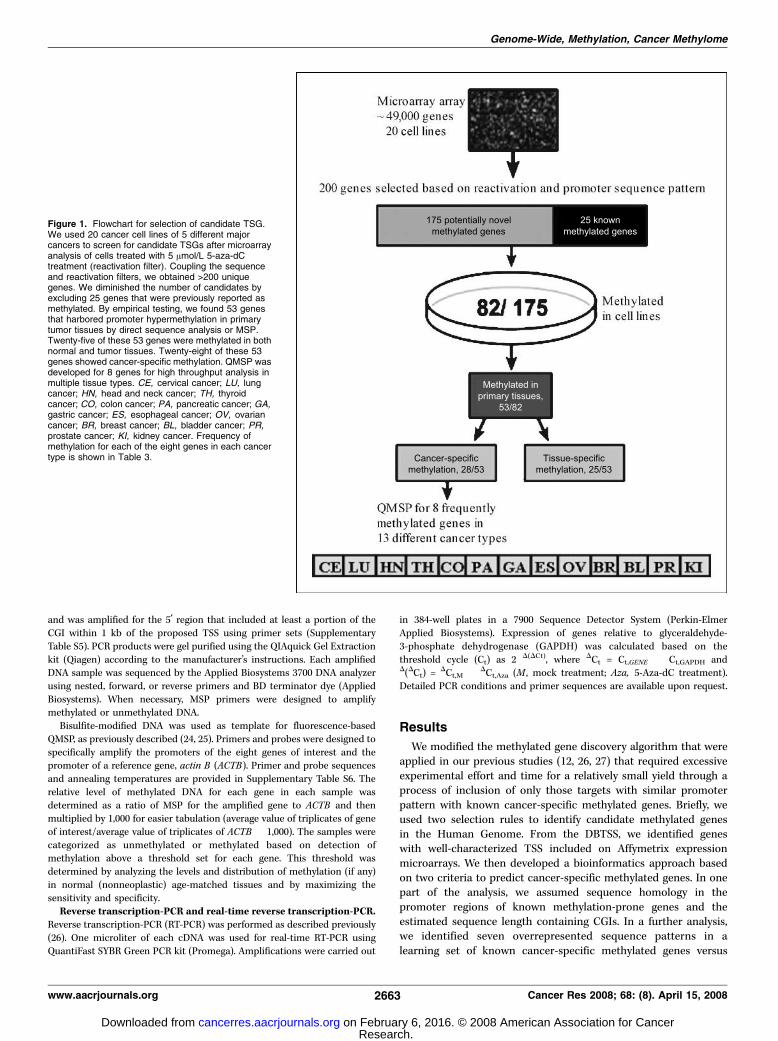
Figure 1. Flowchart for selection of candidate TSG.We used 20 cancer cell lines of 5 different majorcancers to screen for candidate TSGs after microarrayanalysis of cells treated with 5 Amol/L 5-aza-dCtreatment (reactivation filter). Coupling the sequenceand reactivation filters, we obtained >200 uniquegenes. We diminished the number of candidates byexcluding 25 genes that were previously reported asmethylated. By empirical testing, we found 53 genesthat harbored promoter hypermethylation in primarytumor tissues by direct sequence analysis or MSP.Twenty-five of these 53 genes were methylated in bothnormal and tumor tissues. Twenty-eight of these 53genes showed cancer-specific methylation. QMSP wasdeveloped for 8 genes for high throughput analysis inmultiple tissue types. CE, cervical cancer; LU, lungcancer; HN, head and neck cancer; TH, thyroidcancer; CO, colon cancer; PA, pancreatic cancer; GA,gastric cancer; ES, esophageal cancer; OV, ovariancancer; BR, breast cancer; BL, bladder cancer; PR,prostate cancer; KI, kidney cancer. Frequency ofmethylation for each of the eight genes in each cancertype is shown in Table 3.
Genome-Wide, Methylation, Cancer Methylome
www.aacrjournals.org 2663 Cancer Res 2008; 68: (8). April 15, 2008
Research. on February 6, 2016. © 2008 American Association for Cancercancerres.aacrjournals.org Downloaded from

Figure 2. Distribution of the predicted 200 methylated genes along the chromosomal map by computational approach. Most of the genes are clustered in limitedchromosomal regions. No genes were found on chromosome Y.
Cancer Research
Cancer Res 2008; 68: (8). April 15, 2008 2664 www.aacrjournals.org
Research. on February 6, 2016. © 2008 American Association for Cancercancerres.aacrjournals.org Downloaded from

tissue-specific methylated genes. We then applied these patterns toreal data sets generated in 20 cancer cell lines from the mostcommon types of cancer treated with 5 Amol/L 5-aza-dC toreactivate gene transcripts silenced by promoter methylation. Thegene filtering approach and data analysis are depicted in Fig. 1.We considered a gene to be reactivated if re-expression occurred
in at least one cell line of any particular cancer type. The 200methylation-prone genes identified from this computationalapproach are shown on a chromosomal map in Fig. 2.Validation of modified approach in cell lines. Out of the 200
genes predicted to be methylated by our modified approach, 25genes were identified as reported to harbor cancer-specificmethylation after a literature search [Pubmed search words,(particular gene name) and (methylation)]. To validate theremaining 175 genes, we designed primers for each gene and testedeach one by bisulfite sequence analysis, combined bisulfiterestriction analysis (COBRA), and/or MSP in one or more cell linethat exhibited re-expression after demethylation treatment. Pro-moter methylation of 82 genes (82 of 175; 47%) was documentedbased on identification of z50% methylated CpG sites in the CGI incontrast to 10% to 20% in previous algorithm (12, 27).Promoter hypermethylation in normal and primary tumor
tissues. To determine if the methylated genes in cancer cell lineswere cancer specific, we investigated promoter methylation in alimited number (n = 10–15 for tumors; n = 2–12 for normals) ofvarious primary tumors and age-matched normal tissues bybisulfite sequence analysis, COBRA, and/or MSP (SupplementaryTable S8). Out of 82 genes that showed methylation in cell lines,promoter methylation was detected in 53 (65%) genes in primarytumor tissues. After testing corresponding age-matched normaltissues, 28 of these genes were identified to be methylated in acancer-specific manner. Thus, 28 of 175 (16%) new cancer-specificmethylated genes were identified through our combination of acomputational approach and empirical studies. We used age-matched normal tissue as a control. If the frequency of methylationis higher in cancer and absent or lower level/frequency in normaltissue at an optimal cutoff, we considered it as a cancer-specificmethylation. A summary of our analysis of all 200 genes is detailedin Table 1.No methylation was detected in the remaining 93 genes (175–82)
in cell lines or primary tissues. However, we empirically analyzed
only 200 to 300 bp of a potential 1- to 2-kb region of the pro-moter by bisulfite sequencing or MSP for the majority of the genes.Figure 3A shows the chromatogram of bisulfite sequencing ofpromoter methylation of representative candidate genes in primarytissues and cancer cell lines of different cancer types. The cell linesexamined showed methylation of target gene and exhibitedsilencing of mRNA expression (Fig. 3B). This suggests that mRNAexpression of these genes were regulated by promoter hyper-methylation.Candidate cancer genes. The cancer-specific methylated genes
identified in this study are listed in Table 2. By modified approach,we selected 200 genes, and after empirical testing, 28 were newlyidentified candidate cancer genes (methylated in primary tumorsbut not in progenitor cells). Overall, between 2 and 12 newlymethylated cancer genes were identified in lung, breast, colon,prostate, and cervical cancer.The 200 candidate cancer genes identified in this study fell into
three categories: (a) genes previously observed to be altered inhuman cancer by methylation (e.g., APC, SFRP1, FHIT, andTWIST). The reidentification of genes previously shown to bemethylated in human cancers represents a critical validation ofour modified approach in this study; (b) Genes in which noprevious methylation in human cancers was discovered but hadbeen linked to cancer through functional studies (e.g., PAPSS2,TUBG2 , and DLL4). Although genetic and epigenetic alterationscurrently provide the most reliable indicator of the importance ofa gene in human neoplasia (7, 28, 29), there are many other genesthat are thought to play key roles on the basis of functional orexpression studies; (c) Genes with no previous connection withneoplasia (e.g., NTRK2, ASMLTL , and TFP12). In addition, cancer-specific methylation was observed in genes for which nobiological role has yet been established, such as OGDHL,C1ORF166 , and ARMC7 .New targets of aberrant methylation in major types of
cancer by QMSP. We noted that some of the cancer-specificmethylated genes were reactivated and methylated in more thanone type of cell line. To determine the frequency of methylation in alarger set of samples and in multiple cancer types, we selected 8 ofthe most frequently cancer-specific methylated genes from our listof newly identified 28 genes and developed a QMSP assay. Wefound cancer-specific methylation at various frequencies for each
Table 1. Summary of findings on 200 candidate methylated
Tissue types Potential
methylated
genes*
Reported
methylation in
cancer
Number of
genes tested
Methylated in
cell lines
Methylated in
tumor tissues
Methylation in
normal tissue
Cancer-specific
methylation
Lung Squamous 36 7 21 9/21 6/9 3/6 3/6
Lung adeno 18 5 13 6/13 2/6 1/2 1/2Breast 31 1 28 6/28 5/6 3/5 2/5
Prostate 45 9 36 16/36 8/16 5/8 3/8
Colorectal 48 4 44 23/44 16/23 10/16 6/16
Cervical 45 4 33 22/33 16/22 3/16 13/16Total 233
c30
b175 82/175 53/82 25/53 28/53
NOTE: Total experimentally tested genes: 200 � 25 = 175.
*Number of genes selected by modified approach.c33 overlapping genes across the all cancer types.b5 overlapping in different types of cancer. Thus, number of known cancer-specific methylated genes is 25 (30–5).
Genome-Wide, Methylation, Cancer Methylome
www.aacrjournals.org 2665 Cancer Res 2008; 68: (8). April 15, 2008
Research. on February 6, 2016. © 2008 American Association for Cancercancerres.aacrjournals.org Downloaded from

gene in multiple types of cancer (Table 3). A high frequency of
cancer-specific methylation for at least one gene was identified in
every cancer type, supporting the notion that methylated genes are
likely to play a role across multiple cancer types.
Discussion
Most studies on DNA methylation in cancer have focused on acandidate gene approach where a tumor suppressor or previouslyreported methylated gene is tested in another type of cancer.
Figure 3. A, promoter methylation ofrepresentative candidate genes. a, methylationof KIF1A and OSMR by conventionalmethylation-specific PCR in cancer cell lines andprimary tissues; M, methylated; U, unmethylated;NBT, normal breast tissues from noncancerpatients; BTT, breast tumor tissues; NN, normalcolon epithelium from noncancer patients; PN,paired normal colon tissues from colon cancerpatients; PT, paired colon cancer tissues.b, representative sequencing results of the PAK3and NISCH in cancer cell lines, normal, andcancer tissues. Normal tissues were taken fromnoncancer patients. Arrows, all guaninespresent after sequencing are complementary tomethyl cytosines on the opposite DNA strand.B, re-expression of representative genesanalyzed by semiquantitaive RT-PCR or real-timeRT-PCR. a and b, reactivated PAK3 and OGDHLwere observed by the 5-Aza-dC treatment inH23 and Siha cell lines by semiquantitativeRT-PCR. c, overexpression of OSMR wasobserved by the 5-Aza-dC treatment analyzed byreal-time quantitative RT-PCR. Relative fold wascalculated by the expression of OSMR mRNAto GAPDH (an internal control). Fold increase ofOSMR ranged from 5.2 (HCT116) to 2,868(DLD-1). Experiments were performed induplicate; columns, mean; bars, SD. *, P < 0.05.d, the amplification plot of OSMR transcript inDLD-1. A, 5-Aza-dC treated; M, mock treated.
Cancer Research
Cancer Res 2008; 68: (8). April 15, 2008 2666 www.aacrjournals.org
Research. on February 6, 2016. © 2008 American Association for Cancercancerres.aacrjournals.org Downloaded from

Although a number of studies have attempted to detect additionalgene targets, in general, the gene selection methodologies have notbeen sensitive enough to identify target genes with comparativelyless time and labor. By developing a new tool to analyze genepromoters in combination with a relatively large expressionmicroarray data set, it has been possible for the first time toidentify a large number of target genes. In our experience, this is amajor advance over previous empirical techniques that requiredexcessive experimental effort and yielded only a few (<0.5%)cancer-specific methylated genes (12, 27). Our yield, based on acombination of re-expression arrays and promoter sequencepattern, provided a nearly 500-fold higher yield of genes harboringpromoter methylation.We found that 47% (82 of 175) of the genes tested in cell lines
were methylated by bisulfite sequencing and/or MSP, and 65%(53 of 82) of these genes were methylated in primary tumors. Ourresults are consistent with previous studies (12, 26, 27), where the
frequency of methylation of any particular gene in primary tumors isgenerally less than that observed in cell lines. The discrepancybetween the computationally and pharmacologically predicted (175)and experimentally (82) identified methylated genes in cell linesmay be partially due to the analysis of limited regions (f 200–300 bpfor most of the genes) by bisulfite sequencing or MSP.To compare the overall pattern of methylated CGIs among
tumors, we tested 300 primary tumors of 13 different types with8 frequently cancer-specific methylated genes identified from ourapproach. Pancreas, gastric, thyroid, and ovary cancers displayedrelatively low levels of methylation. Colon, prostate, esophagus, andkidney tumors, however, displayed a much higher frequency ofmethylation overall. Some tumors within a type displayed highinherent levels of methylation, whereas others within the sametumor type displayed low levels (data not shown). The data are notconsistent with chance variation from tumor to tumor because inthe absence of heterogeneity, the variance of the methylation
Table 2. Cancer-specific methylated genes and their proposed functions
Genes Loci Gene name Proposed function
NISCH 3p21.1 Nischarin Human I(l)-imidazdine receptor candidate gene, IRAS
PIGH 14q11-q24 Phosphatidylinositol glycan, class H
cyclin-dependent kinaseinhibitor 1A (p21, Cip1) activated
Encodes an endoplasmic reticulum associated–protein
that is invovled in GPI-anchor biosynthesis
PAK3 Xq22.3-q23 Kinase 3 Critical effectors that link Rho GTPases to cytoskeleton
reorganization and nuclear signalingTUBB4 19p13.3 Tubulin, h4 h-tubulinKIF1A 2q37.3 Kineasin family member 1A Anterograde motor protein that transports
membranous organelles along axonal microtubules
MAL 2cen-q13 T-cell proliferation protein A candidate linker protein in T-cell signal transductionENPEP 4q25 Glutamyl aminpeptidase Type II integral membrane protein
MCAM 11q23.3 Melanoma cell adhasion molecule Cell adhesion
SSBP2 5q13.3 ssDNA-binding protein 2 DNA binding
B4GALT1 9p21 h 1,4-galactosyltransferase, polypeptide 1 Elevated level in highly metastatic human lung cancer cellsOSMR 5p12 Oncostatin M receptor Induces apoptosis in adrenocortical Y-1 tumor cells
NTRK2 9q22.1 Neurotrophic tyrosine kinase, receptor, type 2 Receptor for brain-derived neurotrophic factor,
neurotrophin-3
PAPSS2 10q23.1-q23.2 3¶-phosphoadenosis5¶-phosphosulfate synthetase 2
Bifunctional enzyme with both atp sulfurylaseand aps kinase activity
SFRP4 7p14.1 Secreted frizzled-related protein 4 Modulators of Wnt signaling
TUBG2 17q21 g-Tubulin gene Ostomalacia-associated tumors and endometrialand breast carcinomas
PTGS 2 1q25.2-q25.3 Prostaglandin-endoperoxide synthase 2 Prostaglandin biosynthesis
NPTX1 17q25.1-q25.2 Neuronal pentraxin I Calcium ion binding, transport, and synaptic transmission
CCNA1 13q12.3-q13 Cyclin A1 Cell cycleASMTL Xp22.3; Yp11.3 Acetylserotonin O-methyltransferase like O-methyltransferase activity,
S-adenosylmethionine-dependent
methyltransferase activity
TFP12 7q22 Tissue factor pathway inhibitor 2 Serine-type endopeptidase inhibitor activity, extracellularmatrix structural constituent
OGDHL 10q11.23 Oxoglutarate dehydrogenase like Plays a major role in the citric acid cycle, converting
a-ketoglutarate to succiynl CoADLL4 15q14 y-like 4 (Drosophila) Notch binding, calciumion binding
GDAP1L1 20q12 Ganglioside-induced differentiation-associated
protein 1-like 1
Mutation in human GDAP1 are associated with
Charcot-Marie-Tooth–type 4A disease
C1ORF166 1p36.12 Hypothetical protein LOC79594 Involved in mediating protein-protein interactionsARMC7 17q25.1 Amadillo repeat containing 7 Binding
C13orf18 13q14.12 Protein chromosome 13 open reading frame 18 Protein phosphatase inhibitor activity
C9orf19 9p13-p12 Golgi-associated plant pathogenesis-related
protein 1
Extracellular region
HCP1 17q11.2 Home carrier protein 1 Transporter activity
Genome-Wide, Methylation, Cancer Methylome
www.aacrjournals.org 2667 Cancer Res 2008; 68: (8). April 15, 2008
Research. on February 6, 2016. © 2008 American Association for Cancercancerres.aacrjournals.org Downloaded from

frequency would not be expected to be greater than the mean.Therefore, aberrant methylation of CGIs can be quantitativelydifferent in individual tumors within a tumor type and morepronounced in particular tumor types.We found cancer-specific and tissue-specific methylation events
in different tissue types. For example, PAK3 cancer-specificmethylation was found in esophagus, lung, cervix, head and neck,and bladder cancers with high frequency. PAK3 was alsooccasionally methylated in other normal tissues. PAK3 is locatedin the X chromosome; thus, it is likely that there will always bemethylated signal in samples from female patients. However, weconsider PAK3 as cancer-specific methylation as we also foundhigh frequency of methylation in samples from male cancerpatients. Like PAK3, some other genes showed either cancer-specific or tissue-specific methylation in multiple organs (Table 3).Although there have been reports of MCAM overexpression inmelanoma, we found a high frequency of MCAM promotermethylation in prostate cancer. Oncostatin M receptor (OSMR)showed cancer-specific methylation only in colon cancer and waspreviously shown to have a major functional role in breast andother cancers (30, 31). Liang et al. (32) reported loss of expressionof SSBP2 in 50% of myeloid leukemia cell lines and concluded thatloss of SSBP2 expression may underlie the impaired differentiationseen in human myeloid leukemia. However, before this report,there was no reported mechanism for loss of expression of thisDNA-binding protein. b4GalT-1 is constitutively expressed in alltissues, with the exception of the brain (33), as a Golgi-residentprotein. We found a high frequency of cancer-specific methylationof b4GalT-1 in esophagus, lung, colon, and prostate. NISCH[imidazoline receptor antisera selected (IRAS)] was first isolatedas an imidazoline-1 receptor candidate cloned by an IRAS cDNAapproach (34) and was independently shown to be an interactingpartner for insulin receptor substrate 4 (35). IRAS was recentlyreported to protect transfected PC12 cells from apoptosis (36, 37),whereas its mouse homologue, Nischarin, which lacks the NH2-terminal PX domain, was identified as a cytosolic-interacting
protein for a5 integrin and shown to inhibit cell migration byinhibiting the ability of PAK1 to phosphorylate substrates (37, 38).We found a high frequency of cancer-specific methylation of thisgene in lung, head and neck, and gastric cancer. KIF1A is a memberof the KIF1/Unc104 family, and targeted deletion of the KIF1A genein mice causes accumulation of clear small vesicles in the cell bodyof neurons as well as marked neuronal death (39). We report for thefirst time a high frequency of cancer-specific methylation of KIF1Ain majority of human tumors.The frequency of methylation within a tumor type of the
individual CGIs affected in at least three different tumor types isshown (Table 3). Some targets were methylated at a high frequencyin one tumor type but infrequently in others (e.g., OSMR; Table 3),whereas other targets (e.g., KIF1A) were methylated at relativelyhigh frequencies in the majority of tumor types. Thus, whereassome CGIs targets are shared by multiple tumor types, others aremethylated in a tumor-type–specific manner. It has beendocumented that virtually all biochemical, biological, and clinicalattributes are heterogeneous within human cancers of the samehistologic subtypes (40). Our data suggest that differences in themethylated genes in various tumors could account for a major partof this heterogeneity.Like any global genomic and epigenomic approach, our study has
limitations. First, we were not able to test all the known and newlydiscovered methylated genes in all the 13 types of cancer included inthis study. Second, although mosaic methylation occurred in mostof the cases, focal methylation for some genes was also reported,and methylation in 5¶ untranslated regions would not be detectableby the methods we used. Future studies using a combination ofdifferent technologies will be able to address these issues.The results of this study inform future cancer methylome
discovery effort in several important ways:
A major technical challenge of such studies will be discerningcancer-specific methylation from the large number of tissue-specific methylated genes. In our study, using modified gene
Table 3. Methylation frequency in different cancer types
Number of methylation positive/number of total cases (%)
PAK3 NISCH KIF1A OGDHL
Tissue types T N T N T N T N
Breast 13/29 (45) 1/10 (10) 8/24 (33) 1/10 (10) 12/29 (41) 0/9 (0) 8/25 (32) 0/10 (0)Esophagus 5/19 (26) 0/10 (0) 4/19 (21) 0/10 (0) 6/19 (32) 0/8 (0) 2/19 (11) 0/13 (0)
Lung 16/34 (47) 1/17 (6) 12/34 (35) 1/16 (6) 8/34 (24) 1/17 (6) 2/34 (6) 0/17 (0)
Pancreas 7/32 (22) 0/2 (0) 0/32 (0) 2/2 (100) 24/30 (80) 0/2 (0) 6/32 (19) 0/2 (0)
Colon 9/20 (45) 2/13 (15) 1/20 (5) 0/13 (0) 7/19 (37) 0/13 (0) 4/20 (20) 0/13 (0)Prostate 10/20 (50) 2/20 (10) 1/20 (5) 2/20 (10) 3/20 (15) 7/20 (35) 0/20 (0) 1/20 (5)
Gastric 4/14 (29) 10/10 (100) 6/14 (43) 0/10 (0) 9/12 (75) 10/10 (100) 3/14 (21) 10/10 (100)
Cervix 13/24 (54) 0/10 (0) 3/24 (13) 1/10 (10) 7/24 (29) 0/10 (0) 4/24 (17) 0/10 (0)Thyroid 8/20 (40) ND 4/20 (20) ND 4/20 (20) ND 0/20 (0) ND
Kidney 7/24 (29) ND 13/20 (65) ND 1/20 (5) ND 0/20 (0) ND
Head & Neck 13/25 (52) 0/8 (0) 9/25 (36) 0/8 (0) 9/25 (35) 0/8 (0) 2/25 (8) 0/8 (0)
Ovary 10/19 (53) ND 9/19 (47) ND 2/19 (11) ND 0/19 (0) NDBladder 3/20 (15) 0/6 (0) 5/20 (25) 1/6 (17) 4/20 (20) 0/6 (0) 0/20 (0) 0/6 (0)
Abbreviation: ND, not done.
Cancer Research
Cancer Res 2008; 68: (8). April 15, 2008 2668 www.aacrjournals.org
Research. on February 6, 2016. © 2008 American Association for Cancercancerres.aacrjournals.org Downloaded from

selection criteria in pharmalogical unmasking strategy, weidentified 47% methylated genes in contrast to 10% to 20% byprevious criteria. In the future, improvements in gene selectionstrategy for prediction of methylation-prone gene should resultin less labor and less empirical experimentation.Another technical issue is the development of high throughputassays for the analysis of large numbers of samples. In this study,we developed QMSP assay for eight novel cancer-specificmethylated genes and similar real-time assays could bedeveloped individually for newly identified methylated targets.Once a methylation target set is known for a particular cancer,or even if the entire cancer ‘‘methylome’’ is discovered, othergenomic approaches such as chip arrays may facilitate largescale research and clinical efforts.Although it is likely that studies of other solid tumor types willalso identify a large number of methylated genes, it will beimportant to apply rigorous approaches to identify the specificmethylated genes that have been selected for during tumori-genesis. Our modified approach can predict for cancer-specificmethylated genes and reduce empirical testing.There has been much discussion about which genes should bethe focus of future efforts for methylation analysis. Our resultssuggest that many genes not previously implicated in cancer aremethylated at significant levels and may provide novel clues tocancer pathogenesis.
Adding these data to previous reports, perhaps up to one third(f300 genes total) of the cancer methylome has now beendiscovered, compared with the identification of perhaps 200mutated genes over the past 2 decades and recent genome-widemutation analysis in primary tumors (41). An emerging picture ofgenetic and epigenetic changes and their relationship is unraveling
the biological networks responsible for human cancer. The geneticand epigenetic alterations in different cancer types are diverse(42, 43), and we and others previously found unique inverserelationships between genetic/epigenetic changes (27, 44, 45). How-ever, 26 genes obtained in the Vogelstein’s last mutation screeningare also methylated in our study (41, 46). Ultimately, the epigenomeof all cancer tissues will be mapped out even as we now approach atotal molecular signature of cancer. According to Dr. Peter Jones(as reviewed in ref. 47), each differentiated cell has a differentepigenome. Our comprehensive analysis contributes greatly to theemerging epigenomic map of DNA methylation in the humangenome. Additional studies using similar and complementarygenomic strategies should yield further insights into the dynamicsand hierarchy of epigenetic regulation during tumorigenesis. Thesedata define the epigenetic landscape of major human cancer types,provide new targets for diagnostic and therapeutic intervention,and open fertile avenues for basic research in tumor biology.
Acknowledgments
Received 10/17/2007; revised 1/16/2008; accepted 2/7/2008.Grant support: National Cancer Institute U01-CA84986, Oncomethylome Sciences,
SA, and Dutch Cancer Society (project number RUG 2004-3161). Under a licensingagreement between Oncomethylome Sciences, SA and the Johns Hopkins University,D. Sidransky is entitled to a share of royalty received by the University upon sales ofany products described in this article. D. Sidransky owns Oncomethylome Sciences, SAstock, which is subject to certain restrictions under University policy. D. Sidransky is apaid consultant to Oncomethylome Sciences, SA and is a paid member of thecompany’s Scientific Advisory Board. The Johns Hopkins University in accordancewith its conflict of interest policies is managing the terms of this agreement. A. van derZee is a paid consultant for OncoMethylome Sciences, SA, Liege, Belgium. M.O. Hoqueis a recipient of the FAMRI Young Clinical Scientist Award and aYoung InvestigatorAward from the International Association for the Study of Lung Cancer.
The costs of publication of this article were defrayed in part by the payment of pagecharges. This article must therefore be hereby marked advertisement in accordancewith 18 U.S.C. Section 1734 solely to indicate this fact.
Table 3. Methylation frequency in different cancer types (Cont’d)
Number of methylation positive/number of total cases (%)
OSMR B4GALT1 MCAM SSBP2
T N T N T N T N
1/28 (0) 0/10 (0) 11/28 (39) 0/10 (0) 28/29 (97) 7/1 (70) 20/29 (69) 6/10 (60)1/20 (5) 0/10 (0) 12/20 (60) 1/10 (10) 19/19 (100) 0/10 (0) 15/18 (83) 0/10 (0)
1/34 (0) 0/17 (0) 21/34 (60) 0/17 (0) 24/30 (80) 15/17 (88) 23/34 (68) 12/17 (71)
2/32 (6) 0/2 (0) 9/32 (28) 0/2 (0) 28/32 (88) 1/2 (50) 1/32 (3) 1/2 (50)
16/20 (75) 0/13 (0) 18/20 (90) 0/13 (0) 18/20 (90) 3/12 (25) 20/20 (100) 3/12 (25)0/20 (0) 0/20 (0) 20/20 (100) 0/20 (0) 17/20 (85) 0/20 (0) 16/20 (80) 2/20 (10)
0/14 (0) 12/12 (100) 11/14 (79) 8/12 (67) 12/14 (86) 12/12 (100) 13/14 (93) 12/12 (100)
1/24 (4) 0/10 (0) 4/24 (17) 0/10 (0) 14/23 (61) 7/10 (70) 9/23 (39) 4/10 (40)0/19 (0) ND 5/19 (26) ND 16/21 (76) ND 9/17 (53) ND
0/23 (0) ND 10/23 (43) ND 23/23 (100) ND 18/23 (78) ND
0/26 (0) 0/8 (0) 3/26 (12) 2/8 (25) 10/26 (38) 5/7 (71) 9/26 (35) 4/7 (57)
2/19 (11) ND 9/19 (47) ND 19/19 (100) ND 7/19 (37) ND0/20 (0) 0/6 (0) 9/20 (45) 0/6 (0) 18/20 (90) 3/6 (50) 5/20 (25) 0/6 (0)
References1. Hanahan D, Weinberg RA. The hallmarks of cancer.Cell 2000;100:57–70.
2. Holliday R, Pugh JE. DNA modification mechanismsand gene activity during development. Science 1975;187:226–32.
3. Riggs AD. X inactivation, differentiation, and DNAmethylation. Cytogenet Cell Genet 1975;14:9–25.4. Hark AT, Schoenherr CJ, Katz DJ, Ingram RS, LevorseJM, Tilghman SM. CTCF mediates methylation-sensitiveenhancer-blocking activity at the H19/Igf2 locus. Nature2000;405:486–9.5. Jaenisch R, Bird A. Epigenetic regulation of gene
expression: how the genome integrates intrinsicand environmental signals. Nat Genet 2003;33Suppl:245–54.6. Leonhardt H, Rahn HP, Cardoso MC. Functional linksbetween nuclear structure, gene expression, DNAreplication, and methylation. Crit Rev Eukaryot GeneExpr 1999;9:345–51.
Genome-Wide, Methylation, Cancer Methylome
www.aacrjournals.org 2669 Cancer Res 2008; 68: (8). April 15, 2008
Research. on February 6, 2016. © 2008 American Association for Cancercancerres.aacrjournals.org Downloaded from

7. Momparler RL. Cancer epigenetics. Oncogene 2003;22:6479–83.8. Ehrlich M. DNA hypomethylation, cancer, the immu-nodeficiency, centromeric region instability, facialanomalies syndrome and chromosomal rearrangements.J Nutr 2002;132:2424–9S.9. Belinsky SA, Nikula KJ, Palmisano WA, et al. Aberrantmethylation of p16 (INK4a) is an early event in lungcancer and a potential biomarker for early diagnosis.Proc Natl Acad Sci U S A 1998;95:11891–6.10. Hoque MO, Rosenbaum E, Westra WH, et al.Quantitative assessment of promoter methylation pro-files in thyroid neoplasms. J Clin Endocrinol Metab 2005;90:4011–8.11. Esteller M, Sparks A, Toyota M, et al. Analysis ofadenomatous polyposis coli promoter hypermethylationin human cancer. Cancer Res 2000;60:4366–71.12. Yamashita K, Upadhyay S, Osada M, et al. Pharma-cologic unmasking of epigenetically silenced tumorsuppressor genes in esophageal squamous cell carcino-ma. Cancer Cell 2002;2:485–95.13. Suzuki H, Gabrielson E, Chen W, et al. A genomicscreen for genes upregulated by demethylation andhistone deacetylase inhibition in human colorectalcancer. Nat Genet 2002;31:141–9.14. Gentleman RC, Carey VJ, Bates DM, et al.Bioconductor: open software development for compu-tational biology and bioinformatics. Genome Biol 2004;5:R80.15. Suzuki Y, Yamashita R, Sugano S, Nakai K. DBTSS,DataBase of Transcriptional Start Sites: progress report2004. Nucleic Acids Res 2004;32:D78–81.16. Suzuki Y, Yamashita R, Nakai K, Sugano S. DBTSS:DataBase of human Transcriptional Start Sites and full-length cDNAs. Nucleic Acids Res 2002;30:328–31.17. Olson SA. EMBOSS opens up sequence analysis.European Molecular Biology Open Software Suite. BriefBioinform 2002;3:87–91.18. Gardiner-Garden M, Frommer M. CpG islands invertebrate genomes. J Mol Biol 1987;196:261–82.19. Takai D, Jones PA. Comprehensive analysis of CpGislands in human chromosomes 21 and 22. Proc NatlAcad Sci U S A 2002;99:3740–5.20. Burgard AP, Moore GL, Maranas CD. Review of theTEIRESIAS-based tools of the IBM Bioinformatics andPattern Discovery Group. Metab Eng 2001;3:285–8.
21. Rigoutsos I, Floratos A. Combinatorial patterndiscovery in biological sequences: The TEIRESIASalgorithm. Bioinformatics 1998;14:55–67.22. Frank E, Hall M, Trigg L, Holmes G, Witten IH. Datamining in bioinformatics using Weka. Bioinformatics2004;20:2479–81.23. Hoque MO, Lee CC, Cairns P, Schoenberg M,Sidransky D. Genome-wide genetic characterization ofbladder cancer: a comparison of high-density single-nucleotide polymorphism arrays and PCR-based micro-satellite analysis. Cancer Res 2003;63:2216–22.24. Hoque MO, Begum S, Topaloglu O, et al. Quantitationof promoter methylation of multiple genes in urine DNAand bladder cancer detection. J Natl Cancer Inst 2006;98:996–1004.25. Hoque MO, Topaloglu O, Begum S, et al. Quantitativemethylation-specific polymerase chain reaction genepatterns in urine sediment distinguish prostate cancerpatients from control subjects. J Clin Oncol 2005;23:6569–75.26. Kim MS, Yamashita K, Baek JH, et al. N-methyl-D-aspartate receptor type 2B is epigenetically inactivatedand exhibits tumor-suppressive activity in humanesophageal cancer. Cancer Res 2006;66:3409–18.27. Tokumaru Y, Yamashita K, Osada M, et al. Inversecorrelation between cyclin A1 hypermethylation andp53 mutation in head and neck cancer identified byreversal of epigenetic silencing. Cancer Res 2004;64:5982–7.28. Vogelstein B, Kinzler KW. Cancer genes and thepathways they control. Nat Med 2004;10:789–99.29. Varmus H. The new era in cancer research. Science2006;312:1162–5.30. Liu J, Hadjokas N, Mosley B, Estrov Z, Spence MJ,Vestal RE. Oncostatin M-specific receptor expressionand function in regulating cell proliferation of normaland malignant mammary epithelial cells. Cytokine 1998;10:295–302.31. Savarese TM, Campbell CL, McQuain C, et al.Coexpression of oncostatin M and its receptors andevidence for STAT3 activation in human ovariancarcinomas. Cytokine 2002;17:324–34.32. Liang H, Samanta S, Nagarajan L. SSBP2, a candidatetumor suppressor gene, induces growth arrest anddifferentiation of myeloid leukemia cells. Oncogene2005;24:2625–34.
33. Lo NW, Shaper JH, Pevsner J, Shaper NL. Theexpanding h 4-galactosyltransferase gene family: mes-sages from the databanks. Glycobiology 1998;8:517–26.34. Wang Y, Zhou Y, Szabo K, Haft CR, Trejo J. Down-regulation of protease-activated receptor-1 is regulatedby sorting nexin 1. Mol Biol Cell 2002;13:1965–76.35. Piletz JE, Ivanov TR, Sharp JD, et al. Imidazolinereceptor antisera-selected (IRAS) cDNA: cloning andcharacterization. DNA Cell Biol 2000;19:319–29.36. Sano H, Liu SC, Lane WS, Piletz JE, Lienhard GE.Insulin receptor substrate 4 associates with the proteinIRAS. J Biol Chem 2002;277:19439–47.37. Dontenwill M, Pascal G, Piletz JE, et al. IRAS, thehuman homologue of Nischarin, prolongs survival oftransfected PC12 cells. Cell Death Differ 2003;10:933–5.38. Alahari SK, Nasrallah H. A membrane proximalregion of the integrin a5 subunit is important for itsinteraction with nischarin. Biochem J 2004;377:449–57.39. Yonekawa Y, Harada A, Okada Y, et al. Defect insynaptic vesicle precursor transport and neuronal celldeath in KIF1A motor protein-deficient mice. J Cell Biol1998;141:431–41.40. Shapiro JR, Shapiro WR. Clonal tumor cell heteroge-neity. Prog Exp Tumor Res 1984;27:49–66.41. Sjoblom T, Jones S, Wood LD, et al. The consensuscoding sequences of human breast and colorectalcancers. Science 2006;314:268–74.42. Costello JF, Fruhwald MC, Smiraglia DJ, et al.Aberrant CpG-island methylation has non-random andtumour-type-specific patterns. Nat Genet 2000;24:132–8.43. Esteller M, Corn PG, Baylin SB, Herman JG. A genehypermethylation profile of human cancer. Cancer Res2001;61:3225–9.44. Xing M, Cohen Y, Mambo E, et al. Early occurrence ofRASSF1A hypermethylation and its mutual exclusionwith BRAF mutation in thyroid tumorigenesis. CancerRes 2004;64:1664–8.45. Toyooka S, Tokumo M, Shigematsu H, et al.Mutational and epigenetic evidence for independentpathways for lung adenocarcinomas arising in smokersand never smokers. Cancer Res 2006;66:1371–5.46. Wood LD, Parsons DW, Jones S, et al. The genomiclandscapes of human breast and colorectal cancers.Science 2007;318:1108–13.47. Garber K. Momentum building for human epige-nome project. J Natl Cancer Inst 2006;98:84–6.
Cancer Research
Cancer Res 2008; 68: (8). April 15, 2008 2670 www.aacrjournals.org
Research. on February 6, 2016. © 2008 American Association for Cancercancerres.aacrjournals.org Downloaded from

2008;68:2661-2670. Cancer Res Mohammad Obaidul Hoque, Myoung Sook Kim, Kimberly Laskie Ostrow, et al. Cancer MethylomeGenome-Wide Promoter Analysis Uncovers Portions of the
Updated version
http://cancerres.aacrjournals.org/content/68/8/2661
Access the most recent version of this article at:
Material
Supplementary
http://cancerres.aacrjournals.org/content/suppl/2008/04/15/68.8.2661.DC1.html
Access the most recent supplemental material at:
Cited articles
http://cancerres.aacrjournals.org/content/68/8/2661.full.html#ref-list-1
This article cites 47 articles, 25 of which you can access for free at:
Citing articles
http://cancerres.aacrjournals.org/content/68/8/2661.full.html#related-urls
This article has been cited by 16 HighWire-hosted articles. Access the articles at:
E-mail alerts related to this article or journal.Sign up to receive free email-alerts
Subscriptions
Reprints and
To order reprints of this article or to subscribe to the journal, contact the AACR Publications
Permissions
To request permission to re-use all or part of this article, contact the AACR Publications
Research. on February 6, 2016. © 2008 American Association for Cancercancerres.aacrjournals.org Downloaded from