Molecular Analysis of Aedes aegypti Classical Protein Tyrosine Phosphatases Uncovers an Ortholog of...
13
Molecular Analysis of Aedes aegypti Classical Protein Tyrosine Phosphatases Uncovers an Ortholog of Mammalian PTP-1B Implicated in the Control of Egg Production in Mosquitoes Debora Monteiro Moretti 1,6 , Lalima Gagan Ahuja 2 , Rodrigo Dutra Nunes 1,6 , Cecı´lia Oliveira Cudischevitch 1,6 , Carlos Renato Oliveira Daumas-Filho 1,6 , Priscilla Medeiros-Castro 1,6 , Guilherme Ventura-Martins 1,6 , Willy Jablonka 1,6 , Felipe Gazos-Lopes 1,6 , Raquel Senna 1,6 , Marcos Henrique Ferreira Sorgine 1,6 , Klaus Hartfelder 3 , Margareth Capurro 4 , Georgia Correa Atella 1,6 , Rafael Dias Mesquita 5,6 , Ma ´ rio Alberto Cardoso Silva-Neto 1,6 * 1 Laborato ´ rio de Sinalizac ¸a ˜ o Celular (LabSiCel), Instituto de Bioquı ´mica Me ´ dica Leopoldo de Meis, Universidade Federal do Rio de Janeiro, Rio de Janeiro, RJ, Brazil, 2 Department of Pharmacology, University of California San Diego, San Diego, California, United States of America, 3 Departamento de Biologia Celular e Molecular e Bioagentes Patoge ˆ nicos, Faculdade de Medicina de Ribeira ˜o Preto, Universidade de Sa ˜o Paulo, Ribeira ˜ o Preto, Brazil, 4 Departamento de Parasitologia, Instituto de Cie ˆ ncias Biome ´ dicas, Universidade de Sa ˜o Paulo, Sa ˜o Paulo, Brazil, 5 Departamento de Bioquı ´mica, Instituto de Quı ´mica, Universidade Federal do Rio de Janeiro, Rio de Janeiro, RJ, Brazil, 6 Instituto Nacional de Cie ˆ ncia e Tecnologia em Entomologia Molecular (INCT-EM), Rio de Janeiro, RJ, Brazil Abstract Background: Protein Tyrosine Phosphatases (PTPs) are enzymes that catalyze phosphotyrosine dephosphorylation and modulate cell differentiation, growth and metabolism. In mammals, PTPs play a key role in the modulation of canonical pathways involved in metabolism and immunity. PTP1B is the prototype member of classical PTPs and a major target for treating human diseases, such as cancer, obesity and diabetes. These signaling enzymes are, hence, targets of a wide array of inhibitors. Anautogenous mosquitoes rely on blood meals to lay eggs and are vectors of the most prevalent human diseases. Identifying the mosquito ortholog of PTP1B and determining its involvement in egg production is, therefore, important in the search for a novel and crucial target for vector control. Methodology/Principal Findings: We conducted an analysis to identify the ortholog of mammalian PTP1B in the Aedes aegypti genome. We identified eight genes coding for classical PTPs. In silico structural and functional analyses of proteins coded by such genes revealed that four of these code for catalytically active enzymes. Among the four genes coding for active PTPs, AAEL001919 exhibits the greatest degree of homology with the mammalian PTP1B. Next, we evaluated the role of this enzyme in egg formation. Blood feeding largely affects AAEL001919 expression, especially in the fat body and ovaries. These tissues are critically involved in the synthesis and storage of vitellogenin, the major yolk protein. Including the classical PTP inhibitor sodium orthovanadate or the PTP substrate DiFMUP in the blood meal decreased vitellogenin synthesis and egg production. Similarly, silencing AAEL001919 using RNA interference (RNAi) assays resulted in 30% suppression of egg production. Conclusions/Significance: The data reported herein implicate, for the first time, a gene that codes for a classical PTP in mosquito egg formation. These findings raise the possibility that this class of enzymes may be used as novel targets to block egg formation in mosquitoes. Citation: Moretti DM, Ahuja LG, Nunes RD, Cudischevitch CO, Daumas-Filho CRO, et al. (2014) Molecular Analysis of Aedes aegypti Classical Protein Tyrosine Phosphatases Uncovers an Ortholog of Mammalian PTP-1B Implicated in the Control of Egg Production in Mosquitoes. PLoS ONE 9(8): e104878. doi:10.1371/ journal.pone.0104878 Editor: Luciano A. Moreira, Centro de Pesquisas Rene ´ Rachou, Brazil Received December 30, 2013; Accepted July 18, 2014; Published August 19, 2014 Copyright: ß 2014 Moretti et al. This is an open-access article distributed under the terms of the Creative Commons Attribution License, which permits unrestricted use, distribution, and reproduction in any medium, provided the original author and source are credited. Funding: This work was supported by FAPERJ (Fundacao Carlos Chagas Filho de Apoio a Pesquisa do Estado do Rio de Janeiro), CNPq (Conselho Nacional de Desenvolvimento Cientifico e Tecnologico) and INCT-EM (Instituto Nacional de Ciencia e Tecnologia em Entomologia Molecular). The funders had no role in study design, data collection and analysis, decision to publish, or preparation of the manuscript. Competing Interests: The authors have declared that no competing interests exist. * Email: [email protected] Introduction Tyrosine phosphorylation is part of a complex cell repertoire that first appeared nearly 600-million years ago and is largely responsible for the emergence of the first multicellular animals [1]. Protein tyrosine phosphatases (PTPs) are enzymes that catalyze tyrosine dephosphorylation and regulate central steps of cell biology. The PTP family is composed of four different subfamilies. The active sites of classes I, II and III each harbor a cysteine, which is involved in catalysis. In Class IV, this cysteine is replaced PLOS ONE | www.plosone.org 1 August 2014 | Volume 9 | Issue 8 | e104878
Transcript of Molecular Analysis of Aedes aegypti Classical Protein Tyrosine Phosphatases Uncovers an Ortholog of...

Molecular Analysis of Aedes aegypti Classical ProteinTyrosine Phosphatases Uncovers an Ortholog ofMammalian PTP-1B Implicated in the Control of EggProduction in MosquitoesDebora Monteiro Moretti1,6, Lalima Gagan Ahuja2, Rodrigo Dutra Nunes1,6,
Cecılia Oliveira Cudischevitch1,6, Carlos Renato Oliveira Daumas-Filho1,6, Priscilla Medeiros-Castro1,6,
Guilherme Ventura-Martins1,6, Willy Jablonka1,6, Felipe Gazos-Lopes1,6, Raquel Senna1,6,
Marcos Henrique Ferreira Sorgine1,6, Klaus Hartfelder3, Margareth Capurro4, Georgia Correa Atella1,6,
Rafael Dias Mesquita5,6, Mario Alberto Cardoso Silva-Neto1,6*
1 Laboratorio de Sinalizacao Celular (LabSiCel), Instituto de Bioquımica Medica Leopoldo de Meis, Universidade Federal do Rio de Janeiro, Rio de Janeiro, RJ, Brazil,
2 Department of Pharmacology, University of California San Diego, San Diego, California, United States of America, 3 Departamento de Biologia Celular e Molecular e
Bioagentes Patogenicos, Faculdade de Medicina de Ribeirao Preto, Universidade de Sao Paulo, Ribeirao Preto, Brazil, 4 Departamento de Parasitologia, Instituto de
Ciencias Biomedicas, Universidade de Sao Paulo, Sao Paulo, Brazil, 5 Departamento de Bioquımica, Instituto de Quımica, Universidade Federal do Rio de Janeiro, Rio de
Janeiro, RJ, Brazil, 6 Instituto Nacional de Ciencia e Tecnologia em Entomologia Molecular (INCT-EM), Rio de Janeiro, RJ, Brazil
Abstract
Background: Protein Tyrosine Phosphatases (PTPs) are enzymes that catalyze phosphotyrosine dephosphorylation andmodulate cell differentiation, growth and metabolism. In mammals, PTPs play a key role in the modulation of canonicalpathways involved in metabolism and immunity. PTP1B is the prototype member of classical PTPs and a major target fortreating human diseases, such as cancer, obesity and diabetes. These signaling enzymes are, hence, targets of a wide arrayof inhibitors. Anautogenous mosquitoes rely on blood meals to lay eggs and are vectors of the most prevalent humandiseases. Identifying the mosquito ortholog of PTP1B and determining its involvement in egg production is, therefore,important in the search for a novel and crucial target for vector control.
Methodology/Principal Findings: We conducted an analysis to identify the ortholog of mammalian PTP1B in the Aedesaegypti genome. We identified eight genes coding for classical PTPs. In silico structural and functional analyses of proteinscoded by such genes revealed that four of these code for catalytically active enzymes. Among the four genes coding foractive PTPs, AAEL001919 exhibits the greatest degree of homology with the mammalian PTP1B. Next, we evaluated the roleof this enzyme in egg formation. Blood feeding largely affects AAEL001919 expression, especially in the fat body andovaries. These tissues are critically involved in the synthesis and storage of vitellogenin, the major yolk protein. Including theclassical PTP inhibitor sodium orthovanadate or the PTP substrate DiFMUP in the blood meal decreased vitellogeninsynthesis and egg production. Similarly, silencing AAEL001919 using RNA interference (RNAi) assays resulted in 30%suppression of egg production.
Conclusions/Significance: The data reported herein implicate, for the first time, a gene that codes for a classical PTP inmosquito egg formation. These findings raise the possibility that this class of enzymes may be used as novel targets to blockegg formation in mosquitoes.
Citation: Moretti DM, Ahuja LG, Nunes RD, Cudischevitch CO, Daumas-Filho CRO, et al. (2014) Molecular Analysis of Aedes aegypti Classical Protein TyrosinePhosphatases Uncovers an Ortholog of Mammalian PTP-1B Implicated in the Control of Egg Production in Mosquitoes. PLoS ONE 9(8): e104878. doi:10.1371/journal.pone.0104878
Editor: Luciano A. Moreira, Centro de Pesquisas Rene Rachou, Brazil
Received December 30, 2013; Accepted July 18, 2014; Published August 19, 2014
Copyright: � 2014 Moretti et al. This is an open-access article distributed under the terms of the Creative Commons Attribution License, which permitsunrestricted use, distribution, and reproduction in any medium, provided the original author and source are credited.
Funding: This work was supported by FAPERJ (Fundacao Carlos Chagas Filho de Apoio a Pesquisa do Estado do Rio de Janeiro), CNPq (Conselho Nacional deDesenvolvimento Cientifico e Tecnologico) and INCT-EM (Instituto Nacional de Ciencia e Tecnologia em Entomologia Molecular). The funders had no role in studydesign, data collection and analysis, decision to publish, or preparation of the manuscript.
Competing Interests: The authors have declared that no competing interests exist.
* Email: [email protected]
Introduction
Tyrosine phosphorylation is part of a complex cell repertoire
that first appeared nearly 600-million years ago and is largely
responsible for the emergence of the first multicellular animals [1].
Protein tyrosine phosphatases (PTPs) are enzymes that catalyze
tyrosine dephosphorylation and regulate central steps of cell
biology. The PTP family is composed of four different subfamilies.
The active sites of classes I, II and III each harbor a cysteine,
which is involved in catalysis. In Class IV, this cysteine is replaced
PLOS ONE | www.plosone.org 1 August 2014 | Volume 9 | Issue 8 | e104878

by aspartic acid [2]. Class I Cys-PTPs are the largest group of
PTPs and are divided into ‘‘classical’’ and dual specificity
phosphatases. Classical phosphatases are enzymes that are strictly
devoted to the dephosphorylation of phosphotyrosine residues.
Classical PTPs have been further subdivided into receptor PTPs
and soluble or non-transmembrane PTPs [3].
Aedes aegypti is the vector of Dengue and yellow fever. Once it
feeds on blood, a complex series of signaling events lead to yolk
synthesis and egg formation. Synthesized by the female mosquito
fat body, vitellogenin (Vg), the main yolk protein, is the ultimate
result of a chain of endocrine and signaling events that are still not
completely understood. It has been shown that, after a blood meal,
the amino-acid concentration in the hemolymph increases and the
synthesis of brain-derived signaling molecules, such as insulin-like
peptides, is induced [4–6]. Such peptides stimulate the ovaries to
produce ecdysone, which then induces the fat body to produce Vg.
Vg production by the fat body also relies on amino acids derived
from blood digestion, which activate the TOR/S6k signaling
cascade [7,8]. Vg is then secreted by the fat body into the
hemolymph and taken up by the developing follicles via receptor-
mediated endocytosis. In mosquitoes, the interaction of insulin or
insulin-like peptides with the mosquito insulin receptor (MIR)
triggers the PI3K/Akt pathway and promotes the production of
ecdysteroids, the regulation of egg formation and immunity [9,10].
Furthermore, inhibition of PTPs an antagonist of the insulin
pathway decreases ecdysteroid production by mosquito ovaries
[9]. Silencing of the Phosphatase and Tensin homologue (PTEN),
an antagonist of the PI3K pathway, leads to an increase in egg
formation [10]. The above studies suggest the presence of PTPs as
modulators of egg formation in mosquitoes, but the genes coding
for these enzymes have not yet been identified. It is possible that
the inhibition of PTP activity encoded by such genes may
ultimately reduce or impair the ability of female mosquitoes to lay
eggs, as demonstrated for other components involved in vitello-
genesis [11,12].
In the study reported herein, we conducted a bioinformatics
analysis of the A. aegypti genome to identify the mosquito ortholog
of PTP1B and determine its involvement in egg formation [13].
The inhibition of these regulators or the blocking of proteins under
their transcriptional control can potentially provide new targets for
suppression of egg formation and pathogen transmission by
mosquitoes [14]. Among the genes that encode mosquito PTPs,
AAEL001919 has the highest (53%) identity with human PTP1B.
The treatment of blood-fed mosquitoes with classical PTP
inhibitors or the silencing of this gene through RNAi partially
blocked egg production. Thus, AAEL001919 may present a
potential target for the control of tyrosine phosphorylation in
mosquitoes and may ultimately be used to decrease mosquito
reproduction.
Results
In silico analysis of soluble PTP sequencesThe search for PTP sequences in the Aedes aegypti genome has
led to the discovery of 8 PTP genes coding for 10 proteins
belonging to Class I soluble PTPs: AAEL001046, AAEL001919-
PA, AAEL001919-PB, AAEL003108, AAEL005492,
AAEL008528-PA, AAEL008528-PB, AAEL010234,
AAEL010914 and AAEL011434 (Figure 1). Among the genes
that code for classical PTPs in Aedes, two, AAEEL001919 and
AAEL008528, presented mRNA splice variants denoted as RA or
RB. For each of these genes, the distinguishing character was the
presence of at least one classical PTP domain (Figure 1). Some
PTPs showed accessory domains, including AAEL003108, that
harbored adaptor domains, including FERM and PDZ, which are
crucial for signal transduction (Figure 1). Several of these
sequences also showed the presence of inactive or absent PTP
domains, the significance of which can be linked to the role of
pseudo-phosphatases in signaling. Therefore, these sequences may
function as adaptor molecules that link pathways rather than fill
catalytic roles [15].
Analysis of PTP sequences and their specific motifsAs the PTP domain is defined by ten sequence and structural
motifs [16], the sequences of interest were analyzed in detail for
PTP motifs. Of the ten motifs, special attention was given to the
active-site motifs, motifs 1, 8, 9 and 10. The other six motifs (2, 3,
4, 5, 6, 7) form the core of the PTP protein and are implicated in
the thermodynamic stabilities of the molecules. Sequence conser-
vation in the four active-site motifs was, therefore, used as a tool to
classify the PTP sequences into active or inactive PTP domains
(Table 1 and Table 2). PTP activity is critically dependent on the
nucleophilic cysteine present in Motif 9 (also called the HCS motif)
[17,18]. Motif 1 (also called the KNRY loop), while not directly
involved in the reaction per se, is crucial for recruiting the
phosphotyrosine into the PTP active site and distinguishes PTPs
from serine/threonine phosphatases [19].
Of the ten PTP sequences analyzed, the following seven showed
the presence of at least one active PTP domain: AAEL001919-PA,
AAEL001919-PB, AAEL005492, AAEL011434, AAEL008528-
Figure 1. Domain architecture of A. aegypti Protein TyrosinePhosphatases. The ten Protein Tyrosine Phosphatase sequences fromA. aegypti were analyzed for the presence biologically active domains.The domains were identified using a conserved sequence search asmentioned in the methods. Domain architecture was mapped onto thesequence using the DOG 1.0 protein-structure illustrator (DOG 1.0:illustrator of protein domain structures). Nomenclature used is asfollows: PTP - Protein Tyrosine Phosphatase domain, PTP (br) - AbsentProtein Tyrosine Phosphatase domain, SEC14 - Phosphatidylinositoltransfer protein domain, FERM - F for 4.1 protein, E for ezrin, R forradixin and M for moesin, cytoskeletal association domain (includes theFA: FERM-associated domain) PDZ - also called DHR or GLGF, adaptordomain.doi:10.1371/journal.pone.0104878.g001
The Mosquito PTP1B Regulates Egg Formation
PLOS ONE | www.plosone.org 2 August 2014 | Volume 9 | Issue 8 | e104878

PA, AAEL008528-PB and AAEL003108 (Table 1 and Figure 2).
The active domains of the splicing variants of AAEL001919 and
AAEL008528 were identical (Table 1 and Figure 2). A multiple
sequence alignment of these sequences with human PTP1B
showed the presence of all ten conserved PTP motifs (Figures 1
and 4). The sequences of these proteins were subsequently used to
obtain molecular models to enable further study of their structure-
function relationships.
Structural analysis of mosquito PTP domainsMolecular models of the five active PTP domains from the
above seven different proteins were obtained using the previously
solved homologous PTP-1B structure as a template (Figure 3,
Table 1). Each of the structures showed the classical PTP fold with
a twisted b-sheet composed of eight b-strands at the center flanked
by eight a-helices. The active-site cysteine was observed at the
center of the active site flanked by the general acid aspartate from
Motif 8. Motif 10 glutamines were also observed at the active site
(Figure 4). A superposition of these structures with human PTP1B
showed that the geometry of the central active site is extremely
conserved (Figure 4 and Table 2). This result is consistent with the
information obtained from multiple sequence alignments which
showed that the ten motifs to be extremely conserved (Figure 2).
Evolutionary analysis of mosquito PTP sequencesA phylogram of the A. aegypti PTP domains was made using the
previously characterized PTPs of humans (Homo sapiens) and flies
(Drosophila melanogaster) (Figure 5). These genomes were chosen
Table 1. Analysis of PTP catalytic sequences.
ProteinAccessionnumber
Numberof PTPdomains
PTP domainBoundary(amino acidsequence number)
Sequence of Motif 1, Motif 8,Motif 9 and Motif 10 Motifs active/inactive
PTP domainpredictedactive/inactive
AAEL001046 2 PTP ABSENT (1–182) INACTIVE/DOMAIN ABSENT INACTIVE INACTIVE
PTP ABSENT (189–281) INACTIVE/DOMAIN ABSENT INACTIVE INACTIVE
AAEL001919-PA 1 1–206 (ABSENT) YTTWPDFGIP PIIHCSAGIGRSGT IQTVDQLYF INACTIVE ACTIVE ACTIVEACTIVE
ACTIVE
AAEL001919-PB 1 1–206 (ABSENT) YTTWPDFGIP PIIHCSAGIGRSGT IQTVDQLYF INACTIVE ACTIVE ACTIVEACTIVE
ACTIVE
AAEL003108 1 603–979 NLNKNRY YLAWPDHGVP PIIHCSAGIGRTG VQNVSQYRF ACTIVE ACTIVE ACTIVEACTIVE
ACTIVE
AAEL005492 1 410–681 NLAKNRY FTSWPDYGVP PMVVHCSAGIGRT IQMPDQYVF ACTIVE ACTIVE ACTIVEACTIVE
ACTIVE
AAEL008528-PA 2 PTP (ABSENT) 103–167 INACTIVE/DOMAIN ABSENT INACTIVE INACTIVE
PTP (205–445) NESKHKR FQVWPDHGVP PICVHCSAGIGRT VQTEAQYKF INACTIVE ACTIVE ACTIVEACTIVE
ACTIVE
AAEL008528-PB 2 PTP (ABSENT) 103–167 INACTIVE/DOMAIN ABSENT INACTIVE INACTIVE
PTP (205–445) NESKHKR FQVWPDHGVP PICVHCSAGIGRT VQTEAQYKF INACTIVE ACTIVE ACTIVEACTIVE
ACTIVE
AAEL010234 1 991–1228 NKARNF YNEWGDQNCP PPVLIHCNEGGGRTPSLAQYKF ACTIVE INACTIVE INACTIVEINACTIVE
INACTIVE
AAEL010914 1 307–573 KNRSID LWPKQSA NCLNGSDRSC DPNHMQL INACTIVE INACTIVEINACTIVE INACTIVE
INACTIVE
AAEL011434 1 781–1181 QSKNRY FPDWPDHRSP PIIHCSAGIGRTG VQNSEQYEL ACTIVE ACTIVE ACTIVEACTIVE
ACTIVE
doi:10.1371/journal.pone.0104878.t001
Table 2. Analysis of the superposition Mosquito PTPs with human PTP1B.
ProteinAmino AcidsModeled Template PTP structure Name/PDB ID
Percentage SequenceIdentity
Root Mean SquareDeviation (RMSD) fromPTP1B structure(2AZR)(C-a RMSD) (A)
AAEL001919 229 TCPTP (2I1Y) 34% 0.283
AAEL005492 299 MEG2/PTPN9(2PA5) 47% 1.546
AAEL011434 283 PCPTP1/PTPR (2A8B) 29% 2.323
AAEL003108 262 PTPH1/PTPN3(2B49) 46% 1.510
AAEL008528 309 (PTP domain) SHP2/PTPN11 (3B7O) 60% 1.1412
188 (Absent PTPdomain)
CD45 (1YGU) 19% ---
doi:10.1371/journal.pone.0104878.t002
The Mosquito PTP1B Regulates Egg Formation
PLOS ONE | www.plosone.org 3 August 2014 | Volume 9 | Issue 8 | e104878

because of their closeness with the mosquito genome (D.melanogaster) and for their extensive biochemical and structural
characterizations (H. sapiens). The phylogram was generated
using a multiple sequence alignment at the CLUSTALW web-
server. The neighbor-joining algorithm was used to obtain the
evolutionary tree using the TREEVIEW software package [20].
From the phylogram, it was evident that both active and inactive
A. aegypti PTP domains were closely related to the vertebrate and
fly PTP domains. The well-formed PTP domain of AAEL008528
was observed to be closest to the D. melanogaster Corkscrew PTP.
Belonging to the SHP1 family, these PTPs are characterized by
the presence of SH2 in addition to their tyrosine phosphatase
domains. Interestingly, the absent PTP domain of AAEL008528
also clustered near these proteins (along with the absent N-
terminal PTP domain of AAEL001046), indicating a plausible role
for these absent PTP domains as adapter modules. The single PTP
domains AAEL05492, AAEL003108 and AAEL001919 clustered
separately into three different groups, which were closest to the D.melanogaster DPTP52F, dm PTPMEG, dmPTP61F and H.sapiens MAG2, MEG1, PTP1B, respectively. The phylogram
also indicated that the inactive and active PTPs from A. aegypticlustered separately, but the proximity of the inactive PTP
sequences to the other PTP homologues indicates that these could
function as signaling molecules, much like adaptor proteins. The
gene AAEL011434 was also described in the analysis conducted
by Hatzihristidis et al. 2013 [21]. However, in Drosophila, this
enzyme is a negative modulator of the MAPK pathway and is,
thus, not considered in this study as a true member of the classical
PTP group [22].
PTP involvement in mosquito egg formationThe involvement of a complex set of intracellular signaling
pathways in the regulation of Vg synthesis and egg formation has
been described in the literature [23]. To determine the involve-
ment of PTPs in mosquito reproduction, we initially evaluated the
expression in different tissues of the 4 PTP genes that code for
active enzymes (AAEL005492, AAEL008528, AAEL003108, and
AAEL001919). AAEL011434 was not analyzed because this gene
was identified as a PTP after the major experiments were
completed and because it seems to be a dual-specificity phospha-
tase that modulates MAPKs [22]. AAEL003108 and
AAEL001919 are the only PTPs detectable in all tissues isolated
from sucrose-fed mosquitoes (Figure 6). In contrast, AAEL005492
had very low concentration in all tissues. Curiously, AAEL008528
is significantly expressed in the mosquito head, concomitant with a
transient increase in phosphotyrosine phosphorylation that may
involve fluctuations in the expression levels of AAEL008528 [24].
AAEL008528 is orthologous to Drosophila corkscrew, a PTP that
was discovered in mutations related to defects in embryonic
development [25,26]. AAEL003108 clusters with the DrosophiladmPTPMEG. PTPMEG is a cytoplasmic PTP containing FERM
(F for 4.1 protein, E for ezrin, R for radixin and M for moesin) and
PDZ (P for postsynaptic density protein (PSD95)), D for
Drosophila disc large-tumor suppressor (Dlg1), and Z for zonula
occludens-1 protein (zo-1)) domains. AAEL001919 clusters with
the Drosophila PTP dmPTP61F [29,30,31]. In mammalian T
cells, PTP (TCPTP) and PTP1B share a high level of structural
similarity but display different functions, which are defined
through the presence of specific non-catalytic domains that alter
enzyme localization within the cell.
AAEL003108 clusters with the Drosophila dmPTPMEG.
PTPMEG is a cytoplasmic PTP containing FERM (F for 4.1
protein, E for ezrin, R for radixin and M for moesin) and PDZ (P
for postsynaptic density protein (PSD95)), D for Drosophila disc
large-tumor suppressor (Dlg1), and Z for zonula occludens-1
protein (zo-1)) domains. In Drosophila, the vertebrate homologs of
Figure 2. Multiple sequence alignments of the A. aegypti PTP domains with the PTP domain of human PTP1B. Regions of highsequence similarity (.90\%) are highlighted in red; regions of moderate sequence identity (50–90%) are highlighted in blue. The ten motifs defininga PTP domain are marked in red boxes. These are in accordance with the nomenclature of Andersen et al. [59].doi:10.1371/journal.pone.0104878.g002
The Mosquito PTP1B Regulates Egg Formation
PLOS ONE | www.plosone.org 4 August 2014 | Volume 9 | Issue 8 | e104878

PTPN3 (PTPH1) and PTPN4 (MEG1) are expressed in the
nervous system, where they are involved in the regulation of axon
projections [27,28]. PTP 003108 was not further addressed in the
following experiments no matter it is clearly more expressed in the
ovaries due to two reasons: its lack of function in the mammalian
Figure 3. Structural models of the ‘active’ PTP sequences from A. aegypti. All proteins showed the classical PTP fold with a twisted b sheet atthe center surrounded by a helices. Because of its low sequence conservation, the absent PTP domain of AAEL008528 could not be modelledcompletely. Only one model is shown for genes with splicing variants because their PTP domains are identical. For additional details, please checkFigure 4.doi:10.1371/journal.pone.0104878.g003
The Mosquito PTP1B Regulates Egg Formation
PLOS ONE | www.plosone.org 5 August 2014 | Volume 9 | Issue 8 | e104878

models and its lack of effect on early silencing experiments of egg
formation and [28]
AAEL001919 clusters with the Drosophila PTP dmPTP61F
[29,30,31]. In mammalian T cells, PTP (TCPTP) and PTP1B
share a high level of structural similarity but display different
functions, which are defined through the presence of specific non-
catalytic domains that alter enzyme localization within the cell.
PTP activity against pNPP using tissues from sucrose-fed
mosquitoes is highly sensitive to sodium orthovanadate (data not
shown). In addition, in our pNPP assays with mosquito tissues, the
inclusion of a peptide that contains a phosphotyrosine dephos-
phorylated by the mammalian PTP1B leads to a 40–90%
inhibition of total PTP activity (Figure S1), indicating the presence
of catalytically active mosquito enzymes possessing the same
biochemical properties as mammalian PTPs.
The above overall analysis suggests that AAEL001919 is likely
to be the mosquito ortholog of mammalian PTP1B and DrosophiladmPTP61F. To determine the role of AAEL001919 in egg
formation, we performed several experiments. Because Aedes is an
anautogenous mosquito, we evaluated the impact of blood feeding
on aspects of PTP function. Initially, we evaluated the effect of
blood feeding on total PTP activity using pNPP as substrate
(Figure 7). Blood feeding induced a decrease in total enzyme
activity at 24 hours in all tissues except the mosquito head. Major
changes in PTP activity were detected in both the mosquito
midgut and the fat body during the first 72 hours following the
blood meal.
Subsequently, we analyzed the expression of AAEL001919 in
mosquito tissues. Blood feeding induced a parallel increase in
AAEL001919 expression in the ovaries and the fat body, with
major peaks at 48 hours in both tissues (Figure 8). Finally,
including the classical PTP inhibitor vanadate or the PTP
substrate DiFMUP in a blood meal modified the dynamics of
Vg synthesis in the mosquito fat body (Figure 9A). It also
decreased the total number of eggs laid by females treated in
this manner (Figure 9B). This effect on egg formation was
mimicked by RNAi silencing of AAEL001919 (Figures 9C and
9D). Taken together, these results indicate the involvement of the
PTP gene AAEL001919 in egg formation in A. aegypti.
Discussion
In mammalian cells, the down regulation of insulin pathway is
promoted by the activation of either PTEN or PTP1B. PTP1B is
classically described as an enzyme involved in the dephosphory-
lation of phosphotyrosine-containing substrates, especially the
insulin receptors in tissues such as liver, muscle and adipocytes.
PTP1B usually associates with IR and IRS1 and dephosphorylates
these targets. Other classical targets of mammalian PTP1B are the
substrates downstream of the leptin receptor. These findings have
established PTP1B as a major target for the treatment of diabetes
and obesity and have triggered great efforts from many
laboratories to develop specific inhibitors of this enzyme [3].
Moreover the combination of trapping mutants with chips
containing a collection of cellular peptides has confirmed the
prediction of the phosphorylation of the following five high-
Figure 4. Superimposition of A. aegypti ‘active’ PTP model structures with PTP1B as the reference. (A) The classical PTP-domain as seenin PTP1B (PDB ID: 2AZR). The ten motifs defining the conserved PTP domain are highlighted (B) Superimposition of the active PTP-domains of Aedesagypti onto the structure of PTP1B (C) Sequence conservation defining the conserved motifs of the PTP domain. Four motifs viz., M1, M8, M9 and M10harbour the active site residues of the PTP domain. The sequence of these motifs as seen in the Aedes aypti PTP domains are mentioned in Table 1.(D) Active site of the PTP-domain as seen in PTP1B.doi:10.1371/journal.pone.0104878.g004
The Mosquito PTP1B Regulates Egg Formation
PLOS ONE | www.plosone.org 6 August 2014 | Volume 9 | Issue 8 | e104878

ranking PTP1B substrates in mammalian cells: PLC-c1, Gab1,
SHP2, EGFR and SHP1 [32,33]. Furthermore, this strategy has
demonstrated, for the first time, that TC-PTP, an enzyme that is
closely related to PTP1B, differs functionally from PTP1B and that
their substrate specificities overlap only partially. Thus, these
findings imply a broader range of functions for PTP1B and
PTP1B-like enzymes, indicating that they are not solely involved
in the regulation of glucose homeostasis. Several strategies have
been employed in the literature to evaluate the role of PTP1B in
cell biology [32,33]. These strategies include overexpression of the
enzyme in cultured cells, its knockout in mice, or the physical
association of proteins with the active site of the enzyme using
substrate-trapping mutants and analysis of the results using mass
spectrometry [32–35]. Thus, PTP1B is, in fact, a pleiotropic
enzyme that acts as a master regulator of several cell-signaling
networks [32,33]. It is possible that vector biology may benefit
from such findings by using a similar approach to block PTP1B
activity in mosquitoes and, thus, avoid pathogen transmission.
The insulin pathway is involved in diverse aspects of Dipteran
biology, including lifespan, immunity and egg formation
[29,30,31]. As mentioned above, the insulin pathway in mosqui-
toes acts as an ambivalent modulator of metabolism and immunity
[9,36–39]. Several members of the canonical pathways activated
by insulin in mosquitoes have been described in recent years, but
little is known about negative regulators of this pathway [6,40].
Such regulators may include PTPs, which are master regulators of
tyrosine dephosphorylation. In mosquitos, insulin and insulin-like
peptides also modulate the proliferation of immune cells known as
hemocytes [39]. The inhibition of these regulators or blocking the
expression of proteins under their transcriptional control can
potentially provide new targets for molecular modeling and for
blocking egg formation and pathogen transmission by mosquitoes
[14]. Thus, identifying negative modulators of such pathways is an
epidemiologically relevant task. Activation of the PI3K/Akt
pathway by down-regulation of a specific Aedes PTEN (AaegP-
TEN6) leads to an increase in oviposition [10]. Furthermore,
PTEN overexpression has been shown to extend the mosquito
lifespan and increase resistance to P. falciparum development
[40].
The pharmacological inhibition of PTPs by pervanadate and
the silencing of AAEL001919 by RNAi decreased egg production
(Figure 9). It is important to mention that pervanadate has been
previously shown to affect ecdysone production by mimicking
insulin effects on mosquito ovaries [9]. However, the same study
showed that pervanadate treatment exhibits a bell-shaped activity
curve, whereby either low or high doses of the compound can
inhibit ecdysone production [9]. These results suggest that PTPs
modulate the phosphorylation states of proteins in this pathway.
This effect is different from the effects of PTEN silencing, which
ultimately activate the PI3K-Akt branch of the insulin signaling
pathway and increase egg production. PTENs are lipid phospha-
tases whose predominant enzymatic activity appears to be the
dephosphorylation of phosphoinositides, ultimately leading to the
shutdown of the insulin pathway. This is not the case with PTP1B,
whose substrates are largely within but not restricted to
components of the insulin pathway [32,33].
In the present study, we evaluated mosquito PTP structure. We
have started with all the genes coding for classical enzymes from
this family, but four enzymes were more specifically addressed
(Figures 1 to 6). AAEL008528 is involved in transducing signals
from the receptor tyrosine kinase Torso. Corkscrew and
AAEL008528 are also orthologs of the human gene PTPN11,
which encodes the PTP SHP2 protein. Mutations in PTPN11 are
related to the development of human diseases known as
NOONAN and LEOPARD syndromes. These two syndromes
have similar effects but differ in their underlying mechanisms,
which result in a gain of function in the case of NOONAN and a
loss of function in LEOPARD. The role of this mosquito PTP
through differently available phenotypes should be investigated in
the futures. Regarding AAEL003108 in Drosophila, the vertebrate
homologs of PTPN3 (PTPH1) and PTPN4 (MEG1) are expressed
in the nervous system, where they are involved in the regulation of
axon projections [27,28]. Despite their presence in mammalian T
cells, the roles of PTPN3 and PTN4 are not clear, but their
Figure 5. Evolutionary analysis of Aedes PTP sequences withthe well-characterized PTPs from H. sapiens and D. melanoga-ster. PTP domains from A. aegypti and from sequences derived fromhuman and fly (names starting with ‘‘h’’ and ‘‘dm’’) were clusteredgenomes. The A. aegypti active PTP domains are highlighted in red,while the closest human and fly PTPs are highlighted in blue and green,respectively.doi:10.1371/journal.pone.0104878.g005
The Mosquito PTP1B Regulates Egg Formation
PLOS ONE | www.plosone.org 7 August 2014 | Volume 9 | Issue 8 | e104878

silencing does not affect proper signaling in this model [27,28].
The Drosophila TCPTP/PTP1B ortholog dmPTP61F codes for
two splicing variants that are differentially localized in the cell.
The isoform localized in the endoplasmic reticulum is shown to be
able to regulate insulin signaling in vivo [29]. Mutants lacking both
PTP61F variants display a decrease both in mean life span and in
female fecundity [29]. Furthermore, it has been shown previously
that PTP61F dephosphorylates the Drosophila insulin receptor in
S2 cells in vitro and acts as a negative regulator of the DrosophilaJAK/STAT pathway [30,31].
To ensure that our observations can be interpreted as the
enzymatic behavior of PTPs, appropriate controls were performed
including enzyme activity initially using pNPP, a generic substrate
for phosphatases [24,39–46]. Assays were performed in the
presence or absence of micromolar levels of orthovanadate, a
classical PTP inhibitor. Under these conditions, this inhibitor
always blocked 40–90% of the overall activity, either with pNPP or
a specific peptide substrate for PTP1B (Figure S1). To reveal
specific effects of PTP1B on mosquito biology, particularly in
events during the vitellogenic phase, we studied egg laying in the
absence or presence of vanadate and detected a vanadate-induced
decrease in egg formation. Surprisingly, even at high concentra-
tions of the inhibitor, only 15–25% of the PTP activity was
abolished in the presence of this molecule. Despite this, no
structurally or conformationally relevant differences exist between
PTP1B in human and A. aegypti, an observation also supported by
the structural approach conducted at the beginning of this study
(Figures 1 to 4, Tables 1 and 2). This inhibitor-susceptibility
difference may open paths for the design of specific inhibitors for
different groups of organisms. Furthermore, these results reinforce
the idea that PTPs play a key role in the oogenesis of A. aegypti.
Analysis of tyrosine phosphoproteomics in Drosophila S2 or the
knockdown of PTP61F in Drosophila Schneider cells led to the
identification of several dPTP61F substrates [34]. Comparison of
those results with another study addressing the role of Albeson
tyrosine kinase (Abl) and Abl interacting protein (Abi) in
Drosophila oogenesis revealed Abi/Abl complex as a direct target
of dPTP61F [34,47]. Abl phosphorylates Abi, which is also a direct
target of dPTP61F-mediated dephosphorylation. Thus, the
silencing of dPTP61F resulted in an increase of Abl phosphory-
lation and activity and an increase in Abi activity. Abi functions as
a substrate adaptor protein for Abl and a core member of the
SCAR/WAVE complex, relaying signals from Rac to Arp2/3 and
regulating actin dynamics. Tyrosine phosphorylation of SCAR/
WAVE by Abl is required for actin polymerization [47]. Thus, the
silencing of AAEL001919 in Aedes may lead to an impairment of
cytoskeleton dynamics associated with egg formation that
ultimately blocked egg production. Regarding the effect of
AAEL001919 on Vg synthesis (Figure 9) we speculate that such
effect may occur through the up regulation of enzyme expression
(Figure 8B). RT-PCR data obtained here were also analyzed in
comparison with the RNA-seq results published by another group
[48]. Such results regarding the expression of PTP genes described
in the present study match each other. Thus the analysis of all PTP
genes reported here indicate a similar increase on AAEL001919
after a blood meal but the exact point in time when this occurs is
difficult to define due to different approaches used by both studies.
Nevertheless, AAEL001919 as reported by Bonizzoni et al. 2011 is
the major PTP gene affected by a blood meal [48].
In conclusion the analysis of Aedes classical PTPs reported here
led to the identification of a protein encoded by the AAEL001919
gene and showing great structural similarity to human and
Drosophila PTP1B. This protein may be a major negative
Figure 6. Expression levels of classical PTPs in Aedes tissues. Female mosquitoes (20–30 insects) were maintained on 10% sucrose ad libitumand were collected at four and seven days after emergence from the pupal stage. They were dissected, and RNA was extracted from different tissues.Expression levels of PTPs were measured using qPCR. The following tissues were evaluated: (A) Ovaries; (B) Fat body; (C) Midgut and (D) Head.Expression levels of the following genes were evaluated: AAEL003108, AAEL001919, AAEL008528, AAEL005492. The latter were normalized againstthe expression of the mosquito Rp49 gene. Data are the means 6 S.E.M of three different experiments.doi:10.1371/journal.pone.0104878.g006
The Mosquito PTP1B Regulates Egg Formation
PLOS ONE | www.plosone.org 8 August 2014 | Volume 9 | Issue 8 | e104878
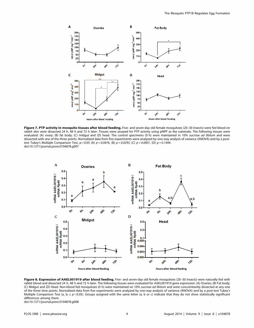
Figure 7. PTP activity in mosquito tissues after blood feeding. Five- and seven-day old female mosquitoes (20–30 insects) were fed blood onrabbit skin were dissected 24 h, 48 h and 72 h later. Tissues were assayed for PTP activity using pNPP as the substrate. The following tissues wereevaluated: (A) ovary; (B) fat body; (C) midgut and (D) head. The control specimens (0 h) were maintained in 10% sucrose ad libitum and weredissected with one of the three points. Normalized data from five experiments were analyzed by one-way analysis of variance (ANOVA) and by a post-test Tukey’s Multiple Comparison Test, p,0.05 (A) p = 0.0476, (B) p = 0.0293, (C) p = 0.0001, (D) p = 0.1494.doi:10.1371/journal.pone.0104878.g007
Figure 8. Expression of AAEL001919 after blood feeding. Five- and seven-day old female mosquitoes (20–30 insects) were naturally fed withrabbit blood and dissected 24 h, 48 h and 72 h later. The following tissues were evaluated for AAEL001919 gene expression: (A) Ovaries; (B) Fat body;(C) Midgut and (D) Head. Non-blood fed mosquitoes (0 h) were maintained on 10% sucrose ad libitum and were concomitantly dissected at any oneof the three time points. Normalized data from five experiments were analyzed by one-way analysis of variance (ANOVA) and by a post-test Tukey’sMultiple Comparison Test (a, b, c p,0.05). Groups assigned with the same letter (a, b or c) indicate that they do not show statistically significantdifferences among them.doi:10.1371/journal.pone.0104878.g008
The Mosquito PTP1B Regulates Egg Formation
PLOS ONE | www.plosone.org 9 August 2014 | Volume 9 | Issue 8 | e104878

regulator of the insulin pathway in mosquitoes, but its direct
targets remain unknown, and their identity was not addressed in
the present investigation. Insulin receptors (IRs) usually present
high conservation, especially in the kinase catalytic domain that is
essential for their activation, but the PTP1B target region (that
contains tyrosine phosphorylation sites) is considered the most
variable part of these receptors. The high level of similarity in
PTP1B is interesting because the IR variable region would be
expected to guide selective pressure on PTP1B for the purpose of
concomitant changes [41]. The differences in inhibitor suscepti-
bility among PTPs may allow drug design through a strategy that
generates a drug that only affects the insect phosphatase or its
substrates, such that it is inactive in mammals. In D. melanogaster,
the ortholog of PTP1B, the dmPTP61F gene, generates two
products that differ only by a small C-terminal sequence and that
contain the same membrane-binding domain. This difference
allows the product containing this specific domain to become
anchored to the endoplasmic reticulum, whilst the product that
lacks the specific domain exhibits nuclear localization [41,42].
Interestingly, in mammals, the membrane-bound PTP1B has been
reported to have an inhibitory effect on the insulin signaling
pathway, leading to insulin resistance, and has also been shown to
be capable of causing an increase in blood glucose [41,42]. In
contrast, the PTP1B protein with the truncated membrane-
binding C-terminal domain can still lead to insulin resistance but is
unable to activate SREBP-1 [42–44]. In conclusion, while it is
likely that AAEL001919 regulates glucose metabolism and
immunity in mosquitoes, the present study clearly demonstrates
its involvement in egg formation in Aedes aegypti females.
Furthermore, this enzyme demonstrates potential use as a target
for designing specific inhibitors that block mosquito egg develop-
ment and vector-mediated disease transmission.
Materials and Methods
ReagentsTris, glycine, acrylamide, bis-acrylamide, TEMED, DMSO,
DTT, bovine serum albumin, sodium vanadate, okadaic acid,
Folin reagent and pNPP were obtained from Sigma-Aldrich
Company (St. Louis, MO, USA). Prestained Full-Range Rainbow
Figure 9. Effect of PTP inhibitors and silencing action of the AAEL001919 gene on egg formation in Aedes aegypti. (A) Five- and seven-day old females were artificially fed with rabbit blood supplemented with the PTP inhibitor vanadate or the PTP substrate Difmup. Following a bloodmeal, fat bodies were dissected from females at the indicated times, and the expression of Vg mRNA was measured by qPCR and normalized againstRp49 gene expression. (B) Females from the experiments shown in A were placed in individual tubes, and the eggs laid were quantified onsubsequent days after the blood meal. (C) One- and two-day old females (5 insects) were injected with either 140 ng of AAEL001919 RNAi or dsMal.AAEL001919 expression levels were quantified three days after RNAi injection as shown. (D) Injected mosquitoes from the experiment shown in panelC were naturally blood-fed three days later, and the number of eggs laid following the blood meal was quantified. Normalized data from fourexperiments were analyzed by One-way ANOVA (panels A and B) and Student’s t-test (Panel D) with significance levels set at p,0.05. Groupsassigned with the same letter (a, b or c) indicate that they do not show statistically significant difference among them. Different letters in differentgroups indicate that such groups show significant differences among themselves (Panels A and B). Asterisks indicate (Panel B) indicate a significantdifference among groups (student T- test, p,0.05).doi:10.1371/journal.pone.0104878.g009
The Mosquito PTP1B Regulates Egg Formation
PLOS ONE | www.plosone.org 10 August 2014 | Volume 9 | Issue 8 | e104878

molecular weight standards were obtained from Amersham
Biosciences (Buckinghamshire, England). Ethanol, Triton X-100
and DMSO were obtained from Merck (Darmstadt, Germany).
Ethics statementAll animal care and experimental protocols were conducted
following the guidelines of the institutional care and use committee
(Committee for Evaluation of Animal Use for Research from the
Federal University of Rio de Janeiro, CAUAP-UFRJ) and the
NIH Guide for the Care and Use of Laboratory Animals (ISBN 0-
309-05377-3). The protocols were approved by CAUAP-UFRJ
under registry IBQM067-05/16. Technicians dedicated to the
animal facility at the Institute of Medical Biochemistry (UFRJ)
carried out all aspects related to rabbit husbandry under strict
guidelines to insure careful and consistent handling of the animals.
Mosquito rearing and blood mealsAedes aegypti (Liverpool Black Eye strain) were raised in an
insectary at the Federal University of Rio de Janeiro, Brazil, under
a 12-h light/dark cycle at 28uC and 60–80% relative humidity.
Larvae were fed with dog food (Pedigree Junior), and adults were
maintained in a cage and given a solution of 10% sucrose adlibitum. Five- to seven-day old females were used in the
experiments. Mosquitoes were naturally fed on rabbits’ ear veins
or were artificially fed with heparinized rabbit blood. Artificial
feeding was performed using water-jacketed artificial feeders
maintained at 37uC and sealed with parafilm membranes.
Whenever indicated blood was supplemented with PTP inhibitors.
Phosphatase assay using para-Nitrophenylphosphate(pNPP)
The tissues were homogenized in buffer containing 20 mM of
sodium acetate (pH 4.0). The protein concentration of each
extract was determined by the Lowry method. The homogenates
were used to perform the phosphatase assays, using 0.5–3 mg of
protein, depending on the tissue, and using p-nitrophenylpho-
sphate (pNPP) as substrate. Enzyme activity was measured in the
presence or absence of 100 mM sodium orthovanadate, used as
tyrosine phosphatase inhibitor, to determine the specific activities
of PTPs. The reactions were conducted at 37uC for 60 minutes
and were stopped by adding 2 N NaOH. In some experiments, we
included 0.1 mM of PTP 1B substrate II (Glu-Leu-Glu-Phe-pTyr-
Met-Asp-Tyr-Asp-Tyr-Glu) (Calbiochem) in addition to pNPP.
Further conditions were as described previously [24,49].
In silico analysis of Aedes PTPsIdentification and classification of tyrosine phosphatases (PTPs) in
mosquitoes were accomplished by searching conserved domains from
Pfam. The search for conserved domains (CDs) employed FAT [50],
a program developed by our group. It is a HMMER filter and blast
manager. First, proteins predicted (version 1.3) by Vector-Base in the
A. aegypti genome were downloaded (https://www.vectorbase.org/
download/aedes-aegypti-liverpoolpeptidesaaegl13fagz). Using FAT,
the sequences were filtered using characteristic domains of PTP
families (Class I soluble classical PTPs used Y_phosphatase domain
PF00102). The sequences obtained were compared using blast with
databases such as nr, Swiss-prot and a custom human tyrosine
phosphatases database. Lastly, hmmscan searched for other
conserved domains in these previously filtered proteins. While this
manuscript was in preparation, we became aware of another
publication that described a specific sequence-based method for the
automatic classification of PTPs present in 65 genomes, including A.
aegypti [21]. The same classical PTPs described in that study were
confirmed by the strategy described here.
Analysis of PTP sequences, protein models and active-site modeling
In silico approaches were used to ascertain the function and the
evolutionary homologs of Aedes PTPs. Because proteins with high
sequence and structural similarity may possess the same biological
functions, the in silico approach was used to find the closest
homologs of PTPs in an effort to obtain homology gene models
[50]. Sequences were used to search the homologue sequence
space for related proteins using the NCBI BLAST server [50]. A
BLOSUM62 matrix was used for coring of the sequences, with an
expected threshold of 10 and a sampling size of 3 amino-acids at a
time. The non-redundant protein database was used for searching
the sequence space without filtering out low-complexity regions.
To identify the homologues with known protein structures, the
PSI-BLAST tool was used against the freely available Protein Data
Bank. PSI-BLAST allowed for better sensitivity and enabled us to
find the closest five structures, which could be used in homology
modeling. These five sequences were used for a multiple sequence
alignment using the MULTALIGN server [51–53]. In several
instances, the Aedes PTP and the homologous PTP sequences had
gaps, so the modeler could not be effectively used for homology
modeling. In these cases, the automated Protein Homology/
analogy Recognition Engine was used to find the respective
homology models [54]. The reliability of the models was checked
via submission to the WHAT IF server [55,56]. The protein
structures were visualized and superimposed using PyMOL
software (DeLano Scientific LLC). The various domains in the
sequences were identified using a combination of online servers,
including the Conserved residue Function Prediction Server [56]
and the Conserved domain Database at the NCBI [57]. The active
site of the Protein-Tyrosine-Phosphatase domain was ascertained
using the Catalytic Site Atlas [58] as well as the siteFinder web-
based tool [59]. The ten motifs that define the PTP domain were
identified and used, as defined in the PTP database [60].
Secondary-structure prediction for the sequence corresponding
to the PTP domain was obtained from the Psipred server [61].
Sequences of human and fly (Drosophila melanogaster) PTPs were
obtained from the non-redundant Protein Database at NCBI and
used for construction of dendrograms using the CLUSTALW web
server and the TREEVIEW software package.
RNA extraction and qPCR analysisRNA was extracted from each sample using TRIzol reagent
(Life Technologies). RNA quantification was accomplished using a
NanoDrop-3300 (Thermo Scientific) device before cDNA synthe-
sis was performed. Briefly, 1 mg of RNA was treated with RNase-
free DNase (Fermentas) to avoid genomic DNA contamination.
Subsequently, cDNA synthesis was performed using the High
Capacity cDNA Reverse Transcription kit (Applied Biosystems),
according to the manufacturer’s protocol. Transcript levels were
determined by Real Time PCR using a SYBR-Green-based
method. The quantitative PCR assays were employed a StepO-
nePlus Real Time PCR System (Applied Biosystems) using
PowerSYBR Green PCR Master Mix (Applied Biosystems). Gene
expressions, as evaluated by mRNA levels, were calculated by
normalization to the levels of Ribosomal protein 49 (rp49) mRNA
(accession number AAT45939), which served as the endogenous
control in each individual sample, and taking into consideration
the respective efficiency of each pair of primers. The comparative
DDCt method was then used to calculate relative gene expression
levels, and all standard errors were calculated based on DCt, as
The Mosquito PTP1B Regulates Egg Formation
PLOS ONE | www.plosone.org 11 August 2014 | Volume 9 | Issue 8 | e104878

described in the Applied Biosystems User Bulletin #2 (http://www3.
appliedbiosystems.com/cms/groups/mcb_support/documents/
generaldocuments/cms_040980.pdf). The primers used presented
an efficiency of at least 95%. The primer pairs corresponding to the
analyzed genes were as follows: AAEL001919 (fwd 59 GATTGGCG
AAGAGCACAAATTG, rvs 59 TAATCGGAACGTCCTTTTGC
39), AAEL005492 (fwd 59 GTGATAGTAATGACCACTCG 39, rvs
59 ACCTGATAGCATCCATATTC 39), AAEL008528 (fwd 59
GGGTCCAATTTGTGTCCAC 39, rvs 59 GATCTCGCAGTC-
CAGACC 39), AAEL003108 (fwd 59ATGGTACAACAGGAAAG-
CAG 39, rvs 59GATGGAGAATCCTTCAGACA 39), AAEL010234
(fwd 59 TAGATTTACGGTGGCGGAC 39, rvs 59 GCTTG-
CGTCTGGTTCTTTTC 39), AAEL010914 (fwd 59 TGGCAG-
TATGGTGGATAGC 39, rvs 59 CAACCGTTCAACCTCCTTG
39), AAEL001046(fwd 59AAGCAACCGAACTTTGTTGG 39, rvs
59CCCGTTGAGTTGGTCT GATT 39), Rp49(fwd 59TCAAC-
CCCCGTTCGAACA 39, rvs 59CCGTAACCGATGTTTGGC39),
Vg(fwd 59 TGAATTTGTCACCCCCGATC 39, rvs 59 TTC-
ACGCTTGACACATTCCTG 39).
dsRNA synthesis and injectionsDouble-stranded RNA was synthesized using a MEGAscript
RNAi kit (Ambion, Austin, TX, USA), according to manufactur-
er’s instructions. 138 nL of 3 mg/mL dsRNA solution re-suspended
in water was injected into the thorax of cold-anesthetized 1-to-2-
day-old female mosquitoes. dsRNA injections employed a
nanoject II nanoliter injector (Drummond Scientific). Primers’
sequences used for template amplification for dsRNA synthesis
were as follows: RNAi1919F 59 TAATACGACTCACTA-
TAGGGTGCCACCGTTACCTAAGGAC 39 and RNAi1919R
59 TAATACGACTCACTATAGGGCTGGGCTAGACACTG-
CTTCC 39. These primers contained a T7 polymerase binding
sequence, required for dsRNA synthesis. As a control, we used
maltose-binding protein (mal) from Escherichia coli dsRNA
(dsmal). This sequence was inserted into a pBlueScript KS+(Stratagene) and was amplified using the T7 minimal promoter
primer (59 TAATACGACTCACTATAGGG 39) for template
generation and dsRNA synthesis.
Evaluation of the Role of PTP on Egg OvipositionIn some experiments mosquitoes were fed with 10% sucrose
enriched either with the 0.1 mM of the PTP inhibitor sodium
orthovanadate or the PTP substrate DifMUP. In these experi-
ments cages with 10–15 mosquitoes were fed with blood in the
presence or absence of the mentioned PTP modulators and 72 hrs
later the number of laid eggs was evaluated for the whole cage. At
least three different cages were used in each experiment. Therefore
each plotted point on the graphics (Figure 9B) is the number of
eggs in a single cage divided by the number of females present on
that cage. RNAi silencing of AAEL001919 was also used as a
strategy to block PTP activity. In each of these experiments a total
of 30 mosquitoes where either injected with dsMal or ds1919 as
indicated in figure legend. Following injection groups of 2
mosquitoes were kept in separate in Falcon tubes in order to
decrease mortality. The plotted results indicate the total number of
eggs laid in each Falcon divided by 2 female mosquitoes.
Statistical AnalysisAll experiments were performed at least in triplicate. The results
are presented as the means and standard errors of the mean.
Normalized data were analyzed by One-way ANOVA or
Student’s t-test using GraphPad Prism software.
Supporting Information
Figure S1 Effect of a tyrosine phosphorylated peptideon PTP activity towards pNPP on different tissues of A.aegypti. Tissues dissected from 10% sucrose-fed females (were
homogenized and enzyme activity towards pNPP was assayed in
the presence or absence of 0.1 mM of PTP 1B substrate II. After
30 minutes at 37uC, the reactions were halted by adding 1:8,5
volumes of 2 M NaOH. Results represent the standard two
determinations in triplicate and mean deviation.
(TIF)
Acknowledgments
We acknowledge the technical support of Katia Anastacio Laia, Jaciara
Miranda Freire and Geane Cleia Braz. This work is dedicated to the
memory of Dr. Alexandre A. Peixoto.
Author Contributions
Conceived and designed the experiments: DMM MACSN. Performed the
experiments: DMM RDN COC WJ CRODF FGL LGA KH RDM RS
GVM PMC. Analyzed the data: DMM COC LGA MHFS RDM WJ
MACSN. Contributed reagents/materials/analysis tools: LGA RDM MC
GCA MACSN. Wrote the paper: LGA MACSN.
References
1. Lim WA, Pawson T (2010) Phosphotyrosine signaling: evolving a new cellular
communication system. Cell 142(5):661–667.
2. Sacco F, Perfetto L, Castagnoli L, Cesareni G (2012) The human phosphatase
interactome: An intricate family portrait. FEBS Lett 586(17):2732–2739.
3. Alonso A, Sasin J, Bottini N, Friedberg I, Friedberg I, et al. (2004) Protein
tyrosine phosphatases in the human genome. Cell 117(6):699–711.
4. Brown MR, Graf R, Swiderek KM, Fendley D, Stracker TH, et al. (1998)
Identification of a steroidogenic neurohormone in female mosquitoes. J Biol
Chem 273(7):3967–3971.
5. Brown MR, Clark KD, Gulia M, Zhao Z, Garczynski SF, et al. (2008) An
insulin-like peptide regulates egg maturation and metabolism in the mosquito
Aedes aegypti. Proc Natl Acad Sci U S A 105(15):5716–5721.
6. Gulia-Nuss M, Robertson AE, Brown MR, Strand MR (2011) Insulin-like
peptides and the target of rapamycin pathway coordinately regulate blood
digestion and egg maturation in the mosquito Aedes aegypti. PLoS One
6(5):e20401.
7. Hansen IA, Sieglaff DH, Munro JB, Shiao SH, Cruz J, et al. (2007) Forkhead
transcription factors regulate mosquito reproduction. Insect Biochem Mol Biol
37(9):985–997.
8. Hansen IA, Attardo GM, Roy SG, Raikhel AS (2005) Target of rapamycin-
dependent activation of S6 kinase is a central step in the transduction of
nutritional signals during egg development in a mosquito. J Biol Chem
280(21):20565–20572.
9. Riehle MA, Brown MR (1999) Insulin stimulates ecdysteroid production
through a conserved signaling cascade in the mosquito Aedes aegypti. InsectBiochem Mol Biol 29(10):855–860.
10. Arik AJ, Rasgon JL, Quicke KM, Riehle MA (2009) Manipulating insulinsignaling to enhance mosquito reproduction. BMC Physiol 9:15.
11. Kokoza VA, Raikhel AS (2011) Targeted gene expression in the transgenic
Aedes aegypti using the binary Gal4-UAS system. Insect Biochem Mol Biol41(8):637–644.
12. Kokoza V, Ahmed A, Cho WL, Jasinskiene N, James AA, et al. (2000)
Engineering blood meal-activated systemic immunity in the yellow fevermosquito, Aedes aegypti. Proc Natl Acad Sci U S A 97(16):9144–9149.
13. Barr AJ, Ugochukwu E, Lee WH, King ON, Filippakopoulos P, et al. (2009)
Large-scale structural analysis of the classical human protein tyrosinephosphatome. Cell 136(2):352–363.
14. Franz AW, Sanchez-Vargas I, Adelman ZN, Blair CD, Beaty BJ, et al. (2006)Engineering RNA interference-based resistance to dengue virus type 2 in
genetically modified Aedes aegypti. Proc Natl Acad Sci U S A 103(11):4198–
4203.
15. Tonks NK (2009) Pseudophosphatases: grab and hold on. Cell 139(3):464–5.
16. Larkin MA, Blackshields G, Brown NP, Chenna R, McGettigan PA, et al. (2007)
Clustal W and Clustal X version 2.0. Bioinformatics 23(21):2947–2948.
17. Asthagiri D, Dillet V, Liu T, Noodleman L, Van Etten RL, et al. (2002) Density
functional study of the mechanism of a tyrosine phosphatase: I. Intermediate
formation. J Am Chem Soc 124(34):10225–10235.
The Mosquito PTP1B Regulates Egg Formation
PLOS ONE | www.plosone.org 12 August 2014 | Volume 9 | Issue 8 | e104878
18. Zhang ZY (1998) Protein-tyrosine phosphatases: biological function, structural
characteristics, and mechanism of catalysis. Crit Rev Biochem Mol Biol 33(1):1–52.
19. Madan LL, Gopal B (2011) Conformational basis for substrate recruitment in
protein tyrosine phosphatase 10D. Biochemistry 50(46):10114–10125.20. Page RDM (1996) TreeView: An application to display phylogenetic trees on
personal computers. Computer Applications in the Biosciences 12(4): 357–35821. Hatzihristidis T, Liu S, Pryszcz L, Hutchins AP, Gabaldon T, et al. (2013) PTP-
central: A comprehensive resource of protein tyrosine phosphatases in eukaryotic
genomes. Methods 65(2):156–164.22. Karim FD, Rubin GM (1999) PTP-ER, a novel tyrosine phosphatase, functions
downstream of Ras1 to downregulate MAP kinase during Drosophila eyedevelopment. Mol Cell 3(6):741–750.
23. Roy SG, Raikhel AS (2011) The small GTPase Rheb is a key component linkingamino acid signaling and TOR in the nutritional pathway that controls
mosquito egg development. Insect Biochem Mol Biol 41(1):62–69.
24. Jablonka W, Senna R, Nahu T, Ventura G, Menezes L, et al. (2011) A transientincrease in total head phosphotyrosine levels is observed upon the emergence of
Aedes aegypti from the pupal stage. Mem Inst Oswaldo Cruz 106(5):546–552.25. Perkins LA, Larsen I, Perrimon N (1992) Corkscrew encodes a putative protein
tyrosine phosphatase that functions to transduce the terminal signal from the
receptor tyrosine kinase torso. Cell 70(2):225–236.26. Oishi K, Zhang H, Gault WJ, Wang CJ, Tan CC, et al. (2009) Phosphatase-
defective LEOPARD syndrome mutations in PTPN11 gene have gain-of-function effects during Drosophila development. Hum Mol Genet 18(1):193–
201.27. Whited JL, Robichaux MB, Yang JC, Garrity PA (2007) Ptpmeg is required for
the proper establishment and maintenance of axon projections in the central
brain of Drosophila. Development 134(1):43–53.28. Bauler TJ, Hendriks WJ, King PD (2008) The FERM and PDZ domain-
containing protein tyrosine phosphatases, PTPN4 and PTPN3, are bothdispensable for T cell receptor signal transduction. PLoS One 3(12):e4014.
29. Buszard BJ, Johnson TK, Meng TC, Burke R, Warr CG, et al. (2013) The
nucleus- and endoplasmic reticulum-targeted forms of protein tyrosinephosphatase 61F regulate Drosophila growth, life span, and fecundity. Mol Cell
Biol 33(7):1345–1356.30. Wu CL, Buszard B, Teng CH, Chen WL, Warr CG, et al. (2011) Dock/Nck
facilitates PTP61F/PTP1B regulation of insulin signalling. Biochem J439(1):151–159.
31. Baeg GH, Zhou R, Perrimon N (2005) Genome-wide RNAi analysis of JAK/
STAT signaling components in Drosophila. Genes Dev 19(16):1861–1870.32. Ferrari E, Tinti M, Costa S, Corallino S, Nardozza AP, et al. (2011)
Identification of new substrates of the protein-tyrosine phosphatase PTP1B byBayesian integration of proteome evidence. J Biol Chem 286(6):4173–4185.
33. Mertins P, Eberl HC, Renkawitz J, Olsen JV, Tremblay, et al. (2008)
Investigation of protein-tyrosine phosphatase 1B function by quantitativeproteomics. Mol Cell Proteomics 7(9):1763–1777.
34. Chang YC, Lin SY, Liang SY, Pan KT, Chou CC, et al. (2008) Tyrosinephosphoproteomics and identification of substrates of protein tyrosine
phosphatase dPTP61F in Drosophila S2 cells by mass spectrometry-basedsubstrate trapping strategy. J Proteome Res 7(3):1055–1066.
35. Hilger M, Bonaldi T, Gnad F, Mann M (2009) Systems-wide analysis of a
phosphatase knock-down by quantitative proteomics and phosphoproteomics.Mol Cell Proteomics(8):1908–1920.
36. Riehle MA, Fan Y, Cao C, Brown MR (2006) Molecular characterization ofinsulin-like peptides in the yellow fever mosquito, Aedes aegypti: expression,
cellular localization, and phylogeny. Peptides 27(11):2547–2560.
37. Pakpour N, Corby-Harris V, Green GP, Smithers HM, Cheung KW, et al.(2012) Ingested human insulin inhibits the mosquito NF-kB-dependent immune
response to Plasmodium falciparum. Infect Immun 80(6):2141–2149.38. Helbling P, Graf R (1998) Localization of the mosquito insulin receptor homolog
(MIR) in reproducing yellow fever mosquitoes (Aedes aegypti). J Insect Physiol
44(12):1127–1135.
39. Castillo J, Brown MR, Strand MR (2011) Blood feeding and insulin-like peptide
3 stimulate proliferation of hemocytes in the mosquito Aedes aegypti. PLoS
Pathog 7(10):e1002274.
40. Hauck ES, Antonova-Koch Y, Drexler A, Pietri J, Pakpour N, et al. (2013)
Overexpression of phosphatase and tensin homolog improves fitness and
decreases Plasmodium falciparum development in Anopheles stephensi. MicrobesInfect 15(12):775–787.
41. Tiganis T (2013) PTP1B and TCPTP–nonredundant phosphatases in insulin
signaling and glucose homeostasis. FEBS J 280(2):445–458.
42. McLaughlin S, Dixon JE (1993) Alternative splicing gives rise to a nuclear
protein tyrosine phosphatase in Drosophila. J Biol Chem 268(10):6839–6842.
43. Shimizu S, Ugi S, Maegawa H, Egawa K, Nishio Y, et al. (2003) Protein-tyrosinephosphatase 1B as new activator for hepatic lipogenesis via sterol regulatory
element-binding protein-1 gene expression. J Biol Chem 278(44):43095–43101.
44. Ugi S, Shi K, Nishio Y, Shimizu S, Guo B, et al. (2009) Membrane localizationof protein-tyrosine phosphatase 1B is essential for its activation of sterol
regulatory element-binding protein-1 gene expression and consequent hypertri-
glyceridaemia. J Biochem 146(4):541–547.
45. Silveira AB, Castro-Santos J, Senna R, Logullo C, Fialho E, et al. (2006) Tick
vitellin is dephosphorylated by a protein tyrosine phosphatase during egg
development: effect of dephosphorylation on VT proteolysis. Insect BiochemMol Biol 36(3):200–209.
46. Fialho E, Silveira AB, Masuda H, Silva-Neto MA (2002) Oocyte fertilization
triggers acid phosphatase activity during Rhodnius prolixus embryogenesis.Insect Biochem Mol Biol 32(8):871–280.
47. Huang CH, Lin TY, Pan RL, Juang JL (2007) The involvement of Abl andPTP61F in the regulation of Abi protein localization and stability and lamella
formation in Drosophila S2 cells. J Biol Chem 282(44):32442–32452.
48. Bonizzoni M, Dunn WA, Campbell CL, Olson KE, Dimon MT, et al. (2011)RNA-seq analyses of blood-induced changes in gene expression in the mosquito
vector species, Aedes aegypti. BMC Genomics 12:82.
49. Gazos-Lopes F, Mesquita RD, Silva-Cardoso L, Senna R, Silveira AB, et al.(2012) Glycoinositolphospholipids from Trypanosomatids subvert nitric oxide
production in Rhodnius prolixus salivary glands. PLoS One 7(10):e47285.
50. Mesquita RD, Seabra Junior ES, Matos ES (2011) FAT-Functional AnalysisTool; patented at the Brazilian national trade mark office, # 11083–6.
51. Wood TC, Pearson WR (1999) Evolution of protein sequences and structures.
J Mol Biol 291(4):977–995.
52. Altschul SF, Madden TL, Schaffer AA, Zhang J, Zhang Z, et al. (1997) GappedBLAST and PSI-BLAST: a new generation of protein database search
programs. Nucleic Acids Res 25(17):3389–3402.
53. Corpet F (1988) Multiple sequence alignment with hierarchical clustering.Nucleic Acids Res 16(22):10881–10890.
54. Kelley LA, Sternberg MJ (2009) Protein structure prediction on the Web: a case
study using the Phyre server. Nat Protoc 4(3):363–371.
55. Vriend G (1990) WHAT IF: a molecular modeling and drug design program.J Mol Graph 8(1):52–6, 29.
56. Wass MN, Sternberg MJ (2008) ConFunc–functional annotation in the twilight
zone. Bioinformatics 24(6):798–806.
57. Marchler-Bauer A, Anderson JB, Chitsaz F, Derbyshire MK, DeWeese-Scott C,
et al. (2009) CDD: specific functional annotation with the Conserved Domain
Database. Nucleic Acids Res 37(Database issue): D205–210.
58. Porter CT, Bartlett GJ, Thornton JM (2004) The Catalytic Site Atlas: a resource
of catalytic sites and residues identified in enzymes using structural data. Nucleic
Acids Res 32(Database issue):D129–133.
59. Innis CA (2007) siteFiNDER|3D: a web-based tool for predicting the location of
functional sites in proteins. Nucleic Acids Res 35(Web Server issue):W489–494.
60. Andersen JN, Mortensen OH, Peters GH, Drake PG, Iversen LF, et al. (2001)Structural and evolutionary relationships among protein tyrosine phosphatase
domains. Mol Cell Biol 21(21):7117–7136.
61. McGuffin LJ, Bryson K, Jones DT (2000) The PSIPRED protein structureprediction server. Bioinformatics 16(4):404–405.
The Mosquito PTP1B Regulates Egg Formation
PLOS ONE | www.plosone.org 13 August 2014 | Volume 9 | Issue 8 | e104878