Cross-Linking/Mass Spectrometry Uncovers Details of Insulin ...
10
ORIGINAL RESEARCH published: 09 October 2019 doi: 10.3389/fendo.2019.00695 Frontiers in Endocrinology | www.frontiersin.org 1 October 2019 | Volume 10 | Article 695 Edited by: Michael Lawrence, Walter and Eliza Hall Institute of Medical Research, Australia Reviewed by: Briony Forbes, Flinders University, Australia Alexander Leitner, Eidgenössische Technische Hochschule Zurich, Switzerland *Correspondence: Irena Selicharová [email protected] † Present address: Jelena Radosavljevi ´ c, Department of Biochemistry, Faculty of Chemistry, University of Belgrade, Belgrade, Serbia Specialty section: This article was submitted to Molecular and Structural Endocrinology, a section of the journal Frontiers in Endocrinology Received: 23 July 2019 Accepted: 25 September 2019 Published: 09 October 2019 Citation: Pompach P, Viola CM, Radosavljevi ´ c J, Lin J, Jirá ˇ cek J, Brzozowski AM and Selicharová I (2019) Cross-Linking/Mass Spectrometry Uncovers Details of Insulin-Like Growth Factor Interaction With Insect Insulin Binding Protein Imp-L2. Front. Endocrinol. 10:695. doi: 10.3389/fendo.2019.00695 Cross-Linking/Mass Spectrometry Uncovers Details of Insulin-Like Growth Factor Interaction With Insect Insulin Binding Protein Imp-L2 Petr Pompach 1 , Cristina M. Viola 2 , Jelena Radosavljevi ´ c 3† , Jingjing Lin 3 , Jiˇ rí Jirá ˇ cek 3 , Andrzej M. Brzozowski 2 and Irena Selicharová 3 * 1 Institute of Microbiology, Czech Academy of Sciences, Prague, Czechia, 2 York Structural Biology Laboratory, Department of Chemistry, The University of York, York, United Kingdom, 3 Institute of Organic Chemistry and Biochemistry, Czech Academy of Sciences, Prague, Czechia Structural details of changes accompanying interaction between insulin-related hormones and their binding partners are often enigmatic. Here, cross-linking/mass spectrometry could complement structural techniques and reveal details of these protein- protein interfaces. We used such approach to clarify missing structural description of the interface in human insulin-like growth factor (IGF-1): Drosophila melanogaster imaginal morphogenesis protein-late 2 protein (Imp-L2) complex which we studied previously by X-ray crystallography. We crosslinked these proteins by heterobifunctional cross- linker sulfosuccinimidyl 4,4 ′ -azidopentanoate (Sulfo-SDA) for the subsequent mass spectrometry (MS) analysis. The MS analysis revealed IGF-1:Imp-L2 interactions which were not resolved in the crystal structure of this assembly, and they converged with X-ray results, indicating the importance of the IGF-1 N-terminus interaction with the C-terminal (185–242) part of the Imp-L2 for stability of this complex. Here, we also showed the advantage and reliability of MS approach in solving details of protein-protein interactions that are too flexible for solid state structural methods. Keywords: IGF-1, Imp-L2, cross-linking, diazirine ring, mass spectrometry INTRODUCTION The insulin/insulin-like growth factor signaling axis is an evolutionary ancient and highly conserved hormonal system involved in the regulation of metabolism, growth and lifespan in animals. In humans this axis contains insulin and two insulin-like growth factors (IGF-1 and IGF- 2) eliciting their function through activation of tyrosine kinase-type receptors (1). Secretion of insulin from pancreatic β-cells is regulated in response to blood glucose levels. Subsequently, the active monomeric form of this hormone circulates freely in the blood and is readily cleared away in minutes. In contrast, IGFs are secreted by many tissues in endocrine or paracrine mode and their bioavailability is controlled by several IGF binding proteins (IGFBP 1–6) (2) and the non-signaling IGF-2/mannose-6-phosphate receptor (3). There are also IGFBP-related proteins (IGFBP-rP) that carry the N-terminal domain of IGFBPs hence are classified as part of the IGFBP superfamily. However, IGFBP-rPs have low or no affinity for IGFs/insulin, and their functions are not fully elucidated being, most probably, IGF/insulin independent (4). brought to you by CORE View metadata, citation and similar papers at core.ac.uk provided by Faculty of Chemistry Repository - Cherry
-
Upload
khangminh22 -
Category
Documents
-
view
0 -
download
0
Transcript of Cross-Linking/Mass Spectrometry Uncovers Details of Insulin ...
ORIGINAL RESEARCHpublished: 09 October 2019
doi: 10.3389/fendo.2019.00695
Frontiers in Endocrinology | www.frontiersin.org 1 October 2019 | Volume 10 | Article 695
Edited by:
Michael Lawrence,
Walter and Eliza Hall Institute of
Medical Research, Australia
Reviewed by:
Briony Forbes,
Flinders University, Australia
Alexander Leitner,
Eidgenössische Technische
Hochschule Zurich, Switzerland
*Correspondence:
Irena Selicharová
†Present address:
Jelena Radosavljevic,
Department of Biochemistry, Faculty
of Chemistry, University of Belgrade,
Belgrade, Serbia
Specialty section:
This article was submitted to
Molecular and Structural
Endocrinology,
a section of the journal
Frontiers in Endocrinology
Received: 23 July 2019
Accepted: 25 September 2019
Published: 09 October 2019
Citation:
Pompach P, Viola CM,
Radosavljevic J, Lin J, Jirácek J,
Brzozowski AM and Selicharová I
(2019) Cross-Linking/Mass
Spectrometry Uncovers Details of
Insulin-Like Growth Factor Interaction
With Insect Insulin Binding Protein
Imp-L2. Front. Endocrinol. 10:695.
doi: 10.3389/fendo.2019.00695
Cross-Linking/Mass SpectrometryUncovers Details of Insulin-LikeGrowth Factor Interaction WithInsect Insulin Binding Protein Imp-L2
Petr Pompach 1, Cristina M. Viola 2, Jelena Radosavljevic 3†, Jingjing Lin 3, Jirí Jirácek 3,
Andrzej M. Brzozowski 2 and Irena Selicharová 3*
1 Institute of Microbiology, Czech Academy of Sciences, Prague, Czechia, 2 York Structural Biology Laboratory, Department of
Chemistry, The University of York, York, United Kingdom, 3 Institute of Organic Chemistry and Biochemistry, Czech Academy
of Sciences, Prague, Czechia
Structural details of changes accompanying interaction between insulin-related
hormones and their binding partners are often enigmatic. Here, cross-linking/mass
spectrometry could complement structural techniques and reveal details of these protein-
protein interfaces. We used such approach to clarify missing structural description of the
interface in human insulin-like growth factor (IGF-1): Drosophila melanogaster imaginal
morphogenesis protein-late 2 protein (Imp-L2) complex which we studied previously
by X-ray crystallography. We crosslinked these proteins by heterobifunctional cross-
linker sulfosuccinimidyl 4,4′-azidopentanoate (Sulfo-SDA) for the subsequent mass
spectrometry (MS) analysis. The MS analysis revealed IGF-1:Imp-L2 interactions which
were not resolved in the crystal structure of this assembly, and they converged with X-ray
results, indicating the importance of the IGF-1 N-terminus interaction with the C-terminal
(185–242) part of the Imp-L2 for stability of this complex. Here, we also showed the
advantage and reliability of MS approach in solving details of protein-protein interactions
that are too flexible for solid state structural methods.
Keywords: IGF-1, Imp-L2, cross-linking, diazirine ring, mass spectrometry
INTRODUCTION
The insulin/insulin-like growth factor signaling axis is an evolutionary ancient and highlyconserved hormonal system involved in the regulation of metabolism, growth and lifespan inanimals. In humans this axis contains insulin and two insulin-like growth factors (IGF-1 and IGF-2) eliciting their function through activation of tyrosine kinase-type receptors (1). Secretion ofinsulin from pancreatic β-cells is regulated in response to blood glucose levels. Subsequently, theactive monomeric form of this hormone circulates freely in the blood and is readily cleared away inminutes. In contrast, IGFs are secreted by many tissues in endocrine or paracrine mode and theirbioavailability is controlled by several IGF binding proteins (IGFBP 1–6) (2) and the non-signalingIGF-2/mannose-6-phosphate receptor (3). There are also IGFBP-related proteins (IGFBP-rP) thatcarry the N-terminal domain of IGFBPs hence are classified as part of the IGFBP superfamily.However, IGFBP-rPs have low or no affinity for IGFs/insulin, and their functions are not fullyelucidated being, most probably, IGF/insulin independent (4).
brought to you by COREView metadata, citation and similar papers at core.ac.uk
provided by Faculty of Chemistry Repository - Cherry
Pompach et al. IGF-1 Interaction With Insect IBP
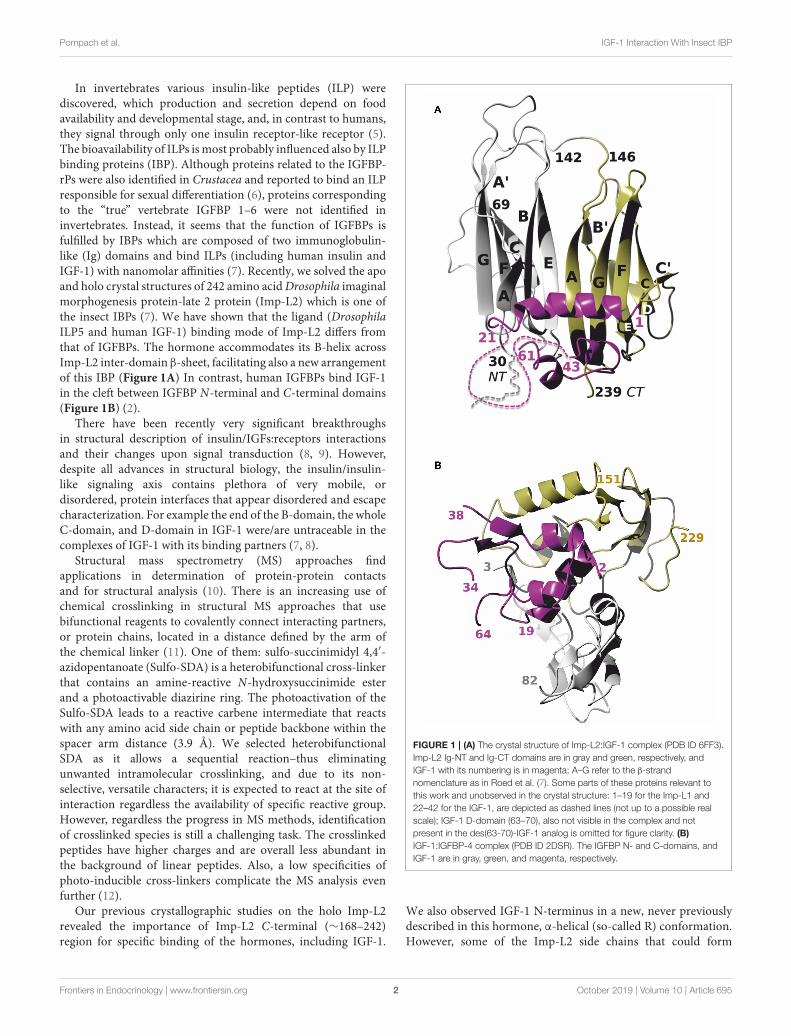
In invertebrates various insulin-like peptides (ILP) werediscovered, which production and secretion depend on foodavailability and developmental stage, and, in contrast to humans,they signal through only one insulin receptor-like receptor (5).The bioavailability of ILPs ismost probably influenced also by ILPbinding proteins (IBP). Although proteins related to the IGFBP-rPs were also identified in Crustacea and reported to bind an ILPresponsible for sexual differentiation (6), proteins correspondingto the “true” vertebrate IGFBP 1–6 were not identified ininvertebrates. Instead, it seems that the function of IGFBPs isfulfilled by IBPs which are composed of two immunoglobulin-like (Ig) domains and bind ILPs (including human insulin andIGF-1) with nanomolar affinities (7). Recently, we solved the apoand holo crystal structures of 242 amino acidDrosophila imaginalmorphogenesis protein-late 2 protein (Imp-L2) which is one ofthe insect IBPs (7). We have shown that the ligand (DrosophilaILP5 and human IGF-1) binding mode of Imp-L2 differs fromthat of IGFBPs. The hormone accommodates its B-helix acrossImp-L2 inter-domain β-sheet, facilitating also a new arrangementof this IBP (Figure 1A) In contrast, human IGFBPs bind IGF-1in the cleft between IGFBP N-terminal and C-terminal domains(Figure 1B) (2).
There have been recently very significant breakthroughsin structural description of insulin/IGFs:receptors interactionsand their changes upon signal transduction (8, 9). However,despite all advances in structural biology, the insulin/insulin-like signaling axis contains plethora of very mobile, ordisordered, protein interfaces that appear disordered and escapecharacterization. For example the end of the B-domain, the wholeC-domain, and D-domain in IGF-1 were/are untraceable in thecomplexes of IGF-1 with its binding partners (7, 8).
Structural mass spectrometry (MS) approaches findapplications in determination of protein-protein contactsand for structural analysis (10). There is an increasing use ofchemical crosslinking in structural MS approaches that usebifunctional reagents to covalently connect interacting partners,or protein chains, located in a distance defined by the arm ofthe chemical linker (11). One of them: sulfo-succinimidyl 4,4′-azidopentanoate (Sulfo-SDA) is a heterobifunctional cross-linkerthat contains an amine-reactive N-hydroxysuccinimide esterand a photoactivable diazirine ring. The photoactivation of theSulfo-SDA leads to a reactive carbene intermediate that reactswith any amino acid side chain or peptide backbone within thespacer arm distance (3.9 Å). We selected heterobifunctionalSDA as it allows a sequential reaction–thus eliminatingunwanted intramolecular crosslinking, and due to its non-selective, versatile characters; it is expected to react at the site ofinteraction regardless the availability of specific reactive group.However, regardless the progress in MS methods, identificationof crosslinked species is still a challenging task. The crosslinkedpeptides have higher charges and are overall less abundant inthe background of linear peptides. Also, a low specificities ofphoto-inducible cross-linkers complicate the MS analysis evenfurther (12).
Our previous crystallographic studies on the holo Imp-L2revealed the importance of Imp-L2 C-terminal (∼168–242)region for specific binding of the hormones, including IGF-1.
FIGURE 1 | (A) The crystal structure of Imp-L2:IGF-1 complex (PDB ID 6FF3).
Imp-L2 Ig-NT and Ig-CT domains are in gray and green, respectively, and
IGF-1 with its numbering is in magenta; A–G refer to the β-strand
nomenclature as in Roed et al. (7). Some parts of these proteins relevant to
this work and unobserved in the crystal structure: 1–19 for the Imp-L1 and
22–42 for the IGF-1, are depicted as dashed lines (not up to a possible real
scale); IGF-1 D-domain (63–70), also not visible in the complex and not
present in the des(63-70)-IGF-1 analog is omitted for figure clarity. (B)
IGF-1:IGFBP-4 complex (PDB ID 2DSR). The IGFBP N- and C-domains, and
IGF-1 are in gray, green, and magenta, respectively.
We also observed IGF-1 N-terminus in a new, never previouslydescribed in this hormone, α-helical (so-called R) conformation.However, some of the Imp-L2 side chains that could form
Frontiers in Endocrinology | www.frontiersin.org 2 October 2019 | Volume 10 | Article 695
Pompach et al. IGF-1 Interaction With Insect IBP
tighter interactions within the IBP region and contribute to thestabilization of this conformation, were disordered. Therefore,we explored the MS approach with the use of Sulfo-SDA towardidentification of crosslinked peptides derived from IGF-1:Imp-L2 complex interface, in order to investigate the in-solutionproximity of these, possibly, structurally important side-chains,and to verify our previous observation.
MATERIALS AND METHODS
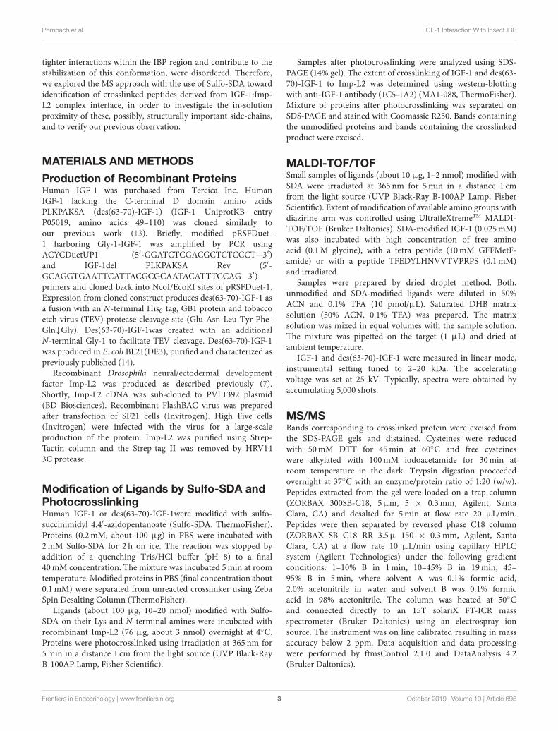
Production of Recombinant ProteinsHuman IGF-1 was purchased from Tercica Inc. HumanIGF-1 lacking the C-terminal D domain amino acidsPLKPAKSA (des(63-70)-IGF-1) (IGF-1 UniprotKB entryP05019, amino acids 49–110) was cloned similarly toour previous work (13). Briefly, modified pRSFDuet-1 harboring Gly-1-IGF-1 was amplified by PCR usingACYCDuetUP1 (5′-GGATCTCGACGCTCTCCCT−3′)and IGF-1del PLKPAKSA Rev (5′-GCAGGTGAATTCATTACGCGCAATACATTTCCAG−3′)primers and cloned back into NcoI/EcoRI sites of pRSFDuet-1.Expression from cloned construct produces des(63-70)-IGF-1 asa fusion with an N-terminal His6 tag, GB1 protein and tobaccoetch virus (TEV) protease cleavage site (Glu-Asn-Leu-Tyr-Phe-Gln↓Gly). Des(63-70)-IGF-1was created with an additionalN-terminal Gly-1 to facilitate TEV cleavage. Des(63-70)-IGF-1was produced in E. coli BL21(DE3), purified and characterized aspreviously published (14).
Recombinant Drosophila neural/ectodermal developmentfactor Imp-L2 was produced as described previously (7).Shortly, Imp-L2 cDNA was sub-cloned to PVL1392 plasmid(BD Biosciences). Recombinant FlashBAC virus was preparedafter transfection of SF21 cells (Invitrogen). High Five cells(Invitrogen) were infected with the virus for a large-scaleproduction of the protein. Imp-L2 was purified using Strep-Tactin column and the Strep-tag II was removed by HRV143C protease.
Modification of Ligands by Sulfo-SDA andPhotocrosslinkingHuman IGF-1 or des(63-70)-IGF-1were modified with sulfo-succinimidyl 4,4′-azidopentanoate (Sulfo-SDA, ThermoFisher).Proteins (0.2mM, about 100 µg) in PBS were incubated with2mM Sulfo-SDA for 2 h on ice. The reaction was stopped byaddition of a quenching Tris/HCl buffer (pH 8) to a final40mM concentration. The mixture was incubated 5min at roomtemperature. Modified proteins in PBS (final concentration about0.1mM) were separated from unreacted crosslinker using ZebaSpin Desalting Column (ThermoFisher).
Ligands (about 100 µg, 10–20 nmol) modified with Sulfo-SDA on their Lys and N-terminal amines were incubated withrecombinant Imp-L2 (76 µg, about 3 nmol) overnight at 4◦C.Proteins were photocrosslinked using irradiation at 365 nm for5min in a distance 1 cm from the light source (UVP Black-RayB-100AP Lamp, Fisher Scientific).
Samples after photocrosslinking were analyzed using SDS-PAGE (14% gel). The extent of crosslinking of IGF-1 and des(63-70)-IGF-1 to Imp-L2 was determined using western-blottingwith anti-IGF-1 antibody (1C5-1A2) (MA1-088, ThermoFisher).Mixture of proteins after photocrosslinking was separated onSDS-PAGE and stained with Coomassie R250. Bands containingthe unmodified proteins and bands containing the crosslinkedproduct were excised.
MALDI-TOF/TOFSmall samples of ligands (about 10 µg, 1–2 nmol) modified withSDA were irradiated at 365 nm for 5min in a distance 1 cmfrom the light source (UVP Black-Ray B-100AP Lamp, FisherScientific). Extent of modification of available amino groups withdiazirine arm was controlled using UltrafleXtremeTM MALDI-TOF/TOF (Bruker Daltonics). SDA-modified IGF-1 (0.025mM)was also incubated with high concentration of free aminoacid (0.1M glycine), with a tetra peptide (10mM GFFMetF-amide) or with a peptide TFEDYLHNVVTVPRPS (0.1mM)and irradiated.
Samples were prepared by dried droplet method. Both,unmodified and SDA-modified ligands were diluted in 50%ACN and 0.1% TFA (10 pmol/µL). Saturated DHB matrixsolution (50% ACN, 0.1% TFA) was prepared. The matrixsolution was mixed in equal volumes with the sample solution.The mixture was pipetted on the target (1 µL) and dried atambient temperature.
IGF-1 and des(63-70)-IGF-1 were measured in linear mode,instrumental setting tuned to 2–20 kDa. The acceleratingvoltage was set at 25 kV. Typically, spectra were obtained byaccumulating 5,000 shots.
MS/MSBands corresponding to crosslinked protein were excised fromthe SDS-PAGE gels and distained. Cysteines were reducedwith 50mM DTT for 45min at 60◦C and free cysteineswere alkylated with 100mM iodoacetamide for 30min atroom temperature in the dark. Trypsin digestion proceededovernight at 37◦C with an enzyme/protein ratio of 1:20 (w/w).Peptides extracted from the gel were loaded on a trap column(ZORBAX 300SB-C18, 5µm, 5 × 0.3mm, Agilent, SantaClara, CA) and desalted for 5min at flow rate 20 µL/min.Peptides were then separated by reversed phase C18 column(ZORBAX SB C18 RR 3.5µ 150 × 0.3mm, Agilent, SantaClara, CA) at a flow rate 10 µL/min using capillary HPLCsystem (Agilent Technologies) under the following gradientconditions: 1–10% B in 1min, 10–45% B in 19min, 45–95% B in 5min, where solvent A was 0.1% formic acid,2.0% acetonitrile in water and solvent B was 0.1% formicacid in 98% acetonitrile. The column was heated at 50◦Cand connected directly to an 15T solariX FT-ICR massspectrometer (Bruker Daltonics) using an electrospray ionsource. The instrument was on line calibrated resulting in massaccuracy below 2 ppm. Data acquisition and data processingwere performed by ftmsControl 2.1.0 and DataAnalysis 4.2(Bruker Daltonics).
Frontiers in Endocrinology | www.frontiersin.org 3 October 2019 | Volume 10 | Article 695

Pompach et al. IGF-1 Interaction With Insect IBP
FIGURE 2 | Amino-acid sequences of IGF-1 (top) and des(63-70)-IGF-1 (bottom). IGF-1 domains are highlighted by background colors (B domain yellow, A domain
blue, C domain gray and D domain violet). Trypsin cleavage sites and expected peptides are shown. Peptides containing amino acid residues with primary amines
(red) susceptible to succinimide reaction are named N-terminal, B-C and A-D.
Strategy for Identification of CrosslinkedPeptidesMS spectra were searched for peptides corresponding to thesequence of Imp-L2 and IGF-1 or des(63-70)-IGF-1 usingLinks software (15). The Links algorithm was set to considerthe carbamidomethylation of cysteine and the possible singleoxidation of methionine. The mass error threshold was keptbelow 2 ppm and assigned fragments were verified manually.Crosslinked peptides were searched between the two sequencesas N-terminus or Lysine connected anywhere by an arm withmolecular mass +82.0419 Da (16). Suggested crosslinks foundin several (n ≥ 4) successive scans were considered as reliableand the spectra were manually searched for theoretical MS/MSfragments of peptides originating from the two proteins.
Further, the data were exported to mgf format andloaded into the StavroX software (version 3.6.0.1). The cross-linked peptides were searched using following parameters:fixed caramidomethylation of cysteines, variable oxidationof methionines, specificity of SDA for site 1 “K,S,T,Y,”specificity for site 2 “A,I,L,M,S,T,W,H,D,E,N,K,P,G,V,Q,m,C,B”(where “m” represents oxidized methionine and “B” representscaramidomethylated cysteine). Precursor precision was set to 2.0ppm and fragment ion precision was set to 20.0 ppm. FDR cutoff was below 5%. Decoy analysis was performed by shufflingfasta database while keeping the amino acids of protease sites inplace (17).
RESULTS
Photocrosslinking of IGF-1 ordes(63-70)-IGF-1 to Imp-L2We prepared the IGF-1 lacking the D domain residuesPLKPAKSA (des(63-70)-IGF-1) with the aim to reduce thenumber of primary amines available for reaction with thesuccinimide ester in Sulfo-SDA and thus simplify theMS analysis(Figure 2). Also, an additional N-terminal glycine residueGly(-1) was incorporated into IGF-1 to facilitate production ofdes(63-70)-IGF-1 (13).
The extent of modification of the wt and truncated IGF-1 by SDA was tested using MALDI-TOF/TOF (Figure 3).The SDA-modified IGF-1 yielded triple modified productand, in slightly lower amount, quadruple modified IGF-1as well (Figure 3C). As expected, the prevailing products of
the des(63-70)-IGF-1 modification were only double modifiedspecies with two diazirine arms (Figure 3D). Species withhigher extend of modification that are observed in thechromatograms (Figures 3C,D) resulted from limited reactivityof N-hydroxysuccinimide ester with hydroxyl group in sidechains of serines, threonines, and tyrosines.
To check specificity of the crosslinking reaction, we performedcontrol experiments where SDA-modified IGF-1 was incubatedwith excess of free amino acid/test peptides and irradiated.MALDI-TOF/TOF analysis was used to search for the products.Either alkene or alcohol were formed (same as in Figure 3C)after irradiation of diazirine, and no visible crosslinked productswere detected in these experiments despite high concentrationof peptides.
Efficiency of Imp-L2 crosslinking reactions with IGF-1and des(63-70)-IGF-1 were analyzed using SDS-PAGE andwestern-blot. Unreacted hormones and Imp-L2, together with acrosslinked product, were clearly visible on the Coomassie bluestained gels. The crosslinked product was detected with an IGF-1antibody (Figure 4). Only about 10–20% of the starting amountof Imp-L2 was crosslinked to the hormones in both, IGF-1 anddes(63-70)-IGF-1 reactions.
Identification of Crosslinked PeptidesCrosslinked peptides were searched using Links algorithm(dealing with MS data) and StavroX software (dealing withMS/MS data). Both approaches identified the same crosslinks.The results are shown in Supplementary Material (“StavroXresults for crosslinks between IGF-1 or des (63-70)-IGF-1 andImp-L2.pdf” and “Links results for crosslinks between IGF-1or des(63-70)-IGF-1 and Imp-L2.xlsx”). Crosslinked peptidesare listed in Table 1 for des(63-70)-IGF-1:Imp-L2 complex andin Table 2 for IGF-1:Imp-L2 complex and diagrams showingfragments observed in respective MS/MS spectra of each peptideare drawn in Figure 5.
As expected, the crosslinking of des(63-70)-IGF-1 providedsimpler data and was considered as decisive when assessing theLinks data of IGF-1 analysis (see Supplementary Material). Onlythe crosslinks found in multiple successive scans and confirmedby manual searches for MS/MS fragments were considered asreliable. The other hits were excluded as false positive. Allthe selected identified crosslinks were subsequently also foundthrough automated StavroX analysis.
Frontiers in Endocrinology | www.frontiersin.org 4 October 2019 | Volume 10 | Article 695

Pompach et al. IGF-1 Interaction With Insect IBP
FIGURE 3 | MALDI-TOF/TOF analysis of (A) IGF-1 and (B) des(63-70)-IGF-1 after coupling with Sulfo-SDA. Carbene was formed after irradiation of diazirine moiety
and was either eliminated by formation of double bound resulting in the increase of Mr of about 82 Da or it reacted with one molecule of water resulting in the increase
of Mr of about 100 Da. (C) SDA modified IGF-1. (D) SDA modified des(63-70)-IGF-1.
We have not identified any reliable crosslinked product ofLys residues within the IGF-1 D domain. Although there weremultiple suggested crosslinks in the Links search none fulfilledthe criteria of reliability, and no ions confirming the crosslinkwere found. No potential crosslinks in D domain were identifiedusing the StavroX. The D domain probably did not contribute tothe ligand interaction.
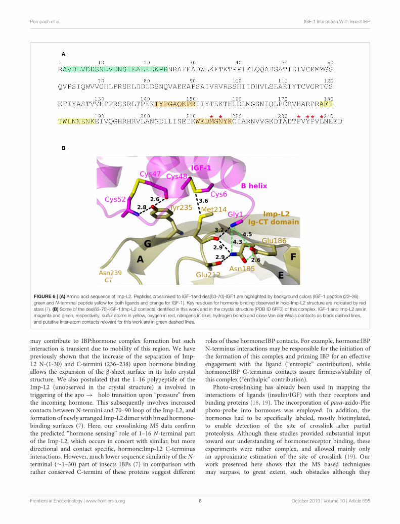
The des(63-70)-IGF-1 N-terminal Gly(-1) was found tobe bound to Imp-L2 peptide 178–188 (AEITWLNNENK).Identical crosslinked peptide was found for IGF-1:Imp-L2experiment (Figure 6). We observed the b6 ion of peptide178–188 Imp-L2 which would suggest that the crosslink wasformed within the sequence 184–188 of Imp-L2 (Figure 5and Supplementary Material). This finding corresponds to thecrystallography data as IGF-1 Gly1 is very close to Imp-L2Asn185 (∼4.3 Å) and Glu186 (∼4.5 Å) (Figure 6B).
Besides, other contacts of IGF-1 Gly1 were found (Table 2and Figure 6B). The N-terminal peptide was crosslinked tothe peptide 211–218 (WEDMGNYK) which was identified asmajor binding interaction site in the X-ray structure. It wasalso crosslinked to peptide 142–150 (TYPGAQKPR). All three
Imp-L2 peptides found to be crosslinked to IGF-1 N-terminusform neighboring β-strands A, G, and F (Figures 1A, 6B) ofImp-L2 Ig C-terminal domain.
Next, we identified crosslinked peptideconnecting IGF-1 23–36 with Imp-L2 peptide 2–24(AVDLVDDSNDVDNSIEAEEEKPR) (Tables 1, 2, Figure 5
and Supplementary Material). The crosslinked peptidewas identified in both IGF-1:Imp-L2 and des(63-70)-IGF-1:Imp-L2 crosslinking experiments. The 22–44 region ofIGF-1 and the N-terminal part of Imp-L2 (1-29) werenot visible in the crystal structure despite their spatialproximity (Figure 1A) (7). However, a high number ofscans (see Supplementary Material) in which this crosslinkedpeptide was identified suggests its relative abundance and,consequently, would indicate strong interactions withinthese sequences. As shown in our experiments with testpeptides, the carbene formed upon photoactivation ofdiazirine would react readily with water to create alcohol,or rearrange to give an alkene if there is no close contact withan interacting partner thus reducing the yield of the crosslinkingreaction (16).
Frontiers in Endocrinology | www.frontiersin.org 5 October 2019 | Volume 10 | Article 695

Pompach et al. IGF-1 Interaction With Insect IBP
FIGURE 4 | SDS-PAGE and western blot analysis of photocrosslinked IGF-1 or des(63-70)-IGF-1 to Imp-L2. Imp-L2 (line 1) was photocrosslinked to SDA-modified
IGF-1 (line 2) or des(63-70)-IGF-1 (line 3). Samples were resolved by SDS-PAGE on 14% gels and stained either by Coomassie blue or blotted to PVDF membrane
and detected with anti-IGF-1 antibody (1C5-1A2) (MA1-088, ThermoFisher). Molecular weight standards (BioRad) are shown in line 4.
TABLE 1 | Crosslinked peptides between des(63-70)-IGF-1 and Imp-L2.
Crosslinked peptide (des(63-70)-IGF-1)–(Imp-L2) Exp. mass Thr. mass Error StavroX score
(−1, 21)–(178, 188) 3776.7892 3776.7894 −0.06 67
(22, 36)–(2, 24) 4307.9865 4307.9851 +0.31 103
TABLE 2 | Crosslinked peptides between IGF-1 and Imp-L2.
Crosslinked peptide (IGF-1)–(Imp-L2) Exp. mass Thr. mass Error StavroX score
(1, 21)–(178, 188) 3,719.7681 3719.7680 −0.04 79
(1, 21)–(211, 218) 3430.5381 3430.5388 −0.2 24
(1, 21)–(142, 150) 3405.6567 3405.6566 +0.03 61
(22, 36)–(2, 24) 4307.9859 4307.9851 +0.19 41
DISCUSSION
Structural mass spectrometry (MS) approaches find utilizationin determination of protein-protein contacts and for structuralanalysis. We aimed to explore the cross-linking/massspectrometry approach toward revealing the in-solutionproximity of protein-protein interface. We utilized a IGF-1:Imp-L2 complex that we previously studied using X-ray
crystallography. The knowledge of the structure allowed us tocritically evaluate reliability of crosslinked peptides proposedby the scoring algorithm of MS spectra but also to verify ourprevious observation. To simplify the MS analysis we preparedthe IGF-1 lacking the D domain. As anticipated, theMS spectrumof des(63-70)-IGF-1 crosslinked to Imp-L2 was simpler, clearerand contained less false-positive hits compared to the IGF-1:Imp-L2 spectrum. Nevertheless, identical crosslinked peptides
Frontiers in Endocrinology | www.frontiersin.org 6 October 2019 | Volume 10 | Article 695

Pompach et al. IGF-1 Interaction With Insect IBP
FIGURE 5 | Diagrams of identified crosslinked peptides (A) between IGF-1:Imp-L2 and (B) between des(63-70)-IGF-1:Imp-L2. MS/MS fragments and sites of
crosslinks found in StavroX search are shown in blue. Manually assigned fragments confirming crosslinks suggested by Links algorithm are shown in violet.
were identified for both wt and truncated IGF-1 bound toImp-L2 which further supported reliability of the approach.
We previously described a unique R-state like rearrangementof N-terminus of IGF-1 firmly anchored to Imp-L2 β-sheet surface in the IGF-1:Imp-L2 crystal structure (7). Thisarrangement was supported by the crosslinking experiments
(Figure 6) where we identified the N-terminal peptide of IGF-1crosslinked to the C-terminal part of Imp-L2.
Surprisingly the most distinctive crosslinked peptide wasfound within the N-terminal peptide 2–24 of Imp-L2 and 23–36 region of IGF-1. These parts of the proteins were notobserved in the crystal structure. This would suggest that they
Frontiers in Endocrinology | www.frontiersin.org 7 October 2019 | Volume 10 | Article 695
Pompach et al. IGF-1 Interaction With Insect IBP
FIGURE 6 | (A) Amino acid sequence of Imp-L2. Peptides crosslinked to IGF-1and des(63-70)-IGF1 are highlighted by background colors (IGF-1 peptide (22–36)
green and N-terminal peptide yellow for both ligands and orange for IGF-1). Key residues for hormone binding observed in holo-Imp-L2 structure are indicated by red
stars (7). (B) Some of the des(63-70)-IGF-1:Imp-L2 contacts identified in this work and in the crystal structure (PDB ID 6FF3) of this complex. IGF-1 and Imp-L2 are in
magenta and green, respectively; sulfur atoms in yellow, oxygen in red, nitrogens in blue; hydrogen bonds and close Van der Waals contacts as black dashed lines,
and putative inter-atom contacts relevant for this work are in green dashed lines.
may contribute to IBP:hormone complex formation but suchinteraction is transient due to mobility of this region. We havepreviously shown that the increase of the separation of Imp-L2 N-(1-30) and C-termini (236–238) upon hormone bindingallows the expansion of the β-sheet surface in its holo crystalstructure. We also postulated that the 1–16 polypeptide of theImp-L2 (unobserved in the crystal structure) is involved intriggering of the apo → holo transition upon “pressure” fromthe incoming hormone. This subsequently involves increasedcontacts between N-termini and 70–90 loop of the Imp-L2, andformation of newly arranged Imp-L2 dimer with broad hormone-binding surfaces (7). Here, our crosslinking MS data confirmthe predicted “hormone sensing” role of 1–16 N-terminal partof the Imp-L2, which occurs in concert with similar, but moredirectional and contact specific, hormone:Imp-L2 C-terminusinteractions. However, much lower sequence similarity of the N-terminal (∼1–30) part of insects IBPs (7) in comparison withrather conserved C-termini of these proteins suggest different
roles of these hormone:IBP contacts. For example, hormone:IBPN-terminus interactions may be responsible for the initiation ofthe formation of this complex and priming IBP for an effectiveengagement with the ligand (“entropic” contribution), whilehormone:IBP C-terminus contacts assure firmness/stability ofthis complex (“enthalpic” contribution).
Photo-crosslinking has already been used in mapping theinteractions of ligands (insulin/IGF) with their receptors andbinding proteins (18, 19). The incorporation of para-azido-Phephoto-probe into hormones was employed. In addition, thehormones had to be specifically labeled, mostly biotinylated,to enable detection of the site of crosslink after partialproteolysis. Although these studies provided substantial inputtoward our understanding of hormone:receptor binding, theseexperiments were rather complex, and allowed mainly onlyan approximate estimation of the site of crosslink (19). Ourwork presented here shows that the MS based techniquesmay surpass, to great extent, such obstacles although they
Frontiers in Endocrinology | www.frontiersin.org 8 October 2019 | Volume 10 | Article 695
Pompach et al. IGF-1 Interaction With Insect IBP
still present some—but different–limitations. It seems that themain bottleneck here is the detection and identification ofthe crosslinked peptides. The applications of various scoringprocedures with statistical methods in automated data analysisdoes not prevent misassignment of the crosslinked products(12). To address this issue we utilized our knowledge ofthe mass of the crosslinker arm and the span/length of theexpected peptides originating from the ligand. These wereexploited in the input for the search of reliable outputs,allowing us to eliminate some false-positive hits. Obtainedcrosslinked peptides were confirmed by MS/MS data. However,even though crosslinked proteins were separated on SDS-PAGE, the abundance of crosslinked peptides in the spectrawas low. Consequently the suggested sites of crosslink given byStavroX (Figure 5) should be considered with caution as MS/MSsequence coverage of the peptides was insufficient in most ofthe cases.
To summarize, we have shown nice convergence ofcrystallographic and crosslinking data in solving the interactionbetween IGF-1 and Imp-L2, and in clarification of postulated,but not observed in solid state, hormone:IBP interfaces. Hencethis MS-based approach and methodology can be utilized inother systems where contributing peptides from one interactingpartner can be clearly defined and the crosslinked product couldbe isolated.
DATA AVAILABILITY STATEMENT
The datasets generated for this study are available on request tothe corresponding author.
AUTHOR CONTRIBUTIONS
PP performed the MS and analyzed the data. CV preparedthe recombinant Imp-L2. JR and JL prepared the recombinantdes(63-70)-IGF-1. JJ designed the experiment and editedthe manuscript. AB correlated the crosslinking data withcrystallography data and wrote the manuscript. IS designed andperformed the experiments, evaluated and interpreted the data,and wrote the manuscript.
FUNDING
Medical Research Council Grants MR/K000179/1 andMR/R009066/1, European Regional Development Fund; OPRDE; Project: Chemical biology for drugging undruggable targets(ChemBioDrug) (No. CZ.02.1.01/0.0/0.0/16_019/0000729).Czech Infrastructure for Integrative Structural Biology(LM2015043 CIISB for CMS BIOCEV funded by MEYS CR)Institutional support was provided by project RVO 61388963.
ACKNOWLEDGMENTS
We thank Katerina Nováková from the IOCB for performing MSanalysis on MALDI-TOF/TOF.
SUPPLEMENTARY MATERIAL
The Supplementary Material for this article can be foundonline at: https://www.frontiersin.org/articles/10.3389/fendo.2019.00695/full#supplementary-material
REFERENCES
1. Belfiore A, Malaguarnera R, Vella V, Lawrence MC, Sciacca L, Frasca F, et al.Insulin receptor isoforms in physiology and disease: an updated view. EndocrRev. (2017) 38:379–431 doi: 10.1210/er.2017-00073
2. Forbes BE, McCarthy P, Norton RS Insulin-like growth factor bindingproteins: a structural perspective. Front Endocrinol. (2012) 3:38.doi: 10.3389/fendo.2012.00038
3. Williams C, Rezgui D, Prince SN, Zaccheo OJ, Foulstone EJ, ForbesBE, et al. Structural insights into the interaction of insulin-likegrowth factor 2 with IGF2R domain 11. Structure. (2007) 15:1065–78.doi: 10.1016/j.str.2007.07.007
4. Hwa V, Oh Y, Rosenfeld RG. The insulin-like growth factor-binding protein(IGFBP) superfamily. Endocr Rev. (1999) 20:761–87. doi: 10.1210/er.20.6.761
5. Nässel DR, Liu Y, Luo J. Insulin/IGF signaling and its regulation inDrosophila.Gen Comp Endocrinol. (2015) 221:255–66. doi: 10.1016/j.ygcen.2014.11.021
6. Herran B, Cerveau N, Houdelet C, Bernier C, Debenest C, Delaunay C, etal. IGFBP-rP1, a strongly conserved member of the androgenic hormonesignalling pathway in Isopoda. Gen Comp Endocrinol. (2019) 272:9–19.doi: 10.1016/j.ygcen.2018.11.006
7. Roed NK, Viola CM, Kristensen O, Schluckebier G, Norrman M, Sajid W, etal. Structures of insect Imp-L2 suggest an alternative strategy for regulatingthe bioavailability of insulin-like hormones. Nat Commun. (2018) 9:3860.doi: 10.1038/s41467-018-06192-3
8. Xu Y, Kong GK,Menting JG, Margetts MB, Delaine CA, Jenkin LM, et al. Howligand binds to the type 1 insulin-like growth factor receptor. Nat Commun.(2018) 9:821. doi: 10.1038/s41467-018-03219-7
9. Weis F, Menting JG, Margetts MB, Chan SJ, Xu Y, Tennagels N, et al. Thesignalling conformation of the insulin receptor ectodomain. Nat Commun.(2018) 9:4420. doi: 10.1038/s41467-018-06826-6
10. Sinz A. Cross-linking/mass spectrometry for studying protein structuresand protein-protein interactions: where are we now and whereshould we go from here? Angew Chem Int Ed Engl. (2018) 57:6390–6.doi: 10.1002/anie.201709559
11. O’Reilly FJ, Rappsilber J. Cross-linking mass spectrometry: methods andapplications in structural, molecular and systems biology. Nat Struct Mol Biol.(2018) 25:1000–8. doi: 10.1038/s41594-018-0147-0
12. Iacobucci C, Sinz A. To be or not to be? five guidelines to avoidmisassignments in cross-linking/mass spectrometry. Anal Chem. (2017)89:7832–5. doi: 10.1021/acs.analchem.7b02316
13. Machácková K, Chrudinová M, Radosavljevic J, Potalitsyn P, KrížkováK, Fábry M, et al. Converting insulin-like growth factors 1 and 2 intohigh-affinity ligands for insulin receptor isoform A by the introductionof an evolutionarily divergent mutation. Biochemistry. (2018) 57:2373–82doi: 10.1021/acs.biochem.7b01260
14. Hexnerova R, Krizkova K, Fabry M, Sieglova I, Kedrova K, Collinsova M, etal. Probing receptor specificity by sampling the conformational space of theinsulin-like growth factor II C-domain. J Biol Chem. (2016) 291:21234–45.doi: 10.1074/jbc.M116.741041
15. Young MM, Tang N, Hempel JC, Oshiro CM, Taylor EW, Kuntz ID,et al. High throughput protein fold identification by using experimentalconstraints derived from intramolecular cross-links and mass spectrometry.Proc Natl Acad Sci USA. (2000) 97:5802–6. doi: 10.1073/pnas.090099097
Frontiers in Endocrinology | www.frontiersin.org 9 October 2019 | Volume 10 | Article 695
Pompach et al. IGF-1 Interaction With Insect IBP
16. Giese SH, Belsom A, Rappsilber J. Optimized fragmentation regime fordiazirine photo-cross-linked peptides. Anal Chem. (2016) 88:8239–47.doi: 10.1021/acs.analchem.6b02082
17. Götze M, Pettelkau J, Schaks S, Bosse K, Ihling CH, Krauth F, et al.StavroX–a software for analyzing crosslinked products in protein interactionstudies. J Am Soc Mass Spectrom. (2012) 23:76–87. doi: 10.1007/s13361-011-0261-2
18. Horney MJ, Evangelista CA, Rosenzweig SA. Synthesis and characterizationof insulin-like growth factor (IGF)-1 photoprobes selective for the IGF-binding proteins (IGFBPS). photoaffinity labeling of the IGF-binding domainon IGFBP-2. J Biol Chem. (2001) 276:2880–9. doi: 10.1074/jbc.M007526200
19. Xu B, Huang K, Chu YC, Hu SQ, Nakagawa S, Wang S, et al. Decoding thecryptic active conformation of a protein by synthetic photoscanning: insulin
inserts a detachable arm between receptor domains. J Biol Chem. (2009)284:14597–608. doi: 10.1074/jbc.M900087200
Conflict of Interest: The authors declare that the research was conducted in theabsence of any commercial or financial relationships that could be construed as apotential conflict of interest.
Copyright © 2019 Pompach, Viola, Radosavljevic, Lin, Jirácek, Brzozowski and
Selicharová. This is an open-access article distributed under the terms of the Creative
Commons Attribution License (CC BY). The use, distribution or reproduction in
other forums is permitted, provided the original author(s) and the copyright owner(s)
are credited and that the original publication in this journal is cited, in accordance
with accepted academic practice. No use, distribution or reproduction is permitted
which does not comply with these terms.
Frontiers in Endocrinology | www.frontiersin.org 10 October 2019 | Volume 10 | Article 695