Choosing Conceptions of Realism: The Case of the Brains in a Vat
Upload
independentCategory
view
6download
0
NeuroToxicology 46 (2015) 92–100
Sex- and tissue-specific methylome changes in brains of miceperinatally exposed to lead
Francisco Javier Sanchez-Martın a, Diana M. Lindquist b, Julio Landero-Figueroa c,Xiang Zhang a, Jing Chen a, Kim M. Cecil b, Mario Medvedovic a, Alvaro Puga a,*a Department of Environmental Health and Center for Environmental Genetics, University of Cincinnati College of Medicine, Cincinnati, OH 45267, USAb Imaging Research Center, Department of Radiology, Cincinnati Children’s Hospital Medical Center, Cincinnati, OH 45229, USAc Metallomics Center of the Americas, Department of Chemistry, University of Cincinnati, Cincinnati, OH 45219, USA
A R T I C L E I N F O
Article history:
Received 15 October 2014
Received in revised form 9 December 2014
Accepted 10 December 2014
Available online 18 December 2014
Keywords:
DNA methylation
Brain
Gene expression
Lead
Heavy metals
A B S T R A C T
Changes in DNA methylation and subsequent changes in gene expression regulation are the hallmarks of
age- and tissue-dependent epigenetic drift and plasticity resulting from the combinatorial integration of
genetic determinants and environmental cues. To determine whether perinatal lead exposure caused
persistent DNA methylation changes in target tissues, we exposed mouse dams to 0, 3 or 30 ppm of lead
acetate in drinking water for a period extending from 2 months prior to mating, through gestation, until
weaning of pups at postnatal day-21, and analyzed whole-genome DNA methylation in brain cortex
and hippocampus of 2-month old exposed and unexposed progeny. Lead exposure resulted in
hypermethylation of three differentially methylated regions in the hippocampus of females, but not
males. These regions mapped to Rn4.5s, Sfi1, and Rn45s loci in mouse chromosomes 2, 11 and 17,
respectively. At a conservative fdr < 0.001, 1623 additional CpG sites were differentially methylated in
female hippocampus, corresponding to 117 unique genes. Sixty of these genes were tested for mRNA
expression and showed a trend toward negative correlation between mRNA expression and methylation
in exposed females but not males. No statistically significant methylome changes were detected in male
hippocampus or in cortex of either sex. We conclude that exposure to lead during embryonic life, a time
when the organism is most sensitive to environmental cues, appears to have a sex- and tissue-specific
effect on DNA methylation that may produce pathological or physiological deviations from the
epigenetic plasticity operative in unexposed mice.
� 2014 Elsevier Inc. All rights reserved.
Contents lists available at ScienceDirect
NeuroToxicology
1. Introduction
Environmental signals enable organisms to react and adapt tochanging living conditions. More than a filter that selects potentialphenotypic variations, the environment is itself the source of thevariation through cues that enable the developing organism toincrease its fitness in that particular environment (West-Eberhard,2005). Because embryonic life is a time when the organism is mostsensitive to environmental signals (Yamazaki et al., 2003), thisphenotypic plasticity is particularly critical during development.Developmental plasticity, however, is not always adaptive andoften gives rise to maladaptive pathophysiological consequenceseither in the embryo or in later adult life, as is the case with theresponses to lead exposure. There is a good agreement that the
* Corresponding author. Tel.: +1 5135580916.
http://dx.doi.org/10.1016/j.neuro.2014.12.004
0161-813X/� 2014 Elsevier Inc. All rights reserved.
most important cognitive, behavioral and psychiatric health effectsof lead exposure are manifest long after exposure has ceased(Wright et al., 2008; Yuan et al., 2006), suggestive of either agenetic (mutational) or an epigenetic component. However, thecauses of the long-term morbidity associated with prenatal andearly postnatal exposure to lead are poorly understood. Thevariability in genetic or epigenetic factors as exacerbating orprotective agents of human neurodevelopmental morbidity hasnot been adequately examined in relationship to early exposure tolead. Studies linking attention deficits, aggressive and disruptivebehavior, and poor self-regulation have shown that early exposureto lead results in an increased likelihood of engaging in antisocialbehavior in later life (Dietrich et al., 2001; Needleman et al., 1996,2002; Wright et al., 2008). Current debate centers on theidentification of the developmental periods during which theorganism is most vulnerable to the effects of lead and on theexposure level and duration that produce adverse effects. Risk
F.J. Sanchez-Martın et al. / NeuroToxicology 46 (2015) 92–100 93
factors and biomarkers are needed to identify individuals at highrisk for lead-associated maldevelopment.
In humans, early life exposure to lead can produce persistentalterations in the brain structure of adults, including loss of graymatter in the cortex (Brubaker et al., 2009; Cecil et al., 2008),changes in myelin structure in white matter (Brubaker et al.,2009), and low level of activation in brain areas associated withlanguage function, such as left frontal cortex and left middletemporal gyrus (Yuan et al., 2006). Additionally, mice exposed tolead in utero have also neurochemistry alterations in thehippocampus, including increment of myoinositol/creatine(Ins/Cr) and glutamine (Gln) (Lindquist, unpublished). Recently,gestational lead exposure in Wistar rats was shown to reduce thenumber of pyramidal cells in the hippocampus (Baranowska-Bosiacka et al., 2013). In addition, differentiation of embryonicstem cells into glutamatergic neurons in the presence of leadcaused alterations in the expression of glutamate receptorsubunits Grin1, Grin2D, Grik5, Gria4, and Grm6 that were alsoobserved in hippocampus and cortex of mice gestationallyexposed to this metal (Sanchez-Martin et al., 2013). Usingprimary rat hippocampal cultures, lead was found to negativelymodify important neuronal pathways implicated in synapticplasticity, such as learning, memory, and cell survival (Guilarteand McGlothan, 2003; Neal et al., 2011). These in vivo and in vitro
findings suggest that cortex and hippocampus are the key targettissues of lead toxicity in the brain.
Heavy metals such as lead elicit environmental signals thatmodulate epigenetic mechanisms often associated with regulationof gene expression, of which DNA methylation at CpG sites is themost common (Rountree et al., 2001). Expression and activity ofDNA methyltransferases (DNMTs) are highly regulated in thecentral nervous system (CNS) (Feng et al., 2005). Important genestriggered during memory formation and synaptic plasticity, suchas Reelin and brain-derived neurotrophic factor (BDNF), showdramatic changes in promoter methylation when DNMT activity isinhibited in hippocampus of young adult mice (Levenson et al.,2006), leading to the hypothesis that DNMT activity may be crucialto regulate brain function. Consistent with this hypothesis, 700-day old mice exposed during gestation to lead showed changes inthe induction or repression of 150 genes that correlated with theirDNA methylation profiles (Dosunmu et al., 2012). Strikingly,Macaca fascicularis monkeys exposed to lead during infancyshowed epigenetic changes twenty-three years later that causedreduced levels of total DNA methylation, DNA methyltransferases-1 and -3A, methyl CpG binding protein-2, and modified histonemarks critical for the regulation of gene expression. As a result ofthese changes, the aging brains of these monkeys showedelevated expression of Alzheimer disease-related genes, includingb-amyloid precursor protein (APP) and b-site APP cleavingenzyme 1 (BACE1), as well as an increase of total amyloid plaquesin the cortex (Bihaqi et al., 2011; Wu et al., 2008).
We used DNA methylation analyses to determine whetherprenatal to early postnatal exposure to lead acetate wereassociated with persistent DNA methylation changes in the braintissues of exposed mice. Our results show that, at a time pointwhen blood lead levels of perinatally exposed and unexposed adultmice are undistinguishable from each other, there is a highlysignificant change in DNA methylation in the specific brain regionsof the exposed mice, with a trend to be negatively correlated togene expression levels. The effect is sex- and tissue-dependent,with females showing greater hypermethylation than males, andmore so in hippocampus than cortex. Exposure to lead duringembryonic life appears to have a sex- and tissue-specific effect thatmay produce pathological or physiological deviations from theepigenetic plasticity operative in unexposed mice. Furtheranalyses to correlate DNA methylation and regulatory gene
expression changes will be crucial to understand the mechanismsof lead neurotoxicity.
2. Materials and methods
2.1. Animals and lead exposure
C57BL/6 mice (Charles River) were housed in the Vivarium atthe Cincinnati Children’s Hospital Medical Center under standardconditions (10 h light/14 h dark) and given ad libitum access tofood and water. The Animal Care and Use Committees of theCincinnati Children’s Hospital Medical Center and the University ofCincinnati approved all experimental procedures conducted withthese mice. Mice were treated humanely and with regard foralleviation of suffering. Female mice were given drinking watercontaining 0, 3 (low dose), or 30 ppm (high dose) lead acetate(Sigma Aldrich, St. Louis, MO), approximately 60- and 600-fold,respectively the current action level, for a minimum of 2 monthsprior to mating to stabilize their circulating lead levels and weremaintained on this water through weaning. Male breeding micewere exposed to leaded water only while they were with thefemales. Pups were weaned at post-natal day 21 and were put onnormal drinking water for the duration of the experiment. Drinkingpatterns and water consumption showed no appreciable differ-ences between the groups. One male and one female from each offour litters were evaluated at post-natal day 60 (�1 day) at a timewhen blood was collected for blood lead analysis and brain regionswere collected for DNA methylation analyses. Blood and tissuesamples were stored at �80 8C until analyzed.
2.2. Tissue collection and total DNA isolation
Brain cortex and hippocampus were collected by dissectionusing a rodent brain slicer matrix (Zivic Instruments, Pittsburgh,PA). Total DNA from the two brain areas was isolated using aDNeasy blood and tissue kit from Qiagen following the manufac-turer’s protocols. The incubation time for lysis was 60 and 150 minfor cortex and hippocampus, respectively.
2.3. Global methyl-seq analysis
To prepare the sequencing library we used the SureSelectMethyl-Seq Target Enrichment System for mouse kit (Agilent,Santa Clara, CA). Following Agilent recommended conditions, weused 2–3 mg of high quality mouse genomic DNA sheared in aCovaris S2 focused-ultrasonicator (Covaris, Woburn, MA) to a sizeof 150–200 bp, as validated by 2100 Bioanalyzer (Agilent). TheDNA fragments were end-repaired, 30-end adenylated and ligatedto the methylated adaptor. The size of the ligated libraries wasvalidated in the Bioanalyzer, followed by hybridization withbiotin-labeled RNA-baits to capture the regions where methylationimpacts gene regulation, including CpG islands, CpG island shores,undermethylated regions, promoters, and differentially methylat-ed regions. After hybridization, the libraries were captured withstreptavidin beads, bisulfite-modified with the EZ DNA Methyl-ation-Gold kit (Zymo, Irvine, CA), and enriched by 8 cycles of PCR.Individually amplified libraries were labeled with unique indicesby 6 cycles of PCR and purified and size-selected using AMPure XPbeads (Beckman Coulter, Indianapolis, IN). The quality andquantity of the libraries were assessed by the Bioanalyzer HighSensitivity DNA assay. To accurately quantify the library concen-tration for clustering generation, the libraries were diluted 1:106 ina buffer containing 10 mM Tris–HCl, pH 8.0 and 0.05% Tween 20,and analyzed by qPCR using a Kapa Library Quantification kit(Kapabiosystem, Woburn, MA) in the ABI’s 9700HT Fast Real-TimePCR System (Lifetech, Grand Island, NY).

F.J. Sanchez-Martın et al. / NeuroToxicology 46 (2015) 92–10094
2.4. Cluster generation and HiSeq sequencing
Equal amounts of four uniquely indexed libraries were pooledto fill one lane of the flow cell for clustering in the cBot system(Illumina, San Diego, CA). The pooled libraries at a finalconcentration of 15 pM were clustered onto a flow cell usingIllumina’s TruSeq PE Cluster kit v3, and sequenced for paired-end2� 100 cycles using TruSeq SBS kit v3 on Illumina HiSeq systemaccording to the manufacturer’s recommended protocol. All librarypreparation, clustering and sequencing steps were performed bythe Genomics, Epigenomics and Sequencing Core of the Universityof Cincinnati.
2.5. Statistical differential methylation analysis
Methyl-seq data was demultiplexed and converted to fastq filesusing Illumina’s CASAVA 1.8. The sequencing quality was assessedby FastQC software (Andrews, 2014). Paired-end sequence wasaligned to UCSC mm10 mouse genome using Bismark (Krueger andAndrews, 2011) with default parameters. Bismark was then used toextract methylation status from each read. This resulted in countsof methylated and un-methylated reads at each CpG site in everysample. In order to identify differentially methylated CpG sitescaused by lead exposure, the male and female lead-exposedsamples were separately compared to control samples of the samesex. The beta-binomial test from ibb R package (Pham et al., 2010)was used to test for differential methylation on the counts.
(A)
(B)
(C
-log 10
(p)
Chromosome1 2 3 242319171615 184 5 6 7 8 9 10 11 12 13 14
2
0
4
6
8
-log 10
(p)
Chromosome1 2 3 242319171615 184 5 6 7 8 9 10 11 12 13 14
2
0
4
6
8
Differen�ally hypermethylated reg ions
(D
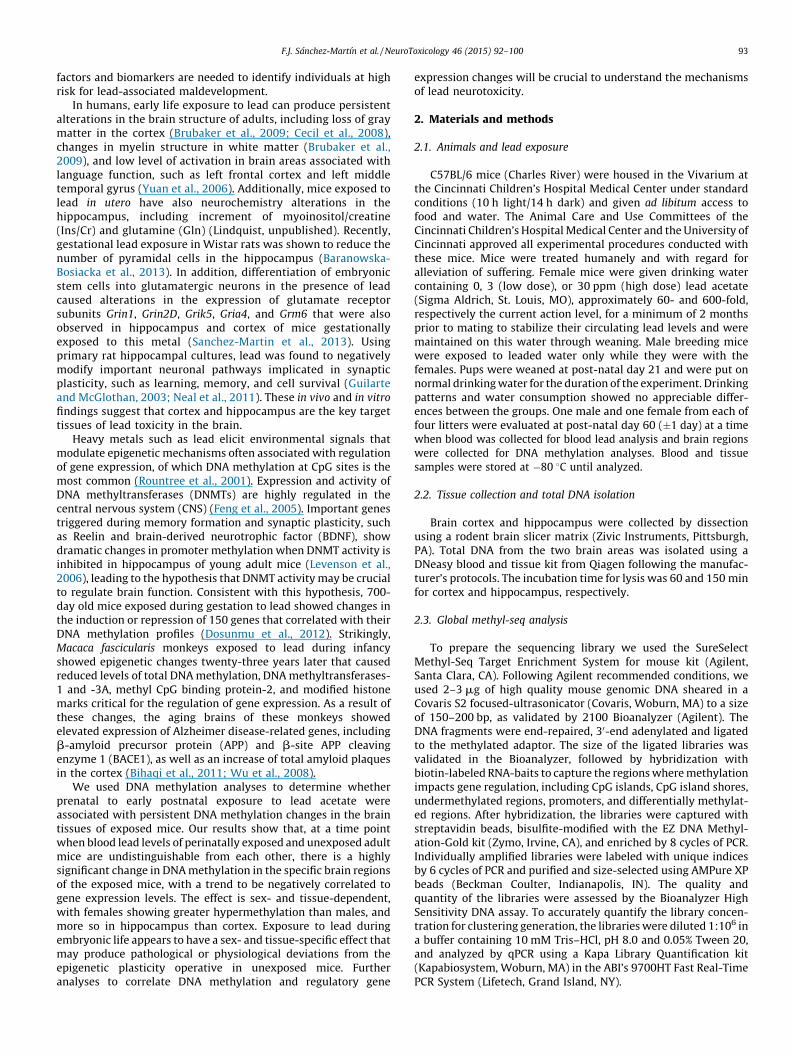
Fig. 1. Manhattan plot of �log10 p-values of differential methylation across the mouse ge
hypermethylated regions in mice exposed to 3 ppm (A) and 30 ppm (B) of lead. (C, D) Dif
(D) of lead.
Basically, the count of methylated C of a CpG site was distributedaccording to a binomial distribution with success probability p in n
total reads of the site, and p was modeled as a random variablefrom a beta distribution. Since the differential methylation analysiswas performed on raw counts, no filtering on coverage wasperformed. A Manhattan plot was used to visualize the distributionof differentially methylated CpG sites on the genome. Significantdifferentially methylated CpG sites were selected with fdr adjustedp-values of <0.001, <0.01, or <0.1. Two adjacent significant CpGsites were combined into one significant region if they were lessthan 1000 bp apart from each other. The significant region wasextended until there were no more significant CpG sites within1000 bp from the current region. The closest genes of significantsites and regions were identified using Bioconductor packageVariantAnnotation (Obenchain et al., 2014).
2.6. Reverse transcription and real-time PCR
Total RNA was isolated from the tissue samples by using theRNeasy Mini kit (Qiagen). Reverse transcription was performedusing random hexamer primers and SuperScript III transcriptase(Invitrogen) as indicated previously (Wang et al., 2010). Real-timeqPCR was performed to quantify the expression levels of differentgenes which were normalized to Gapdh mRNA. SupplementalTable S1 shows a list of primers used for each gene. Raw data wasanalyzed using the 2
DDCt method and it is shown as the log2 of thenormalized level.
)Differen�ally hypomethylated reg ions
-log 10
(p)
Chromosome1 2 3 242319171615 184 5 6 7 8 9 10 11 12 13 14
2
0
4
6
8
-log 10
(p)
Chromosome1 2 3 242319171615 184 5 6 7 8 9 10 11 12 13 14
2
0
4
6
8
)
nome induced by lead acetate in the hippocampus of male mice. (A, B) Differentially
ferentially hypomethylated regions in mice were exposed to 3 ppm (C) and 30 ppm
F.J. Sanchez-Martın et al. / NeuroToxicology 46 (2015) 92–100 95
3. Results
3.1. Blood lead levels in PND60 mice
We exposed dams to 0, 3, or 30 ppm lead acetate in drinkingwater for a minimum of 2 months prior to mating and maintainedthese conditions through weaning, while male breeding mice wereexposed to leaded water only while they were with the females.Pups were weaned at post-natal day 21 and were given drinkingwater without lead for the duration of the experiment. At PND60,the time when they were subjected to DNA methylation analyses,we found no significant difference in blood lead values among thethree groups of pups. Blood lead levels in pups (n = 16 per group) ofcontrol, 3 ppm- and 30 ppm-exposed dams were 1.2 � 1.1,0.9 � 0.8 and 1.3 � 3.1 mg/dl respectively, well below the currentaction level of 5 mg/dl, in good agreement with values observed inunexposed mice fed a normal diet (Ercal et al., 1996; Iavicoli et al.,2003) and consistent with prior observations by others (Virgoliniet al., 2008; Widzowski et al., 1994).
3.2. CpG site coverage
Bismark alignment was performed after bisulfite sequencing toanalyze the coverage of CpG data reads obtained. The number ofCpG sites covered was higher in cortex than in hippocampussamples, ranging between 1.5 million for males and 1.2 million forfemales in both groups of lead exposure. In the female hippocam-pus, sequencing covered slightly more than 0.8 million CpG sitesfor mice exposed to 3 ppm and 1.5 million CpG sites when exposed
(A)
(B)
(C
(D
Differen�ally hypermethylated reg ions
-log 10
(p)
Chromosome1 2 3 242319171615 184 5 6 7 8 9 10 11 12 13 14
2
0
4
6
8
-log
10(p
)
Chromosome1 2 3 242319171615 184 5 6 7 8 9 10 11 12 13 14
2
0
4
6
8
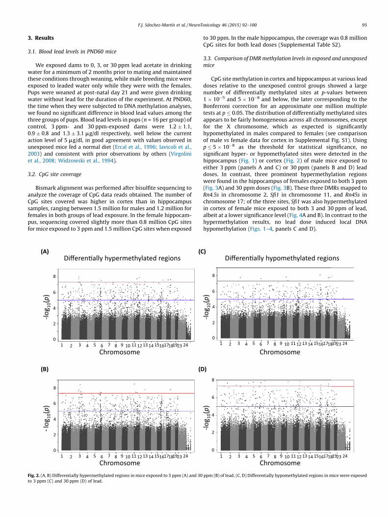
Fig. 2. (A, B) Differentially hypermethylated regions in mice exposed to 3 ppm (A) and 30
to 3 ppm (C) and 30 ppm (D) of lead.
to 30 ppm. In the male hippocampus, the coverage was 0.8 millionCpG sites for both lead doses (Supplemental Table S2).
3.3. Comparison of DMR methylation levels in exposed and unexposed
mice
CpG site methylation in cortex and hippocampus at various leaddoses relative to the unexposed control groups showed a largenumber of differentially methylated sites at p-values between1 � 10�5 and 5 � 10�8 and below, the later corresponding to theBonferroni correction for an approximate one million multipletests at p � 0.05. The distribution of differentially methylated sitesappears to be fairly homogeneous across all chromosomes, exceptfor the X chromosome, which as expected is significantlyhypomethylated in males compared to females (see comparisonof male vs female data for cortex in Supplemental Fig. S1). Usingp � 5 � 10�8 as the threshold for statistical significance, nosignificant hyper- or hypomethylated sites were detected in thehippocampus (Fig. 1) or cortex (Fig. 2) of male mice exposed toeither 3 ppm (panels A and C) or 30 ppm (panels B and D) leaddoses. In contrast, three prominent hypermethylation regionswere found in the hippocampus of females exposed to both 3 ppm(Fig. 3A) and 30 ppm doses (Fig. 3B). These three DMRs mapped toRn4.5s in chromosome 2, Sfi1 in chromosome 11, and Rn45s inchromosome 17; of the three sites, Sfi1 was also hypermethylatedin cortex of female mice exposed to both 3 and 30 ppm of lead,albeit at a lower significance level (Fig. 4A and B). In contrast to thehypermethylation results, no lead dose induced local DNAhypomethylation (Figs. 1–4, panels C and D).
)
)
Differen�ally hypomethylated reg ions
-log 10
( p)
Chromosome1 2 3 242319171615 184 5 6 7 8 9 10 11 12 13 14
2
0
4
6
8
-log 10
(p)
Chromosome1 2 3 242319171615 184 5 6 7 8 9 10 11 12 13 14
2
0
4
6
8
ppm (B) of lead. (C, D) Differentially hypomethylated regions in mice were exposed

(A)
(B)
(C)
(D)
Differen�ally hypomethylated reg ions
-log 10
(p)
Chromosome1 2 3 242319171615 184 5 6 7 8 9 10 11 12 13 14
2
0
4
6
8
-log 10
(p)
Chromosome1 2 3 242319171615 184 5 6 7 8 9 10 11 12 13 14
2
0
4
6
8
Rn4.5sSfi1
Rn45 s
Differen�ally hypermethylated reg ions-lo
g 10(p
)
Chromosome1 2 3 242319171615 184 5 6 7 8 9 10 11 12 13 14
2
0
4
6
8
10
-log 10
(p)
Chromosome
Rn4.5s
Sfi1
Rn45 s
1 2 3 242319171615 184 5 6 7 8 9 10 11 12 13 14
2
0
4
6
8
10
Fig. 3. Manhattan plot of �log10 p-values of differential methylation across the mouse genome induced by lead acetate in the hippocampus of female mice. (A, B) Differentially
hypermethylated regions in mice exposed to 3 ppm (A) and 30 ppm (B) of lead. (C, D) Differentially hypomethylated regions in mice were exposed to 3 ppm (C) and 30 ppm
(D) of lead.
F.J. Sanchez-Martın et al. / NeuroToxicology 46 (2015) 92–10096
In addition to the three prominent regions described above, anumber of hyper- and hypomethylated sites were evident in theManhattan plots in Figs. 1–4 with p-values comprised between the1 � 10�5 and 5 � 10�8 thresholds indicated earlier. We set threedifferent false discovery rate (fdr) values to categorize thesignificance of these sites and determine whether specific regionsof the genome showed statistically significant changes inmethylation patterns due to lead exposure. For this analysis, weconsidered that a group of contiguous sites plus 500 bp at eitherside constituted a genome region associated with its correspond-ing gene locus. At fdr � 0.1, most of the female genome showedmethylation changes after exposure to 3 or 30 ppm of lead, withgreater than 160,000 CpG sites, 60,000 genome regions and 16,000genes differentially methylated in hippocampus. No significantnumber of sites were affected in the hippocampus of exposedmales (Table 1). At this cut-off level, cortices in both sexes showedsignificant differential methylation, 10 times higher in femalesthan in males (Supplemental Table S3).
At fdr � 0.01, we found 20,129 differentially methylated sites infemale hippocampus, corresponding to 12,435 genome regionsencompassing 6180 genes (Table 1). At this cut-off value, neitherthe hippocampus of males nor the cortex of females exposed tolead presented significant sites with changes in the methylationpattern, but we found 112 hypermethylated sites in the cortex ofmales exposed to 3 ppm and 181 when exposed to 30 ppm(Supplemental Table S3).
At fdr � 0.001, we found 1623 differentially methylated CpGsites in the hippocampus of exposed females only, and nosignificant sites in cortex of either sex. Of these 1623 CpG sites,
322 corresponded to females exposed to 3 ppm and 1537 (withsome overlap with the previous dose) to 30 ppm exposure,encompassing 222 regions of the genome and affecting 117 uniquegenes (Table 1 and Supplemental Tables S3 and S4). Interestingly,most of the 1623 significant CpG sites observed in the hippocam-pus at this cut-off were hypermethylated after lead exposure withlittle evidence of hypomethylation (Fig. 5 and SupplementalTable S4). In addition, data from male and female differentialmethylation clustered together for tissue and treatment dose, butseparately for sex (Fig. 5), indicating that perinatal lead exposureinduces differential methylation in male and female brain tissues,albeit at very different levels.
3.4. Gene expression analysis by real-time PCR
We used real-time PCR to analyze the expression of 60 of the117 differentially methylated unique genes at fdr � 0.001 in thehippocampus of male and female mice exposed to 0 and 3 ppmlead. The selected genes had differentially methylated CpG siteswith p < 0.05 for females exposed to both lead doses, and the siteswere located between �10 and +10 kb from the transcription startsite (Supplemental Table S4). Expression of these genes in leadexposed mice, whether male or female, was very similar to theirexpression in control mice when the genes were analyzedindividually (Supplemental Fig. S2); however, there was a trendtoward a significant negative correlation between methylationchange and mRNA level (pslope = 0.083; r2 = 0.327) in the hippo-campus of female mice exposed to 3 ppm lead (Fig. 6A) and not inthe males (pslope = 0.996; r2 = 0.002) (Fig. 6B). These data suggest

(A)
(B)
(C)
(D)
Differen�ally hypermethylated reg ions-lo
g 10(p
)
Chromosome
Sfi1
1 2 3 242319171615 184 5 6 7 8 9 10 11 12 13 14
2
0
4
6
8
Differen�ally hypomethylated reg ions
-log 10
(p)
Chromosome1 2 3 242319171615 184 5 6 7 8 9 10 11 12 13 14
2
0
4
6
8
-log 10
(p)
Chromosome1 2 3 242319171615 184 5 6 7 8 9 10 11 12 13 14
2
0
4
6
8
-log 10
(p)
Chromosome
Sfi1
1 2 3 242319171615 184 5 6 7 8 9 10 11 12 13 14
2
0
4
6
8
Fig. 4. Manhattan plot of �log10 p-values of differential methylation across the mouse genome induced by lead acetate in the cortex of female mice. (A, B) Differentially
hypermethylated regions in mice exposed to 3 ppm (A) and 30 ppm (B) of lead. (C, D) Differentially hypomethylated regions in mice were exposed to 3 ppm (C) and 30 ppm
(D) of lead.
Table 1Number of significant CpG sites, genome regions and genes affected by Pb exposure
at different fdr cut-offs.
fdr CpG sites Genome regions Genes
0.001 1623 222 117
0.01 20,129 12,435 6180
0.1 159,619 66,539 16,141
F.J. Sanchez-Martın et al. / NeuroToxicology 46 (2015) 92–100 97
that lead not only induces hypermethylation in the femalehippocampus, but also that it may silence the expression of genesinvolved in brain function as a consequence of epigeneticmodification.
4. Discussion
In this study we show that in utero and lactational lead exposureinduces persistent DNA methylation changes in hippocampus andcortex, two brain tissues that are lead targets in mice. The majorityof changes result from DNA hypermethylation and are highly sexdependent, with female mice substantially more affected thanmales. Of the two brain tissues, the hippocampus had significantlyhigher levels of differentially methylated CpG sites than the cortex.At a restrictive p-value �5 � 10�8, only CpG sites in the Sfi1 genewere hypermethylated in the female cortex at both exposurelevels. In contrast, exposure led to high levels of hypermethylationin the hippocampus of females, that affected sites in three loci,Rn4.5s, Sfi1, and Rn45s, mapping to chromosomes 2, 11, and 17,
respectively, showing a striking degree of DNA hypermethylation.At a conservative fdr � 0.001, we found 1623 differentiallymethylated CpG sites in the hippocampus of exposed females,the majority (>90%) showing hypermethylation. These sitesdefined 222 regions of the genome, corresponding to an additional117 unique genes. After analyzing the expression of 60 from thisgroup, we found a trend toward a significant negative correlationbetween expression and methylation change in exposed femalemice, but not males. This analysis is inherently limited for itexamines only a single time point of expression and does not takeinto consideration the timing of expression of the genes tested.These genes are distributed throughout the genome and do notappear to be related through either regulatory or functionalconnections, even though the methylation changes occur repro-ducibly in multiple mice at the same locations.
Early life exposure to lead may have toxic effects in thedeveloping brain. Lead exposure during early childhood has beenlinked to deficits in cognitive functions and IQ, behavioral effects,and attention deficit hyperactivity disorder (Bellinger et al., 1994;Chen et al., 2007; Froehlich et al., 2009), suggesting that leadneurotoxicity may result from the alteration of mechanisms likeDNA methylation that regulate transcriptional pathways contrib-uting to synapse function, neurogenesis and ultimately, expressionof memory-related genes. Lead has recently been proposed to actin a locus-specific way on the epigenome, depending on thegenomic features in which affected CpG sites are located (Faulket al., 2013, 2014). Our current results are in conceptual agreementwith this hypothesis.

Fig. 5. Heat map of significant CpG sites differentially methylated at a
fdr � 0.001. The values shown for the females correspond to the normalized b-
values calculated from the number of methylated counts divided by the number of
methylated plus unmethylated counts for each one of the the1623 CpG sites in the
females. The male b-values are the corresponding values in the males.
-1.0 -0.5 0.5 1.0
-2
-1
1
2r2 = 0.32 7Pintercep t<0.00 1Pslop e=0.08 3
Log 2
mR
NA
leve
ls
Methylation change
-1.0 -0.5 0.5 1.0
-2
-1
1
2r2=0.00 2Pintercep t=0.22 1Pslop e=0.99 6
Log 2
mR
NA
leve
lsMethylation change
(A)
(B)
Fig. 6. Analysis of the correlation between mRNA levels and methylation change in
hippocampus of females (A) and males (B) exposed to 3 ppm of lead. The log2 mRNA
levels determined by RT-PCR of the 60 genes tested (see Supplemental Fig. S2) is
shown as a function of the corresponding methylation changes (Supplemental
Table S4). Negative methylation change values indicate hypomethylation and
positive, hypermethylation.
F.J. Sanchez-Martın et al. / NeuroToxicology 46 (2015) 92–10098
Lead exposure alters the expression of genes involved in DNAmethylation, such as methyl-cytosine-phosphate-guanine bindingprotein-2 (MeCP2) and DNA methyltransferases-1 and -3A (Bihaqiet al., 2011; Schneider et al., 2013; Wu et al., 2008). Exposure tolead from gestational day 13 to PND20 resulted in significantchanges of gene expression in cortical regions of mouse brain, at20 and 700 days of age; these changes were correlated withchanges in DNA methylation profile and repressed many genesnormally up-regulated during normal aging, suggesting that earlylife exposure to lead disturbs developmental processes in the brainand compromises its ability to cope with adult challenges(Dosunmu et al., 2012). Hypermethylation of genes involved inneurogenesis signaling pathways has also been found in neuronalprecursor cells derived from human embryonic stem cellschronically exposed to lead. These cells exhibit shorter neuritesand less branching, as well as a significant decrease in theexpression levels of the neural marker genes PAX6 and MSI1 (Senutet al., 2014).
Methylation and expression show a strong sex-dependence,with changes more evident in females than in males. Similar sex-specific effects have been observed as the consequence of maternalseparation, which caused repression of BDNF expression inhippocampus of C57BL/6J female mice but not males, andhypermethylation in males but not females (Kundakovic et al.,2013). Brain differences between males and females are a commonphenomenon, since sexual differentiation in the brain takes placeduring a perinatal sensitive window as a result of gonadalhormone-induced developmental organization (Auger and Auger,2013; Chung and Auger, 2013; Menger et al., 2010). Sex differencesin gene expression patterns and in the regulation of genes codingfor DNA methylases have been observed in hippocampus andfrontal cortex of rats exposed to lead (Schneider et al., 2011,
2012b), and shown to be differentially expressed depending on thedevelopmental timing of the exposure (Schneider et al., 2012a).The effect of sex in the regulation of the genome and epigenome islargely unexplored. Sex influences genomic methylation statusalthough the mechanisms underlying this effect are unknown (Liuet al., 2010). Possibly, interactions between sex hormones and leadexposure during development make the female hippocampusmore susceptible to differentially methylation. In contrast to ourresults, observations in humans, have shown that early exposure tolead causes neuropsychological alterations in mid-adolescentmales not, so much in females (Ris et al., 2004). These epigeneticsex differences observed in mice may be independent conse-quences of exposure, underlining the complexity of the sex-specific response to early life adversities that can affect theepigenetic regulation of gene expression in males and females.
Rn4.5s codes for a 98-nucleotide nuclear RNA with unknownfunction that is transcribed by RNA polymerase III (Gogolevskayaet al., 2010) and Rn45s codes for the RNA precursor to 18S, 5.8S and28S rRNA (Grozdanov et al., 2003); both of them show changes inmethylation in the hippocampus of females exposed to 3 ppm oflead (Supplemental Figs. S3 and S4). Although neither of these twogenes has been linked to metal toxicity or lead neurotoxicitypreviously, their hypermethylation by lead exposure maycompromise ribosome structure or overall protein synthesiscapacity in the hippocampus. Changes in expression of Sfi1 havebeen observed in a genetic mouse model of neurodevelopmentaldisorder, being up-regulated in young brain and down-regulatedin older brain (Trent et al., 2014). This gene has two regions thatare differentially methylated in the hippocampus of femalesexposed to 3 ppm of lead (Supplemental Fig. S5) but its expressionis not changed in either sex (Supplemental Fig. S2) possibly
F.J. Sanchez-Martın et al. / NeuroToxicology 46 (2015) 92–100 99
because the gene is already highly methylated and changes inthese two regions are not enough to alter the expression levels.
The promoter of the Dynlt1b (dynein light chain Tctex-type 1B)gene is hypermethylated in the hippocampus of females exposedto 3 and 30 ppm of lead (Supplemental Fig. S6). In the dentate gyrusof the hippocampus, new neurons continue to be generatedthroughout life from progenitor cells at the subgranular zone(Eriksson et al., 1998; Kornack and Rakic, 1999). These newbornneurons serve not only to maintain the pool of neurons, but also tobuild memory (Ernst et al., 2014; Nakashiba et al., 2012). Dynlt1b
transcription is high in these cells (Dedesma et al., 2006), and actsas a regulator for the genesis of neurons (Gauthier-Fisher et al.,2009; Tseng et al., 2010) and possibly for neurite outgrowth andaxon formation as well (Sachdev et al., 2007). Although theexpression level is not changed at PND60 after early life exposureto lead (Supplemental Fig. S2), it is plausible that prenatal and earlypostnatal exposure to lead may inhibit the formation of newneurons, decrease the total pool of neurons, and eventuallycompromise memory formation.
Although the use of lead has been reduced in the last fewdecades, exposure is still an important concern due to its non-biodegradable nature and its ubiquitous presence, which poses apotential health risk as a result of the increased sensitivity ofchildren to lead toxicity (Markowitz, 2000). During the gestationalperiod, lead crosses the placenta and blood–brain barrier reachingthe developing fetal brain (Hu et al., 1998). It is becomingincreasingly evident that early-life exposure to lead may produceenduring changes in the epigenetic mechanisms that regulate geneexpression in the brain, contributing to pathological and physio-logical outcomes.
Conflict of interest
The authors declare that there are no conflicts of interest.
Transparency document
The Transparency document associated with this article can befound in the online version.
Acknowledgments
We thank Ying Xia, Andrew Vonhandorf, Chia-I Ko, HisakaKurita, Vinicius Carreira, Qin Wang and Yunxia Fan for criticallyreading the manuscript and providing helpful criticisms. This workwas supported by NIH grants R21 ES020048 and the Center forEnvironmental Genetics P30 ES06096.
Appendix A. Supplementary data
Supplementary data associated with this article can be found, inthe online version, at http://dx.doi.org/10.1016/j.neuro.2014.12.004.
References
Andrews S. FastQC. A quality control tool for high throughput sequence data., 2014http://www.bioinformatics.babraham.ac.uk/projects/fastqc/.
Auger CJ, Auger AP. Permanent and plastic epigenesis in neuroendocrine systems. FrontNeuroendocrinol 2013;34(3):190–7.
Baranowska-Bosiacka I, Struzynska L, Gutowska I, Machalinska A, Kolasa A, Klos P, et al.Perinatal exposure to lead induces morphological, ultrastructural and molecularalterations in the hippocampus. Toxicology 2013;303:187–200.
Bellinger D, Leviton A, Allred E, Rabinowitz M. Pre- and postnatal lead exposure andbehavior problems in school-aged children. Environ Res 1994;66(1):12–30.
Bihaqi SW, Huang H, Wu J, Zawia NH. Infant exposure to lead (Pb) and epigeneticmodifications in the aging primate brain: implications for Alzheimer’s disease. JAlzheimers Dis 2011;27(4):819–33.
Brubaker CJ, Schmithorst VJ, Haynes EN, Dietrich KN, Egelhoff JC, Lindquist DM, et al.Altered myelination and axonal integrity in adults with childhood lead exposure: adiffusion tensor imaging study. Neurotoxicology 2009;30(6):867–75.
Cecil KM, Brubaker CJ, Adler CM, Dietrich KN, Altaye M, Egelhoff JC, et al. Decreasedbrain volume in adults with childhood lead exposure. PLoS Med 2008;5(5):e112.
Chen A, Cai B, Dietrich KN, Radcliffe J, Rogan WJ. Lead exposure, IQ, and behavior inurban 5- to 7-year-olds: does lead affect behavior only by lowering IQ? Pediatrics2007;119(3):e650–8.
Chung WC, Auger AP. Gender differences in neurodevelopment and epigenetics.Pflugers Arch 2013;465(5):573–84.
Dedesma C, Chuang JZ, Alfinito PD, Sung CH. Dynein light chain Tctex-1 identifiesneural progenitors in adult brain. J Comp Neurol 2006;496(6):773–86.
Dietrich KN, Ris MD, Succop PA, Berger OG, Bornschein RL. Early exposure to lead andjuvenile delinquency. Neurotoxicol Teratol 2001;23:511–8.
Dosunmu R, Alashwal H, Zawia NH. Genome-wide expression and methylation profil-ing in the aged rodent brain due to early-life Pb exposure and its relevance to aging.Mech Ageing Dev 2012;133(6):435–43.
Ercal N, Treeratphan P, Hammond TC, Matthews RH, Grannemann NH, Spitz DR. In vivoindices of oxidative stress in lead-exposed C57BL/6 mice are reduced by treatmentwith meso-2,3-dimercaptosuccinic acid or N-acetylcysteine. Free Rad Biol Med1996;21:157–61.
Eriksson PS, Perfilieva E, Bjork-Eriksson T, Alborn AM, Nordborg C, Peterson DA, et al.Neurogenesis in the adult human hippocampus. Nat Med 1998;4(11):1313–7.
Ernst A, Alkass K, Bernard S, Salehpour M, Perl S, Tisdale J, et al. Neurogenesis in thestriatum of the adult human brain. Cell 2014;156(5):1072–83.
Faulk C, Barks A, Liu K, Goodrich JM, Dolinoy DC. Early-life lead exposure results indose- and sex-specific effects on weight and epigenetic gene regulation in wean-ling mice. Epigenomics 2013;5(5):487–500.
Faulk C, Liu K, Barks A, Goodrich JM, Dolinoy DC. Longitudinal epigenetic drift in miceperinatally exposed to lead. Epigenetics 2014;9(7):934–41.
Feng J, Chang H, Li E, Fan G. Dynamic expression of de novo DNA methyltransferasesDnmt3a and Dnmt3b in the central nervous system. J Neurosci Res2005;79(6):734–46.
Froehlich TE, Lanphear BP, Auinger P, Hornung R, Epstein JN, Braun J, et al. Associationof tobacco and lead exposures with attention-deficit/hyperactivity disorder. Pedi-atrics 2009;124(6):e1054–63.
Gauthier-Fisher A, Lin DC, Greeve M, Kaplan DR, Rottapel R, Miller FD. Lfc and Tctex-1regulate the genesis of neurons from cortical precursor cells. Nat Neurosci2009;12(6):735–44.
Gogolevskaya IK, Veniaminova NA, Kramerov DA. Nucleotide sequences of B1 SINE and4.5S(I) RNA support a close relationship of zokors to blind mole rats (Spalacinae)and bamboo rats (Rhizomyinae). Gene 2010;460(1–2):30–8.
Grozdanov P, Georgiev O, Karagyozov L. Complete sequence of the 45-kbmouse ribosomal DNA repeat: analysis of the intergenic spacer. Genomics2003;82(6):637–43.
Guilarte TR, McGlothan JL. Selective decrease in NR1 subunit splice variant mRNA inthe hippocampus of Pb2+-exposed rats: implications for synaptic targeting andcell surface expression of NMDAR complexes. Brain Res Mol Brain Res2003;113(1–2):37–43.
Hu H, Rabinowitz M, Smith D. Bone lead as a biological marker in epidemiologicstudies of chronic toxicity: conceptual paradigms. Environ Health Perspect1998;106(1):1–8.
Iavicoli I, Carelli G, Stanek EJ, Castellino N, Calabrese EJ. Effects of low doses of dietarylead on red blood cell production in male and female mice. Toxicol Lett2003;137(3):193–9.
Kornack DR, Rakic P. Continuation of neurogenesis in the hippocampus of the adultmacaque monkey. Proc Natl Acad Sci U S A 1999;96(10):5768–73.
Krueger F, Andrews SR. Bismark: a flexible aligner and methylation caller for Bisulfite-Seq applications. Bioinformatics 2011;27(11):1571–2.
Kundakovic M, Lim S, Gudsnuk K, Champagne FA. Sex-specific and strain-dependenteffects of early life adversity on behavioral and epigenetic outcomes. FrontPsychiatry 2013;4:78.
Levenson JM, Roth TL, Lubin FD, Miller CA, Huang IC, Desai P, et al. Evidence thatDNA (cytosine-5) methyltransferase regulates synaptic plasticity in the hippocam-pus. J Biol Chem 2006;281(23):15763–73.
Liu J, Morgan M, Hutchison K, Calhoun VD. A study of the influence of sex on genomewide methylation. PLoS ONE 2010;5(4):e10028.
Markowitz M. Lead poisoning. Pediatr Rev 2000;21(10):327–35.Menger Y, Bettscheider M, Murgatroyd C, Spengler D. Sex differences in brain epige-
netics. Epigenomics 2010;2(6):807–21.Nakashiba T, Cushman JD, Pelkey KA, Renaudineau S, Buhl DL, McHugh TJ, et al. Young
dentate granule cells mediate pattern separation, whereas old granule cellsfacilitate pattern completion. Cell 2012;149(1):188–201.
Neal AP, Worley PF, Guilarte TR. Lead exposure during synaptogenesis alters NMDAreceptor targeting via NMDA receptor inhibition. Neurotoxicology 2011;32(2):281–9.
Needleman HL, Riess JA, Tobin MJ, Biesecker GE, Greenhouse JB. Bone lead levels anddelinquent behavior. JAMA 1996;275(5):363–9.
Needleman HL, McFarland C, Ness RB, Fienberg SE, Tobin MJ. Bone lead levels in adjudi-cated delinquents. A case control study. Neurotoxicol Teratol 2002;24(6):711–7.
Obenchain V, Lawrence M, Carey V, Gogarten S, Shannon P, Morgan M. VariantAnnota-tion: a Bioconductor package for exploration and annotation of genetic variants.Bioinformatics 2014;30(14):2076–8.
F.J. Sanchez-Martın et al. / NeuroToxicology 46 (2015) 92–100100
Pham TV, Piersma SR, Warmoes M, Jimenez CR. On the beta-binomial model foranalysis of spectral count data in label-free tandem mass spectrometry-basedproteomics. Bioinformatics 2010;26(3):363–9.
Ris MD, Dietrich KN, Succop PA, Berger OG, Bornschein RL. Early exposure to lead andneuropsychological outcome in adolescence. J Int Neuropsychol Soc 2004;10(2):261–70.
Rountree MR, Bachman KE, Herman JG, Baylin SB. DNA methylation, chromatininheritance, and cancer. Oncogene 2001;20(24):3156–65.
Sachdev P, Menon S, Kastner DB, Chuang JZ, Yeh TY, Conde C, et al. G protein betagamma subunit interaction with the dynein light-chain component Tctex-1 reg-ulates neurite outgrowth. EMBO J 2007;26(11):2621–32.
Sanchez-Martin FJ, Fan Y, Lindquist DM, Xia Y, Puga A. Lead induces similar geneexpression changes in brains of gestationally exposed adult mice and in neuronsdifferentiated from mouse embryonic stem cells. PLOS ONE 2013;8(11):e80558.
Schneider JS, Anderson DW, Sonnenahalli H, Vadigepalli R. Sex-based differences ingene expression in hippocampus following postnatal lead exposure. Toxicol ApplPharmacol 2011;256(2):179–90.
Schneider JS, Anderson DW, Talsania K, Mettil W, Vadigepalli R. Effects of develop-mental lead exposure on the hippocampal transcriptome: influences of sex,developmental period, and lead exposure level. Toxicol Sci 2012a;129(1):108–25.
Schneider JS, Mettil W, Anderson DW. Differential effect of postnatal lead exposure ongene expression in the hippocampus and frontal cortex. J Mol Neurosci2012b;47(1):76–88.
Schneider JS, Kidd SK, Anderson DW. Influence of developmental lead exposure onexpression of DNA methyltransferases and methyl cytosine-binding proteins inhippocampus. Toxicol Lett 2013;217(1):75–81.
Senut MC, Sen A, Cingolani P, Shaik A, Land SJ, Ruden DM. Lead exposure disrupts globalDNA methylation in human embryonic stem cells and alters their neuronaldifferentiation. Toxicol Sci 2014;139(1):142–61.
Trent S, Fry JP, Ojarikre OA, Davies W. Altered brain gene expression but not steroidbiochemistry in a genetic mouse model of neurodevelopmental disorder. MolAutism 2014;5(1):21.
Tseng YY, Gruzdeva N, Li A, Chuang JZ, Sung CH. Identification of the Tctex-1 regulatoryelement that directs expression to neural stem/progenitor cells in developing andadult brain. J Comp Neurol 2010;518(16):3327–42.
Virgolini MB, Rossi-George A, Weston D, Cory-Slechta DA. Influence of low levelmaternal Pb exposure and prenatal stress on offspring stress challenge respon-sivity. Neurotoxicology 2008;29(6):928–39.
Wang Y, Fan Y, Puga A. Dioxin exposure disrupts the differentiation of mouse embry-onic stem cells into cardiomyocytes. Toxicol Sci 2010;115:225–37.
West-Eberhard MJ. Developmental plasticity and the origin of species differences. ProcNatl Acad Sci U S A 2005;102(Suppl. 1):6543L 6549.
Widzowski DV, Finkelstein JN, Pokora MJ, Cory-Slechta DA. Time course of postnatallead-induced changes in dopamine receptors and their relationship to changes indopamine sensitivity. Neurotoxicology 1994;15(4):853–65.
Wright JP, Dietrich KN, Ris MD, Hornung RW, Wessel SD, Lanphear BP, et al. Associationof prenatal and childhood blood lead concentrations with criminal arrests in earlyadulthood. PLoS Med 2008;5(5):e101.
Wu J, Basha MR, Brock B, Cox DP, Cardozo-Pelaez F, McPherson CA, et al. Alzheimer’sdisease (AD)-like pathology in aged monkeys after infantile exposure to environ-mental metal lead (Pb): evidence for a developmental origin and environmentallink for AD. J Neurosci 2008;28(1):3–9.
Yamazaki Y, Mann MR, Lee SS, Marh J, McCarrey JR, Yanagimachi R, et al. Reprogram-ming of primordial germ cells begins before migration into the genital ridge,making these cells inadequate donors for reproductive cloning. Proc Natl Acad SciU S A 2003;100(21):12207–12.
Yuan W, Holland SK, Cecil KM, Dietrich KN, Wessel SD, Altaye M, et al. The impact ofearly childhood lead exposure on brain organization: a functional magneticresonance imaging study of language function. Pediatrics 2006;118(3):971–7.
Copyright © 2022 FDOKUMEN