Gene profiling suggests a common evolution of bladder cancer subtypes
11
RESEARCH ARTICLE Open Access Gene profiling suggests a common evolution of bladder cancer subtypes Donna E Hansel 1* , Zhongfa Zhang 2 , David Petillo 2 and Bin T Teh 3,4 Abstract Background: Bladder cancer exists as several distinct subtypes, including urothelial carcinoma (UCa), squamous cell carcinoma (SCCa), adenocarcinoma and small cell carcinoma. These entities, despite showing distinct morphology and clinical behavior, arise from the urothelial lining and are often accompanied by similar precursor/in situ findings. The relationship between these subtypes has not been explored in detail. Methods: We compared gene expression analysis of the two most common subtypes of bladder cancer, UCa (n = 10) and SCCa (n = 9), with an additional comparison to normal urothelium from non-cancer patients (n = 8) using Affymetrix GeneChip Human genome arrays (Affymetrix, Santa Clara, CA). The results were stratified by supervised and unsupervised clustering analysis, as well as by overall fold change in gene expression. Results: When compared to normal urothelium, UCa showed differential expression of 155 genes using a 5-fold cut-off. Examples of differentially regulated genes included topoisomerases, cancer-related transcription factors and cell cycle mediators. A second comparison of normal urothelium to SCCa showed differential expression of 503 genes, many of which were related to squamous-specific morphology (desmosomal complex, intermediate filaments present within squamous epithelium, squamous cornifying proteins, and molecules upregulated in squamous carcinomas from other anatomic sites). When compared, 137 genes were commonly dysregulated in both UCa and SCCa as compared to normal urothelium. All dysregulated genes in UCa were shared in common with SCCa, with the exception of only 18 genes. Supervised clustering analysis yielded correct classification of lesions in 26/27 (96%) of cases and unsupervised clustering analysis yielded correct classification in 25/27 (92.6%) of cases. Conclusions: The results from this analysis suggest that bladder SCCa shares a significant number of gene expression changes with conventional UCa, but also demonstrates an additional set of alterations that is unique to this entity that defines the squamous phenotype. The similarity in deregulated gene products suggests that SCCa may be a much more closely related entity at the molecular level to conventional UCa than previously hypothesized. Keywords: Gene expression profiling, Bladder cancer, Urothelial, Squamous, Subtype Background Urothelial carcinoma (UCa) represents the most com- mon form of bladder cancer in the United States (>90%) and is characterized by frequent mutations in TP53, RB and PTEN [1]. Morphologically, UCa consists of invasive nests of carcinoma cells with variable atypia and fre- quent surrounding retraction artifact (Figure 1A), al- though this appearance can vary significantly. Less common forms of bladder cancer in the United States include squamous cell carcinoma (SCCa; Figure 1B), adenocarcinoma and small cell carcinoma, which are de- fined as pure morphologic entities that lack a typical urothelial component [2]. Our understanding of the mo- lecular relationships between these other forms of blad- der cancer that arise from the urothelial lining has been limited and may be based on the low number of cases available for study and/or lack of significant attention paid to this topic. It appears, however, that despite a similar origin from the surface urothelium, these various * Correspondence: [email protected] 1 Department of Pathology, University of California at San Diego, 9500 Gilman Drive, MC 0612, La Jolla, CA 92093, USA Full list of author information is available at the end of the article © 2013 Hansel et al.; licensee BioMed Central Ltd. This is an open access article distributed under the terms of the Creative Commons Attribution License (http://creativecommons.org/licenses/by/2.0), which permits unrestricted use, distribution, and reproduction in any medium, provided the original work is properly cited. Hansel et al. BMC Medical Genomics 2013, 6:42 http://www.biomedcentral.com/1755-8794/6/42
Transcript of Gene profiling suggests a common evolution of bladder cancer subtypes

Hansel et al. BMC Medical Genomics 2013, 6:42http://www.biomedcentral.com/1755-8794/6/42
RESEARCH ARTICLE Open Access
Gene profiling suggests a common evolution ofbladder cancer subtypesDonna E Hansel1*, Zhongfa Zhang2, David Petillo2 and Bin T Teh3,4
Abstract
Background: Bladder cancer exists as several distinct subtypes, including urothelial carcinoma (UCa), squamous cellcarcinoma (SCCa), adenocarcinoma and small cell carcinoma. These entities, despite showing distinct morphologyand clinical behavior, arise from the urothelial lining and are often accompanied by similar precursor/in situfindings. The relationship between these subtypes has not been explored in detail.
Methods: We compared gene expression analysis of the two most common subtypes of bladder cancer, UCa (n =10) and SCCa (n = 9), with an additional comparison to normal urothelium from non-cancer patients (n = 8) usingAffymetrix GeneChip Human genome arrays (Affymetrix, Santa Clara, CA). The results were stratified by supervisedand unsupervised clustering analysis, as well as by overall fold change in gene expression.
Results: When compared to normal urothelium, UCa showed differential expression of 155 genes using a 5-foldcut-off. Examples of differentially regulated genes included topoisomerases, cancer-related transcription factors andcell cycle mediators. A second comparison of normal urothelium to SCCa showed differential expression of 503genes, many of which were related to squamous-specific morphology (desmosomal complex, intermediatefilaments present within squamous epithelium, squamous cornifying proteins, and molecules upregulated insquamous carcinomas from other anatomic sites). When compared, 137 genes were commonly dysregulated inboth UCa and SCCa as compared to normal urothelium. All dysregulated genes in UCa were shared in commonwith SCCa, with the exception of only 18 genes. Supervised clustering analysis yielded correct classification oflesions in 26/27 (96%) of cases and unsupervised clustering analysis yielded correct classification in 25/27 (92.6%) ofcases.
Conclusions: The results from this analysis suggest that bladder SCCa shares a significant number of geneexpression changes with conventional UCa, but also demonstrates an additional set of alterations that is unique tothis entity that defines the squamous phenotype. The similarity in deregulated gene products suggests that SCCamay be a much more closely related entity at the molecular level to conventional UCa than previouslyhypothesized.
Keywords: Gene expression profiling, Bladder cancer, Urothelial, Squamous, Subtype
BackgroundUrothelial carcinoma (UCa) represents the most com-mon form of bladder cancer in the United States (>90%)and is characterized by frequent mutations in TP53, RBand PTEN [1]. Morphologically, UCa consists of invasivenests of carcinoma cells with variable atypia and fre-quent surrounding retraction artifact (Figure 1A), al-though this appearance can vary significantly. Less
* Correspondence: [email protected] of Pathology, University of California at San Diego, 9500 GilmanDrive, MC 0612, La Jolla, CA 92093, USAFull list of author information is available at the end of the article
© 2013 Hansel et al.; licensee BioMed CentralCommons Attribution License (http://creativecreproduction in any medium, provided the or
common forms of bladder cancer in the United Statesinclude squamous cell carcinoma (SCCa; Figure 1B),adenocarcinoma and small cell carcinoma, which are de-fined as pure morphologic entities that lack a typicalurothelial component [2]. Our understanding of the mo-lecular relationships between these other forms of blad-der cancer that arise from the urothelial lining has beenlimited and may be based on the low number of casesavailable for study and/or lack of significant attentionpaid to this topic. It appears, however, that despite asimilar origin from the surface urothelium, these various
Ltd. This is an open access article distributed under the terms of the Creativeommons.org/licenses/by/2.0), which permits unrestricted use, distribution, andiginal work is properly cited.

Figure 1 Two major subtypes of bladder cancer include (A)urothelial carcinoma, which is the most common form ofbladder cancer and contains variably sized nests and (B)squamous cell carcinoma, which is characterized bydesmosomes and keratin pearls (arrow).
Hansel et al. BMC Medical Genomics 2013, 6:42 Page 2 of 11http://www.biomedcentral.com/1755-8794/6/42
forms of bladder cancer show differential clinical be-havior, morphologic appearances, immunohistochemicalmarkers and response to chemotherapy.SCCa represents the second most common form of
bladder cancer in the United States (5-8% of bladder can-cer cases) and is distinguished by invasive squamous car-cinoma cells containing desmosomes and often keratinformation [3]. A higher incidence of bladder SCCa hasbeen reported in parts of the Middle East and Africa, how-ever, where chronic infection with the water-bourne para-site Schistosoma haematobium, represents a major riskfactor in the development of this disease [4]. Other poten-tial risk factors in the development of SCCa include long-term catheterization, calculi that form in the urinary tract,and a nonfunctioning bladder, amongst others, and theserisk factors may account for many instances of SCCa iden-tified in the United States and Europe.Despite the distinct appearance of UCa and SCCa, the
cellular origin of these two forms of bladder cancer has
been debated. Whereas UCa can arise in associationwith surface high-grade changes of the urothelium (e.g.,carcinoma in situ) and SCCa can arise in associationwith squamous dysplasia and squamous carcinoma insitu of the bladder, there are numerous cases that showoverlap of surface changes. For example, SCCa canoccur in the absence of any surface squamous metaplasiaand may only be associated with urothelial carcinoma insitu [3]. Furthermore, UCa itself has been shown todemonstrate “divergent” differentiation with the abilityof UCa tumor cells to develop squamous or glandularfeatures. These findings suggest that 1) the molecular re-lationship between historically distinct subtypes of blad-der cancer may be more similar than previouslyhypothesized and 2) the surface urothelium in a bladderundergoing neoplastic alterations may be unusuallysuited to give rise to divergent phenotypes in the settingof both in situ and invasive disease.To date, much of the molecular analysis on bladder
SCCa has been limited due to a predominant focus oninfectious, Schistosomal-derived cases as well as use ofimmortalized bladder cancer cell lines in a number ofstudies [5-8]. In this setting, the understanding of therelationship between UCa and SCCa, as well as the dis-tinction between primary molecular changes and thosesecondarily induced by infection-specific responses, be-comes difficult. We sought to compare the two mostcommon forms of pure bladder cancer in the US (UCaand non-infectious SCCa) using multi-level gene expres-sion analysis to determine the relationship and possiblehierarchy of these two examples of bladder cancer. Theresults from our study suggest a closer relationship be-tween these neoplastic entities than previously proposed;a shared evolution of these cancers may represent an op-portunity for targeting bladder cancer along commonpathways early in the disease process.
MethodsSpecimen collectionSpecimens were collected with Institutional ReviewBoard approval. The existing bladder cancer biobank(1971 onwards) was searched for snap-frozen tissueobtained from non-neoplastic bladder and/or ureter andfrom patients with either UCa or SCCa. Frozen sectionswere obtained from all specimens and reviewed; speci-mens with any necrosis or <90% tumor or normal cellnuclei were excluded from analysis. H&E slides corre-sponding to the initial pathology specimen associatedwith each sample were re-reviewed for accuracy oftumor classification. The clinical records for any patientswith normal urothelium were reviewed; any patient witha precedent or subsequent occurrence of urinary tractneoplasia was excluded from analysis. This resulted in 8normal urothelium specimens, 10 UCa specimens and 9

Hansel et al. BMC Medical Genomics 2013, 6:42 Page 3 of 11http://www.biomedcentral.com/1755-8794/6/42
SCCa specimens (total in archive) used for analysis. Nopatient with SCCa had a precedent or concurrent historyof Schistosomal infection. This study was approved bythe Cleveland Clinic IRB.
Raw gene expression levelsTen micrograms of total RNA from each sample wasprocessed using the Affymetrix GeneChip one-cycle targetlabeling kit (Affymetrix, Santa Clara, CA). The resultantbiotinylated cDNA was fragmented and subsequentlyhybridized to the GeneChip Human genome (54,675 probesets including more than 35,000 human genes; Affymetrix).Arrays were washed, stained, and scanned using theAffymetrixModel450 Fluidics Station and AffymetrixModel3000 scanner per manufacturer’s recommended protocols.Expression values were generated using Microarray
Suite (MAS) v5.0 software (Affymetrix). The probes wereredefined according to a new study (http://brainarray.mhri.med.umich.edu/Brainarray/Database/CustomCDF/version10) to combine probes representing the same genefor a single profile per gene. The hybridizations were nor-malized using the robust multichip averaging (rma) algo-rithm in the Bioconductor package affy (see http://www.bioconductor.org/) in order to obtain summary expressionvalues for each probe set [9,10]. This resulted in morethan 17,000 genes, each of which then has one numericnumber to represent its relative gene expression intensityin the sample.
Clustering studyA hierarchical clustering algorithm was used to identifyunsupervised clusters based on the Euclidean distancefor dissimilarities between the data samples. The slightlymodified “plot.phylo” program from analyses of phyloge-netics and evolution (ape) package of R was used toshow the clustering results [11,12]. The interquartilerange (IQR) and coefficient of variation (CV) were usedto filter identified genes in the unsupervised clusteringstudy. IQR was defined to be the distance between the
Figure 2 Boxplot of the 27 samples used for study presented in log2
third and first quartiles of the data; the CV of a vectorwas defined to be the standard deviation divided by itsmean value. We used IQR > 0.3 and CV > 0.05 as ourfiltering criteria. This resulted in a data set of approxi-mately 13600 genes. Other cutoff values provided similarclustering results. We also used the limma package toidentify genes for supervised clustering analysis. Whenmore than two classes of genes were present in the studygroup, the comparison was made between all pairs ofclasses. When comparison was made between two con-ditions, we used a fold change of 5 as a cutoff value todeclare a gene significant.We set 0.05 as our significance level for all tests.
All calculations were implemented in R environment(R > 2.15.0, see http://www.r-project.org).
ResultsComparative analysisDespite the shared urothelium from which SCCa andUCa arises, it is unclear whether these two morphologic-ally distinct forms of bladder cancer share significantmolecular overlap and, if so, whether a hierarchy intumor types exists. In order to address this question, weperformed a four-way interrogation of gene expressionprofiles: 1) normal urothelium versus SCCa, 2) normalurothelium versus UCa, 3) normal urothelium versusSCCa and UCa combined (shared alterations) and 4)UCa versus SCCa (divergent alterations). We includedfor analysis 8 samples of normal urothelium, 10 samplesof invasive high-grade UCa and 9 samples of invasiveSCCa. A boxplot of the data set shows that all sampleshave a roughly comparable distribution of the gene ex-pression values, except only one sample (normal sample1; Figure 2). When analyzed by subsequent unsupervisedor supervised clustering studies, sample 1 did correctlysegregate into the normal urothelial cluster; we thereforeretained this sample in our study set.Unexpectedly, the gene expression profiles revealed a
large number of shared gene expression differences in
scale.

Figure 3 Heatmap comparing UCa and SCCa to normal urothelium for all 27 samples using supervised clustering and a fold change of5, with 262 genes represented.
Hansel et al. BMC Medical Genomics 2013, 6:42 Page 4 of 11http://www.biomedcentral.com/1755-8794/6/42
UCa and SCCa relative to the normal urothelium whenusing a 5-fold cut-off (n = 137, Figure 3). In addition tothese shared gene expression differences, SCCa demon-strated an additional 366 uniquely dysregulated genesrelative to normal urothelium, whereas UCa demon-strated only an additional 18 genes that were uniquely
Figure 4 Supervised clustering of the 27 samples using the topmost significant 877 genes (limma method from Bioconductorusing adjusted p value <=0.001; error rate 1/27=3.7%; black =UCa; red = SCCa; green = normal).
dysregulated relative to normal urothelium. Using super-vised clustering (Figure 4) and unsupervised clustering(Figure 5A, B) analysis, we were able to reproduciblysegregate normal urothelium, UCa and SCCa specimens,although two specimens (UCa23 and UCa24) appearedslightly different than other tumors in the UCa category,but could correctly segregate with other UCa specimenswhen a lower threshold value was applied to the analysis;specifically, no morphological difference was appreciatedin these two specimens.All differentially expressed genes were used to obtain
fold changes (in log2 scale) to compare UCa versus nor-mal and SCCa versus normal (Figure 6A). The majorityof genes have fold change differences within 2 (17,468,99.25%, grey). A relatively larger number of genes havefold change differences above 2 (184, red) than the num-ber of genes with fold change differences below −2 (47,blue). Overall, the fold change vectors correlated wellwith each other (cor. coefficient greater than 0.73), withthe exception of the 184 genes located above the se-lected area (red), which are significantly higher in SCCawhen compared to normal urothelium. A summary ofthe 4-way analysis performed with total gene expressiondifferences is presented in Figure 6B.
Commonly dysregulated genes in UCa and SCCa versusnormal urotheliumWe next sought to determine commonalities in gene ex-pression changes in UCa and SCCa versus normalurothelium. As normal urothelium lines the urinary tractthroughout its length, and represents the common epi-thelium from which any form of bladder cancer (with
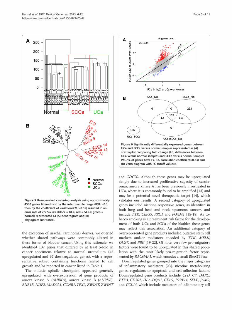
Figure 5 Unsupervised clustering analysis using approximately4500 genes filtered first by the interquantile range (IQR, >0.3)then by the coefficient of variation (CV, >0.05) resulted in anerror rate of 2/27=7.4% (black = UCa; red = SCCa; green =normal) represented as (A) dendrogram and (B)phylogram (unrooted).
Figure 6 Significantly differentially expressed genes betweenUCa and SCCa versus normal samples represented as (A)scatterplot comparing fold change (FC) differences betweenUCa versus normal samples and SCCa versus normal samples(98.7% of genes have FC ≤2, correlation coefficient=0.73) and(B) Venn diagram with FC cutoff value=5.
Hansel et al. BMC Medical Genomics 2013, 6:42 Page 5 of 11http://www.biomedcentral.com/1755-8794/6/42
the exception of urachal carcinoma) derives, we queriedwhether shared pathways were commonly altered inthese forms of bladder cancer. Using this rationale, weidentified 137 genes that differed by at least 5-fold incancer specimens relative to normal urothelium (45upregulated and 92 downregulated genes), with a repre-sentative subset containing functions related to cellgrowth and/or reported in cancer listed in Table 1.The mitotic spindle checkpoint appeared generally
upregulated, with overexpression of gene products ofaurora kinase A (AURKA), aurora kinase B (AURKB),BUB1B, NUF2, MAD2L1, CCNB1,TPX2, ZWINT, ZWINT
and CDC20. Although these genes may be upregulatedsimply due to increased proliferative capacity of carcin-omas, aurora kinase A has been previously investigated inUCa, where it is commonly found to be amplified [13] andmay be a potential novel therapeutic target [14], whichvalidates our results. A second category of upregulatedgenes included nicotine-responsive genes, as identified inboth lung and head and neck squamous cancers, andinclude TTK, CEP55, PRC1 and FOXM1 [15-18]. As to-bacco smoking is a preeminent risk factor for the develop-ment of both UCa and SCCa of the bladder, these genesmay reflect this association. An additional category ofoverrepresented gene products included putative stem cellmarkers and/or mediators encoded by TTK, MELK,DLG7, and PBK [19-22]. Of note, very few pro-migratoryfactors were found to be upregulated in this shared popu-lation with the most likely pro-migration factor repre-sented by RACGAP1, which encodes a small RhoGTPase.Downregulated genes grouped into the major categories
of inflammatory mediators [23], nicotine metabolizinggenes, regulators or apoptosis and cell adhesion factors.Downregulated gene products include CFD, C7, DARC,PTX3, CD302, HLA-DQA1, CD69, P2RY14, SELE, JAM2,and CCL14, which include mediators of inflammatory cell

Table 1 Commonly dysregulated genes shared by urothelial and squamous carcinoma versus normal rothelium
Gene Fold change UCa Fold change SCCa Function
UBE2C 15.1 15.1 Ubiquitin-conjugating enzyme; promotes cell cycle; amplified in UCC
TOP2A 13 14.8 DNA topoisomerase
CDC20 11.4 21.1 Regulates RUNX1; interacts with Aurora A; cell cycle regulation
CCNB2 9.49 12.5 Cyclin B2; TGF-beta mediated cell cyle control
CEP55 9.32 15.1 Centromeric protein regulated by FOXM1
TPX2 9.32 11.6 Microtubule formation at kinetochores
TTK 9.08 10.7 Mitotic spindle checkpoint protein; smoking responsive gene
FOXM1 8.99 7.66 Can increase levels of cyclin family members; smoking responsive gene
ZWINT 8.87 7.37 Kinetochore interactor
BUB1B 8.83 9.06 Kinase involved in spindle checkpoint function
MELK 8.58 12.4 AMPK related kinase; implicated in stem cell function
DLG7 7.98 12.3 Putative stem cell factor that interacts with kinetochore; increased in UCC
NUF2 7.64 7.97 Kinetochore-associated protein involved in chromosome segregation
MAD2L1 7.61 9.06 Spindle checkpoint protein; regulates start of anaphase
AURKB 6.62 9.53 Microtubule-associated protein involved in chromosome segregation
CCNB1 6.43 9.89 Cyclin B1; complexes with p34 to regulate mitotic activity
RACGAP1 6.24 5.89 RhoGTPase encoding gene
AURKA 5.64 8.51 Kinetochore-associated protein found at spindle poles during mitosis
ADH1B −42.3 −70.1 Involved in tobacco smoke detoxification; implicated in esophageal cancer
UPK1A −14.4 −22.4 Urothelium-associated protein
FHL1 −12.5 −8.37 LIM-protein that regulates apoptosis and proliferation
ANXA10 −12.1 −18.8 Regulates cell growth; synergizes with p53 mutation for worse outcomes
ADAMTS1 −10.0 −8.56 Matrix metalloproteinase
DARC −9.93 −15.9 Binds cytokines; can influence tumor cell binding to endothelial cells
MYH11 −8.96 −18.8 Smooth muscle myosin
DMN −8.81 −11.0 Intermediate filament associated with desmin
TCF21 −7.43 −9.92 Tumor suppressor gene; undergoes methylation in some cancers
HLA-DQA1 −7.01 −8.38 Expression may be altered following repeated BCG exposure
CD69 −6.63 −5.48 Lectin superfamily; reduced in head and neck SCC patients
ANK2 −6.28 −8.84 Links integral membrane proteins to the underlying cytoskeleton
CLU −6.18 −5.72 Apoptotic mediators; expression decreased in many cancers
SELE −6.14 −7.75 Inflammatory mediator; regulates immune cell-endothelial interaction
JAM2 −6.07 −7.61 Tight junction protein; regulates immune cell binding
AOX1 −5.76 −7.12 Nicotine metabolizing protein
PROM1 −5.27 −8.29 Expressed in adult stem cells; mediates differentiation
CCL14 −5.09 −8.40 Cytokine
Differentially expressed genes have an adjusted p value of <0.05.
Hansel et al. BMC Medical Genomics 2013, 6:42 Page 6 of 11http://www.biomedcentral.com/1755-8794/6/42
adhesion, humoral response and monocyte activity. HLA-DQ1 has been evaluated previously in UCa and its expres-sion is associated with repeated exposure (and response)to BCG [24].ADH1B and AOX1 are involved in the metabolism of
nicotine, with the former gene implicated in the risk ofesophageal carcinoma [25]. Apoptotic mediators that aredownregulated include CLU, FHL1 and PCP4, whereas
cell adhesion and cytoskeletal mediators that are down-regulated include UPK1A, MYH11, DMN, MFAP4, ITM2A,ANK2, JAM2, MYLK, PROM1, DPT, and FBLN5.Of the 137 genes differentially expressed between
bladder UCa and SCCa versus normal urothelium, 18have been previously reported to be up/down regulatedin UCa and 35 have been reported in SCCa arising fromnon-bladder sites. Due to the rarity of profiling papers

Hansel et al. BMC Medical Genomics 2013, 6:42 Page 7 of 11http://www.biomedcentral.com/1755-8794/6/42
available on bladder SCCa, however, these factors havenot been studied in this entity to date.
A limited subset of uniquely dysregulated genes definesUCaOne of the most surprising results from this study are thevery small number of genes that were found to beuniquely dysregulated in UCa versus normal urothelium(n = 18; representative subset Table 2). The remainder ofdysregulated genes (n = 137) are found in common withthose altered in bladder SCCa. Uniquely dysregulatedgenes in UCa include CLCA4 (chloride channel), IL33,GPR171 (G protein coupled receptor), CENPF (centro-mere protein F) and CD36 (thrombospondin receptor).EZH2 has been reported to be upregulated in UCa andrepresents a putative stem cell marker [26] and a repressorof E-cadherin expression [27]; of relevance, E-cadherin isfrequently lost in high-grade UCa [28].
Well-categorized squamous factors are uniquelyupregulated in SCCaFinally, we analyzed uniquely dysregulated genes in SCCaversus normal urothelium and identified 185 upregulatedand 181 downregulated unique genes that differed by atleast 5-fold between these two groups (representative sub-set presented in Table 3). The majority of dysregulatedgenes are factors that have been associated with the squa-mous phenotype and histology, with many of these factorsidentified in squamous carcinomas arising at other sites.Upregulated gene products include keratins that are
Table 2 Uniquely dysregulated genes in grothelial garcinoma
Gene Fold change UCa Function
MEST 8.14 Encodes a me
EZH2 5.57 Stem cell relat
CENPF 5.54 Component o
GINS2 5.43 Psf2 homolog
CLCA4 −11.9 Member of th
POU2AF1 −8.96 Regulates TH1
IL33 −7.99 Member of IL1
SEPP1 −6.94 Selenoprotein
IL6 −6.57 Inflammatory
GPR171 −6.45 G protein-cou
FOSB −6.26 One member
ITK −6.05 Regulator of T
AGR3 −5.62 May regulate p
CCL19 −5.45 Cytokine affec
CD36 −5.42 Thrombospon
DKK1 −5.33 Inhibits WNT s
Differentially expressed genes have an adjusted p value of <0.05.
specific for squamous epithelium (KRT6B, KRT16, KRT5,KRT20), the family of S100 calcium binding proteins com-monly upregulated in SCCa from various anatomical sites(S100A7, S100A8, S100A9, S100A12, S100A14, S100A16),the serpin family (SERPINB1-7), desmosome-associatedproteins that characterize squamous epithelium (DSG1,DSG3, PKP1, PKP3), numerous peptides (PI3, SPINK5,KLK7, SLPI), and a variety of pro-motility factors. Down-regulated gene products include putative tumor suppres-sor genes (SCUBE2), factors previously reported as lost inaggressive bladder cancer (FOXA1, GATA3, UPK3A), andmetabolizing enzymes with polymorphisms affecting can-cer risk (UGT1A10, UGT1A7).
DiscussionCurrent pathological classification distinguishes UCa andSCCa as distinct diagnostic entities [4]. This has resultedin numerous publications that have evaluated the differ-ences in clinical outcomes, treatment response and mo-lecular profiles that distinguish these two bladder cancertypes, with mixed results [29]. Although some studieshave suggested that when compared stage-for-stage, theoutcomes are similar for patients with bladder UCa andSCCa [3,29,30], other studies have implicated that SCCaand/or UCa with squamous differentiation may present ata higher stage and behave more aggressively [31-33]. Oneconsistency amongst studies is the limited response ofbladder SCCa of the bladder to conventional chemother-apy and/or radiation therapy that is administered in thesetting of UCa and may relate to the squamous phenotype
versus normal urothelium
mber of alpha/beta hydrolase superfamily; imprinting in cancer
ed gene; previously reported as upregulated in UCa
f centromere/kinetochore complex; affects chromosome segregation
; complex component that regulates DNA replication in yeast
e calcium sensitive chloride conductance superfamily
and TH2 immune responses
family; enhances TH2 cytokine production
; may affect oxidative stress response
cytokine; increased after BCG administration in the bladder
pled receptor; may regulate hematopoietic progenitor cells
of the AP-1 transcription factor complex; implicated in ovarian carcinoma
cell proliferation
rotein folding; may regulate cisplatin resistance in ovarian cancer
ting B and T cell migration; may enhance B-cell mediated immunity;
din receptor; may affect tumor vascularity and matrix content
ignaling; may inhibit invasive behavior and self-renewal in some cancers
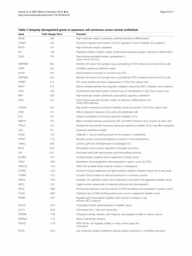
Table 3 Uniquely dysregulated genes in squamous cell carcinoma versus normal urothelium
Gene Fold change SCCa Function
KRT6B 289 High molecular weight cytokeratin; stratified epithelium differentiation
S100A7 242 Promotes migration and invasion of SCCa; reported in urine of bladder SCCa patients
KRT16 147 High molecular weight cytokeratin
PI3 140 Peptidase inhibitor 3/elafin; marker of abnormal squamous growth; induced by inflammation
DSG3 77.6 Desmosome-associated protein; upregulated invarious forms of SCCa
SERPINB4 58.3 Member of human SCCa antigen locus; activated by STAT3; enhances survival of SCCa cells
CNFN 56.3 Cornifelin; squamous epithelial marker
KLK10 53.9 Serine protease; increased in oral and lung SCCa
SERPINB3 42.5 Member of human SCCa antigen locus; activated by STAT3; enhances survival of SCCa cells
FGFBP1 31.3 FGF carrier protein; promotes angiogenesis in SCCa from various sites
MMP1 31.0 Matrix metalloproteinase that degrades collagens; induced by EGF in bladder cancer patients
DSG1 29.6 Desmosome-associated protein; enhances loss of cell adhesion in SCCa from various sites
KRT5 25.8 High-molecular weight cytokeratin; expressed by squamous epithelium
DSC3 25.1 Desmosome-associated protein; marker of squamous differentiation; caninhibit EGFR pathway
S100A8 22.9 May predict metastatic potential of bladder cancer; increased in SCCa from various sites
LY6D 21.1 Affects interaction between SCCa cells and endothelial cells
CA2 19.3 Carbonic anhydrase II; Previously reported in bladder SCCa
MMP10 18.7 Matrix metalloproteinase; activated by EGF and STAT3; inhances SCCa invasion at other sites
PTHLH 15.5 Parathyroid hormone-like hormone; previously reported in bladder SCCa; may affect apoptosis
SCEL 15.1 Squamous epithelium marker
CALB1 12.9 Calbindin 1; calcium binding protein of the troponin C superfamily
PTPRZ1 11.9 Receptor protein tyrosine phosphatase; involved in CNS development
LAMC2 8.95 Laminin, gamma2; Overexpressed in esophageal SCC
KIF14 6.58 Microtubule motor protein; reported in laryngeal carcinoma
JUP 6.31 Associated with both desmosomes and intermediate junctions
SCUBE2 −27.1 Secreted protein; putative tumor suppressor in breast cancer
HPGD −20.6 Metabolism of prostaglandins; downregulated in gastric cancer by COX2
HMGCS2 −15.6 HMG-CoA synthase family; enzyme involved in ketogenesis
CYP4B1 −15.0 Involved in drug metabolism and lipid synthesis; related to bladder cancer risk in one study
TGFBR3 −13.1 Encodes TGF-β receptor III; reduced expression in numerous cancers
UPK3A −10.9 Uroplakin 3A; urothelial marker; loss of expression associated with aggressive bladder cancer
AOC3 −10.1 Copper amine oxidase; aids in leukocyte adhesion and transmigration
FBLN1 −9.86 Secreted glycoprotein; may be involved in ECM remodeling; downregulated in gastric cancer
FOXA1 −9.82 Forkhead class of DNA binding proteins; loss occurs in aggressive bladder cancer
PPARG −9.27 Regulate gene transcription together with retinoid X receptors; mayinfluence BCG response
GATA3 −8.97 Transcription factor; downregulated in bladder cancer
CCL15 −8.63 Chemotactic for T cells and monocytes;
TSPAN8 −7.96 Tetraspanin family; interacts with integrins; may regulate motility in various cancers
ADRA2A −7.75 Alpha-2-adrenergic receptor;
PDGFD −7.74 PDGF family; can regulate motility in many cancer types andchemotaxis
KRT20 −6.32 Low molecular weight cytokeratin; typically patchy expression in urothelial carcinoma
Hansel et al. BMC Medical Genomics 2013, 6:42 Page 8 of 11http://www.biomedcentral.com/1755-8794/6/42

Table 3 Uniquely dysregulated genes in squamous cell carcinoma versus normal urothelium (Continued)
CYP1A1 −5.7 Hypermethylated in bladder cancer; polymorphisms related to bladdercancer risk
CYP1B1 −5.61 Polymorphisms associated with bladder cancer risk
UGT1A10 −5.28 UDP-glucuronosyltransferase; involved in detoxification of carcinogens
UGT1A7 −5.28 UDP-glucuronosyltransferase; polymorphisms associated with cancer risk
Differentially expressed genes have an adjusted p value of <0.05.
Hansel et al. BMC Medical Genomics 2013, 6:42 Page 9 of 11http://www.biomedcentral.com/1755-8794/6/42
[34,35]. To date, however, the relationship between thesetwo forms of bladder cancer that arise from the urotheliallining of the bladder has not been clearly delineated.The results from our study suggest that UCa generally
shares the majority of its dysregulated genes relative tonormal urothelium in common with SCCa, with veryfew uniquely dysregulated genes; in contrast, SCCa –while sharing many genes in common with UCa – showsa much larger category of dysregulated genes that areoften in common with SCCa arising at other sites. Whenconsidering the relationship between these two closelyrelated entities, two possibilities emerge. First, invasiveUCa may represent a default pathway of bladder cancerdevelopment, with clonal change resulting in SCCa de-velopment and overgrowth of a pre-existent UCa. Thishypothesis is supported by the not infrequent findingof mixed morphology bladder cancers, where a well-documented UCa contains areas of squamous and/orglandular differentiation [4]. Further supporting thishypothesis is a prior paper that has examined the rela-tionship of co-existent small cell carcinoma and UCa ofthe bladder: the results from this prior study suggest thatthe small cell carcinoma in this setting represented aclonal outgrowth from the background invasive UCa[36] a finding that might not be dissimilar across allother bladder cancer “subtypes” and which can be sup-ported by the findings in this paper. A second possibilityis that an early bladder cancer stem cell exists, eitherprior to invasion or early in the course of invasion,which gives rise to distinct morphological entities alongdiscrete molecular lineages that are considered pure sub-types. Specifically, early molecular changes define anumber of shared alterations between various bladdercancer subtypes that subsequently diverge along differ-ent morphologic lines [36]. In such a scenario, the lim-ited number of additional alterations identified in UCawould suggest this to be a “default” pathway in bladdercarcinogenesis, with significant additional alterations re-quired to develop the squamous phenotype.Regardless of the model proposed, the current data sup-
ports a close evolution between UCa and SCCa, with geneexpression changes in the latter primarily reflecting mor-phological correlates of the squamous phenotype seen inSCCa arising from different sites. Our data also suggestthat proliferative changes, including deregulation (and in
some cases amplification) of mitotic spindle checkpointcomponents may be critical in the early stages of bladdertumorigenesis. Further validation of our findings usingother “pure” types of bladder cancer – such as adenocar-cinoma and small cell carcinoma – will further strengthenthe implications of our results, although the rare nature ofthese other forms of bladder cancer may make such astudy challenging.Although we have used only one technique to analyze
the relationship between UCa and SCCa, our ability toreproducibly segregate the entities in our study usingboth supervised and unsupervised clustering analysissuggest that our data is robust. A second limitation isthe use of a limited number of specimens for analysis,although the use of 10 SCCa samples is relatively highgiven the rarity of this disease entity. The overall geneexpression profiles between our two bladder cancerentities suggest that the development of these bladdercancer forms occurs along similar lines. However, it isclear that the magnitude of expression changes differs insome instances; for example, reduction in ADH1B oc-curs by a factor of 40-fold in UCa and 70-fold in SCCa.The importance of relative fold change (versus direction-ality) in these cancer subtypes was not a primary focusof investigation in this study. However, the relativeincrease or decrease in mRNA expression may have arelevant biological role when studied in a whole cellsystem.Our study has identified numerous categories of genes
that may be of relevance to the development of UCa,including mitotic spindle regulators, putative stem cellfactors, nicotine metabolizing enzymes and inflamma-tory regulators. The vast majority of these genes appearto be similarly dysregulated in bladder SCCa. One ex-ception is a large category of inflammatory mediators(POU2AF1, IL33, IL6, ITK, CCL19) that are altered andmay reflect the administration of BCG, which is fre-quently given intravesically for superficial UCa but notSCCa. In contrast, SCCa appears to overlap significantlyin gene expression differences with UCa and additionallycontains a large number of additional up- and down-regulated gene products. Perhaps not surprisingly, manyof these gene transcripts have been reported in SCCafrom the head and neck region, oral cavity, lung andskin. As SCCa is considered to have limited response to

Hansel et al. BMC Medical Genomics 2013, 6:42 Page 10 of 11http://www.biomedcentral.com/1755-8794/6/42
therapies conventionally employed for UCa, the broadlist of discrete targets – some of which are currentlyundergoing clinical trials for targeted therapy in otherforms of SCCa – may provide an alternative treatmentfor patients with either pure or mixed SCCa of thebladder.
ConclusionsIn summary, we found that UCa and SCCa of the blad-der share a number of differentially regulated genes,suggesting a close evolution of these two major subtypesof bladder cancer. Future studies that seek to furtherdelineate the relationship and, thus, pathogenesis of vari-ous forms of bladder cancer will provide additionalinsight into the development of bladder cancer. Ultim-ately, the finding of shared molecular changes may allowinvestigators to develop targeted therapy that may beused either earlier in the course of disease or treat abroader range of cancer morphologies with success.
Competing interestsThe authors declare that they have no competing interests.
Authors’ contributionsDEH collected the specimens, performed pathologic analysis, interpreted thedata and drafted the manuscript; ZZ performed data analysis and drafted themanuscript; DP performed gene expression analysis and reviewed themanuscript; BTT analyzed the data and reviewed the manuscript. All authorsread and approve of the final manuscript.
AcknowledgmentsWe thank the Cleveland Clinic for providing bladder samples for analysis. Wethank Jill Barnholtz-Sloan, PhD, for careful review and input to thismanuscript.
Author details1Department of Pathology, University of California at San Diego, 9500 GilmanDrive, MC 0612, La Jolla, CA 92093, USA. 2Van Andel Research Institute, GrandRapids, MI, USA. 3National Cancer Centre of Singapore-VARI, Singapore,Singapore. 4Duke-NUS Graduate Medical School, Singapore, Singapore.
Received: 6 June 2013 Accepted: 11 October 2013Published: 17 October 2013
References1. Wu XR: Urothelial tumorigenesis: a tale of divergent pathways. Nat Rev
Cancer 2005, 5:713–725.2. Amin MB, McKenney JK, Paner GP, Hansel DE, Grignon DJ, Montironi R, Lin
O, Jorda M, Jenkins LC, Soloway M, et al: ICUD-EAU internationalconsultation on bladder cancer 2012: pathology. Eur Urol 2013, 63:16–35.
3. Lagwinski N, Thomas A, Stephenson AJ, Campbell S, Hoschar AP, El-Gabry E,Dreicer R, Hansel DE: Squamous cell carcinoma of the bladder: aclinicopathologic analysis of 45 cases. Am J Surg Pathol 2007, 31:1777–1787.
4. Eble JN, Sauter G, Epstein JI, Sesterhenn IA: Pathology and genetics oftumours of the urinary system and male genital organs. Lyon, France: IARCPress; 2004.
5. Ewis AA, El-Samman E, Ali N, Kajimoto K, Shinohara Y, Ishikawa M,Kanayama HO, Baba Y: Gene expression profile in squamous cellcarcinoma of the urinary bladder using complementarydeoxyribonucleic acid microarray. Urol Oncol 2007, 25:120–127.
6. Blaveri E, Simko JP, Korkola JE, Brewer JL, Baehner F, Mehta K, Devries S,Koppie T, Pejavar S, Carroll P, et al: Bladder cancer outcome and subtypeclassification by gene expression. Clin Cancer Res 2005, 11:4044–4055.
7. Sanchez-Carbayo M, Socci ND, Charytonowicz E, Lu M, Prystowsky M, ChildsG, Cordon-Cardo C: Molecular profiling of bladder cancer using cDNA
microarrays: defining histogenesis and biological phenotypes. Cancer Res2002, 62:6973–6980.
8. Ostergaard M, Rasmussen HH, Nielsen HV, Vorum H, Orntoft TF, Wolf H,Celis JE: Proteome profiling of bladder squamous cell carcinomas:identification of markers that define their degree of differentiation.Cancer Res 1997, 57:4111–4117.
9. Zhang Z, Furge KA, Yang XJ, Teh BT, Hansel DE: Comparative geneexpression profiling analysis of urothelial carcinoma of the renal pelvisand bladder. BMC Med Genomics 2010, 3:58.
10. Bolstad BM, Irizarry RA, Astrand M, Speed TP: A comparison ofnormalization methods for high density oligonucleotide array databased on variance and bias. Bioinformatics 2003, 19:185–193.
11. Irizarry RA, Bolstad BM, Collin F, Cope LM, Hobbs B, Speed TP: Summariesof affymetrix GeneChip probe level data. Nucleic Acids Res 2003, 31:e15.
12. Paradis E, Claude J, Strimmer K: APE: analyses of Phylogenetics andEvolution in R language. Bioinformatics 2004, 20:289–290.
13. Sen S, Zhou H, Zhang RD, Yoon DS, Vakar-Lopez F, Ito S, Jiang F, Johnston D,Grossman HB, Ruifrok AC, et al: Amplification/overexpression of a mitotickinase gene in human bladder cancer. J Natl Cancer Inst 2002, 94:1320–1329.
14. Zhou N, Singh K, Mir MC, Parker Y, Lindner D, Dreicer R, Ecsedy JA, Zhang Z,Teh BT, Almasan A, et al: The investigational aurora kinase a inhibitorMLN8237 induces defects in cell viability and cell-cycle progression inmalignant bladder cancer cells in vitro and in vivo. Clin Cancer Res 2013,19:1717–1728.
15. Landi MT, Dracheva T, Rotunno M, Figueroa JD, Liu H, Dasgupta A, MannFE, Fukuoka J, Hames M, Bergen AW, et al: Gene expression signature ofcigarette smoking and its role in lung adenocarcinoma developmentand survival. PLoS One 2008, 3:e1651.
16. Kim IM, Ackerson T, Ramakrishna S, Tretiakova M, Wang IC, Kalin TV, MajorML, Gusarova GA, Yoder HM, Costa RH, et al: The forkhead Box m1transcription factor stimulates the proliferation of tumor cells duringdevelopment of lung cancer. Cancer Res 2006, 66:2153–2161.
17. Gemenetzidis E, Bose A, Riaz AM, Chaplin T, Young BD, Ali M, Sugden D,Thurlow JK, Cheong SC, Teo SH, et al: FOXM1 upregulation is an earlyevent in human squamous cell carcinoma and it is enhanced bynicotine during malignant transformation. PLoS One 2009, 4:e4849.
18. Waseem A, Ali M, Odell EW, Fortune F, Teh MT: Downstream targets ofFOXM1: CEP55 and HELLS are cancer progression markers of head andneck squamous cell carcinoma. Oral Oncol 2010, 46:536–542.
19. Nakano I, Paucar AA, Bajpai R, Dougherty JD, Zewail A, Kelly TK, Kim KJ, Ou J,Groszer M, Imura T, et al: Maternal embryonic leucine zipper kinase (MELK)regulates multipotent neural progenitor proliferation. J Cell Biol 2005,170:413–427.
20. Gudmundsson KO, Thorsteinsson L, Sigurjonsson OE, Keller JR, Olafsson K,Egeland T, Gudmundsson S, Rafson T: Gene expression analysis ofhematopoietic progenitor cells identifies Dlg7 as a potential stem cellgene. Stem Cells 2007, 25:1498–1506.
21. Dougherty JD, Garcia AD, Nakano I, Livingstone M, Norris B, Polakiewicz R,Wexler EM, Sofroniew MW, Kornblum HL, Geschwind DH: PBK/TOPK, aproliferating neural progenitor-specific mitogen-activated protein kinasekinase. J Neurosci 2005, 25:10773–10785.
22. Badenhorst P: Tramtrack controls glial number and identity in thedrosophila embryonic CNS. Development 2001, 128:4093–4101.
23. Yang H, Gu J, Lin X, Grossman HB, Ye Y, Dinney CP, Wu X: Profiling ofgenetic variations in inflammation pathway genes in relation to bladdercancer predisposition. Clin Cancer Res 2008, 14:2236–2244.
24. Saban MR, Hellmich HL, Simpson C, Davis CA, Lang ML, Ihnat MA, O’DonnellMA, Wu XR, Saban R: Repeated BCG treatment of mouse bladderselectively stimulates small GTPases and HLA antigens and inhibitssingle-spanning uroplakins. BMC Cancer 2007, 7:204.
25. Tanaka F, Yamamoto K, Suzuki S, Inoue H, Tsurumaru M, Kajiyama Y, Kato H,Igaki H, Furuta K, Fujita H, et al: Strong interaction between the effects ofalcohol consumption and smoking on oesophageal squamous cellcarcinoma among individuals with ADH1B and/or ALDH2 risk alleles.Gut 2010, 59:1457–1464.
26. Wang H, Albadine R, Magheli A, Guzzo TJ, Ball MW, Hinz S, Schoenberg MP,Netto GJ, Gonzalgo ML: Increased EZH2 protein expression is associated withinvasive urothelial carcinoma of the bladder. Urol Oncol 2012, 30:428–433.
27. Cao Q, Yu J, Dhanasekaran SM, Kim JH, Mani RS, Tomlins SA, Mehra R,Laxman B, Cao X, Yu J: Repression of E-cadherin by the polycomb groupprotein EZH2 in cancer. Oncogene 2008, 27:7274–7284.

Hansel et al. BMC Medical Genomics 2013, 6:42 Page 11 of 11http://www.biomedcentral.com/1755-8794/6/42
28. Bryan RT, Tselepis C: Cadherin switching and bladder cancer. J Urol 2010,184:423–431.
29. Rogers CG, Palapattu GS, Shariat SF, Karakiewicz PI, Bastian PJ, Lotan Y,Gupta A, Vazina A, Gilad A, Sagalowsky AI, et al: Clinical outcomesfollowing radical cystectomy for primary nontransitional cell carcinomaof the bladder compared to transitional cell carcinoma of the bladder.J Urol 2006, 175:2048–2053. discussion 2053.
30. Nishiyama H, Habuchi T, Watanabe J, Teramukai S, Tada H, Ono Y, Ohshima S,Fujimoto K, Hirao Y, Fukushima M, et al: Clinical outcome of a large-scalemulti-institutional retrospective study for locally advanced bladder cancer:a survey including 1131 patients treated during 1990–2000 in Japan.Eur Urol 2004, 45:176–181.
31. Debbagh A, Bennani S, Hafiani M, el Mrini M, Benjelloun S: Epidermoidcarcinoma of the bladder. Apropos of 14 cases. Ann Urol (Paris) 1997,31:199–203.
32. Johnson DE, Schoenwald MB, Ayala AG, Miller LS: Squamous cellcarcinoma of the bladder. J Urol 1976, 115:542–544.
33. Scosyrev E, Yao J, Messing E: Urothelial carcinoma versus squamous cellcarcinoma of bladder: is survival different with stage adjustment?Urology 2009, 73:822–827.
34. Rundle JS, Hart AJ, McGeorge A, Smith JS, Malcolm AJ, Smith PM:Squamous cell carcinoma of bladder. A review of 114 patients. Br J Urol1982, 54:522–526.
35. Khaled HM, Hamza MR, Mansour O, Gaafar R, Zaghloul MS: A phase II studyof gemcitabine plus cisplatin chemotherapy in advanced bilharzialbladder carcinoma. Eur J Cancer 2000, 36(Suppl 2):34–37.
36. Cheng L, Jones TD, McCarthy RP, Eble JN, Wang M, MacLennan GT,Lopez-Beltran A, Yang XJ, Koch MO, Zhang S, et al: Molecular geneticevidence for a common clonal origin of urinary bladder small cellcarcinoma and coexisting urothelial carcinoma. Am J Pathol 2005,166:1533–1539.
doi:10.1186/1755-8794-6-42Cite this article as: Hansel et al.: Gene profiling suggests a commonevolution of bladder cancer subtypes. BMC Medical Genomics 2013 6:42.
Submit your next manuscript to BioMed Centraland take full advantage of:
• Convenient online submission
• Thorough peer review
• No space constraints or color figure charges
• Immediate publication on acceptance
• Inclusion in PubMed, CAS, Scopus and Google Scholar
• Research which is freely available for redistribution
Submit your manuscript at www.biomedcentral.com/submit