Gastrointestinal - Nature
40
134A ANNUAL MEETING ABSTRACTS EGFR gene and Chromosome 7 copy number were assessed by fluorescence in situ hybridization (FISH); polysomy was defined as 3 or more copies in >=10% of cells. Results: EGFR was over-expressed (3+) in 11 of 49 (22.4%) tumors (4/20 FC, 3/15 MTC, 1/9 PTC, 2/5 ATC). Ten of the 11 (90.9%) tumors with high EGFR expression also showed chromosome 7 polysomy (p=0.0002). Chromosome 7 polysomy was present in 20 of 49 (40.8%) tumors (8/20 FC, 7/15 MTC, 3/9 PTC, 2/5 ATC). High polysomy (>40% of cells with 3+ copies) was present in 9 of the 20 (45.0%) polysomic tumors. EGFR gene amplification was not identified. Conclusions: EGFR is 1) over-expressed in a subset of thyroid neoplasms (follicular and c-cell origin), and correlates with chromosome 7 polysomy. 2) Polysomy was not limited to high EGFR expressing tumors. 3) As polysomy maybe a marker of response to targeted therapies, further evaluation of EGFR and polysomy by FISH in advanced thyroid patients is warranted. 590 The Value of Thyroid Atypia of Undetermined Significance (AUS) Terminology and Its Follow-Up by Repeat FNA SH Zydowicz, A Kemp, E Lucas, X Lin, R Nayar. Northwestern University, Chicago, IL. Background: Since 2001, we have used a 6-tier system to report thyroid FNA with 3 “indeterminate” categories and repeat FNA (rFNA) for follow-up of the Atypia of Undetermined significance (AUS) category. We further sub-categorize AUS as “morphologic” (AUS-M) and “adequacy related” (AUS-A). With increasing acceptance of rFNA for AUS, we assessed compliance with follow up and the malignancy outcomes of AUS, Neoplasm and Suspicious. Design: All thyroid biopsy reports between June 2006 and June 2009 were retrieved from our files. Data was analyzed and correlated with surgical outcomes. Results: 4242 adequate thyroid FNA’s were categorized as shown in the Table. Follow- up FNA was done in 319 (37%) of patients first diagnosed as AUS. A definitive diagnosis was made in 192 (60%) with rFNA. On rFNA, 113 (35%) remained as AUS of which 46 had surgery and 12 (26%) were malignant. In comparison, 66% Neoplasm and 84% Suspicious cases underwent surgery. Malignancy outcomes were AUS-overall 7%, Neoplasm 17% and SUSP 63%. When AUS outcomes were assessed by subtype and whether rFNA was done prior to surgery, the malignancy rate was 21% in AUS-A versus 6% in AUS-M cases, and 26% in AUS resected after an AUS rFNA versus 2% in AUS cases that went directly to surgery after the first AUS diagnosis. Distribution of Thyroid Biopsies from June 2006 through June 2009 Total Number of Cases Number of resected cases Negative for Malignancy Neoplasm Positive for Malignancy Negative 2940 (70%) 190 (65%) 153 (80%) 33 (17%) 3 (2%) Other 855 (20%) 332 (39%) 189 (57%) 119 (36%) 24 (7%) “Morphologic” 695 298 (43%) 168 (57%) 113 (38%) 17 (6%) “Adequacy” 160 34 (21%) 21 (62%) 6 (18%) 7 (21%) Neoplasm 212 (5%) 140 (66%) 43 (20%) 68 (32%) 25 (17%) Suspicious 49 (1%) 41 (84%) 5 (12%) 10 (24%) 26 (63%) Positive 186 (4%) 152 (82%) 0 3 (2%) 153 (98%) Conclusions: (1) AUS is a valuable subcategory in thyroid FNA reporting with a lower malignancy outcome (7%) than Neoplasm (17%) and Suspicious (63%). (2) Repeat FNA for AUS definitively categorizes over half of cases first interpreted as AUS (59%) (3) Over half (53%) of AUS on repeat FNA were Negative and did not need surgery (4) The malignancy rate in cases diagnosed as AUS on repeat FNA (26%) is higher than that of the Neoplasm category (17%) and thus these cases need resection (5) “Adequacy related” AUS cases have a higher malignancy rate than “morphologic” AUS (21% versus 6%), emphasizing the importance of not downgrading suboptimal cases to Negative. Gastrointestinal 591 Histologic Subtypes of Microsatellite Instability-High (MSI-H) Colorectal Adenocarcinomas (CRCs) and Their Association with Clinicopathologic Features and Prognosis A Agarwal, S Sethi, E Lin, R Luthra, A Rashid, SR Hamilton, C Eng, DM Maru. MD Anderson Cancer Center, Houston. Background: MSI-H CRCs are seen in hereditary non polyposis colon cancers and have better patient outcome in sporadic CRC. Mucinous/variegated/undifferentiated histology is associated with MSI. Small number of MSI-H CRCs have histology similar to conventional MSI-stable moderately differentiated adenocarcinoma, significance of which is unclear. We studied the histologic subtypes of MSI-H CRC and correlated them with clinicopathologic features and disease-free survival (DFS). Design: Patients with CRC who underwent resection without neo-adjuvant therapy (1998-2008) and had MSI-H status confirmed by immunohistochemistry or molecular assay were included. All tumors were analyzed for known histologic features of MSI-H CRC including intraepithelial lymphocytosis (IEL)/high power field (hpf). Classic MSI-H (n=89) Undifferentiated carcinoma (n=17) > 50% undifferentiated component Mucinous carcinoma (n=8) > 50% mucinous component Signet ring cell carcinoma (n=8) > 10% signet ring component Variegated (n=48) Two or more components each >5% Cribriform/poorly differentiated (n=8) Cribriform architecture > 20% or any focus of sheets of cells Conventional histology with/without IEL (n=35) Moderately differentiated adenocarcinoma with absence of classic MSI-H features, neuroendocrine differentiation and tumor budding The IEL/hpf in classic MSI-H histology group were >4 in 58 tumors, 2-4 in 30 and <2 in 1. The IEL/hpf in conventional histology group were >4 in 8 tumors, 2-4 in 12 and <2 in 15. Details of adjuvant therapy, recurrence status, and DFS were obtained from the medical record. The statistical analysis was performed with Chi-square/Fisher’s exact test and Cox proportional hazards models. Results: CRCs with classic MSI-H histology were right sided (P=0.03), had higher T(P<0.01), N(P<0.01),and M(P=0.01) stage at time of surgery and higher 5 year recurrence with stage I-III patients (P=0.04) than did CRCs with conventional histology with or without IEL. In the univariate analysis, classic MSI-H histology (P=0.02), higher N stage (P=0.02) and age <40 years were associated with worse DFS in stage I-III patients. Using the multivariate Cox proportional hazards model, after adjusting for age, classic MSI-H histology was associated with worse DFS (P=0.02). Conclusions: This study describes a subset of MSI-H CRC with histology similar to MSI-stable CRC, with lower TNM stage, and longer DFS than MSI-H CRC with classic MSI-H histology. 592 Tumor Thickness at Tumor-Normal Interface (TNI): A Novel Pathologic Indicator of Chemotherapy Response in Hepatic Colorectal Metastases (HCRM) A Agarwal, S Kopetz, P Boonsirikamchai, YS Chun, H Wang, JN Vauthey, EM Loyer, DM Maru. MD Anderson Cancer Center, Houston. Background: Progress in treatment of HCRM demands pathologic indicators of therapy response. Pathologic response;one of the best predictor of disease free survival (DFS) has limitation of low reproducibly among pathologists. Based on observation of majority of residual tumor cells seen at the TNI, we hypothesized that the tumor thickness at the TNI correlate with radiologic and pathologic response and DFS. Design: This study included 119 patients (M/F= 1.8, median age 56 years) with resected HCRM (moderately differentiated adenocarcinoma) following preoperative chemotherapy ± Bevacizumab. Imaging response was assessed by the RECIST criteriae in 50 patients. The pathologic response was categorized as complete (no tumor cells), major (<50% residual tumor cells) and minor (>50% residual tumor cells), as published previously. All H&E sections from the tumors were reviewed by two pathologists and maximum tumor thickness comprised of uninterrupted layers of tumor cells was measured perpendicular to the TNI in millimeter. In tumors where entire section has tumor without stroma, the highest thickness measured on a glass slide was utilized for analysis. The maximum tumor thickness <0.5mm was considered as <0.5mm without additional measurement. For specimen with >1 tumor, average residual tumor and maximum thickness at TNI were used. Results: Seventy-six received oxaliplatin based chemotherapy, 43 received irinotecan based chemotherapy and 90 received Bevacizumab. The imaging response was complete in 14, partial in 32 and progression in 4. The complete pathologic response was seen in 9, major response in 52 and minor response in 58. Median tumor thickness at TNI was 2mm (IQR 0.5 to 6mm). The tumor thickness correlated with pathologic (Spearman r=0.81, p<0.01) and radiologic response (Spearman r=0.37, p<0.01). Cut of thickness of 3mm differentiated minor vs. major/complete pathologic response (sensitivity 0.85, specificity 0.89). Tumor thickness correlated with the DFS as continuous variable in log-transformed analysis and lower thickness predicted better DFS (p=0.02). In the Cox regression analysis tumor thickness was a better predictor of DFS than pathologic response. The tumor thickness did not correlate with the type of cytotoxic chemotherapy, but was smaller in patients treated with bevacizumab (p=0.04). Conclusions: Tumor thickness measured at TNI is potentially a new prognostic factor for therapy response and survival outcome in patients with HCRM. 593 Evidence That Dysplasia Begins in the Bases of the Pits in the Pathogenesis of Gastric Cancer AT Agoston, GY Lauwers, RD Odze. Brigham & Womens Hospital, Boston, MA; Massachusetts General Hospital, Boston, MA. Background: Intestinal-type gastric cancer is believed to develop via an intestinal metaplasia-dysplasia-carcinoma pathway. Anecdotally, we have noted that dysplasia-like atypia may be limited to the deep pit epithelium, without surface epithelium involvement, particularly in the stomach of patients with gastric cancer. We hypothesized that this type of epithelial alteration may represent an early form of dysplasia [Pit Dysplasia (PD)]. The aim of this study was to evaluate the clinical, pathologic, and biologic features of PD in an attempt to determine if it is a significant precursor to gastric cancer. Design: Routinely processed tissue sections from 102 randomly selected resection specimens from patients with gastric cancer (mean age 67; M/F ratio 1.6), and from 22 patients with chronic gastritis (mean age 56.8; M/F ratio 1.8) but without cancer (controls), were evaluated for a wide variety of gross and microscopic features. A subset of 30 study cases were also immunostained for Ki67, E-Cadherin, and p53, and evaluated for the presence and degree of positivity in areas of intestinal metaplasia (IM), PD, and carcinoma. Results: PD was present in 50/102 (49%) study patients compared to only 9% of controls (p<0.05). Patients with PD showed an older mean age at diagnosis (71 vs. 64 years, p=0.02), but a similar M/F ratio compared to patients without PD. Pathologically, gastric cancers with PD showed a significantly increased proportion of intestinal-type adenocarcinomas (82% vs. 37%, p<0.01), a higher degree of tumor differentiation (p<0.01), lower overall pathologic stage (p=0.04), an increased association with chronic gastritis (p<0.01) and a significantly higher proportion of cases with IM (40% vs. 13%, p<0.01) and conventional (full pit) dysplasia (44% vs. 4%, p<0.01) compared to study cases without PD. PD was situated adjacent to neoplasia in 72% of cases (low-grade: 24%, high-grade dysplasia: 26%, carcinoma: 66%) and distant from the neoplasia in 28%. The degree of Ki67 and p53 staining increased progressively, and significantly, in areas of IM, PD, and cancer, whereas E-Cadherin staining decreased. Conclusions: Dysplasia-like changes limited to the deep portions of the pits, without surface epithelium involvement, probably represents an important histologically identifiable precursor to gastric cancer. Further prospective biopsy studies are needed to determine the risk of neoplastic progression in patients with PD detected in mucosal biopsy specimens.
-
Upload
khangminh22 -
Category
Documents
-
view
0 -
download
0
Transcript of Gastrointestinal - Nature

134A ANNUAL MEETING ABSTRACTS EGFR gene and Chromosome 7 copy number were assessed by fluorescence in situ hybridization (FISH); polysomy was defined as 3 or more copies in >=10% of cells.Results: EGFR was over-expressed (3+) in 11 of 49 (22.4%) tumors (4/20 FC, 3/15 MTC, 1/9 PTC, 2/5 ATC). Ten of the 11 (90.9%) tumors with high EGFR expression also showed chromosome 7 polysomy (p=0.0002). Chromosome 7 polysomy was present in 20 of 49 (40.8%) tumors (8/20 FC, 7/15 MTC, 3/9 PTC, 2/5 ATC). High polysomy (>40% of cells with 3+ copies) was present in 9 of the 20 (45.0%) polysomic tumors. EGFR gene amplification was not identified.Conclusions: EGFR is 1) over-expressed in a subset of thyroid neoplasms (follicular and c-cell origin), and correlates with chromosome 7 polysomy. 2) Polysomy was not limited to high EGFR expressing tumors. 3) As polysomy maybe a marker of response to targeted therapies, further evaluation of EGFR and polysomy by FISH in advanced thyroid patients is warranted.
590 The Value of Thyroid Atypia of Undetermined Significance (AUS) Terminology and Its Follow-Up by Repeat FNASH Zydowicz, A Kemp, E Lucas, X Lin, R Nayar. Northwestern University, Chicago, IL.Background: Since 2001, we have used a 6-tier system to report thyroid FNA with 3 “indeterminate” categories and repeat FNA (rFNA) for follow-up of the Atypia of Undetermined significance (AUS) category. We further sub-categorize AUS as “morphologic” (AUS-M) and “adequacy related” (AUS-A). With increasing acceptance of rFNA for AUS, we assessed compliance with follow up and the malignancy outcomes of AUS, Neoplasm and Suspicious.Design: All thyroid biopsy reports between June 2006 and June 2009 were retrieved from our files. Data was analyzed and correlated with surgical outcomes.Results: 4242 adequate thyroid FNA’s were categorized as shown in the Table. Follow-up FNA was done in 319 (37%) of patients first diagnosed as AUS. A definitive diagnosis was made in 192 (60%) with rFNA. On rFNA, 113 (35%) remained as AUS of which 46 had surgery and 12 (26%) were malignant. In comparison, 66% Neoplasm and 84% Suspicious cases underwent surgery. Malignancy outcomes were AUS-overall 7%, Neoplasm 17% and SUSP 63%. When AUS outcomes were assessed by subtype and whether rFNA was done prior to surgery, the malignancy rate was 21% in AUS-A versus 6% in AUS-M cases, and 26% in AUS resected after an AUS rFNA versus 2% in AUS cases that went directly to surgery after the first AUS diagnosis.
Distribution of Thyroid Biopsies from June 2006 through June 2009Total Number of Cases
Number of resected cases
Negative for Malignancy Neoplasm Positive for
MalignancyNegative 2940 (70%) 190 (65%) 153 (80%) 33 (17%) 3 (2%)Other 855 (20%) 332 (39%) 189 (57%) 119 (36%) 24 (7%)“Morphologic” 695 298 (43%) 168 (57%) 113 (38%) 17 (6%)“Adequacy” 160 34 (21%) 21 (62%) 6 (18%) 7 (21%)Neoplasm 212 (5%) 140 (66%) 43 (20%) 68 (32%) 25 (17%)Suspicious 49 (1%) 41 (84%) 5 (12%) 10 (24%) 26 (63%)Positive 186 (4%) 152 (82%) 0 3 (2%) 153 (98%)
Conclusions: (1) AUS is a valuable subcategory in thyroid FNA reporting with a lower malignancy outcome (7%) than Neoplasm (17%) and Suspicious (63%). (2) Repeat FNA for AUS definitively categorizes over half of cases first interpreted as AUS (59%) (3) Over half (53%) of AUS on repeat FNA were Negative and did not need surgery (4) The malignancy rate in cases diagnosed as AUS on repeat FNA (26%) is higher than that of the Neoplasm category (17%) and thus these cases need resection (5) “Adequacy related” AUS cases have a higher malignancy rate than “morphologic” AUS (21% versus 6%), emphasizing the importance of not downgrading suboptimal cases to Negative.
Gastrointestinal591 Histologic Subtypes of Microsatellite Instability-High (MSI-H) Colorectal Adenocarcinomas (CRCs) and Their Association with Clinicopathologic Features and PrognosisA Agarwal, S Sethi, E Lin, R Luthra, A Rashid, SR Hamilton, C Eng, DM Maru. MD Anderson Cancer Center, Houston.Background: MSI-H CRCs are seen in hereditary non polyposis colon cancers and have better patient outcome in sporadic CRC. Mucinous/variegated/undifferentiated histology is associated with MSI. Small number of MSI-H CRCs have histology similar to conventional MSI-stable moderately differentiated adenocarcinoma, significance of which is unclear. We studied the histologic subtypes of MSI-H CRC and correlated them with clinicopathologic features and disease-free survival (DFS).Design: Patients with CRC who underwent resection without neo-adjuvant therapy (1998-2008) and had MSI-H status confirmed by immunohistochemistry or molecular assay were included. All tumors were analyzed for known histologic features of MSI-H CRC including intraepithelial lymphocytosis (IEL)/high power field (hpf).Classic MSI-H (n=89)Undifferentiated carcinoma (n=17) > 50% undifferentiated componentMucinous carcinoma (n=8) > 50% mucinous componentSignet ring cell carcinoma (n=8) > 10% signet ring componentVariegated (n=48) Two or more components each >5%
Cribriform/poorly differentiated (n=8) Cribriform architecture > 20% or any focus of sheets of cells
Conventional histology with/without IEL (n=35) Moderately differentiated adenocarcinoma with absence of classic MSI-H features, neuroendocrine differentiation and tumor buddingThe IEL/hpf in classic MSI-H histology group were >4 in 58 tumors, 2-4 in 30 and <2 in 1. The IEL/hpf in conventional histology group were >4 in 8 tumors, 2-4 in 12 and <2 in 15. Details of adjuvant therapy, recurrence status, and DFS were obtained from the medical record. The statistical analysis was performed with Chi-square/Fisher’s exact test and Cox proportional hazards models.
Results: CRCs with classic MSI-H histology were right sided (P=0.03), had higher T(P<0.01), N(P<0.01),and M(P=0.01) stage at time of surgery and higher 5 year recurrence with stage I-III patients (P=0.04) than did CRCs with conventional histology with or without IEL. In the univariate analysis, classic MSI-H histology (P=0.02), higher N stage (P=0.02) and age <40 years were associated with worse DFS in stage I-III patients. Using the multivariate Cox proportional hazards model, after adjusting for age, classic MSI-H histology was associated with worse DFS (P=0.02).Conclusions: This study describes a subset of MSI-H CRC with histology similar to MSI-stable CRC, with lower TNM stage, and longer DFS than MSI-H CRC with classic MSI-H histology.
592 Tumor Thickness at Tumor-Normal Interface (TNI): A Novel Pathologic Indicator of Chemotherapy Response in Hepatic Colorectal Metastases (HCRM)A Agarwal, S Kopetz, P Boonsirikamchai, YS Chun, H Wang, JN Vauthey, EM Loyer, DM Maru. MD Anderson Cancer Center, Houston.Background: Progress in treatment of HCRM demands pathologic indicators of therapy response. Pathologic response;one of the best predictor of disease free survival (DFS) has limitation of low reproducibly among pathologists. Based on observation of majority of residual tumor cells seen at the TNI, we hypothesized that the tumor thickness at the TNI correlate with radiologic and pathologic response and DFS.Design: This study included 119 patients (M/F= 1.8, median age 56 years) with resected HCRM (moderately differentiated adenocarcinoma) following preoperative chemotherapy ± Bevacizumab. Imaging response was assessed by the RECIST criteriae in 50 patients. The pathologic response was categorized as complete (no tumor cells), major (<50% residual tumor cells) and minor (>50% residual tumor cells), as published previously. All H&E sections from the tumors were reviewed by two pathologists and maximum tumor thickness comprised of uninterrupted layers of tumor cells was measured perpendicular to the TNI in millimeter. In tumors where entire section has tumor without stroma, the highest thickness measured on a glass slide was utilized for analysis. The maximum tumor thickness <0.5mm was considered as <0.5mm without additional measurement. For specimen with >1 tumor, average residual tumor and maximum thickness at TNI were used.Results: Seventy-six received oxaliplatin based chemotherapy, 43 received irinotecan based chemotherapy and 90 received Bevacizumab. The imaging response was complete in 14, partial in 32 and progression in 4. The complete pathologic response was seen in 9, major response in 52 and minor response in 58. Median tumor thickness at TNI was 2mm (IQR 0.5 to 6mm). The tumor thickness correlated with pathologic (Spearman r=0.81, p<0.01) and radiologic response (Spearman r=0.37, p<0.01). Cut of thickness of 3mm differentiated minor vs. major/complete pathologic response (sensitivity 0.85, specificity 0.89). Tumor thickness correlated with the DFS as continuous variable in log-transformed analysis and lower thickness predicted better DFS (p=0.02). In the Cox regression analysis tumor thickness was a better predictor of DFS than pathologic response. The tumor thickness did not correlate with the type of cytotoxic chemotherapy, but was smaller in patients treated with bevacizumab (p=0.04).Conclusions: Tumor thickness measured at TNI is potentially a new prognostic factor for therapy response and survival outcome in patients with HCRM.
593 Evidence That Dysplasia Begins in the Bases of the Pits in the Pathogenesis of Gastric CancerAT Agoston, GY Lauwers, RD Odze. Brigham & Womens Hospital, Boston, MA; Massachusetts General Hospital, Boston, MA.Background: Intestinal-type gastric cancer is believed to develop via an intestinal metaplasia-dysplasia-carcinoma pathway. Anecdotally, we have noted that dysplasia-like atypia may be limited to the deep pit epithelium, without surface epithelium involvement, particularly in the stomach of patients with gastric cancer. We hypothesized that this type of epithelial alteration may represent an early form of dysplasia [Pit Dysplasia (PD)]. The aim of this study was to evaluate the clinical, pathologic, and biologic features of PD in an attempt to determine if it is a significant precursor to gastric cancer.Design: Routinely processed tissue sections from 102 randomly selected resection specimens from patients with gastric cancer (mean age 67; M/F ratio 1.6), and from 22 patients with chronic gastritis (mean age 56.8; M/F ratio 1.8) but without cancer (controls), were evaluated for a wide variety of gross and microscopic features. A subset of 30 study cases were also immunostained for Ki67, E-Cadherin, and p53, and evaluated for the presence and degree of positivity in areas of intestinal metaplasia (IM), PD, and carcinoma.Results: PD was present in 50/102 (49%) study patients compared to only 9% of controls (p<0.05). Patients with PD showed an older mean age at diagnosis (71 vs. 64 years, p=0.02), but a similar M/F ratio compared to patients without PD. Pathologically, gastric cancers with PD showed a significantly increased proportion of intestinal-type adenocarcinomas (82% vs. 37%, p<0.01), a higher degree of tumor differentiation (p<0.01), lower overall pathologic stage (p=0.04), an increased association with chronic gastritis (p<0.01) and a significantly higher proportion of cases with IM (40% vs. 13%, p<0.01) and conventional (full pit) dysplasia (44% vs. 4%, p<0.01) compared to study cases without PD. PD was situated adjacent to neoplasia in 72% of cases (low-grade: 24%, high-grade dysplasia: 26%, carcinoma: 66%) and distant from the neoplasia in 28%. The degree of Ki67 and p53 staining increased progressively, and significantly, in areas of IM, PD, and cancer, whereas E-Cadherin staining decreased.Conclusions: Dysplasia-like changes limited to the deep portions of the pits, without surface epithelium involvement, probably represents an important histologically identifiable precursor to gastric cancer. Further prospective biopsy studies are needed to determine the risk of neoplastic progression in patients with PD detected in mucosal biopsy specimens.

ANNUAL MEETING ABSTRACTS 135A
594 Immunohistochemical Detection of IgG4 Plasma Cells in Lymphocytic and Collagenous Gastritis and ColitisCE Aguilar, R Mertens, F Chung, HL Wang. Cedars-Sinai Medical Center, Los Angeles, CA.Background: Elevation of serum IgG4 level and increased number of IgG4-positive plasma cells in tissues have been reported in patients with autoimmune pancreatitis and other potentially autoimmune-mediated disorders, which often respond to steroid therapy. The current study was undertaken to determine whether IgG4-positive plasma cells were increased in lymphocytic and collagenous gastritis and colitis, conditions which are of unknown etiology.Design: A total of 64 endoscopic biopsies with histologic diagnoses of lymphocytic gastritis (n=15), lymphocytic colitis (n=21), collagenous gastritis (n=5) and collagenous colitis (n=23) were immunohistochemically stained for IgG4 using a monoclonal antibody. The number of IgG4-positive plasma cells in the lamina propria in each case was determined by averaging the numbers identified in 3 high power fields (HPF) that showed highest concentrations of IgG4-positive cells.Results: The number of IgG4-positive plasma cells per HPF ranged from 0-4 in lymphocytic gastritis (0.44±1.2), 0-6.3 in lymphocytic colitis (0.87±1.6), 0-40.3 in collagenous gastritis (10.1±17.3), and 0-5.7 in collagenous colitis (0.72±1.4). The mean numbers of IgG4-positive cells were not significantly different between lymphocytic gastritis and lymphocytic colitis (p=0.37) or between lymphocytic colitis and collagenous colitis (p=0.74). However, the mean numbers of IgG4-positive cells was significantly higher in collagenous gastritis in comparison to lymphocytic gastritis (p=0.034) or collagenous colitis (p=0.010). This difference was attributable mainly to 2 of the 5 cases of collagenous gastritis that showed a marked increase in the mean numbers of IgG4-positive cells (8.7 and 40.3, respectively). Only 1 case of lymphocytic colitis and 1 case of collagenous colitis showed a mean number >5 (6.3 and 5.7, respectively).Conclusions: Tissue IgG4-positive plasma cells are sparse in general in lymphocytic and collagenous gastritis and colitis, arguing against an IgG4-related disease process in the majority of such cases. However, an increase in its number (>5 per HPF) is demonstrated in a small fraction of the cases (4/64), particularly in collagenous gastritis (2/5). Further study of a larger number of cases of collagenous gastritis is warranted.
595 Does IgG4 Immunostain on Endoscopic Biliary and Ampullary Biopsies Predict the Likelihood of Autoimmune Pancreatitis?CE Aguilar, JC Bucobo, B Boulay, SK Lo, D Dhall, D Frishberg, M Kahn, R Mertens, J Zhai, HL Wang. Cedars-Sinai Medical Center, Los Angeles, CA.Background: Autoimmune pancreatitis (AIP) is characterized by elevation of serum IgG4 level, increased numbers of IgG4-positive plasma cells in tissue, and responsiveness to steroid therapy. The diagnosis can be challenging, and it can be difficult to distinguish AIP from pancreatic neoplasms clinically and radiographically. The current study was undertaken to determine whether IgG4 immunostains performed on endoscopic biliary and ampullary biopsies could help establish the diagnosis of AIP.Design: Endoscopic biliary and ampullary biopsies from 40 patients with clinical differential diagnosis including AIP were included in this study. All biopsies were immunohistochemically stained for IgG4 at the time of endoscopic examination using a monoclonal antibody. The number of IgG4-positive plasma cells in each case was determined by averaging the numbers identified in 3 high power fields (HPF) that showed highest concentrations of IgG4-positive cells. Clinical followup data were obtained and correlated with immunohistochemical findings.Results: Clinical followup showed 5 cases to be AIP (including 1 presumed) and 35 cases to be non-autoimmune pancreatitis or various other pancreatic disorders including 6 cases of neoplasms (non-AIP). The number of IgG4-positive plasma cells per HPF ranged from 0.33 to 65.7 (40.1±25.8) in the AIP group and from 0 to 22 (3.1±6.0) in the non-AIP group (p<0.0001). In the AIP group, 4 (except for the presumed case) showed IgG4 counts ranging from 29.3 to 65.7 (50±13.2). In the non-AIP group, only 6 (17%) cases showed ≥5 IgG4-positive cells/HPF, ranging from 7.7 to 22 (15.1±5.4). Three (50%) of these cases had clinical followup diagnoses of idiopathic pancreatitis, chronic pancreatitis and primary sclerosing cholangitis. Overall, the likelihood of AIP with ≥5 IgG4-positive cells per HPF was 0.4. The likelihood of AIP increased to 0.8 if the mean IgG4 count was ≥ 20.Conclusions: Immunostain for IgG4 plasma cells in biliary and ampullary biopsies can serve as a useful adjunct tool in the diagnosis of AIP.
596 Helicobacter Pylori Infection Is Associated with an Increase in the Stem Cell Population in Gastric MucosaQ Ahmed, R Tawil, P Sochacki, N Khoury, J Du, B Patel, AP Majumdar, E Levi. Wayne State University, Detroit, MI; Karmanos Cancer Institute, Detroit, MI; John D. Dingell VAMC, Detroit, MI.Background: Evidence supporting a causal relationship between gastric dysplasia, carcinoma and Helicobacter pylori (HP) infection has been demonstrated previously. Growing evidence firmly supports that Cancer stem cells (CSCs) exist in a wide array of solid tumors. However the presence and origin of these CSCs in gastric cancer remains to be elucidated. We conducted a pilot study at our institution to possibly identify these CSCs by immunohistochemical (IHC) markers, in precursor lesions of gastric cancer, including early events such as HP gastritis.Design: Gastric biopsies and resection specimens representing normal (n=10), HP without intestinal metaplasia (IM) (n=12), HP with IM (n=10) and gastric adenocarcinoma (n=12) were identified. These cases were evaluated for stem cell markers CD166, ALDH1 and LGR5 by IHC. We defined positivity in normal and HP cases as membranous and cytoplasmic staining in number of cells per gland (CPG). For cancer cases we used percentage of tumor cells to define positivity. Statistical significance was assessed by Chi Square test.Results: Of the normal cases 8 /10 showed 1-2 CPG positive for ALDH1, 10/10 showed 1-2 CPG positive for LGR5, however CD166 did not stain any glandular cells. 12/12
cases of HP without IM showed 5-6 CPG positive for ALDH1, 4-5 CPG positive for CD166 and 4-5 CPG positive for LGR5. 10/10 cases of HP with IM showed 7-8 CPG positive for ALDH1, 5-6 CPG positive for CD166 and LGR5. Of the 12 cancer cases 85% were positive for ALDH1, 75% were positive for CD166 and 70% were positive for LGR5. The level of positivity ranged from 5 to 40% of the tumor cells. Staining was more prominent in the invasive fronts and lymph node metastases.
n ALDH1 CD166 LGR5Normal 10 1-2 0 1-2HP without IM 12 5-6* 4-5* 4-5*HP with IM 10 7-8* 5-6* 5-6*Gastric adenocarcinoma 12 85% 75% 70%*p<0.05 compared to normalConclusions: The increased number of ALDH1, CD166 and LGR5 positive cells in HP gastritis indicates that activation of stem cells which presumably constitute the clonogenic cells of future cancers in the field of inflammation is an early event and constitutes an important step in the progression of advanced disease. The next step would be to determine whether the stem cells that are activated carry genetic hits that will progress to full blown cancer.
597 Tryptophan Hydroxylase Autoantibodies (TPHAbs) in Autoimmune Polyendocrine Syndrome Type 1 (APS1): An Immunohistochemical StudyR Alaggio, R Scarpa, J Furmaniak, S Chen, B Rees Smith, L Morlin, S Masiero, L Norberto, C Betterle. University of Padua, Padua, Italy; FIRS Laboratories, Cardiff, United Kingdom.Background: APS1 is a rare hereditary disorder characterized by autoimmune manifestations involving endocrine and non-endocrine tissues. Recently, TPHAbs direct against serotonin-producing enterochromaffin cells (EC) have been found in a subset of patients in association with gastrointestinal dysfunction (GID).Design: The aim of the study was to investigate the relationship between morphologic alterations in gastrointestinal tract and clinical features in TPHAbs positive patients (TPHAbs+). Serum TPHAbs were measured in 60 APS1 patients. Thirteen out of 36 TPHAbs+ (5 with and 8 without GID) undewent gastroduodenal endoscopy with gastric (antrum and body) and duodenal biopsies. In 2 cases colonic biopsies were also available. Bioptic specimens from 2 TPHAbs negative APS1 patients (TPHAbs-) and 6 healthy patients were used as controls. HE stained sections were evaluated. Immunoistains to study endocrine cells [Serotonin, Chromogranin A (CGA), Gastrin] and inflammatory infiltrate (CD3, CD4, CD8, CD20) were performed.Results: Histology revealed a mild to moderate lympho-plasmacytic infiltrate in both antrum and body within the lamina propria in the 13 TPHAbs+ with increased CD3+/CD8+ intraepithelial lymphocytes (IELs). Eight duodenal biopsies, respectively from 4 patients with GID (80%) and 4 asymptomatic (50%), showed a significant infiltrate with increased IELs. Focal atrophy of oxyntic mucosa was seen in 4 cases (31%), intestinal metaplasia in 6 (46%). EC-like hyperplasia was absent. Serotonin immunostaining showed no EC in 12 antral, 11 body and 5 duodenal biopsies. Interestingly duodenum from 4/5 patients with GID lacked EC, with only 2 positive cells in the remaining case. Colonic samples displayed both inflammatory infiltrate and paucity of EC. Gastrin was negative in 5 antral and 7 duodenal samples. CGA+ cells were absent in 5 antral, 5 body and 2 duodenal biopsies, in keeping with reduction of enteroendocrine cells. Controls lacked inflammatory infiltrate and alterations of endocrine cells.Conclusions: TPHAbs in APS1 patients are associated with a peculiar morphologic pattern of autoimmune gastritis, with involvement of antrum and diffuse disruption of EC. The extension of the process to the duodenum and colon is associated with clinical symptoms.
598 Microsatellite Instability (MSI) in Gastrointestinal Adenomas: Does It Answer the Germline Versus Sporadic Cancer Question?DS Allende, C Fraser, D Patil, B Leach, MP Bronner, TP Plesec. Cleveland Clinic, Cleveland, OH.Background: High microsatellite instability (MSI-H) is present in ∼90% of hereditary non-polyposis colorectal cancers (HNPCC) and ∼10-15% of sporadic-type colorectal cancers. The MSI status in adenomas may be helpful in differentiating germline versus sporadic cancers.Design: MSI status was determined by PCR and capillary gel electrophoresis of five mononucleotide repeats (BAT25, BAT26, NR21, NR24 and MONO27) from formalin-fixed paraffin-embedded tumor and paired normal samples. MSI-H was defined as instability in 2 or more markers. Immunohistochemistry (IHC) for MLH-1, MSH-2, MSH-6 and PMS-2 was performed on MSI-H cases. Clinical information, results of germline mutation testing (GT) and genetic history were assessed. Features at least suggestive of HNPCC included confirmed deleterious mutation in a DNA mismatch repair gene, multiple family members and generations with HNPCC-related cancers, or loss of MSH-2, or isolated loss of PMS-2 or MSH-6 by IHC.Results: Of the 217 adenomas, 155 were accompanied by adenocarcinoma. Of these, 140 (90%) had concordant MSI status, and 26/140 (18%) were MSI-H. IHC was performed on 25 of the 26 concordant cases and showed matching protein loss in all cases with MLH-1 loss in 18; MSH-2 loss in 5; and isolated MSH-6 loss in 2. All 14 discordant cases showed MSI-H in the cancer, but not in the adenoma; IHC on the discordant cancers revealed MLH-1 loss in 11/13 cases and MSH-2 loss in 1/13 cases. One of 62 (1.6%) adenomas not associated with adenocarcinoma was MSI-H. No adenomas were MSI-low. Correlation between MSI status in adenomas associated with MSI-H carcinomas, results of GT, family history, and IHC analysis is summarized below.
136A ANNUAL MEETING ABSTRACTS Microsatellite Stability Phenotype In Adenomas Associated With MSI-H Carcinomas And
Corresponding HNPCC FeaturesAdenoma Phenotype
Deleterious Mutation
Family History Suggestive of HNPCC
MSH-2 + isolated MSH-6 IHC Loss
Total Independent Features of HNPCC
MSS 0/2 0/12 1/14 1/14MSI-H 9/10 5/17 7/27 15/27P-value 0.045 0.059 0.23 0.003
Conclusions: Microsatellite instability-high phenotype in adenomas associated with MSI-H cancers is significantly associated with clinical and genetic features of HNPCC. When possible, adenomas should be included in MSI analysis of carcinomas to help stratify a patient’s risk of HNPCC.
599 Immunohistochemical Expression of ER, PR and Ki67 in Pancreatic and Small Intestinal Neuroendocrine TumorsAT Arnason, PJ Barnes, D Rayson, M Drewniak, HL Sapp. Dalhousie University, Halifax, NS, Canada.Background: There has been limited study of ER and PR expression in GI neuroendocrine tumors (GI NETs) despite emerging evidence for hormone receptor regulation of pancreatic islet cells. Normal pancreatic islet cells express PR and progesterone has been implicated in the pathogenesis of gestational diabetes. Islet cell proliferation has been demonstrated in PR gene knockout mice. Additionally, a recent study has shown frequent ER and PR expression in primary lung NETs. Investigation of ER and PR expression in GI NETs is therefore warranted.Design: Pathology records were searched to identify patients who had a GI NET diagnosed from Sept. 1991 to Jan. 2009. An IHC panel including Ki67, ER, and PR was applied to tissue microarrays for 74 primary resections and to 9 biopsy specimens. ER and PR were interpreted as negative, 1+ (weak intensity and/or <75% of cells) or 2+ (moderate or strong intensity and >75% of cells).Results: IHC results for 83 cases (38 male, 45 female, mean age 60 years) are summarized below.Site ER - ER + PR - PR + Ki67<2% Ki67>2%SI (non-duodenal) [n=31] 13 (42%) 18 (58%) 26 (84%) 5 (16%) 28 (90%) 3 (10%)Duodenum [n=9] 7 (78%) 2 (22%) 1 (11%) 8 (89%) 5 (56%) 4 (44%)Pancreas [n=43] 37 (86%) 6 (14%) 11(26%) 32 (74%) 33 (77%) 10 (23%)Number (%) of cases showing ER/PR expression (negative [-], positive 1+ or 2+ [+]) and Ki67 index according to primary site.Small intestinal (non-duodenal [SI]) NETs were significantly more likely to express ER than pancreatic primaries (58% vs 14%, p<0.001). Pancreatic NETs were more likely to be PR + than SI tumors (74% vs. 16%, p<0.001). Duodenal tumors showed similar ER/PR expression to pancreatic NETs, however, only the PR + frequency was significantly different from other SI sites (89% vs. 16%, p<0.001). Only pancreatic (25) and duodenal (4) primaries showed 2+ PR staining. There was more frequent 2+ PR staining (p<0.001) in WHO Class 1 pancreatic NETs (20/23=87%) compared to Class 2&3 NETs (5/20=25%). Ki67 was more often >2% (p<0.01) in WHO Class 2&3 tumors (9/20=45%) than in WHO Class 1 tumors (1/23=4%). There was a non-significant trend towards metastasis in duodenal tumors without 2+ PR and with Ki67>2%. There was a non-significant trend towards increased ER expression and Ki67>2% in SI tumors with metastates.Conclusions: SI NETs show a distinct ER/PR expression profile from pancreatic and duodenal primaries. Pancreatic NETs with strong PR expression (2+) are less likely to show metastatic behavior.
600 Intratumoral Heterogeneity of KRAS Mutations in Colorectal Carcinoma: It Is Important To Submit Which Tumor Tissue Sample for Mutation AnalysisH Baloglu, Z Kucukodaci, N Yigit, I Yilmaz, A Haholu, HA Simsek. GATA-HEH, Istanbul, Turkey; MSKCC, New York.Background: The epidermal growth factor receptor (EGFR) plays an important role in tumorigenesis and tumor progression of colorectal cancer. As a result, the EGFR has evolved as a relevant target in the treatment of metastatic colorectal cancer. KRAS serves as a mediator between extracellular ligand binding and intracellular transduction of signals from the EGFR to the nucleus. The presence of activating KRAS mutations has been identified as a potent predictor of resistance to EGFR-directed antibodies such as cetuximab or panitumumab. These agents should therefore be applied only in tumors with a wild-type status of the KRAS gene. Therefore, it is important to elucidate which patients are most likely to benefit from specific targeted therapies. The mutation analysis of only one small area from a FFPE tumor tissue could not be representative of the whole tumor because of the possibility of the heterogeneity for the occurrence of KRAS mutation. So it is an important consideration to choose the tumor sample for mutation analysis.Design: The study population consisted of 28 patients who were treated surgically for colorectal adenocarcinoma at GATA Haydarpasa Teaching Hospital. Selective laser microdissection were performed to all paraffin blocks with tumor (total block number: 3-6, average block number: 5) and extracted DNA was amplified by the polymerase chain reaction (PCR). The PCR products were sequenced with the ABI3100 sequence analyzer (Applied Biosystems, Foster City, CA). The mutation of the KRAS gene was detected in 12 of 28 cases. Then to detect tumor heterogeneity for KRAS mutation among these 12 cases, DNA extracted with and without selective laser microdissection from each paraffin block of the tumir and independent PCR-sequencing reactions were performed. The histology of the same tumor in different blocks was not significantly different.Results: Mutations identical to those found from all paraffin blocks with selective laser microdissection were confirmed in average of 2.8 blocks with selective laser microdissection and in average of 0.9 blocks without selective laser microdissection.
Conclusions: This study revealed that KRAS gene mutation were significantly heterogeneous in colorectal adenocarcinomas parafin blocks and indicate that it is important to select the paraffin block and to collect enough tumor cells for mutation analysis.
601 Neoadjuvant Therapy Induces Loss of MSH6 Expression in Colorectal CarcinomaF Bao, H Rennert, RK Yantiss. Weill Cornell Medical College, New York, NY.Background: The use of immunostains to detect abnormal DNA repair protein expression has become routine practice in the evaluation of patients with colorectal carcinoma, particularly when they have clinical features that raise the possibility of hereditary non-polyposis colon cancer (HNPCC). However, many distally located adenocarcinomas are neoadjuvantly treated prior to definitive surgical treatment, and we have noted that some treated rectal cancers display loss of DNA repair protein expression, despite the lack of underlying microsatellite instability (MSI). The purpose of this study was to determine the frequency and clinical significance of decreased DNA repair protein expression among neoadjuvantly treated colorectal carcinomas.Design: We identified 51 patients with neoadjuvantly treated adenocarcinoma of the distal colorectum, all of whom underwent surgical resection of their tumors. Immunostains for MLH1, MSH2, MSH6, and PMS2 were performed on tissue sections obtained from post-treatment tumor samples. Cases that showed loss of staining for any of the DNA repair proteins were subjected to molecular testing for MSI. Pretreatment biopsy samples from these tumors were also evaluated for DNA repair protein expression using the abovementioned panel of immunohistochemical stains.Results: The study group included 26 males and 25 females (mean age: 58 years). Ten (20%) tumors showed decreased MSH-6 nuclear staining, including 9 with loss of staining in greater than 75% of the tumor cells and 1 that showed complete loss of MSH6 staining. Loss of MSH6 staining did not reflect MSI in any of the cases and was not associated with patient age, gender, tumor grade, stage, or degree of post-treatment tumor regression. All 9 post-treatment tumors that showed near-complete loss of MSH6 staining displayed strong, diffuse MSH6 expression in pretreatment biopsy samples, but the one with complete loss also lacked MSH6 expression in the pretreatment biopsy sample. All of the cases showed preserved nuclear staining for MLH1, MSH2 and PMS2.Conclusions: Neoadjuvant therapy frequently induces partial loss of MSH6 immunoexpression in colorectal carcinomas, which may be extensive in nearly 20% of cases. This finding does not correlate with underlying MSI. Rather, it probably reflects a therapeutic effect, such as cell cycle dysregulation, in mismatch repair proficient tissues. When loss of MSH6 expression is noted in neoadjuvantly treated tumors, evaluation of pretreatment biopsy samples for DNA repair protein expression may be considered prior to further assessment for HNPCC.
602 MicroRNA Expression during the Colorectal Adenoma-Adenocarcinoma SequenceAN Bartley, KA Patel, BA Barkoh, B Mishra, H Yao, A Rashid, R Luthra, SR Hamilton. The University of Texas MD Anderson Cancer Center, Houston, TX.Background: Micro-RNAs (miRNAs) are small (22 nucleotide) non-coding RNAs that regulate gene expression, and their expression is altered in most tumor types including colorectal adenocarcinomas (CAC). Although several studies have identified miRNAs differentially expressed in CAC compared to non-neoplastic colorectal mucosa (NNM), limited information is available on miRNAs associated with progression in the adenoma-CAC sequence. We therefore profiled CAC, high-grade and low-grade intra-epithelial neoplasia/dysplasia in adenomas where they arose, and adjoining NNM.Design: RNA was isolated with RecoverAll (Applied Biosystems, CA) from 34 samples of manually microdissected formalin-fixed paraffin-embedded tissue from 10 patients with resected stage I, II, or III CAC. Expression levels of 416 miRNAs were determined with Agilent miRNA microarray. Statistical analyses were performed with analysis of variance, Fisher’s exact test and Tukey honest significant difference test.Results: Unsupervised cluster analysis segregated the samples into three groups based on disease progression in 88 differentially expressed miRNAs, (p<0.001). A class comparison analysis identified 87 of 88 miRNAs had differential expression between NNM and CAC, 77 between NNM and LGD, 42 between NNM and HGD, 3 between LGD and HGD, 20 between LGD and CAC, and 19 between HGD and CAC. Nine of 88 miRNAs were differentially expressed between NNM and LGD, LGD and CAC, and NNM and CAC (p < 0.05), four of which are upregulated (miR-34a, 34b, 99a, and 224) and five downregulated (miR-206, 516a-5p, 617, 1208, and 1285). miR-34b, -99a, -206, -224, and -516a-5p were differentially expressed between NNM and HGD (p < 0.01), and miR-99a, -516a-5p, -617 and -1285 were differentially expressed between HGD and CAC (p < 0.01). miR-1285 was also significantly different between LGD and HGD (p<0.05).Conclusions: We identified nine miRNAs differentially expressed in the histopathologic lesions of colorectal carcinogenesis; six not described previously in neoplastic progression in the large bowel. A separate confirmation set from 10 additional patients is currently being profiled for validation, and RT-PCR confirmation in all samples is in progress. Our findings suggest that various miRNAs play specific roles in the sequences of events leading to colorectal adenocarcinoma, and their use as biomarkers or as therapeutic targets therefore may depend upon the timing of their use during progression.

ANNUAL MEETING ABSTRACTS 137A
603 Molecular Correlates with “Corkscrew” and Microglandular Architecture in Colorectal AdenomasAN Bartley, JA Buckmeier, P Thompson, P Lance, SR Hamilton. The University of Texas MD. Anderson Cancer Center, Houston, TX; University of Arizona, Tucson, AZ.Background: The classification of polyps of the colorectum is important; particularly the differences in the histopathologic and molecular characteristics of advanced adenomas and serrated polyps relative to subsequent development of colorectal adenocarcinoma. We evaluated the molecular characteristics of a subset of conventional adenomas with “corkscrew” or microglandular budding architecture.Design: Adenomas with an infolded corkscrew glandular pattern or microglandular budding were selected from 3500 participants in a phase III chemoprevention trial. Areas of tubular adenoma within the same polyps and separate conventional tubular adenomas (TA), traditional serrated adenomas (TSA), and adjacent and separate non-neoplastic mucosa were selected as controls. Sixty-eight polyps from 56 patients were studied, including 40 adenomas with corkscrew or microglandular budding architecture, six TSA, and 22 conventional TA. Polyps ranged from 3 to 32 mm (mean 14.7 mm, SD 6.9). Histopathologic characteristics also included dilated glands with mucinous epithelium, dystrophic goblet cells, and gastric foveolar-type epithelium. Expression of p53, caspase-3 apoptosis marker, Ki-67, beta-catenin with membranous, cytoplasmic and nuclear localization, and MLH1, MSH2, MSH6, and PMS2 mismatch repair gene products were evaluated by immunohistochemistry in the pre-selected histopathologic regions.Results: There were significant differences among subtypes of polyps for expression of p53, MLH1, and beta-catenin among the markers evaluated. p53 expression was significantly greater in the corkscrew and microglandular budding areas than in mucosa (p<.006), and in microglandular budding as compared to TSA (p<.03). MLH1 expression was greater in corkscrew areas than in mucosa (p=.04), but less in microglandular budding and dilated glands with mucinous epithelium than in TSA (p=.02). Membranous beta-catenin expression was lower in corkscrew areas than in TSA (p=.05), and cytoplasmic beta-catenin was slightly greater in corkscrew areas than in mucosa (p=.05).Conclusions: Corkscrew configuration and microglandular budding in conventional adenomas are associated with altered expression of p53 and MLH1 and compartmental localization of beta-catenin. These differences suggest divergence in key molecular progression pathways within conventional adenomas that influence glandular architecture.
604 Dedifferentiated Goblet Cell Carcinoid (Adenocarcinoma Ex GCC) Is a Morphologically Distinctive and Aggressive Neoplasm with Peritoneal Dissemination: An Analysis of 35 CasesO Basturk, J Cheng, P Philip, B El-Rayes, I Coban, S Bandyopadhyay, N Cuhaci, NV Adsay. NYU, New York; Emory U, Atlanta; WSU, Detroit.Background: Recently, adenocarcinomatously transformed examples of GCC are being recognized increasingly and have been reported variably as ovarian metastasis of appendiceal tumors with GCC-like and signet ring patterns(Hristov, Young&Ronnett et al AJSP 2007,31:1502) or adenocarcinoma ex GCC(Tang&Klimstra et al AJSP 2008;32:1429).Design: Here, we document 35 examples of this entity (largest series to date) and discuss their clinicopathologic features.Results: Clinical: F/M:27/8. Mean age=53(31-81). 26 had disseminated tumor in the abdomen (18 peritoneal&17 GYN-tract involvement), in addition to the tumor in the appendix. 12 were initially explored with the diagnosis of ovarian ca. Path: All had at least some foci of conventional GCC pattern but also had a dedifferentiated component composed of one or more of the followings: I Goblet-type cells in non-glandular, diffuse-infiltrative pattern (cords or individual signet-ring cells; n=23). II Non-mucinous cells in diffuse-infiltrative pattern(n=20). III Microglandular pattern without goblet cells(small, round rosette-like tubules lined by well-polarized cuboidal-columnar cells; n=16). IV Mixed component: ordinary intestinal pattern(n=9) or extracellular mucin(n=12). V GCC pattern with marked nuclear atypia(n=10). IHC: In dedifferentiated components, chromogranin was + in scattered cells in 19, abundant in 1. The tumors were also CK20+++, CK7--+, MUC2+++, MUC1++-, E-cadherin +++, nuclear -catenin-, and Ki67 >70%. Outcome: 26 patients had abdominal carcinomatosis but only 1 had hepatic/pulmonary metastases composed of intestinal-type. F/U was available in 28: 14 died of disease(2-45 mos), 11 are alive with disease(2-79 mos) and 3 alive without disease(2.5-23 mos).Conclusions: Dedifferentiated GCCs are aggressive neoplasms that primarily affect females, often present with carcinomatosis, and mimic ovarian ca. The prognosis is significantly worse (median surv, 20.5 mos) than that of ordinary GCC. They appear to be biologically different from intestinal adenoca, by showing a propensity to spread along the peritoneal surfaces, but seldom to liver/lung. Small tubular formations and the distinctive pattern formed by the goblet-like cells are identifiable in most examples even in metastatic sites, allowing the recognition of its primary appendiceal origin, which has significant management implications.
605 IgG4+ Plasma Cells in Gastric Biopsies Specific for Patients with Autoimmune Atrophic Gastritis/Pernicious AnemiaAS Bedeir, JG Lash, MB Ray. Caris Diagnostics, Irving, TX.Background: Although initially described in autoimmune pancreatitis, the presence of increased IgG4 + plasma cells has also been noted in other autoimmune diseases and other organs. Autoimmune gastritis is characterized by anti-parietal and anti-intrinsic factor antibodies that can cause pernicious anemia (PA). Histologically, autoimmune gastritis manifests as a chronic atrophic gastritis (CATG) restricted to the corpus, but there are other causes of CATG, including Helicobacter infection. This study investigates the presence of IgG4+ plasma cells in gastric mucosal biopsies from patients with a variety of gastric histopathology, including CATG with and without PA.
Design: Archives were searched for patients with the histologic diagnosis of CATG and a history of PA (CATG+PA+, n=46). As controls, archives were searched for 25 patients each with the histologic diagnosis of CATG but no history of PA (CATG+ PA-), patients with normal histology, those with mild chronic inactive gastritis (MCIG), and those with the diagnosis of Helicobacter pylori gastritis (HP).Quantification of Ig G4+ plasma cells were obtained via two methods. 1. Immunohistochemical stain for IgG4 on formalin fixed paraffin embedded tissue. Areas with the highest density of IgG4+ plasma cells were selected, and the average of the 3 most cellular HPFs were counted. 2. To ensure that the number of IgG4+ cells was not simply related to the degree of inflammation (density of plasma cells), dual chromagen immunohistochemical stains for CD138/IgG4 were performed. Areas with the highest density of CD138+ plasma cells were selected, and the number of IgG4+ cells / 200 CD138+ plasma cells was counted. For both methods, the results are interpreted as follows: < 5 considered negative, 5 or more considered positive.Results: Identical results were obtained by both staining methods. 17/46 (37%) PA patients were positive for increased IgG 4+ plasma cells in. All other groups were negative.Table1
number PositiveCATG+PA+ 46 17 (37%)CATG+PA- 25 0Normal 25 0MCIG 25 0HP 25 0CATG+PA+:chronic atrophic gastritis & pernicious anemia. CATG+PA-: chronic atrophic gastritis & NO pernicious anemia. MCIG: mild chronic inactive gastritis, HP: Helicobacter pylori gastritis.Conclusions: Increased IgG4+ plasma cells was present in 37% of patients with CATG+ PA+ but not in those with CATG+PA-, MCIG, HP or normal gastric biopsies (100% specific in this study). IgG4+ plasma cells may play a role in the pathogenesis of PA.
606 Mismatch Repair Status in a Cohort of Rectal Adenocarcinomas before and after ChemoradiationAM Bellizzi, CD Crowder, WL Marsh, H Hampel, WL Frankel. The Ohio State University Medical Center, Columbus, OH; Brigham and Women’s Hospital, Boston, MA.Background: At our institution we have performed mismatch repair immunohistochemistry (MMR IHC) as a screen for Lynch syndrome on all colorectal cancer resections since 2006. Anecdotally we have observed decreased staining in neoadjuvant-treated rectal adenocarcinomas (NTx RA). This diminution is particularly prominent with MSH6, often resulting in an “equivocal” interpretation. We performed this study to systematically evaluate MMR IHC in NTx RA.Design: We assembled a cohort of 65 matched pretreatment biopsies (bx) and resections (res) from patients receiving NTx for RA. MMR IHC for MLH1, PMS2, MSH2, and MSH6 was performed. Bx and res tissue were stained on the same slide. Tumor IHC was evaluated for intensity (0, 1: less staining than is typical/less staining than internal control, 2: robust staining) and quantity (0-100%) and a score representing the product of these calculated. Intact staining in internal control was required for interpretation. In 14 (22%), res demonstrated a complete response. Mean data was analyzed using Wilcoxon matched-pairs tests, with P < 0.05 considered significant.Results: Quantitative IHC data is summarized in the table.
Quantitative Mismatch Repair Immunohistochemistry (Score ± SD)Biopsy (n=65) Resection (n=51) P
MLH1 191 ± 34 171 ± 52 0.002PMS2 189 ± 36 169 ± 54 0.0005MSH2 191 ± 38 157 ± 67 0.0002MSH6 165 ± 54 81 ± 64 <0.0001score = intensity * percent; sd, standard deviationThree (4.6%) cases displayed abnormal (absent) MMR IHC (2 MSH2/MSH6, 1 MLH1/PMS2/MSH6). Eight (16%) res demonstrated between 1-5% MSH6 staining (considered equivocal); the corresponding bx revealed 70-100% MSH6 staining. Equivocal results were often seen in tumors with atrophic glands/prominent nucleoli, and although nuclear staining was sparse, diffuse nucleolar staining was frequently observed.Conclusions: NTx RA demonstrate significantly decreased MMR IHC expression, compared to matched pretreatment controls. MSH6 is especially prone to this phenomenon, not infrequently resulting in equivocal results (decrease did not alter interpretation of other stains). This finding may be due to altered morphology/proliferative activity in treated tumor. Also, limited residual tumor in about a quarter of cases precludes MMR IHC assessment. Before embarking on expensive molecular testing in these cases, staining the pretreatment biopsy represents an attractive alternative.
607 Treated Rectal Adenocarcinomas Are Associated with Increased Expression of CK20 and ChromograninAM Bellizzi, CD Crowder, WL Marsh, H Hampel, WL Frankel. The Ohio State University Medical Center, Columbus, OH; Brigham and Women’s Hospital, Boston, MA.Background: Neoadjuvant chemoradiation (NTx) in stage II/III rectal adenocarcinoma (RA) has become standard of care, associated with less toxicity and better local control than adjuvant therapy. Morphologic changes in these cases, including induction of endocrine differentiation, are well-described, and the immunophenotype of untreated RA (e.g. frequent CK7/CK20 co-expression) is established. Less is known about the immunophenotype of treated RA. This information would be useful, as we occasionally struggle to separate scant residual tumor from benign mimics (e.g. anal glands, endometriosis).Design: We assembled a group of 64 matched pre-treatment biopsies (bx) and resections (res) from patients receiving NTx for RA. Matched bx and res were placed on a single slide for staining. CK7, CK20, CDX2, and chromogranin immunohistochemistry (IHC) was performed. Cases were assessed for qualitative (yes/no) and quantitative [intensity
138A ANNUAL MEETING ABSTRACTS (0, 1, 2) and percent (0-100)] tumor staining and scores representing the product of intensity and percent calculated. In 14, res demonstrated a complete response (no IHC data for these). Quantitative data was analyzed using Wilcoxon matched-pairs tests, with P’s < 0.05 considered significant.Results: Qualitative and quantitative IHC results are summarized in the tables.Qualitative Immunohistochemical Results (%)
Biopsy (n=64) Resection (n=50)CK7-/CK20+ 73 82CK7+/CK20+ 14 16CK7+/CK20- 5 0CK7-/CK20- 8 2CDX2 100 98Chromogranin 19 34
Quantitative Immunohistochemical Results (Score ± SD)Biopsy (n=64) Resection (n=50) P
CK7 7 ± 27 11 ± 36 0.8CK20 78 ± 63 127 ± 52 < 0.0001CDX2 181 ± 34 158 ± 57 < 0.0001Chromogranin 0.7 ± 2 1.7 ± 3.2 0.02score = intensity * quantity; sd, standard deviationConclusions: Treated RA is associated with significantly increased CK20 and chromogranin expression compared to pre-treatment controls. Diminution in CDX2 may be explained by focal staining in a few res with little residual tumor (ie. only rare glands). Aside from increased chromogranin expression, the qualitative immunophenotype of treated RA is largely unchanged, with about 15% co-expressing CK7/CK20. Increased CK20 expression represents an unexpected result, and future directions include correlation of this finding with morphology, tumor characteristics, and outcome.
608 The Use of Molecular Markers as a Method of Predicting Response to Combined Modality Therapy for Advanced Stage Rectal CancersME Berho, SD Wexner. Cleveland Clinic Florida, Weston, FL.Background: Response to combined modality therapy (CMT) of advanced stage rectal cancers is predictive of outcome. In addition to clinical and pathologic features, expression of a variety of molecules may provide another method of identifying tumor responsiveness to CMT. Our study aim was to evaluate several markers in the apoptotic pathway as well as expression of COX-2 and VGEF to determine their ability to predict response to CMT.Design: 152 patients with advanced rectal cancer treated with CMT followed by radical resection were included in the analysis. Paraffin-embedded sections obtained before and after therapy were assessed by immunohistochemical staining for COX-2, VGEF, p53, p21, p27, Bax, BCL2 and Apoptosis Protease-Activating Factor 1 (APAF-1) and correlated with tumor regression grade, complete pathological response and T-downstaging. Clinical and pathologic data was also collected. Data was analyzed using Chi-square and Spearman correlation.Results: Pathological complete response was seen in 24.5% of patients. Amongst the apoptosis associated markers, only APAF-1 expression was found to be significantly associated with tumor regression grade (p<0.001), complete pathologic response (p<0.031), and T-downstaging (p<0.004). On multivariate analysis, APAF-1 expression was found to be independently associated with tumor regression grade. In contrast, overexpression of COX2 and VGEF in pre-treatment biopsies was related to less tumor regression (p<0.003) and less likelyhood of T-downstaging (p<0.03)Conclusions: Immunohistochemical evaluation of initial biopsy specimens of rectal cancer with APAF-1, COX2 and VGEF may predict tumor response to CMT in patients with advanced rectal cancer Those with an expected limited response may be considered for other investigational neoadjuvant protocols.
609 Luminal Distribution of Eosinophils in Initial Esophageal Biopsies and Symptoms of Reflux at Presentation are Predictive of Response to Inhaled Corticosteroid Treatment in Adult Patients with Eosinophilic EsophagitisJI Bernstein, DA Katzka, EE Furth. University of Pennsylvania School of Medicine, Philadelphia, PA.Background: The standard management of adult patients with eosinophilic esophagitis (EE) includes inhaled corticosteroids with variable responses to treatment. While enumeration of eosinophils in esophageal biopsies is standard of care for initial diagnosis, the utility of this and other features as predictive markers of treatment response is unknown.Design: The surgical pathology database of our institution was queried for cases of EE diagnosed between 1998 and 2008. All patients with at least two esophageal biopsies reviewed in that time period were included in the study if the first biopsy available was retrieved prior to initiation of corticosteroid treatment. 120 biopsies from 44 patients were reviewed and the number of eosinophils per high power field (hpf) was quantified for each fragment of tissue obtained. A patient was classified as a responder if one of the patient’s biopsies following initiation of treatment had a maximum of 5 eosinophils per hpf. In addition to the microscopic review, patient demographics and endoscopic findings at the time of each biopsy were recorded.Results: Twenty-nine patients with EE were male and fifteen were female. Their median age was 34 years (range 18 - 57). Seventeen patients (39%) were defined as responders and twenty-seven as non-responders (61%). No difference in age or gender was found between the responders and non-responders, p=.33 and p =.54, respectively. Patients with symptoms of reflux were more likely to respond to treatment (p=.03). No endoscopic findings were significantly correlated with response. Patients with eosinophils concentrated at the luminal surface were more likely to respond to treatment (p=.05). However, there was no significant difference in the maximum number of eosinophils between responders and non-responders (p=.53).
Conclusions: Our results suggest that luminal distribution of eosinophils in initial esophageal biopsies, rather than the concentration or patchiness of these cells, is predictive of response to treatment. In addition, patients complaining of symptoms consistent with reflux at time of presentation are also more likely to respond to inhaled corticosteroids. Thus, the biology of EE may be more diverse than originally thought. These findings have important implications for pathology reporting and patient management.
610 Does Testing of KRAS in Patients with Metastatic Colorectal Cancer Offer Valuable Information in Deciding Treatment Options?C Bossard, P Jamet, S Bezieau, S Kury, F Airaud, H Senellart, T Matysiak-Budnik, JF Mosnier. CHU Hotel Dieu, Nantes, France; Faculte de Medecine, Nantes, France; Centre Rene Gauducheau, Nantes, France.Background: Several studies demonstrated that patients with metastatic colorectal cancer (mCRC) without KRAS mutation benefit from therapy with monoclonal antibodies directed against the EGFR. However, only 20 to 50% of patients with KRAS wild-type status respond to this treatment. To explore whether this “resistance” could be in part explained by a molecular heterogeneity, we compared KRAS and BRAF gene status between primary tumor and matched metastases in patients with mCRC.Design: Mutational status of KRAS (codons 12, 13) and BRAF (codon 600) genes was evaluated retrospectively in primary CRC (n=24) and matched synchronous and/or metachronous metastases (n=33) or in local recurrence (n=1) from 20 patients. The number of metastases available per patient was comprised between 1 and 4. Mutation analysis was performed by means of direct sequencing of PCR products obtained from genomic DNA extracted from histologicaly macrodissected paraffin-embedded tissue sections. Only tissue samples containing at least 50% of tumor cells were studied.Results: KRAS mutation was found in 10 of 20 (50%) primary CRC, 16 of 31 (51%) metastases, and in one local recurrence. No BRAF V600E mutation was found. In 10 of 17 (58%) available pairs, KRAS status was concordant between primary CRC and metastases. In 7 of 17 (41%) pairs, KRAS status was discordant : in 2 cases, mutation was present in primary CRC but absent in synchronous metastasis; in 1 case, mutation was detected only in local recurrence; in 1 case, mutation was absent in primary CRC and in 1 synchronous metastasis but present in two others, synchronous and metachronous; in 1 case, mutation was found in primary CRC and in synchronous metastasis but absent in metachronous one; in 2 patients with synchronous multifocal CRC, discordant results were found between primary CRC, between metastases, and between CRC and metastases.Conclusions: This study demonstrates a significant genotypic heterogeneity in CRC between primary tumor and matched metastases. These data raise the question of whether testing of KRAS in patients with mCRC offers really valuable information in deciding treatment options.
611 The GCTM-5 Monoclonal Antibody Is a Biomarker for Progenitors in the Columnar Lined EsophagusDR Braxton, LM Petrovic, PT Chandrasoma, MF Pera. Eli and Edythe Broad Center for Regenerative Medicine and Stem Cell Research, Los Angeles; Los Angeles; Keck School of Medicine of the University of Southern California, Los Angeles.Background: Barrett’s esophagus (BE) is a premalignant condition characterized by the presence of Intestinal Metaplasia (IM). The exact pathogenesis and cell of origin of IM has not been elucidated. Recently, a multilayered epithelium has been proposed to be the precursor of IM. The GCTM-5 monoclonal antibody reacts with adult tissues derived from the foregut endoderm such as the pancreatic and biliary ductal epithelium but not with normal gastric or esophageal mucosa. The purpose of this study is to evaluate the ability of the GCTM-5 antibody to identify a progenitor population in the reflux-induced metaplasia to adenocarcinoma sequence of BE.Design: A total of 20 biopsy specimens from patients who had undergone evaluation for GERD and were categorized as containing foci of Cardiac mucosa (CM), Intestinal metaplasia (IM), Dysplasia (D), and Adenocarcinoma (EAC). The proliferative and maturation characteristics (Sialo- versus Sulphomucin) of the GCTM-5 reactive epithelia were assessed by Ki-67 and the High Iron Diamine /Alcian Blue (HID/AB) biochemical stain.Results: GCTM-5 did not react with squamous epithelium. GCTM-5 marked a subpopulation of cells in cardiac mucosa (3/8) and intestinal metaplasia (14/14) foci, and was more widely reactive in areas of dysplasia (5/7) and adenocarcinoma (3/5). GCTM-5 was also reactive with multilayered epithelium (2/4) and focally reactive with the ductal epithelium of submucosal glands. Histochemical evaluation of mucin characteristics using the HID/AB method found that GCTM-5 was expressed to a high degree of concordance with sialomucins. Cells that failed to react with HID/AB did not react with GCTM-5. The ME was found to be proliferating in 100% of the GCTM-5+
foci. The GCTM-5+ CM was found to be proliferating in 83% of foci. Foci of GCTM-5+
IM were proliferating in 94% of foci.Conclusions: Our data indicate that the GCTM-5+ epithelia are proliferative and immature as evidenced by Ki-67 and sialomucin co-staining respectively. This suggests that GCTM-5 marks progenitors of the metaplastic epithelia in the esophagus. Therefore, we propose that the GCTM-5+ Multilayered epithelium is a multipotent progenitor of the metaplastic epithelia found in the esophagus. These finding contribute to the understanding of the pathogenesis BE and may lead to the use of GCTM-5 as a diagnostic or prognostic biomarker.

ANNUAL MEETING ABSTRACTS 139A
612 CRM1 Expression Is Associated with EGFR in Colorectal CarcinomaAC Buckendahl, W Weichert, A Kasajima, C Denkert, M Dietel, A Noske. Charité University Hospital, Berlin, Germany; University Hospital, Zurich, Switzerland.Background: Chromosomal region maintenance/ exportin1 /Xpo1 (CRM1) is an important nuclear export factor for several proteins and mRNAs (e.g. p53, AKT, FOXO) relevant in tumor biology. Epidermal growth factor receptor (EGFR) belongs to the family of ErbB receptor tyrosine kinases and is deregulated in colorectal carcinomas and many other human tumors. Receptor activation stimulates several intracellular pathways that influence proliferation, cell migration, and survival. No data exists concerning the relation of CRM1 and EGFR in colorectal carcinomas to date. We aimed to investigate the expression patterns of CRM1 in colorectal cancer and to elucidate possible in vitro and in vivo interactions of CRM1 with EGFR.Design: Expression of CRM1 was investigated by immunohistochemistry in 336 human colorectal carcinomas. Data was correlated to clinico-pathological factors, patient survival as well as to the expression levels of EGFR. CRM1 and EGFR protein levels in colon carcinoma cell lines were determined by western blotting. To investigate the role of CRM1 in the regulation of EGFR, colon cancer cell lines were incubated with Leptomycin B (LMB), a specific CRM1 inhibitor. EGFR expression patterns were analyzed by Western Blot and Immunofluorescence.Results: Of the 336 colorectal carcinomas 143 cases (42.6%) showed nuclear CRM1 expression. Cytoplasmic staining was present in 127 cases (37.8%). No associations with clinico-pathological features and patient survival were found. High nuclear and cytoplasmic CRM1 expression was significantly associated with elevated EGFR protein levels. CRM1 as well as EGFR protein expression was observed in all investigated colon cancer cell lines. Inhibition of CRM1 in colon cancer cell lines resulted in a suppression of EGFR protein expression.Conclusions: CRM1 is expressed in a subset of colorectal carcinomas and expression levels are related to EGFR expression in vivo. In vitro inhibition of CRM1 with LMB resulted in a suppression of EGFR protein expression. These data suggest a role for CRM1 in the regulation of EGFR expression in colorectal cancer.
613 Digestive Lesions in Systemic Mastocytosis. A Study of 23 PatientsD Canioni, H Sokol, N Hoyeau, S Barete, F Soppelsa, P Dubreuil, O Lortholary, O Hermine. Hopital Necker, Paris, France; Hopital Saint Antoine, Paris, France; Hopital La Pitié Salpetriere, Paris, France; AFIRMM, Paris, France.Background: Systemic Mastocytosis (SM) is characterized by accumulation and abnormal activation of mast cells in several organs. Gastro-intestinal (GI) symptoms and lesions have been poorly studied. The aim of this study is to describe the GI clinical symptoms and histological lesions in 23 patients with SM and to compare them to digestive biopsies of 19 healthy subjects (controls) and 17 patients with inflammatory bowel diseases (IBD) in order to evaluate their specificity.Design: The 23 GI tract biopsies of SM patients from different sites (oesophagus to rectum) were studied on H&E staining and with CD117 and Tryptase antibodies and the number of mast cells (MC) was counted on 5 fields on high power view. Then, they were compared with the GI histology and immunohistochemistry the HS and IBD patients following same procedure.Results: Digestive symptoms of SM patients were flush (45%), nauseas (45%), abdominal pain (59%) bloating (73%) and liquid stools (77%). Presence of GI symptoms was not correlated with c-kit mutation or high tryptase level. The number MC was increased in 78% of SM GI biopsies and this increase was slight in 27% of cases. Mast cells were also increased in controls and IBD (89% and 100%) but slightly comparing to SM patients (82% and 41%). The striking feature in SM GI biopsies was the repartition of MC in nests and their topography in the lamina propria (frequently around crypts or in villi tops). This feature was different from controls and IBD in which MC were scattered in lamina propria. The ratio MC/inflammatory infiltrate was also helpful for distinguishing SM from IBD patients since this ratio was in favour of MC in 74% of SM patients and in only 10% of IBD patients.Conclusions: GI symptoms are highly prevalent in systemic mastocytosis. Histological lesions are not correlated with clinical symptoms in SM patients but they can be distinguished from those of controls and mainly IBD patients.
614 Bile Acid Reflux Contributes to the Progression from Barrett’s Esophagus to Esophageal Adenocarcinoma Via Activation of a Novel Bile Acid Receptor TGR5 and NADPH Oxidase NOX5-SW Cao, J Hong, J Behar, J Wands, LJ Wang, RA DeLellis, D Lambeth, M Resnick. Rhode Island Hospital and Warren Alpert Medical School of Brown University, Providence, RI; Emory University School of Medicine, Atlanta, GA.Background: Gastroesophageal reflux disease complicated by Barrett’s esophagus (BE) is a major risk factor for esophageal adenocarcinoma (EA). However, the mechanisms of the progression from BE to EA are not fully understood. Bile acids may play an important role in this progression. The aim of this study is to determine the expression of a novel bile acid receptor TGR5 in BE and to examine the role of NADPH oxidase NOX5-S and TGR5 in taurodeoxycholic acid (TDCA)-induced increased cell proliferation.Design: Immunohistochemical and RT-PCR assessment of TGR5 expression were performed on biopsy samples of BE and EA. The human Barrett’s EA cell line FLO was transfected with siRNA or plasmids using Lipofectamine 2000 or Amaxa-Nucleofector-System respectively. NOX5 and TGR5 mRNA were measured by real-time PCR. H2O2 was measured by using the Amplex® Red H2O2 assay kit. Cell proliferation was determined by measurement of thymidine incorporation.Results: Immunohistochemical staining showed that TGR5 was present in BE and EA, and almost undetectable in esophageal squamous epithelial cells. TGR5 mRNA levels were significantly higher in EA tissues than in normal esophageal mucosa or Barrett’s mucosa. In FLO cells, NADPH oxidase NOX5-S was present. TDCA significantly
increased NOX5-S expression, H2O2 production and thymidine incorporation. This increase in thymidine incorporation was significantly reduced by knockdown of NOX5-S. Knockdown of TGR5 markedly inhibited TDCA-induced increase in NOX5-S expression, H2O2 production and thymidine incorporation. Conversely overexpression of TGR5 significantly enhanced TDCA’s effects. TGR5 receptors were coupled with Gαq and Gαi-3 proteins, but only Gαq mediated TDCA-induced increase in NOX5-S expression, H2O2 production and thymidine incorporation.Conclusions: TDCA-induced increase in cell proliferation depends on upregulation of NOX5-S expression. TDCA-induced NOX5-S expression may be mediated by activation of the TGR5 receptor and Gαq protein in FLO EA cells. Supported by NIH NIDDK R01 DK080703.
615 The Role of Bile Acid in the Development of Gastric Adenocarcinoma Via Activation of a Novel Bile Acid Receptor TGR5W Cao, W Tian, J Hong, LJ Wang, R DeLellis, S Moss, M Resnick. Rhode Island Hospital, Providence, RI.Background: Bile reflux is a risk factor in the development of intestinal metaplasia in the stomach and is believed to function as an initiator of gastric carcinogenesis. However, the mechanisms whereby bile reflux promotes gastric tumor formation are not fully understood. In this study we determined the expression of a novel G-protein coupled bile acid receptor TGR5 in gastric adenocarcinoma and examined the role of TGR5 in cell proliferation.Design: Tissue microarrays were created from paraffinized blocks from 171 patients with gastric adenocarcinomas (86 intestinal and 85 diffuse subtypes). In addition, cores of normal mucosa and intestinal metaplasia were taken from most cases. The microarrays were stained for TGR5, and the intensity of staining was determined using a 3-point scale. Cell proliferation was measured in the gastric adenocarcinoma cell line AGS treated with taurodeoxycholic acid (TDCA, a bile acid). The effects of TGR5 knockdown by siRNA and overexpression on cell proliferation were also determined.Results: Strong TGR5 membranous staining was present in 12% of the cores with intestinal metaplasia but was not detected in normal gastric epithelium (p<0.01). Moderate to strong TGR5 membranous staining was present in 52% of the intestinal but in only 25% of the diffuse subtype of adenocarcinomas (p<0.001). Kaplan-Meier univariate survival analysis revealed that moderate to strong TGR5 staining was associated with decreased patient survival (p<0.05). TDCA treatment significantly increased thymidine incorporation in AGS cells. This increase was significantly decreased by knockdown of TGR5 with TGR5 siRNA. In addition, overexpression of TGR5 significantly enhanced TDCA-induced increase in thymidine incorporation.Conclusions: TGR5 is moderately to strongly expressed in most gastric intestinal-type adenocarcinomas and is also associated with poor prognosis. Bile acid may activate TGR5 receptors and thereby increase cell proliferation, thus contributing to the development of gastric adenocarcinoma, particularly the intestinal type. Supported by NIH NIDDK R01 DK080703.
616 Correlation between ATG16L1 and NOD2 Gene Crohn’s Disease-Associated Risk Alleles with Abnormal Ileal Paneth Cell MorphologyW Cao, EC Boedecker, DN Frank, K Cadwell, L Mayer, TS Stappenbeck, RB Sartor, E Li, N Harpaz. Mt. Sinai School of Medicine, New York; U. of Washington, St. Louis; U. of Colorado, Denver; U. of New Mexico, Albequerque; U. of N. Carolina, Chapel Hill.Background: ATG16L1, an autophagy gene, and NOD2, an intracellular pattern recognition receptor, are 2 of >30 known susceptibility genes for Crohn’s disease (CD). Although the relationships between their respective functions and the pathology of CD are not yet fully understood, they have been associated with defective Paneth cell (PC) bacterial defense and/or homeostasis. The association between ATG16L1 (T300A) homozygosity and impaired PC function in humans is reflected in morphologic alterations that include disorganized or diminished cytoplasmic granules. In this study we investigated whether NOD2 risk alleles alone or in combination with ATG16L1 result in PC abnormalities and whether they depend on the patient’s IBD status.Design: We evaluated the morphologic features of uninflamed H&E-stained ileal sections from surgical resection specimens of 60 patients (24 CD, 17 ulcerative colitis (UC) and 19 non-IBD controls) with known genotypes for >30 CD-associated risk alleles including the 3 major NOD2 SNPs (Leu1007fs, R702W and G908R) and the ATG16L1 T300A SNP. These features included the density, homogeneity and intensity of staining of PC granules, which were graded as normal or abnormal.Results: Among 19 ATG16L1 homozygotes (8/24 CD, 4/17 UC, 7/19 controls), PC abnormalities occurred in 8 (100%) with CD, 3 (75%) with UC and 3 (43%) controls. PC abnormalities occurred in all of 3 patients who were homozygous for NOD2 risk alleles and wild type for ATG16L1 (all CD), in 1 patient (UC) who was homozygous for both risk alleles, and in all of 5 NOD2/ATG16L1 double heterozygotes (3 CD, 2 UC). Of 16 wild type patients (5 CD, 4 UC and 7 NM), PC abnormalities occurred in 2 (1 UC, 1 control; p=0.004). No enterocyte abnormalities were observed in any of the cases.Conclusions: Abnormal PC morphology is associated with the ATG16L1 and NOD2 CD-associated risk alleles either alone or in combination. Importantly, this finding does not correlate with the presence or absence of IBD.
617 Use of an Elastic Connective Tissue Stain Significantly Increases the Detection of Venous Invasion in Esophageal Adenocarcinoma and Is Associated with Adverse Clinical OutcomesMC Castonguay, DK Driman. University of Western Ontario and London Health Sciences Centre, London, ON, Canada.Background: The incidence of esophageal adenocarcinoma is increasing at 2.1% per year in the western world, faster than any other malignancy. Most (62%) patients present with regional or distant spread at diagnosis, resulting in only a 17% five-year survival. Detection of venous invasion (VI) may potentially lead to increased survival
140A ANNUAL MEETING ABSTRACTS if it were to identify a subgroup of patients who would benefit from adjuvant therapy, as for some patients with colorectal carcinoma.Design: Seventy surgical resection cases of esophageal adenocarcinomas, all reported as negative for VI, were selected sequentially from our departmental archives. The absence or presence of VI was confirmed upon review of H&E-stained tumor slides. All tumor blocks were subsequently stained with Movat pentachrome, and re-examined for venous invasion. Tumor stage (TNM 7), grade, and follow-up clinical data were collected. Statistical analysis was done using chi square, t tests, and logistic regression analysis.Results: Nine cases were excluded, based on detection of VI with H&E stain alone. Of the remaining 61 cases, VI was detected in thirty-nine (64%) using Movat pentachrome. Twenty (51%) patients with VI developed visceral metastases during long-term follow-up, compared to 3 (14%) without VI (p=0.004). Patients with VI presented at higher T stage (p<0.001) and N stage (p<0.001), with a trend toward shortened survival (mean 26.6 vs 33.7 months, p=0.26), compared to VI-negative cases; with logistic regression, adjusting for T and N stages, patients with VI were more likely to develop visceral metastases (OR 2.6, p=0.277). Both groups showed equal sex and age distribution.Conclusions: The use of an elastic stain significantly improves the detection of VI in cases of esophageal adenocarcinoma, compared to H&E staining. Our study confirms the adverse prognosis associated with VI, reflecting its relationship to increased T and N stages, and highlights the importance of documenting this histological parameter.
618 Her2 Status in Small Intestinal AdenocarcinomaOTM Chan, Z Chen, DC Phan, E Himmelfarb, HL Wang. Cedars-Sinai Medical Center, Los Angeles, CA; Northwestern University Feinberg School of Medicine, Chicago, IL.Background: The amplification of Her2, a type I receptor tyrosine kinase, has been described in breast, gastric, colorectal, biliary tract, lung, and head and neck cancers. This finding has important clinical significance, as the overexpression of this protein may enable these tumors to be susceptible to targeted monoclonal antibody therapy. Our study investigates the incidence of Her2 overexpression in primary small intestinal adenocarcinoma to assess the potential role for antibody therapy.Design: The study involved 50 primary small intestinal adenocarcinoma resection specimens. Immunohistochemistry was performed on formalin-fixed, paraffin-embedded sections using anti-human Her2 protein. All slides were interpreted based on the DAKO HercepTest scoring guidelines, as follows: 0, no staining is observed or membrane staining is observed in less than 10% of the tumor cells; 1+, a faint/barely perceptible membrane staining is detected in more than 10% of the tumor cells; 2+, a weak to moderate complete membrane staining is observed in more than 10% of the tumor cells; 3+, a strong complete membrane staining is observed in more than 10% of the tumor cells. Protein overexpression was considered to have 3+ staining. For FISH, probes to Her2 and the chromosome 17 centromere were utilized. At least 100 cells were counted from all tumor areas. More than a mean number of four fluorescence signals per two signals of the chromosome 17 centromere were considered amplified. Specimens were considered negative when less than 10% of tumor cells showed amplification of HER-2.Results: 88% (44/50) of the specimens had no observable immunohistochemical staining (score of 0). The remaining 12% (6/50) demonstrated 1+ staining. No Her2 amplification was detected by FISH in any of the specimens.Conclusions: Primary small intestinal adenocarcinoma did not exhibit Her2 amplification in our sample. Therefore, monoclonal antibody therapy may not have therapeutic effect on these tumors.
619 Correlation of KRAS Mutation and Villous Adenoma in Colorectal Adenocarcinoma Guides Pretreatment Screening and Targeted TherapyH Chen, JA Lefferts, GJ Tsongalis, A Suriawinata. Dartmouth-Hitchcock Medical Center, Lebanon, NH.Background: Cetuximab is an EGFR inhibitor effective in treating advanced colorectal adenocarcinoma (CA). However, patients with downstream mutations, such as KRAS mutants at codon 12 or 13, respond poorly to Cetuximab. KRAS mutations had been previously reported in 16-40% of villous adenoma. In this study, we investigate the presence of KRAS mutations in a large number of colon cases, including villous adenoma.Design: Archived surgical resection specimens of villous adenoma (n=9), CA arising from villous adenoma (n=10), and CA not otherwise specified (NOS) (n=25) were collected from the Pathology files at DHMC. The patient demography, gross, and histopathological features of these lesions were thoroughly reviewed. DNA was extracted from formalin-fixed paraffin-embedded sections. All of the DNA samples were subjected to a Taqman real-time PCR assay designed to detect one of the seven point mutations commonly seen in KRAS at codons 12 and 13. Two-thirds of the DNA samples were also subjected to PCR sequencing to confirm the real time PCR results.Results: Patients with CA arising from villous adenoma were predominantly female (F:M 4:1) and patients with CA NOS had a M:F ratio of 1:1. KRAS mutations were found in 7 cases (78%) of villous adenoma and 9 cases (90%) of CA arising from villous adenoma. KRAS mutation was undetectable in CA NOS. Interestingly, all villous adenomas and 9 cases (90%) of CA arising from villous adenoma were grossly polypoid, whereas the gross appearance of CA NOS was predominately ulcerated (15/25, 60%). Furthermore, all cases with KRAS mutation showed villous architecture and no other histologic features were associated with KRAS mutation. KRAS mutations (G12D, G12V, G12C, G12S, and G13D) detected in this study were consistent with previously identified mutations that predicted poor response to Cetuximab treatment.Conclusions: KRAS mutations have a strong association with grossly polypoid lesions especially those with villous architecture such as villous adenoma and CA with persistent preexisting villous adenoma. Patients with a large villous adenoma that progresses to
invasive adenocarcinoma may require pretreatment screening for KRAS mutation as these patients may not respond to Cetuximab.
620 Loss of Cytokeratin 20 Expression in Small Bowel Carcinoma with High Level of Microsatellite InstabilityS Chen, MJ Overman, JL Abbruzzese, RA Wolff, HM Wang. The University of Texas MD Anderson Cancer Center, Texas.Background: Cytokeratin 20 (CK20) expression is commonly detected in colorectal carcinoma (CRC) and small bowel carcinoma. It has been reported that a substantial fraction of CRC cases with negative CK20 expression were related to high level of microsatellite instability (MSI-H). Microsatellite instability is caused by both MLH1 promoter hypermethylation and germline mutation in the mismatch repair genes. Although small bowel carcinomas with MSI-H have distinctive clinical-pathologic features, the immunophenotype has not been studied extensively. In this study, we are examining the cytokeratin 7 (CK7) and CK 20 expression in small bowel carcinoma with MSI-H and microsatellite stable (MSS).Design: CK7 and CK20 expression was evaluated in sporadic small bowel carcinomas with 14 cases of MSI-H and 25 cases of MSS cases (total 39 cases) using immunohistochemical staining. MSI-H and MSS were defined by PCR assay of microsatellite markers and immunohistochemical staining of mismatch repair gene products.Results: 85.7% (12/14) of MSI-H small bowel carcinomas were CK20-, in contrast with 44% (11/25, P = 0.017) of MSS small bowel carcinomas. CK7 loss in small bowel carcinoma showed no significant differences between MSI-H (69%, 9/13) and MSS (73.9%, 17/23, P=1.000).Conclusions: Our study shows that loss of CK20 expression is a phenotypic feature of MSI-H small bowel carcinoma. MSI-H explains much of the subset of small bowel carcinoma that lack CK20 expression.
621 Interobserver Variability in the Diagnosis of Crypt Dysplasia in Barrett’s EsophagusD Coco, J Goldblum, J Hornick, GY Lauwers, E Montgomery, A Srivastava, H Wang, RD Odze. Brigham & Women’s Hospital, Boston, MA; Cleveland Clinic, Cleveland, OH; Massachusetts General Hospital, Boston, MA; John Hopkins Hospital, Baltimore, MD; Dartmouth Medical Center, Lebanon, NH; Beth Israel Deaconess Medical Center, Boston, MA.Background: Recent studies suggest that dysplasia in Barrett’s esophagus (BE) begins in the crypt bases [(“crypt dysplasia”) (CD)] and then extends to the surface epithelium with progression. The aim of this study was to evaluate the interobserver reproducibility of CD among gastrointestinal pathologists with research interest in BE. Diagnostic reproducibility is important if future biology and natural history studies are to be performed accurately.Design: Glass slides of 40 routinely processed H&E stained mucosal biopsies of BE and related neoplasms [(10 BE without dysplasia, 9 CD, 10 low-grade dysplasia (LGD), 9 high-grade dysplasia (HGD), 2 intramucosal adenocarcinoma (IMCA)] diagnosed by the index pathologist) were sent to 5 other GI pathologists for a blinded evaluation of the grade of neoplasia using pre-determined criteria. A second review was performed on the original 40, plus an additional 23, cases after a meeting in which the criteria were modified. Analysis was performed by Kappa (K) statistics.Results: The K value for interobserver agreement of all cases was moderate (K=0.44). The degree of agreement was highest for IMCa (K=0.65) and BE without dysplasia (0.57), and lowest for LGD (K=0.31). Notably, no significant differences were observed in the degree of reproducibility in the diagnosis of CD (K=0.44) compared to LGD (0.31) or HGD (0.46). Regarding CD cases, all 5 observers agreed in 50% of cases, and 4 in 69% of cases. In general, when there was a disagreement with the index pathologist regarding a diagnosis of CD (N= 26/80 readings), the majority diagnosed either LGD or HGD (62%) rather than BE without dysplasia (23%). After a second review, K values for all grades, except Barrett’s without dysplasia and IMCa, increased.Conclusions: The level of agreement for a diagnosis of CD is similar to that observed for other grades of dysplasia, both in this and in previous interobserver reproducibility studies in BE. Thus, similar to LGD and HGD, further biology and natural history studies may be performed to elucidate the role of CD in the pathogenesis of cancer in BE.
622 Surface Epithelium Mitoses Are a Strong Predictor of Cancer Progression in Barrett’s EsophagusDP Coco, A Srivastava, CA Sanchez, DS Cowan, BJ Reid, PL Blount, RD Odze. Brigham & Women’s Hospital, Boston; Dartmouth Hitchcock Medical Center, Lebanon; Fred Hutchinson Cancer Center & University of Washington, Seattle.Background: At present, dysplasia is the clinical standard for risk stratification for progression to cancer in patients with Barrett’s esophagus (BE). However, dysplasia interpretation suffers from a high degree of interobserver variability, and only a small proportion of BE patients develop esophageal adenocarcinoma (EA). Cell cycle abnormalities, especially those affecting G2 and mitosis (increased 4N), have been associated with EA in BE. In a previous pilot study, we reported an association between the presence of surface epithelium mitoses and progression to adenocarcinoma in BE. The aim of this study was to evaluate the prognostic significance of surface mitoses in a large prospective cohort of BE patients with long-term follow-up.Design: 3999 routinely processed mucosal biopsies from 214 BE patients (M/F ratio:170/44, mean age: 63 years, mean BE segment length: 5.7 cm) followed for a mean of 90.4 months (range: 2.3-176 months), all of whom had a baseline (“index”) endoscopy between 1995 and 1999 and at least one follow-up endoscopy, were included. The development of adenocarcinoma was the primary outcome. All biopsies were evaluated in a blinded fashion for the number and percent of typical and atypical mitoses, in the surface and crypt epithelium, in both dysplastic and non-dysplastic

ANNUAL MEETING ABSTRACTS 141Aepithelium. Data were analyzed using a Cox regression model to account for follow-up intervals and censored data.Results: A strong positive correlation was noted between the presence of surface epithelium mitoses, both typical [p=0.0005, Hazard ratio (HR): 4.5, confidence interval (CI) 1.9-10.6] and atypical (p=<0.0001, HR: 7.9, CI 3.8-16.8) and the development of adenocarcinoma. In contrast, typical crypt mitoses showed no correlation with progression to cancer. However, the presence of atypical crypt mitoses showed a strong correlation with cancer outcome (p=0.0046, HR: 2.8, CI 1.4-5.9). All of these data were statistically significant regardless of the presence or absence of dysplasia.Conclusions: The presence of surface epithelium mitoses, both typical and atypical, are a highly significant morphologic biomarker of cancer progression in BE. This is consistent with the previously well-known association between cell cycle abnormalities affecting the G2/M intervals and cancer progression in BE.
623 Gastrointestinal Pathology of Autologous Graft-Versus-Host Disease Following Hematopoietic Progenitor Cell TransplantationCH Cogbill, WR Drobyski, RA Komorowski. Medical College of Wisconsin, Milwaukee, WI.Background: Graft-versus-host disease (GVHD) is the major complication after allogeneic hematopoietic progenitor cell transplantation (HPCT) and is attributable to donor T-cell recognition of recipient alloantigens. In patients undergoing autologous HPCT where there is no genetic disparity to induce an alloresponse, a syndrome similar to allogeneic GVHD has been described. This syndrome, termed autologous GVHD, has been reported to cause minimal morbidity in this patient population. Recent data, however, indicate that autologous GVHD can cause significant disease in the gastrointestinal tract, although the disease incidence and pathological spectrum of this complication are uncertain.Design: Case series, retrospective review.Results: In this study, we report the development of autologous GVHD involving the gastrointestinal tract in sixteen patients treated with HPCT. Fifteen patients had multiple myeloma and one patient had mediastinal diffuse large B-cell lymphoma. Based on 538 autologous stem cell transplants done at our institution over a 9½ year period, this represents a total incidence rate of 3.0%. Of multiple myeloma patients undergoing autologous transplantation, the incidence was 4.2%. Fifteen of the sixteen patients had colonic biopsies performed for persistent diarrhea and showed pathological evidence for GVHD using standard criteria for allogeneic GVHD. The histological grade of GVHD ranged from mild (grade 1/4) to severe (grade 4/4). Changes secondary to medication or infection were excluded. Involvement of the skin and liver was variable. Responses to steroid and immunosuppressive therapy ranged from full resolution of symptoms to death secondary to complications of the immunosuppressive therapy.
Patient CharacteristicsDiagnosis GVHD grade Biopsy date*MM 1 +195MM 4 +26MM 2 +41MM 4 +13MM 1 +25MM 3 +14MM 2 +14Mediastinal DLBCL 2 +15MM 1 +16MM 2 +14MM 3-4 +15MM 4 +16MM 2 +27MM 3 +15MM 3 +14MM 4 +14MM = multiple myeloma; DLBCL = diffuse large B-cell lymphoma*Days following autologous transplant
Conclusions: Patients treated with autologous HPCT, particularly those with multiple myeloma, can develop a potentially life-threatening syndrome pathologically identical to allogeneic GVHD. This diagnosis should be considered in the differential diagnosis when interpreting changes in biopsies from autologous HPCT patients with gastrointestinal symptoms.
624 Endoscopic Treatment Is Safe and Effective for High-Grade Dysplasia in Barrett’s EsophagusTC Cornish, D Lam-Himlin, JY Park, C Shi, E Montgomery. Johns Hopkins University, Baltimore, MD.Background: Although the American College of Gastroenterologists no longer suggests reflex esophagectomy for high-grade dysplasia (HGD) in Barrett’s esophagus, some institutions continue to offer esophagectomy as the primary treatment for HGD. At our large teaching center, endoscopic (rather than surgical) treatment has been the mainstay of treatment for many patients with HGD. We assessed outcomes on patients with HGD in Barrett’s esophagus treated with endoscopic mucosal resection (EMR) with or without other mucosal ablation.Design: Patients with an initial biopsy diagnosis of HGD in Barrett’s esophagus treated with EMR were identified by searching clinical records of a large teaching hospital from 1/1/1999 to 9/30/2006 (to achieve a potential for minimum follow-up of 3 years). Patients with a prior biopsy showing intramucosal or deeper invasive carcinoma were excluded. Follow-up information was obtained from pathology and clinical records.Results: Forty-three patients were identified, comprising 42 whites (97.7%) and 1 African-American (2.3%) with a male predominance [36 men (83.7%), 7 women (16.3%) ], a median age at the time of initial EMR of 62.2 years, and a median follow-up of 39.6 months. In addition to EMR, 23 patients (53.5%) received photodynamic therapy, 6 (14.0%) had other ablative therapy, and 5 (11.6%) received cryogenic therapy. 18 patients (41.9%) had one or more subsequent EMRs during the follow-up period.
The initial EMR showed no Barrett’s epithelium in 3 cases (6.98%), was negative for dysplasia in 2 cases (4.65%), indefinite for dysplasia in 2 cases (4.65%), had low grade dysplasia in 5 cases (11.63%), HGD in 24 cases (55.81%) and intramucosal carcinoma in 7 cases (16.28%). None of the EMR specimens had adenocarcinoma invading the submucosa. At the end of the follow-up interval, 36 (83.72%) patients showed improved pathology: 19 patients (44.19%) had no evidence of Barrett’s epithelium, 13 patients (30.23%) had Barrett’s mucosa without dysplasia, 2 patients (4.65%) had epithelial changes indefinite for dysplasia, and 2 patients (4.65%) had low grade dysplasia. 5 patients (11.6%) had residual high grade dysplasia at the end of the study, and 2 patients (4.6%) had progressed beyond high grade dysplasia: one underwent esophagectomy for esophageal carcinoma (T2a N1 M0) and is living, while the other is deceased from widely metastatic esophageal carcinoma.Conclusions: Endoscopic mucosal resection is a safe and effective method for long term management of high grade dysplasia in Barrett’s esophagus.
625 Lack of ERCC1 and Olfactomedin 4 (Olfm4) Protein Expression Predicts Neoadjuvant Therapy Sensitivity in GE Junction Adenocarcinoma (GEJAd)EL Courville, A Srivastava, TS Hong, OH Yilmaz, LN Calvo, BI Zaki, M Mino-Kenudson. Massachusetts General Hospital, Boston; Dartmouth-Hitchcock Medical Center, Lebanon.Background: GEJAds are often treated with platinum-based neoadjuvant regimens but the overall survival remains poor. Biomarkers that predict response to neoadjuvant therapy can be of clinical importance by guiding appropriate chemoradiation therapy protocols with increased efficacy. ERCC1 is an enzyme involved in DNA repair mechanisms and may be a predictor of resistance to platinum-based chemotherapy. Olfm4 is a marker of intestinal stem cells that has been linked to therapeutic resistance in colorectal cancers. In this study, we retrospectively examined the role of ERCC1 and Olfm4 protein expression in predicting response to neoadjuvant therapy in GEJAds.Design: 51 patients with GEJAd (average age 62 yrs, M:F = 44:7), treated with a platinum-based agent with (n=48) or without (n=3) radiation, and followed by resection were analyzed. The tumors were graded as well, moderate, or poorly differentiated in the pre-treatment biopsy. The biopsies were stained with ERCC1 and Olfm4 by immunohistochemistry. ERCC1 and Olfm4 expressions were scored as positive or negative based on previously published methods. The response to neoadjuvant therapy was evaluated by AJCC TNM staging in the resection specimen.Results: ERCC1 and Olfm4 immunoreactivity did not correlate with post-treatment T and N stages, completeness of eradication (Table 1) or with tumor grade. However, tumors negative for both ERCC1 and Olfm4 were more sensitive to the neoadjuvant therapy than tumors positive for one or both markers. Sensitivity, Specificity, PPV, and NPV of ERCC1 and Olfm4 double negative lesions for the neoadjuvant therapy response were 80%, 58%, 17% and 96%, respectively.Conclusions: The results of our study indicate that lack of expression of both ERCC1 and Olfm4 proteins may predict sensitivity to platinum-based neoadjuvant regimens in GEJAds and this finding warrants further investigation in future prospective studies.
Table 1ypT ERCC1+ (%) Olfm4+ (%) ERCC1- and Olfm4- (%)T0 (n=7) 1 (14) 1 (14) 5 (71)T1(n=11) 5 (46) 2 (18) 5 (46)T2 (n=13) 3 (23) 5 (39) 5 (38)T3 (n=20) 10 (50) 2 (10) 8 (40)ypN*N0 (n=30) 9 (30) 8 (27) 14 (47)N1 (n=20) 10 (50) 2 (10) 8 (40)ypT0N0*Yes (n=5) 1 (20) 0 (0) 4 (80)No (n=45) 18 (40) 10 (22) 18 (40)* No lymph nodes were found in 1 case.
626 Hyperplastic Polyposis Syndrome Is an Underdiagnosed & Unclear Entity: An Opportunity for the Surgical Pathologist?CD Crowder, K Sweet, AM Bellizzi, WL Frankel. Ohio State University, Columbus, OH; Brigham & Women’s Hospital, Boston, MA.Background: Hyperplastic polyposis syndrome (HPS) is poorly defined and has an increased but unclear risk of colorectal carcinoma (CRC). Its clinical importance relates to its frequent association with the serrated pathway to CRC, characterized by BRAF mutations, CpG island methylation and CRC with microsatellite instability. We determined the frequency of HPS in individuals in whom serrated polyps (SPs) were detected at colonoscopy, and assessed whether surgical pathologists can help identify those at risk of HPS during routine sign out.Design: We reviewed pathology reports with a diagnosis of hyperplastic polyp (HP) or sessile serrated adenoma (SSA) within a large, university centered system over a 6 month period. Previous pathology reports (routinely available during sign out) were reviewed, and current and total number of polyps and polyp types were recorded. For individuals with ≥ 5 cumulative SPs, endoscopy reports were reviewed for polyp number, size and location. We determined which individuals met current WHO criteria for HPS.Results: 951 cases with a diagnosis of HP or SSA were identified. Of these, 124 had ≥ 5 cumulative SPs, prompting review of endoscopy reports. 17 (1.8 %) met WHO criteria. This group included 7 men and 10 women, with an average age of 59.1 years. 14 cases met WHO criteria of ≥ 5 proximal SPs, 2 of which are > 1.0 cm; 1 met the criteria of ≥ 30 SPs distributed throughout the colon; and 2 met both criteria. Among this group, mean cumulative SP count was 18.9 (total) and 13.4 (proximal to sigmoid); the mean SP in the current colonoscopy was 4.1. Nine (52.9%) of these individuals had ≥ 3 SPs detected at the current colonoscopy. 2 additional individuals (50 and 78 years) did not fulfill criteria but had > 25 cumulative SPs throughout the colon.
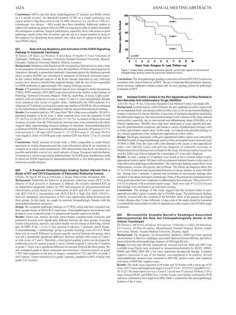
142A ANNUAL MEETING ABSTRACTS Conclusions: HPS is rare but likely underdiagnosed (17 patients met WHO criteria in a 6 month review). No threshold number of SPs in a single endoscopy was clearly useful to flag those most at risk for HPS. However, if a cut-off of 3 SPs at 1 colonoscopy was chosen, > 50% would have been identified. Additional studies to address the underlying genetic basis for HPS are ongoing in order to further illuminate this ambiguous syndrome. Surgical pathologists, especially those with access to prior pathology reports at the time of routine sign out, are in a unique position to assist in this endeavor by identifying those patients who either meet or appear at high risk to meet WHO criteria.
627 Role of K-ras Mutations and Activation of the EGFR-Signalling Pathway in Colorectal CarcinomaM Dantes, CW Hann von Weyhern, R Rosenberg, H Hoefler, F Fend. University of Tuebingen, Tuebingen, Germany; University Hospital Technical University, Munich, Germany; Technical University Munich, Munich, Germany.Background: Mutations in the Kirsten-ras (K-ras) gene have been known as early events in carcinogenesis of colorectal cancer since the hypothesis of linear cancerogenesis by Vogelstein. Recently a monoclonal antibody Cetuximab against the Epidermal growth factor receptor (EGFR) was introduced in treatment of advanced colorectal cancer. In this context molecular analysis of the K-ras became important as only wild-type carriers have shown to benefit from targeted therapy. Still the rate of non-responders to EGFR-antibodies is approximately 45% among wild-type carriers.Design: 274 primarily resected colorectal cancers were arranged to tissue microarrays (TMA). FFPE material (1993-2003) were retrieved from the archive of the Institute of Pathology Technical University Munich. With the small flaps of tissue, Light-cycler-analysis of K-ras-mutations in exon 2, codons 12 and 13, were accomplished. Results were compared with results of regular slides. Additionally the TMA-material of a subgroup of 53 patients was tested for gene copy number of EGFR by silver-enhanced in-situ hybridization (SISH) and compared with the immunohistochemical staining.Results: For mutation analysis, 207 cases from TMA-slides were evaluable. 102 (49%) presented mutation in K-ras exon 2. Most common ones were the mutations G13D (27=26.5%), G12D (26=25.5%) and G12V (17=16.7%). As control of effectiveness and accuracy of results from the TMA material, selected cases were compatred with whole original slides demonstrating identical findings of mutations. Immunohistochemical evaluation of EGFR shows even distribution for staining intensities: 80 tumors (33.1%) recieved score 1+, 69 cases (28.5%) score 2+, 31 (12.8%) score 3+. 62 cases (25.6%) were negative. Ratio of EGFR gene copy number/CEP was below 1.5 in 53 selected cases.Conclusions: Small amounts of tissue from a TMA comparable to small biopsy specimens in routine diagnostics provide exact information about K-ras mutations in comparison to whole tumor preparations. This demonstrates that the K-ras analysis is possible and reliable even in very small amounts of tissue. Frequency and distribution of mutations is similar to previously published data. No EGFR gene amplification could be observed. EGFR-expression by immunohistochemistry is very heterogenious, even in between needle-biopsies.
628 A Proposed Histopathologic Grading System Derived from a Study of KIT and CK19 Expression in Pancreatic Endocrine TumorsLN Dao, TC Smyrk, RV Lloyd, CM Lohse, L Zhang. Mayo Clinic, Rochester, MN.Background: Predicting the behavior of pancreatic endocrine tumor (PET) in the absence of local invasion or metastasis is difficult. We recently identified KIT as an independent prognostic marker for PET and proposed an immunohistochemical classification system based on a combination of KIT and CK19 expression: low risk (KIT-/CK19-); intermediate risk (KIT-/CK19+); high risk (KIT+/CK19+). Survival, metastases, and recurrence of PET were significantly different between the three groups. In this study, we sought to correlate histopathologic features with the immunohistochemical categories.Design: We compared pathologic findings of 97 PETs which had been classified into three groups based on KIT/CK19 expression. Clinicopathological associations with prognosis were evaluated using Cox proportional hazards regression models.Results: Tumor size, mitosis, necrosis, tumor border, extrapancreatic extension, and perineural invasion were significantly different between the three groups. A scoring system using mitosis, necrosis, and tumor border was developed as following: mitosis (per 50 HPF): 0 (0), 1 (1-3), 2 (≥4); necrosis: 0 (absent), 1 (present); tumor border: 0 (non-infiltrating), 1 (infiltrating), giving a possible histology score of 0 to 4. While there was an overall difference in disease-specific survival between the groups, there was not a statistically significant difference between patients with scores of 0 and 1 or with scores of 3 and 4. Therefore, a three-tiered grading system was developed by combining score 0/1 tumors as grade 1, score 2 tumors as grade 2, and score 3/4 tumors as grade 3. There was a significant difference in outcome between the three grades. We also compared grade to tumor metastasis and recurrence. Nineteen percent of grade 1 PET had metastasis at the time of surgery, compared to 73% and 99% of grade 2 and 3 tumors. Tumor recurred in 6% grade 1 patients, compared to 46% of those with grade 2 or 3 tumors.
Conclusions: This histopathologic grading system derived from KIT/CK19 expression correlates with clinical behavior of PET. The use of reproducible parameters (mitosis, tumor necrosis, infiltrative border) makes this an easy grading system for pathologic evaluation of PET.
629 Isolated Colitis Limited to the Peri-Appendiceal Orifice Related to Non-Steroidal Anti-Inflammatory Drugs (NSAIDs)A Del Rio Perez, W Xin. University Hospitals Case Medical Center, Cleveland, OH.Background: Isolated chronic colitis limited to the peri-appendiceal orifice region with no accompanied ileitis and chronic colitis in other sites, is not an uncommon finding in routine evaluation for chronic diarrhea. A spectrum of etiologies should be included in the differential diagnosis: the most common being Crohn’s disease (CD), drug-induced enterocolitis, especially due to non-steroid anti-inflammatory drugs (NSAIDs) or no clinical significance. NSAIDs have long been implicated to cause specific and non-specific gastrointestinal symptoms, and induce a variety of pathological changes, such as ileitis and isolated colonic ulcer. In this study, we looked at the possible etiology or any clinical significant of the isolated peri-appendiceal orifice colitis.Design: The biopsy specimens of the peri-appendiceal orifice region were selected by SNOMED search of surgical pathology archives from our hospital system between years of 2006 to 2008. Only the cases with colitis limited to the cecum or peri-appendiceal orifice were selected. Cases with previous diagnosis of colorectal carcinoma or inflammatory bowel diseases were excluded in this study. Electronic medical record was used for follow-up of these patients for a maximum of 3 years after diagnosis.Results: In total, a group of 23 patients were found to have isolated colitis in peri-appendiceal orifice region. All cases with accompanied terminal ileum or other parts of colon showed no pathological abnormalities. The endoscopic findings ranged from: focal inflammation, erosion, ulcerations, and erythema. The follow-up diagnoses show that 21 % (5/23) of patients had documented histories of routine chronic or acute NSAIDs use. Among these 5 patients, 3 patients had resolution of microscopic findings after cessation of the drugs and repeat colonoscopy. None of the patients developed persistent lower gastrointestinal tract symptoms or inflammatory bowel disease. There were 62 % (14/23) of patients with occasional aspirin usage. There were only 4 % (1/23) of cases that findings were attributed to an infections etiology.Conclusions: The findings of this study suggest that the isolated colitis in peri-appendiceal orifice region is mainly due to NSAIDs usage. The pathological findings could be reversed after the cessation of the NSAIDs. None of the patients developed Crohn’s disease after 3 years followup. A large scale of the study should be warranted to establish the relationship of colitis in appendiceal orifice region with NSAIDs usage in humans.
630 Microsatellite Unstable Barrett’s Esophagus-Associated Adenocarcinomas Are Rare, but Clinicopathologically Similar to the Colonic CounterpartEG Demicco, AB Farris, AJ Iafrate, HA Gaissert, J Fukuoka, C Cohen, LR Zukerberg, GY Lauwers, M Mino-Kenudson. Massachusetts General Hospital, Boston; Emory University, Atlanta; Toyama Medical University, Toyama, Japan.Background: The frequency of microsatellite instability (MSI) has been reported inconsistently in Barrett’s esophagus-associated adenocarcinoma (BEAd), and little is known about the clinicopathologic features of MSI high BEAds.Design: Seventy-nine BEAds consecutively resected between 2000 and 2007 with available tissue blocks were evaluated by immunohistochemistry for MLH1, MSH2, MSH6 and PMS2 (MSI IHC). No cases underwent neoadjuvant therapy. Complete negative expression in any of the markers was considered to be positive. Several clinicopathologic features were examined in MSI IHC positive cases, and compared with those of MSI IHC negative cases.Results: The study cases consisted of 69 males and 10 females with a mean age of 65 years. AJCC tumor and lymph node stages were pT1/2/3/4 = 35/10/33/1 and pN0/1/2/x = 45/31/2/1; the mean tumor size was 3.6 cm (4.7 cm for non-T1 tumors). Of those, 5 (6%) cases showed MLH1 and PMS2 loss. 3 of the 5 cases were further examined by PCR and were confirmed to have high-level MSI. Table 1 summarizes the clinicopathologic features of the 5 cases.

ANNUAL MEETING ABSTRACTS 143A
Table 1
Age (yrs) Gender Tumor size (cm) pT pN Histology Follow-up
(mo) Status
80 F 8.0 4 2 heterogeneous with medullary 14.8 dead of disease
79 M 5.5 3 1heterogeneous with mucinous and signet ring cells
22.7 dead of disease
75 M 7.5 3 1 medullary 0.5 dead of complication
67 M 2.8 2 0 poorly differentiated with signet ring cells 10.7 dead of unknown cause
78 F 6.0 3 0 medullary 3.0 lost to follow-up
Compared to MSI IHC negative cases, the MSI IHC positive BEAds were associated with older age (p < 0.005), larger tumor size (p = 0.15 for non-T1 tumors) and more advanced T stage (p = 0.15). The morphology of the MSI IHC positive tumors was heterogeneous with medullary, mucinous and/or signet ring cell features recapitulating the colonic counterpart.Conclusions: Microsatellite instability is rare in BEAds, but is associated with clinicopathologic features similar to the colonic counterpart such as mucinous, medullary, or signet ring cell features. Because of its scarcity, the survival impact of MSI is yet undetermined. Additional larger scale studies are warranted.
631 Validation of a Topographic-Anatomic Subclassification for Adenocarcinoma of the GE Junction (GEJ)EG Demicco, AB Farris, B Agbor-Etang, J Fukuoka, D Daives, M Snimizu, HA Gaissert, M Mino-Kenudson. Massachusetts General Hospital, Boston; Emory University, Atlanta; Toyama University Hospital, Toyama, Japan; Saitama Medical University, Saitama, Japan.Background: The incidence of adenocarcinoma of the GEJ is increasing more rapidly than any other cancer in the US. Due to the location, staging and treatment for GEJ tumors varies among institutions. The Siewert classification system differentiates between adenocarcinoma of the esophagus (type 1) and that of “cardia” (type 2). The aim of this study was to evaluate whether there was a difference in biologic properties between the 2 types.Design: We evaluated 149 untreated GEJ type 1 (n=49) and type 2 (n=100) tumors consecutively resected between 2000 and 2008. Demographic data, clinical and/or pathologic evidence of BE, tumor stage, tumor histology (adenosquamous cell carcinoma [AdSq], conventional adenocarcinoma [Ad], mucinous/signet ring cells [muc], medullary/undifferentiated [med], heterogenous [mix]) and outcome were compared between the 2 types. A subset of cases with available blocks (type 1: 40, type 2: 74) was examined by immunohistochemistry using tissue microarray and classified into 4 mucin phenotypes (intestinal [I], gastric [G], combined intestinal and gastric [M], and null [N]).Results: Type 1 was more likely associated with male gender (p = 0.060), BE background (p = 0.00072) and mixed mucin phenotype (p = 0.028) (Table 1).
Table 1Type 1 (n=49) Type 2 (n=100)
Age (yrs) 65.7 +/- 11.1 66.0 +/- 10.7M:F 44:5a 77:23a
BE 42 (89%)b 58 (58%)b
Tumor size (cm) 4.2 +/- 2.1 3.9 +/- 2.2pT1/2/3/4 22/7/20/0 31/13/55/1pN0/1/2/3 26/20/2/0 44/50/4/1pMx/1 47/2 97/3ASq/Ad/muc/med/mix 1/20/11/5/12 0/50/14/7/29I/G/M/N 9/9/19/3c 13/29/18/14c
a: p=0.06, b: p < 0.001, c: p < 0.05While the 5 year survival rate was better in type 1 (27.3% vs. 11.1%), the difference was not statistically significant (p = 0.11). On multivariate analysis, cardiac location (type 2) trended to independently predict poor survival (p = 0.0783), as did older age (p = 0.0419), advanced T stage (p = 0.027), LN metastasis (p = 0.005), and tumor size (p = 0.027).Conclusions: Although type 1 and type 2 tumors are morphologically similar, type 2 tumors are associated with a lower incidence of BE background and mucin phenotypes different from those of type 1, and appear to predict poor prognosis. Our results suggest that type 1 and type 2 tumors have different biologic properties.
632 Role of pan-T and Cytotoxic T Cells in the Pathogenesis of Adult Eosinophilic Esophagitis – A Single Institutional Study of 325 PatientsP Dhakras, D Patil, J Liao, HN Li, SM Rao, G-Y Yang. Northwestern University, Chicago, IL.Background: Eosinophilic esophagitis (EE) has evolved as a distinct clinic pathologic entity which is increasingly recognized by pathologists and gastroenterologists worldwide. The difference from pediatric EE (predominant due to allergy), is that only 1/3 of adult EE is in association with allergy and the etiology predominantly remains unknown.It has been shown that T cell and T regulatory cells (T reg) may be involved in the interaction with eosinophils and in that sense, may play a pivotal role in pathogenesis of eosinophilic esophagitis. This study is designed to characterize T cell-mediated inflammation in adult EE patients in comparison with reflux esophagitis and normal esophagus and explore the role of T cells in pathogenesis of EE.Design: Large cohort of 325 patients with EE was analyzed which were classified as distal predominant type and diffuse type of EE according to pathologic diagnostic criteria.Concurrent infiltration of pan-T cell, cytotoxic T cells (CD8),CD4 T cells and T regulatory lymphocyte (Fox p3 labeled cells) was quantitatively analyzed immunohistochemically in the selected different types of EE biopsy specimens (n=20 for each type) in comparison with reflux esophagitis (n=10) and morphologically
normal esophagus (n=5) with proper positive and negative controls. Average numbers of immunostaining-labeled T cells were counted for at least five high power fields in the areas of EE with highest eosinophil infiltration.Results: Significant increase of pan T cells (CD3)and cytotoxic T cells (CD8) was observed in both distal predominant and diffuse types of EE as compared to normal and reflux esophagitis (P<0.05). The results showed there were average of 31.75+ 7.47 and 34.93+ 26.19 intraepithelial CD8 T cells, respectively in distal-predominant and diffuse type EE compared to 18.13+ 7.66 in normal esophagus and 22.60+ 14.20 in reflux esophagitis. But, there is no statistically significance for CD8 T cells between distal predominant and diffuse types of EE. Similar results were noted for CD3 labeled pan-T cells, with 37.35+ 7.45 in distal predominant, 39.38+9.30 in diffuse type.T regs (Foxp3 labeled cells) in EE showed marked difference in the ratio of CD3 to Treg cells between EE and normal esophagus.Conclusions: We demonstrate that pan-T cell and cytotoxic T cells play an important role in the pathogenesis of EE.T cells would be also a biomarker to differentiate EE from reflux esophagitis and the role of T cell-mediated inflammation needs to be investigated further.
633 Expression of Stem Cell Markers in Human Gastric Adenocarcinoma and Non-Neoplastic Gastric Mucosa, a Study of 209 CasesS Dhingra, W Feng, D Zhou, T Khoury, RE Brown, D Tan. MD Anderson Cancer Center, Houston; UT Health Science Center, Houston; University of Rochester, Rochester; Roswell Park Cancer Institute, Buffalo.Background: Cancer stem cells (CSCs) are a unique subpopulation that possesses the capacity to repopulate tumors, drive malignant progression and mediate radio/chemoresistance. Stem cell markers, CD44 and nestin, are known to play a role in disease progression in colorectal carcinoma. Recent studies suggest that CD44 expressing gastric cancer cells show increased resistance for chemotherapy or radiation-induced cell death. Role of nestin as a CSC in gastric adenocarcinoma (GAC) is largely unknown. In this study we evaluated the expression of CD44 and nestin in GAC.Design: Tissue microarray blocks from 168 cases of GAC and 41 adjacent non-neoplastic gastric mucosa (NNGM) were assembled. Immunohistochemical stains were performed using antibody against CD44 and nestin. The intensity (1-3+) and percentage of chromogenic signal was determined, and a composite score (CS), the product of the intensity and percentage was calculated. The significance of difference in means was determined by paired student’s t-test.Results: Membranous CD44 was positive in 52% (79/151) cases of GAC. Majority of CD44 positive cases (60/79 [75%])} showed strong (2-3+) signal intensity with 49% (39/79) displaying CS > 100 (range 5-270). In contrast, adjacent NNGM showed expression of CD44 in 24 % (10/41) cases. Nestin was localized in the cytoplasm and 23% (38/168) cases of GAC were positive. Strong (2-3+) intensity of staining was seen in 92% (35/38) cases and 57% (22/38) showed CS>100 (range 5-240). A subset of nestin positive cases (16/38 [42%]) showed (1-3+) membranous staining of CD44. Some cases with intestinal metaplasia (IM) showed either CD44 (3/6) or nestin (2/6) expression. Few NNGM (2/41 [5%]) showed nestin positivity. GAC showed statistically higher expression of nestin compared to NNGM (p<0.001).Conclusions: This study reveals that overexpression of stem cell marker, nestin, in GAC is significantly increased versus NNGM. In addition, IM, a common premalignant lesion, also displays increased expression of stem cell markers. These findings suggest that elevated expression of stem cell markers may be associated with development and progression of GAC. Further studies using animal models and cell culture will elucidate the potential of using specific agents to target stem cells in management of GAC.
634 Pyloric Gland Adenoma of the Stomach – A Clinicopathologic Study of 8 CasesIG Do, KM Kim, CK Park. Samsung Medical Center, Sungkyunkwan University School of Medicine, Seoul, Korea.Background: Pyloric gland adenoma (PGA) is a rarely described neoplasia of stomach accounting for 2.7% of all gastric polyps. Similar adenomas of the gastric type have been reported in gastric heterotopic mucosa in the duodenum, gallbladder, main pancreatic duct, and uterine cervix. Apart from case reports, only a few clinical follow-up data are available on PGA in the stomach and their relationship to malignant transformation was not fully explored.Design: In a large comprehensive cancer center located in a gastric cancer prevalent area, 8 PGAs from 8 patients were retrieved. Clinicopathologic features and immunohistochemical panel consisting of mucin core peptides (MUC) 2, MUC6, MUC5AC and CD10 were studied. Clinical follow-up information was obtained in 4 cases of PGA. PGAs were divided into 3 categories: those with low-grade dysplasia, those with high-grade dysplasia, and those in association with adenocarcinoma.Results: The mean age of PGAs was 62 years with a male predominance over female by 7: 1. The locations within the stomach were cardia (n=3), fundus (n=3), high body (n=1) and lower body (n=1). Microscopically, all 8 cases were classified as 5 PGAs with low-grade dysplasia, 1 PGA with high-grade dysplasia, and 2 PGAs in association with adenocarcinoma. PGAs were mainly composed of closely packed pyloric type glands with monolayer of cuboidal or columnar cells containing round nuclei and pale to eosinophilic cytoplasm and forming some ectactic foveolae. Immunohistochemically, all PGAs were strongly positive for MUC 6 and negative for MUC 5AC, MUC 2 and CD10. Interestingly, two PGAs showed continuous transition to well differentiated gastric-type adenocarcinoma at the time of diagnosis and one PGA with high-grade dysplasia progressed to invasive tubular adenocarcinoma during the follow-up.Conclusions: PGAs have a characteristic histologic appearance and mucin phenotype. In our experience, 37.5% of gastric PGAs showed transition or progression to adenocarcinoma. PGAs are true neoplasms with a high potential for malignant transformation and require complete removal and close clinical follow-up.

144A ANNUAL MEETING ABSTRACTS 635 Intravesicular Papillary-Tubular Neoplasm (IVPN) as a Unifying Category for Mass-Forming Preinvasive Neoplasms of the Gallbladder: An Analysis of 87 CasesN Dursun, JC Roa, O Tapia, A Cakir, I Coban, O Basturk, D Akdemir, J Sarmiento, H Losada, NV Adsay. Emory U, GA; U de La Frontera, Temuco, Chile; NYU, NY; Ohio Northern U, OH.Background: Mass-forming (polypoid) preinvasive papillary and tubular neoplasms of gallbladder (GB) mucosa, which we propose to refer to as IVPN, have been poorly characterized.Design: 87 IVPNs, defined as non-invasive neoplasm forming a well-defined exophytic mass measuring >1cm (the criterion employed for clinical significance in pancreatic IPMNs, and cholecystectomy indication for GB polyps), were analyzed.Results: M/F=20/62, mean age 64. Median tumor size=2.1 cm (range, 1-7.7). I. Variable growth patterns were evident: Papillary (46%), tubulopapillary (39%), and tubular (14%). II. Various cellular lineages including intestinal (17%), gastric (16%), pancreatobiliary (30%) could be discerned; however, transitional/mixed forms were common (36%). III. 4% of the lesions had no high-grade dysplasia (HGD), while 30% had focal, and 66% extensive. HGD was commonly extensive in papillary examples (10/12) but not in tubular ones (5/12). However, even the cases that would have qualified as “pyloric gland adenoma” (previously considered an innocuous lesion), had at least focal HGD (14/16), and 75% occurred in association with invasive carcinoma (12/16). Conversely, some cases that might have qualified as “papillary ca” displayed foci of low-GD (18/52) or had no identifiable invasion (7/52). IV. 75% had an associated invasive ca (ordinary adenoca-55/63; others-8, including 3 mucinous, 2 neuroendocrine). Invasion was focal in 4/63 and extensive in 36/63. Among systematically analyzed invasive carcinomas, IVPN was detected in 10% (63/606). V. 1- and 3-yr actuarial survival were 92% and 77% for non-invasive IVPNs vs 68% and 45% for IVPNs with an associated invasive ca (p=0.05). However, even invasive ones had a better clinical outcome than ordinary GB carcinomas (3-yr survival 45% vs 28%; p<0.001).Conclusions: IVPNs are analogues of mass-forming preinvasive neoplasms in other organs such as pancreatobiliary IPMNs, showing variable cellular lineages, spectrum of dysplasia (adenoma-carcinoma sequence), and a mixture of tubular-papillary growth patterns, often with significant overlaps, warranting the classification of these lesions under one unified category. These are relatively indolent neoplasia with significantly better prognosis than ordinary GB carcinomas; however, invasive examples can be fairly aggressive, and even non-invasive ones may be fatal.
636 The Significance of Mucosal Eosinophilia in the Pediatric ColonSM El Jamal, T Gibbons, E Davis, AG Saad. University of Arkansas for Medical Sciences, Little Rock, AR.Background: The exact number and pattern of colonic mucosal eosinophilia (CME) that is often associated with colonic disorders is largely unknown. The aim of this study is to evaluate the significance of CME in the pediatric population.Design: Pediatric patients with colonic biopsies interpreted as normal (control) or increased mucosal eosinophilia (patients) seen in the pediatrics service are included in this study. Clinical presentations included abdominal pain, vomiting, abnormal bowel movements, and blood in stool. In each part of the colon, eosinophils were evaluated for various parameters including number, location (superficial vs. basal), distribution [clustering (EC) vs. scattered] and intraepithelial eosinophils (IEE). The number of eosinophils was generated by manually counting the number of eosinophils per hot spot at 40X magnification. A cluster of eosinophils is defined as a cluster of ≥5 eosinophils.Results: The study consisted of 29 patients (14 patients and 15 controls) with complete clinical follow-up. Details of histologic findings are in table 1. Follow-up of the patients group found that 10 patients met the criteria of recurrent abdominal pain (RAP), 2 patients developed IBD and 2 patients were found to have parasitic infection. Of the control group, 13 patients were found to have extracolonic pathology such as H. pylori infection and gastro-esophageal reflux disorder; one patient developed IBD and another was found to have parasitic infection. Analysis of the data showed strong correlation between the presence of EC and IEE and the development of colonic diseases. the number of CME was a poor predictor of colonic diseases. There was no correlation between the distribution (superficial vs. basal) of eosinophils and the development of colonic diseases.Parameter Patients (14) Controls (15)Eosinophilic clustering 13 (92.8%) 1 (6.7%)Epithelial eosinophils 10 (71.4%) 3 (20%)Basally located eosinophils 5 (35.7%) 6 (40%)Superficially located eosinophils 4 (28.6%) 5 (3.3%)Number of eosinophils (mean ± SD): Right colon (cecum and ascending) 31.2 ± 12.1 24.1 ± 8.9Transverse colon 29.9 ± 10.9 24.3 ± 6.8Left colon (descending and rectosigmoid) 30.8 ± 19.5 21.4 ± 6.1
Conclusions: Our results show that the pattern of distribution of eosinophils within the colonic mucosa, in particular the presence of EC, is more important than the absolute number of eosinophils in predicting the existence of colonic diseases. The is a strong correlation between colonic eosinophilia and patients who carry a diagnosis of RAP.
637 “In Situ” Growth of Metastatic Tumors to the Gastrointestinal Tract: A Potential Mimic of Primary NeoplasiaJS Estrella, A Rashid, SC Abraham. MD Anderson Cancer Center, Houston.Background: It can be difficult to distinguish primary from metastatic neoplasms to the gastrointestinal (GI) tract, particularly in the small bowel where metastatic tumors outnumber primaries. The presence of an adenomatous precursor is usually taken as strong evidence for primary neoplasia. Gross configuration of the tumor and regional lymph node involvement are also features commonly used in this distinction.
Design: We studied GI resections from 1987-2009 that were reported to show mucosal involvement by metastatic carcinoma. Gross descriptions and histologic sections were evaluated for the following: 1) “in situ” growth (i.e., growth within the basement membrane of villi/crypts), 2) resemblance to a precursor/adenoma, 3) maturation of tumor cells toward the lumen (i.e, better differentiated tumor within mucosa and less differentiated tumor - including presence of tumor budding - in the wall), 4) gross configuration of the metastasis, and 5) lymph node involvement in the metastatic site.Results: Detailed results are presented in Table 1. The study population comprised 99 resections from small bowel (n=75), colorectum (n=14) or both (n=10). Many (74%) metastatic carcinomas lacked a classic serosal-based configuration and showed lymph node involvement in the metastatic site (64%). Carcinomas originating from the GI tract were significantly more likely to show in situ growth (p<0.0001), resemblance to a precursor (p<0.0001), and maturation toward the mucosa (p<0.0001). In 5 cases (3 GI and 2 GYN origin), metastatic tumors were initially interpreted as new primaries by the pathologist (n=4) or radiologist (n=1).Conclusions: Metastatic carcinomas involving the intestinal mucosa can exhibit histologic features mimicking second primaries, particularly when they originate from the GI or GYN tract. Presence of an apparent adenoma cannot be taken as prima facie evidence of a primary neoplasm.
Table 1
Primary site In situ growth Precursor resemblance
Maturation toward mucosa
Non-serosal based configuration
Lymph node involvement
GI tract (n=56) 34 (61%) 26 (46%) 28 (50%) 45 (80%) 11 of 22 (50%)GYN tract (n-28) 6 (21%) 2 (7%) 1 (4%) 15 (54%) 14 of 19 (74%)Lung (n=7) 2 (29%) 0 (0%) 0 (0%) 6 (86%) 4 of 5 (80%)GU tract (n=5) 0 (0%) 0 (0%) 0 (0%) 5 (100%) 1 of 2 (50%)Head & neck (n=2) 0 (0%) 0 (0%) 0 (0%) 1 (50%) 2 of 2 (100%)
Breast (n=1) 1 (100%) 0 (0%) 0 (0%) 1 (100%) Not sampled
638 Immunohistochemical Staining for Smoothelin Differentiates the Duplicated Muscularis Mucosa of Barrett’s Esophagus from the True Muscularis MucosaHF Faragalla, CJ Streutker. University of Toronto, Toronto, ON, Canada; Saint Micheal Hospital, Toronto, ON, Canada.Background: The muscularis mucosa underlying the metaplastic mucosa of Barrett’s esophagus is frequently duplicated, with an intervening layer of lamina propria between the superficial muscle layer (neo-muscularis mucosa, NMM) and deep muscularis mucosa (true muscularis mucosa, TMM). This duplication causes difficulties with accurate staging of superficially invasive carcinoma in biopsy specimens and endoscopic mucosal resections (EMR), as invasion underneath the superficial muscle layers may be mistaken for submucosal invasion. Intramucosal carcinoma (pT1a) can often be treated by EMR resection or other non-surgical modalities, whereas patients with submucosal (pT1b) invasion are recommended for esophagectomy. Therefore, the accurate staging of such specimens is crucial. Smoothelin is a novel smooth muscle protein expressed only by fully differentiated smooth muscle cells and not by proliferative or noncontractile smooth muscle cells and fibroblasts: it has been suggested that in the bladder it can separate hyperplastic muscularis mucosa from the true muscularis propria. We hypothesized that immunohistochemistry for smoothelin would differentiate the NMM from the TMM.Design: Eleven cases of endoscopic mucosal resections for Barrett’s related neoplasia were retrieved from the archives of the pathology department. Immunohistochemical staining for smoothelin, smooth muscle actin and smooth muscle myosin were performed to evaluate differential staining in the TMM versus NMM. The staining score was evaluated as follows: 0 < 5% of cells staining, +1 or focal =5%-10%, +2 or moderate =11% to 50%, +3 or strong, diffuse >50% muscle cell positivity.Results: With SMA, strong and diffuse staining(+3) was observed with similar intensity and pattern in NMM(11/11) and TMM (11/11).With smoothelin, the NMM showed weak focal staining (+1) in 8/11 cases (73%), and moderate staining +2 in 3/11 cases (17%), and the TMM showed very strong and diffuse staining(+3) in 11/11cases (100%). With smooth muscle myosin, strong and diffuse staining was observed with similar intensity in both the TMM and NMM in 11/11 cases.Conclusions: In our study, smoothelin staining in the NMM is significantly weaker than that seen in the true/deep muscularis mucosa. This suggests that this antibody may be of use in evaluation of esophageal biopsies or endoscopic resections to more accurately stage early stage adenocarcinomas.
639 Expression of S100p in Human Esophageal Adenocarcinoma and Its Precursor LesionsW Feng, Z Zhou, D Tan. The University of Texas Medical School at Houston, Houston; University of Rochester Medical Center, Rochester; The University of Texas M.D. Anderson Cancer Center, Houston.Background: S100p is a 95-amino acid protein with a restricted cellular distribution that was first purified from placenta. It has been shown to be expressed in prostate adenocarcinoma and gastrointestinal malignancies, such as colon and pancreatic adenocarcinoma. It is also a poor prognostic factor for lung and breast cancer. The expression of S100p in esophageal adenocarcinoma (EAC) and its precursor lesions, including distinctive-type Barrett mucosa (BM) (intestinal metaplasia) and esophageal columnar dysplasia (ECD) is largely unknown. Therefore, in this study, we evaluated the expression of S100p in EAC and its precursor lesions.Design: We examined samples from 135 cases of esophageal adenocarcinoma, 40 ECD (15 high grade and 25 low grade), 37 BM without dysplasia, and 164 non-neoplastic esophageal mucosa (NNEM) without dysplasia or BM, in formalin-fixed, paraffin-embedded tissue microarray blocks. Tissues were stained with antibody against S100P (1:20, MAB2957, R&D Systems) and incubated for 30 minutes at ambient temperature.

ANNUAL MEETING ABSTRACTS 145AThe intensity (1-3+) and percentage of positive nuclear-staining cells were determined, and a composite score (CS), the product of the intensity and percentage of positive cells (0-300) was calculated. The significance of difference in means was determined by two-tailed student’s t-test.Results: Some EAC cases (48 of 135 [36%]) showed 1-3+ nuclear S100p staining. Poorly differentiated EAC had statistically significant lower S100p expression than did well and moderately differentiated EAC (p<0.001). Furthermore, patients with high S100p-expressing EAC (76% one year survival) had better survival than did those with low S100p-expressing EAC (56% one year survival)(p=0.06). A subset of ECD cases (23 of 40 [64%]), including 14 low grade dysplasia and 9 high grade dysplasia cases showed weak 1+ staining for nuclear S100p. NNEM without BM (53 of 164 [32%]) and NNEM with BM (27 of 37 [73%]) showed weak 1+ staining for S100p within the superficial columnar epithelium.Conclusions: In this study, we showed that S100p is overexpressed in EAC, ECD, and columnar epithelium with and without distinctive-type BM. These results suggest that S100p overexpression plays a role in EAC carcinogenesis and is a prognostic marker for EAC.
640 Stem Cell Markers CD44 and Nestin in Human Esophageal Adenocarcinoma and Its Precursor LesionsW Feng, Z Zhou, D Tan. The University of Texas Medical School at Houston, Houston; University of Rochester Medical Center, Rochester; The University of Texas M.D. Anderson Cancer Center, Houston.Background: Recently, there has been mounting evidence for the existence of cancer stem cells within solid tumors of the colon, pancreas, breast, and prostate. CD44 and nestin are two stem cell markers that are known to be present in colorectal carcinoma and gastrointestinal stromal tumors, respectively. The expression of these two stem cell markers in esophageal adenocarcinoma (EAC) and its precursor lesions, including distinctive-type Barrett mucosa (BM) (intestinal metaplasia) and esophageal columnar dysplasia (ECD), is largely unknown. In this study, we evaluated the expression of CD44 and nestin in EAC and its precursor lesions.Design: We evaluated samples from 135 cases of EAC, 40 ECD (15 high grade and 25 low grade), 37 BM without ECD, and 164 non-neoplastic esophageal mucosa (NNEM), without ECD or BM, in formalin-fixed, paraffin-embedded tissue microarray blocks. Tissues were stained with antibodies against CD44 and nestin. The intensity (1-3+) and percentage of positive nuclear-staining cells were determined. The significance of difference in means was determined by student’s t-test.Results: Most EAC cases showed 1-3+ membranous staining for CD44 (91 of 135 [67%]), and a subset of CD44-positive cases showed 1-3+ cytoplasmic staining for nestin (40 of 135 [30%]). Overall, EAC showed higher nestin expression compared to NNEM (p<0.005). Poorly differentiated EAC had statistically significant higher expression of both CD44 and nestin than did moderately and well differentiated EAC (p < 0.05). Most ECD cases (35 of 40 [87%], 22 low grade and 13 high grade) showed 1-2+ membranous staining for CD44. A subset of ECD cases (13 of 40 [33%], 9 low grade and 4 high grade) showed 1+ cytoplasmic staining for nestin. Most NNEM tissues showed 1-3+ membranous staining for CD44 (128 of 164 [78%]) and a few showed 1-3+ cytoplasmic staining for nestin (8 of 164 [5%]) predominantly in basal/parabasal squamous epithelial cells. Most BM showed 1-2+ membranous staining for CD44 (32 of 37 [86%]), and a subset showed 1-2+ cytoplasmic staining for nestin (7 of 37 [19%]).Conclusions: In this study, we identified stem cell markers in EAC and its precursor lesions. These markers may serve as therapeutic targets for EAC. Further study is needed to elucidate the prognostic implications of stem cells in EAC.
641 Prevalence of Upper Gastrointestinal Tract Inflammation in Patients with Ulcerative ColitisAJ Gersten, D Beneck, D Blanco, RK Yantiss. Weill Cornell Medical College, New York, NY.Background: Ulcerative colitis (UC) is a form of inflammatory bowel disease (IBD) that diffusely affects the colonic mucosa in a retrograde fashion from the rectum. Classic teaching emphasizes that UC spares the upper gastrointestinal (UGI) tract and, in fact, its distribution aids distinction from Crohn’s disease (CD), particularly among pediatric patients. Some authors have reported rare UC patients to have UGI involvement, although the frequency and severity with which this occurs is not known. The aim of this study was to determine the prevalence of UGI inflammation among UC patients. We chose to evaluate pediatric patients because these children often undergo routine endoscopic examination of the UGI tract during evaluation for colitic symptoms, prior to initiation of therapy.Design: We identified 81 pediatric patients with newly diagnosed IBD, all of whom underwent endoscopy and biopsy of the UGI tract and colon prior to initiation of therapy. Patients with hard criteria for CD (granulomas, chronic ileitis, strictures, anal disease) were excluded from the study, as were those with confounding inflammatory conditions (e.g. H. pylori infection), yielding a final study group of 24 patients with presumed UC. Endoscopic and mucosal biopsy findings were reviewed, and the presence, distribution, and severity of inflammation were recorded.Results: There were 10 males and 14 females in the study group (mean: 12.1 years). Colonoscopy revealed colitis in all patients, which, on biopsy analysis, proved to be mild, moderate, or severe in 5, 11, and 8 patients, respectively. Two patients had endoscopic and histologic mild duodenitis. Fifteen (63%) patients had chronic gastritis, which was inactive in 11 (46%) and active in 4 (17%) patients. Giemsa stains were negative for H. pylori in all cases. Seven (29%) patients had esophagitis, including 5 (21%) with distal esophageal inflammation and 2 (8%) with features of eosinophilic esophagitis. At follow-up (mean: 21 months), 20 patients were classified as definite UC and 4 were considered to have probable UC.Conclusions: Pediatric patients with new-onset UC frequently have UGI tract inflammation, which does not correlate with severity of colonic disease. Most patients
with this finding have a clinical course typical of UC, provided they do not have other features to suggest CD. Therefore, the presence of UGI tract inflammation by itself should not be an exclusion criterion for UC in the pediatric population.
642 Overexpression of X-Linked Inhibitor of Apoptosis Protein (XIAP) in Gastrointestinal Dsyplasia and AdenocarcinomaK Gilbert-Lewis, J Heitzman, W Feng, W Li. University of Texas Health Science Center at Houston, Houston, TX.Background: X-linked inhibitor of apoptosis (XIAP) is a human inhibitor of apoptosis protein involved in the blockage of intrinsic and extrinsic apoptotic pathways. XIAP has been implicated in cancer metastasis and progression in a variety of neoplasms. The aim of this study is to determine the expression pattern of XIAP in gastric and colorectal carcinoma and correlate its expression with clinicopathologic variables.Design: Formalin-fixed paraffin-embedded tissue sections of 18 cases of gastric adenocarcinoma and 28 cases of colorectal carcinoma were immunostained for XIAP using standard avidin-biotin techniques. The level of XIAP expression was categorized into four grades: 0 (negative), 1+ (weak), 2+ (moderate), or 3+ (strong) based on intensity of staining.Results: There was significantly higher expression of XIAP in the neoplastic areas compared to normal matched tissue, independent of tumor grade in 18 cases of gastric adenocarcinoma and 28 cases of colorectal carcinoma. All tumor cells were moderate to strongly positive, with an average staining intensity grade of 2.8±0.3 compared to normal epithelium that stained weakly with an average grade 1.1±0.5. Areas with dysplastic epithelium displayed similar XIAP staining to neoplastic areas. The results show significant XIAP over-expression in neoplastic tissue compared to normal matched epithelium (p<0.0001).Conclusions: Significant over-expression of XIAP in gastrointestinal adenocarcinomas suggests XIAP as an integral role in the spectrum of tumor progression. While normal epithelium retains the low-level expression of XIAP in an effort to maintain homeostasis or avoid inappropriate cell death, dysplastic epithelium and adenocarcinomatous cells fail to turn off XIAP signals. The up-regulation of XIAP expression suggests its possible role in gastric and colorectal carcinogenesis and its potential utility as a target for innovative therapy.
643 Immunohistochemistry for SDHB Distinguishes Carney Triad and Carney-Stratakis Syndrome Associated GISTs (Type 2 GISTs) from Usual Sporadic GISTs (Type 1 GISTs)AJ Gill, A Chou, A Clarkson, RP Eckstein, A Shi, C Smith, L Siosson, DE Benn, RJ Clifton-Bligh. Royal North Shore Hospital, Pacific Highway, St Leonards, Australia; Kolling Institute of Medical Research Royal North Shore Hospital and University of Sydney, Sydney, NSW, Australia.Background: The Carney Triad (CT) is GIST, paraganglioma (PG), and pulmonary chondroma (PC). The Carney Stratakis Syndrome (CSS) is GIST and PG with mutations of succinate dehydrogenase (SDH) B, C or D. No mutations in KIT, PDGFRA or SDH- have been described in CT. Non CT hereditary PGs may be associated with germline SDHB, SDHC or SDHD mutations which form the mitochondrial complex II (MCII). These PGs show negative or weak staining for SDHB regardless of whether the B,C or D subunit is mutated. There is evidence that other yet to be described mutations may cause instability of MCII and also result in negative SDHB staining. We postulated that instability of MCII will occur and thus SDHB IHC will be negative in CT GISTs.Design: We performed IHC for SDHB on formalin fixed paraffin embedded blocks from 3 GISTs arising in CT and on 104 consecutive apparently sporadic GISTs from 98 subjects.Results: All 3 CT GISTS showed completely negative staining. Of the sporadic GISTs 99 (95%) were strongly positive, 3 (3%) weakly positive and 2 (2%) negative. Subsequent investigation revealed that 1 of the negative GISTS arose in a young female who had previously had multiple GISTS resected at prior operations over the preceding 17 years. This was unknown to us or the reporting pathologists, but clearly indicates an underlying tumour diathesis (perhaps a forme fruste of CT or CSS). The other negative case was from a young woman who presented with heavy gastric bleeding and hepatic metastases (features seen in CT).Conclusions: We conclude that negative staining for SDHB occurs commonly in the GISTS of CT and presumably in GISTs of CSS but rarely to never in isolated benign disease. We classify usual SDHB positive GISTS as type 1. The great majority of these GISTS will be driven by KIT and PDGFRA mutations. We classify SDHB negative cases as type 2 GISTs. Type 2 GISTS likely form a heterogeneous group but all are characterized by mitochondrial complex II instability. IHC for SDHB can be used diagnostically to classify GISTS as type1 or type 2. Type 2 GISTs should be offered genetic testing for SDH and if negative follow up as for CT. Provisional data suggests that their natural history and response to treatment may vary from type 1 GISTs.
644 Utility and Limitations of Mesothelin Immunohistochemistry for Pancreatic Neoplasia in Both Surgical and Fine Needle Aspiration MaterialsJP Glass, RE Donohue, LA Cerilli. University of New Mexico, Albuquerque, NM.Background: Previous studies have shown that mesothelin overexpression can be detected by immunohistochemistry in pancreatic adenocarcinoma. Our study attempts to confirm these results and further characterize immunohistochemical mesothelin staining in both fine needle aspirates and surgical materials from a spectrum of pancreatic neoplasms.Design: Immunohistochemical staining for mesothelin was performed on resection or biopsy specimens from: 23 ductal adenocarcinomas, 15 ampullary adenocarcinomas, 4 distal common bile duct adenocarcinomas, 6 mucinous neoplasms, 3 serous cystadenomas, 4 benign cysts, 1 solid pseudopapillary tumor, and 2 chronic pancreatitis

146A ANNUAL MEETING ABSTRACTS cases. Mesothelin staining was also performed on cell block material from 37 fine needle aspirates of pancreatic adenocarcinoma. Positive staining in the surgicals was defined as at least 2+ (moderate intensity) apical staining in at least 5% of cells. Positive staining in the aspirations was defined as any adenocarcinoma cells with at least 2+ apical staining.Results: 24 of 42 resected or biopsied adenocarcinomas (ductal, ampullary, and distal common bile duct) stained for mesothelin, while none of the non-carcinoma cases (0 of 16) showed positive staining (p = 0.0000784). In addition, only 3 of the 15 ampullary adenocarcinomas were positive for mesothelin, compared with 21 of 27 of the non-ampullary adenocarcinomas (p = 0.0002884). 27 of the 37 pancreatic adenocarcinoma fine needle aspirates were also positive for mesothelin. Of the 10 negative cases, limited material was present for evaluation in 3 cases (30%).
TOTAL MESOTHELIN + MESOTHELIN -ALL ADENOCARCINOMA 42 24 18PANCREATIC DUCTAL ADENOCARCINOMA 23 18 5AMPULLARY ADENOCARCINOMA 15 3 12BILIARY (BILE DUCT) ADENOCARCINOMA 4 3 1ALL MUCINOUS NEOPLASMS 6 0 6INTRADUCTAL PAPILLARY MUCINOUS NEOPLASM 4 0 4
MUCINOUS CYSTIC NEOPLASM 2 0 2SEROUS CYSTADENOMA 3 0 3SOLID-PSEUDOPAPILLARY NEOPLASM 1 0 1CHRONIC PANCREATITIS 2 0 2SIMPLE CYST OR PSEUDOCYST 4 0 4FINE NEEDLE ASPIRATIONS OF PANCREATIC ADENOCARCINOMA 37 27 10
Conclusions: This study confirms that mesothelin expression in pancreatic adenocarcinoma can be detected by immunohistochemistry in both surgical and fine needle aspiration specimens. In addition, this study supports the novel finding that adenocarcinoma arising from the ampulla of Vater does not usually express mesothelin and further suggests that mesothelin is not expressed by other pancreatic neoplasms, such as pancreatic mucinous neoplasms.
645 Loss of CDH1 Expression in Colorectal Cancer Is Independently Associated with Microsatellite Instability and Female GenderKL Golden, Y Baba, CS Fuchs, S Ogino. Brigham and Women’s Hospital/Harvard Medical School, Boston, MA; Dana-Farber Cancer Institute, Boston, MA.Background: E-cadherin (CDH1) is important for maintaining the adhesive properties of epithelial cells, while its loss may contribute to the invasive properties observed in colorectal cancers. No study has comprehensively investigated the association of CDH1 loss in colorectal cancers with clinical, prognostic, pathologic, and molecular features, including microsatellite instability (MSI) and the CpG island methylator phenotype (CIMP).Design: Among 525 colorectal cancers with clinical outcome data, we detected loss of CDH1 membrane expression in 281 (54%) tumors by immunohistochemistry. We analyzed for CIMP by real-time PCR, and BRAF, KRAS, and PIK3CA mutations and LINE-1 methylation by Pyrosequencing.Results: In multivariate logistic regression analysis with a stepwise selection procedure, CDH1 loss was significantly associated with female gender and MSI-high (Table 1), but not with age at diagnosis, body mass index, family history of colorectal cancer, tumor location, stage, tumor grade, mucinous or signet ring cell components, CIMP, BRAF, KRAS, PIK3CA, LINE-1 hypomethylation, p53, or COX-2. Compared to patients with CDH1-expressing colon cancers, those with CDH1-lost colon cancers showed a non-significant tendency for a high overall mortality (Figure 1) [hazard ratio 1.28; 95% CI, 0.99-1.66].
Multivariate analysis of the independent relations with CDH1 loss in colorectal cancerVariables in the final model for CDH1 loss Multivariate OR (95% CI) P valueFemale gender (vs. male gender) 1.89 (1.28-2.81) 0.0014MSI-high (vs. MSI-low/MSS) 1.89 (1.12-3.21) 0.017Mucinous component (>0% vs. 0%) 1.54 (1.00-2.39) 0.053Signet ring cell component (>0% vs. 0%) 2.19 (0.92-5.20) 0.077Stage IV (vs. stage I-III) 1.53 (0.88-2.67) 0.14 PIK3CA mutation 0.66 (0.36-1.21) 0.18OR, odds ratio; CI, confidence interval; MSI, microsatellite instability
Conclusions: CDH1 loss in colorectal cancer is independently associated with MSI-high and female gender.
646 KRAS Mutations Are Associated with Specific Morphologic Features in Colon CancerA Gunal, B Kenney, P Hui, S Kilic, R Xu, D Jain, K Mitchell, M Robert. Gulhane Military Medical School, Ankara, Turkey; Yale University School of Medicine, New Haven, CT; New York University School of Medicine, New York, NY.Background: Genetic mutations of the KRAS gene occur at an early stage in colon cancer. KRAS mutations predict resistance to anti-EGFR therapy in Stage IV disease. While histology in colon cancer has been associated with DNA mismatch repair gene mutations, no comprehensive analysis of morphological correlates for KRAS mutations exists. As it is now the standard of care to assess KRAS mutations in Stage IV colon cancer, morphological correlates could be useful in clinical practice. The aim of the current study is to correlate histological features of colon cancer with KRAS mutations.Design: Tumor tissue from 145 colon cancer resections performed over a two year period were tested for KRAS mutations. KRAS mutation status was correlated with histological characteristics, including age, gender, tumor site, size, gross configuration, histological type, grade, mucinous component, lymphovascular invasion, extramural venous/perineural invasion, peritumoral lymphocytic response, infiltrative vs pushing growth, TNM status and overall stage. Statistical analysis was performed using Pearson chi-square and multivariate analysis.Results: KRAS mutations were present in 55/145 cases (37.9%), consistent with reported rates. KRAS mutations were significantly associated with usual adenocarcinoma morphology (chi square p=0.055; multivariate p=0.014), peritumoral lymphocytic response (chi square p=0.028; multivariate p=0.017), T3-T4 status (chi square p=0.012; multivariate p=0.015), right colon tumors (multivariate p= 0.027), absent lymphovascular invasion (multivariate p=0.008), and metastases at the time of resection (multivariate p=0.034). No association was found between KRAS mutational status and the other factors.
Effects of histology on KRAS mutation positivityTumor characteristics Odds Ratio 95% CIRight colon location 2.51 1.11-5.71Type (usual adenocarcinoma) 8.40 1.53-45.45Tumor depth T3,T4 7.52 1.48-38.17Distant metastasis at presentation 3.01 1.08-8.37Absent lymphovascular invasion 3.11 1.35-7.14Lymphocytic response present 2.73 1.19-6.23
Conclusions: Specific morphological features in colon cancer suggest a higher likelihood of the presence of KRAS mutations. These morphological features overlap partially with those associated with DNA mismatch repair gene mutations. If confirmed, these results may suggest a paradigm for directed KRAS testing.
647 Duodenal Lymphocytosis with Normal Villous Architecture: How Often Is It Celiac Disease?STG Hammer, M Maneerattanaporn, K Rude, W Chey, J Greenson. University of Michigan Health System, Ann Arbor, MI.Background: Increased intraepithelial lymphocytes (IELs) with normal duodenal architecture is a common finding (Marsh 1). Many diseases have been implicated in such lesions, including celiac sprue (CS), infection, NSAIDs, and autoimmune conditions. CS incidence in patients with Marsh 1 lesions has varied widely from 9 to 40%. We

ANNUAL MEETING ABSTRACTS 147Acorrelated histologic features with clinical outcome to determine the incidence of CS in Marsh 1 lesions and identify unique, predictive histology.Design: We identified 134 patients diagnosed with intraepithelial lymphocytosis over 4 years. Inclusion criteria required at least 2 well-oriented biopsies, increased IELs, and available CS serologic data. The remaining 79 cases were evaluated for marsh class, number of IELs, lamina propria inflammation, neutrophils, hemosiderin, lymphoid aggregates, and location (duodenal bulb versus not). Serology results and final clinical diagnoses were obtained from the clinical team and correlated to the histologic findings. Pathologists and clinicians were blinded to each other’s collected data.Results: Six percent (5) of biopsies showed <35 IELs/100 epithelial cells, 13% (10) showed 35-40 IELs, 23% (18) showed 40-50 IELs, 28% (22) had 50-60 IELs, 23% (18) 60-70, and 9% (7) had > than 70 IELs. The diagnoses most commonly associated with IELs were CS (23%, 18), Unknown (29%, 23), NSAID use (24%, 19), and Irritable Bowel Syndrome (13%, 10). Other diagnoses associated with IELs included bacterial overgrowth, common-variable immunodeficiency syndrome, type 1 and 2 diabetes, graft versus host disease, helicobacter pylori infection, inflammatory bowel disease, juvenile rheumatoid arthritis, collagenous colitis, systemic lupus erythematosis, and tropical sprue (each 5% or less of cases). All disease categories were equally distributed amongst the various gradations of intraepithelial lymphocytosis. No other pathologic features were diagnostic of a specific etiology.Conclusions: The diagnostic categories most commonly associated with IELs were CS, unknown disease, NSAID use, and irritable bowel syndrome. Other conditions including autoimmune disease and immune dysregulatory disorders were also associated in a minority of cases. There was no difference in the amount of lymphocytosis for any given category of disease. A variety of conditions present with Marsh 1 lesions, and no definite histologic differences can be identified among this group. We found that 23% of Marsh 1 lesions were due to CS, a finding that is significantly higher when compared to similar studies.
648 Reproducibility of the Rapid Bud Count Method for Assessment of Tumour Budding in Stage II Colorectal CancerBD Hayes, A Maguire, N Conlon, D Gibbons, LM Wang, K Sheahan. St Vincent’s University Hospital and University College Dublin, Dublin, Ireland.Background: We recently described a rapid assessment method for the characterisation of tumour budding in colorectal cancer which was independently associated with a poor outcome (hazard ratio = 4.76). Although highly reproducible in the initial study (Wang et al, Am J Surg Pathol 2009;33:134), the reproducibility of the method between pathologists within an academic department with a training programme has not been evaluated.Design: A test set of forty stage II colorectal cancer cases (twenty each of low and high budding) was used to assess reproducibility. Each of the four study participants underwent brief training in the rapid bud count method comprising review of a PowerPoint presentation, review of the initial study manuscript and review of sample cases, not included in the study, with one of the authors (KS). Five 200x fields at the invasive tumour front were examined per slide (3-9 slides per case). The total number of fields containing at least one tumour bud (defined as an isolated cluster of less than five cells) was noted. Cases with more than 50% of the fields positive were classed as high budding. Agreement was measured using percentages and free-marginal kappa statistics for multiple raters of dichotomous data.Results: There was substantial agreement between the pathologists (kappa = 0.617) on the presence of low or high budding. There was total agreement (4:0) in 62.5% of cases (25/40), and total disagreement (2:2) in one case (2.5%). In the remaining 14 cases (35%) there was three-way agreement: ten cases with one pathologist underestimating high budding, and four with overestimation of low budding. The mean deviation in the number of positive fields by the disagreeing pathologist was low and was similar in cases of underestimation (4.1 or 28%) and overestimation (4.125 or 33%). In three of the ten cases of underestimation, the degree of deviation in the number of positive fields was marginal (less than 1, or 4%), such that a single additional positive microscopic field would have classed the case as high budding.Conclusions: There is substantial reproducibility of the rapid bud count method for assessment of tumour budding in this cohort of pathologists, despite minimal training and experience of the technique. Borderline-low tumour budding cases may benefit from additional studies, either through cytokeratin staining or repeat analysis by a second pathologist.
649 Sporadic Tubular Adenomas of the Proximal Colon May Harbor BRAF MutationsJT Henderson, RK Yantiss. Weill Cornell Medical College, New York, NY.Background: Colonic tubular and/or villous (conventional) adenomas typically display APC/B-catenin/Wnt signaling pathway abnormalities, and likely precede carcinomas that show similar molecular changes. However, up to 25% of colon cancers harbor BRAF mutations, which are rarely (<2%) detected in these polyps, leading some investigators to propose that BRAF-mutated cancers develop from a “non-adenomatous” precursor, namely, sessile serrated polyp/adenoma (SSP/SSA). We have noted that some patients with SSP/SSA also have conventional adenomas in nearby mucosa. We hypothesized that the latter may show similar molecular alterations to those of SSP/SSA, namely BRAF mutations. Thus, the purpose of this study was to evaluate the BRAF mutational status of conventional adenomas that developed in patients with SSP/SSAs, in order to determine whether they showed molecular features of the “serrated neoplastic pathway.”Design: We prospectively collected routinely processed biopsy samples containing conventional adenomas from 29 patients. All patients had at least one SSP/SSA proximal to the splenic flexure, as well as a conventional adenoma in the same area, but discontinuous with the serrated polyp. DNA was extracted from manually dissected
adenomas and amplified. BRAF mutational analysis was performed using bidirectional PCR primers for the BRAF activation segment (exon 15), and the complete coding sequence was analyzed.Results: The study group contained 16 females and 13 males (mean age: 67 years). All of the adenomas had conventional histology (27 tubular and 2 tubulovillous) and none showed serrated architecture. Eighteen patients had additional polyps in the colon (mean 2.8, range: 1-7), including hyperplastic polyps, SSP/SSAs, and either conventional or serrated adenomas. None of the patients met diagnostic criteria for hyperplastic polyposis. Twenty-eight (97%) cases were successfully analyzed. Of these, 3 (11%) adenomas harbored BRAF mutations, including 2 with T1799A (V600E) and 1 with G1798C (V600L) amino acid substitutions.Conclusions: Patients with SSP/SSAs commonly have conventional (non-serrated) adenomas of the proximal colon. Interestingly, these adenomas appear to have a substantially higher prevalence (11%) of BRAF mutations than has been reported for conventional adenomas. Although we evaluated a limited number of cases in this series, our data raise the possibility that some conventional adenomas could represent precursors to BRAF-mutated colorectal carcinomas.
650 Prevalence of Serrated Polyp Subtypes and Predictors of Advanced Serrated Histology in a Large Colorectal Cancer Screening CohortJT Hetzel, S Cerda, J Coukos, FA Farraye, CS Huang, K Omstead, S Hammond, S Yang, MJ O’Brien. Boston University Medical Center, Boston, MA.Background: The aim of the present study was to determine the prevalence of serrated polyps in a large average risk screening cohort using prospectively defined classification criteria and to clarify demographic risk factors associated with more advanced pathology.Design: Pathology reports were reviewed for all screening colonoscopies at Boston Medical Center from 2006 to 2008. Histological diagnoses were abstracted and classified as adenomas, serrated polyps and other. All cases classified as serrated polyps were reviewed jointly by 2 GI pathologists using prospectively defined classification criteria (Torlakovic et al) and reclassified: Microvesicular Serrated polyp (MVSP), Goblet Cell Serrated Polyp (GCSP), Sessile Serrated Adenoma (SSA), or Serrated Adenoma nos (dysplastic serrated polyp). Prevalence was summarized as patients with at least one polyp per 100 patients. Predictors of histological diagnosis among serrated polyps were modeled with multivariate logistic regression.Results: The study included 7116 average risk screening colonoscopies. Specimens of 4272 polyps were received from 2365 colonoscopies (33.2%). Prevalence of all polyps is shown in table 1. Among serrated polyps the distribution of subtypes was as follows: GCSP 51.8% (n=575); MVSP 40.6% (n=451); SSA 6.4% (n=71); SA 1.2% (n=13). In the multivariate analysis predictors of SSA histology among serrated polyps were proximal location (Adj OR 13.7; CI 7.8 - 24.1) and larger size (Adj OR 6.2; CI 2.2 - 17.7); predictors of dysplasia were age in years (adj OR 1.1; CI 1.1 - 1.2), larger size (OR 8.7; CI 1.7 - 44.6) and female gender (marginally significant).
Table 1 - Serrated polyp prevalenceProximal Distal Proximal and distal% Prevalence % Prevalence N % of Serrated % Prevalence
Adenoma 14.6 10.6 2523 22.1Serrated 2.4 6.9 1110 100.0 9.1MVSP 1.1 3.3 451 40.6 4.4GCSP 0.8 3.9 575 51.8 4.7SSA 0.7 0.2 71 6.4 0.9SA 0.0 0.1 13 1.2 0.2Other 3.5 3.1 639 7.9
Conclusions: This study provides an estimate of serrated polyp prevalence in a large urban population-based cohort. Advanced serrated polyp categories had a prevalence 20 fold lower than that of adenomatous polyps. Risk factors for dysplastic serrated polyps included older age and larger size, suggesting that higher rates may be expected in older cohorts.
651 Atypical Histopathologic Features in Total Colectomy Specimens from Patients with Followup Proven Ulcerative ColitisE Himmelfarb, O Chan, D Phan, D Dhall, P Fleshner, G Melmed, HL Wang. Cedars-Sinai Medical Center, Los Angeles, CA.Background: Strict application of the classic histopathologic assessment criteria for ulcerative colitis (UC) leaves many patients in the category of indeterminate or unclassifiable colitis even after total colectomy. About 5-10% of patients undergoing ileal pouch-anal anastomosis (IPAA) for UC develop Crohn Disease (CD) after IPAA. In this study, extensive clinical followup data was used to limit this bias by restricting assessments of colectomies to those patients who remained CD-free in the followup period.Design: A total of 147 patients with initial diagnoses of UC or unclassifiable/indeterminate colitis who underwent total colectomy and completed at least 3 months of postoperative clinical followup were selected for the study. A list of histopathologic features atypical for UC was developed by literature study and discussion among a colorectal surgeon, a gastroenterologist and gastrointestinal pathologists specialized in inflammatory bowel disease. Detailed histopathologic reassessment of the colectomy specimens was carried out by two pathologists who were blinded to long-term patient outcomes. All patients who developed CD after colectomy were excluded from the analysis.Results: The clinical followup time after total colectomy ranged from 3 to 144 months (mean: 37 months). During followup, 14 patients developed clinical CD and were excluded from the study. In the remaining 133 patients who remained CD-free, the following atypical histopathologic features were identified (n; %): discontinuous chronic changes in crypts (15; 11%), discontinuous active inflammation (36; 27%), focal transmural inflammation away from deeply ulcerated area (6; 5%), slit-like fissure (26; 20%), V-shaped ulcer (39; 29%), crypt granuloma (32; 24%), neural

148A ANNUAL MEETING ABSTRACTS hypertrophy (7; 5%), muscle hypertrophy (5; 4%), discontinuous ileitis (8; 6%), ileal villous architectural distortion (10; 8%), and ileal ulcer (4; 3%). No true granuloma was identified in any of the cases.Conclusions: Atypical histopathologic features are frequently found in colectomy specimens even in patients who remain CD-free during followup. Identification of these features should not be used to exclude a diagnosis of UC. Interaction between clinicians and pathologists proves helpful in resolving the diagnostic conundrum of colonic inflammatory bowel disease.
652 Calretinin Immunostaining as an Adjunct in the Diagnosis to Hirschsprung DiseaseSK Holland, P Ramalingam, RH Pololsky, M Reid-Nicholson, JR Lee. Medical College of Georgia, Augusta, GA; Charlie Norwood VAMC, Augusta, GA.Background: Calretinin immunostaining in Hirschsprung Disease (HD) has recently been reported. Earlier this year, Kapur, et al compared calretinin immunostaining with acetylcholinesterase staining and determined that calretinin is potentially superior (Pediatr Dev Pathol 12:6). They found differential calretinin staining within intrinsic nerve fibers (INFs) in HD and non-HD. They also suggested that positive calretinin staining may avoid repeat biopsies in cases that appear to have been obtained from the physiologic aganglionic segment.Design: Our cohort of HD cases has been previously reported (Mod Path 22:133A). 4 patients with ganglion cells (GCs) had neural hypertrophy (NH)(> 40 µm diameter), and 6 patients that required repeat biopsies due to transitional or low columnar mucosa. Paraffin blocks with diagnostic material were retrieved on 23 patients with proven HD, 4 patients with GCs and NH, 14 randomly selected patients with GCs, and the original biopsies on 5 patients that required repeat biopsies. Replicate slides were immunostained with calretinin. Four pathologists scored each case for the presence of INFs and the presence of GCs. The difference in scores was assessed using logistic regression with case/control status for Hirschsprung disease as the dependent variable and number of pathologists scoring positive as the independent variable.Results: All patients with HD were negative for INFs. 83% of non-HD immunos were deemed positive by 3 or 4 pathologists.
Number of Pathologist saying “YES” to presence of intrinsic nervesNumber 0 1 2 3 4Hirschsprung 23 0 0 0 0Non-Hirschsprung 2 1 1 4 15p=<.000180% of the original biopsies deemed insufficient for diagnosis were positive for INFs by 3 or 4 pathologists. All four of the cases with GCs and NH had INF. 3 or 4 pathologist identified GCs in 70% of non-HD cases. One pathologist interpreted light staining of individual cells as GCs in 2 cases of HD. Based on statistical analysis, the association between disease status and pathologist rating was significant (p < 0.0001), with a specificity of 0.83 (95% CI: 0.60, 0.94) and a sensitivity of 1 (95% CI: 0.821902, 1).Conclusions: Calretinin immunostaining appears to be a credible adjunctive modality in the diagnosis of HD, particularly in cases that are suspicious for sampling of the aganglionic segment.
653 Telomeres Are Shortened in Acinar to Ductal Metaplasia (ADM) Lesions Associated with Pancreatic Intraepithelial Neoplasias (PanINs) but Not in Isolated ADMsS-M Hong, CM Heaphy, C Shi, HJ Cho, AK Meeker, JR Eshleman, RH Hruban, M Goggins. Johns Hopkins Medical Institutions, Baltimore, MD; Korea University, Seoul, Republic of Korea.Background: Telomeres protect against chromosomal breakage, fusion, and interchromosome bridges during cell division. Shortened telomeres have been observed in the earliest grade of pancreatic intraepithelial neoplasia (PanIN). Recent studies with genetically engineered mouse models suggested acinar ductal metaplasias (ADMs) could be an early precursor lesion to pancreatic cancer. However, studies in human tissues have suggested that ADM lesions may represent retrograde extension of PanINs. Telomere length of ADM lesions arising in the context of noninvasive precursor lesions has not been elucidated.Design: We assessed telomere length using fluorescent in situ hybridization from multiple non-invasive precursor lesions from 22 patients, including lesions from 20 isolated ADMs, 13 ADM associated with PanINs, and12 PanINs and normal regions, including 22 areas with acinar cells, 12 with ductal cells, and 22 with fibroblasts. Quantitative image analyses were performed to measure relative telomere length.Results: Relative telomere length was significantly different among non-invasive lesions; 15.5+22.0 in normal acinar cells, 10.7+8.3 in ductal cells, 10.0+13.0 in fibroblasts, 10.4+12.4 in isolated ADMs, 5.5+9.3 in ADM with PanINs, and 2.5+4.9 in PanINs, respectively (p=0.006, Unbalanced 1-way ANOVA). Telomeres are significantly shorter in ADM lesions associated with PanINs (p=0.04,T-test) and in PanINs (p=0.004, T-test), than they are in acinar cells. However there was no significant difference of relative telomere length among other cell types.Conclusions: Shortened telomeres are found in ADM lesions associated with PanINs suggesting that most isolated ADMs are not genetically unstable and are not a precursor to PanIN, as defined by telomere length. Further investigation is required to determine if ADMs associated with PanIN are a precursor of PanINs or if such ADMs arise as a consequence of retrograde neoplastic growth of PanIN cells with shortened telomeres.
654 PEComas of the Gastrointestinal Tract and Mesentery: Clinicopathologic Study of 25 Cases with Evaluation of Prognostic ParametersJL Hornick, CDM Fletcher. Brigham & Women’s Hospital, Boston.Background: PEComas (perivascular epithelioid cell tumors) are distinctive mesenchymal neoplasms that most often arise in retroperitoneum, visceral organs, and abdominopelvic sites and usually show reactivity for melanocytic and smooth muscle markers. Fewer than 20 PEComas of the gastrointestinal (GI) tract have been reported, and behavior and criteria for malignancy are incompletely defined. The purpose of this study was to examine the clinicopathologic features of GI PEComas and to evaluate prognostic parameters.Design: 25 PEComas of the GI tract and mesentery were retrieved from consult and surgical files. Clinical and pathologic features were evaluated, and immunohistochemistry was performed. Clinical follow-up was obtained from medical records and referring physicians. Statistical analysis was performed using Fisher exact test.Results: 13 patients were female and 12 male (median age 46 yr; range 7–70 yr). One patient had tuberous sclerosis. 14 tumors arose in the colon, 8 small bowel, and 1 each stomach, omentum, and duodenum. Median size was 7 cm (range 0.8–22 cm). Two tumors were limited to mucosa and submucosa, 3 extended to muscularis propria, 12 to subserosa, and 7 into mesentery; 2 tumors had lymph node metastases. Mucosal ulceration was present in 10 cases. The tumors were composed of nests and sheets of usually epithelioid cells with abundant granular eosinophilic to clear cytoplasm, surrounded by a delicate capillary vasculature. Nine tumors had mixed epithelioid and spindle cell components, and 1 was predominantly spindled. 13 tumors showed marked nuclear atypia. 12 tumors contained occasional pleomorphic cells, and 9 showed diffuse pleomorphism. Median mitotic rate was 5 per 10 HPF (range 0-36). Vascular invasion was present in 2 cases, and 13 had necrosis. By IHC, 16/25 were positive for HMB-45, 18/23 melan-A, 13/21 MiTF, 13/25 SMA, 18/25 desmin, and 4/24 focal cytoplasmic S-100. One case each was positive for KIT, EMA, keratin, and TFE3. Follow-up was available for 21 patients (median 3 yrs; range 2-111 months). Eight patients developed metastases (6 liver, 3 peritoneum, 2 lung, 1 bone). Thus far, 4 patients died of disease. Metastases were significantly associated with tumor size ≥7 cm (p=0.02), marked atypia (p<0.001), diffuse pleomorphism (p=0.01), and mitoses ≥5 per 10 HPF (p<0.001).Conclusions: The colon is the most common site for GI PEComas, which range from benign lesions to aggressive, high grade sarcomas. The presence of marked nuclear atypia and mitotic activity are the strongest predictors of malignant behavior.
655 Metastatic CRCs KRAS and BRAF Genotyping in Routine Diagnosis: Results and PitfallsS Houlle, A Lamy, F Blanchard, J Bossut, E Fiant, F Le Pessot, F Di Fiore, R Sesboue, JC Sabourin. Rouen University Hospital, Rouen, France; Faculty of Medicine, Rouen, France.Background: KRAS and BRAF are both downstream in the EGFR signaling cascade. KRAS and BRAF activating mutations have respectively been reported in 30-40% and 5-10% of metastatic colorectal cancers (mCRCs). The discovery that KRAS activating mutations were predictive of resistance to monoclonal antibody EGFR-targeted therapies has led to restraining cetuximab and panitunimab in the treatment of wild-type KRAS mCRCs. Recently it has also been shown that BRAF V600E mutation had the same predictive effect on these therapies. KRAS and BRAF mutation screening of mCRCs is now performed in routine diagnosis but reliable guidelines are still to be defined.Design: KRAS mutations in codons 12 and 13 were assessed in 910 formalin-fixed paraffin embedded mCRCs using a primer extension approach with SNaPshot® technology. BRAF V600E mutation was searched for in a subset of these patients (238). Tailed SNaPshot® primers were designed on both DNA strands. Extended primers were then separated and visualized in an automated DNA sequencer.Results: KRAS mutational status was successfully assessed in 910 cases (772 primary tumors and 138 metastases). KRAS mutations were present in 40.4% and BRAF mutation in 6.3% of all analyzed mCRCs : as expected, KRAS and BRAF mutations were mutually exclusive. In the 138 metastases screened, KRAS mutations were found in 52 cases (37.7%) and BRAF mutations in only one. Among the 37 paired primary tumors/metastases analyzed, 4 (10%) presented a discordance. Variations in KRAS status were also observed due to tumoral heterogeneity in 5 patients. No discordance was found with BRAF mutations. Finally, artefactual (non reproducible in multiple analyses) KRAS mutations were observed in 29 patients (41 analyses). Fixation and paraffin embedding leading to DNA alterations might explain these results.Conclusions: Our data show that variations of KRAS status occurred in a small subset of mCRCs during neoplastic dissemination, suggesting the assessment of KRAS mutation in metastasis when accessible. The mutual exclusiveness of KRAS/BRAF mutations is also a guarantee of the quality of analysis. Furthermore, KRAS mutations must be confirmed by two independent analyses in order to differentiate DNA changes due to carcinogenesis or formalin fixation and paraffin embedding.
656 HER-2/Neu Overexpression and Amplification in Esophageal Adenocarcinoma and High Grade DysplasiaY Hu, D Hicks, T Godfrey, J Peters, D Tan, X Qiu, Z Zhou. University of Rochester Medical Center, Rochester, NY; MD Anderson Cancer Center, Houston, NY.Background: Her-2 oncogene is localized to chromosome 17q and recently is reported to have an amplification in esophageal adenocarcinoma (EAC). Clinical trials using trastuzumab for EAC are performed in several institutes. However, the prevalence and clinical criteria of overexpression and amplification in EAC and its precursor lesions are controversial.Design: Samples from 116 cases of EAC, 15 high grade dysplasia (HGD), 18 low grade dysplasia (LGD), 34 Barrett’s esophagus (BE) without dysplasia, 283 columnar cell change (CCM), and 89 squamous mucosa (SM) within formalin-fixed paraffin-embedded tissue microarray blocks were examined. The tissue microarray was stained by routine
ANNUAL MEETING ABSTRACTS 149Apractical techniques in our laboratory for breast carcinoma with mouse monoclonal anti-Her-2 antibody (Dako) and CISH (Dako) with adequate positive and negative controls. The percentages (0-100%) and intensities (1-3+) of positive membrane Her-2 staining cells were determined. The ratio between HER2/neu and 17 chromosome copy numbers was counted. The cutoff of the positive overexpression and amplification is ≥ 2.Results: With the IHC studies, EAC (11, 9.48%) and HGD (1, 6.67%) showed positive membrane Her-2 staining (2-3+). No LGD, BE, CCM and normal SE showed positive overexpression. With the CISH studies, EAC (21, 18.10%) and HGD (1, 6.67%) showed positive amplification (≥2). No LGD, BE, CCM and normal SE (0, 0%) showed positive amplification. All Her-2 overexpression cases (2-3+) with IHC study showed the amplification with CISH study. Five Her2 expression cases (1+) with IHC study also showed the amplification with the CISH study. However, five cases with Her-2 amplification with CISH study show no expression with IHC studies. EAC showed statistically significantly higher overexpression and amplification compared to the LGD, BE, CCM, and normal SM groups.Conclusions: This study confirms that Her2 is overexpressed and amplified in EAC and HGD with routine practice techniques in our IHC and molecular laboratory. It is negative in LGD, BE, CCM and normal SM. However, the CISH method showed higher sensitivity than the IHC to detect Her-2 change. Clinically, CISH test is recommended for all patients with 1+ Her2 IHC staining. Her2 amplification only in EAC and HGD suggests that the cases with Her2 overexpression or amplification would be a good treatment target for a clinical trial.
657 Opportunistic Diseases of the Gastrointestinal Tract in the Age of HAARTAR Huppmann, JM Orenstein. George Washington University, Washington, DC.Background: Opportunistic diseases of the GI tract have been a serious problem for the HIV/AIDS population since the very beginning of the epidemic. The introduction of highly active antiretroviral therapy (HAART) has significantly reduced their incidence in the developed world. However, for many reasons, GI symptoms still persist at a significant level. This study describes the experience of a teaching hospital pathology department with GI biopsies from symptomatic HIV/AIDS patients since the advent of HAART.Design: GI biopsies from upper (EGD) and lower GI endoscopies were prepared at 3 levels and examined by the same pathologist who examined all of our GI biopsies from this patient population since the beginning of the epidemic. Special stains or IHC, i.e., Giemsa, GMS, PAS, CMV, and HSV-1, were performed in select cases.Results: Between 1996 and 2008, 992 individual GI biopsies were taken during 442 endoscopies performed on symptomatic HIV-positive patients. The highest yield of specific diagnoses was from the esophagus (48%), followed, in order, by the stomach (20%), small intestine (19%), colon (14%), and rectum (9%). As before HAART, a variety of diagnoses were rendered, most commonly CMV and esophageal candidiasis, followed by H. pylori, cryptosporidiosis, reflux esophagitis, and atypical mycobacterial infection. Symptoms that were most likely to result in a diagnostic biopsy were odynophagia/dysphagia, followed by abdominal pain, nausea/vomiting, and diarrhea.
Specific diagnoses by specimen site
Diagnosis (# patients) Esophagus Stomach Small intestine Colon Rectum Total #
biopsiesCMV (32) 15 4 2 19 5 45Candida (32) 38 1 39HSV (5) 4 1 5Adenovirus (4) 4Cryptosporidiosis (10) 1 2 7 8 18Isospora (3) 3 3Microsporidiosis (2) 3 3Cryptococcus (1) 1 1Giardia (1) 1 1MAC (7) 5 5 10Spirochetosis (5) 5 1 6Pseudomembranous colitis (2) 1 1 2H. pylori (18) 23 23Bacteria (4) 5 5Kaposi sarcoma (7) 4 1 1 1 7Lymphoma (4) 1 1 3 1 6Carcinoma (3) 1 1 1 3Reflux (19) 21 21
Conclusions: Even in the post-HAART era, GI endoscopy with biopsy remains an important diagnostic procedure in symptomatic HIV-positive patients. Esophageal biopsy was the most diagnostically fruitful in our institution. H. pylori is now the most often diagnosed gastric process. The pathologist must still be capable of diagnosing a wide variety of opportunistic processes in this patient population.
658 Overexpression of Transcription Intermediary Factor 1 γ (TIF1γ) Is an Early Event in Colorectal Carcinogenesis and Inversely Related to Smad4 Inactivation in Colorectal CarcinomaS Jain, M Yu, S Singhal, F Francis,, C Hajdu, S Suriawinata, M Zhang, N Aladhamy, L Chiriboga, R Pan, P Lee, J Wang, R Xu. New York University School of Medicine, New York, NY; Brooklyn Hospital Center, Brooklyn, NY; Dartmouth-Hitchcock Medical Center, Lebanon, NH; NYU School of Medicine, New York, NY.Background: TIF1γ is a cofactor or competitor of Smad4 on TGFβ pathway. Prior studies have shown that Smad4 deletion occurs in 30-50% of colorectal cancer and there is inverse relationship between the expression of Smad4 and TIF1γ in vitro. It is unknown whether TIF1γ has an opposite effect compared to Smad4 in colorectal carcinogenesis and about its significance of overexpression in colorectal neoplasms.Design: Tissue microarrays were prepared from paraffin-embedded tissue, including 51 colorectal carcinoma (CRC), 25 tubular adenoma (TA), 12 hyperplastic polyps (HP) with matched normal epithelium (NE). Immunohistochemistry was performed using antibodies against TIF1γ, Smad4 and TGFβII to characterize their expression patterns
in CRC, TA, HP and NE. The expression levels were scored semi-quantitatively: 0 negative, 1+ weak, 2+ moderate and 3+ strong expression. Statistical analyses were performed with student t-test and Fisher exact test.Results: Overexpression of TIF1γ is more frequently found in neoplasms, 15/25 (60%) TA and 24/51 (47%) CRC compared to 3/12 (25%) HP (p<0.05), but there is no statistical difference between TA and CRC. Inactivation of SMAD4 is seen in 22/51 (43%) CRC, but none in TA and HP. Overexpression of TGFβII is also more commonly seen in neoplasms, 13/25 (52%) TA and 29/51(57%) CRC compared to 3/12 (25%) HP(P<0.05); however, no statistical difference is found between CRC and TA. Furthermore, there is a correlation between TIF1γ overexpression and Smad4 inactivation as well as between Smad4 inactivation and TGFβII overexpression (p<0.05), but no correlation between TIF1γ and TGFβII overexpression. There is no statistical difference in TIF1γ and TGFβII overexpression or Smad4 inactivation between paired primary CRC and metastasis and between CRCs with and without metastasis.Conclusions: Overexpression of TIF1γ occurs in early stage of colorectal carcinogenesis, and shows an inverse relationship with Smad4 inactivation. The findings suggest that TIF1γ may have a role independent from, as well as a collaborative effect with Smad4 in colorectal carcinogenesis.
659 Clinicopathologic Significance of Cyclooxygenase-2 Overexpression in Colorectal AdenocarcinomaSM Jang, KS Jang, KH Lee, SS Paik. Hanyang University, Seoul, Republic of Korea.Background: Cyclooxygenase-2 (COX-2) plays an important role in colorectal cancer development and is frequently up-regulated in colorectal cancer. The purpose of this study was to investigate COX-2 overexpression in colorectal adenocarcinoma and to evaluate the correlation with the clinicopathological parameters and p53 expression, as well as its effect on patient survival.Design: We evaluated the expression of COX-2 and p53 on the tissue microarray of 414 colorectal adenocarcinomas by immunohistochemistry. Data was analyzed by Fisher’s exact test, chi-square test, one-way ANOVA, Cox regression hazards model and log-rank test with Kaplan-Meier curves.Results: The cytoplasmic COX-2 overexpression was detected in 56.3% of colorectal adenocarcinoma samples. COX-2 overexpression was correlated with favorable clinicopathologic factors in lymph node metastasis (p = 0.002), AJCC and Dukes’ stage (p = 0.008 and p = 0.017, respectively), and lymphatic invasion (p = 0.001). Other characteristics associated with COX-2 overexpression were colonic site of tumor (p = 0.008) and poor differentiation (p = 0.017). There was no correlation between COX-2 overexpression and p53 expression (p = 0.485). In univariate survival analysis, patients with COX-2 overexpression revealed better overall survival and disease-free survival (p = 0.021 and p = 0.017, respectively, log-rank test). In multivariate survival analysis with the Cox proportional hazards model, COX-2 overexpression was an independent prognostic factor of overall survival and disease-free survival (p = 0.029 and p = 0.039, respectively).Conclusions: COX-2 overexpression was significantly associated with favorable clinicopathologic phenotype and an indicator of better survival in our cohort of colorectal cancer patients.
660 Autoimmune-Associated Pouchitis (AAP): A Histologic Evaluation and Comparison to Antibiotic-Responsive Pouchitis (ARP) and Normal PouchesW Jiang, B Shen, JR Goldblum. Cleveland Clinic, Cleveland, OH.Background: Restorative proctocolectomy with ileal pouch-anal anastomosis (IPAA) is the treatment of choice in patients with presumed ulcerative colitis/indeterminate colitis who require surgical excision. Autoimmune disorders frequently coexist with inflammatory bowel disease, and they appear to be associated with an increased risk for chronic antibiotic-refractory pouchitis (CARP). The aim of this study was to determine whether histologic features could reliably distinguish patients with a clinical diagnosis of AAP from ARP.Design: 16 patients were identified from our Pouchitis Clinic with the clinical diagnosis of AAP. Clinical diagnostic criteria for AAP included (1) CARP with response to corticosteroids, immunomodulators or biologics, (2) positive serum autoantibodies (ANA, RF, anti-microsomal, etc.), but negative anti-endomysial or tissue transglutaminase antibodies, and (3) concurrent autoimmune disorders (RA, asthma, SLE, etc.). The control groups included 39 patients with ARP and 19 patients with normal pouches. Each pouch biopsy was examined, and various histologic features were evaluated (Table 1).Results: When compared with the ARP group, the AAP group showed significant increase in deep crypt apoptosis and pyloric gland metaplasia (PGM; Table 1). No difference was seen in other histopathologic parameters between these two groups. When compared to the normal group, AAP group also showed significantly more villous blunting, architectural distortion, ulceration, LP mononuclear cellularity and neutrophilic activity.

150A ANNUAL MEETING ABSTRACTS
Conclusions: AAP has distinctive histologic features (increased crypt apoptosis and PGM) when compared with ARP, and this can aid in the diagnosis of this group of patients in the setting of CARP, which in turn would prompt appropriate treatment. Some of the histologic features of AAP have also been described in adult autoimmune enteropathy. Further studies are warranted to study these autoimmune-associated entities.
661 Beta-Catenin Immunolabeling in Serrated Epithelial Change in the Setting of Inflammatory Bowel DiseaseK Jockovic, EA Montgomery, CA Iacobuzio-Donahue. Johns Hopkins University, Baltimore, MD.Background: The significance of serrated epithelial change (SEC) in the setting of inflammatory bowel disease (IBD) is not well understood. However, some interpret this finding as a preneoplastic lesion indicating further characterizations are needed. SEC is difficult to distinguish on morphologic grounds from the serrated appearance of sessile serrated adenomas, traditional serrated adenomas and hyperplastic polyps. Recently, aberrant beta-catenin nuclear labeling has been described in both sessile serrated adenomas and traditional serrated adenomas, but not hyperplastic polyps, indicating it may have value in classifying serrated epithelial lesions with neoplastic potential in the setting of IBD.Design: Immunohistochemical labeling for beta-catenin was performed on a tissue microarray (TMA) containing 34 samples of SEC from colonic biopsies or resection specimens from 28 patients with IBD. Beta-catenin labeling was characterized with respect to membranous (normal) vs. nuclear (abnormal) location. Membranous labeling was considered normal. Nuclear labeling, accompanied by a loss of membranous labeling, was considered abnormal when observed outside the crypt bases (bottom third of crypts).Results: The mean age of all patients was 49.0±15.5, 15 patients (54%) were male and 31 patients (93%) were Caucasian. The patients were evenly distributed between Crohn’s disease (14/28, 50%) and ulcerative colitis (12/28, 43%). Two patients were classified as having indeterminate colitis. In this set of patients, 71% (24/34) of SEC lesions occurred in isolation, 9% (3/34) were noted adjacent to columnar epithelial changes indefinite for dysplasia, 6% (2/34) adjacent to low grade dysplasia, and 15% (5/34) were adjacent to with high grade dysplasia or carcinoma. Twenty-three of 34 (68%) examples of SEC were present in the left colon, 7/34 (21%) in the right colon, and three were not designated. Abnormal beta-catenin labeling was seen in 6% (2/34) of cases with SEC, both of which were located adjacent to either high-grade dysplasia or invasive adenocarcinoma. Moreover, when stratified by colonic location, 2/7 (28%) samples of SEC from the right colon had abnormal nuclear accumulation of beta-catenin, compared to 0/24 samples from the left colon (p=0.04).Conclusions: The frequency of abnormal beta-catenin labeling in SEC parallels that recently reported for sessile serrated polyps. In some patients with IBD, the finding of SEC in the right colon may indicate the development of a sessile serrated adenoma.
662 Epithelial Expression of MHC Class II Is Associated with Intraepithelial Lymphocytosis in Helicobacter pylori GastritisM Joo, SH Chang, HS Kim, KM Kim. Ilsan Paik Hospital, Inje Univerisity, Goyang, Gyeonggi, Republic of Korea; Samsung Medical Center, Sungkyunkwan University School of Medicine, Seoul, Republic of Korea.Background: Intraepithelial lymphocytosis in the stomach can occur in association with a variety of conditions, including Helicobacter pylori (H. pylori) infection, celiac sprue and crohn’s disease. H. pylori is believed to be causally related to the development of lymphocytic gastritis (LG) characterized by a marked increase in the number of intraepithelial lymphocytes (IELs). However, it remains unknown how H. pylori infection contributes to increase gastric IELs. In the gastric mucosa with H. pylori infection, MHC class II or monocyte chemoattractant protein-1 (MCP-1) can be aberrantly expressed in the gastric epithelium, which modulates T cell response. The aims of this study were to evaluate the changes of gastric IELs in response to H. pylori infection and to investigate the role of expression of MHC class II or MCP-1 in the gastric intraepithelial lymphocytosis.
Design: The clinical and pathological findings, the number of IELs (on CD3, CD8, CD4, T cell restricted intracellular antigen-1 (TIA-1), and granzyme B (GrB) immunostainings), and expressions of MHC class II and MCP-1 were evaluated in 34 normal gastric mucosa (control group), 49 H. pylori gastritis (HPG) and 37 LG with H. pylori infection.Results: Gastric IELs in association to H. pylori infection consisted of mixed populations of latent cytotoxic T cells (CTLs) (CD8+/TIA-1+/GrB-), activated CTLs (CD8+/TIA-1+/GrB+) and CD4+ T cells. Compared to normal controls, HPGs showed a significantly higher number of CD3+, CD8+, CD4+ or GrB+ IELs. Epithelial MHC class II expression showed significant differences between groups, whereas a significant difference in MHC class II expression in lamina propria or MCP-1 expression (either in epithelium or in lamina propria) was found between normal controls and HPGs, but not between HPGs and LGs. On univariate analysis, the IEL counts correlated with patient’s age, H. pylori infection, mononuclear cell infiltration, and expressions of MHC class II and MCP-1. Multiple regression analysis demonstrated that epithelial expression of MHC class II was the most important factor associated with an increase of the IELs.Conclusions: Expression of MHC class II or MCP-1 is associated with gastric intraepithelial lymphocytosis in patients with H. pylori infection, and epithelial expression of MHC class II may play an important role in the increase of gastric IELs.
663 Assessment of Molecular Biomarkers To Predict Response to Chemoradiation in Rectal Adenocarcinomas: A Tissue Microarray Based StudyP Kapur, D Saha, S Huerta. UT Southwestern Medical Center, Dallas, TX.Background: It is estimated that rectal cancer will affect 40,870 Americans in 2009. Preoperative chemoradiation has emerged as the standard treatment for patients with stage II and stage III rectal cancer. However, preoperative chemoradiation results in a wide spectrum of clinical response. Although standard pathologic features such as tumor grade and stage are important for the management of rectal cancer patients, they have limited ability to predict which patients will experience disease recurrence and/or progression. The purpose of this study was to identify an immunohistochemical panel predictive of response to ionizing radiation in rectal cancer.Design: TMA constructs were prepared from surgically resected stage II/III rectal adenocarcinomas and matching adjacent mucosa from patients treated with preoperative chemoradiation (n = 46) between 2000 and 2008. Immunohistochemistry was performed, on residual viable tumors, for proteins associated with cell cycle (MIB-1, Cyclin E, p21, p27, and p53) and apoptosis (survivin, Bcl-2, and BAX). Immunoreactivity data were subjected to univariate and forward stepwise logistic regression analyses.Results: Complete pathological response was seen in 8 patients (17.4%). These patients could not be assessed for immunoreactivity because there were no viable tumor cells. Good response (50%-99% reduction in tumor) was seen in 19 patients (41.3%). Poor response (<50% reduction in tumor) was seen in 19 patients (41.3%). Univariate analysis showed that the immunoreactivities for MIB-1 (OR 0.33, p < 0.001), p53 (OR 0.16, p < 0.001), Bcl-2 (OR 0.35, p < 0.001) were inversely associated with pathologic response; and the immunoreactivity for BAX (OR 46, p < 0.001) was directly associated with pathological response. Forward stepwise logistic regression analysis demonstrated that MIB-1 was an independent predictor of response to chemoradiation (p = 0.001).Conclusions: The use of a panel of biomarkers associated with cell cycle (MIB-1, p53) and apoptosis (Bcl-2, BAX) may predict response to chemoradiation. Further prospective studies should be performed on preoperative biopsy samples of patients with rectal adenocarcinoma.
664 Tumoral Downregulation of the Antigen Processing Machinery Is Tightly Linked to a Prognostically Unfavorable Loss of Inflammatory Response in Human Colorectal CarcinomaA Kasajima, C Sers, H Sasano, K Johrens, A Stenzinger, A Noske, AC Buckendahl, S Darb-Esfahani, BM Muller, J Budczies, A Lehman, M Dietel, C Denkert, W Weichert. Charité - University of Medicine, Berlin, Germany; Tohoku University Graduate School of Medicine, Sendai, Japan.Background: Antitumor inflammatory response is known to inhibit tumor growth in colorectal carcinoma (CRC). The density and functionality of tumor infiltrating lymphocytes (TIL) in turn is regulated by the antigen presentation machinery (APM) through regulator proteins such as transporters associated with antigen processing (TAPs) and major histocompatibility complex (MHC class I) antigen. We sought to investigate the in vivo association of those factors and their impact on prognosis in colorectal cancer.Design: TAP1, TAP2 and MHC class I expression as well as inflammatory infiltrate and TILs (CD4, CD8 and CD20) were assessed by immunohistochemistry in 336 sporadic CRCs. The factors were correlated with each other as well as with clinico-pathological parameters and patient outcome.Results: TAP1 and TAP2 expression was significantly associated with MHC class I expression (TAP1; r = 0.363, p < 0.001, TAP2; r = 0.393, p < 0.001). Density of CD8+ TIL was predominantly found in TAP1, TAP2 and MHC class I positive cases. Density of CD4+ TIL was linked with TAP1 and TAP2, but not with MHC class I. High CD4+ and CD8+ cell count but not TAP1, TAP2 and MHC class I expression had favorable prognostic impact in colorectal cancer (p=0.003 and p=0.003, respectively).Conclusions: Our data show that the expression of key components of the APM is tightly linked to the density of TILs, which are positive prognostic factors in colorectal cancer in vivo. This implies that modulation of these factors may help to enhance antitumor inflammatory response which in turn may improve patient prognosis.

ANNUAL MEETING ABSTRACTS 151A
665 Polyps Detected by CT Colonography: A Histologic ReviewJD Kemp, PE Young, BD Cash, SM Hussey. National Naval Medical Center, Bethesda, MD; National Naval Medical Center and Uniformed Services University of Health Sciences, Bethesda, MD.Background: Computed tomographic colonography (CTC) is a minimally-invasive diagnostic modality comparable to the gold standard colonoscopy for the detection of neoplastic colonic lesions in asymptomatic patients. Though it has been widely endorsed as a screening tool for colorectal cancer (CRC), little information exists on the radiologic-histologic correlation of polyps discovered by CTC. Our study aims to address this by reviewing the histopathology of these polyps.Design: Asymptomatic, average-risk patients referred for colon cancer screening underwent CTC and were categorized into two groups based on the findings. Patients with polyps > 10 mm or with > 3 polyps exceeding 6 mm were referred for immediate clearing optical colonoscopy (OC) and all polyps were biopsied (Category A). Patients with 1 or 2 polyps measuring 6-9.9 mm had follow-up CTC and clearing OC at 1 year (Category B). All biopsies from patients in both categories from 2004-2009 were reviewed retrospectively by a pathologist (SH). Patient demographic information was reviewed using the electronic chart system.Results: Category A included 40 patients (28% female, 72% male) with a mean age of 57 years and a total of 109 polyps (2.7 polyps per patient). 84 polyps (77%) were adenomatous, including 69 tubular adenomas (TA), 13 tubulovillous adenomas (TVA), and 2 sessile serrated adenomas (SSA). Six of these (4 TAs and 2 TVAs) contained foci of high grade dysplasia. Another 23 polyps (21%) were non-adenomatous, including 19 hyperplastic polyps (HP), 1 carcinoid tumor, 1 juvenile polyp, 1 inflammatory polyp, and 1 lipoma. 2 cases of colonic adenocarcinoma (2%) were identified. Category B included 60 patients (23% female, 76% male) with a mean age of 57 years and a total of 124 polyps (2.1 polyps per patient). 96 polyps (77%) were adenomatous, including 90 TAs, 4 TVAs, 1 villous adenoma (VA), and 1 SSA. 1 TA contained high grade dysplasia. 28 polyps (23%) were non-adenomatous (26 HPs, 2 inflammatory polyps). There were no malignancies identified in Category B.Conclusions: Our findings correlate with previous studies in identifying rates of adenomatous change, “advanced histology” (high grade dysplasia and/or villous architecture) and invasive carcinoma in polyps found at OC. Advanced histology was seen more commonly in polyps greater than 10 mm. Observation for one year in patients with polyps between 6 and 9.9 mm did not result in increased rates of dysplasia or neoplasia.
666 Ipilimumab Induced Enterocolitis: A Clinico-Pathologic StudyB Kenney, C Loeser, M Sznol, H Kluger, D Jain, K Mitchell. Yale University School of Medicine, New Haven; Yale University School of Medicince, New Haven.Background: Ipilimumab (IP), a monoclonal antibody to cytotoxic T-cell associated antigen 4 is under investigation for treatment of metastatic melanoma (MM) and other malignancies. Enterocolitis (EC) observed in 25-35% of patients responds to steroid and anti-TNF therapy and is believed to be immune mediated. Limited descriptions of gastrointestinal (GI) involvement exist. The study goal was to perform a histopathologic analysis of IP induced EC and correlate with clinical features.Design: All patients with MM treated at our institution with IP 10mg/kg q3week x 4 then q3months with persistent diarrhea (> grade 2) and had endoscopy were included. All GI biopsies were retrospectively reviewed for histologic changes. Correlation with clinical presentation and response to treatment was performed.Results: 20 of 54 patients treated with IP (37%) developed diarrhea. 14 patients had endoscopy, 11 of which were biopsied. A total of 75 biopsies (large bowel-53, ileum-3, duodenum-8, stomach-6, esophagus-5) were reviewed. Proctocolitis was most frequent (34) {mild (8), moderate (25) or severe (1)}; most consistently seen as increased lamina propria lymphoplasmacytic inflammation, cryptitis, crypt abscesses, mucin depletion, crypt atrophy and erosions/ulcers. Crypt branching, Paneth cell metaplasia and basal plasmacytosis were infrequent. General patterns on initial biopsy were autoimmune (AI) enteropathy (4), infectious EC (4) and non-specific/ overlapping features (3). 1 patient with deep ulcers and intramural abscesses later had colonic perforation with tapering of steroids. 2 patients had enteritis with no colitis (villous blunting of duodenal/ileal mucosa, without significant intraepithelial lymphocytes). Gastritis was always accompanied by duodenitis (6). Esophagitis was not seen. Follow up biopsies showed histologic progression in 4, improvement in 2, and no change in 3. 18 of the 20 with diarrhea were evaluable for tumor response; 4 (22%) had clinical objective tumor responses (ongoing), 5 had mixed/ transient tumor regression; there was no correlation with severity of diarrhea or colitis.Conclusions: The spectrum of GI inflammatory changes induced by IP therapy is wide and may mimic self-limited colitis, inflammatory and AI enteritis, however, subtle differences exist. While mechanisms of GI toxicity are unclear, awareness of the spectrum of changes is necessary to avoid diagnostic errors.
667 COX2 and P27 Expression in Young Patients with Colorectal CancerBC Kenney, ME Robert, D Jain, KA Mitchell. Yale University School of Medicine, New Haven, CT.Background: Despite an overall decrease in incidence, colorectal cancer (CRC) rates are increasing in patients under 50. Specific pathologic, and now molecular, features have been described in these patients. Recent studies in CRC involving p27 and COX2 expression have shown potential for therapeutic use of COX2 inhibitors. We investigated CRC patients aged <40, <50, and 50-60, and analyzed morphologic data, MLH1 and MSH2 expression and p27 and COX2 staining among subgroups.Design: Our database was searched for CRC resections in patients <40 (n=20), 40-50 (n=53) and 50-60 (n=89) from 1985-2009. Morphologic data, including tumor grade/differentiation, stage, size and lymph node status were determined from all available
slides and reports. MLH1, MSH2, p27, and COX2 immunostains were done in patients <50 and graded on a binary or semi-quantitative scale. Statistical analysis was performed using one-way ANOVA and the f-test.Results: Compared to reported rates in all CRC, patients <50 had less COX2 staining and greater incidence of MSH2 loss, but similar loss of p27 and MLH1.
MSI and P27 Loss (%)<40 40-50 Reported
MSH2 11.8 4.1 1-2MLH1 11.1 12.5 10-15P27 12.5 12.2 11-33
COX2 expression (%)Score <40 40-50 Reported0 40 25 161+ 47 44 192+ 13 31 65
In our cohort, those <40 had even greater MSH2 loss and less COX2 expression. In all patients <50, MSI-H tumors were more likely to lose p27 expression (p<0.0015). Loss of p27 was associated with female gender, higher tumor grade and lymph node positivity. Increased COX2 staining was associated with lower tumor grade and stage, smaller size and with less invasion, lymph node positivity and mucinous/ signet ring differentiation. Patients <40 had more poorly differentiated and left-sided/rectal tumors, more retrieved lymph nodes, deeper invasion, more mucinous and signet ring differentiation, Crohns-like inflammation and AJCC stage II and III tumors (range p<0.0001 to 0.03). Other morphologic factors were not significantly different.Conclusions: Our data show that CRC in young patients has a higher incidence of MSH2 loss and lower COX2 expression, while loss of MLH1 and p27 is similar to that seen in the older population. Increased COX2 staining was associated with favorable morphologic features, while loss of p27 was associated with unfavorable ones. These data suggest that COX2 may play a different role in young patients with CRC, and that COX2 inhibitors may be less relevant as a prophylactic or therapeutic modality in these patients.
668 IQ Motif-Containing GTPase-Activating Proteins (IQGAPs) Are an Extremely Sensitive and Specific Biomarker of Hepatocellular CarcinomaH Khurana, CD White, DB Sacks, VA Schmidt, RD Odze. Brigham and Women’s Hospital, Boston, MA; State University of New York, New York, NY.Background: IQGAPs are multidomain proteins that integrate Rho GTPase and Ca2+/calmodulin signaling, and are involved in cell adhesion and cytoskeletal reorganization. In mammals, three homologous IQGAPs (IQGAP1, IQGAP2 and IQGAP3) have been identified. In a mouse model, IQGAP2 downregulation results in IQGAP1 amplification and the development of hepatocellular carcinoma (HCC). The aim of the study was to determine the role of these proteins in human hepatocellular carcinogenesis, and as tissue biomarkers for this tumor.Design: A total of 20 biopsy and 56 surgical resection specimens from 76 patients (M/F ratio: 2/1, Mean age: 60 years) were selected. These included 36 HCC, 4 benign hepatic adenomas (HA), 23 cirrhosis cases (mostly hepatitis C related) and 13 normal liver samples. Immunohistochemical staining for IQGAP1 and IQGAP2 was performed using well-characterized, highly specific antibodies. The rabbit polyclonal IQGAP1 antibody was generated in our laboratory by injecting a purified fusion protein against the N-terminal region of IQGAP1 into rabbits prior to exsanguination (Open Biosystems, AL). The mouse monoclonal antibody against IQGAP2 was purchased from Upstate Biotechnology (MA). The specimens were evaluated for the presence and degree of staining (grade 0: less than 5% tumor cells positive, grade 1: 5% to 25%, grade 2: 26% to 50%, grade 3: 51% to 75%, grade 4: > 75%) and compared between the different specimens.Results: Cytoplasmic staining for IQGAP1 was detected in 36/36 (100%) HCCs, and all cases exhibited strongly diffuse positivity (grade 4). In contrast, none of the hepatic adenomas, cirrhosis or normal liver tissues showed IQGAP1 staining (100% grade 0; p= <0.000001). IQGAP2 was not expressed in any of the 36 HCC cases (0%) whereas 4 (100%) HA, 23 (100%) cirrhosis and 13 (100%) normal liver tissue samples showed IQGAP2 immunoreactivity (p<0.000001 for all comparisons versus HCC). The sensitivity and specificity of positive IQGAP1 staining for detection of HCC was 100 % and 100% respectively.Conclusions: Upregulation of IQGAP1 and down-regulation of IQGAP2 play a role in hepatocellular carcinogenesis. IQGAP1 immunostaining is a highly sensitive and specific marker of HCC in tissue specimens. Pharmacological manipulation of IQGAPs may provide a novel therapeutic approach for the treatment of HCC.
669 HER2 Overexpression in Primary and Metastatic Carcinomas in pT2b Gastric Adenocarcinoma: Comparison of Antibodies A4085 and CB11 with Correlation to Results of Fluorescent In Situ HybridizationJY Kim, KM Park, EY Cho, KM Kim, CK Park. Samsung Medical Center, Sungkyunkwan University School of Medicine, Seoul, Korea; Inje University Sanggye-Paik Hospital, Seoul, Korea; Chungnam National University Hospital, Daejeon, Korea.Background: Recently a phase III trial has shown a significant improvement in overall survival when trastuzumab is added in patients with HER2+ advanced gastric cancer. However, a limited numbers of HER2 overexpression and/or gene amplification in gastric carcinoma (GC) have been reported and comparison of different anti-HER2 antibodies in primary and paired metastatic GCs has not been conducted.Design: For this purpose, quadruplicated TMAs from the primary tumor obtained from surgically removed 193 pT2b gastric cancer patients (pN0 in 96 and pN2 in 97 cases), and matching duplicated TMAs from metastatic lymph nodes were used. Immunohistochemistry (IHC) with A4085 (HercepTestTM, Dako) and CB11 (1:400,

152A ANNUAL MEETING ABSTRACTS Novocastra) were performed and compared with fluorescence in situ hybridization (FISH) (Vysis Inc). Three pathologists blinded to FISH results independently interpreted tumor cell membranous staining on a scale of 0 to +3.Results: The sensitivities and specificities of IHC compared with FISH are as follows: A4085,85.7/95.7 (primary) and 92.3/96.1 (metastatic); CB11, 57.1/99.4 (primary) and 84.6/100 (metastatic). With A4085, 4 (2.6%) of 155 GC cases negative by IHC showed HER2 amplification by FISH, whereas 27 (71.1%) of 38 cases with HER2 protein overexpression showed HER2 amplification by FISH. With CB11, 12 (6.9%) of 175 GC cases negative by IHC showed HER2 amplification by FISH, whereas 1 (5.9%) of 17 cases with HER2 protein overexpression showed HER2 amplification by FISH. Significant concordance between IHC and FISH was observed with A4085 than CB11 (p<0.00). Discrepancies in HER2 status between primary tumors and metastases were observed in 25 cases (13%) by IHC and 20 cases by FISH. Patients with HER2 overexpression by IHC showed significantly worse overall survival [relative risk: 0.706 (95% CI, 0.317-0.375), p = 0.01], whereas it was not statistically significant in patients with HER2 amplification by FISH (log-rank test, p=0.32).Conclusions: Unlike in breast carcinoma, A4085 predicts HER2 gene amplification with great sensitivity in GC than CB11. Discrepancies in HER2 status between primary tumors and metastases were also observed in GCs. Furthermore, we found that HER2 overexpression may serve as a significant prognostic factor in patients with T2b GC.
670 KIT-Negative Gastrointestinal Stromal Tumors with Special Reference to Diagnostic ApproachYE Kim, GH Kang, CK Park, DY Kang, KM Kim. Samsung Medical Center, Sungkyunkwan University School of Medicine, Seoul, Korea; Chungnam National University Hospital, Daejeon, Korea.Background: The pathological diagnosis of gastrointestinal stromal tumor (GIST) is based on histologic findings and immunohistochemical demonstration of KIT protein. However, KIT-negative GISTs account for ∼5% of GIST cases and cause diagnostic difficulties. Recently, in addition to CD34 and PKC-theta, DOG1 is introduced as an important immunohistochemical marker with greater sensitivity and specificity. Expression of DOG1, PKC-theta and CD34 was explored in KIT- negative GISTs to evaluate their diagnostic utility.Design: Immunohistochemistry for DOG1, PKC-theta, CD34 and mutation analyses for KIT exon 9, 11, 13, 17 and PDGFRA exon 12, 14, 18 were performed in 26 KIT-negative GIST cases. For a control, 114 KIT-positive GISTs were used.Results: Of 26 KIT-negative GISTs, 25 were located in the stomach and one in the rectum. The histologic subtypes included 12 spindle, 10 epithelioid, and 4 mixed types. Among 26 KIT-negative GISTs, DOG1 was positive in 21 cases (81%), PKC-theta in 24 cases (92%), and CD34 in 20 cases (77%). All KIT-negative GISTs were positive for either DOG1 or PKC-theta. However, there was no statistical significance between DOG1 expression and PKC-theta expression in the diagnosis of KIT-negative GISTs (Mcnemar test, P=0.219). Mutation analyses showed PDGFRA exon 18 mutation in 14 (54%) KIT-negative GISTs. Mutations of KIT gene were not observed. KIT-negative GISTs lacking KIT/PDGFRA mutation also expressed either DOG1 or PKC-theta. In 114 KIT-positive GISTs, DOG1, PKC-theta and CD34 was positive in 89 (78%), 95 (83%), and 76 (67%) cases, respectively. Negative for both DOG1 and PKC-theta was observed in 8 KIT-positive GISTs (7%).Conclusions: Combination of DOG1 and PKC-theta immunohistochemistry is useful in the diagnosis of KIT-negative GISTs. Although DOG-1 is a promising marker in a subset of KIT-negative GISTs, the limitation in its sensitivity should be recognized.
671 Neutrophilic Infiltration in Gluten Sensitive Enteropathy (GSE): A Series of 100 Pediatric PatientsOK Kolman, I Brown, CJ Moran, GJ Russell, M Mino-Kenudson. Massachusetts General Hospital and Harvard Medical School, Boston, MA; Sullivan Nicolaides Pathology, Taringa, Australia.Background: While several small studies postulate that neutrophils constitute a part of the duodenal inflammatory infiltrate in GSE, neutrophils are commonly seen in other duodenal pathology, such as peptic injury, and may complicate the diagnosis of GSE. To define the spectrum of neutrophilic activity in GSE, we reviewed a series of 100 pediatric patients with GSE, the largest in the literature to date.Design: 114 duodenal and 100 antral biopsies obtained as part of initial assessment for GSE of 100 patients with positive Celiac serology and symptomatic response to a gluten-free diet were semiquantitatively evaluated for histologic parameters of disease activity [intraepithelial lymphocytes per 100 enterocytes (IEL), lymphoplasmacytic infiltration of the lamina propria (LPI), crypt hyperplasia and villous blunting combined as “disease activity score”(DAS: 0-12)], neutrophilic activity [lamina propria infiltration, surface exocytosis, and cryptitis, combined as “neutrophilic activity score”(NAS: 0-6)], eosinophilic infiltration (0-3), foveolar metaplasia (0-2), and Brunner gland hyperplasia (BGH; 0-1). Antral biopsies were evaluated for the presence and type of gastritis.Results: The patients were 63% female with average age of 9.3 years (range: 1.5-20). 28% of patients had mild and 59% moderate villous blunting, with mean IEL of 67 (range 10-140). 78% showed neutrophilic infiltration, with neutrophilic exocytosis in 47%, and cryptitis in 20%. In multivariate analysis, NAS correlated inversely with age (p= .003) and positively with the DAS (p=.004), but correlation with IEL (p=.10) and eosinophilic infiltration (p=.09) were not significant. Mean age of low NAS (45% of patients, score: 0-1) was 11.0, while mean age of high NAS (55%, score: 2-5) was 7.9 (p=.002). In univariate analysis, NAS correlated with the presence of H. pylori negative lymphocytic gastritis (18% of patients, p=.002). NAS showed no significant relationship to presence of foveolar metaplasia (11% of patients, p=.28) or BGH (16%, p=.36).Conclusions: Our study shows that neutrophilic infiltration is not uncommon in duodenal biopsies of patients with GSE, correlates with the overall disease activity, and is more frequently seen in younger patients. Thus, prominent duodenal neutrophilic infiltration
is not diagnostic of superimposed peptic injury or other entities, especially in young children with clinical findings supportive of GSE.
672 Frequent CpG Island Hypermethylation in Serrated Polyps, Contrasted with Low Frequency of CpG Island Hypermethylation in Traditional Adenomatous PolypsHJ Kwon, JM Bae, NY Cho, GH Kang. Seoul National University College of Medicine, Seoul, Korea; Cancer Research Institute, Seoul National University, Seoul, Korea.Background: Three known molecular mechanisms of colorectal carcinogenesis include chromosomal instability (CIN), CpG island methylator phenotype (CIMP), and microsatellite instability (MSI). Traditional adenoma-carcinoma sequence is a multistep model which explains the carcinogenesis of CIN+ CRCs that are in an exclusive relationship with CIMP+ CRCs or MSI+ CRCs. In contrast to CIN+ CRCs, we don’t know well regarding the premalignant lesions of CIMP+ CRCs or MSI+ CRCs. Previous studies investigating CpG island hypermethylation in colonic epithelial polyps used methylation-specific PCR which is a qualitative study and produces inconsistent results in detection of low level methylation. There has been no study which perform quantitative methylation analysis in colonic epithelial neoplastic polyps for their methylation status in reference CpG island loci of CIMP marker panel.Design: In the present study, we analyzed a total of 198 epithelial polyps, including sessile serrated adenoma (SSA), traditional serrated adenoma (TSA), hyperplastic polyp (HYP), villous adenoma (VA), tubulovillous adenoma (TVA), and tubular adenoma (TA), for their methylation status in eight-CIMP panel markers using real-time PCR-based methylation specific PCR (MethyLight assay) in order to determine which polyp might be premalignant lesions of CIMP+ CRCs.Results: The mean number of methylated genes was 4.9, 4.0, 2.2, 1.8, 1.1, 0.7, 0.3, and 0.2 for SSA, TSA, HYP, VA, TVA, low grade TA, high grade TA, and normal non-neoplastic mucosa, respectively. The number of genes methylated was significantly higher in TSA and SSA than in HYP and significantly higher in HYP than in TVA, TA, and IEADC. CIMP+ lesions (defined as lesions having methylation at five or more markers) were noted in SSA (73% of 22 cases), TSA (52% of 29), VA (14% of 21), HYP (13% of 30), TVA (5% of 29), and none of low grade and high grade TA and normal colonic mucosa samples. Comparison between right and left sidedness revealed that proximal location was closely associated with enhanced hypermethylation in SSA and HYP.Conclusion: Our results indicate that TSA, SSA, and HYP are epigenetically altered lesions and suggest that these lesions might be precursor lesions of CIMP+ CRCs.Conclusions: Our results indicate that TSA, SSA, and HYP are epigenetically altered lesions and suggest that these lesions might be precursor lesions of CIMP+ CRCs.
673 Lipid-Rich Variant of Appendiceal Well Differentiated Endocrine Tumors (Carcinoid)S La Rosa, G Finzi, G Puppa, C Capella. Ospedale di Circolo, Varese, Italy; G. Fracastoro City Hospital, Verona, Italy; University of Insubria, Varese, Italy.Background: Well differentiated endocrine tumors (WDETs) of the appendix show characteristic and well recognized morphology including proliferation of well differentiated cells with no or mild nuclear atypia and finely granulated eosinophilic cytoplasm, arranged in rounded solid nests. Clear cell WDETs of the appendix, which can represent a diagnostic challenge, have been sporadically described in the literature. However, it is not know if they represent only a morphological variant or if they are associated to different clinical characteristics respect to conventional appendiceal WDETs. Moreover, the reason of the clear appearance of the cytoplasm of tumor cells has never been explained.Design: In the present study we have analyzed the clinicopathological, immunohistochemical and ultrastructural features of 13 WDETs of the appendix composed of clear cells with foamy cytoplasm selected in the file of our department and representing about 20% of our series of 70 appendiceal well differentiated WDETs.Results: Five neoplasms were exclusively composed of clear cells, while in the other eight neoplasms clear cells represented 25% to 50% of tumor cells. No patients showed the von Hippel-Lindau syndrome, known to be associated with clear cell WDETs of different sites. Patients’ survival rate was excellent and overlapped that of conventional appendiceal WDETs. Tumor cells showed an immunophenotype similar to that of conventional appendiceal WDETs and the ultrastructural examination demonstrated that the clear cell appearance of the cytoplasm was due to lipid accumulation.Conclusions: Lipid-rich variant of appendiceal WDETs, which represent 20% of our series of appendiceal WDETs, are not very rare if they are thoroughly searched for and they do not show different relevant clinical characteristics respect to conventional WDETs of the appendix. However, it is important to know their existence for the differential diagnosis with more aggressive neoplasms including goblet cell carcinoid and appendiceal metastases from adenocarcinomas with clear cells, as clear cell carcinoma of the kidney.
674 Morphologic Characterization of Gastric Polyps in Juvenile Polyposis and Peutz-Jeghers’ Syndromes Versus Gastric Hyperplastic PolypsD Lam-Himlin, JY Park, T Cornish, C Shi, E Montgomery. Johns Hopkins Hospital, Baltimore, MD.Background: Hamartomatous polyps from patients with Juvenile Polyposis Syndrome (JPS) and Peutz-Jeghers’ Syndrome (PJS) are morphologically distinct in the small and large bowel, often aiding in diagnosis of these syndromes. Such syndromic gastric polyps, however, are not well characterized. We investigated the histologic features of gastric polyps in patients with JPS or PJS to develop improved histologic criteria to distinguish these from gastric hyperplastic (HP) polyps.Design: Patients with clinically confirmed hamartomatous polyposis syndromes were identified, including 26 patients with JPS (both familial and sporadic) and 17 patients

ANNUAL MEETING ABSTRACTS 153Awith PJS. All gastric polyps (n= 30) from these patients were intermixed with gastric HP polyps from non-syndromic patients (n=26) and subsequently blindly reviewed by a panel of gastrointestinal pathologists. A consensus diagnosis was rendered. Following re-review of the syndromic cases, the panel established histologic criteria for distinguishing JPS, PJS, and HP gastric polyps based on epithelial changes, glandular architecture, changes in lamina propria, and smooth muscle qualities. A “sleeping period” of two weeks lapsed before the same cases were re-numbered and blindly re-reviewed. Diagnoses were then rendered while adhering to the suggested criteria. Cases that the reviewers recalled were discarded from the study (n=8).Results: On initial review, accuracy in diagnosis of gastric polyps in JPS was 50% and PJS 18%, compared to HP gastric polyps at 92%. Adherence to the recommended histologic criteria showed accuracy of JPS 20% and PJS 44%, compared to HP gastric polyps at 96%. Accuracy in diagnosis in antral mucosa was 77%, oxyntic mucosa 79%, and transitional-type mucosa (mixed antral and oxyntic) 25%. Diagnostic accuracy based on size was 70% for polyps ≤3mm, 69% 4–9mm, and 67% ≥10mm.Conclusions: Identification of gastric polyps from JPS and PJS patients without the context of clinical history of these syndromes remains poor, even with adherence to a set of morphologic criteria developed by gastrointestinal pathologists reviewing such cases. Abiding by such criteria improved recognition of PJS polyps by more than double (p<0.19), but yielded an accuracy of only 44%. Accuracy did not improve when results were stratified for polyp location or biopsy size. Whereas these syndromic polyps are readily diagnosed in the small bowel and colon, histologic features to distinguish JPS and PJS from gastric HP polyps are unreliable in the gastric location.
675 Cell Signaling Pathways and KRAS Status in Advanced Colorectal CancerS Landolfi, J Hernandez, T Moline, C Romagosa, I de Torres, S Ramon y Cajal. HU Vall Hebron, Barcelona, Spain.Background: Epidermal growth factor receptor (EGFR) and its signaling pathways are known to be altered in colorectal cancer (CRC). The main pathways involved are AKT-mTOR and RAS-RAF-MAPK pathways with their final effectors 4E-BP1 and S6, which regulate mRNA translation. Novel therapeutic agents targeting EGFR have improved outcomes in CRC, althought they are effective only in a subset of patients. KRAS gene mutations (found in 30-40% of CRCs) are associated with poor response to target therapies. On the other hand, the absence of KRAS mutation does not guarantee a better response. Investigation of other genetic/epigenetic biomarkers is necessary to refine the responder population. The aim of this work is to study the role of the factors involved in these pathways and their cross-talk in patients with wild-type (WT) or mutated KRAS, in order to describe their different oncogenic activities.Design: Fifty metastatic CRCs were selected and KRAS status was determined by real-time PCR. Immunohistochemistry (IHC) was performed for EGFR, Ki-67, p-EGFR, p-MAPK, p-AKT, p-mTOR, p-4E-BP1 and p-S6.Results: KRAS mutation was found in 40% of the cases. Patients with WT KRAS showed a significant high expression of p-EGFR (p=0.003) and low expression of p-MAPK (p=0.009), compared with mutated KRAS tumors. The study of the cross-talk between the different markers highlighted, in both mutated and WT KRAS tumors, a significant association between p-EGFR levels and p-mTOR levels (p<0.05 in WT KRAS tumors and p<0.01 in mutated KRAS tumors). Additionally, in both groups, the expression levels of p-AKT and Ki-67 correlated (p<0.05 in WT KRAS tumors and p<0.01 in mutated KRAS tumors). Moreover, p-mTOR expression related to p-S6 in both groups (p=0.05). On the other hand, in WT KRAS tumors p-MAPK levels correlated with p-mTOR and p-4E-BP1 (p<0.01), but not with p-S6, while in mutated KRAS tumors there was a correlation between p-S6 and p-MAPK (p<0.01).Conclusions: In this study we have looked at some of the main up/downstream factors that are involved in the activation of KRAS pathways in CRC, in order to describe the different oncogenic activities of the molecules. p-MAPK was up-regulated in mutated KRAS tumors indicating its role in promoting cell cycle regulation. On the other hand, WT KRAS tumors showed activation of p-EGFR and the AKT – mTOR – 4E-BP1 pathway. These results may suggest that deregulations along this signaling pathway, including others as yet unknown genetic alterations, could be the cause of anti-EGFR target therapy resistance in WT KRAS patients.
676 PAS Is Superior to H&E for Screening Prophylactic Gastrectomies from CDH1 Mutation CarriersAF Lee, H Rees, DA Owen, DG Huntsman. University of British Columbia, Vancouver, BC, Canada; Saskatoon City Hospital, Saskatoon, SK, Canada; Vancouver General Hospital, Vancouver, BC, Canada; British Columbia Cancer Agency, Vancouver, BC, Canada.Background: Hereditary diffuse gastric cancer is an autosomal dominant cancer susceptibility syndrome caused by germline mutations in the E-cadherin (CDH1) gene, with a penetrance of 70%. Because endoscopy can not be relied upon to detect early diffuse gastric cancers, prophylactic gastrectomy is the cancer risk reduction strategy of choice for many unaffected mutation carriers. Examination of the entire mucosa of prophylactic gastrectomy specimens is essential and has revealed occult gastric cancers in >90% of cases. We hypothesized that diagnostic accuracy and speed of microscopic analysis of prophylactic gastrectomy specimens could be improved by staining entire cases with PAS rather than H&E for primary screening.Design: Formalin-fixed paraffin embedded tissue sections from six total gastrectomy cases, all from molecularly confirmed CDH1 mutation carriers (108-164 blocks per case), were subjected to PAS staining; an alternate level from each block was stained with H&E. PAS and H&E-stained slides for each case were combined and randomized. For each slide, the microscopic presence or absence of invasive signet-ring cells, and the time taken to examine the slide, were recorded by an observer blinded to the original pathologic diagnoses. Lesions present in both PAS and H&E-stained slides on review, but identified only in one slide at primary screening, were scored as missed lesions.
Results: In three gastrectomy specimens examined to date, significantly fewer lesions were missed on PAS-stained slides (5 missed lesions) than on H&E-stained slides (13 missed lesions); p<0.05 by Fisher’s exact test. In addition, the average time taken to examine each case was significantly less for the PAS-stained condition (2 hr 50 min +/- 31 min) than for the H&E-stained condition (4hr 18 min +/- 34 min); p<0.05 by Student’s t-test.Conclusions: Our data to date suggest that diagnostic accuracy and speed for detecting invasive signet ring carcinomas in prophylactic total gastrectomies from CDH1 mutation carriers may be improved by performing PAS staining on the entire case instead of H&E.
677 Metastatic Lymph Node Ratio in Advanced Gastric Carcinoma: A Better Prognostic Factor Than Number of Metastatic Lymph Nodes?SY Lee, IS Hwang, YS Park, J Gardner, JY Ro. University of Ulsan College of Medicine, Asan Medical, Center, Seoul, Democratic Peoples Republic of Korea; University of Keimyung College of Medicine, Daegu, Democratic Peoples Republic of Korea; The Methodist Hospital, Weill Medical College, Cornell University, Houston, TX.Background: Gastric carcinoma is the most common cancer and the second most common cause of cancer-related death in Korea. Among the category I prognostic factors of gastric carcinoma, lymph node metastasis (nodal status) is considered to be the strongest prognostic factor. In the present study, we aimed to evaluate which lymph node assessment method, metastatic lymph node number vs. ratio of metastasis, was better to predict survival in comparison with known prognostic factors in advanced gastric carcinoma. In addition, we tried to determine what level of MLR was a statistically significant factor to get a meaningful separation of survival.Design: 342 advanced gastric carcinoma patients who underwent radical gastrectomy and more than 15 lymph node dissection at Asan Medical Center, Korea in 2002 were included. The metastatic lymph node ratio (MLR) was calculated as the ratio between the number of metastatic lymph nodes and total dissected lymph nodes, and compared with conventional pN stage. The survival analysis was calculated according to the Kaplan-Meier method and compared by log-rank test.Results: We demonstrated that the MLR was a simple and reproducible prognostic factor that supplemented the limitation of the conventional N staging system, and provided more accurate prognostic stratification in advanced gastric cancer. In addition to patients’ age, tumor size, and chemotherapy, MLR was a strong prognostic factor in multivariate analysis, although the number of lymph node metastases,was not a strong factor. Moreover, with the relationship to pT stage, MLR showed better survival information than that of ordinary AJCC pN stage. We also proposed that the optimal cutoff values for MLR should be classified into four groups as follows: MLR0: 0, MLR1: 0-0.3, MLR2: >0.3-0.6, and MLR3: >0.6.Conclusions: In conclusion, the MLR is a simple and reproducible prognostic factor that supplements the limitation of the conventional N staging system, and provides more accurate prognostic stratification in advanced gastric cancer. Based on the results above, we propose replacing pN staging system with MLR to provide both the patients and clinicians more evidence-based and accurate prognostic information.
678 Molecular Profile of Crohn’s Colitis-Associated Colorectal Adenocarcinoma Is More Similar to the ‘Sporadic’ Rather Than Ulcerative Colitis-Associated ProfileJK Lennerz, A Srivastava, J Batten, JH Chen, AJ Iafrate, G Lauwers. Massachusetts General Hospital, Boston; Dartmouth-Hitchcock Medical Center, Lebanon; Indiana University, Indianapolis.Background: It is now recognized that Crohn’s disease (CD) carries a 20-fold higher cancer risk and similarily to ulcerative colitis (UC), one of the major long-term complications is the development of colorectal adenocarcinoma (CRC). Molecular profiling of CRC in UC demonstrated ∼25% microsatellite instability (MSI) and ∼10% BRAF mutations. In contrast, much less is known about Crohn’s colitis-associated CRC. The aim is to examine MSI- and BRAF status in a large series of CRC arising in Crohn’s colitis.Design: Based on a previous Patient Database Registry analysis (IBD 2006;12:491-496) we identified patients with CD that showed a) involvement of at least one third of the colon and b) a confirmed diagnosis of CRC. Formalin-fixed paraffin embedded material was analyzed for BRAF and MSI. BRAF analysis employed chain termination sequencing with assessment of exon 15; in particular the common V600E mutation (GTG>GAG). Microsatellite analysis combined a) multiplex PCR with fluorescent primers for the following five MSI consensus microsatellite loci (‘Bethesda markers’): BAT25, BAT26, D2S123, D5S346, D17S250 and b) IHC for MLH1, MSH2, MSH6 and PMS2.Results: In a series of 227 patients of Crohn’s colitis we identified 33 cases of CRC (∼14%). The 33 patients were 24 males, average age 59 (range 34-77), and mean time since initial diagnosis of CD 26 years. The CRC characteristics were: 22 left-sided (66%) and tumor staging: 0(n=2); I(n=10); IIA(n=7); IIB(n=3); IIIB(n=4); IIIC(n=4); IV(n=3). There was no family history of IBD in 26/27 cases. All informative cases were BRAF-WT (=GTG; n=20/20). Combined PCR/IHC MSI assessment showed 2 of 27 cases with MSI-H (>2 unstable markers); the remainder of cases were microsatellite stable (0 unstable markers).Conclusions: CRC complicating Crohn’s colitis are BRAF wild-type and the vast majority (∼93%; 25/27) are microsatellite-stable. Thus, the molecular profile of these tumors is more similar to the ‘sporadic pathway’ of CRC rather than that observed in the setting of UC. These findings may suggest a role of CD-associated inflammation, epithelial injury and repair in carcinogenesis. Nonetheless, the genetic similarity to sporadic CRC does not lessen the importance of surveillance programs for CD-patients since the incidence of CRC is still higher than the general population.

154A ANNUAL MEETING ABSTRACTS 679 Nuclear Overxpression of Phosphorylated Epidermal Growth Factor Receptor as an Independent Poor Prognosticator in Gallbladder CarcinomaCF Li, FM Fang, JM Wang, HY Huang. Chi-Mei Hosp., Tainan, Taiwan; Chang Gung Memorial Hosp., Kaohsiung, Taiwan; NCKU, Tainan, Taiwan.Background: Advanced gallbladder carcinoma (GBCA) is scarcely curable, requiring better understanding of molecular aberrations to aid in its management. Despite small-molecule inhibitors of EGFR proved useful in treating lung adenocarcinomas, littlie is known about the precise mechanism of deregulated EGFR signaling in GBCA. Moreover, prior study of GBCA never examined the occurrence and significance of EGFR nuclear import, a novel pathway found in other cancers to regulate gene expression.Design: Immunoexpression localization and level of EGFR and phosphorylated EGFR (pEGFR) were assessable for 104 GBCAs on tissue microarrays, 76 of which were successfully analyzed for EGFR gene status by both chromogenic in situ hybridization and mutant-enriched PCR targeting exons 19 and 21, respectively. The results were correlated with clinicopathological factors and disease-specific survival (DSS). Fractional western blotting and confocal immunofluoresce were used to substantiate the genuine nuclear localization of EGFR in 3 GBCA cell lines (SNU308, RCB1129, and RCB1130).Results: EGFR and pEGFR were overexpressed in the cytoplasm (C) in 22 and 72 cases and in the nuclei (N) in 25 and 62 cases, respectively. EGFR amplification (amp) was identified in 11 cases (15%), while no case showed deletion of exon 19 or L858R mutation of exon 21. To varying extent, all cell lines demonstrated endogenous nuclear localization of EGFR. EGFR amp and overexpressed EGFR and pEGFR were variably associated with significant clinicopathological prognosticators, including AJCC stage II-IV with C-EGFR (p=0.036) and N-EGFR (p=0.010), pT3-4 stage with N-EGFR (p=0.015) and EGFR amp (p=0.045), and vascular invasion with N-EGFR (p=0.003) and N-pEGFR (p=0.031). Notably, EGFR amp was strongly related to overexpressed C-EGFR alone (p<0.001) but not correlated with DSS. Although both N-EGFR (p=0.0014) and N-pEGFR (p=0.0017) were adverse poor prognosticators univariately, only pEGFR independently predict worse DSS (p=0.0468, HR=2.024), together with AJCC stage II-IV (p=0.0132, HR=2.216) and old age (p=0.0428, HR=1.785).Conclusions: EGFR amplification is present in a minor subset of GBCA but not prognostically useful. It is not rational to treat refractory GBCA with small-molecule EGFR inhibitor, given absence of mutation in exons 19 and 21. Nuclear import of EGFR truly occurs in GBCA and confers clinical aggressiveness with N-pEGFR overexpression being identified as an independent prognosticator.
680 Morphological Findings in Upper Gastrointestinal Biopsies of Patients with Ulcerative Colitis: A Controlled StudyJ Lin, HD Appelman, BJ McKenna. University of Michigan, Ann Arbor, MI.Background: Gastric inflammation in inflammatory bowel disease, both Crohn’s disease and ulcerative colitis (UC), is known to occur, and duodenal involvement by ulcerative colitis has been described. However, the frequency and types of inflammation in upper gastrointestinal biopsies in patients with ulcerative colitis have not been well studied.Design: Esophageal (24), gastric (59) and duodenal (38) biopsies from 70 UC patients who visited the University of Michigan Health System in 2005 were reviewed. These were compared with biopsies from a control group of 97 consecutive patients of similar age and gender distribution having biopsies of the esophagus (35), stomach (66) and duodenum (46). The pattern and extent of inflammation were noted in each biopsy.Results: The most common upper gastrointestinal inflammation in UC patients was focal gastritis, present in 17 (28.8%) of 59 patients with gastric biopsies, compared to 9.1% of controls (p<0.01). Thirteen (22.0%) and 12 (20.3%) UC patients had gastric basal mixed inflammation or superficial plasmacytosis respectively, compared with 7.6% and 6.1% of controls (p<0.05 for both features). There were no esophageal inflammations that occurred more commonly in UC than controls. Four UC patients and no controls had diffuse chronic duodenitis (p<0.05). All 4 UC-duodenitis patients were among the 11 with previous colectomies, and all had histories of pouchitis (follow 16-97 mo., avg. 53.7 mo.). Only one of the UC-colectomy patients without duodenitis developed pouchitis (follow up 15-87 mo., avg. 38.6 mo.).Conclusions: Most UC patients have no upper gastrointestinal inflammation in biopsies, and most of the inflammations they have are not unique. The most common upper gastrointestinal inflammation in patients with UC is focal gastritis (28.8%), followed by gastric basal mixed inflammation (22%) and superficial plasmacytosis (20.3%). The one unique upper gastrointestinal inflammation in UC patients is diffuse chronic duodenitis, present in 9.8% of all patients in the study, and 36.3% of UC patients with colectomy. UC-colectomy patients with diffuse chronic duodenitis are more likely to have pouchitis than those without duodenitis.
681 Esophageal Adenocarcinoma Shows Upregulation of Skp2 and Cks1 and Downregulation of p27JY Liu, AM Bellizzi, IS Hatzaras, M Bloomston, WL Frankel. Ohio State University, Columbus, OH; Brigham and Women’s Hospital, Boston, MA.Background: P27 is a cyclin dependent kinase inhibitor involved in cell cycle regulation. Skp2 and Cks1 are components of the complex responsible for ubiquitylation of p27, leading to its subsequent proteosomal degradation. P27, Skp2 and Cks1 have been implicated in cancer development. We compared these proteins in esophageal adenocarcinoma (Ad), Barrett esophagus with dysplasia (BD) and without dysplasia (B), and normal glands (NG) to investigate their role in carcinogenesis.Design: Archival files were searched for esophageal Ad resections. 63 cases were reviewed and cores were taken from Ad (63), BD (22), B (23), and NG (35). Tissue microarrays with 2 cores per area, 2.0 mm each were constructed. Sections were stained for p27, Skp2 and Cks1 and evaluated for percentage of positive staining and intensity (1, weakly positive and 2, strongly positive). A score was determined by multiplying
percentage by intensity. Statistical analyses were performed using Mann Whitney U test, Cox regression, and Pearsons correlation.Results: Skp2 and Cks1 were significantly upregulated in Ad, BD and B as compared to NG (p<0.001), while Cks1 was also significantly increased in BD compared to B (p<0.001). P27 was significantly downregulated in Ad as compared with all other groups (p<0.001). Expression of Cks1 and Skp2 directly correlated (r=0.46, p=0.0001), while the correlation of p27 to Cks1 and Skp2 was not statistically significant. Survival did not correlate with any markers.Expression of p27, Skp2 and Cks1
Normal Glands Barrett Barrett with Dysplasia Adenocarcinomap27 (mean SD) 44.6 ±119 20.7 ±21.4 36.2 ±45.1 5.6 ±11Skp2 (mean SD) 1.4 ±2.8 14.3 ±23.1 20.4 ±26.8 35.9 ±31.5Cks1 (mean SD) 4.4 ±4.4 19.6 ±17.7 61.1 ±42.8 59 ±42
Mann Whitney U TestNG vs. B NG vs. BD NG vs. Ad B vs. BD BD vs. Ad
p27 0.8372 0.7084 <0.0001 0.7689 0.0006skp2 <0.0001 <0.0001 <0.0001 0.0923 0.3Cks1 <0.0001 <0.0001 <0.0001 0.0008 0.9
Conclusions: Skp2 and Cks1 are overexpressed in Ad, BD and B suggesting they play an early role in the progression to B and Ad. P27 was only downregulated in Ad indicating that it is involved in later stages of carcinogenesis. Additional studies are warranted to elucidate the mechanism of p27 downregulation in esophageal Ad.
682 Gastroesophageal Junction Hyperplastic (Inflammatory) Polyps: A Clinical and Pathologic Study of 46 CasesKB Long, RD Odze. Brigham and Women’s Hospital, Boston, MA.Background: Hyperplastic (inflammatory) polyps (HPs) of the gastric corpus and antrum typically develop as a result of an underlying chronic inflammatory condition. However, little is known regarding the etiology of HPs of the gastroesophageal junction (GEJ). We have noted, anecdotally, that GEJ HPs often occur in patients without gastric pathology. The aim of this study was to evaluate the clinical and pathologic features of HPs from the GEJ, and to compare the data to HPs that occur in the distal stomach.Design: 134 consecutive polyps of the GEJ were identified by a 5-year search through the pathology files of a major tertiary care hospital. Of these, 46 (34%) polyps from 46 patients met the pathologic criteria for HPs and form the basis of this study. The 46 study patients, and their polyps, were evaluated for a wide variety of clinical, endoscopic, and pathologic features, including their association with Barrett’s esophagus (BE) and chronic gastritis, among others. The findings were compared to 46 HPs (from 46 patients) of the distal stomach that were obtained randomly from the same 5-year time period.Results: Compared to patients with distal gastric HPs, patients with HPs of the GEJ were younger (mean age: 55.9 years vs 63.0 years; p=0.04) and showed a higher M:F ratio (52.2% vs 34.8%). Pathologically, GEJ HPs showed a significantly higher rate of positivity for multilayered epithelium (p=0.006) and association with BE (p=0.03). All BE-associated GEJ HPs were associated with either ultrashort (<1 cm) or short-segment (1-3 cm) BE. All other pathologic variables, including intestinal metaplasia, were similar to those of distal stomach HPs. In a subanalysis, BE-associated GEJ HPs (33% of all GEJ HPs) showed a higher M:F ratio, higher incidence of GERD symptoms (p=0.05), and lack of association with chronic gastritis or H. pylori infection, compared to all other HPs. Only one HP (from the GEJ) from both patient groups was associated with a neoplasm (signet ring cell carcinoma). Upon follow up (17% of patients; mean follow up time: 15 months), only one patient with a GEJ HP and 4 with distal stomach HPs developed recurrent HPs, but none of the patients developed dysplasia or carcinoma.Conclusions: Unlike HPs of the distal stomach, a significant number of HPs of the GEJ arise in association with BE, and without gastric pathology. In patients with BE, the columnar-lined segment is often ultrashort, and thus a HP may be the first clinical/endoscopic manifestation of this disorder.
683 Longitudinal Outcome Study of Sessile Serrated Adenomas of the Colorectum: Sessile Serrated Adenomas of the Colorectum Are Associated with Increased Risk for Subsequent Right-Sided Colorectal AdenocarcinomaF Lu, DL Webber, D van Niekerk, D Owen, S Tha, D Turbin. University of British Columbia, Vancouver, Canada.Background: Colonic sessile serrated adenomas (SSA) have a significant incidence of high microsatellite instability (MSI-H), and are associated with MSI-H colorectal carcinoma (CRC), developing through the ‘mutator pathway’ of carcinogenesis. SSA is currently managed in the same fashion as adenomatous polyps (AP). Our aim was to study the natural history of SSA by analyzing retrospectively the outcome of previously undiagnosed SSA and compare it to that of hyperplastic polyps (HP) and AP.Design: All colorectal polyps diagnosed between 1980 and 2001 as HP were selected from the UBC Hospital archive. Cases chosen as possible SSA were additionally reviewed by 3 pathologists and only those diagnosed as SSA by all 3 pathologists were included. For each subject with SSA, control HP and AP subjects were chosen. Control subjects were matched for age, gender and year-of-diagnosis. Control subjects were also free of a previous or concomitant diagnosis of CRC or AP with high grade dysplasia (HGD). Clinical follow-up was obtained for each SSA and control subject.Results: A total of 1402 colorectal polyps diagnosed as HP were examined. 81 (5.8%) polyps in 55 subjects were diagnosed as SSA. Of the 40 SSA subjects without a history of CRC or AP with HGD, 6 (15%) developed subsequent CRC or AP with HGD, 5 of these being CRC. The average time to subsequent CRC or AP with HGD was 8.3 years. The incidence of subsequent CRC was significantly higher in the SSA subjects than in the control HP (12.5% vs. 1.8%, p=0.021, Barnard’s exact, one sided) and AP subjects (12.5% vs. 1.8%, p=0.021). The SSA associated with subsequent CRC or AP with HGD were mostly located in the distal colon. However, in contrast to the control

ANNUAL MEETING ABSTRACTS 155AHP and AP, all of the subsequent CRC or AP with HGD associated with SSA developed in the proximal colon, with 4 of the 5 CRC having a MSI-H phenotype.Conclusions: The SSA in this study were high risk lesions, with 15% of the SSA subjects developing subsequent CRC or AP with HGD, the majority being right-sided CRC of MSI-H phenotype. This incidence was higher than that of the control HP and AP subjects, and would support the current follow-up of SSA similar to AP, with a strong suggestion that patients harboring them be followed even more closely, and with special attention to examination of the proximal colon.
684 Hypoxia Inducible Adenosine A2B Receptor Modulates Proliferation of Colon Carcinoma CellsD Ma, T Kondo, T Nakazawa, D Niu, T Kawasaki, K Mochizuki, T Yamane, R Katoh. University of Yamanashi, Chuo, Yamanashi, Japan.Background: Extracellular adenosine regulates a wide variety of physiological processes interacting with four adenosine receptor-A1, -A2A, -A2B, and -A3 subtypes. However, little is known of their pathophysiological roles in human cancers.Design: In this study, we examined the expression pattern of adenosine receptors in various colorectal tissues (nomal colon mucosa, tubular adenomas, and tubular adenocarcinomas) and human colon carcinoma cell lines (DLD-1, SW480, HCT-15, LOVO, and COLO205). Moreover, we studied the effect of selective antagonist against adenosine receptors on cancer cell proliferation.Results: Using RT-PCR and Western blotting, we found that adenosine receptor A2B (ADORA2B) was consistently upregulated in colorectal carcinoma tissues and colon cancer cell lines compared with normal colorectal mucosa. In immunohistochemistry, diffuse immunopositivity of ADORA2B was observed in 64% of colorectal adenocarcinomas (27/42), while 15% in tubular adenomas (3/20) and 0% in normal colon glands (0/62). We also found significant induction of ADORA2B expression by hypoxia state in mRNA level at 8 hour incubation and in protein level at 24 hour incubation in colon carcinoma cell lines. To examine the function of ADORA2B, we applied ADORA2B selective antagonist (MRS1754) in colon carcinoma cells. Then, significant growth inhibitory effect of MRS1754 was demonstrated with dose-dependent manner by MTT cell proliferation assay.Conclusions: In conclusion, ADORA2B was overexpressed in colon carcinomas relevant with hypoxic state, presumably promoting cancer cell growth. Our date suggested adenosine receptor is a potential therapeutic target for colorectal cancer.
685 Levels of Lipid Peroxidation Are an Important Stimulus in Driving the Adenoma-Carcinoma Sequence in Colorectal CancerA Maguire, M Biniecka, R Geraghty, J Hyland, D O’Donoghue, H Mulcahy, J O’Sullivan, K Sheahan. St. Vincent’s University Hospital, Dublin, Ireland; University College Dublin, Dublin, Ireland.Background: Oxidative stress can result in lipid peroxidation. 4-Hydroxy-2-nonenal (4-HNE) is the main aldehyde formed during lipid peroxidation of 6-polyunsaturated fatty acids by superoxide and is a crucial mediator of oxidative stress. 4-HNE is known to be mutagenic in mammalian cells. Mitochondria are also targets of 4-HNE where 4-HNE adduct formation can alter many mitochondrial functions. The role of lipid peroxidation in driving the adenoma-carcinoma sequence in colorectal cancer (CRC) is unknown.Design: Tissue microarrays were constructed for 39 contiguous adenomas, 51 synchronous adenomas and their matched colorectal cancer and non-neoplastic tissue. 41 adenomas from patients with no history of colorectal carcinoma were also examined. Immunohistochemistry for 4-HNE was performed and semi-quantitatively scored for both epithelial and stromal cell staining. Percentage positivity and intensity of nuclear and cytoplasmic staining 4HNE was scored for both cell types in all tissues. Data was statistically analysed using the Wilcoxon signed rank test.Results: Epithelial nuclear expression of 4-HNE is higher in non-neoplastic tissue and synchronous adenomas compared to matched carcinomas (p values all <0.02). Epithelial nuclear intensity of 4-HNE is significantly higher in synchronous adenomas and non-neoplastic tissue compared to carcinomas (p values all <0.002). Expression of cytoplasmic (but not nuclear) 4-HNE is lowest in epithelium of non-neoplastic tissue (10%), increased in synchronous adenomas (56%) and contiguous adenomas (62%) and highest in carcinomas (75%) (p values all <0.05).Conclusions: Levels of nuclear 4-HNE are higher in non-neoplastic tissue and adenomas compared to matched carcinomas. This may point to a role for nuclear 4-HNE in the initiation of the adenoma-carcinoma sequence. Levels of cytoplasmic 4-HNE which is mitochondrial in nature, increase during the adenoma-carcinoma sequence suggesting a potential role for lipid peroxidation and it’s effect on mitochondria in neoplastic progression.
686 Establishing Quantitative Colonic Enteroendocrine Cell Controls in the Study of Colonic EndocrinopathiesA Manuyakorn, HM Yang, GR Cortina. UCLA, Los Angeles; Faculty of Medicine Siriraj Hospital, Bangkok, Thailand.Background: Abnormal enteroendocrine (EE) cell numbers have been described in congenital and acquired diarrheal disorders. EE cell numbers 2 standard deviations below controls have been used to quantitatively define paucity of EE cells in rare conditions. This requires a defined set of controls. Aim 1: Establish controls for adult, pediatric, male, female, resection and biopsy derived colonic mucosa. Aim 2: Quantitatively study EE abnormalities in more common conditions such as microscopic and ulcerative colitis.Design: 40 samples of normal mucosa were selected from pediatric/adult; right/left colon; and biopsy/resection specimens. 15 lymphocytic colitis, 7 collagenous colitis, and 5 ulcerative colitis cases were also collected. The populations of EE cells and enterochromaffin (EC) cells were enumerated in a defined segment of mucosa. Statistics
were performed using two-sample t-tests. P values less than 0.05 were considered significant. This study was approved by the institutional review board.Results: In normal colon there were significantly fewer EE and EC cells in the right vs left (EE cells: 58+26 vs. 121+47, p < 0.0001; EC cells: 39+22 vs. 62+34, p 0.0191). There was no significant difference in number of EE and EC cells between pediatric vs. adult population, male vs female and biopsy vs resection specimen. Numbers of EE and EC cells in microscopic colitis were not different from normal controls. Numbers of EC cells in ulcerative colitis were higher than controls (Table 1).
EE and EC cells in microscopic colitis, ulcerative colitis, and controlsEE cells EC cellsX+SD p value X+SD p value
Right colon control 58+26 39+22LC, right 60+15 0.7948 50+15 0.1244CC, right 62+29 0.7164 54+28 0.1696Left colon control 121+47 62+34LC, left 103+56 0.3470 75+42 0.3317CC, left 73+25 0.0180 66+22 0.7748UC, left 154+67 0.1739 129+62 0.0022
Conclusions: In studying colonic endocrinopathies, right sided test specimens need to be restricted to right sided controls, and likewise left sided specimens. No similar restrictions need be made based on gender, age, biopsy or resection specimens. Microscopic colitis does not have abnormal number of endocrine cells. Ulcerative colitis has a quantitatively higher number of EC cells.
687 The Loss/Decreased Expression of GPR43 in Colorectal Carcinogenesis: A Highly Possible Tumor Suppressor Gene and Novel Therapeutic TargetKA Matkowskyj, J Liao, H Li, SM Rao, G-Y Yang. Northwestern University, Chicago, IL.Background: Short chain fatty acids (SCFAs) are an essential fuel source for colonocytes and are important for the functional integrity of the epithelium. The effects of SCFAs on cancer cell differentiation have been extensively studied. SCFAs can activate 2 closely related G protein-coupled receptors, GPR41 and GPR43. The role of GPR receptors, particularly in colon carcinogenesis, are unknown. The aim of this study is to analyze GPR43 expression in normal colonic epithelium, tubular adenomas, primary and metastatic colonic adenocarcinomas and to explore its potential role in carcinogenesis.Design: Four tissue microarrays were created. Three of these arrays included 15 cases from colonic resection specimens with normal, primary and metastatic colonic adenocarcinoma in either lymph nodes or liver from each patient. The last microarray contained 10 colonic specimens with 5 tubular adenomas (TA) and 5 adenocarcinomas arising in TA. Immunohistochemistry was performed using a polyclonal rabbit GPR43 antibody and the avidin-biotin-peroxidase approach. The staining intensity was classified as no staining, low intensity staining, or high intensity staining. Next, protein was extracted from 11 colonic cells lines including the normal human colonic epithelial cell line NCM460 and the colonic adenocarcinoma cell line HT29, followed by western blotting using GPR43 antibody.Results: High intensity expression for GPR43 was identified as punctuate, cytoplasmic staining in 100% of normal colonic epithelium (n=15). Loss of expression was found in 65% (13/20) of adenocarcinomas; specifically, all moderately and poorly differentiated carcinomas showed no expression (n=13), compared to 100% (7/7) of well-differentiated carcinomas which showed identical levels of GPR43 expression as seen in normal colonic mucosa (p<0.01). Interestingly, metastatic adenocarcinoma showed a 93% loss of expression for GPR43 (n=15) regardless of differentiation status. All tubular adenomas (n=5) demonstrated identical levels of GPR43 expression as in normal mucosa. Western blot revealed high intensity expression of GPR43 in the NCM460 cell line and in only 1 of the 10 adenocarcinoma cell lines (HT29).Conclusions: This study demonstrates a significant decrease/loss of GPR43 receptor expression in moderate/poorly differentiated colonic adenocarcinomas and associated metastatic lesions, indicating it may serve as a useful malignant biomarker. Further studies are warranted exploring this possible tumor suppressor gene in colonic adenocarcinoma.
688 Persistent Esophagitis and Eosinophilic Esophagitis in Barrett’s EsophagusM McIntire, CA Sanchez, DS Cowan, BJ Reid, PL Blount, RD Odze. Brigham and Women’s Hospital, Boston, MA; Fred Hutchinson Cancer Research Center, Seattle, WA.Background: Some patients with Barrett’s esophagus (BE) show persistent squamous esophagitis despite proton pump inhibitor therapy (PPI). In addition, some investigators have recently proposed that BE may increase the incidence of idiopathic eosinophilic esophagitis (EOE). The aim of this study was to evaluate the clinical, pathologic, and biologic features of BE patients with persistent squamous esophagitis, and to determine the prevalence rate of EOE in this patient population.Design: Mucosal biopsies of squamous epithelium proximal to areas of BE were evaluated from 184 consecutive patients who are part of a long-term prospective surveillance program of high risk BE patients. Biopsies were evaluated for the number [per high power field (HPF)] and location of eosinophils (eos) and the data was correlated with clinical, endoscopic, and pathologic features, such as length of BE, dysplasia occurrence, and aneuploidy, as measured by flow cytometry.Results: The mean number of eos per patient was 6.7 (range 0-48.7). 60 patients revealed persistent squamous esophagitis, characterized by biopsies with inflammatory features and ≥ 1 eos/HPF, and 124 patients had no evidence of persistent esophagitis. Patients with persistent squamous esophagitis were significantly younger in age (63 years versus 68, p=0.003), but did not differ from patients without persistent squamous esophagitis for any other clinical, endoscopic, or pathologic variable, including the incidence of

156A ANNUAL MEETING ABSTRACTS dysplasia or aneuploidy. Linear trend analysis showed a positive correlation between the mean number of eos per patient and length of BE in patients with persistent esophagitis. Only 5 patients fulfilled the pathologic criteria for EOE (> 15 eos/HPF), but none of these patients showed eosinophilic microabscesses, or surface layering of eosinophils typical of EOE, and none showed endoscopic evidence of EOE as well. Patients with > 15 eos/HPF did not differ from the rest of the cohort with regard to any of the demographic, clinical, or pathologic features.Conclusions: In this high-risk BE cohort, 33% of patients showed persistent squamous esophagitis proximal to BE despite long term PPI therapy. These patients are younger in age and show longer lengths of BE, but the risk of dysplasia or aneuploidy was not influenced by persistent esophagitis. The incidence of EOE in this patient population was 0%, and this does not support the theory that BE is a risk factor for EOE.
689 MUC2 Is a Sensitive and Specific Marker of Goblet Cell Metaplasia in the Distal Esophagus and Gastroesophageal Junction (GEJ)M McIntire, G Soucy, TL Vaughan, RD Odze. Brigham and Women’s Hospital, Boston, MA; Fred Hutchinson Cancer Research Center, Seattle, WA.Background: MUC2 is a mucin core glycoprotein expressed exclusively in goblet cells of the intestinal tract. A previous study suggested that MUC2 expression in non-goblet columnar cells from the esophagus represents an intermediate stage in the conversion of squamous to columnar epithelium in patients with Barrett’s esophagus (BE) and is highly associated with goblet cell (GC) metaplasia. The aim of this study was to evaluate the prevalence rate and association of MUC2 positivity with GC, and with risk factors of BE, in GEJ biopsies from patients with GERD symptoms.Design: 100 patients (50 with GC and 50 without) selected from a large group of patients (N=552) who were prospectively endoscoped and interviewed as part of a community clinic-based study of GERD patients in Washington state were immunostained with MUC2 and evaluated for the presence and degree of positivity in GC and non-goblet cells (NGC). The findings were also examined according to key clinical risk factors for BE, including gender, race, waist-to-hip ratio, smoking history, and body mass index (BMI).Results: Overall, MUC2 staining was positive in GC from all patients (100%) and NGC from 41/100 (41%) patients. The presence of MUC2 expression in NGC correlated strongly with the presence of GC. 39/50 (78%) patients with GC were positive for MUC2 in NGC compared to only 2/50 (4%) patients without GC (p<0.0001). Both of the latter patients who showed MUC2 positivity in the absence of GC showed endoscopic evidence of esophageal columnar metaplasia. In a sub-analysis, both GC, and MUC2 staining in NGC, correlated with endoscopic evidence of columnar metaplasia (p=0.05, p=0.02, respectively). Interestingly, the prevalence rate of MUC2 staining was significantly more common in patients ≥ 50 years of age (p=0.0003), Caucasians (p=<0.0001), those with a BMI of > 25 (p=0.00006), and with increased frequency of heartburn (≥ weekly) (p=0.0003), all of which have been previously shown to be risk factors for BE.Conclusions: MUC2 in NGC is highly predictive of GC metaplasia in the distal esophagus and GEJ. MUC2 staining in GEJ biopsies is associated with known risk factors for BE and with endoscopic evidence of BE. These results confirm that MUC2 expression in NGC represents an intermediate step in the development of GC metaplasia in the esophagus and GEJ region.
690 Evidence That Gastric Heterotopia and Gastric Metaplasia Are Distinct EntitiesSD Melton, RS Kinsey, A Singhal, S Suterwala, RM Genta. Caris Diagnostics, Irving, TX; Dallas VAMC - UTSW, Dallas, TX.Background: Gastric-type epithelium seen amidst small intestinal mucosa in duodenal biopsies is not uncommon. These changes can be seen with and without accompanying inflammation. Gastric mucosa in the duodenum, originally believed to be heterotopic tissue (i.e., congenital), is now considered by some authors to be part of a spectrum of peptic-related metaplastic change. This study was designed to assess the prevalence and the clinicopathologic associations of metaplastic-heterotopic gastric mucosa in the duodenum.Design: Using a national database, demographic, clinical, and histopathologic data were collected from all patients who had an esophagogastroduodenoscopy with a biopsy over a 12-month period. Demographic, clinical, endoscopic, and histopathologic information was then analyzed in separately built databases. “Gastric heterotopia” was diagnosed when islands of oxyntic glands were lodged within the mucosa of the duodenum. “Peptic duodenopathy” implies the presence of gastric foveolar metaplasia (without gastric glands, parietal, or chief cells) and no active inflammation. “Peptic duodenitis” was defined as peptic duodenopathy with active inflammation.Results: Duodenal biopsies were available from 28,210 patients (median age 53 years, range 0 - 95; 65.5% women). Peptic duodenopathy was diagnosed in 1,372 patients; peptic duodenitis in 585; and gastric heterotopia in 543. H. pylori infection was documented in 9.8% of patients with a normal duodenal mucosa; in 9.7% of those with duodenopathy; and in 25% of those with peptic duodenitis. In contrast, only 2.2% of the patients with gastric heterotopia had concurrent H. pylori gastritis. Patients with gastric heterotopia had also a significantly lower prevalence of gastric intestinal metaplasia (p<.001) and a much higher prevalence (2.5 times, p<.001) of fundic gland polyps in the gastric mucosa.Conclusions: Although duodenal inflammation was more common in patients with H. pylori infection, gastric foveolar metaplasia was not; therefore, we submit that there is not enough evidence to ascribe all cases of duodenitis to a “peptic” disorder, as the names “peptic duodenopathy” and “peptic duodenitis” seem to imply. Gastric heterotopia is not related to any of these, but its association with gastric fundic gland polyps suggests that may be a congenital oxyntic island made more prominent by proton pump inhibitors use.
691 Pathologic and Clinical Findings in over 10,000 Biopsies of the Terminal IleumSD Melton, MH Saboorian, RM Genta. Dallas VAMC - UT Southwestern Medical Center, Dallas, TX; Caris Diagnostics, Irving, TX.Background: Biopsy of the terminal ileum (TI) is a common modality for evaluating Crohn’s disease (CD), certain infections, and any abnormalities localized by imaging. Previous reports have shown normal histology in 82% to 97.3% of TI biopsies, depending on the patient population. Our study aims to address the TI biopsy in the private setting, correlating the clinical and histologic findings in more than 10,000 patients.Design: Using a nation-wide database, we extracted all patients who had a colonoscopy with at least one biopsy in a 12-month period. A separate database was then prepared to include only patients in whom ileal tissue was identified histologically. Demographic, clinical, and histopathologic information was then analyzed.Results: Of 178,963 unique patients who underwent a colonoscopy with at least one biopsy specimen, biopsies from the TI were available in 10,279 (median age 46 years, 59.0% female). The most common clinical indications for colonoscopy in these patients were abdominal pain (26.4%); diarrhea (21.1%); and suspicion of inflammatory bowel disease (22.8%), with CD accounting for 9.9%; and screening (7.5%). Overall, ileal biopsies were unremarkable in 76.2% of cases. Of patients with suspected CD; only 50.3% of biopsies were normal, while 19.1% had chronic active ileitis and 15.9% active ileitis. Only 63.2% of patients undergoing screening colonoscopy had a histologically unremarkable TI. Active ileitis was seen in 10.4% of TI biopsies; occurring in 16.8% of screened patients, 12.3% of patients with anemia, and 9.9% of those with abdominal pain. Concurrent duodenal biopsies were also available in 2051 (19.9%) patients. Duodenal intraepithelial lymphocytosis (DIL) was diagnosed in 116 (5.7%) celiac disease in 24 (1.2%) patients. Of the 24 patients diagnosed with sprue in the duodenum, 10 had lymphocytosis also in the TI, 9 had unremarkable ileal mucosa, and 4 had sprue. Pathologic changes in the TI were present in 17.2% of patients with DIL.Conclusions: This study shows that in a nation-wide population who had colonoscopy in a private setting, the value of TI biopsies is not limited to the investigation of CD. Although only a small percentage of patients undergoing screening colonoscopy had TI biopsies, 37.7% of these showed significant and unexpected pathology.
692 Genome-Wide DNA Methylation Profiling of Sporadic and Familial Colorectal CancerMA Milewski, DT Leahy, J Conroy, S Ennis, H Mulcahy, J Hyland, D O’Donoghue, K Sheahan. University College Dublin, Dublin, Ireland; St. Vincent’s University Hospital, Dublin, Ireland.Background: Evolution of familial and sporadic colorectal cancers is variously driven by the processes of microsatellite instability (MSI), chromosomal instability and the epigenetic consequences of aberrant DNA methylation. In this study, we seek to determine how genome wide methylation patterns relate to the currently accepted classifications of colorectal cancer.Design: We studied a cohort of 72 cases of colorectal cancer with matching adjacent normal tissue. This cohort was well characterised in terms of clinicopathological features including MSI, and BRAF and KRAS mutation status. Sixteen confirmed cases of hereditary non-polyposis colorectal cancer were included in the study. Genomic DNA was extracted from formalin fixed paraffin embedded tissue and subjected to sodium bisulfite modification. The Illumina GoldenGate Methylation array with Cancer Panel was used to interrogate the methylation status at 1,505 CpG loci from 807 cancer associated genes.Results: Cluster analysis using BeadStudio Methylation software showed distinct epigenetic profiles underlying the traditional group classifications of colorectal cancer. More than seventy genes showed marked differential methylation across the groups. The discriminant methylated genes included some which have been previously described in this regard, such as BMP3, p16, MGMT and MLH1. The adjacent normal samples clustered separately to the tumors. Validation of array results was performed using methylation specific PCR.Conclusions: The identification of methylation patterns that further discriminate between sporadic and familial subsets of colorectal cancer should lead to improved diagnosis and prognosis and also provide a basis for future molecular target-based intervention studies.
693 Characterization of Rectal, Proximal and Distal Colon Cancers Based on Clinicopathological, Molecular and Protein ProfilesP Minoo, I Zlobec, M Peterson, L Terracciano, A Lugli. University of California San Diego, San Diego, CA; Institute of Pathology, University Hospital of Basel, Basel, Switzerland.Background: Accumulating evidence suggests that colorectal cancer should be viewed as a heterogeneous disease, with proximal and distal colorectal cancers showing multiple biological and clinical differences. The aim of this study was to perform a clinicopathological, molecular and protein characterization of proximal and distal colon carcinomas and rectal cancers in order to develop a profile of these tumors and thus providing insight into their pathogenesis and oncogenic behavior.Design: 399 colorectal cancer patients were evaluated for clinicopathologic and molecular features including K-RAS, BRAF and MSI status. These tumors were also screened for expression of 36 tumor-associated and 14 lymphocyte/inflammatory-associated protein markers.Results: Proximally located tumors show significantly larger tumor size, higher T-stage, higher tumor grade (p<0.001 each) and more frequent mucinous histologic subtype (p=0.038) compared to the distal colon and rectum. The frequency of BRAF mutation and MSI-high phenotype were significantly higher in proximal colon cancers (p=0.002 each). Expression of 11 tumor-associated markers (CDX2, CD44v6, CD44s, TOPK, nuclear beta-catenin, pERK, APAF-1, E-Cadherin, MUC2, p21 and bcl2) and 4 tumor immunologic markers (CD68, CD163, FoxP3 and TIA-1) was found to be significantly

ANNUAL MEETING ABSTRACTS 157Adifferent between rectum, left- and right-sided tumors. In multivariate analysis CD44s, CD44v6, nuclear beta-catenin and CD68 expression was found to best discriminate left- versus right sided colon cancers (p=0.037, p<0.001, 0.001 and 0.017). Tumor diameter, pT stage and MSI status best distinguish right-sided colon cancers from rectal cancers (p=0.005, 0.002 and 0.033) and pT stage and E-Cadherin best discriminate left-sided colon cancers and rectal cancers (p=0.004 and 0.016).Conclusions: Rectal cancers, proximal and distal colon cancers vary significantly with respect to their clinicopathological, molecular characteristics and protein expression profiles. This data along with existing evidence for the presence of distinct regional embryological origin and gene expression profile is highly supportive of the concept that proximal and distal CRCs are distinct clinicopathologic entities. This concept has practical implications in prevention and treatment of both familial and sporadic CRCs.
694 Gallbladder Adenomas Frequently Harbor Mutations Which Are Uncommon in Gallbladder AdenocarcinomasA Mojtahed, RK Pai, RK Pai. Stanford University, Stanford, CA; Washington University, St. Louis, MO.Background: The role of gallbladder adenomas in the pathogenesis of gallbladder adenocarcinoma is still controversial. The purpose of this study was to evaluate gallbladder adenocarcinomas, adenoma, and dysplastic lesions for BRAF and KRAS mutations, as well as mismatch repair protein (MMR) abnormalities, to provide insight into the potential role of adenomas as precursor lesions to adenocarcinoma.Design: We analyzed 29 gallbladder carcinomas (9 papillary and 20 non-papillary), 16 adenomas (6 pyloric, 3 intestinal, 3 biliary, 3 mixed pyloric-biliary, and 1 mixed pyloric-intestinal), and 5 high-grade dysplastic lesions. DNA was extracted from paraffin sections, using the DNease Tissue Kit (Qiagen, CA). Manual microdissection was performed to exclude overabundance of non-lesional tissues. Mutant KRAS was detected using a validated KRAS mutation kit (Mutector II, TrimGen, MD) that identifies somatic mutations located in codons 12 and 13. PCR amplification was performed on PTC-200 cyler (MJ Research, MA). The mutation was detected on an Applied Biosytems 3100. Mutations in codon 600 of BRAF were detected by real-time PCR and post-PCR allelic discrimination melting curve analysis. MMR immunohistochemistry for MLH1, MSH2, MSH6, and PMS2 was also performed.Results: Gallbladder adenocarcinomas and high-grade dysplastic lesions infrequently harbored KRAS (2/29 and 0/5) or BRAF (0/29 and 0/5) mutations (Table 1). Compared with adenocarcinomas, adenomas frequently harbored KRAS codon 12 mutations (5/16) and BRAF mutations (1/16) (p = 0.01). Adenomas with pyloric-type histology more often harbored KRAS mutations (4/10) compared with other histologic subtypes (1/6). Both adenomas and adenocarcinomas displayed intact expression of MMR proteins.
Table 1. Summary of KRAS, BRAF, and Mismatch Repair Protein Analysis
Histologic Diagnosis No. of Cases KRAS Codon 12 Mutation
KRAS Codon 13 Mutation
BRAF V600E Mutation
Intact MMR IHC
Adenoma 16 5 0 1 16Pyloric or mixed pyloric-type 10 4 0 1 10Intestinal-type 3 1 0 0 3Biliary-type 3 0 0 0 3High-grade Dysplasia 5 0 0 0 Not DoneAdenocarcinoma 29 2 0 0 29Papillary 9 1 0 0 9Non-papillary 20 1 0 0 20
Conclusions: The presence of frequent KRAS and BRAF mutations in gallbladder adenomas compared with gallbladder adenocarcinomas suggests that adenomas and adenocarcinomas arise through distinct molecular pathways.
695 Lymphoid Proliferations in Terminal Ileum Biopsies: A Potential Mimic of Malignant Lymphoma. Morphologic, Immunohistochemical, and Genotypic Evaluation of 38 CasesA Mojtahed, R Pai, K Seo, G Fisher, DA Arber, TA Longacre. Stanford University, Stanford, CA.Background: Endoscopic biopsies of the terminal ileum that are small and distorted may be difficult to distinguish from early involvement by lymphoma. A full immunophenotypic and genotypic analysis is occasionally sought to exclude lymphoma at this site, but the performance of these studies in this setting has not been well defined.Design: Terminal ileal biopsies from 38 patients (36 without lymphoma and 2 involved by lymphoma) were evaluated by immunohistochemistry (IHC) for CD3, CD5, CD43, CD20, CD21, and CD10 and by PCR for (IGH@) gene rearrangement using BIOMED-2 primers. A case was considered PCR positive if an identical clone was detected in duplicate runs. Abnormal IHC patterns were defined as co-expression of CD43, CD10 or CD5, atypical CD20 cells outside of a follicular dendritic cell network, extrafollicular CD21 follicular dendritic cells, or atypical pattern of CD10 cells. Each patient had clinical follow up including a second terminal ileal biopsy.Results: 27/38 biopsies had a normal phenotype on IHC and no evidence of reproducible IGH@ clonality by PCR. One case had an abnormal phenotype with atypical CD20 cells outside of the follicular network on IHC, but was non-clonal by IGH@ PCR. Two biopsies exhibited an expanded B cell population with aberrant CD5 expression and had a monoclonal PCR pattern. Both had a history of mantle cell lymphoma. Eight biopsies with a normal phenotypic pattern by IHC were monoclonal by PCR with identically sized clones detected on repeat studies. Follow up terminal ileal biopsies from these 8 patients (4 ulcerative colitis, 2 Crohn disease, 2 irritable bowel syndrome) showed a monoclonal pattern in only one; in this case a different sized clone was detected, suggesting an oligoclonal (reactive) process; all other follow up biopsies studied were non-clonal. None of the patients developed clinical evidence of lymphoma on follow up (range 3-47 months, mean 18 months).
Conclusions: Terminal ileal biopsies may simulate low-grade B-cell lymphoma on small biopsies. Immunophenotypic and genotypic evaluation may be difficult to interpret in these biopsies. To avoid overdiagnosis and overtreatment, morphologic, immunophenotypic, and genotypic studies should be carefully interpreted in conjunction with the clinical setting; clonality studies on repeat biopsy specimens may be useful to identify the absence of a reproducible clone in cases that do not have other clinical evidence of lymphoma.
696 Smoothelin Is a Specific Marker To Distinguish Muscularis Propria and Muscularis Mucosae in the Gastrointestinal TractMA Montani, T Thiesler, G Kristiansen. University Hospital Zurich, Zurich, Switzerland.Background: The unequivocal recognition of anatomical structures relevant for staging is increasingly challenging as tumor specimens and biopsies become ever smaller. So far no marker is available to discriminate reliably between muscularis propria (MP) and muscularis mucosae (MM) of the gastrointestinal tract. Smoothelin has been proposed to differ in MP and MM of the urinary bladder and to aid in diagnostics. We aimed to analyze the expression of smoothelin in MP and MM in order to define a novel diagnostic tool to help identifying MM bundles in the gastrointestinal tract.Design: Expression of smoothelin and α-smooth muscle actin was analyzed immunohistochemically in gastrointestinal specimens from colon, stomach and esophagus (n=107). For statistics, a two sided Wilcoxon rank test to compare expression levels was used.Results: In contrast to α-smooth muscle actin which stained MM and MP equally strong, smoothelin expression in MM was either absent or significantly weaker, which was particularly valid in gastric and colon specimens. In samples of the esophagus, however, smoothelin was consistently positive even in the MM, although in direct comparison to MP a slight decrease of intensity was noted (a-c: colon, d-f: stomach, g-i: esophagus; b,e,h: α-smooth muscle actin, c,f,i: smoothelin).
Conclusions: The combination of smoothelin and SMA represents a robust marker combination to discriminate MM from MP in the gastroinstestinal tract, since smoothelin expression in MM is absent or weaker than in MP.
697 Lynch Syndrome Screening in a Series of 869 Prospective Colorectal CarcinomasE Musulen, C Sanz, A Munoz-Marmol, L Benito, JL Mate, A Ariza. Hospital Universitari Germans Trias i Pujol, Badalona, Barcelona, Spain; Institut Catala d’Oncologia, L’Hospitalet de Llobregat, Barcelona, Spain.Background: According to the Bethesda guidelines, presentation of colorectal carcinoma (CRC) before age 50 and presentation of mucinous or medullary (M/M) CRC before age 60 are both criteria to suspect Lynch syndrome (LS). The aim of the present study was to evaluate the efficiency of these two criteria.Design: A series of 869 prospectively resected primary CRCs was studied. In all cases the following scheme was applied: (i) immunohistochemistry for mismatch repair protein genes (MLH1, MSH2, and MSH6); (ii) microsatellite instability (MSI) analysis amplifying five different microsatellites (NR21, NR24, NR27, bat25, and bat26) when all three MLH1, MSH2, and MSH6 were positive in CRC patients younger than 50 and in M/M CRC patients younger than 60; and (iii) sequencing study of the V600E B-raf gene mutation in MLH1(-) cases. CRC was considered to be probably LS-related when showing one of the following profiles: (i) MSH2 (-) and MSH6 (-); (ii) MSH6 (-); (iii) MLH1 (-) with wild type B-raf gene; or (iv) MSI-H (≥ than 2 mutated microsatellites).Results: Of the patients studied, 81 were under 50 years of age, 77 showed M/M CRC, 93 met suspicious criteria, and 45 showed probably LS-related CRC. Young age (<50 years) and M/M CRC proved to be strong risk factors with statistical significance (p=0.029 and p=0.000). Of the 45 patients with probably LS-related CRC, only 10 met Bethesda criteria.Conclusions: Screening for LS should not be limited to patients meeting the Bethesda criteria, but should be performed in every case of CRC.
158A ANNUAL MEETING ABSTRACTS 698 A Histopathologic Analysis of Ileitis in Pediatric Ulcerative Colitis (UC)RM Najarian, A Bousvaros, L Ashworth, JD Goldsmith. Beth Israel Deaconess Medical Center and Harvard Medical School, Boston, MA; Children’s Hospital Boston and Harvard Medical School, Boston, MA.Background: So-called ‘backwash ileitis’ has been described in adults with UC, especially in those with severely active, proximal colon disease. However, no studies have rigorously characterized such changes in pediatric UC patients. This study investigates the prevalence of ileitis in pediatric UC and the potential association with the severity, distribution, and extent of colonic inflammatory changes.Design: 48 UC patients were identified who had concurrent, pretreatment ileal and colonic endoscopic mucosal biopsies. Each ileal biopsy was then examined for the presence of ulceration, severity of acute inflammation, features of chronic injury, and granulomas. Concurrent colon biopsies were then reviewed similarly to the ileal biopsies. Additionally, the overall extent and geographic distribution of activity and chronicity in the colon was analyzed. Clinical and endoscopic followup were performed by investigators blinded to the histologic results. Patients with endoscopic features of Crohn’s disease or those found to have Crohn’s disease in the follow-up period were excluded.Results: 49 ileal biopsies and 320 colon biopsies were reviewed in 48 patients ranging from 3 to 20 years of age (48% female). Ileitis was found in 12 patients (25% of population). Acute inflammatory (AI) changes were the only findings seen in the inflamed ileal biopsies (surface/crypt AI: 10 cases, crypt abscess: 1 case, erosion: 1 case). Pancolitis or subtotal colitis (pancolitis with cecal sparing) was found in 100% of patients with ileitis and 73% of patients without ileitis (p=0.065); distal colonic disease was found in 17% and 34% of patients with and without ileitis, respectively (p=0.322). Active inflammation was noted in the cecum in 84% of patients with ileitis and in 62% of patients without ileitis (p=0.134); the severity of cecal active inflammation was not associated with ileitis. The presence of neutrophilic crypt abscesses or ulceration was found in 42% and 50% of patients with and without ileitis, respectively (p=0.433).Conclusions: The severity, extent, or distribution of active inflammation in the colon is not associated with the presence of ileitis in these pediatric UC patients. Thus, the proposed mechanism of “backwash ileitis” is likely not the sole mechanism for ileal inflammation in pediatric UC.
699 Loss or Decreased Expression of Aldo-Keto Reductase Family 1 B10 Protein in Ulcerative Colitis: A Potential Susceptibility Gene to Ulcerative ColitisSD Norwood, J Liao, HN Li, MS Rao, G-Y Yang. Northwestern University Feinberg School of Medicine, Chicago, IL.Background: Aldo-keto reductase family 1 B10 (AKR1B10) protein acts as an enzyme that detoxifies reactive free radical carbonyl compounds. Expression of AKR1B10 protein in intestinal surface epithelium protects against cytotoxic carbonyl compounds derived from intracellular lipid peroxidation, the diet and gut microbes. Free radicals, such as carbonyls, play a key pathogenic process in ulcerative colitis (UC), however the role of AKR1B10 in UC is not known. Here, we studied the expression of AKR1B10 in colonic epithelium of active chronic UC compared to normal colonic epithelium, and showed that decreased expression of this protective enzyme may contribute to cellular damage.Design: A one-to-one case-control cohort, matched for age and gender, was designed to compare active chronic UC versus normal colon. 50 colon specimens (from biopsies & resections) with the diagnosis of “active chronic ulcerative colitis” (19 male and 31 female) were collected; and 50 colon specimens, matched for age and gender, with histologically normal colonic epithelium were also collected. Immunohistochemistry was performed on the 100 specimens with AKR1B10 antibody. The staining of the colonic surface epithelium was classified as no staining/loss of expression (0), low intensity staining/decreased expression (L) or high intensity staining/high expression (H).Results: AKR1B10 was expressed as cytoplasmic staining in the epithelial cells of the top 1/3 of colonic epithelium but not in colonic crypts. The results showed that 20% (n=10) of UC specimens displayed loss of expression of AKR1B10, and 64% (n=32) and 16% (n=8) exhibited low and high intensity staining. In the normal colon, 2% (n=1) of specimens showed loss expression of AKR1B10, and 38% (n=19) and 60% (n=30) showed low and high intensity staining. The differences between no staining and low/high staining (0 vs L/H) as well as low staining and high staining (L vs H) in UC vs normal colon was statistically significant, (p<0.0078) and (p< 0.0001), using Fisher’s exact test.Conclusions: The decreased expression of AKR1B10 in UC, as shown by no staining and low staining, compared to normal colon is suggestive of a genetic alteration either leading to no expression or decreased expression of AKR1B10. This finding suggests that AKR1B10 is a possible susceptibility gene for UC, and that loss of expression could lead to loss of protection from damaging free radicals. Further, AKR1B10 could also be a useful biomarker for UC.
700 Eosinophilic Proctocolitis in InfantsAM O’Shea, G Mazzoleni, RH Riddell. Beaumont Hospital, Dublin, Ireland; Azienda Sanitaria dell’Alto Adige, Bolzano, Italy; Mount Sinai Hospital, Toronto, Canada.Background: Allergic/eosinophilic proctocolitis(EP) is a significant cause of proctocolitis in the early years of life. The clinical findings are often non-specific & include bloody diarrhoea. Accurate diagnosis relies on the exclusion of other causes & the appropriate response to dietary elimination of the relevant protein. Examples in neonates are thought to be related to ingested cow’s milk proteins in maternal breast milk, but in older weaning infants to direct ingestion. Studies of histologic changes in EP describe increased numbers of eosinophils in the lamina propria(LP), crypts & muscularis mucosae(MM) & focality of changes. In this study we characterise the spectrum of histologic findings in EP in neonates, older infants & also those in whom
cow’s milk was not clearly involved in the underlying disease.Design: 91 biopsies from 53 infants (24 females; 29 males) were retrieved from the pathology departments of two institutions and grouped as follows: Group 1: 0-7months with a diagnosis of EP; Group 2: >7 (18-59)months with a diagnosis of EP & Group 3: 2-50 months with varied non-EP aetiologies. The slides and report from each biopsy were reviewed, the site recorded and a number of parameters assessed including: Architectural distortion(AD); average eosinophil(Eos) count per hpf in the LP; Eos. distribution; Eos. in crypts, MM & surface epithelium(SE); neutrophils(N) in the LP & crypts; & chronic inflammatory cells(CI) in the LP.Results: 54/55 biopsies were left sided/rectal in Group1; 11/19 were left sided in Group2 & 16/17 in Group3. The mean no. Eos/hpf was: Group1: 10; Group2: 13; Group3: 14. Other results are as follows:
Table 1.Group1(n/%) Group2(n/%) Group3(n/%)
AD 2(4)* 5(26)* 12(70)*Focal increase in Eos. in LP 48(87)* 13(68)* 7(41)*Eos present in crypts 41(74) 11(58) 12(70)Eos present in MM (MM not included in all cases) 27(69) 11(92) 8(73)
N present in LP 6(11)* 4(21)* 17(100)*N present in crypts 5(9)* 4(21)* 17(100)*Increase in CI in LP 0(0)* 4(21)* 13(76)*Eos. SE 27(49) 3(15) 17(100)TOTAL NO. BIOPSIES 55(100) 19(100) 17(100)
*p<0.05; Group1 or 2 vs. Group3; Fisher’s exact test Conclusions: This study characterises the changes of EP and confirms the significance of focality of raised eosinophils in the LP. The first 2 groups are similar morphologically, but group 3 appears to be a more severe disease, but currently of uncertain aetiology.
701 Lymphoid Reaction to Colorectal Cancer Predicts Patient Survival Independent of Lymph Node Count and MSI, and Explains the Association between CIMP and Good PrognosisS Ogino, K Nosho, JN Glickman, M Mino-Kenudson, CS Fuchs. Brigham and Women’s Hospital, Boston, MA; Dana-Farber Cancer Institute, Boston, MA; Harvard Medical School, Boston, MA; Massachusetts General Hospital, Boston, MA.Background: Host immune response to tumor may be an important prognostic factor for colon cancer patients. However, little is known on prognostic significance of histopathologic lymphoid reaction, independent of the lymph node count and tumoral molecular alterations, both of which are associated with lymphocytic reaction and prognosis.Design: Among 843 colorectal cancers, we examined 4 components of lymphocytic reaction (0-3) (i.e., Crohn’s-like reaction, peritumoral reaction, intratumoral periglandular reaction and tumor infiltrating lymphocytes) and an overall lymphocytic reaction score (0-12). We used Cox regression to compute hazard ratio (HR) for mortality, adjusted for clinical and tumoral characteristics, including the positive and negative lymph node counts, microsatellite instability (MSI), CpG island methylator phenotype (CIMP) and KRAS and BRAF mutations.Results: Increasing overall lymphocytic reaction score was associated with a significant improvement in colorectal cancer-specific and overall survival (log-rank p<0.003).
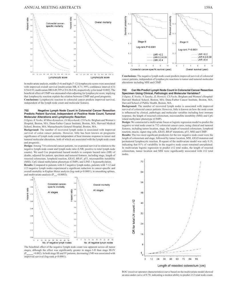
ANNUAL MEETING ABSTRACTS 159A
In multivariate analysis, middle (3-6) and high (7-12) lymphocytic scores were associated with improved overall survival [multivariate HR, 0.71; 95% confidence interval (CI) 0.54-0.93; multivariate HR 0.49; 95% CI 0.28-0.86, respectively; p for trend=0.002]. The beneficial effect of CIMP was attenuated when adjusting for lymphocytic score, implying that lymphocytic reaction explain the relation between CIMP and good prognosis.Conclusions: Lymphocytic reaction to colorectal cancer predicts improved survival, independent of the lymph node count and molecular features.
702 Negative Lymph Node Count in Colorectal Cancer Resection Predicts Patient Survival, Independent of Positive Node Count, Tumoral Molecular Alterations and Lymphocytic ReactionS Ogino, K Nosho, M Mino-Kenudson, JA Meyerhardt, CS Fuchs. Brigham and Women’s Hospital, Boston, MA; Dana-Farber Cancer Institute, Boston, MA; Harvard Medical School, Boston, MA; Massachusetts General Hospital, Boston, MA.Background: The number of recovered lymph nodes is associated with improved survival of colon cancer patients. However, little has been known on prognostic significance of lymph node count independent of host immune response to tumor and tumoral molecular alterations, both of which are associated with the lymph node count and prognosis.Design: Among 716 colorectal cancer patients, we examined survival in relation to the negative lymph node count and lymph node ratio (LNR; positive to total lymph node counts). We used Cox proportional hazard models to compute hazard ratio (HR) of deaths, adjusted for patient, specimen and tumoral features, including stage, length of resected colorectum, lymphoid reaction, KRAS, BRAF, p53, microsatellite instability (MSI), CpG island methylation phenotype (CIMP), and LINE-1 hypomethylation.Results: Compared to patients with 0-3 negative lymph nodes, patients with 7-12 and ≥13 negative lymph nodes experienced a significant reduction in cancer-specific and overall mortality in Kaplan-Meier analysis (log-rank p<0.0001), in smoothing splines, and multivariate analysis (Ptrend<0.0003).
The benefitial effect of the negative lymph node count was apparent across all tumor stages, although the effect was significantly greater in stages I-II than stage III-IV (Pinteraction=0.002). In both stage III and IV patients, decreasing LNR was associated with improved survival (log-rank p<0.0001).
Conclusions: The negative lymph node count predicts improved survival of colorectal cancer patients, independent of lymphocytic reactions to tumor and tumoral molecular alterations including MSI and CIMP.
703 Can We Predict Lymph Node Count in Colorectal Cancer Resection Specimen Using Clinical, Pathologic and Molecular Variables?S Ogino, K Nosho, N Tanaka, JL Hornick, CS Fuchs. Brigham and Women’s Hospital/Harvard Medical School, Boston, MA; Dana-Farber Cancer Institute, Boston, MA; Harvard School of Public Health, Boston, MA.Background: The number of recovered lymph nodes is associated with improved survival of colorectal cancer patients. However, little is known on how the node count is influenced by clinical, pathologic and molecular variables including host immune response, the length of resected colorectum, microsatellite instability (MSI) and CpG island methylator phenotype (CIMP).Design: We constructed a multivariate linear or logistic regression model to predict the negative or total node count in 732 colorectal cancer cases, using clinical and tumoral features, including tumor location, stage, the length of resected colorectum, lymphoid reaction, mucin, signet ring cells, KRAS, BRAF mutations, p53, MSI and CIMP.Results: The two most significant predictors for the raw negative node count were the length of colorectum and stage, followed by tumor location, MSI, KRAS mutation and peritumoral lymphocytic reaction. R-square of the multivariate model was only 0.19, indicating that 81% of variability in the negative node count remained unexplained. In multivariate logistic regression to predict ≥12 total nodes, the length of resected colorectum, tumor location and MSI were significantly associated with ≥12 total nodes.
ROC (receiver operator characteristics) curve based on the multivariate model showed an area under curve of 0.70, indicating a modest ability to predict ≥12 total node count.

160A ANNUAL MEETING ABSTRACTS
Conclusions: The length of colorectum, marked peritumoral lymphoid reaction, stage II and MSI-high are associated with a high negative node count. The negative or total lymph node counts vary greatly even after accounting for clinical, pathologic and molecular variables in a multivariate linear or logistic regression model.
704 Intraampullary Papi l lo-Tubular Neoplasms ( IAPN): A Clinicopathologic Analysis of 85 Cases of Mass-Forming Preinvasive Neoplasm Occuring within the AmpullaN Ohike, GE Kim, I Coban, T Tajiri, A Krasinskas, S Bandyopadhyay, O Basturk, T Morohoshi, Y Shimada, D Kooby, C Staley, NV Adsay. Emory U, GA; USCF, CA; Showa U, Tokyo, Japan; U of Pittsburg, PA; WSU, MI; NYU, NY; Georgia Tech, GA.Background: There has been no systematic analysis or uniform terminology for mass-forming preinvasive neoplasms occurring within the ampulla, which we refer to as IAPN.Design: 85 resected IAPNs were analyzed. Cell lineage was designated intestinal (IN) or gastric/pancreatobiliary (GPB) for preinvasive component, and intestinal (InvIN) or pancreatobiliary (InvPB) for associated invasive carcinoma (inv ca).Results: M/F=58/26; mean age 64; mean size 2.7cm. I. 27% had papillary growth, 57% papillary & tubular and 20% tubular. II. 8% had no high-grade dysplasia, 38% had focal, 27% substantial and 26% extensive. III. 75% were IN; 25% GPB. Sensitivity & specificity of cell lineage markers: IN: CDX2(74%, 89%), MUC2(31, 100) and CK20(82, 72); GPB: MUC1(67%, 92%), MUC5AC(56, 95), MUC6(39, 97), and CK7(94, 46). 40% were CK7+/CK20+; 64% were P53+ (IHC). IV. 75% had inv ca: 53% were InvIN, 41% InvPB and 4% others. Invasion was <1 cm in 24 cases and >2cm in 16. Preinvasive cell lineage persisted into invasive component: All InvIN arose from IN and 62% of InvPB from GPB. V: Overall, IAPNs had better survival than other ampullary carcinomas with 3 and 5-yr surv rates as 71% and 44% vs 44 and 28, respectively. Even those with invasion had a significantly better survival (3-yr 69 vs 44%, p<0.01). Although IN and InvIN showed a trend for better prognosis than GPB and InvPB; the difference did not reach statistical significance (5-yr; p=0.25, p=0.07, respectively). Tumor budding (58%) had a significantly worse prognosis (p<0.05).Conclusions: Intraampullary neoplasms characterized by papillary and tubular preinvasive growth, which we term IAPNs, are analogous to pancreatobiliary IPMNs or intraductal tubulopapillary neoplasms, by showing variable cell lineages & spectrum of dysplastic changes (adenoma-ca sequence). Morphologic classification of IAPNs into IN vs GPB is also supported by IHC panel, with CDX2/MUC2 as markers for IN and MUC1/MUC2/MUC5AC for GPB. CK7/CK20 profile is non-discriminatory. IAPNs are indolent neoplasms; even those with invasion have a better prognosis than pancreatic or other ampullary carcinoma. Tumor budding is associated with aggressive behavior.
705 Does Neoadjuvant Therapy Alter KRAS and/or MSI Status in Rectal Adenocarcinoma?SO Ondrejka, DF Schaeffer, MA Jakubowski, DA Owen, MP Bronner. Cleveland Clinic, Cleveland, OH; The University of British Columbia, Vancouver, BC, Canada.Background: To our knowledge, the genotoxic effect of neoadjuvant chemoradiation therapy on tumor molecular diagnostic testing is unknown. This study examines rectal adenocarcinomas both pre and post neoadjuvant treatment to assess whether it alters MSI and KRAS results. These two tests significantly impact rectal cancer management, based on the hereditary and prognostic aspects of MSI status and because both KRAS and MSI predict response to therapy. If genotoxic neoadjuvant treatment were to modify the results of these molecular tests, then the resultant testing inaccuracy could mislead clinical decision-making. This study examines this unexplored issue.Design: A total of 18 rectal adenocarcinoma patients with available pre and post treatment material were selected for this multi institutional study. DNA was extracted from microdissected rectal adenocarcinomas and paired normal tissues from both pretreatment biopsies and post-treatment resection specimens. MSI testing utilized the revised NCI panel of 5 mononucleotide repeat markers, comparing cancers to matched normal control tissues and defining microsatellite stable (MSS) as 5 stable markers and microsatellite instability high (MSI-H) as >2 unstable markers. KRAS codon 12 and 13 point mutations were examined by PCR and bidirectional sequencing.Results: MSI and KRAS results were unchanged comparing rectal cancers before and after chemoradiotherapy in all 18 patients (p= 1.000; 95% CI: 0.3969 to 2.520). All 18 tumors (100%) were MSS. KRAS testing identified 12 (67%) wild-type tumors
and 6 (33%) mutated tumors with identical KRAS mutations pre and post treatment. These MSI and KRAS mutational prevalences parallel those in the reported rectal cancer literature.Conclusions: Neoadjuvant therapy does not appear to cause significant artifactual genetic alterations in treated rectal adenocarcinoma, indicating that either untreated rectal cancer biopsies or post-treatment resection specimens are appropriate for testing.
706 Expression of Aldehyde Dehydrogenase Is Associated with High-Grade Histology and Poor Survival in Patients with Primary Small Bowel AdenocarcinomasML Othman, A Rashid, JL Abbruzzese, RA Wolff, H Wang. M.D. Anderson Cancer Center, Houston, TX; M.D.Anderson Cancer Center, Houston, TX.Background: Aldehyde dehydrogenase (ALDH) has been identified as highly expressed in embryonal tissue as well as in adult stem cells isolated from bone marrow, brain, breast and other tissues. Recent studies have shown that ALDH is also overexpressed in cancer stem cells and ALDH activity has been used as a marker for cancer stem cells. However the expression of ALDH in primary small bowel adenocarcinoma has not been examined.Design: To determine the significance of ALDH as a stem cell marker in primary small bowel adenocarcinoma, a tissue microarray (TMA) consisted of 50 cases of small bowel adenocarcinoma were utilized with appropriate controls. ALDH expression were analyzed by immunohistochemistry and the staining results were graded as positive or negative using strong positive staining in more than 5% of the tumor cells as a cut off. Clinical information and follow-up were obtained from reviewing patients’ medical records. The ALDH expression was correlated with tumor site, grade, and overall survival. Comparisons were performed by chi-square and overall survival was analyzed by log-rank method.Results: Twenty-one of 50 tumors had cytoplasm staining for ALDH. The expression of ALDH correlated significantly with high-grade histology and poor survival. 9 of 11 (82%) poorly differentiated adenocarcinomas was positive for ALDH compared to 12/39 (31%) positive cases seen in well to moderately differentiated carcinoma group (p=0.004). The mean survival of patients whose tumors were positive for ALDH was 50.5 ± 12.9 months compared to 104.3 ± 14.4 months for patients whose tumors were negative for ALDH (p= 0.03).Conclusions: Our study showed that ALDH is expressed in 42% of primary small bowel adenocarcinomas. ALDH expression in small bowel Adenocarcinomas correlates with high-grade histology and poor survival. ALDH may be a prognostic marker for primary small bowel adenocarcinomas.
707 Increased Intraepithelial Eosinophils in Patients Undergoing Photodynamic TherapyVL Owens, TC Smyrk, KK Wang. Mayo Clinic, Rochester, MN.Background: Markedly increased esophageal intraepithelial eosinophils (IEE) are usually associated with the clinicopathologic syndrome of eosinophilic esophagitis (EE), a syndrome dominated by dysphagia. We noted markedly increased IEE in some patients undergoing photodynamic therapy (PDT) for esophageal dysplasia/neoplasia and were surprised to learn that the patients did not have symptoms or endoscopic features of EE. We proceeded to review the esophageal findings in our entire cohort of patients undergoing PDT.Design: 385 patients underwent PDT between the years 1991-2009. The pathology reports from all endoscopic biopsies were reviewed for increased IEE. 187 patients had at least some esophageal eosinophils, and 17 patients had biopsies read as having “numerous/many” eosinophils or more than 20 IEE/high power field. We reviewed all endoscopic biopsies from these 17 patients and assessed the maximum density of IEE/HPF, presence or absence of degranulation, basal zone thickness, papillary height, spongiosis, and density of eosinophils in the stomach. The patient’s date of PDT was correlated with symptoms and endoscopic findings at the time of biopsy.Results: In 14/17 patients, there was a temporal relationship between increased IEE and PDT. The number of days after PDT to the first significant increase in IEE (≥20 eosinophils/HPF) ranged from 83-692 days. The highest peak density of IEE ranged from 30-200/HPF (mean=96). In 6/14 biopsies, the highest peak density was in the first post-PDT biopsy, and in many patients increased IEE persisted for years. The majority of cases showed eosinophilic degranulation, spongiosis, increased papillary height, and basal zone thickening. In most cases, eosinophil density was greatest in the basal 2/3’s of the squamous epithelium. All patients had at least some gastric eosinophils, with densities ranging from 1-75/HPF (mean=26). No patients had endoscopic findings of EE. One patient had dysphagia, but had a post-therapy related stricture; another patient complained of rare episodes of dysphagia.Conclusions: 14 of 385 patients undergoing PDT developed increased IEE, but they did not have endoscopic findings or symptoms of EE. There may be subtle histologic clues to the diagnosis of PDT-related IEE (basally distributed eosinophils; gastric involvement by eosinophils). The fact that eosinophils can be quite numerous and are degranulated in PDT patients without dysphagia suggests that other effectors play a role in the development of dysphagia in EE.
708 Gastric Foveolar Hyperplasia, Foveolar Dysplasia and Pit Dysplasia in 414 Gastric Cancers-Prevalence and Clinicopathological SignificanceDY Park, HJ Kang, DH Shin, KU Choi, CH Lee, GY Huh, MY Sol, GY Lauwers. Pusan National University Hospital, Busan, Republic of Korea; Massachusetts General Hospital and Harvard Medical School, Boston, MA.Background: Despite wide acceptance of the gastritis-intestinal metaplasia-dysplasia-carcinoma sequence, especially in intestinal type gastric adenocarcinoma, the precise detailed nature of precursor lesions of gastric adenocarcinoma remains to be delineated.

ANNUAL MEETING ABSTRACTS 161AGastric foveolar hyperplasia, fovoelar dysplasia and pit dysplasia are not well-defined and their prevalence and clinicopathological significances are unclear.Design: On this background, we have evaluated gastric foveolar hyperplasia (thickening of mucosa as much as 1.5 times compared to adjacent gastric foveolar mucosa), foveolar dysplasia and pit dysplasia (dysplasia in pit with surface foveolar maturation) in 414 gastric cancers and investigated relationship between various clinicopathological features of gastric cancers and features of adjacent mucosa (extent of inflammation, atrophy, intestinal metaplasia).Results: Of the 414 gastric cancers, 221 cases (53.4%), 105 cases (25.4%), 87 cases (21.0%) showed gastric foveolar hyperplasia, foveolar dysplasia and pit dysplasia in adjacent gastric mucosa, respectively. Gastric adenocarcinoma with foveolar hyperplasia were significantly associated with older age (p=0.000), male gender (p=0.000), body/fundic location (p=0.000), intestinal histologic type (p=0.000), advanced gastric cancer (p=0.008), lymphovascular tumor emboli (p=0.004), tendency of lymph node metastasis (p=0.062) and microsatellite instability (MSI-H) (p=0.042). Gastric adenocarcinoma with foveolar dysplasia and pit dysplasia were highly associated with older age (p=0.001, 0.002), male gender (p=0.028, 0.050), body/fundic location (p=0.000, 0.000), intestinal histologic type (p=0.000, 0.000). Interestingly, gastric mucin-containing intestinal metaplasia (incomplete IM) in adjacent mucosa was highly associated with gastric adenocarcinoma with gastric foveolar hyperplasia, pit dysplasia (p=0.000,0.000). Also muc6 expression of gastric adenocarcinoma has associated with association of pit dysplasia in adjacent mucosa phenotypes (p=0.036).Conclusions: Taken together, we suggest that gastric fovolear hyperplasia, foveolar dysplasia and pit dysplasia are important candidate precursor of gastric adenocarcinoma, especially in intestinal type gastric adenocarcinoma and more detailed prospective studies are needed to determine precise clinical impact in clinical GI pathology routine practice.
709 Lesions Arising in the Setting of Autoimmune Metaplastic Atrophic Gastritis (AMAG) in an Urban Tertiary Care SettingJY Park, T Cornish, D Lam-Himlin, C Shi, E Montgomery. Johns Hopkins Hospital, Baltimore, MD.Background: Autoimmune Metaplastic Atrophic Gastritis (AMAG) is an early manifestation of pernicious anemia, preceding the hematological changes of pernicious anemia by years to decades. Autoimmune atrophic gastritis manifests in 1 to 5% of the general population. It has been associated with the development of intestinal metaplasia and neoplastic lesions. Pyloric gland adenoma (PGA) is a newly recognized lesion that is highly associated with AMAG. We investigated the presence of PGA and other lesions arising in the setting of AMAG in our urban tertiary care center.Design: We searched all non-consultation gastric biopsies from 1988 to 2008 using our laboratory information system and further analyzed the histopathologic features of all cases coded as AMAG. In addition, we further selected cases that had been confirmed as AMAG by immunohistochemical staining (IHC) for the body of the stomach (negative gastrin) and the presence of linear and nodular ECL-cell hyperplasia (chromogranin). All polyps and/or neoplastic lesions from this subset were reviewed.Results: The gastric biopsies (n=54,814) were from 45.5% male (M) and 54.5% female (F) patients comprising 66.4% white, 23.6% African American (AA) and 10% other (non-white Hispanic, Asian, etc.) patients. Gastritis as a general category accounted for 69.3% of gastric biopsies; AMAG was diagnosed in 619 cases of which 527 cases from 380 patients had confirmatory IHC. Based on these cases, AMAG occurred in 0.7% of biopsies with a 2:1 F:M ratio. It was diagnosed in 0.7% of gastric biopsies from white patients, and in 0.8% of gastric biopsies from AA patients. For white males and females the average age was 68.6 and 63.9, respectively; for AA males and females the average age was 64.6 and 62.6, respectively. A review of polyps and/or other neoplastic lesions showed 147 polyps (107 hyperplastic, 15 inflammatory, 11 gastric adenomas, intestinal type, 11 oxyntic gland pseudopolyps, and 3 PGAs), 9 adenocarcinomas, 3 lymphomas, and 23 carcinoids. One of the PGAs had been previously diagnosed as a gastric hyperplastic polyp with changes indefinite for dysplasia.Conclusions: Consistent with recent epidemiologic surveys of pernicious anemia, the histopathologic diagnosis of AMAG occurs with similar frequency in AA and white patients. The review of prior cases has revealed a single case of PGA that was not previously recognized. Although PGA is strongly associated with AMAG, the current investigation reveals that even in the setting of AMAG, PGA is a rare entity.
710 CpG Island Hypermethylation in Gastric Carcinoma and Its Clinicopathological ImplicationSY Park, MC Kook, YW Kim, NY Cho, GH Kang. National Cancer Center, Goyang-si, Korea; Seoul National University College of Medicine, Seoul, Korea.Background: Gastric carcinoma is one of the human cancers in which promoter CpG island hypermethylation is frequently found. EBV-positive gastric cancer and microsatellite instability-positive gastric carcinoma are well known to harbor frequent promoter CpG island hypermethylation. However, in gastric carcinomas negative for microsatellite instability and EBV, clinicopathological features associated with CpG island hypermethylation remain unclear.Design: We analyzed 196 cases of gastric carcinoma for their methylation status in 16 cancer-specific methylation markers using MethyLight assay and correlated their methylation with clinicopathological features.Results: The number of genes methylated was 14.4, 10.3, and 6.2 for EBV-positive gastric cancer (n=16), microsatellite instability-positive gastric cancer (n=30), and EBV-negative/ microsatellite instability-negative gastric cancer (n=150), respectively (P<0.001). With exclusion of EBV-positive or microsatellite instability-positive gastric cancers from the analysis, higher number of methylated genes was found in gastric cancers of female patients than in those of male patients (7.4 vs. 5.6, P=0.004), in infiltrative gross types (Borrman III or IV) than in fungating types (I or II) (6.4 vs. 4.8, P=0.051), in high grade differentiation than in low grade differentiation (6.7 vs. 4.4,
P=0.001), in high stage (III or IV) than in low stage (I or II) (6.7 vs. 5.3, P=0.016). Mixed-type, diffuse-type, and intestinal-type gastric cancers were in a decreasing order of the number of methylated genes (9.2, 6.9, and 4.8; P<0.001). Hypermethylation at 14 markers or more was closely associated with poor clinical outcome and found to be an independent prognostic factor.Conclusions: The findings indicate a close relationship of frequent CpG island hypermethylation with specific clinicopathological features and suggest that aberrant CpG island hypermethylation might be involved in the genesis of mixed-type gastric cancer.
711 Can Pathologists Distinguish Barrett’s Gastric-Type Dysplasia from Reactive Gastric Cardiac Mucosa in GERD?DT Patil, AE Bennett, D Mahajan, D Allende, C Fraser, MP Bronner. Cleveland Clinic Foundation, Cleveland.Background: Morphologic identification of dysplasia remains the gold standard marker of cancer risk in Barrett’s esophagus. Despite this, reproducible histologic diagnosis is difficult due in part to obscuring inflammation. Gastric-type Barrett’s dysplasia is a recently recognized dysplasia subtype that is characterized by non-stratified, basally oriented but enlarged nuclei with only mild pleomorphism. The distinction between Barrett’s gastric-type dysplasia and reactive gastric cardiac mucosa in gastroesophageal reflux disease (GERD) has not been systematically studied and forms the basis of this report.Design: A total of 3,698 endoscopic biopsies from a cohort of 461 Barrett’s patients were reviewed to identify 43 patients (80 biopsies) with gastric-type Barrett’s dysplasia (13 LGD, 30 HGD) using previously defined criteria (Mahajan D, et al. Mod Pathol, 2009). These were compared histologically to biopsies from 60 GERD patients with markedly inflamed and reactive epithelial changes of gastric cardiac mucosa.Results: Surface nuclear stratification was exclusively found in reactive epithelium (80% vs 0%, p<0.00001). Nuclear atypia was restricted to the upper/surface epithelium (top-heavy atypia) in reactive cardia, compared to full thickness atypia in gastric-type Barrett’s dysplasia (0% vs 80%, p<0.00001). Crowded glandular architecture was significantly associated with gastric-type dysplasia (78% vs 0%, p<0.00001), while villiform architecture was more common in reactive cardia (53% vs 6%, p=0.0006). Cytologically, gastric-type dysplasia showed a trend toward mild nuclear pleomorphism (35% vs 10%, p=0.09), while prominent nucleoli were more commonly noted in gastric-type dysplasia (33% vs 79%) (p=0.0003). Considerable overlap of nuclear size existed, rendering this final feature unhelpful in the differential diagnosis.Conclusions: Nuclear stratification and surface predominant or “top-heavy” atypia accompanied by non-crowded, villiform architecture are features that most reliably distinguish marked reactive cardiac atypia in GERD from gastric-type Barrett’s dysplasia.
712 Prediction of Adenocarcinoma (ADC) on Esophagectomy from Pre-Resection Biopsies of Barrett’s Esophagus (BE) with at Least High Grade Dysplasia (HGD): A Comparison of Two SystemsDT Patil, JR Goldblum, T Plesec, A Bennett, W Henricks, L Schoenfield, L Yerian, MP Bronner, L Rybicki, T Rice, E Downs-Kelly. Cleveland Clinic, Cleveland.Background: Distinguishing BE-HGD from intramucosal carcinoma (IMC) and submucosal carcinoma (SMC) on biopsies is important due to different therapeutic options, some of which are reserved for “cancer”. In regards to pre-resection biopsies, Downs-Kelly et al [Am J Gastroenterol 2008 (CC)]outlined 4 diagnostic categories: HGD, HGD with marked glandular architectural distortion, cannot exclude IMC (HGD/MAD), IMC and SMC while Zhu et al [Am J Clin Path 2009 (UM)] described criteria for HGD “suspicious” for ADC (HGD/S). This study evaluates pre-resection biopsies from patients who underwent esophagectomy for BE with at least HGD to:1) compare CC and UM systems, 2) correlate pre and final resection diagnosis, and 3) identify histologic features in biopsies that might be predictive of ADC on esophagectomy.Design: Using the CC system, 7 GI pathologists reviewed 168 pre-resection biopsies with at least HGD; those biopsies with a consensus diagnosis (agreement by ≥4 pathologists) were included. Logistic regression analysis was used to identify endoscopic and histologic features that predicted ADC on esophagectomy.Results: Of the112 pre-resection biopsies with a consensus diagnosis, 83 (74%) had ADC on esophagectomy (59 IMC, 24 SMC). Pre-resection diagnoses were: 32 HGD (29%), 32 HGD/MAD (29%), 45 IMC (40%) and 3 SMC (2%). Applying UM system to the biopsy series showed excellent overall agreement with CC system (k=0.86). Both systems showed significant correlation between preop and esophagectomy diagnosis (p<0.001). The likelihood of finding ADC on resection was significantly higher with HGD/MAD (OR 2.9, p=0.04) or HGD/S (OR 5, p=0.002), compared to HGD. Presence of an endoscopic lesion (OR 4.7, p=0.002), “never-ending” gland pattern (OR 3.7, p=0.016), sheet-like growth (chi2 p<0.001), angulated glands (OR 8.5, p=0.001), ≥3 dilated glands with intraluminal debris (OR 2.6, p=0.045), and >1 focus of single cell infiltration into lamina propria (OR 8.9, p<0.001) increased the odds of finding ADC on resection. The latter two variables remained independent predictors of ADC in multivariable analysis.Conclusions: The CC and UM systems had excellent agreement and both were predictive of resection diagnosis. The finding of ≥3 dilated glands with intraluminal debris and >1 focus of single cell infiltration into the lamina propria were independent histologic predictors of ADC on resection.

162A ANNUAL MEETING ABSTRACTS 713 Prevalence of Human Papillomavirus in Esophageal Squamous Papilloma: A Polymerase Chain Reaction StudyDC Phan, RC Chan, WS Nichols, P Manna, S Kerley, OTM Chan, E Himmelfarb, D Dhall, HL Wang. Cedars-Sinai Medical Center, Los Angeles, CA; Physicians Reference Laboratory, Overland Park, KS.Background: Human papillomavirus (HPV) has been implicated in the etiopathogenesis of a variety of benign and malignant squamous lesions, including squamous papilloma and squamous cell carcinoma of the upper aerodigestive tract. However, the role of HPV in the development of esophageal squamous papilloma is poorly understood. Studies over the past two decades have generated highly controversial results, which may be due to differences in the techniques used to detect viral antigens or DNA and/or geographic variations. We aimed to reinvestigate this question using advanced polymerase chain reaction (PCR) technique for the detection of both low and high risk HPV DNA.Design: Forty-five cases of esophageal squamous papilloma were included in this study. DNA was extracted from formalin-fixed, paraffin-embedded esophageal biopsies. The detection of HPV DNA was performed using MY11/MY09 and GP5+/GP6+ primers in a nested PCR assay, which is capable of amplifying a 455-bp fragment within the conserved L1 region common to different HPV types. Positive cases were further characterized employing type-specific PCR. The quality of sample DNA was evaluated by amplification of a 289-bp fragment of the housekeeping gene b-actin.Results: The patients included 22 males and 23 females with a mean age of 50 years (range: 17-84 years). HPV DNA was detected in only 2 (4%) cases. Further typing of these 2 cases demonstrated HPV subtype 59 in 1 case but was indeterminate in the other. Interestingly, both patients with positive HPV detection were immunocompromised and the one with subtype 59 also had a history of anal squamous cell carcinoma in situ (HPV testing was not performed on this lesion). None of the 45 patients developed squamous cell carcinoma in the esophagus.Conclusions: Unlike other squamous lesions in the upper aerodigestive tract, squamous papilloma of the esophagus is infrequently associated with HPV infection and carries an insignificant risk to evolve into squamous cell carcinoma.
714 P16 and Ki-67 Immunostaining as Markers of Anal Intraepithelial Neoplasia and Condyloma – Correlation with Human Papillomavirus Detection by PCREC Pirog, KD Quint, RK Yantiss. Weill Medical College of Cornell University, New York, NY; DDL Diagnostic Laboratory, Voorburg, Netherlands.Background: The classification of anal intraepithelial neoplasia (AIN) in mucosal biopsies is subject to considerable interobserver variability. Ki-67 and p16 immunostains have been shown to aid the diagnosis of squamous neoplasia in biopsy samples from the uterine cervix. Thus, the aim of this study was to evaluate the utility of Ki-67 and p16 immunostaining as adjunct tests to increase diagnostic accuracy.Design: 75 consecutive anal biopsies originally diagnosed as benign anal transitional zone (NEG, n=15), fibroepithelial polyp (FEP, n=10), condyloma accuminatum (CONA, n=10), low-grade AIN (AIN1, n=20), and high-grade AIN (10 cases AIN2 and 10 cases AIN3) were collected. All cases were re-reviewed to obtain a consensus diagnosis and all cases were tested for HPV DNA by SPF10 PCR and LIPA25 genotyping assay. The results of HPV testing were correlated with consensus diagnosis to reach a final classification. All cases were immunostained for Ki-67 and p16. Ki-67 positivity was defined as a cluster of at least two positive nuclei in the upper two-thirds of the epithelial thickness. A postitive result for p16 was defined as moderate to strong intensity nuclear and cytoplasmic staining.Results: Histologic review and HPV testing led to reclassification of 16 cases, so that the final study group included 17 NEG, 14 FEP, 6 CONA, 11 AIN1, 16 AIN2, and 11 AIN3. All cases of CONA and AIN were positive for HPV, whereas all FEPs were negative for HPV by PCR. All NEG cases and FEPs were negative for both Ki-67 and p16. All CONA and AIN1 cases were positive for Ki-67, but negative for p16. All cases of either AIN2 or AIN3 were positive for Ki-67 and p16. Overall, the sensitivity and specificity of Ki-67 for detection of anal condyloma or AIN was 1.0 and 1.0, respectively. The sensitivity and specificity of p16 for detection of AIN2 or AIN3 was 1.0 and 1.0, respectively.Conclusions: Ki-67 is a highly sensitive and specific marker for detection of HPV-related changes and neoplasia in the anal mucosa, whereas p16 staining is strongly associated with high-grade squamous neoplasia. These results indicate that a combination of these markers may aid interpretation of anal mucosal biopsy samples.
715 A Combination of NR-21, BAT25, and MONO27 Run in One Polymerase Chain Reaction (PCR) Accurately Detects Microsatellite Instability High (MSI-H) TumorsBP Portier, M Bronner, X Liu. Cleveland Clinic, Cleveland, OH.Background: Microsatellite instability (MSI) testing is increasingly performed on tumor tissue to screen for hereditary non-polyposis colorectal cancer. However, it remains expensive, laborious, and difficult to standardize. Several quasi-monomorphic markers were included in a MSI panel recently developed by Promega (Madison, WI). This study was undertaken to determine if the combination of three markers (NR-21, BAT25, Mono27), run in one PCR reaction, could correctly characterize MSI status in all tumors tested.Design: The MSI database from 11/2006 to 03/2009 was reviewed. Selection criteria for MSI testing included patient age <50, right-sided colonic location, small bowel or gastric neoplasm, MSI-associated tumor morphology, and family history. The 5 marker panel of quasi-monomorphic mononucleotide repeats (BAT26, NR-21, BAT25, Mono27, and NR24) was compared to the 3-marker panel (NR-21, BAT25, Mono27) for association with MSI-H (defined as the presence of ≥ 2 unstable markers out of the 5 tested markers) or MSS (no unstable markers out of the 5 tested markers). In addition, mismatch repair protein immunohistochemistry for MLH-1, MSH-2, MSH-6, or PMS-2 was performed on 99 tumors (88 MSI-H and 11 MSS).
Results: A total of 487 tumors were tested during the study period, including 305 colorectal carcinomas, 157 colorectal adenomas, and 25 non-colorectal tumors [small bowel (13), appendix (3), stomach (2), uterus (2) and others (5)]. Out of 487 tumors tested, 10 (2.05%) had insufficient DNA. The overall MSI-H rate was 22.5% (27.6% for colorectal carcinomas, 13.3% for colorectal adenomas, and 12.4% for non-colorectal tumors). Each MSI marker, BAT26, NR-21, BAT25, Mono27, and NR-24 alone correctly identified 98%, 98%, 95%, 98%, and 96% of MSI-H tumors (N=105) and all MSS tumors (N=372). A combination of NR-21, BAT25, and Mono27 run in one PCR reaction correctly identified all 477 MSI-H and MSS tumors. Among 99 tumors (11MSS and 88 MSI-H) tested for mismatch repair proteins by immunohistochemistry, the MSI status and IHC results showed 99% concordance (1 MSI-H case failed to show loss of mismatch repair proteins by IHC).Conclusions: A combination of NR-21, BAT25, and Mono27 (three quasi-monomorphic mononucleotide repeat markers) run in a single multiplexed PCR reaction was sufficient to determine MSI status with 100% accuracy (n=477). Whether this more limited MSI panel will reduce cost, turnaround time, or facilitate inclusion of additional molecular tests remains to be investigated.
716 Revised Bethesda Guidelines Directing MSI Testing of Colorectal Cancers in Routine Surgical Pathology Practice: A Single Institution ExperienceJC Post, M Bronner, B Leach, E Downs-Kelly, X Liu. Cleveland Clinic, Cleveland, OH.Background: Lynch syndrome (LS) is the most common hereditary form of colorectal cancer (CRC) with a known genetic basis. The revised 2004 Bethesda guidelines were developed to identify potential LS patients by guiding reflex testing of CRCs for microsatellite instability (MSI). This study was undertaken to retrospectively evaluate guideline use in routine surgical pathology practice, hypothesizing that a significant number of potential LS patients who are ≥60 years would be missed by guideline recommendations, when no additional clinical data are available to the pathologists.Design: All CRC cases surgically resected from 11/06 to 3/09 were retrieved from the pathology database and analyzed for MSI testing rates in the following patient groups: group A: ≥60 yrs (n=286); group B: <50 yrs (n=62); and group C: 50-59 yrs (n=97). MSI testing was performed using a panel of 5 quasi-monomorphic mononucleotide repeat markers comparing cancers to matched normal control tissue with MSI-H defined as 2 markers showing instability. Reflex MSI testing was performed for MSI-H phenotype CRC morphology (groups A & C), or age <50 (group B).Results: CRCs were diagnosed in 447 patients during the study period. The cumulative reflex MSI testing rate was 35.1% (157/447), and 24.2% of those patients tested had MSI-H CRCs (38/157). In groups A, B & C respectively, the rate of reflex MSI testing was 32.2% (92/286), 41.9% (26/62), and 38.1% (37/97). MSI-H was identified in groups A, B, and C at a rate of 28.2% (26/92), 19.2% (5/26). and 18.9% (7/37), respectively. The number of MSI-H CRCs with non-hMLH-1 loss by immunohistochemistry, and therefore strongly implicating LS, for groups A-C were 5 (19.2)%, 1 (20%), and 3 (42.8%), respectively.Conclusions: A significant number of probable LS patients (5/9; 55.5%) were ≥60 years in the above testing paradigm based on MSI-H phenotype CRC morphology and age. Therefore, guideline #3 of the revised Bethesda guidelines, which recommends testing CRCs with the MSI-H histology diagnosed in a patient who is <60 years, proved inadequate for LS screening in our patient population. Adherence by pathologists to the revised Bethesda guidelines, which recommend testing all CRCs in patients younger than 50 and CRCs with the MSI-H histology in a patient between 50-59 years deserves further consideration, as our practice-based data show that 55.5% of probable LS patients were ≥ 60 years.
717 Nodal Scar but Not Proliferation Index Predicts Worse Survival in Treated Esophageal AdenocarcinomaJK Purdy, SK Bihlmeyer, HD Appelman, AC Chang, L Zhao, MB Orringer, SG Urba, BJ McKenna. University of Michigan, Ann Arbor, MI.Background: Previous studies of esophageal adenocarcinoma (EA) treated with chemoradiation (CRT) have found post-treatment pathologic stage, quantity of residual tumor (tumor regression grade, TRG), nodal metastases and pre-CRT proliferation index (PI) in biopsies to be predictive of survival. However, these studies combined patients with EA and squamous cell carcinoma without standard CRT. No study has examined whether scar depth in areas of regressed tumor or nodal scar without tumor (presumably evidence of treated tumor) or post-CRT PI predicts survival in EA patients.Design: 52 esophagectomies for EA without distant metastases, all receiving the same CRT, from March 2002 to April 2006 were evaluated. Cases were analyzed for depth of scar, nodal scar and amount of residual tumor using a 4-tiered TRG system and were classified according to TNM and pathologic stage. 44 cases with residual EA were stained with Ki67 to calculate post-treatment PI. Kaplan-Meier methods were used to estimate median survival. Cox regression models were performed to assess the impact of different factors on survival.Results: After controlling for TRG and stage, patients who had 1 additional lymph node with scar were 20% more likely to die (HR=1.205, 95% CI=1.049-1.384) and 27% more likely to have disease progression (HR=1.274, 95% CI= 1.096-1.480) than patients without nodal scar (p=0.0082 and p=0.0016 respectively). Post-CRT PI did not predict overall or disease-free survival. TRG showed a trend toward predicting survival but this did not reach statistical significance. 12 patients staged as T2 according to depth of residual tumor were upstaged to T3 using scar depth, but T stage based on scar depth had no better correlation with survival than that based on tumor depth.Conclusions: In post-CRT esophageal adenocarcinoma, nodal scar predicts worse survival and disease progression. Unlike pre-CRT EA, PI in post-CRT EA does not predict survival. While some previous studies have suggested that post-CRT TRG predicts survival, this was not statistically significant, possibly due to lack of a standard

ANNUAL MEETING ABSTRACTS 163ACRT protocol in those studies or to small sample size in our study. T stage based on scar depth was not a better predictor of survival than T stage based on tumor depth.
718 Detection of Chromosomal Anomalies in Colon Biopsy Specimens Using Fluorescence In Situ Hybridization (FISH) – A Pilot StudyJ Qian, D Hossain, G Sadasivan, DG Bostwick. Bostwick Laboratories, Glen Allen, VA.Background: Colorectal carcinoma is the third most common cancer diagnosed in the United Sates. Using current histological techniques, differentiating between adenoma and carcinoma can be challenging in certain cases, and identifying those adenomas with a high risk for progression to cancer is not possible. In this study, we sought to develop a FISH test to aid in the differential diagnosis of adenoma and adenocarcinoma.Design: We used FISH with centromere-specific probes for chromosomes 7, 15, 18, and 20 to evaluate chromosomal anomalies in colon biopsy specimens diagnosed as benign (31), adenoma (25) and adenocarcinoma (26). The FISH signals were enumerated in 50 cells per case, and the chromosomal anomalies were correlated with pathologic findings.Results: Numeric chromosomal anomalies were found in 0% (0/31) of benign, 64% of adenoma (16/25), and 92% (24/26) of carcinoma specimens (for each pair The mean percentage of cells with chromosomal changes was 55% in cancer specimens, significantly higher than that in adenoma (18%, p< 0.0001) and benign mucosa (2%, p< 0.0001). The most frequent chromosomal anomaly was gain of chromosome 7. FISH anomalies had an overall sensitivity of 81% and specificity of 88% for the distinction of adenoma from carcinoma.
[table1]Sensitivity Specificity ROC
Cancer vs. Benign 92% 100% 0.96Cancer vs. Adenoma 81% 88% 0.84Cancer vs. Non-Cancer 92% 71% 0.82Cancer/Adenoma vs. Benign 78% 100% 0.89Adenoma vs. Benign 64% 100% 0.82
Sensitivity Specificity ROC Cancer vs. Benign 92% 100% 0.96 Cancer vs. Adenoma 81% 88% 0.84 Cancer vs. Non-Cancer 92% 71% 0.82 Cancer/Adenoma vs. Benign 78% 100% 0.89 Adenoma vs. Benign 64% 100% 0.82Conclusions: Multi-target FISH appeared to be useful for the differential diagnosis of colonic adenoma from adenocarcinoma, with a high level of sensitivity and specificity. In addition, adenoma with high level of FISH-detected chromosomal anomalies may require close clinical follow-up.
719 DNA Methyltransferases 1, 3a and 3b Overexpression and Clinical Significance in Gastroenteropancreatic Neuroendocrine TumorsZR Qian, MM Rahman, EL Wang, M Nakasono, T Hayashi, R Haba, M Ishida, H Okabe, T Sano. Institute of Health Biosciences, University of Tokushima Graduate School, Tokushima, Japan; Tsurugi Municipal Handa-Hospital, Tokushima, Japan; Faculty of Medicine, Kagawa University, Kagawa, Japan; Shiga University of Medical Science, Shiga, Japan.Background: Alteration of DNA methylation is one of the most consistent epigenetic changes in human cancers. Three genes, namely DNA methyltransferase (DNMT) 1, 3a, and 3b, coding for DNMTs that affect promoter methylation status are thought to play an important role in the development of cancers and may be good anticancer therapy targets. Methylation of tumor suppressor genes has been reported in gastroenteropancreatic (GEP) neuroendocrine tumors (NETs); however, there are no studies about DNMTs protein expression and their clinical significance in GEP NETs.Design: In this work, using immunohistochemistry the expression of DNMT1, 3a and 3b were studied in 63 GEP NETs included well-differentiated NET (WNET, of either benign or uncertain behavior; n = 31), well-differentiated neuroendocrine carcinoma (WNEC; n = 16), and poorly-differentiated neuroendocrine carcinoma (PNEC; n = 16).Results: Expression of DNMT1, 3a and 3b was frequently detected in GEP NETs (87%, 81%, 75%, respectively). DNMT3a expression level was significantly higher in poorly-differentiated neuroendocrine carcinomas than well-differentiated NETs or well-differentiated neuroendocrine carcinoma s (P < 0.01, P < 0.05, respectively). Expression of DNMT1, 3a and 3b showed significantly higher levels in stage IV tumors than stage I and II tumors. In addition, expression of DNMT1, 3a and 3b was positively correlated with MIB-1 labeling index in GEP NETs (R = 0.293, P = 0.019; R = 0.457, P = 0.001; R = 0.249, P = 0.049; respectively). In addition, expression levels and frequencies of positive immunostaining of DNMT3a and 3b were significantly lower in midgut NETs than foregut and hindgut NETs.Conclusions: These results suggest that DNMTs may have important roles in tumorigenesis and development of GEP NETs and epigenetic therapy directed against DNMTs may be a considerable therapeutic approach.
720 Sensitivity of Morphologic Predictors of Colorectal Carcinomas (CRC) with High Microsatellite Instability (MSI-H) in Different Ethnic Groups and Their Molecular Characteristics – A Study of 42 CasesM Ramineni, J Punia, L Niemeier, L Whiteley, M Cankovic, DA Chitale. Herny Ford Hospital, Detroit, MI.Background: MSI-H is observed in 15% of sporadic CRC. Identification of CRC with MSI-H is important because of the prognostic and therapeutic significance associated with MSI as they are less likely to benefit from fluorouracil-based chemotherapy. Limited data exists in the literature on sensitivity of morphologic predictors of CRC with MSI-H in different ethnic groups and their molecular features and we sought to investigate this in 42 candidate cases.Design: Consecutive cases of CRC having morphologic predictors of MSI-H (>2 tumor-infiltrating lymphocyte cells per hpf, lack of dirty necrosis,Crohn-like reaction, mucinous
component and well or poor differentiation) were selected. DNA was extracted from blocks containing more than 80% tumor. MSI testing was done using commercial kit for PCR and immunohistochemistry for MMR protein expression (MLH1, MSH2, MSH6). Promoter methylation of hMLH1(hMLH1-M) was analyzed with a methylation-specific PCR. Sequencing for BRAFV600E and KRAS codon 12,13 was carried out using standard protocol. Relevant pathologic and clinical data was recorded.Results: 42 cases met above histologic criteria, 20 (47.6%) in right colon (RC), 11 (26.2%) left colon (LC), 9 (21.4%) rectal and 2 (4.8%) synchronous tumors in RC & LC. 2 cases had medullary features and 5 with mucinous component. 6/42 (14.3%) tumors showed MSI-H, 4 involving the RC and 3 positive for BRAF V600E mutation and hMLH1-M.14/42 (33.3%) were KRAS mutants CRC with all except 1(which was MSI-H with hMLH1-M) negative for other mutations and had proficient MMR. All the BRAF V600E were found in Caucasian (CA) patients mainly elderly women while KRAS mutations predominated African Americans (AA) (Fisher exact p value: 0.03; 0.003 respectively).
Positive results by ethnicityMSI-H+ BRAF+ KRAS+ hMLH1
AA (19) 2 0 11 1CA(19) 4 4 3 3Others(4) 0 0 0 0Total(42) 6 4 14 4
Conclusions: In our cohort, the overall sensitivity of morphologic predictors of MSI-H was lower (∼20%) even when only CRC in RC were included as one of the predictors of MSI-H, but sensitivity was slightly better in CA than AA. This most likely is due to different ethnic mix in our cohort. Most MSI-H tumors were sporadic CRC with frequent BRAF mutations, hMLH1-M and occurred in RC of elderly CA women. In contrast, in AA KRAS mutation was the most frequent genetic alteration with no other significant mutations.
721 Pancreatic Endocrine Tumors: A Possible Interaction of Utrophin and p53 in Malignant ProgressionTA Rege, V Nose. Brigham & Women’s Hospital, Boston, MA.Background: Pancreatic endocrine tumors (PET) are rare tumors with unpredictable clinical behavior. No histologic features or immunohistochemical markers have reliably predicted malignant progression. The 2004 World Health Organization (WHO) classification uses size, angioinvasion, mitotic activity and Ki-67 proliferative index as prognostic criteria. Recently, cytokeratin 19 and KIT have been reported as prognostic markers. Deletion of long arm of chromosome 6 is a common aberration in tumors. The cytoskeletal protein utrophin encoded on 6q is frequently lost in malignant PET. Prior studies raised the possibility of an important role of utrophin in the malignant progression of PET. P53 is a known oncogene implicated in a variety of tumors. The purpose of this study was to analyze the expression of utrophin and p53 in PET in a series of known malignant cases and cases with an unknown malignant behavior for its possible role in malignant progression.Design: Sixty cases of PET were identified from our pathology files. The mean age at presentation was 57.6 years (range 25 to 88); the M:F ratio was 1:1, and the mean size was 4.3 cm (0.6 to 14 cm). The clinical follow-up data were examined and tumors classified according with the 2004 WHO classification and then separated in two groups as known malignant cases and cases with an unknown malignant behavior. Histopathologic and immunohistochemical stains were evaluated and utrophin (MANCHO3 clone 8A4; Dr. G. Morris, University of Iowa), p53, Ki-67 expression were scored semiquantitatively as 1+ (<5%), 2+ (5-10%), 3+ (10-50%), and 4+ (>50%).Results: Utrophin expression was present in malignant tumors and more frequently associated with p53 expression than with cases where p53 was negative. Utrophin and p53 expression was also associated with local invasion, size of tumor, and mitotic rate. WHO criteria, node metastases, mitotic count, and Ki-67 proliferative index, infiltrative border, necrosis, perineural invasion, extrapancreatic expression and tumor size were associated with poor prognosis.Conclusions: We demonstrated that utrophin expression is associated with expression of p53 in known malignant PET. Our findings suggest a potential role for utrophin interaction with p53 overexpression in malignant progression and metastasis. Further studies are warranted to clarify the possible role in the malignant progression of PET, the regulation, and putative therapeutic applications of these proteins.
722 Prognostic Implications of Cytoplasmic and Nuclear Overexpression of Lipocalin-2/NGAL in Colorectal Adenocarcinoma (CRC)KA Robstad, JD Choate, CE Sheehan, JS Ross, DM Jones. Albany Medical College, Albany, NY.Background: The lipocalin family is a diverse group of secreted soluble proteins that bind hydrophobic ligands and act as small molecule transporters. Lipocalin-2, also known as neutrophil gelatinase-associated lipocalin (NGAL), is an inflammatory cytokine upregulated in acute inflammatory conditions which has been found to be over-expressed in various human malignancies including carcinomas of the breast, ovary, pancreas and colon. The prognostic significance of Lipocalin-2 expression in CRC has not been previously investigated.Design: Formalin-fixed, paraffin-embedded sections from 156 colorectal adenocarcinomas (CRCs) were immunostained by an automated method (Ventana Medical Systems; Tucson, AZ) using rat monoclonal lipocalin-2/NGAL (R&D Systems, Minneapolis, MN). Cytoplasmic and nuclear immunoreactivity was semi quantitatively evaluated based on both intensity (weak, moderate and intense) and distribution (focal <10%, regional 10 to 50% and diffuse >50%) and results were correlated with clinicopathologic variables.Results: Lipocalin-2 immunoreactivity was predominately cytoplasmic, however, significant nuclear immunoreactivity was noted in a subset of cases. Intense diffuse

164A ANNUAL MEETING ABSTRACTS cytoplasmic overexpression of lipocalin-2 was observed in 30/156 (19%) of CRC and correlated with early AJCC stage (28% of stage I/II vs 11% of stage III/IV; p=0.007) and presence of concomitant Crohn’s disease (100% with Crohn’s vs 0% without Crohn’s). Nuclear lipocalin-2 immunoreactivity was noted in 6 cases, all 6 of which (100%) were lymph node negative (p=0.005), early stage (p=0.015), and moderately differentiated/grade 2 (p=0.102) tumors. Lipocalin-2 over-expression did not correlate with disease recurrence or overall survival. On multivariate analysis, pathologic stage at diagnosis independently predicted patient survival.Conclusions: Cytoplasmic lipocalin-2 over-expression is associated with both early AJCC tumor stage as well as the presence of pre-existing Crohn’s disease potentially reflecting its role as an inflammatory cytokine. Nuclear expression, only identified in a small subset of CR, was found to correlate significantly with low-stage, moderately differentiated, lymph node-negative tumors. Further studies of both nuclear and cytoplasmic lipocalin-2 expression in CRC appear warranted.
723 PCHK2 Is Associated with Differentiation and Survival in Gastric AdenocarcinomaJB Rock, IS Hatzaras, M Bloomston, K Huebner, WL Frankel. The Ohio State University, Columbus, OH.Background: Checkpoint kinase 2 (Chk2) is an effector kinase central in the control of cell cycle checkpoints and associated with the p53 pathways. Phosphorylation at Thr68 (pChk2) in response to DNA damage activates Chk2 leading to autophosphorylation, transient dimerization and upregulation of kinase activity. Alterations in pChk2 have been described in several cancers and recent studies have shown the potential of its inhibitors in sensitizing tumor cells to DNA-damaging agents. Our purpose was to study the correlation between pChk2 tumor grade and survival.Design: Gastric Adenocarcinomas (168) were retrieved and age, gender, tumor grade and outcome were reviewed. Controls were obtained from uninvolved stomach in 33 cases. Tissue microarrays with 1 mm cores were stained with the pChk2 antibody. Staining was graded as strongly positive (2), weakly positive (1) and negative (0). Comparisons were made by two-tailed student’s t-test for continuous data and contingency table analysis (chi-square and Fisher’s exact test) where appropriate. Survival was analyzed using the Kaplan-Meier time to even method. A logistic regression model was used to identify predictors of poor differentiation. Cox proportional hazards regression was used to evaluate predictors of survival. Statistical significance was accepted at p<0.05.Results: Mean patient age was 65.8 years with a male to female ratio 1.2:1. Most controls were negative for pChk2 (88%) with weak positive staining noted in 4 cases. Tumors stained 2, 1 and 0 in 11 (7%), 77 (46%) and 80 (48%) cases respectively. By univariate analysis with an Odds Ratio of 9.07 (95% CI 1.13, 17.23) pChk2 was statistically associated with poor differentiation (p = 0.037); by multivariate analysis the Odds Ratio was 13.34 (95% CI 1.40, 16.75; p = 0.024). Age and gender were not associated with poor differentiation. Weak pChk2 expression conferred a significant survival benefit over strong expression (p = 0.001).
Conclusions: pChk2 is expressed in many gastric cancers and strong expression is associated with poor differentiation. Weaker expression may predict longer survival. Studies are warranted to further evaluate pChk2 for prognosis and possibly chemotherapy.
724 WWOX, FHIT, β-Catenin and 14-3-3 Sigma in 171 Gastric AdenocarcinomasJB Rock, IS Hatzaras, M Bloomston, J Liu, WL Frankel. The Ohio State University, Columbus, OH.Background: Loss of Fragile Histidine triad (FHIT) and WW domain-containing oxidoreductase (WWOX) are seen in many cancers. 14-3-3 sigma prevents malignant transformation at the G2/M cell cycle checkpoint; both over-expression and loss have been associated with poor outcomes. Nuclear β-Catenin (βcat) has been linked to poor prognosis. We evaluated the correlation between these markers and survival in gastric adenocarcinoma (GAd).Design: GAd (171) were retrieved and age, gender, tumor grade and outcome were reviewed. Tissue microarrays of tumor and benign stomach (33 controls) were stained with FHIT, WWOX, βcat, and 14-3-3 Sigma. FHIT, WWOX and 14-3-3 Sigma were graded as strongly positive (2), weakly positive (1) and negative (0). βcat was scored membranous (1), nuclear (2) or absent (0). Comparisons of groups were by 2-tailed student’s t-test and contingency tables. Survival was analyzed with Kaplan-Meier
time to event method and log-rank test. Spearman’s correlation coefficiency, a logistic regression model, and Cox proportional hazards regression were used. Statistical significance was p <0.05.Results: Mean age was 65.8 years with male: female ratio 1.2:1. FHIT and WWOX (p = 0.001) as well as FHIT and βcat (p = 0.01) showed significant correlations. Decreased FHIT and WWOX and expression of 14-3-3 Sigma were associated with poor differentiation with Odds Ratios of 2.9, 5.7 and 6.3 respectively; age, gender and βcat showed no association. Intact expression (2) of WWOX confers a survival benefit while increased age and poor differentiation are associated with shorter survival.
Percent Staining for MarkersNormal Well & Mod Diff Poorly Diff p
βcat 0.010 0 5 91 97 83 612 3 12 30FHIT 0.060 0 44 321 6 41 592 94 16 9WWOX 0.010 0 8 31 0 66 852 100 27 1214-3-3 Sigma 0.090 0 8 41 12 84 782 88 8 19
Conclusions: FHIT and WWOX expression show coordinate loss and predict poor differentiation. Strong expression of WWOX confers a survival benefit. 14-3-3 Sigma over-expression correlated with poor differentiation. These markers may be useful in predicting outcome in GAd.
725 The Prognostic Value of Oncocytic Change and Dvorak Grade of Regression in Rectal Adenocarcinoma Treated with Neo-Adjuvant ChemoradiotherapyP Roy, S Serra, R Chetty. University Health Network, Toronto, ON, Canada.Background: Neo-adjuvant chemoradiation (NACRT) is a standard treatment option for locally advanced rectal adenocarcinoma. Oncocytic change (OC) has been described in this context as a reflection of cytotoxic damage or cellular hypoxia induced by chemoradiation. The aim of this study was to asses the incidence of oncocytic change in the residual tumor and its possible significance on clinical outcome.Design: 40 cases of rectal adenocarcinomas treated with NACRT followed by surgery were examined for degree of regression (DR) using Dvorak’s regression scoring system and OC. The latter was expressed as a percentage of the entire tumor population. Progression-free survival (PFS) and overall survival (OS) were assessed using the Kaplan Meier curve. The impact of pre-operative and post-operative T and N status as well as DR and OC were assessed using the log-rank test.Results: Only one case had a complete response (DR4), 12/40 and 10/40 had a partial response with DR3 and DR2, respectively, while 17/40 had poor response with DR1. OC content was <35% in 29/40 case and >35% in 11 cases. A definite predictive trend showed that patients with OC > 35% in residual tumor had a poorer prognosis. However, it was not significant in our small series (3yr PFS 75% in <35% vs 65% in >35%, p=0.2851). A similar non-significant relation was also noted with the DR and PFS (3yr PFS 59% in DR1/2 vs 90% in DR3/4, p=0.3495). The pre-operative T and N stage were not significant predictors of PFS or OS. However, the post-operative T stage showed a statistically significant impact (p=0.0018).Conclusions: Our study is the first to correlate OC as a possible prognostic marker in rectal adenocarcinoma patients receiving NACRT. Though we found that post-operative T stage was the only significant prognostic factor, OC and DR showed a definite trend to predict outcome. Although non-significant in our study, this may have significance in a larger cohort of patients.

ANNUAL MEETING ABSTRACTS 165A
726 Interobserver Variability in Grading of Response to Neoadjuvant Chemoradiotherapy in Rectal Adenocarcinomas Using Five Grading SystemsP Roy, S Serra, R Chetty. University Health Network, Toronto, ON, Canada.Background: Neo-adjuvant chemoradiation (NACRT) is a standard treatment option for locally advanced rectal adenocarcinoma. Various scoring systems are currently in use to assess the regression of the tumour following CT/RT. The robustness of any scoring system depends on interobserver agreement based on defined criteria. There is a paucity of studies that test the interobserver agreement of the grading systems commonly used in clinical practice for rectal adenocarcinoma.Design: Cases of rectal adenocarcinomas treated with NACRT followed by surgery were identified from the archives of the Department of Pathology at the Toronto General Hospital. Three observers independently examined the slides to assess the degree of regression using five different scoring systems: the Dvorak’s regression scoring system (DRS), Royal College of Pathologist Cancer Dataset method (RCPath), the Rectal Cancer Regression Grade(RCRG), the modified Rectal Cancer Regression Grade(m-RCRG), and the Tumour Regression Grade (TRG) . The crude percentage of overall agreement was calculated and the inter-observer variability for each system was calculated using the Fleiss’ modification of Cohen’s kappa κ.Results: 41 cases of rectal adenocarcinoma were graded for degree of regression. The calculated percentage of overall agreement and the corresponding κ values for interobserver agreement were 71.5% and 0.57 for DRS; 70.7% and 0.45 for RCPath; 69.1% and 0.50 for RCRG; 62.6% and 0.42 for m-RCRG; 55.2% and 0.38 for TRG. Using Landis and Koch’s method for interpreting κ values, a moderate agreement (0.41-0.60) was noted with all the systems except the TRG for which the agreement was fair (0.21-0.40). We found the Dvorak’s system the easiest to apply and with the highest degree of agreement among the 5 systems.Conclusions: The interobserver variability of the systems for assessing degree of regression in rectal adenocarcinomas post NARCT must be factored into their interpretation. The Dvorak’s system is the most reproducible of the systems currently used in clinical practice.
727 Sessile Serrated Polyps Associated with Lipoma: A Clinical, Pathologic, and Immunohistochemical Study of 35 CasesMB Ruzinova, RD Odze. Brigham and Women’s Hospital, Boston, MA.Background: We have noted, anecdotally, that some sessile serrated polyps (SSP) develop in association with a submucosal lipoma (SSP-L). The significance of this epithelial-mesenchymal interaction is unknown. The aim of this study was to evaluate the clinical, pathologic and immunohistochemical features of (SSP-L), and to determine if they are biologically different from SSPs that develop de novo.Design: 321 consecutive SSPs from 276 patients were identified from the pathology files and evaluated for their association with an underlying submucosal lipoma. We defined lipoma as the presence of a well-circumscribed homogeneous nodular collection of submucosal adipose tissue. 35 polyps from 32 patients were identified and form the basis of this study. The clinical (age, gender, location), pathologic (size, dysplasia, etc.) and immunohistochemical (expression of MUC2, MUC6 and Ki-67) features were evaluated in all SSP-L and compared to SSP samples not associated with a submucosal lipoma.Results: The prevalence rate of SSP-L was 10.9% of the cohort of SSPs. Patients with SSP-L showed a mean age of 55 years (range; 38-83), and 18 (58%) were female and 14 (42%) were male. 71% were present in the right colon, 6% in the transverse colon, and 20% in the left colon. 6 (17%) had conventional adenomatous dysplasia. Compared to patients with SSPs, (233 samples from 230 patients), no differences were observed with regard to any of the clinical or pathological features, including the type and frequency of dysplasia. There were also no differences noted in expression of MUC2 and MUC6, or in Ki-67 proliferation rate, in SSP-L as compared to age-and gender-matched samples of SSPs. Follow up information was available for 28% of patients (mean follow up time: 11.5 months). None of the patients developed recurrence of their polyp, dysplasia or cancer.Conclusions: A small proportion of SSPs (10.9%) are associated with an underlying lipoma. However, the clinical, pathologic and selected immunophenotypic features of these polyps are similar to SSPs without an underlying lipoma. More studies are needed to determine the mechanism and significance of the epithelial/mesenchymal interaction that occurs in a small subset of SSPs.
728 Evaluation of Epidermal Growth Factor Receptor Expression and Gene Amplification in Colorectal Carcinoma: An Immunohistochemical and Chromogenic In Situ Hybridization Study Using Tissue MicroarrayRS Saad, C Rowsell, MA Khalifa. Sunnybrook Health Sciences Center/University of Toronto, Toronto, ON, Canada.Background: Recent data has shown that immunohistochemistry (IHC) for detection of EGFR protein is not useful for prediction of response of colorectal cancer to anti-EGFR drugs. Chromogenic in situ hybridization (CISH) is shown to be a potential alternative to FISH. In this study, we assess EGFR gene amplification in colorectal adenocarcinoma using CISH in comparison to protein expression detection by IHC in a tissue microarray (TMA).Design: One hundred and twenty five (n=125) paraffin-embedded tissue blocks of histologically-confirmed primary colorectal adenocarcinoma were cored twice at a diameter of 1.5 mm, using TMA technology (TMArrayer 100). We also included 50 cases of metastatic colorectal carcinoma to the liver. The cores were re-embedded into the final recipient block. IHC was performed using anti-EGFR monoclonal antibody (31G7). CISH protocol was applied based on the use of EGFR gene. EGFR IHC was scored in 4-tiered system based on the percentage of positive cells: 0 no stain, 1+ (1-10%), 2+ (11-50%) and 3+ (> 50%).
Results: With IHC, EGFR was detected in 86/175 (49%) cases: strong membrane staining (3+) in 31/175 (18%) cases, moderate (2+) in 21/175 (12%) and weak staining (1+) in 34/175 (19%). The remaining cases, 89/175 (51%), were negative. CISH demonstrated gene amplification (>4 copies/nucleus) in 46/175 (26%): 19/31 (61%) of (3+) IHC cases, 5/21 (24%) of (2+) IHC, 7/34 (21%) of (1+) IHC cases, and 15/89 (17%) of negative cases. High level gene amplification (>10 copies/nucleus) was only seen in 3/175 cases. There was a significant correlation between IHC and CISH staining (r=0.28, P< 0.05). IHC (3+) predicted a higher percentage of CISH amplification than other grades of staining with a statistically significant difference. In metastatic carcinoma, CISH showed gene amplification in 26/50 (54%) cases, in comparison to 20/125 (16%) in primary colorectal carcinoma, with a significant difference (P< 0.05).Conclusions: Strong IHC staining (3+) is predictive of EGFR gene amplification, which may still be clinically significant. In some cases gene amplification was only focal offering a potential explanation for poor response to targeted therapy in EGFR-positive tumors. CISH EGFR may represent a more effective tool for patient selection for targeted treatment. This study also shows that only a small fraction of EGFR-positive colorectal carcinomas detected by IHC truly have gene amplification.
729 Expression of the microRNA-200 Family; a New Role for miRNAs in the Morphological Changes of Epstein-Barr Virus Associated Gastric CarcinomaT Sakatani, A Shinozaki, T Ushiku, M Isogai, S Ishikawa, H Uozaki, M Fukayama. The University of Tokyo, Tokyo, Japan.Background: MicroRNAs (miRNAs) are small non-coding RNA molecules involved in the post-transcriptional regulation of gene expression, and their aberrant expression has been implicated in a wide variety of complex biological processes. Epstein-Barr virus (EBV)-associated gastric carcinoma especially stands out by its unique morphology, however, associations with specific miRNAs have yet to be elucidated.Design: Thirty-one primary gastric carcinoma cases, as well as 31 samples corresponding normal tissues were investigated for the expression levels of miR-200a and miR-200b by the real time quantitative RT-PCR, and assessed the associations with EBV infection, E-cadherin expression, and other clinicopathological parameters. We established EBV-infected cell lines as models of EBV-asssociated gastric carcinoma and evaluated the effect of EBV infection in the aberrant expression of the miR-200 family and E-cadherin in vitro.Results: There were 13 EBV-associated gastric carcinomas and 18 EBV-negative gastric carcinomas in this study. EBV-associated gastric carcinoma showed decreased expression levels of the miR-200 family compared with EBV-negative gastric carcinoma. The downregulation of the miR-200 family was observed in several gastric carcinoma cell lines infected recombinant EBV, compared with their original cell lines. Among them, one of the EBV-infected cell lines revealed a dramatic cell-to-cell adhesion, along with the reduction of E-cadherin expression and upregulation of its repressors ZEB1 and ZEB2. Transfection of these EBV-infected cells with precursors of the miR-200 family caused partial restortion of E-cadherin expression. Additionally, transfection of the original cell line with EBV-encoded small RNA (EBER)-gene resulted in the similar morphological changes.Conclusions: Our results indicate that EBV infection to the epithelial cells causes down regulation of the miR-200 family, and leads to the reduction of the E-cadherin by upregulating its repressors ZEB1 and ZEB2. The loss of cell-to-cell adhesion is essential for the tumor to progress and EBERs play a role in this carcinogenic process. These findings provide some clues clarifying the viral oncogenesis in EBV-associated gastric carcinoma.
730 Sessile Serrated Adenomas: High-Risk Adenomas?SN Salaria, CA Iacobuzio-Donahue, E Montgomery. Johns Hopkins Hospital, Baltimore, MD.Background: Although sessile serrated adenomas (SSAs) were largely unrecognized in general pathology and gastroenterology practice until about 2005, we have diagnosed them since late 2001, allowing up to 8 years of follow-up on such patients. Evidence supports the recognition of SSAs as precursors to some sporadic colorectal carcinomas. It has been proposed that patients with SSA undergo surveillance similar to that for patients with tubular adenomas (TAs). We evaluated the follow up that patients with SSA diagnosed between 2002 and 2004 received in our large teaching institution and compared it to follow-up of randomly selected TAs.Design: Slides and records of patients diagnosed with SSA from 01/01/2002 to 12/31/2004 from a large hospital were reviewed. A control group of patients with sporadic TAs was randomly selected from the same time period.Results: A total of 94 patients were diagnosed with SSA between 01/01/2002 - 12/31/2004. Their average age was 61.45 years. One patient presented with a left sided adenocarcinoma arising from an SSA. Forty (42.5%) of the patients (22 men, 18 women) had follow-up colonoscopy. The average interval from diagnosis to initial follow up was 2.47 years (range 0.5-6 years). Follow up showed colonic adenocarcinoma in 2/40 (5%) patients. 1/40 (2%) patient had high grade dysplasia in a TA. 21/40 (52%) patients had additional or persistent SSA and 16/40 (40%) patients had TAs. Of the two adenocarcinomas detected in the follow-up period, one was a right sided moderately differentiated carcinoma and the other a left sided mucinous adenocarcinoma. Forty (21 men, 19 women) randomly chosen patients diagnosed with TAs between 01/01/2002 and 12/31/2004 formed the control group. Their average age was 70.37 years. The average time between the original diagnosis and follow up was 2.95 years (range 0.5-6 years). All 40 (100%) patients had follow up colonoscopy. 35/40 (87.5%) patients had TAs on follow up, 3/40 (7%) patients had hyperplastic polyps, and 1/40 (2%) patient diagnosed with a SSA.Conclusions: Although the follow-up of SSAs from the study period (2002-2004) was less rigorous than that for sporadic TAs (in fact, patients with sporadic TAs were followed more aggressively than suggested by current guidelines), our clinicians were
166A ANNUAL MEETING ABSTRACTS aware of the need for follow-up of SSA and our data support the view that SSAs are preneoplastic or markers for risk of neoplasia. Those with follow-up were managed as per high-risk adenomas; their clinical outcomes supported this. These results suggest that guidelines for following patients with SSAs are warranted.
731 Cyclooxygenase-2 (Cox-2) Expression during Carcinogenesis in Ulcerative Colitis-Associated NeoplasiaB Santoni, M Berho. Cleveland Clinic Florida, Weston.Background: Cox-2 is commonly expressed in sporadic colorectal cancer, but little is known regarding its role in ulcerative colitis (UC) - related carcinogenesis. The aim of this study was to investigate the presence of Cox-2 over-expression in the UC-associated dysplasia-carcinoma lesions and surrounding inflamed tissue.Design: Institutional Review Board approval was obtained for this study, A total of 190 patients were divided into 5 groups as follows: (1) UC-associated colorectal carcinoma (n=48), (2) UC and dysplasia (n=62), (3) long standing (>10 years) UC without carcinoma or dysplasia (n=34), (4) sporadic colorectal cancer (n=30), (5) non-UC and non-colorectal cancer (constipation, diverticulosis) (n=16). All patients had multiple tissue specimens evaluated for Cox-2 expression sampled from five locations: normal colorectal mucosa (INF-), inflamed mucosa (INF+), high-grade dysplasia (HGD), low-grade dysplasia (LGD), and carcinoma. Cox-2 over-expression was detected by immunohistochemistry in paraffin-embedded sections using a monoclonal Cox-2 antibody. Degree of staining was graded semi-quantitatively using criteria previously described. Generalized Estimating Equations (GEEs) were used to model predictors of Cox-2 expression.Results: COX-2 expression varied by tissue location (p<0.0001), and disease (p=0.018). Over expression of cox-2 was significantly higher in UC-related carcinoma than in inflamed and non inflamed mucosa (p<0.0001), but not significantly different from areas of HGD or LGD. There was also evidence suggesting that HGD and LGD are significantly higher than inflamed and non inflamed mucosa. Over expression of Cox-2 was significantly higher in inflamed mucosa than in non-inflamed mucosa (p=0.008). Tissue location is a stronger predictor of Cox-2 expression than is disease. Duration of Ulcerative colitis is a statistically significant predictor of Cox-2 expression.Conclusions: These results suggest that Cox-2 overexpression may be involved in the progression from inflamed mucosa to dysplasia to carcinoma in UC This finding may impact on the therapeutic management of UC, as COX-2 inhibitors could reduce and/or slow the risk of cancer development in these patients.
732 Absolute Increase in Endocrine Cells in Pediatric Gastric Biopsies Following Long-Term Proton Pump Inhibitor TherapyDF Schaeffer, E Hassal, T Sturby, DA Owen. The University of British Columbia, Vancouver, BC, Canada.Background: Long term gastric acid inhibition using proton pump inhibitor (PPI) therapy produces a marked increase in plasma gastrin, leading to expansion of the gastric oxyntic mucosa and hyperplasia of enterochromaffin-like (ECL) cells. Whether this leads to neuroendocrine neoplasms is unclear. Endocrine cell proliferative lesions have traditionally been classified as pseudohyperplasia, hyperplasia (diffuse, linear, micronodular, adenomatoid), dysplasia, and neoplasia (intramucosal or invasive carcinoids). This study was aimed at determining the ECL response to long term PPI therapy by measuring the absolute number of endocrine cells in the gastric mucosa pre- and peri- therapy.Design: 20 pediatric patients (mean age: 8.2yr) received a pretreatment (baseline) gastric body biopsy and a ‘treatment’ biopsy following PPI therapy for at least 9 month (median: 26.95 months). Immunohistochemical staining for synpathophysin and chromogranin A was performed on all cases. Positive cells were counted in a blinded fashion in at least 5 crypts/biospy and averaged. The Spearman rank-order correlation coefficient (r) was used to assess bivariate association.Results: ‘Treatment’ biopsies showed a 72% increase in Synapthophysin positive cells [pre: 649 (range/crypt 2-19); post: 1191(range/crypt 5-25)] and a 100% increase in Chromogranin A positive cells [pre: 563 (range/crypt 1-15); post: 1124 (range/crypt 3-20)]. Three cases showed discordant results by Synaptohysin staining (decrease in ECL cells during treatment), while no discordant cases were identified in the Chromogranin A stained cases. Synapthophysin expression correlated positively with Chromogranin A expression (r=0.07). Histomorphologically, all cases showed simple diffuse hyperplasia, without evidence of nodular changes.Conclusions: The present study introduces a simple, accurate and reproducible method for counting ECL cells in the gastric mucosa and classifying them accordingly. We demonstrated an increase in endocrine cells in the majority of pediatric patients undergoing long-term PPI treatment. While the ECL increase was predominantly present as simple hyperplasia, the striking increase of ECL cells in this patient cohort, in conjunction with an increase in PPI treatment throughout our society, warrants careful monitoring of ECL hyperplasia and further investigation into the long-term outcomes.
733 KRAS Mutation Analysis in Colonic Adenocarcinomas: Correlation between Biopsy and Resection SpecimensJF Schmidt, G Soucy, RD Odze, L Dejesa-Jamanila, K Arnold, Q Yang, C Kuslich, RH Lash. Caris Diagnostics, Irving, TX; Brigham & Women’s Hospital, Boston, MA; Caris Diagnostics, Phoenix, AZ.Background: The KRAS gene is mutated in 30-40% of colorectal adenocarcinomas (CRCs) resulting in a constitutively active protein. Recent clinical trials that demonstrated EGFR inhibitor resistance in CRCs with activating KRAS mutations led to recommendations for KRAS mutation analysis in the pretreatment workup of all patients with stage IV CRC. Unfortunately, in some circumstances only biopsy material is available for analysis prior to treatment. The aim of this study was to determine the
correlation between biopsy and resection tissue regarding the presence and type of KRAS mutations analyzed by sequencing.Design: Pretreatment biopsy and resection specimens from 30 CRC patients were selected (1 block from biopsies and 2 tumor blocks from resections). For each tissue block, the areas with the highest percentage of viable tumor nuclei were chosen for molecular analysis. In the biopsy specimens with sufficient separately identifiable intramucosal carcinoma, the intramucosal and invasive areas were analyzed separately (12 patients). The selected areas were macrodissected, DNA was isolated, and KRAS mutations in codons 12, 13, and 61 were identified by Sanger sequencing using primers flanking the codons of interest.Results: Overall, KRAS mutations were identified in 11/30 (37%) of the tumors. A 100% correlation was noted regarding the presence/absence of KRAS mutations between biopsy and resection specimens (both blocks) for all 30 patients. The same point mutation was identified in biopsy and resection specimens in 11/11 (100%); interestingly, in 2 cases, 2 different point mutations were identified within the biopsy, 1 mutation in the invasive component and a different mutation in the intramucosal component.Conclusions: Our finding of perfect correlation between KRAS mutation status in biopsy and resection specimens from individual patients suggests that biopsy material is sufficient for clinical KRAS mutation analysis. Specific KRAS point mutation differences can be identified within CRC biopsies.
734 DOG-1 Expression in Detection of Gastrointestinal Stromal TumorsAC Schmitt, C Cohen, MT Siddiqui. Emory University Hospital, Atlanta, GA.Background: Gastrointestinal stromal tumors (GIST) are characterized by KIT or PDGFRA activating mutations. The diagnosis of GIST relies on C-Kit immunohistochemical (IHC) detection; however up to 15% of GIST are C-Kit negative. Antibodies with increased sensitivity in the detection of C-kit negative GIST cases could be of value, as they might also benefit from imatinib mesylate therapy. Gene expression profiling studies have identified two novel proteins strongly expressed in GIST: DOG-1 and protein kinase C- theta (PKCθ), a signaling molecule important in T-cell activation. DOG-1 is a protein of unknown function highly expressed in GIST, irrespective of KIT or PDGFRA mutation status. Compared to C-Kit, it is rarely expressed in other soft tissue tumors. We investigated two DOG-1 antibody clones, to determine their use in the diagnosis of GIST, especially C-Kit negative GIST.Design: IHC was performed on 40 GIST, 5 adenoid cystic carcinomas (ACC), 5 mastocytomas (MC), and 5 pure seminomas (SEM). The staining pattern of two DOG-1 mouse monoclonal antibody clones, (Cell Marque SP31 and Leica Microsystems K9); two C-Kit antibody clones, (DakoCytomation polyclonal antibody [pC-Kit], and Novocastra T595 monoclonal antibody [mC-Kit]); and BD Biosciences PKCθ monoclonal antibody was analyzed.Results:
Total of positive staining cases.DOG-1 (SP31) DOG-1 (K9) pC-Kit mC-Kit PKCθ
GIST 38/40 36/40 28/40 22/40 23/40ACC 0 0 5/5 5/5 0MC 0 0 4/5 4/5 0SEM 0 0 5/5 4/5 040 GIST cases studied, only one case negative for all 5 antibodies.
Sensitivity and Specificity of antibody panel used.DOG-1 (SP31) DOG-1 (K9) pC-Kit mC-Kit PKCθ
Sensitivity 0.95 0.90 0.70 0.55 0.57Specificity 1.00 1.00 0.06 0.13 1.00
C-Kit Negative GIST included pC-Kit 12/40), mC-Kit (18/40) and both p and mC-Kit (11/40). Of the 11 totally negative cases, DOG-1 (SP3) was positive in 9/11(82%), DOG-1 (K9) was positive in 9/11 (82%) and both DOG-1 (SP31 and K) were positive in 10/11 (91%) of the cases.Conclusions: Both DOG-1 (SP31 and K9) antibodies have a high sensitivity and specificity for diagnosis of GIST. They are also both more sensitive and specific for the diagnosis of GIST than C-Kit. PKCθ shows a lower sensitivity but an equal specificity to DOG-1. In summary, routine IHC detection of GIST with DOG-1 antibody can be very helpful, especially in C-Kit negative cases.
735 Expression and Prognostic Value of S-100A Proteins in Stage II Colon CancerKT Sciandra, BJ Winn, W Cao, R Tavares, L Noble, MB Resnick. Rhode Island Hospital, The Warren Alpert Medical School of Brown University, Providence, RI.Background: S-100 proteins are a family of calcium binding proteins, which have been implicated in various intracellular and extracellular functions ranging from the control of cell-cycle progression, cell differentitaion, extracellular signaling, cell motility, signal transduction, and intercellular adhesion to invasion and metastasis. Both over and underexpression of these proteins have been reported as independent prognostic factors in certain solid tumors. In this study we evaluated the expression of the S-100A2, A4, and A9 proteins in the normal colon and in Stage II colon cancer and correlated protein expression with patient outcome.Design: Tissue microarrays were created from 144 cases of TNM Stage II colon carcinoma from the institution’s surgical pathology archive. The tissue miroarrays were immunohistochemically stained with S-100A2 (monoclonal, Sigma), S-100A4 (polyclonal, Dako) and S-100A9 (monoclonal, Abnova). The cases were scored for intensity of cytoplasmic staining and presence or absence of nuclear staining.Results: In normal colonic mucosa S-100A2 showed moderate to strong staining in epithelial cells, with stronger staining toward the surface, and positive nuclear staining. S-100A4 and S-100A9 staining of normal colonic tissue was negative in both the cytoplasm and nucleus of the epithelial cells. In the colon carcinoma cases, cytoplasmic staining for S-100A2 was negative in 21%, weakly positive in 25% and strongly positive
ANNUAL MEETING ABSTRACTS 167Ain 55%, with 90% showing positive nuclear staining. Cytoplasmic staining for S-100A4 was negative in 23%, weakly positive in 11% and strongly positive in 67%, with 65% showing positive nuclear staining. Cytoplasmic staining for S-100A9 was negative in 17%, weakly positive in 26% and strongly positive in 58%, with 4% showing positive nuclear staining. Strong cytoplasmic staining with S-100A2, but not with S-100A4 and S-100A9 was associated with both better survival (P=0.012) and lack of recurrence of colon cancer (P=0.0033). There was no significant association between expression of any of the S-100A proteins and tumor differentiation or lymphovascular invasion. There was also no correlation between expression of these proteins with each other on a case per case basis.Conclusions: The S-100A4 and A9 proteins are overexpressed in neoplastic colonic epithelium. Loss of S-100A2 expression is a novel negative prognostic factor in colonic adenocarcinoma.
736 “Clear Cell Change” in Colonic Tubular Adenoma and Corresponding Colonic Clear Cell Adenocarcinoma Is Associated with Decreased MUC2 and MUC5 ExpressionC Shi, JR Scudiere, T Cornish, D Lam-Himlin, JY Park, E Montgomery. Johns Hopkins Hospital, Baltimore, MD; Caris Diagnostics, Boston.Background: Focal clear cell change is seen in a small subset of colonic tubular adenomas (TAs). Adenocarcinomas arising from these lesions can also demonstrate a clear cell phenotype. This clear cell change has not been well characterized.Design: We characterized clear cell changes found in colonic TAs and associated invasive clear cell adenocarcinomas by immunohistochemical labeling. Ten TAs, all with at least focal clear cell change with/without associated invasive adenocarcinoma, from 9 patients formed the basis of this study. Formalin-fixed, paraffin-embedded tumor sections were stained with PAS/PAS-diastase and immunolabeled with antibodies to MUC2, MUC5, MUC6, CK7, CK20 and CDX2.Results: Seven of 10 (70%) TAs with focal clear cell change had focal to extensive high-grade dysplasia. Two of the adenomas had an associated invasive clear cell adenocarcinoma. The adenomas/carcinomas ranged from 0.5 to 3.5 cm. PAS/PAS-diastase stains showed minimal PAS(+) materials in the clear cells. Immunohistochemical studies demonstrated that the clear cells had decreased MUC2 labeling compared to the surrounding conventional adenoma in 8 cases (8/10, 80%), including the two clear cell adenocarcinomas, and decreased MUC5 labeling in 3 of 3 cases (100%) with positive MUC5 labeling in the background TA. No immunoreactivity to MUC6 was observed in either the clear cells or background TAs. Compared to background TAs, both increased and decreased expression of CK7 and CK20 were observed in areas with clear cell change. Decreased CDX2 expression in the clear cells was seen in 4 of 9 (44%) TAs with positive CDX2 labeling of the background adenoma. One of the clear cell carcinomas was CK20+, CK7-, CDX2+ and the other was CK20+. CK7-, CDX2 - focal positive.Conclusions: The clear cells seen in some colonic TAs have different immunophenotypic characteristics than the associated background adenomatous epithelium. Invasive clear cell adenocarcinomas arising from these lesions share morphologic and immunohistochemical features with the clear cell change in the TAs although both clear cell adenocarcinomas retained the typical CK20(+)/CK7(-) profile of conventional adenocarcinomas. Our results indicate that clear cell adenocarcinomas can be primary in the colorectum with identifiable precursors. Awareness of them and their immunoprofile allows distinction from clear cell lesions from other sites.
737 Discordance between MSI-H Histology and DNA Mismatch Repair (MMR) Protein Immunohistochemistry (IHC) in Colorectal Carcinoma of Young Patients: Frequency and Clinical SignificanceJ Shia, C Sempoux, L Tang, N Katabi, E Vakiani, M Guo, M Weiser, Z Stadler, D Klimstra. Memorial Sloan-Kettering Cancer Center, NY.Background: IHC for MMR proteins is being increasingly used for screening colorectal cancer (CRC) patients for Lynch syndrome. MSI-H histology has also been incorporated into the revised Bethesda guidelines (BG) for identifying at risk CRC patients for MSI testing. This study aimed at analyzing the frequency and significance of early-onset CRCs that show discordance between MSI-H histology and MMR IHC staining results.Design: A consecutive series of 280 CRC patients <50 years of age was studied. “Morphology negative – IHC abnormal” was defined as having no MSI-H histology with the highest tumor infiltrating lymphocyte (TIL) count of <5/HPF, but having loss of staining for at least 1 of the 4 major MMR proteins (MLH1, PMS2, MSH2, and MSH6). “Morphology positive – IHC normal” was defined as having a highly characteristic MSI-H histology (any histologic type plus a highest TIL/HPF count of >30) but no loss of staining for any of the 4 MMR proteins. The pathological findings were correlated with clinical characteristics.Results: Of the 280 cases, 60 (21%) showed loss of staining for at least 1 MMR protein. Four of the 60 (7%) did not have any features of MSI-H histology with only focal areas showing a few TILs, the highest TIL/HPF being ≤5. Two of the 4 tumors lost staining for MSH2/MSH6 and 2 lost staining for MLH1/PMS2. A positive cancer family history was noted in all 4 patients, but none except 1 fulfilled the revised BG. Two patients presented with stage IV disease, and 2 stage III. Of the 220 tumors with normal IHC staining for all 4 MMR proteins, 7 (3%) exhibited a highly characteristic MSI-H histology with a median highest TIL/HPF count of 53 (range, 38 – 115). A positive cancer family history was noted in 6 of the 7 cases, but none fulfilled the revised BG. All 7 patients presented with low stage disease (stage I, n=4; stage II, n=3).Conclusions: In this consecutive series of CRC patients < 50 years of age, 7% of the MMR IHC-abnormal tumors had no discriminating morphologic features, and 3% of the IHC-normal tumors had a highly characteristic MSI-H histology. The fact that all morphology negative-IHC abnormal cases presented with advanced disease whereas all
IHC-normal but TIL-rich cases were low stage implies biologic significance of TILs. Further studies on such discrepant cases may also lead to discoveries of as yet unraveled molecular/genetic mechanisms that underlie certain early-onset CRCs.
738 Loss of PPARG Expression Is Common in CpG Island Methylator Phenotype-Low (CIMP-Low) Colorectal CancerK Shima, K Nosho, L Chen, CS Fuchs, S Ogino. Dana-Farber Cancer Institute, Boston, MA; Brigham and Women’s Hospital and Harvard Medical School, Boston, MA.Background: The CpG island methylator phenotype (CIMP) is a distinct phenotype characterized by widespread CpG island methylation and silencing of many tumor suppressor genes (including MLH1), and is a major cause of microsatellite instability (MSI) in colon cancer. The nuclear receptor peroxisome proliferator-activated receptor-γ (PPARG) plays an important role in energy metabolism and inflammatory response, and loss of its expression predicts poor prognosis in colorectal cancer. However, the relationship between PPARG expression and epigenetic alterations remains uncertain.Design: Among 534 colorectal cancers, loss of PPARG expression was detected in 419 (78%) tumors by immunohistochemistry.
We quantified DNA methylation in 8 CIMP-specific markers (CACNA1G, CDKN2A, CRABP1, IGF2, MLH1, NEUROG1, RUNX3, SOCS1); 8 other CpG islands (CHFR, HIC1, IGFBP3, MGMT, MINT1, MINT31, p14, WRN) by MethyLight (real-time PCR); and LINE-1 methylation by Pyrosequencing. Multivariate logistic regression was used to assess independent relations between PPARG loss and epigenetic events.Results: Compared to CIMP-high and CIMP-0 (≥ 6/8 and 0/8 methylated CIMP markers, respectively), PPARG loss was significantly associated with CIMP-low (1/8-5/8 methylated CIMP markers) [vs. CIMP-0; multivariate odds ratio (OR), 2.46; 95% CI, 1.48-4.09; p=0.0005].

168A ANNUAL MEETING ABSTRACTS PPARG loss was not significantly associated with body mass index, LINE-1 methylation, MSI, KRAS, BRAF, PIK3CA, p53, β-catenin and cyclooxygenase-2, but inversely associated with fatty acid synthase overexpression (multivariate OR, 0.41; 95% CI, 0.22-0.74; p=0.0032).Conclusions: PPARG loss is independently associated with CIMP-low in colorectal cancer. Our data provide evidence supporting a molecular difference between CIMP-low, CIMP-0 (CIMP-negative) and CIMP-high.
739 Neuroendocrine Carcinomas of the Gallbladder: A Clinicopathologic Analysis of 23 CasesG Sica, N Dursun, JC Roa, O Tapia, A Cakir, I Coban, J Sarmiento, O Basturk, N Ohike, J Cheng, NV Adsay. Emory U, GA; U de La Frontera, Temuco, Chile; NYU, NY.Background: It is increasingly recognized that high grade neuroendocrine (NE) carcinomas, even when comprising a small proportion of a neoplasm, disproportionately affect the biological behavior. In the gallbladder (GB), data regarding the incidence, significance and types of NEC has been very limited.Design: Among 606 cholecystectomy specimens with carcinoma, 30 showing signs of NE differentiation as defined in the lung (both by pattern and cytomorphologic features) were identified. Dubious cases were analyzed with the 3 NE markers (CD56, synaptophysin and chromogranin) and 7 were excluded due to lack of positivity. Remaining 23 were included for analysis as NEC.Results: M/F=6/1 (vs 3.8 in conventional GB carcinomas). Mean age=62 (range, 26-93). The majority of the cases (20/23) were mixed with an adenocarcinoma component: NE component was <25% of the tumor in 1, 25-75% in 7, and >75% in 15. All 3 pure NECs were pure small cell carcinoma; the remainder were mixed small cell/adenocarcinoma (n=6), and non-small cell NEC (n=14). 3 cases were associated with an intravesicular papillary-tubular neoplasm. Using WHO/ENETs scheme, 22/23 were high-grade (mitosis >20/10HPF) and 1 was intermediate-grade (mitosis >2-20/10 HPF) with focal necrosis. None was low-grade (carcinoid). Interestingly, 1 patient with pure small cell carcinoma had a recurrence (after therapy), which showed a mixed small cell/adenocarcinoma. Patients with NE morphology died faster (median survival 7.7 mos) compared to patients without NE morphology (median survival 14.6 mos; p=0.004). One patient had metastases that remained stable under therapy with adenocarcinoma protocol but had a striking (albeit short lasting) response to small cell carcinoma protocol. The one patient with intermediate-grade tumor was alive at last follow up of 5.5 mos.Conclusions: NE component is present in 3.7% of GB carcinomas, seen predominantly in females. 95% are high-grade. About 40% is of the small-cell, 60%, non-small cell type. Half of the small cell types are pure, while non-small cell ones are uniformly associated with an adenocarcinoma component. The presence of NE morphology is associated with significantly lower median survival (p=0.004) and faster time to death when compared to conventional GB carcinomas, supporting the hypothesis that the high grade NE component drives the biology of the tumor.
740 Colorectal Granular Cell Tumor; a Study of 24 CasesAD Singhi, E Montgomery. Johns Hopkins Hospital, Baltimore, MD.Background: Granular cell tumor (GCT) is commonly located in the subcutaneous tissue and oral cavity, and uncommon in the gastrointestinal tract, where the majority arise in the esophagus with over-representation in African Americans (AA). However, experience with GCTs found in the colorectum is quite limited. We report the clinicopathologic features of the largest series to date of colorectal GCTs.Design: We reviewed the clinicopathologic features of 24 colorectal GCT seen at our institution between 1995-2009, which included 23 biopsies and one low anterior resection.Results: Review of the clinical features of all 24 cases from 22 patients identified an equal gender distribution (12 males and 10 females) ranging in age from 31 to 60 years (mean 51 years; median 53 years) with a modest Caucasian predominance (14/22, 64%; our overall patient population is 67% white). The majority of colorectal GCT involved the right colon (18/24, 75%) ranging in size from 0.2 to 1.8 cm (mean 0.6 cm; median 0.5 cm). Most neoplasms were encountered on routine colonoscopy (14/22, 64%), however three patients presented with hematochezia, three with changing bowel habits, one with diverticular disease and one with appendicitis. Of the eighteen cases available for histologic review, the tumors were predominantly infiltrative (12/18, 67%) involving either the mucosa (7/18, 39%), submucosa (8/18, 44%) or both (3/18, 17%). The microscopic features were similar to those of GCTs found elsewhere, but many of the neoplasms differed by displaying nuclear pleomorphism (8/18, 44%), lymphoid cuffs (8/18, 44%), and focal calcification (5/18, 28%). Some had reactive mucosal surface changes (6/18, 33%), including one initially misdiagnosed as a tubular adenoma. Both mitoses and necrosis were absent. On immunochemistry, sixteen of the neoplasms were stained for S-100 and all cases demonstrated positive staining. Follow-up information was present for seventeen patients (17/22, 77%) with two documented recurrences within the cecum and no metastases.Conclusions: While infrequently found in the colorectum, GCT typically present incidentally on routine colonoscopy and involve the right colon; they are not over-represented in AA patients. The majority of GCTs have an infiltrative growth pattern and tend to display nuclear pleomorphism, a lymphoid cuff, focal calcification and reactive mucosal surface changes, which in our experience, may lead to misdiagnosis on colorectal mucosal biopsies. Although GCTs were benign tumors in this series, we identified two recurrences in our series and therefore, follow-up may be warranted.
741 Pathological Response after Neoadjuvant Treatment in Advanced Gastric Cancer: Impact on SurvivalJJ Sola, C Beorlegui, A Panizo, FJ Pardo, G Aisa, JA Diaz-Gonzalez, JL Hernandez, J Aristu, MA Idoate. Clinica Universidad de Navarra, Pamplona, Navarra, Spain.Background: Combined modality treatment, i.e. neoadjuvant radiochemotherapy (ChRT) plus radical surgery, has been promoted to improve the outcome of patients with operable locally advanced gastric cancer. Grade and type of both tumoral and node response could be prognostic factors for global and disease-free survival (OS-DFS).Design: Sixty one patients with locally advanced gastric cancer (cT3-4/N+) were treated with neoadjuvant ChRT plus radical surgery between 2000 and 2007. For each patient preoperative stage and type of tumor were recorded. Grade of pathological tumor response was performed according Becker’s scheme. Type of node response was recorded using Millers’ scheme for breast carcinoma. Major pathological response (MPR) was defined as Becker’s grade 1 (⟨10% of residual tumor). Statistical studies were performed using SPSS v. 15.0.Results: Forty patients (65%) received ChRT and the rest only chemotherapy (Ch). T- and N-stage down staging were observed in 75,4% and 46,4% of patients. There were significant differences between Ch and ChRT series in T down staging (57,1% vs. 86,1%, p=.014). Complete pathological response was achieved in 7 patients (12,1%), and grade Ia of response was observed in 9 (15,8%), all of them from ChRT group. MPR was observed in 30 patients (52,6%), 33,3% from Ch and 63,9% from ChRT series. Pathological evidence of node response was observed in 29 (52,7%) cases, with only 10 (18,2%) without residual tumor. Median follow-up was 27,3 months (4,8-93). Median OS and DFS were 45,7 and 36,5 months, respectively. Univariate analysis revealed significant differences in DFS for MPR, pN, node complete response and T down staging; there were differences in OS for pT, pN, T and N down staging, MPR and node complete response. In Cox multiple regression only pN (OR=8.85; 95% CI: 2.69-29.4; p<0.001) for DFS and N down staging (OR=4.65; 95% CI: 1.35-15.8; p=0.015) and MPR (OR=2.84; 95% CI: 1.10-7.4 p=0.033) for OS retain the differences.Conclusions: Combined neoadjuvant ChRT were more effective than Ch alone to obtain complete or major pathological response in locally advanced gastric carcinoma, although there were no differences in DFS or OS between both groups. Major pathological response and N down staging were independent prognostic factors for OS while only pN was prognostic factor for DFS.
742 Foveolar and Serrated Dysplasia Are Rare High-Risk Lesions in Barrett’s Esophagus: A Prospective Outcome Analysis of 214 PatientsA Srivastava, CA Sanchez, DS Cowan, RD Odze. Dartmouth Hitchcock Medical Center, Lebanon; Fred Hutchinson Cancer Center & University of Washington, Seattle; Brigham & Women’s Hospital, Boston.Background: Most dysplasia in BE is adenomatous, composed of cells with stratified, pencil shaped hyperchromatic nuclei. Rarely, dysplasia shows cytoplasmic features similar to gastric “foveolar” dysplasia, or serration combined with cytoplasmic eosinophilia reminiscent of serrated adenomas of the colon. The clinical and molecular characteristics, and risk of cancer, of these subtypes of dysplasia are unknown. The aim of this study was to evaluate the prevalence rate and outcome of BE patients with foveolar (FD) or serrated (SD) dysplasia from a large prospective cohort of high-risk BE patients.Design: 3999 mucosal biopsies from 214 BE patients [mean age:62.9 yrs, M/F ratio: 170:44, mean BE segment length 5.69 cm], were evaluated for the type (conventional, FD, SD) and grade of conventional dysplasia. Patients with FD or SD were compared to patients with conventional LGD or HGD for a variety of clinical features and for outcome. Goblet cell density (GCD=mean # goblet cells/crypt) in adjacent non-dysplastic epithelium, and flow cytometric abnormalities (aneuploidy or increased 4N fraction) were evaluated in all cases.Results: Overall, 150 patients (70%) were negative for dysplasia, 22 (10%) had conventional LGD, 16 (7.5%) conventional HGD, 17 (7.9%) had FD and 6 (2.8%) had SD. Three patients (1.4%) had foci of both FD and SD in the same biopsies. The GCD in patients with FD (1.78) was significantly lower than patients with conventional LGD (6.95; p<0.0001) or HGD (4.2; p=0.006). Flow abnormalities were present in 13/17 (76.5%) FD cases which was significantly higher than conventional LGD (5/22; p=0.0013), but similar to HGD (9/16; P=0.28). 8/17 (47.0%) FD patients progressed to cancer, which was also significantly higher than LGD (1/22; p=0.005), but similar to HGD (12/16; p=0.16). Progression to cancer was also high in SD (3/6; 50%) compared to LGD (p=0.02), but similar to HGD (p=0.33). GCD and flow abnormalities in SD were not significantly different from conventional LGD and HGD.Conclusions: Our findings suggest that FD and SD are rare morphological variants of dysplasia in BE that reveal flow abnormalities and a high potential for adenocarcinoma. Based on these data, these rare subtypes may be considered high grade but further studies with larger number of patients with long term follow up are needed to validate these findings.
743 Risk of Conventional Dysplasia and Adenocarcinoma in Patients with Barrett’s Esophagus and Crypt Dysplasia: A Prospective Follow-Up Study of 214 PatientsA Srivastava, DP Coco, CA Sanchez, DS Cowan, RD Odze. Dartmouth Hitchcock Medical Center, Lebanon; Brigham & Women’s Hospital, Boston; Fred Hutchinson Cancer Center & University of Washington, Seattle.Background: We have previously reported clinical and molecular data to support the theory that dysplasia in Barrett’s esophagus (BE) begins in the crypt bases and progresses to evolve the surface epithelium with time. The aim of this study was to evaluate the association of crypt dysplasia (CD) with conventional full-crypt dysplasia, and with esophageal adenocarcinoma (EA), in a large prospective follow up study of high risk BE patients with long term follow up.

ANNUAL MEETING ABSTRACTS 169ADesign: 3999 routinely processed esophageal biopsies from 214 patients (M/F ratio: 170/44, mean age: 63 years, mean BE segment length: 5.7 cm) followed for a mean of 90.4 months (range: 2.3-176 months), all of whom had a baseline endoscopy between 1995-1999 and at least one follow up endoscopy, were included in the study. None of the patients had adenocarcinoma at, or prior to, baseline endoscopy. All biopsies were evaluated in a blinded fashion for the presence and grade of conventional low (LGD) and high grade dysplasia, (HGD), and for CD, defined as per previously published criteria as dysplasia-like changes limited to the crypt bases but without evidence of surface involvement.Results: Of the 214 pts, 20 patients had CD (9.3%), 22 (10.3%) patients had LGD, and 16 (7.5%) patients had HGD at baseline endoscopy. Overall, 13/20 (65%) CD patients had synchronous or metachronous conventional dysplasia or cancer upon follow up. Of the 20 pts with CD at baseline endoscopy, 5 (25%) CD patients had synchronous LGD, 3 (15%) had HGD, and 12 (60%) did not have conventional LGD or HGD. Upon follow-up, 2/12 CD patients without conventional dysplasia developed LGD (16.7%), 2 (16.7%) developed HGD, and 1 (8.3%) developed adenocarcinoma at the time of most recent endoscopy. In addition, 2 of the CD cases with synchronous conventional dysplasia at baseline also developed EA upon follow up.Conclusions: Our data provides evidence that CD represents an early precursor of conventional dysplasia in BE. The finding of CD in a biopsy specimen from patients with BE should alert the pathologist to the possibility of synchronous or metachronous dysplasia or cancer. Larger studies with adenocarcinoma as the principle outcome are needed to validate these findings.
744 Interstitial Cells of Cajal (ICC) and Nerve Fiber Changes in the Gastric Muscle of Patients with Diabetic GastroparesisC Subramony, J Hughes, J Hughes, C Lahr, D Spree, SA Bigler, T Abell. University of Mississippi Medical Center, Jackson, MS.Background: Normal gastric motility depends upon the integrity of the smooth muscle cells richly supplied by a network of neuronal cell processes from the nerve plexuses and the interstitial cells of Cajal (ICC) which in smooth muscle relay electric impulses from the inter-muscular plexus to the smooth muscle cells, thereby modulating gut motility. Animal studies have shown reduced numbers of ICC in gastroparesis. Here, the number of ICC and neuronal cell processes observed in gastric biopsies from patients with diabetes (DM) and gastroparesis (GP) was compared with those seen in gastric tissues obtained post-mortem.Design: Full-thickness gastric biopsies were obtained from thirty patients (23 w,7 m; mean age of 49.8 yrs) with drug-refractory DM GP during surgery to implant a permanent gastric electrical stimulator and gastric tissues were obtained from 19 control patients post-mortem (6 w,13 m, mean age of 43.6 yrs; includes 1 w and 1m with DM). Immunostains with antibodies directed against CD117 (for ICC) and S-100 protein (for neuronal processes) were performed on paraffin-embedded sections. CD117 positive cells and S-100 protein positive fibers in the outer longitudinal (outer) and the inner circular (inner) muscular layers of the muscular is propria were counted in ten high power fields (x400 magnification). The mean number of cells or fibers per high power field was calculated for each patient and control.Results: Our findings differed from those reported for animal studies (Table 1). The number of CD117 cells did not differ between groups. However, significantly fewer S-100 positive nerve fibers were seen in samples from DM GP patients.
Diabetics Controls p valueS-100 outer muscle 6.3 20.7 0.00001S-100 inner muscle 13.4 26.1 0.009CD117 outer muscle 5.36 6.97 0.42CD117 inner muscle 9.9 6.67 0.025Ratio CD117/S100 outer muscle 1.49 0.427 0.008Ratio CD117/S100 inner muscle 1.07 0.364 0.003
Conclusions: Patients with diabetic gastroparesis had significantly fewer nerve fibers in both the inner and outer muscle layers than non-gastroparetic controls. ICC were seen in greater numbers in the inner muscle layer of diabetic patients with gastroparesis than were seen in control patients. Further studies correlating histologic changes with direct gut electrophysiology may reveal clinically useful information.
745 Expression of Mismatch Repair Genes, HMLH1, HMSH2 and HMSH6 in 195 Small Intestinal AdenocarcinomasWJ Sung, YK Bae, S-M Hong, The Korean Small Intestinal Cancer Study Group. Yeungnam University College of Medicine, Daegu, Korea; Johns Hopkins Medical Institutions, Baltimore, MD.Background: Primary adenocarcinomas of the small intestine are uncommon. Although much has been reported about microsatellite instability related to colorectal adenocarcinomas, little is known about the role of mismatch repair genes involved in small intestinal adenocarcinoma (SIAC).Design: A total of 195 SIAC cases were collected from 22 institutions in Korea and tissue microarrays were made. Immunohistochemistry was performed using monoclonal antibodies for hMLH1, hMSH2 and hMSH6.Results: One hundred twenty two male and 73 female patients with median age of 59 years old (range 23-86) presented with SIACs located at the duodenum (106), jejunum (59) and ileum (30). The histologic type consisted of 177 adenocarcinomas, 9 mucinous carcinomas, 4 signet ring cell carcinomas and 5 undifferented carcinomas. Immunohistochemistry revealed loss of expression for hMLH1, hMSH2 and hMSH6 in 25/193 (13%), 25/193 (13%) and 29/195 (15%), respectively. The loss of hMLH1 expression was more frequent in female, while loss of hMSH2 expression was more frequent in male (p=0.05). The loss of hMLH1 expression was associated with depth of invasion (pT) (p=0.038) and expression of hMSH2 and hMSH6 was correlated with histologic type (mucinous and undifferented carcinoma > adenocarcinoma, p<0.001
and p=0.019, respectively) All patients with peritumoral adenoma expressed hMSH6 expression, while 29 of 166 patients without peritumoral adenoma showed loss of hMSH6 expression (p=0.015).Conclusions: The expression of mismatch repair genes in SIACs is associated with depth of invasion, histologic type and the presence of peritumoral adenoma. And the frequencies of aberrant expression of mismatch repair genes in SIACs are similar to those of colorectal adenocarcinomas. Our results suggest that small intestinal carcinogenesis is similar to that of colorectal carcinoma.
746 Small Bowel Allograft Explant Mucosal Alterations Correlate with Vascular ArteriopathyBJ Swanson, WJ Grant, SJ Radio. University of Nebraska Medical Center, Omaha, NE.Background: Chronic allograft rejection (CR) is a significant barrier to small bowel allograft survival. Although CR primarily involves the submucosa, serosa and mesentery, some mucosal alterations have been suggested. The aim of this study was to correlate mucosal alterations of long-term small bowel allograft explants with vascular arteriopathic changes.Design: Archived long-term (minimum 88 days) small bowel allograft explants were retrospectively analyzed (n=26). Common causes for explant included: graft rejection/failure(n= 12), Obstruction (n=5), and Sepsis/Necrotic Bowel (n=5), and perforation (n=4). Specimens were semi-quantitatively assessed for mucosal alterations (gland loss/distortion, villous blunting, cryptitis, epitheliitis, apoptosis, mucin loss, paneth cell loss), neural hyperplasia, submucosal/serosal fibrosis as well as vascular changes (location, size of vessel, amount of luminal narrowing and extent of involvement). Due to total mucosal atrophy/ulceration, 27% of cases could not be evaluated for mucosal alterations.Results: The study included 20 children and 6 adults (age >18 years) with average time to explant of 1113 days (range 88-4457 days). Moderate (<25%) arteriopathy was present in 42% of cases, and severe (>75%) in 35%. Mucosal ulceration was present in 77% of cases. Mild to severe (>25%) mucin loss within the villi was present in 33% of cases when villi were present to evaluate. Moderate villous branching was present in 6% of cases when villi were present to evaluate. Minimal to mild villous blunting was present in 32% of cases while moderate to severe blunting was present in 53% of cases. Paneth cell loss was present in 21% of cases. Submucosal neural hyperplasia was present in 8% of cases. Explants with no vascular arteriopathy were removed on average at 645 days. Mucin loss (>25% villi without goblet cells) was associated with mild (<25%) vascular arteriopathy (p<0.05). Although Paneth cell loss (<76% of crypts) and submucosal hyperplasia were associated with vascular arteriopathy, these associations were not statistically significant (p = 0.11, p = 0.16, respectively).Conclusions: Arteriopathy is common in chronic rejection of small bowel transplantation. Mucin loss is statistically associated with vascular arteriopathy. Furthermore, villous blunting and Paneth cell loss are associated with vascular arteriopathy. These findings may be useful for evaluating endoscopic intestinal mucosal biopsies for chronic rejection.
747 Inflammatory Pseudotumor of the Esophagus: A Report of 5 CasesK Sy, S Al-Haddad, J Parfitt, CJ Streutker. University of Toronto, Toronto, ON, Canada; St. Michael’s Hospital, Toronto, ON, Canada; London Health Sciences Centre, University of Western Ontario, London, ON, Canada.Background: Inflammatory pseudotumors of the esophagus are rare lesions which are frequently misdiagnosed as malignancies, particularly sarcomas, due to the marked atypia of the stromal cells. These lesions most often arise in a background of reflux or pill esophagitis, but their etiology is uncertain. Due to their benign behaviour, it is important to distinguish them from true malignancies.Design: We present 5 cases of inflammatory pseudotumors of the esophagus. The cases occurred in 3 females and 2 males with a mean age of 60.8 years. Of the 5 cases, 4 were consult cases from outside our hospital which had all been diagnosed as sarcomas elsewhere and sent to our centre for second opinion or for consideration of mucosal resection therapy. Clinical presentations were variable with one presenting with difficulty swallowing, one presenting with abdominal pain and weight loss and one with an upper gastrointestinal bleed. All cases on endoscopy identified nodules or masses in the distal esophagus.Results: Microscopically, the lamina propria in all 5 cases contained chronic inflammation, increased vascularity and granulation tissue. 3/5 cases showed mucosal ulceration and acute inflammation within the epithelium. Markedly atypical and pleomorphic stromal cells with prominent nucleoli were identified in 5/5 cases. Immunohistochemistry performed on 4 of the cases (Table 1) showed uniform positivity for vimentin: the cells were negative for all other markers. Follow-up endoscopic mucosal resection was done in one patient and a small area of resolving inflammation and stromal changes was noted.
Immunohistochemical profiles of 5 cases of pseudosarcoma of the esophagusPATIENT VIMENTIN SMA AE1/AE3 CD34 S100/HMB45 CD45 CD301 3+ NA 0 NA 0 0 02 3+ 0 0 NA NA NA NA3 3+ 1+ 0 0 NA NA NA4 2+ NA 0 NA 0 NA NA(NA - not available) Conclusions: Benign reactive lesions in the esophagus can show very marked nuclear atypia in the stromal fibroblasts, which are easily mistaken for mesenchymal malignancies of the esophagus. True sarcomas of the esophagus are extremely rare: pathologists should consider the diagnosis of an inflammatory pseudotumour if the atypia is present in an inflammatory background.
170A ANNUAL MEETING ABSTRACTS 748 HER2 Status in Gastric Cancer: Correlation between IHC and FISHLJ Tafe, M Zaidinski, C Hedvat, L Tang, Y Janjigian, M Shah, V Barbashina. Memorial Sloan-Kettering Cancer Center, New York City, NY.Background: Patients with advanced gastric cancer (GC) have poor survival with current therapy. In a subset of GC, HER2 represents a new promising therapeutic target. HER2 is a proto-oncogene that encodes a transmembrane tyrosine kinase receptor. The oncogenic conversion of HER2 commonly occurs through gene amplification which results in protein overexpression. The results of the ToGA phase III clinical trial (Van Cutsem, 2009 ASCO) demonstrated the benefit of trastuzumab when added to standard chemotherapy in patients with HER2 positive GC, a finding that is expected to make significant impact on clinical practice. The optimal HER2 testing strategy in GC is not yet defined. We sought to evaluate the concordance between IHC and FISH to determine if breast cancer scoring criteria and algorithm are applicable to GC.Design: FFPE tumor samples from advanced stage GC (stomach or gastroesophageal junction/ distal esophagus) were tested by immunohistochemistry (IHC) and fluorescence in situ hybridization (FISH). IHC was performed with Ventana PATHWAY rabbit monoclonal antibody (clone 4B5). FISH was performed using the PathVysion HER2 assay and procedure (Vysis/Abbott). IHC and FISH results were scored according to the ASCO/CAP guidelines for breast cancer.Results: Results on the first 40 cases are available (the study is ongoing). This includes 31 biopsy and 9 resection specimens among which 30 were primary tumors, 1 was a recurrence and 9 were metastases. Seven (17.5%) cases showed HER2 amplification, which correlated well with 3+ IHC scores. The overall concordance rate between IHC and FISH was 93%. There was a trend for higher positivity rate (IHC 3+/ FISH+) in moderately differentiated intestinal type tumors, particularly those located in the gastic antrum and GEJ/distal esophagus. Equivocal IHC (2+) and FISH results (1.8 – 2.2) were relatively uncommon (7.5% and 2.5%, respectively).FISH IHC 0 IHC 1+ IHC 2+ IHC 3+ TotalPositive (HER2/CEP17 ratio > 2.2) - 1 (2.5%) 1 (2.5%) 5 (12.5%) 7 (17.5%)Negative (HER2/CEP17 ratio < 1.8) 20 (50%) 11 (27.5%) 1 (2.5%) - 32 (80%)Equivocal (HER2/CEP17 ratio 1.8 - 2.2) - - 1 (2.5%) - 1 (2.5%)
Conclusions: Our preliminary data show 17.5% HER2 positivity rate in GC with 93% concordance between IHC and FISH. The ASCO/CAP HER2 interpretation guidelines and testing algorithm used for breast cancer may be applicable to GC.
749 Esophageal Leukoplakia: Risk Factors and Relationship to Squamous NeoplasiaMW Taggart, A Rashid, SC Abraham. MD Anderson Cancer Center, Houston, TX.Background: Leukoplakia of the oropharynx is a relatively common lesion that is associated with radiation and tobacco use and is a known risk factor for progression to squamous carcinoma (SCC). In contrast, esophageal keratosis is unusual and its clinical associations are not well-characterized.Design: We studied 42 patients with esophageal hyperkeratosis (with or without leukoplakia by endoscopy) diagnosed over a 15 year period. Clinical parameters including history of head & neck pathology, smoking, alcohol use and reflux disease were obtained from the medical files. Histologic slides were reviewed jointly by 2 pathologists for the following features: hyperkeratosis extent, presence of parakeratosis, infection, squamous dysplasia and SCC.Results: Esophageal hyperkeratosis was seen in 87 biopsy sites from 42 patients (mean age 65 yr, range 36-82 yr). Twelve (29%) had a history of radiation to the chest or head & neck. Parakeratosis was also present in 35 (83%) patients and infection in 19 (45%), including bacterial colonization (n=11), Candida (n=3) or both (n=5). Hyperkeratosis involved nondysplastic mucosa in 28 (67%) patients, squamous dysplasia in 8 (19%) and both dysplastic and nondysplastic mucosa in 6 (14%). These 42 patients appeared to fall into 2 main groups: hyperkeratosis found during surveillance for Barrett esophagus or adenocarcinoma (group 1, n=15) and all others (group 2, n=27). There were no differences in age, presence of parakeratosis, or smoking, but there were trends toward higher F:M ratio, greater extent of hyperkeratosis, and higher prevalence of infection in group 2. Group 2 had significantly greater alcohol history, head & neck pathology (SCC in 7, squamous dysplasia in 4, leukoplakia in 4, and lichen planus in 3), and esophageal squamous dysplasia and /or SCC (see Table 1).Conclusions: Risk factors for esophageal hyperkeratosis appear similar to those of oropharyngeal leukoplakia, particularly in the non-Barrett setting. These patients frquently (44%) have accompanying benign or malignant oral/laryngeal pathology. Because of the frequent association between esophageal hyperkeratosis and esophageal dysplasia/SCC, it may represent a preneoplastic condition.
Hyperkeratosis setting
Sex (male)
*Tobacco (>10 pack yr)
*Alcohol (>2/day)
Head&neck pathology
Extent of hyperkeratosis (>3 foci)
SCCSquamous dysplasia only
BE/adenoCa (n=15) 87% 71% 0% 0% 27% 0% 0%All others (n=27) 59% 67% 41% 44% 52% 35% 23%P 0.09 ns <0.001 0.003 0.2 0.015 0.07
*Current or former use
750 Transformation of Appendiceal Goblet Cell Carcinoid Tumor to Adenocarcinoma Is Correlated with the Status of KRAS MutationLH Tang, C Lau, A Schoepflin, J Shia, S Lassmann, M Ladanyi, M Werner, DS Klimstra. Memorial Sloan-Kettering Cancer Center, New York, NY; Freiburg Institute of Pathology, Freiburg, Germany.Background: Appendiceal tumors of goblet cell carcinoid (GCC) family display a spectrum of histologic features and possess the potential to transform to an adenocarcinoma phenotype. A recent study has proposed a classification of these tumors into typical GCC and adenocarcinoma ex GCC based on evaluation of their morphological features. This approach has provided guidelines for both the diagnosis
and the prediction of prognosis of GCCs. The aim of this study was to carry out further molecular investigations to support the concept of GCC transformation to adenocarcinoma genotype.Design: Cases of morphological variants of typical GCC (n=11) and adenocarcinoma ex GCC (n=15) were dissected using laser-microscopy from formalin fixed and paraffin embedded tissue sections. Genomic DNA was then extracted using a micro-extraction kit from Qiagen. Screening for point mutations in KRAS, N-RAS, HRAS, BRAF, EGFR, PIK3CA, ERBB2, and MEK1 was performed using the Sequenom MassArray System.Results: KRAS mutation was identified at codon 12 in 42% of all cases examined. They were present in 2/11 cases of typical GCC (18%) and in 9/15 cases of adenocarcinoma ex GCC subtype (60%). The most common mutation was KRAS G12V; additional HRAS and NRAS mutations were identified in adenocarcinoma ex GCC only. Of all genes analyzed, more mutations, either by number of cases or by number of mutations per case, were identified in the group of adenocarcinoma ex GCC.
Table 1. Mutation Frequency in Subtypes of GCC Tumors
GCC Subtype Single Mutatins
Multiple Mutations
RAS Mutation
BRAF Mutation
EGFR Mutation
PIK3CA Mutation
MEK1 Mutation
Typical GCC (n=11) 4 (36%) 1 (9%) 2 (18%) 0 2 (18%) 1 (9%) 0Adenocarcinoma ex GCC (n=15) 9 (60%) 6 (40%) 9 (60%) 2 (13%) 5 (33%) 1 (7%) 8 (53%)
Conclusions: KRAS mutation is generally not associated with well differentiated (neuro)endocrine tumors of the gastroenteropancreatic system. We show here that KRAS mutations, commonly associated with conventional adenocarcinoma, are also present in the clinically more aggressive variants of GCC tumors. These findings support the hypothesis that tumors of the GCC family possess the potential to transform to an adenocarcinoma phenotype and genotype, and this pathologic process may be correlated with the presence of KRAS mutation as well as other genetic defects.
751 Pitfalls in Ileal Pouch and Rectal Cuff Biopsy Analysis: Utility of CD10 ExpressionGH Tozbikian, EE Furth. Hospital of the University of Pennsylvania, Philadelphia, PA.Background: Total proctocolectomy with ileal pouch-anal anastomosis (IPAA) is a surgical option for patients with ulcerative colitis (UC). Accurate directed biopsies for the IPAA and rectal cuff are important to assess for inflammation and neoplastic development, respectively. However, there is often confusion as to biopsy locale due to the overlapping clinical symptoms and endoscopic features of pouchitis and UC in the rectal mucosal remnant cuff (cuffitis). Our goal was to utilize the expression of Neprilysin (CD10), a membrane metallopeptidase localized to the luminal brush border of the small bowel and not expressed in and colonic/rectal epithelium, to determine the frequency of errors in biopsy localization.Design: CD10 expression was scored (binary scale) in biopsies as designated at the time of endoscopy (32 IPAA (15 with accompanying adjacent rectal cuff biopsies)) as well in control specimens (7 normal colon, 6 normal small bowel, 10 UC colon and their ileum, 9 Crohn disease (CD) small bowel, and 6 CD colon). Architecture distortion and inflammation were scored as absent or present and 0-3, respectively.Results: All normal and diseased colon controls lacked CD10 expression while all normal and diseased small bowel epithelium equally expressed CD10 showing inflammation did not alter its expression. 9 of 15 cases designated “rectal cuff” were CD10 positive and 4 of 32 cases designated “IPAA” lacked CD10 expression; thus, the prevalence of errors in endoscopic localization was 60% for “rectal cuff” and 13% for “IPAA” biopsies. The frequency of CD10 expression in “rectal cuff” biopsies was significantly less than normal colon, UC colon, CD colon and CD normal small intestine (p<0.05). CD10 negative pouch biopsies had higher inflammatory scores and incidence of crypt architecture distortion compared to CD10 positive biopsies (2.6 vs 1.6, 100% vs 70%).Conclusions: Possible errors in endoscopic localization and histopathologic designation of biopsies from 60% of rectal cuff and 13% of ileal pouch biopsies were found based upon CD10 expression as a marker for small bowel epithelium. Because continued surveillance of the rectal cuff for dysplasia is critical in patients with UC, accurately directed biopsies to this area is needed. Evaluating for CD10 expression may be a useful tool to assure proper biopsy classification.
752 Genetic Mutations in Dysplasia-Carcinoma Sequence Arising in a Background of Barrett’s Esophagus – Does P53 Play a Role?T Varma, ML Nordberg. Louisisna Health Sciences Center, Shreveport, LA; Feist-Weiller Cancer Center, Shreveport, LA.Background: Barrett’s esophagus is the metaplasia of the distal esophageal lining epithelium from squamous to columnar. It usually results as a complication of prolonged gastro-esophageal reflux and is an established precursor of esophageal adenocarcinoma. Grading of dysplasia has prognostic significance and is believed to be a continuum of progressively increasing dysplasia and eventually adenocarcinoma. Several mutation sites have been suggested including P16, P53, c-myc etc.Design: Cases of Barrett’s esophagus between January 2005 and September 2009 diagnosed by light microscopy were retrieved and reviewed to include cases with no dysplasia (3), low-grade dysplasia (13) and high-grade dysplasia (5). Immunohistochemistry (IHC) for P53 and FISH (Fluorescent in-situ hybridization) analysis for Loss of Heterozygosity (LOH) at 17p13.1 (p53 gene) and abnormal copy numbers of Chromosome 17 using centromeric enumeration probe (CEP17) was done on all these cases.Results: All cases negative for dysplasia showed a normal P53 expression on IHC and were normal on FISH. Expression of P53 was increased in five cases of low-grade dysplasia and three cases of high-grade dysplasia though LOH was observed in none of

ANNUAL MEETING ABSTRACTS 171Athe cases by FISH. However, polysomy of chromosome 17 was seen which increased in copy numbers as the degree of dysplasia worsens, ranging on an average of 3-4 in low-grade cases to 4-12 in high-grade cases.Conclusions: No P53 mutation is observed when site-specific probe is analyzed using FISH technology. However, polysomy of chromosome 17 is seen that increases in copy numbers when moving from low-grade to high-grade dysplasia and explains the increased protein expression detected by IHC. But P53 does not appear to be the target gene of interest that is mutated in Barrett’s esophagus with dysplasia and subsequent adenocarcinoma as confirmed by FISH.
753 Pathologic Tumor Regression Grade (TRG) and 10-Year Outcomes in 502 Patients with Rectal Cancer Treated with Preoperative TherapyFM Vecchio, MC Barba, MA Gambacorta, R Ricci, L Santoro, B Diletto, M Balducci, C Coco, GB Doglietto, A Crucitti, B Barbaro, V Valentini. Catholic University, Rome, Italy.Background: Pathologic downstaging in rectal cancer after preoperative therapy is associated with improved long term outcomes. The pathologic assessment of tumor regression grade (TRG) provides valuable prognostic informations and can be used in selecting patients for a more conservative procedure and in identifying high-risk patients who need additional therapy.Design: The present study was undertaken to examine the relationship between TRG and 10-year outcomes in 502 patients with locally advanced rectal cancer treated with preoperative therapy. TRG was defined in five grades (according to the Mandard score, Cancer 1994; 73: 2680-2686) ranging from 1 (complete regression) to 5 (absence of regressive changes). 10-year outcomes were expressed by three parameters: cancer specific survival (CSS), local control (LC) and distant progression (DM).Results: Of the 502 patients, 125 (24.9%) were TRG 1, 90 (17.9%) were TRG 2 (rare residual cancer cells), 177 (35.3%) were TRG 3 (fibrosis outgrowing residual cancer), 102 (20.3%) were TRG 4 (residual cancer outgrowing fibrosis) and 8 (1.6%) were TRG 5. To simplify the analysis, TRG was combined in three groups: TRG1, TRG2-3, TRG4-5. By covariate analysis, TRG1 was associated with 10-year CSS in 89±5.3% of cases at variance of TRG2-3 (84±4.6%) and TRG4-5 (46±10.3%), with a P-value = 0.002. 10-year LC was 97±1.5% in TRG1, 91±2.2% in TRG2-3 and 77±5.6% in TRG4-5, with a P-value = 0.001. 10-year DM was 10.7±3.1% in TRG1, 21,7±3.2% in TRG2-3 and 50.2±6.0% in TRG4-5 (P-value < 0.001).Conclusions: This study, based on a very large group of patients with long term follow-up, demonstrates that TRG is a predictor for cancer specific survival, local control and distant progression after preoperative therapy in locally advanced rectal cancer patients. Moreover, covariate analysis indicates that TRG score can be simplified in three grades: 1, complete regression with absence of tumor cells, 2, incomplete regression with fibrosis predominant on cancer cells, 3, absent or poor regression with residual cancer outgrowing fibrosis .
754 Molecular Characterization of Micropapillary Variant of Colorectal Carcinoma. Review of Sixty CasesM Verdu, R Roman, M Calvo, N Rodon, B Garcia, M Gonzalez, X Puig. BIOPAT, Biopatologia Molecular, SL, Grup Assistencia, Barcelona, Spain; Hospital de Barcelona, SCIAS, Grup Assistencia, Barcelona, Spain; Histopat Laboratoris, Barcelona, Spain; Universitat Barcelona, Barcelona, Spain.Background: Micropapillary carcinoma (MC) is accepted as an aggressive variant of colorectal adenocarcinoma, described for the first time in the breast and later in other organs. At present, there are a limited number of colorectal MC series reported, not enough to define the molecular profile of this variant.Design: Clinicopathological features of a cohort of 379 patients with primary colo-rectal cancer were retrospectively reviewed, looking for the presence, quantification and localization of MC pattern (small papillary cell clusters surrounded by lacunar spaces) respect to tumoral mass. The parameters evaluated in each case included: age, sex, location of primary tumor, tumor size, growing pattern, grade, depth of invasion (pT), lymphovascular and perineural invasion, nodal status (pN) and number of positive lymph node. We also assessed the expression of CK7, CK20, CEA, MUC1, EMA, p53 and Miss Match Repair genes (MMR). Likewise, genetic assessment of microsatellite instability (MIN), chromosome 18q status, p53 and Kras mutation were performed on DNA extracted from formalin-fixed, paraffin-embedded samples.Results: Sixty cases (15.8%) had MC component, ranging from 5 to 95% of the tumor. MC cases presented significantly higher frequency of infiltrative pattern and more positive lymph nodes compared with conventional carcinoma. IHC for MUC1 and EMA makes evident the characteristic inside-out staining pattern of the MC component. Also, these component showed more frequently CK7 expression (16.6 to 3.8%), (p=0.899). In reference to the molecular results, we found statistically significant differences between the two groups on the frequency of p53 alterations (accumulation and/or mutation) (p=0.0343), MIN (p=0.0123), and incidence of RER fenotype (MMR loss and/or MIN) (p=0.0105).Conclusions: With regard to the histological parameters, colorectal carcinoma, having at least a 5% of MC component, appears to be more aggressive than conventional colorectal adenocarcinoma. A remarkable CK7 expression has been observed in the MC component. Also MC are characterized for presenting p53 alterations, lower incidence of microsatellite instability and RER fenotype. These results support the hypothesis of MC carcinogenesis developing through the classical chromosomic instability pathway.
755 High Expression of Toll-Like Receptor 4/Myeloid Differentiation Factor 88 Signals Correlates with Poor Prognosis in Colorectal CancerEL Wang, ZR Qian, M Nakasono, T Tanahashi, Y Bando, E Kudo, M Shimada, T Sano. Institute of Health Biosciences, University of Tokushima Graduate School, Tokushima, Japan; Tsurugi Municipal Handa-Hospital, Tokushima, Japan; Tokushima University Hospital, Tokushima, Japan; Institute of Health Biosciences, Tokushima, Japan.Background: The toll-like receptor (TLR) 4 signaling pathway has been shown to have oncogenic effects in vitro and in vivo. To demonstrate the role of TLR4 signaling in colon tumorigenesis, we examined the expression of TLR4 and myeloid Differentiation Factor 88 (MyD88) in colorectal cancer (CRC).Design: The expression of TLR4 and MyD88 in 108 CRC samples, 15 adenomas, and 15 normal mucosae was evaluated by immunohistochemistry, and the correlations between their immunoscores and clinicopathological variables including disease-free survival (DFS) and overall survival (OS) were analyzed.Results: Compared to normal mucosae and adenomas, 20% cancers displayed high expression of TLR4, and 23% cancers showed high expression of MyD88. The high expression of TLR4 and MyD88 was significantly correlated with liver metastasis (P =0.0001, P = 0.0054). In univariate analysis, the high expression of TLR4 was significantly associated with shorter OS [hazard ratio (HR): 2.17; 95% confidence interval (95% CI): 1.15-4.07; P = 0.015]. High expression of MyD88 expression was significantly associated with poor DFS and OS (HR: 2.33; 95% CI: 1.31-4.13; P = 0.0038 and HR: 3.03; 95% CI: 1.67-5.48; P = 0.0002). High combined expression of TLR4 and MyD88 was also significantly associated with poor DFS and OS (HR: 2.25; 95% CI: 1.27-3.99; P = 0.0053 and HR: 2.97; 95% CI: 1.64-5.38; P = 0.0003). Multivariate analysis showed that high expression of TLR4 (OS: adjusted HR: 1.88; 95% CI: 0.99-3.55; P = 0.0298) and MyD88 (DFS: adjusted HR: 1.93; 95% CI: 1.01-3.67; P = 0.0441; OS: adjusted HR: 2.25; 95% CI: 1.17-4.33; P = 0.0112) were independent prognostic factors of OS. Furthermore, high co-expression of TLR4/MyD88 was strongly associated with both poor DFS and OS.Conclusions: This study showed that high expression of TLR4 and MyD88 are associated with liver metastasis and are independent predictors of poor prognosis in patients with CRC.
756 Incidence of Serrated Lesion and Its Malignant Potential at Five Hospitals in Northern ChinaLP Wang, J Chen, HY Ning, XZ Zhang, J Chen, B Wang. General Hospital of Beijing Military Area, Dongcheng, Beijing, China; 252 PLA Hospital, Baoding, Hebei, China; Navy General Hospital, Haidian, Beijing, China; Julu County Hospital, Julu, Hebei, China; Dongzhimen Hospital, Dongcheng, Beijng, China.Background: There were more studies on molecular pathway and morphologic diagnostic criterion of serrated lesions. However, the incidence of serrated lesion in China has not been established yet with little knowledge on its malignant potential. The purpose of this study was to observe the incidence of serrated lesions in northern China and to compare the malignant potential of traditional serrated adenoma (TSA) with conventional adenoma(CAD).Design: We retrospectively reviewed all the 5347 cases of colorectal polyps and adenoma from five hospital within last five years, All the serrated lesions were classified histologically, and other 187 cases of CAd(160 tubular adenoma and 27 villous adenoma) and 36 cases of invasive adenocarcinoma(ICA) were randomly selected as control from the General Hospital of Beijing Military Area. The degree of dysplasia and the expression of Ki-67, p53 andβ-Catenin were comparied between the TSA and CAD.Results: Totally 5347 cases of colorectal polyps/adenomas were identified. Serrated lesions were found in 258 cases (4.82%), including 112 cases (2.09%) of hyperplastic polyps (HP) ,78 cases (1.57%) of TSA, 26 cases (0.48%) of sessile serrated adenoma (SSA). Sixty-two cases of TSA were identified from 3 hospitals, in which the moderate dysplasia was found in 13 cases, totally high-grade intraepithelial neoplasia and ICA were found in 6 cases (9.6%).Compared with 187 cases of CAD, the moderate dysplasia were found in 27 cases, totally high-grade intraepithelial neoplasia and ICA were found in 25 cases (13.3%) (χ2=19.373, P=0.000<0.05), and there were significant difference between TSA and CAD in degree of dysphasia. The expression of Ki-67, p53 andβ-Catenin had no significant difference between the TSA and CAD (p>0.05).Conclusions: Incidence of serrated lesions is lower in northern China than that in Western countries. TSA has obvious malignant potential, but the rate with high-grade intraepithelial neoplasia and ICA is lower than that in the CAD.
757 Expression of p53, AMACR, β-Catenin, and TFF-1 in Pyloric Gland Adenoma of the Stomach and the DuodenumMC Wang, JR Scudiere, EA Montgomery, ZE Chen. Northwestern University Feinberg School of Medicine, Chicago, IL; Johns Hopkins Medical Institution, Baltimore, MD; Caris Cohen Diagnostics, Newton, MA.Background: Pyloric gland adenomas (PGAs) are recently described rare neoplasms that are clinically and histopathologically distinct from conventional gastric foveolar type adenomas (GFTAs). Despite their bland histologic appearance, PGAs are frequently associated with dysplasia and invasive carcinoma. To date, the molecular tumorigenesis of PGAs has not been elucidated.Design: We studied 14 PGAs (9 from stomach and 5 from duodenum) from 7 patients (mean age of 68), 4 of which had associated invasive carcinoma. For comparison, we also studied 7 GFTAs (all from stomach) from 6 patients (mean age of 48.5). Unstained sections of tumors were obtained from Johns Hopkins Medical Institutions and immunohistochemical stains were performed to detect the expression of p53, AMACR, β-catenin, and TFF-1 in PGAs as compared to GFTAs. Stained sections were reviewed by 2 pathologists. For p53 and AMACR, greater than or equal to 5% staining in the neoplastic cells was considered positive. For β-catenin and TFF-1, abnormal staining patterns were determined by comparison with adjacent normal mucosa. Results were statistically analyzed via Fisher’s exact test.
172A ANNUAL MEETING ABSTRACTS Results: Ten (71.4%) PGAs stained positive for p53, compared to 1 (16.7%) for GFTAs (p=0.02). Additionally, PGAs with associated invasive carcinoma tended to stain p53 more intensely (>50% of neoplastic cells are positive). For β-catenin, all 14 (100%) PGAs were positive for cytoplasmic staining, while only 4 (66.7%) GFTAs were positive (p=0.03). Eight (57.1%) PGAs showed positive nuclear staining, as compared to 2 (33.3 %) GFTAs (p=0.36). In normal gastric mucosa, TFF-1 stains in a linear, apical pattern. However, PGAs demonstrate a diffuse, granular, cytoplasmic staining pattern, which was also seen in associated invasive carcinomas. Seven (50%) PGAs also showed overexpression of AMACR.Conclusions: The majority of PGAs show p53 upregulation that is significantly greater compared to GFTAs. This finding supports that PGAs are a distinct entity, behaving more aggressively than GFTA, given that p53 overexpression is often seen as a late event in malignant transformation. The abnormal staining patterns of β-catenin in PGAs may imply involvement of the Wnt/β-catenin pathway in their tumorigenesis. AMACR overexpression and abnormal TFF-1 staining patterns in PGAs are in keeping with their pre-cancerous nature and warrant further study.
758 Histopathologic Variability in Post Proton Pump Inhibitor (PPI) Trial Biopsies of 31 Patients with Initial Clinicopathological Features Diagnostic of Eosinophilic Esophagitis (EoE)MC Wang, B Zhu, E Toto, I Hirano, ZE Chen. Northwestern University Feinberg School of Medicine, Chicago, IL.Background: Distinguishing EoE from gastroesophageal reflux disease (GERD) can be challenging due to significant overlap in clinical, endoscopic, and histopathologic features between the two entities. To facilitate the distinction, a clinical trial of PPI therapy has been recommended. However, analysis of the histopathologic features and their clinical correlates in post-trial biopsies has not been well documented.Design: 31 patients (24 male and 7 female, mean age of 36) with clinicopathologic features of EoE who had received a PPI clinical trial (double dose, 2 months) were identified. The average interval between pre- and post-trial endoscopic biopsies was 3 months. Clinical presentation and endoscopic findings were reviewed. The histopathologic features of all specimens were analyzed according to the recommended criteria. The data were compared and statistically analyzed via student t-test and Fisher’s exact test.Results: All pre-trial biopsies showed histologic features compatible with EoE (average maximum eosinophil count (MEC) = 36+8.5). Post-trial biopsies showed variable results (average MEC = 5.5+0.7, p<0.05). Eight (25%) patients showed normal histology and reported symptomatic improvement. Ten (32%) patients continued to show histologic features of EoE. Seven (70%) of these patients reported symptomatic improvement, with 2 (20%) also showing endoscopic improvement, despite no difference between the pre- and post-trial MECs (52.5 vs. 60, p>0.05). The remaining 13 patients showed abnormal histologic features but failed to meet the diagnostic criteria for EoE. The pre- and post-trial MECs (36 vs. 5.5; p<0.05) were statistically different in this group, and at least 9 (60%) patients reported symptomatic improvement, with 6 (46%) also showing endoscopic improvement.Conclusions: Our data reveal marked histopathologic variability in the post-trial biopsies of patients with initial clinicopathologic features diagnostic of EoE. The results stress the extremely complex relationship between EoE and GERD and further suggest that even PPI trials may be inadequate in distinguishing between them. The current lack of a standardized approach for interpreting these complex post-trial results can cause potential confusion among patients and clinicians.
759 Routine Immunohistochemistry for H. pylori in Gastric Biopsy Is Neither Cost Effective nor NecessaryXI Wang, S Zhang, J Thomas, F Abreo. LSUHSC, Shreveport, LA.Background: H. pylori infection associates with gastritis, gastric ulcer, gastric adenocarcinoma, and mucosal associated lymphoid tissue (MALT) lymphoma. Documenting the presence of H. pylori in a gastric biopsy is essential for appropriate patient care. Several special stains and immunohistochemistry (IHC) testing methods are routinely used for the evaluation of gastric Hpylori. Our laboratory introduced routine IHC for H. pylori about a year ago, and this study aims to investigate the value of this protocol.Design: Gastric biopsy cases were retrieved from department database during a year period (July 2008 to June 2009) after using routine IHC for H. pylori. Cases with diagnosis of carcinomas and lymphomas were excluded. For calculating the sensitivity and specificity of H&E examination, 40 cases (20 positive and 20 negative) were blindly reviewed by a pathology resident (XW), a junior faculty (SZ), a senior faculty (FA) and a GI pathologist (JOT) independently.Results: A total of 224 patients qualified for the study criteria during this period. The diagnoses were chronic active gastritis (68), chronic gastritis (76), no pathologic abnormality (50), reactive gastropathy (24), and polyps (6). Fifty-four cases were positive for H. pylori on IHC, including 50 cases of chronic active gastritis and 4 cases of chronic gastritis. The IHC positive rate was 73.5% (50/68) in chronic active gastritis, 5.3% (4/76) in chronic gastritis, and 0% (0/80) in other diagnoses. The sensitivity/specificity of finding H. pylori by blindly reviewing H&E slides was 100%/100% for resident, 100%/100% for the junior faculty, 95 %/100% for the senior faculty, and 100%/100% for the GI pathologist.Conclusions: Many gastric biopsies (35.7%, 80/224) in our group had no pathologic abnormality or reactive gastropathy, and did not need a routine IHC for H. pylori. Majority (92.6%, 50/54) IHC H. pylori positive cases were chronic active gastritis, and H. pylori organisms were easily identified on H&E slides due to the presence of numerous organisms. Few chronic gastritis cases (5.3%, 4/76) had H. pylori. H&E slide review had a very good sensitivity and specificity with all levels of observers. In summary, IHC for H. pylori should not be routinely used, especially during this
economically challenging times. IHC should be reserved for unexplained gastritis and previous treated patients with low density of H. pylori organisms.
760 Expression Profiling of Collagenous Colitis in Colonic Epithelium by Laser Capture Microdissection and PCR ArrayBJ Winn, KT Sciandra, P Meitner, R Tavares, MB Resnick. Rhode Island Hospital, The Warren Alpert Medical School of Brown University, Providence, RI.Background: Collagenous colitis (CC) is a disorder characterized by band-like subepithelial collagen deposition. Although the histopathological features of CC are well characterized the pathophysiology of this disorder remains uncertain. Relatively few studies have examined the expression of genes and proteins that play a role in extracellular matrix formation and degradation. Our aim was to determine whether expression profiling could identify factors that may contribute to the pathophysiology of CC.Design: Paraffin blocks from cases of CC and aged matched controls with a history of diarrhea but histologically normal colonic epithelium (NC) were retrieved from the institution’s pathology archive. Laser capture microdissection was utilized to separate surface epithelium from the surrounding stroma. RNA was extracted and reverse transcribed using Paradise FFPE reagents from Molecular Devices. The resulting cDNA was used to probe a ‘Human Extracellular Matrix and Adhesion molecules’ RT2 Profiler PCR Array (SuperArray Bioscience Corp, Frederick, MD) for the genetic expression profile of key genes involved in the deposition of subepithelial collagen. Array results were confirmed in additional cases of each type by QPCR.Results: Two Profiler PCR arrays were run for each subgroup (2CC, 2NC). Ten genes were up-regulated and 15 genes were down-regulated two fold or more in the epithelium from laser-captured collagenous colitis as compared with normal colonic epithelium. Representative genes were confirmed by QPCR in 10 further cases of CC and matched controls (see Tables 1 & 2).
Table 1. Down-regulated genes:Genes ADAMTS1 LAMA2 ADAMTS13 SPP1 THBS1 MMP13 VCANFold change -2.09 -2.15 -2.27 -2.58 -2.7 -2.99 -3.49
Table 2. Up-regulated genes:Genes ITGAL COL12A1 MMP9 MMP3 SELP MMP12 MMP10Fold Change 2.03 2.56 3.94 4.86 5.03 6.25 6.68
Conclusions: All of the up-regulated and down-regulated genes in the array are factors known to be involved in inflammation, fibrosis, or extracellular matrix formation/degradation. Expression profiling by laser capture microdissection of archival pathology specimens is a novel technique by which to identify genes involved in the pathophysiology of microscopic colitis.
761 Is Proximal Microvesicular Serrated Polyp the Precursor of Sessile Serrated Adenoma of the Colon?H Xu, S Yang, O Adjapong, MJ O’Brien. Boston University School of Medicine, Boston, MA.Background: Sessile serrated adenoma (SSA) is a prevalent, mainly proximal, colonic lesion that is a key nexus in the serrated polyp pathway to carcinoma. Whether SSA is a unique lesion de novo or represents progression from a precursor hyperplastic polyp (microvesicular variant - MVSP) is controversial. The aim of this study is to evaluate the degree of concordance of mucin core protein expression, BRAF oncogene mutation and CpG island methylation (CIM) status between proximal MVSP and SSA.Design: 35 endoscopically resected serrated polyps were selected: 25 MVSP (11 proximal and 14 distal) and 10 SSA, classified histologically according to the criteria of Torlakovic et al. Immunohistochemistry staining for MUC2 and MUC6 was performed; staining intensity for cytoplasmic MUC2 and ectopic expression of MUC6 was documented. DNA was extracted from all 35 polyps and KRAS codon 12, 13 mutations and BRAFV600E mutation were assayed. 15 genetic markers for CIM (p16, hMLH1, MGMT, MINT1, MINT2, SOCS1, Neurog1, RUNX3, IGF2, CACNA11G, SFRPS, RASSF2A, Reprimo, 30ST2 and HPP1) were assayed by methylation specific PCR. Groups were compared using Fisher’s Exact and U-Mann Whitney tests as appropriate.Results: All MVSP and SSA polyps shared the common histological features of serrated crypts and microvesicular mucin in cells of the upper crypt. The SSAs were selected on the basis of altered architecture of the basal crypts with inverted T or L-shape crypts. 23 of 25 MVSP (92%) and 8 of 10 (80%) SSA showed BRAF mutation. MUC2 expression was increased in all categories. Ectopic expression of MUC6 was found in 5/9 (55.6%) of the proximal MVSP (p=0.018) and 5/8 (62.5%) of the proximal SSA (p=0.01) compared to 1/14 (7.1%) of distal MVSP. Of the 15 CIMP markers assayed, the median CIM score was 8, 13 and 4 for the proximal MVSP, SSA and the distal MVSP respectively. CIM score of proximal MVSPs and proximal SSAs were significantly higher than those of distal MVSPs (p<0.001, p=0.001).Conclusions: Concordance between histologic features, BRAF mutation, mucin phenotype and CpG island methylation status support the hypothesis that proximal MVSP is the precursor of SSA.
762 Histopathological Analysis of Glandular Lesions in Esophageal Endoscopic Mucosal Resections (EMR) with Emphasis in Follow-Up Tissue FindingsS Yasir, MT Garcia, PA Bejarano. University of Miami/Jackson Memorial Hospital, Miami, FL.Background: EMR is used in the management of Barrett’s esophagus-related superficial neoplasms.The aim of this study is to evaluate features in EMR and correlate their margin status with the findings on follow-up (FU) biopsies.Design: Slides of 50 EMR specimens from 45 patients (39 men and 6 women; mean age 68 y) and their available FU tissues were reviewed.

ANNUAL MEETING ABSTRACTS 173AResults: The mean number of fragments was 2.7. All cases had squamo-columnar mucosa. The mean size of the largest fragment was 0.81cm. The deepest structure in the fragments was superficial muscularis mucosae, deep muscularis mucosae and submucosa in 1, 10 and 39 cases respectively. The most severe abnormality included low grade dysplasia (LGD=10%), high grade dysplasia (HGD=26%), intramucosal carcinoma (IMC=28%) and invasive carcinoma (ICA=10%). In 26% of cases, there was no dysplasia or carcinoma. Intestinal metaplasia (IM) present in 84% (n=42) of resections was associated with LGD (10%), HGD (26%), IMC (24%) and ICA (8%) (p 0.02). In 10% of cases, IM was not associated with dysplasia or malignancy. In 16% (n=8) of resections, IM was not present. However, 4% and 2% of these showed IMC and ICA respectively. FU biopsy tissue diagnosis was obtained in 33 patients at an average of 19.5 weeks. Only one (3%) of the 33 patients had more severe disease on FU (LGD to HGD). In the other 32 patients, 2 (6%) continued with same lesion type, 9 (27%) showed a less severe lesion and 21 (64%) were negative (p 0.2). Fifteen (45%) patients had negative margins at initial EMR. Of those, 3 (9%) showed recurrent disease on FU (2 LGD, I HGD). In the remaining 18 patients with positive margins at initial EMR, 7 (21%) showed the same lesion type, 2 (6%) a less severe lesion, 9 (27%) became negative and none had more severe disease on FU (p 0.3).
Table 1. Comparison of Initial and Follow-up DiagnosisInitial diagnosis at EMR Follow-up Diagnosis
Negative LGD HGD IMC ICANegative (4) 4 0 0 0 0LGD (4) 2 1 1 0 0HGD (10) 5 4 1 0 0IMC (10) 6 1 3 0 0ICA (5) 4 0 0 1 0
Conclusions: The large majority of patients will have histological improvement on FU biopsies. Despite negative margins in EMRs, recurrence of disease may be seen in a minority of patients. On follow up, EMRs with initial positive margins are mostly associated with persistent, less severe or no lesion but not with disease progression. However, longer FU in a larger sample will be needed to confirmation.
763 Olfactomedin-4, a Novel Marker of Intestinal Stem Cell Identity, Is Upregulated in Long-Standing Ulcerative Colitis and Accompanying Low Grade DysplasiaOH Yilmaz, AK Bhan, GY Lauwers, V Deshpande. Massachusetts General Hospital, Boston, MA.Background: Long-standing ulcerative colitis (UC) is associated with a significant increase in the incidence of cancer. Chromosomal instability and p53 mutations are known markers of dysplasia in UC. It has been proposed that the cell-origin for sporadic colorectal carcinoma (CRC) are intestinal cells (ISCs), but to date very little is known about ISCs in chronic colitis or neoplastic transformation that arises in the background of UC. Towards this end, we tested the expression of Olfactomedin-4 (OLMF4), a recently identified novel marker for ISCs, in non-dysplastic and neoplastic epithelium from UC patients.Design: Tissue microarrays were constructed using colectomy specimens from patients with long-standing UC (>10 yrs) and included cores from non-dysplastic mucosa (n=26), low-grade dysplasia (LGD)(n=14), high-grade dysplasia (HGD) (n=5), and carcinoma (CA)(n=4). Furthermore, inflamed colonic mucosa from patients with early UC (biopsy in first year of disease) (n=18) were studied. Immunohistochemical stains for OLMF4 and p53 were performed and scored for intensity, percent positive cells, and percentage of colonic crypt involvement.Results: Expression in normal mucosa: OLMF4 expression was confined to 5-6 cells at the base of the crypts (stem cell zone). Expression in non-neoplastic epithelium: Long-standing UC cases (19/23, 83%) showed OLMF4 expression in the upper half of the colonic crypts, whereas only 1 of 18 (6%) early UC cases showed a similar pattern (p<0.0001). The intensity of OLMF4 expression within the non-dysplastic mucosa of the late UC cohort was significantly stronger than in the early UC cases (2+ staining in 22/26 vs. 1/18, p<0.001). Expression in neoplastic epithelium: While OLMF4 was widely expressed in LGD, it marked fewer but strongly positive (2+) neoplastic cells in high-grade neoplasia.
OLMF4 and p53 expression in neoplastic epitheliumLGD HGD Carcinoma
OLMF4 in >30% of cells 9/13 (69%) 2/5 (40%) 0/4 (0%)p53 in upper 1/2 of crypt 12/14 (86%) 6/6 (100%) 5/5 (100%)
In contrast, p53 was highly expressed in low and high grade neoplasia.Conclusions: These data demonstrate that there is a dramatic increase of phenotypic ISCs in the non-neoplastic epithelium of long-standing UC and that this expansion persists in LGD. The expansion of ISCs in long-standing UC may in part explain the increased incidence of neoplasia in UC, and the presence of rare OLFM4+ cells in high-grade neoplasia may reflect tumor-initiating cells.
764 Withdrawn
765 Folylpolyglutamate Synthase Expression (FPGS) in Colo-Rectal Adenocarcinoma and Its Potential SignificancePJ Zhang, Y Han, TL Pasha, E Furth. Hospital of the University of Pennsylvania, Philadelphia, PA.Background: Folates serve as one-carbon donors in the synthesis of purines and thymidines and therefore are essential for cell growth and replication. FPGS has an important role in folate metabolism by converting folate monoglutamates to polyglutamates which are the preferred substrate for many cellular enzymes. Altered FPGS expression has been linked to oncogenesis, tumor progression and response to antifolates and other chemoreagents in cancer therapy. High FPGS expression has been
shown to be a favorable factor for prognosis and therapeutic response to antigolates and 5-FU in colo-rectal adenocarcinomas (CRAD). However, FPGS has not been evaluated directly on tumor tissue on histologic sections.Design: The study included 58 archival cases of CRAD. The histologic grade and the tumor stage were determined at the time of the diagnosis. Paraffin section was stained with a monoclonal anti-FPGS (C04, 1:100, Leica) on BondMax autostainer (Leica). FPGS immunoreactivity was evaluated under light microscope and scored semiquantitatively for its extent (%).Results: Variable cytoplasmic FPGS reactivity (<2% to 90%) was detected in 50 of 58 cases. In general, FPGS reactivity tended to locate toward the free luminal surface of the tumor. Non-neoplastic colonic epithelium (NNCE, at least 2 mm away from tumor) was present in 54 cases and showed weaker stain in 6 cases. Of the 6 cases with NNCE FPGS stain, 4 were staged 3 or 4 and all were grade 2 or 3. The median % of FPGS stain was 2 for stage 1 (n19), 20 for stage 3 (n23) and 17.5 for stage 4 (n16) tumors; and 3.5 for grade 1 (n8), 10 for grade 2 (n34) and 30 for grade 3 (n15). The difference in FPGS reactivity is significant between stage 3 or 4 vs. stage 1 (p= 0.014 and 0.019) and grade 3 vs. 2 (p=0.02); marginal between grade 3 vs. 1 (p=0.065).Conclusions: FPGS can be detected on histologic sections of colo-rectal adenocarcinomas. The extent of FPGS expression correlates tumor grade and stage. NNCE adjacent to tumor does not normally express FPGS with a few exceptions more commonly seen in high grade and high staged cases. The results of direct immnolocation of FPGS to tumor on histologic section do not imply high FPGS level as a favorable factor as reported previously using different methods. Immunodetection of FPGS in routine histologic material would be practically important for predicting CRAD response to antifolates or other chemoreagents such as 5 FU but needs further validation.
766 Should Patients with Abnormal Anal Cytology Have Anal Biopsy? A Large Retrospective Series on HIV Patients with Abnormal Anal Cytology Who Had Subsequent BiopsyC Zhao, A Domfeh. Magee Womens Hospital, UPMC, Pittsburgh.Background: Anal cancer has been rising with a predilection for HIV infected men. Anal intraepithelial neoplasia (AIN) is a potential precursor of anal squamous cell carcinoma. Unlike the screening of its cervical counterpart, the utility of anal Pap screening for AIN has not been well established.Design: A computer-based search was performed to retrieve all anal Thinprep Pap tests from May 2007 to August 2009 in our databases. Patients’ information, Pap test and biopsy results, and HIV-1 RNA detection results were obtained from Copath system.Results: 688 Anal Pap tests were performed during the study period. 51 (7.4%) were reported as unsatisfactory. 637 Anal tests had diagnosis including 459 abnormal (162 ASC-US, 17 ASC-H, 183 LSIL, 23 ASC-H/LSIL) and 178 negative cases. 167 patients who had abnormal anal smears and also had biopsy were included in this study. All patients except one were male with more than 84% Caucasian. 92.8% patients were HIV-1 positive. The mean age was 46.7 years (22-69).
Correlation between abnormal anal Pap tests and anal biopsy results
No. Total No. Bx AIN 1 AIN 2/3 Negative F/U period Mean mon (range)
ASC-US 162 35 (21.6) 13 (38.2) 18 (51.4) 4 (11.8) 2.7 (0.2-7)ASC-H 17 5 (29.4) 0 4 (80.0) 1(20.0) 4.6 (1-13)LSIL 183 76 (41.5) 31 (40.8) 41(53.9) 4 (5.3) 5.0 (0.4-15)ASC-H/LSIL 23 9 (39.1) 2 (22.2) 7 (77.8) 0 4.9 (0.3-14)HSIL 74 42 (56.8) 5 (11.9) 36 (85.7) 1 (2.4) 3.6 (1-16)Total 459 167 (36.4) 51(30.5) 106 (63.5) 10 (6.0) 4.1 (0.2-16)
Conclusions: 1. Anal cytology screening is an useful tool for HIV patients. However, the significance of abnormal anal Pap tests is much different from cervical Pap tests even though the cytologic features of AIN lesions are similar to CIN lesions. 2. PPV of both ASC-US and LSIL for detection of AIN2/3 was more than 50%.Anal Pap smear is an inaccurate predictor of high-grade anal lesions. Accurate grading of AIN needs anoscopy and biopsy. 3. Any type of abnormal anal cytology may suggest a potential for high grade dysplasia. Our data indicate that all patients with ASC-US and above anal Pap tests should have anoscopy anal biopsy. 4. Unlike cervical cytology, reflex hrHPV testing for HIV patients with anal atypical squamous cytology (ASC) may be unnecessary because 87.5% patients with (ASC) showed AIN in subsequent biopsy. 5. Verification bias may be present because over all only 36% patients with abnormal cytology and only 22% patients with ASC had follow-up biopsy.