GALT deficiency causes UDP-hexose deficit in human ...
10
GALT deficiency causes UDP-hexose deficit in human galactosemic cells K. Lai 1,2,3 , S.D. Langley 3 , F.W. Khwaja 4 , E.W. Schmitt 3 , and L.J. Elsas 3,4 3 Division of Medical Genetics, Department of Pediatrics, Emory University School of Medicine, 2040 Ridgewood Drive, Atlanta, GA 30322, USA; and 4 Genetics and Molecular Biology Program, Graduate Division of Biological Sciences, Emory University School of Medicine, 2040 Ridgewood Drive, Atlanta, GA 30322, USA Received on September 3, 2002; revised on October 28, 2002; accepted on November 22, 2002 Previously we reported that stable transfection of human UDP-glucose pyrophosphorylase (hUGP2) rescued galac- tose-1-phosphate uridyltransferase (GALT)-deficient yeast from ‘‘galactose toxicity.’’ Here we test in human cell lines the hypothesis that galactose toxicity was caused by excess accumulation of galactose-1-phosphate (Gal-1-P), inhibition of hUGP2, and UDP-hexose deficiency. We found that SV40-transformed fibroblasts derived from a galactosemic patient accumulated Gal-1-P from 1.2 0.4 to 5.2 0.5 mM and stopped growing when transferred from 0.1% glucose to 0.1% galactose. Control fibroblasts accumulated little Gal- 1-P and continued to grow. The GALT-deficient cells had 157 10 mmoles UDP-glucose/100 g protein and 25 5 mmoles UDP-galactose/100 g protein when grown in 0.1% glucose. The control cells had 236 25 mmoles UDP- glucose/100 g protein and 82 10 mmoles UDP-galactose/ 100 g protein when grown in identical medium. When we transfected the GALT-deficient cells with either the hUGP2 or GALT gene, their UDP-glucose content increased to 305 28 mmoles/100 g protein (hUGP2-transfected) and 210 13 mmoles/100 g protein (GALT-transfected), respect- ively. Similarly, UDP-galactose content increased to 75 12 mmoles/100 g protein (hUGP2-transfected) and 55 9 mmoles/100 g protein (GALT-transfected), respectively. Though the GALT-transfected cells grew in 0.1% galactose with little accumulation of Gal-1-P (0.2 0.02 mM), the hUGP2-transfected cells grew but accumulated some Gal-1- P (3.1 0.4 mM). We found that 2.5 mM Gal-1-P increased the apparent K M of purified hUGP2 for glucose-1-phosphate from 19.7 mM to 169 mM, without changes in apparent V max . The K i of the reaction was 0.47 mM. Gal-1-P also inhibited UDP-N-acetylglucosamine pyrophosphorylase, which cata- lyzes the formation of UDP-N-acetylglucosamine. We con- clude that intracellular concentrations of Gal-1-P found in classic galactosemia inhibit UDP-hexose pyrophosphorylases and reduce the intracellular concentrations of UDP-hexoses. Reduced Sambucus nigra agglutinin binding to glycoproteins isolated from cells with increased Gal-1-P is consistent with the resultant inhibition of glycoprotein glycosylation. Key words: competitive inhibition/fibroblast/galactosemia/ galactose-1-phosphate uridyltransferase/UDP-glucose pyrophosphorylase Introduction In humans, deficiency of galactose-1-phosphate uridyl- transferase (GALT) (E.C. 2.7.7.12) produces the disorder classic galactosemia (OMIM entry 230,400) (Isselbacher et al., 1956; Segal and Berry, 1995) (Figure 1). In the new- born period, exposure to galactose produces hepatotoxicity, Escherichia coli sepsis, and death in untreated patients. Survivors have long-term complications that include ataxia, verbal dyspraxia, and premature ovarian failure (Waggoner et al., 1990). The mechanisms producing dysfunction of these different organs are complex and remain unknown. In addition to GALT gene mutations, epigenes and envi- ronment are important factors governing the outcome of this disorder (Robertson et al., 2000; Guerrero et al., 2000). Because intracellular accumulation of high level of galac- tose-1-phosphate (Gal-1-P) (up to 3.3 mM) is uniquely observed in GALT deficiency, one hypothesis is that the accumulated Gal-1-P is toxic (Gitzelmann et al., 1984). This hypothesis is based on the clinical observation that patients who suffer from galactokinase deficiency (OMIM 230,200) but have normal GALT activity have cataracts and over- produce galactitol but do not accumulate Gal-1-P. These galactokinase-deficient patients do not have the complica- tions observed in GALT-deficiency galactosemia (Segal and Berry, 1995). Others have proposed that the long-term complications in GALT-deficiency galactosemia result from a deficiency of galactose-containing glycoproteins and/or glycolipids because of deficient production of UDP-galactose (Ng et al., 1989). Biochemical evidence for abnormal galactosy- lation is found in the altered isoform patterns of serum transferrin, b-hexosaminidase, and follicle stimulating hormone when Gal-1-P is accumulated in patients’ cells (Charlwood et al., 1998; Prestoz et al., 1997; Jaeken et al., 1992). The oligosaccharide chains of the circulating trans- ferrin and follicle-stimulating hormone were found deficient in their penultimate galactose and terminal sialic acids. Moreover, lymphocytes and brain lipids of an infant with galactosemia who died from sepsis had reduced N-acetyl- galactosamine and galactosyl residues when compared to a nongalactosemic control (Petry et al., 1991). From these observations GALT-deficiency galactosemia was classi- fied as one of the carbohydrate-deficient glycoprotein 1 Present address: Department of Pediatrics, University of Miami School of Medicine, P.O. Box 016820 (D-820), Miami, FL 33101, USA 2 To whom correspondence should be addressed; e-mail: [email protected] Glycobiology vol. 13 no. 4 # Oxford University Press 2003; all rights reserved. 285 Glycobiology vol. 13 no. 4 pp. 285–294, 2003 DOI: 10.1093/glycob/cwg033 Downloaded from https://academic.oup.com/glycob/article/13/4/285/565762 by guest on 09 March 2022
-
Upload
khangminh22 -
Category
Documents
-
view
0 -
download
0
Transcript of GALT deficiency causes UDP-hexose deficit in human ...

GALT deficiency causes UDP-hexose deficit in human galactosemic cells
K. Lai1,2,3 , S.D. Langley3, F.W. Khwaja4,E.W. Schmitt3, and L.J. Elsas3,4
3Division of Medical Genetics, Department of Pediatrics, EmoryUniversity School of Medicine, 2040 Ridgewood Drive, Atlanta,GA 30322, USA; and 4Genetics and Molecular Biology Program,Graduate Division of Biological Sciences, Emory University Schoolof Medicine, 2040 Ridgewood Drive, Atlanta, GA 30322, USA
Received on September 3, 2002; revised on October 28, 2002; accepted onNovember 22, 2002
Previously we reported that stable transfection of humanUDP-glucose pyrophosphorylase (hUGP2) rescued galac-tose-1-phosphate uridyltransferase (GALT)-deficient yeastfrom ``galactose toxicity.'' Here we test in human cell linesthe hypothesis that galactose toxicity was caused by excessaccumulation of galactose-1-phosphate (Gal-1-P), inhibitionof hUGP2, and UDP-hexose deficiency. We found thatSV40-transformed fibroblasts derived from a galactosemicpatient accumulated Gal-1-P from 1.2� 0.4 to 5.2� 0.5 mMand stopped growing when transferred from 0.1% glucose to0.1% galactose. Control fibroblasts accumulated little Gal-1-P and continued to grow. The GALT-deficient cells had157� 10 mmoles UDP-glucose/100 g protein and 25� 5mmoles UDP-galactose/100 g protein when grown in 0.1%glucose. The control cells had 236� 25 mmoles UDP-glucose/100 g protein and 82� 10 mmoles UDP-galactose/100 g protein when grown in identical medium. When wetransfected the GALT-deficient cells with either the hUGP2or GALT gene, their UDP-glucose content increased to305� 28 mmoles/100 g protein (hUGP2-transfected) and210� 13 mmoles/100 g protein (GALT-transfected), respect-ively. Similarly, UDP-galactose content increased to 75� 12mmoles/100 g protein (hUGP2-transfected) and 55� 9mmoles/100 g protein (GALT-transfected), respectively.Though the GALT-transfected cells grew in 0.1% galactosewith little accumulation of Gal-1-P (0.2� 0.02 mM), thehUGP2-transfected cells grew but accumulated some Gal-1-P (3.1� 0.4 mM). We found that 2.5 mM Gal-1-P increasedthe apparent KM of purified hUGP2 for glucose-1-phosphatefrom 19.7 mM to 169 mM, without changes in apparent Vmax.The Ki of the reaction was 0.47 mM. Gal-1-P also inhibitedUDP-N-acetylglucosamine pyrophosphorylase, which cata-lyzes the formation of UDP-N-acetylglucosamine. We con-clude that intracellular concentrations of Gal-1-P found inclassic galactosemia inhibit UDP-hexose pyrophosphorylasesand reduce the intracellular concentrations of UDP-hexoses.Reduced Sambucus nigra agglutinin binding to glycoproteins
isolated from cells with increased Gal-1-P is consistent withthe resultant inhibition of glycoprotein glycosylation.
Key words: competitive inhibition/fibroblast/galactosemia/galactose-1-phosphate uridyltransferase/UDP-glucosepyrophosphorylase
Introduction
In humans, deficiency of galactose-1-phosphate uridyl-transferase (GALT) (E.C. 2.7.7.12) produces the disorderclassic galactosemia (OMIM entry 230,400) (Isselbacheret al., 1956; Segal and Berry, 1995) (Figure 1). In the new-born period, exposure to galactose produces hepatotoxicity,Escherichia coli sepsis, and death in untreated patients.Survivors have long-term complications that include ataxia,verbal dyspraxia, and premature ovarian failure (Waggoneret al., 1990). The mechanisms producing dysfunction ofthese different organs are complex and remain unknown.
In addition to GALT gene mutations, epigenes and envi-ronment are important factors governing the outcome ofthis disorder (Robertson et al., 2000; Guerrero et al., 2000).Because intracellular accumulation of high level of galac-tose-1-phosphate (Gal-1-P) (up to 3.3 mM) is uniquelyobserved in GALT deficiency, one hypothesis is that theaccumulated Gal-1-P is toxic (Gitzelmann et al., 1984). Thishypothesis is based on the clinical observation that patientswho suffer from galactokinase deficiency (OMIM 230,200)but have normal GALT activity have cataracts and over-produce galactitol but do not accumulate Gal-1-P. Thesegalactokinase-deficient patients do not have the complica-tions observed in GALT-deficiency galactosemia (Segal andBerry, 1995).
Others have proposed that the long-term complications inGALT-deficiency galactosemia result from a deficiency ofgalactose-containing glycoproteins and/or glycolipidsbecause of deficient production of UDP-galactose (Nget al., 1989). Biochemical evidence for abnormal galactosy-lation is found in the altered isoform patterns of serumtransferrin, b-hexosaminidase, and follicle stimulatinghormone when Gal-1-P is accumulated in patients' cells(Charlwood et al., 1998; Prestoz et al., 1997; Jaeken et al.,1992). The oligosaccharide chains of the circulating trans-ferrin and follicle-stimulating hormone were found deficientin their penultimate galactose and terminal sialic acids.Moreover, lymphocytes and brain lipids of an infant withgalactosemia who died from sepsis had reduced N-acetyl-galactosamine and galactosyl residues when compared toa nongalactosemic control (Petry et al., 1991). From theseobservations GALT-deficiency galactosemia was classi-fied as one of the carbohydrate-deficient glycoprotein
1Present address: Department of Pediatrics, University of Miami Schoolof Medicine, P.O. Box 016820 (D-820), Miami, FL 33101, USA2To whom correspondence should be addressed; e-mail:[email protected]
Glycobiology vol. 13 no. 4 # Oxford University Press 2003; all rights reserved. 285
Glycobiology vol. 13 no. 4 pp. 285±294, 2003DOI: 10.1093/glycob/cwg033
Dow
nloaded from https://academ
ic.oup.com/glycob/article/13/4/285/565762 by guest on 09 M
arch 2022

syndromes (Jaeken et al., 1992). The mechanisms for thesebiochemical alterations remained unclear. Because tremor,ataxia, learning disabilities, and premature ovarian failurewere seen in older children with classic galactosemia, it waspossible that elevated levels of Gal-1-P altered surface gly-cosphingolipids (the gangliosides) of the developing brainneurons, as well as the O- or N-linked glycosylation pat-terns of secreted glycoproteins. This pathology could occureither during embryonic or postpartum development.
Animal models of classic galactosemia failed to mimic thehuman pathophysiology of either the neonatal toxicity syn-drome or the long-term complications associated with thedisorder (Leslie et al., 1996). In one study, normal rats fedhigh galactose (40%) diets (Chen et al., 1981) gave birth tonewborns with cataracts and female pups had feweroocytes, but these pups remained fertile and had no long-term complications. The authors contended that the endo-genous GALT genes in these rats might have protectedthem from galactose toxicity. Leslie and co-workers con-structed a GALT double knockout transgenic mouse, butthese mice were healthy and fertile despite being fed withhigh galactose diet (Leslie et al., 1996; Ning et al., 2000).The absence of complications in the GALT-knockout miceindicated the uniqueness of human galactose metabolismand emphasized the need to develop a human cell modelsystem.
In an earlier study, we found that overexpression of thehuman UDP-glucose pyrophosphorylase (hUGP2) gene ina GALT-less yeast strain allowed it to overcome galactosetoxicity and grow on galactose (Lai and Elsas, 2000). In thisstudy, we develop a human cell model that duplicates theyeast system. We used SV40-transformed human fibro-blasts that were derived from a patient with classic galacto-semia. This cell line accumulated Gal-1-P and did not growin galactose. However, when these GALT-deficient cellswere transfected with hUGP2 or GALT they were rescued.We test the hypothesis that in classic galactosemia, Gal-1-Pwas accumulated and reduced UDP-glucose synthesis byinhibiting hUGP2. We quantified UDP-glucose and UDP-galactose and determined the kinetics of hUGP2 inhibitionby Gal-1-P. We conclude that overexpression of hUGP2in human GALT-deficient cells overcame inhibition ofendogenous hUGP2 by Gal-1-P and restored normalUDP-hexose levels.
Results
Galÿ phenotype in human GALT-deficient cells
The inability to grow in galactose (the Galÿ phenotype) isnot limited to GALT-less yeast (Douglas and Hawthorne,1966; Lai and Elsas, 2000) but is also observed in dermalfibroblasts derived from humans with naturally impairedGALT activity (Pourci et al., 1990; this study). There are atleast three possible mechanisms to explain why humanGALT-deficient fibroblasts cannot grow in medium withgalactose as the sole carbohydrate:
1. Lack of energy because galactose is not metabolized toform glucose-1-phosphate;
2. Toxicity resulting from accumulated toxins (e.g., Gal-1-P) in the blocked Leloir pathway;
3. Decreased production of essential uridylated hexoses,such as UDP-galactose.
In this study, we used a human cell model to differentiate1, 2, and 3. We developed a GALT-deficient fibroblast cellline, GM00638A, that ceased to grow when 0.1% galactosewas added to hexose-free Dulbecco's modified Eagle's med-ium (DMEM) supplemented with 10% fetal bovine serum(FBS) (Figure 2). We chose to use 0.1% galactose in thisexperiment because plasma galactose concentrations easilyreach between 10±20 mM (1.8±3.6 mg/ml) in galactosemicinfants after ingestion of a 60-ml bottle of cow milk(Greenman and Rathbun, 1948; Komrower et al., 1956;Segal and Berry, 1995; Siegel et al., 1988). The GALT-deficient fibroblasts began to perish at 24 h after the trans-fer to 0.1% galactose, and over 50% of cells died off by 48 h.By contrast, fibroblast cell line GM00637I with normalGALT activity was unaffected by the addition of galactose.
Because we found that both normal and GALT-deficientcells could grow in hexose-free DMEM supplemented with10% FBS, we postulated that the inability of GALT-deficient cells to grow in galactose was not due to the lackof glucose-1-phosphate (mechanism 1), but rather due totoxicity caused by accumulated toxins (e.g., Gal-1-P)
Fig. 1. Galactose metabolic pathways. Enzyme kinetics and the effect ofGALT deficiency. GALT is deficient in human galactosemia and leads todecrease in UDP-galactose production via the GALT reaction andaccumulation of Gal-1-P. Apparent KM and Vmax of enzymes involved areincluded in boxes. Data for hUGP2 are from this work; those for GALTand GALE are from Komrower et al. (1956), Wells and Fridovich-Keil(1997), and Wholers and Fridovich-Keil (2000).
K. Lai et al.
286
Dow
nloaded from https://academ
ic.oup.com/glycob/article/13/4/285/565762 by guest on 09 M
arch 2022

(mechanism 2) and/or decreased production of UDP-hexoses (mechanism 3). This GALT-deficient cell line hadbeen immortalized with SV40 large T antigen and was thusa model human cell with which to test previous observationsmade in yeast (Lai and Elsas, 2000).
Expression of hUGP2 or GALT allowed GALT-deficientcells to grow in galactose
Previously, we found that overexpression of hUGP2 in aGALT-less yeast strain allowed it to overcome galactosetoxicity and grow on galactose (Lai and Elsas, 2000). Wetransfected the GALT-deficient cell line GM00638A withDNA plasmids expressing either hUGP2 or GALT cDNA.Stable transfectants were selected by a predetermined doseof G-418 as described in Materials and methods. The enzymeactivities of hUGP2 or GALT expressed in the untrans-fected and stable transfectants are shown in Table I.Although there was no GALT activity detected in the galac-tosemic patient's red blood cells, there was detectable but
low GALT activity in the untransfected SV40-transformedfibroblasts derived from this patient. This was increased10-fold when transfected with the GALT gene (Table I).
We found that expression of either GALT or hUGP2gene in the GALT-deficient cells allowed them to grow inhexose-free DMEM supplemented with 10% FBS and 0.1%galactose (Figure 3).
Preliminary biochemical characterization ofnormal, untransfected, and transfectedGALT-deficient cell lines
To explore the mechanisms of rescue in the transfected celllines, we first quantified and compared galactose meta-bolites of the normal, GALT-deficient, and transfected
Fig. 2. Galactose is toxic to human GALT-deficient fibroblasts. HumanGALT-deficient cell line GM00638A grew well in DMEM with 0.1%glucose and 10% FBS (a), but culture in hexose-free DMEM supplementedwith 0.1% galactose and 10% FBS resulted in cessation of growth andeventual cell death after 24 h, as illustrated by the round cells in b. Survivalof the GALT-deficient cells in 0.1% galactose can be prolonged for 24 h if0.01% glucose is added to the medium.
Table I. UGP and GALT activities measured in fibroblast cell extracts
UGP activitya GALT activityb
GM00637I (normal) 16.7� 2.5 (N� 4) 24.3� 1.6 (N� 4)
GM00638A (GALT-deficient) 14.9� 2.0 (N� 4) 1.1� 0.2 (N� 4)
GM00638A transfected withhUGP2
30.0� 3.7 (N� 4) Not done
GM00638A transfected withGALT
Not done 11.0� 0.9 (N � 4)
UGP and GALT activities were determined in cells harvested at 80%confluency in DMEM with 0.1% glucose and 10% FBS.anmol UDP-glucose produced/mg cell protein/min.bnmol UDP-galactose produced/mg cell protein/min.
Fig. 3. Growth of normal, untransfected, and transfected GALT-deficientcell lines on 0.1% galactose. Wild-type (GM00637I), GALT-deficient(GM00638A), and GALT-deficient fibroblasts transfected with eitherGALT or hUGP2 were cultured (in replicates) in DMEM with 0.1%glucose medium and 10% fetal bovine serum prior to transfer to hexose-free DMEM supplemented with 0.1% galactose and 10% FBS at day 1. Cellgrowth (y-axis) was monitored directly by cell counts (expressed as numberof cells per 75-cm2 flask).
GALT deficiency in human galactosemic cells
287
Dow
nloaded from https://academ
ic.oup.com/glycob/article/13/4/285/565762 by guest on 09 M
arch 2022
fibroblast cell lines before and after transfer to 0.1% galac-tose from 0.1% glucose (Table II).
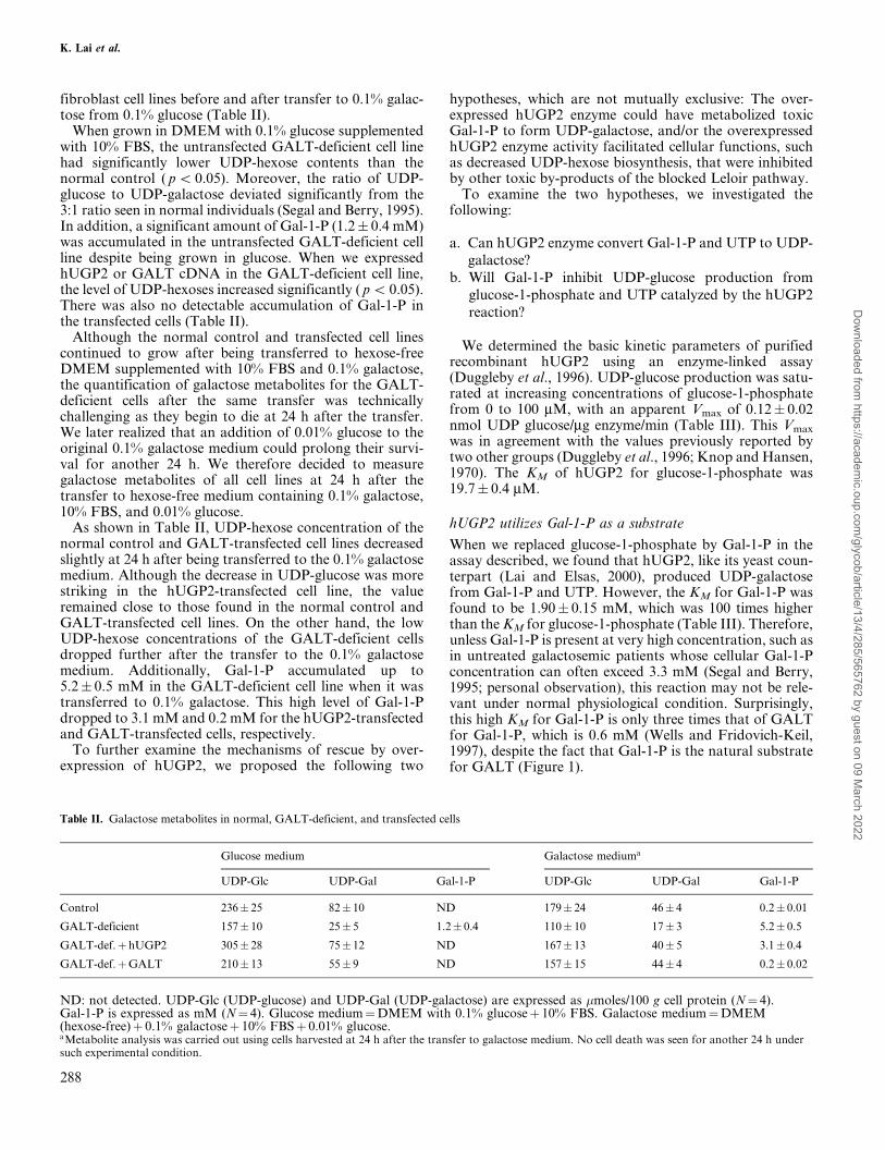
When grown in DMEM with 0.1% glucose supplementedwith 10% FBS, the untransfected GALT-deficient cell linehad significantly lower UDP-hexose contents than thenormal control ( p5 0.05). Moreover, the ratio of UDP-glucose to UDP-galactose deviated significantly from the3:1 ratio seen in normal individuals (Segal and Berry, 1995).In addition, a significant amount of Gal-1-P (1.2� 0.4 mM)was accumulated in the untransfected GALT-deficient cellline despite being grown in glucose. When we expressedhUGP2 or GALT cDNA in the GALT-deficient cell line,the level of UDP-hexoses increased significantly ( p5 0.05).There was also no detectable accumulation of Gal-1-P inthe transfected cells (Table II).
Although the normal control and transfected cell linescontinued to grow after being transferred to hexose-freeDMEM supplemented with 10% FBS and 0.1% galactose,the quantification of galactose metabolites for the GALT-deficient cells after the same transfer was technicallychallenging as they begin to die at 24 h after the transfer.We later realized that an addition of 0.01% glucose to theoriginal 0.1% galactose medium could prolong their survi-val for another 24 h. We therefore decided to measuregalactose metabolites of all cell lines at 24 h after thetransfer to hexose-free medium containing 0.1% galactose,10% FBS, and 0.01% glucose.
As shown in Table II, UDP-hexose concentration of thenormal control and GALT-transfected cell lines decreasedslightly at 24 h after being transferred to the 0.1% galactosemedium. Although the decrease in UDP-glucose was morestriking in the hUGP2-transfected cell line, the valueremained close to those found in the normal control andGALT-transfected cell lines. On the other hand, the lowUDP-hexose concentrations of the GALT-deficient cellsdropped further after the transfer to the 0.1% galactosemedium. Additionally, Gal-1-P accumulated up to5.2� 0.5 mM in the GALT-deficient cell line when it wastransferred to 0.1% galactose. This high level of Gal-1-Pdropped to 3.1 mM and 0.2 mM for the hUGP2-transfectedand GALT-transfected cells, respectively.
To further examine the mechanisms of rescue by over-expression of hUGP2, we proposed the following two
hypotheses, which are not mutually exclusive: The over-expressed hUGP2 enzyme could have metabolized toxicGal-1-P to form UDP-galactose, and/or the overexpressedhUGP2 enzyme activity facilitated cellular functions, suchas decreased UDP-hexose biosynthesis, that were inhibitedby other toxic by-products of the blocked Leloir pathway.
To examine the two hypotheses, we investigated thefollowing:
a. Can hUGP2 enzyme convert Gal-1-P and UTP to UDP-galactose?
b. Will Gal-1-P inhibit UDP-glucose production fromglucose-1-phosphate and UTP catalyzed by the hUGP2reaction?
We determined the basic kinetic parameters of purifiedrecombinant hUGP2 using an enzyme-linked assay(Duggleby et al., 1996). UDP-glucose production was satu-rated at increasing concentrations of glucose-1-phosphatefrom 0 to 100 mM, with an apparent Vmax of 0.12� 0.02nmol UDP glucose/mg enzyme/min (Table III). This Vmax
was in agreement with the values previously reported bytwo other groups (Duggleby et al., 1996; Knop and Hansen,1970). The KM of hUGP2 for glucose-1-phosphate was19.7� 0.4 mM.
hUGP2 utilizes Gal-1-P as a substrate
When we replaced glucose-1-phosphate by Gal-1-P in theassay described, we found that hUGP2, like its yeast coun-terpart (Lai and Elsas, 2000), produced UDP-galactosefrom Gal-1-P and UTP. However, the KM for Gal-1-P wasfound to be 1.90� 0.15 mM, which was 100 times higherthan the KM for glucose-1-phosphate (Table III). Therefore,unless Gal-1-P is present at very high concentration, such asin untreated galactosemic patients whose cellular Gal-1-Pconcentration can often exceed 3.3 mM (Segal and Berry,1995; personal observation), this reaction may not be rele-vant under normal physiological condition. Surprisingly,this high KM for Gal-1-P is only three times that of GALTfor Gal-1-P, which is 0.6 mM (Wells and Fridovich-Keil,1997), despite the fact that Gal-1-P is the natural substratefor GALT (Figure 1).
Table II. Galactose metabolites in normal, GALT-deficient, and transfected cells
Glucose medium Galactose mediuma
UDP-Glc UDP-Gal Gal-1-P UDP-Glc UDP-Gal Gal-1-P
Control 236� 25 82� 10 ND 179� 24 46� 4 0.2� 0.01
GALT-deficient 157� 10 25� 5 1.2� 0.4 110� 10 17� 3 5.2� 0.5
GALT-def.�hUGP2 305� 28 75� 12 ND 167� 13 40� 5 3.1� 0.4
GALT-def.�GALT 210� 13 55� 9 ND 157� 15 44� 4 0.2� 0.02
ND: not detected. UDP-Glc (UDP-glucose) and UDP-Gal (UDP-galactose) are expressed as �moles/100 g cell protein (N� 4).Gal-1-P is expressed as mM (N� 4). Glucose medium�DMEM with 0.1% glucose� 10% FBS. Galactose medium�DMEM(hexose-free)� 0.1% galactose� 10% FBS� 0.01% glucose.aMetabolite analysis was carried out using cells harvested at 24 h after the transfer to galactose medium. No cell death was seen for another 24 h undersuch experimental condition.
K. Lai et al.
288
Dow
nloaded from https://academ
ic.oup.com/glycob/article/13/4/285/565762 by guest on 09 M
arch 2022
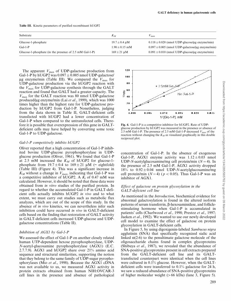
The apparent Vmax of UDP-galactose production fromGal-1-P by hUGP2 was 0.097� 0.005 nmol UDP-galactose/mg enzyme/min (Table III). We compared the Vmax forUDP-galactose production via the hUGP2 reaction withthe Vmax for UDP-galactose synthesis through the GALTreaction and found that GALT had a greater capacity. TheVmax for the GALT reaction was 80 nmol UDP-galactoseproduced/mg enzyme/min (Lai et al., 1999), which was 1000times higher than the highest rate for UDP-galactose pro-duction by hUGP2 from Gal-1-P. Nonetheless, judgingfrom the data shown in Table II, GALT-deficient cellstransfected with hUGP2 had a lower concentration ofGal-1-P when compared to the untransfected cells. There-fore it is possible that overexpression of this gene in GALT-deficient cells may have helped by converting some toxicGal-1-P to UDP-galactose.
Gal-1-P competitively inhibits hUGP2
Oliver reported that a high concentration of Gal-1-P inhib-ited bovine UDP-glucose pyrophosphorylase in UDP-glucose production (Oliver, 1961). We found that Gal-1-Pat 2.5 mM increased the KM of hUGP2 for glucose-1-phosphate from 19.7� 0.4 to 169� 21 mM (~ eightfold)(Table III) (Figure 4). This was a significant increase inKM without a change in Vmax, indicating that Gal-1-P wasa competitive inhibitor of hUGP2. A Ki of 0.47 mM wascalculated. However, it should be noted that these data wereobtained from in vitro studies of the purified protein. Inregard to whether the accumulated Gal-1-P in GALT-defi-cient cells actually inhibits hUGP2 in vivo and to whatextent, we must carry out studies such as metabolic fluxanalyzes, which are out of the scope of this study. In theabsence of in vivo kinetics, we can nevertheless infer suchinhibition could have occurred in vivo in GALT-deficientcells based on the finding that restoration of GALT activityin GALT-deficient cells increased UDP-glucose and UDP-galactose concentrations (Table II).
Inhibition of AGX1 by Gal-1-P
We assessed the effect of Gal-1-P on another closely relatedhuman UTP-dependent hexose pyrophosphorylase, UDP-N-acetyl-glucosamine pyrophosphorylase (AGX1) (E.C.2.7.7.9). AGX1 and hUGP2 share over 21% amino acidsequence and structural similarities, supporting the notionthat they belong to the same family of UDP-sugar pyropho-sphorylases (Mio et al., 1998). Because the AGX1 cDNAwas not available to us, we measured AGX1 activity inprotein extracts obtained from human NIH:OVCAR-3cell lines in the presence and absence of pathological
concentration of Gal-1-P. In the absence of exogenousGal-1-P, AGX1 enzyme activity was 1.12� 0.03 nmolUDP-N-acetylglucosamine/mg cell protein/min (N� 4). Inthe presence of 2.5 mM Gal-1-P, AGX1 activity dropped18% to 0.92� 0.04 nmol UDP-N-acetylglucosamine/mgcell protein/min (N� 4) ( p5 0.05). Thus Gal-1-P was aninhibitor of AGX1.
Effect of galactose on protein glycosylation in theGALT-deficient cell line
As mentioned in the Introduction, biochemical evidence forabnormal galactosylation is found in the altered isoformpatterns of serum transferrin, b-hexosaminidase, and follicle-stimulating hormone when Gal-1-P is accumulated inpatients' cells (Charlwood et al., 1998; Prestoz et al., 1997;Jaeken et al., 1992). We wanted to use our newly developedcell model to examine the effect of galactose on proteinglycosylation in GALT-deficient cells.
In Figure 5, by using digoxigenin-labeled Sambucus nigraagglutinin (SNA) that specifically recognized sialic acidlinked a(2-6) to the penultimate galactose molecule of theoligosaccharide chains found in complex glycoproteins(Shibuya et al., 1987), we revealed that the abundance ofSNA-positive glycoproteins present in cell extracts preparedfrom the GALT-deficient cell line and its GALT-transfected counterpart were identical when the cell lineswere cultured in 0.1% glucose. However, when the GALT-deficient cells were transferred to 0.1% galactose for 24 h,we saw a reduced abundance of SNA-positive glycoproteinsof higher molecular weight (466 kDa) (lane 3, Figure 5).
Table III. Kinetic parameters of purified recombinant hUGP2
Substrate KM Vmax
Glucose-1-phosphate 19.7� 0.4 mM 0.118� 0.020 (nmol UDP-glucose/mg enzyme/min)
Gal-1-P 1.90� 0.15 mM 0.097� 0.005 (nmol UDP-galactose/mg enzyme/min)
Glucose-1-phosphate (in the presence of 2.5 mM Gal-1-P) 169� 21 mM 0.091� 0.010 (nmol UDP-glucose/mg enzyme/min)
Fig. 4. Gal-1-P is a competitive inhibitor for hUGP2. Rate of UDP-glucose production by hUGP2 was measured in the presence or absence of2.5 mM Gal-1-P. The presence of 2.5 mM Gal-1-P decreased Vmax of thereaction without changing the KM as visualized graphically in this doublereciprocal plot.
GALT deficiency in human galactosemic cells
289
Dow
nloaded from https://academ
ic.oup.com/glycob/article/13/4/285/565762 by guest on 09 M
arch 2022
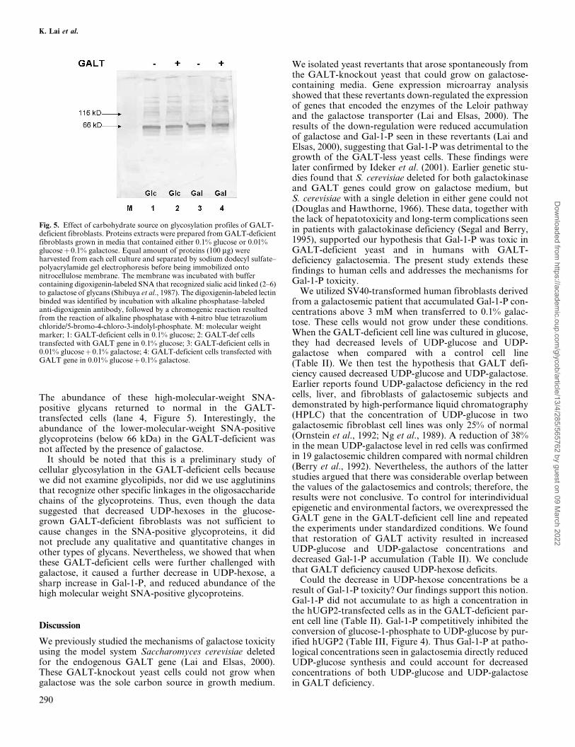
The abundance of these high-molecular-weight SNA-positive glycans returned to normal in the GALT-transfected cells (lane 4, Figure 5). Interestingly, theabundance of the lower-molecular-weight SNA-positiveglycoproteins (below 66 kDa) in the GALT-deficient wasnot affected by the presence of galactose.
It should be noted that this is a preliminary study ofcellular glycosylation in the GALT-deficient cells becausewe did not examine glycolipids, nor did we use agglutininsthat recognize other specific linkages in the oligosaccharidechains of the glycoproteins. Thus, even though the datasuggested that decreased UDP-hexoses in the glucose-grown GALT-deficient fibroblasts was not sufficient tocause changes in the SNA-positive glycoproteins, it didnot preclude any qualitative and quantitative changes inother types of glycans. Nevertheless, we showed that whenthese GALT-deficient cells were further challenged withgalactose, it caused a further decrease in UDP-hexose, asharp increase in Gal-1-P, and reduced abundance of thehigh molecular weight SNA-positive glycoproteins.
Discussion
We previously studied the mechanisms of galactose toxicityusing the model system Saccharomyces cerevisiae deletedfor the endogenous GALT gene (Lai and Elsas, 2000).These GALT-knockout yeast cells could not grow whengalactose was the sole carbon source in growth medium.
We isolated yeast revertants that arose spontaneously fromthe GALT-knockout yeast that could grow on galactose-containing media. Gene expression microarray analysisshowed that these revertants down-regulated the expressionof genes that encoded the enzymes of the Leloir pathwayand the galactose transporter (Lai and Elsas, 2000). Theresults of the down-regulation were reduced accumulationof galactose and Gal-1-P seen in these revertants (Lai andElsas, 2000), suggesting that Gal-1-P was detrimental to thegrowth of the GALT-less yeast cells. These findings werelater confirmed by Ideker et al. (2001). Earlier genetic stu-dies found that S. cerevisiae deleted for both galactokinaseand GALT genes could grow on galactose medium, butS. cerevisiae with a single deletion in either gene could not(Douglas and Hawthorne, 1966). These data, together withthe lack of hepatotoxicity and long-term complications seenin patients with galactokinase deficiency (Segal and Berry,1995), supported our hypothesis that Gal-1-P was toxic inGALT-deficient yeast and in humans with GALT-deficiency galactosemia. The present study extends thesefindings to human cells and addresses the mechanisms forGal-1-P toxicity.
We utilized SV40-transformed human fibroblasts derivedfrom a galactosemic patient that accumulated Gal-1-P con-centrations above 3 mM when transferred to 0.1% galac-tose. These cells would not grow under these conditions.When the GALT-deficient cell line was cultured in glucose,they had decreased levels of UDP-glucose and UDP-galactose when compared with a control cell line(Table II). We then test the hypothesis that GALT defi-ciency caused decreased UDP-glucose and UDP-galactose.Earlier reports found UDP-galactose deficiency in the redcells, liver, and fibroblasts of galactosemic subjects anddemonstrated by high-performance liquid chromatography(HPLC) that the concentration of UDP-glucose in twogalactosemic fibroblast cell lines was only 25% of normal(Ornstein et al., 1992; Ng et al., 1989). A reduction of 38%in the mean UDP-galactose level in red cells was confirmedin 19 galactosemic children compared with normal children(Berry et al., 1992). Nevertheless, the authors of the latterstudies argued that there was considerable overlap betweenthe values of the galactosemics and controls; therefore, theresults were not conclusive. To control for interindividualepigenetic and environmental factors, we overexpressed theGALT gene in the GALT-deficient cell line and repeatedthe experiments under standardized conditions. We foundthat restoration of GALT activity resulted in increasedUDP-glucose and UDP-galactose concentrations anddecreased Gal-1-P accumulation (Table II). We concludethat GALT deficiency caused UDP-hexose deficits.
Could the decrease in UDP-hexose concentrations be aresult of Gal-1-P toxicity? Our findings support this notion.Gal-1-P did not accumulate to as high a concentration inthe hUGP2-transfected cells as in the GALT-deficient par-ent cell line (Table II). Gal-1-P competitively inhibited theconversion of glucose-1-phosphate to UDP-glucose by pur-ified hUGP2 (Table III, Figure 4). Thus Gal-1-P at patho-logical concentrations seen in galactosemia directly reducedUDP-glucose synthesis and could account for decreasedconcentrations of both UDP-glucose and UDP-galactosein GALT deficiency.
Fig. 5. Effect of carbohydrate source on glycosylation profiles of GALT-deficient fibroblasts. Proteins extracts were prepared from GALT-deficientfibroblasts grown in media that contained either 0.1% glucose or 0.01%glucose� 0.1% galactose. Equal amount of proteins (100 mg) wereharvested from each cell culture and separated by sodium dodecyl sulfate±polyacrylamide gel electrophoresis before being immobilized ontonitrocellulose membrane. The membrane was incubated with buffercontaining digoxigenin-labeled SNA that recognized sialic acid linked (2±6)to galactose of glycans (Shibuya et al., 1987). The digoxigenin-labeled lectinbinded was identified by incubation with alkaline phosphatase±labeledanti-digoxigenin antibody, followed by a chromogenic reaction resultedfrom the reaction of alkaline phosphatase with 4-nitro blue tetrazoliumchloride/5-bromo-4-chloro-3-indolyl-phosphate. M: molecular weightmarker; 1: GALT-deficient cells in 0.1% glucose; 2: GALT-def cellstransfected with GALT gene in 0.1% glucose; 3: GALT-deficient cells in0.01% glucose� 0.1% galactose; 4: GALT-deficient cells transfected withGALT gene in 0.01% glucose� 0.1% galactose.
K. Lai et al.
290
Dow
nloaded from https://academ
ic.oup.com/glycob/article/13/4/285/565762 by guest on 09 M
arch 2022
Could a reduction in cellular UDP-glucose and UDP-galactose affect membrane-bound glycoproteins/glyco-lipids? Flores-Diaz and colleagues showed that UDP-glucose deficiency caused by impaired UGP activity in aChinese hamster cell line led to hypersensitivity to the cyto-toxic effect of Clostridium perfringens phospholipase C(1997, 1998). The authors attributed this effect to altera-tions in the composition of plasma membrane glycolipids orglycoproteins. Daran et al. (1997) showed that reducedUDP-glucose formation in S. cerevisiae led to reduction inthe b-glucan and mannan component of the cell wall. Buthow could decreased UDP-glucose synthesis also decreasethe UDP-galactose concentration? In living cells, there aretwo known pathways for the synthesis of UDP-galactose:(1) uridyltransfer of an UMP moiety to Gal-1-P by GALT(Leloir, 1951) (Figure 1), and (2) epimerization of UDP-glucose by UDP-galactose-4-epimerase (GALE) (Salo et al.,1968) (Figure 1). The first reaction is blocked in GALTdeficiency galactosemia (Isselbacher et al., 1956), buthUGP2 (Knop and Hansen, 1970) and GALE reactionscould, in theory, maintain proper UDP-galactose concen-tration if UDP-glucose synthesis were coordinatelyincreased by hUGP2 (Figure 1).
One could argue that UDP-galactose could be formedfrom high concentrations of Gal-1-P in galactosemics viathe hUGP2 reaction (Figures 1 and 4). Although theoreti-cally feasible, our kinetic analysis of the human UGP2enzyme indicated that its apparent Vmax for UDP-galactoseformation is only 0.097� 0.005 nmol UDP-galactoseformed per mg enzyme per minute (Table III). This rate is1000 times slower than that of the GALT reaction, and theproduction of femtomolar amounts of UDP-galactose viathis route would not be sufficient to sustain normal cellulargrowth and posttranslational processing. Thus overexpres-sion of the hUGP2 gene is necessary to rescue the GALT-less yeast and reduce Gal-1-P accumulation (Lai and Elsas,2000). We can thus predict that in human cells culturedfrom patient with galactosemia with Gal-1-P concentra-tions above 2.5 mM endogenous hUGP2 cannot producesufficient UDP-glucose to overcome the reduced rates ofUDP-galactose production caused by deleterious mutationsin the GALT gene (Figure 1).
Although there is no documented information on thecellular concentrations of UDP-glucose and UDP-galactosein the developing fetus, these metabolites are important inposttranslational processing during rapid fetal growth anddifferentiation. The developing brain and ovary of GALT-deficient embryo may well have limited amounts of glyco-proteins and glycolipids. Accumulation of Gal-1-P mayeffectively compete with glucose-1-phosphate or N-acetyl-glucosamine-1-phosphate for UTP-dependent pyropho-sphorylases and reduce the synthesis of theircorresponding UDP-hexoses. The resultant decrease in con-centrations of UDP-glucose and UDP-galactose wouldaffect the properties of membrane glycoproteins/glycoli-pids, as found in patients with uncontrolled galactosemia.Charlwood et al. (1998) and Petry et al. (1991) showed thatgalactose and N-acetylgalactosamine molecules were miss-ing from the oligosaccharide chains of glycoproteins andgangliosides isolated from galactosemic patients with ele-vated Gal-1-P. We add to this notion by observing the
reduced abundance of SNA-positive glycoproteins presentin the GALT-deficient fibroblasts challenged with galactose(Figure 5). Thus, the critical gene (GALT), epigenes(hUGP2), and the environment (carbohydrate source)interact in producing the phenotype (cell growth) inhuman cells.
Materials and methods
Cell culture and transfection of DNA plasmids
We acquired a primary fibroblast cell line derived from aGALT-deficient patient with classic galactosemia who hadno detectable GALT activity in red blood cells (Reichardtand Berg, 1988). The primary cell line (GM00054) andGM00638A, the SV40 large T-antigen transformed cellline derived from GM00054, were obtained from theNIGMS Human Genetic Cell Respository/Coriell Institutefor Medical Research (Camden, NJ).
As a normal control, we obtained another SV40-transformed fibroblast line, GM00637I, which had normalGALT activity. We confirmed that GM00638A (GALT-deficient) manifested the Galÿ phenotype and did notgrow in 0.1% galactose. Fibroblasts were cultured inDMEM (Sigma, St. Louis, MO) at 37�C in 5% CO2.
Human UGP2 and GALT cDNAs were subclonedinto BamHI- and XhoI-digested pCDNA3.0 (Invitrogen,Carlsbad, CA), an episomal mammalian expression vectorwith a CMV promoter. The inserts were verified bysequencing. The resulting recombinant vectors expressingeither the hUGP2 or GALT gene were introduced into theGALT-deficient fibroblasts using the FuGene6 transfectionreagent (Invitrogen). Stable transfectants were selectedthrough resistance to a predetermined dose of 100 mg/mlgeneticin (G-418) over 4±6 weeks. Clones of stable transfec-tants were isolated and expanded subsequently.
Enzyme assays
GALT assay. GALT activity was determined usingmethods previously published (Lai et al., 1999).
hUGP2 assay. Human UGP enzyme activity was assayedat room temperature (25�C) in a 1-ml volume containing 2mM glucose-1-phosphate, 1 mM MgCl2, 1 mM dithiothrei-tol, 0.8 mM Nicotinamide adenosine diphosphate (NAD),0.2 mM UTP, 2.5 ml (0.5 U/ml) UDP-glucose dehydrogen-ase, and 1 mM Tris±HCl (pH 8.0). After incubating thereaction mixture at 25�C for 15 min, the reaction wasstopped by a 3 min incubation at 95�C. The reactions werecentrifuged for 10 min to remove insoluble matter. Theformation of NADH was quantified by the absorbancechange at 340 nm. The quantitative relationship was quan-tified using the Beer-Lambert equation as follows:
D absorbance� D concentration of solutes
� optical path length�molar coefficient of extinction
In this case, optical path length is 1 cm, and the molarcoefficient of extinction is 6220 Mÿ1 cmÿ1.
GALT deficiency in human galactosemic cells
291
Dow
nloaded from https://academ
ic.oup.com/glycob/article/13/4/285/565762 by guest on 09 M
arch 2022

Kinetic studies
All reactions were carried out in a final volume of 1 ml.Each reaction contained 1 mM Tris±HCl, 1 mM MgCl2,0.8 mM NAD, 1 mM dithiothreitol, 0.2 mM UTP, and2.5 ml (0.5 U/ml) of UDP-glucose dehydrogenase.Glucose-1-phosphate, Gal-1-P, or both were added atvarious concentrations as indicated. The reaction wasincubated at room temperature (25�C) after 15 mg of thesemi-purified hUGP2 enzyme was added, and spectropho-tometric readings were taken at 1-, 2-, 3-, 4-, 5-, 7-, 10-, 15-,and 20-min time intervals at 340 nm.
The change in absorbance was used to quantify the for-mation of NADH. The formation of UDP-galactose in thereactions containing Gal-1-P was confirmed by the addi-tion of another enzyme, UDP-galactose-4-epimerase, inaddition to UDP-glucose dehydrogenase and NAD.UDP-galactose-4-epimerase converted the UDP-galactose(produced from the Gal-1-P and UTP) to UDP-glucose,which was subsequently acted on by UDP-glucose dehydro-genase as described.
Competition assay
In all competition assays, Gal-1-P was added at a finalconcentration of 2.5 mM.
Human AGX1 assay
Human AGX1 enzyme activity in cell extracts preparedfrom NIH-OVCAR3 cell lines was assayed using the colorreagent containing 0.03% (w/v) malachite green and 0.2%(w/v) ammonium molybdate described by Mio et al. (1998).NIH-OVCAR3 cell lines were cultured in Corning 75 cm2
flask in DMEM at 37�C in 5% CO2.
Assay of protein concentration
Protein concentration in samples was quantified usingBioRad Protein Assay Reagent (Hercules, CA). Solutionsof bovine serum albumin of known concentrations wereused to construct the standard curve.
Measurement of metabolites
UDP-glucose and UDP-galactose were measured usingenzyme-linked assay described by Ng et al. (1989).Although this method gave variable results when it wasused to measure these metabolites in patients' red bloodcells (Ng et al., 1989; Kirkman, 1992; Segal, 1995; Gibsonet al., 1995), it had been shown to give results identical toHPLC when it was used to measure these metabolites incultured human fibroblasts (Xu et al., 1995).
Gal-1-P contents were determined using methods pre-viously published (Lai and Elsas, 2000). The intracellularvolume was calculated from the equilibrium distribution of3-O-methyl-D-glucose using a protocol established in ourlaboratory by Longo et al. (1988). It was determined thatthe intracellular volume of patient fibroblasts ranged from6.5 to 8 ml/mg cell protein.
Purification of recombinant hUGP2 protein
We purified recombinant hUGP2 enzyme from a bacterialhost BL21 (DE3) (Novagen, Madison, WI). The hUGP2
open reading frame was amplified by PCR (polymerasechain reaction) and primers 5HUGP2 (50-GCCCTCGAG-GATCCATGTCTCAAGATGGTGCT-30) and 3HUGP2(50-GCCCTCGAGTTCGAATCAGTGGTCCAAGATG-CGAAG-30) using plasmid pHC377 (Duggleby et al., 1996)as template. The amplified PCR product was cut withrestriction enzymes BamHI and XhoI and subcloned intothe bacterial expression vector pET30a (� ) (Novagen). Thenucleotide sequence of the subcloned hUGP2 open readingframe in the recombinant vector was determined using anABI 310 automated sequencer (Perkin-Elmer, Boston, MA)to confirm that no mismatch errors were introduced duringPCR. The recombinant vector was transfected into thebacterial strain BL21 (DE3), which has its endogenous galoperon deleted by transposon mutagenesis. The transfectedbacteria were selected on Luria Bertani medium supple-mented with 30 mg/ml kanamycin (Sigma). To inducehUGP2 enzyme production in the bacterial host, bacteriaharboring the recombinant plasmid were grown in LuriaBertani medium with antibiotics to an optical density A600
of 0.6. Ispropylthiogalactoside was added at a final concen-tration of 1 mM, and the culture was grown for another 2 hat 37�C. Cells were harvested by centrifugation at 4000� gfor 20 min. and stored at ±70�C prior to purification of therecombinant hUGP2 protein.
We followed the protocol established by the manufac-turer (Qiagen, Valencia, CA) and purified the hUGP2enzyme based on the principle of affinity chromatography.The bacterial expression vector pET30a(� ) enables co-translation of a hexamer of histidines (His6) in frame atthe amino terminus of the cloned hUGP2 cDNA. Anickel-charged affinity column was used to purify recombi-nant hUGP2 from bacterial cell lysates using modificationsof the manufacturer's method (Qiagen). Lysates of bacterialpellets were incubated with 20 ml/ml nickel-charged resin(Qiagen) at 4�C for 2 h with gentle shaking. Non±His6-tagged proteins were eluted with 20 mM imidazole buffer.The His6-tagged hUGP2 protein was then sequentiallyeluted with buffers containing 50, 100, 150, 200, and250 mM imidazole. Elutes were concentrated and desaltedusing Centricon-300 (Millipore, Bedford, MA) and resus-pended in a small volume of 1 mM Tris buffer (pH 8.0).Protein concentrations were determined by Bradford assay(Bio-Rad) with bovine albumin standards. From 227.5 mgof cell extracts, we recovered 77% of hUGP2 that gave asingle band of 60 kDa on sodium dodecyl sulfate±polyacry-lamide gel electrophoresis. This represented a 111-foldpurification.
Qualitative analysis of SNA-positive glycoproteins
Detection of SNA-positive glycoproteins using digoxigenin-labeled SNA was performed using protocol developed byRoche Molecular Biochemicals (Indianapolis, IN).
Acknowledgment
This research is supported by the Nicholas RochatResearch Foundation for Galactosemia, U.S. Public HealthService Grant MO-1-RR00039 to the General ClinicalResearch Center of Emory University, and a research
K. Lai et al.
292
Dow
nloaded from https://academ
ic.oup.com/glycob/article/13/4/285/565762 by guest on 09 M
arch 2022

grant from the Emory Egleston Children Research Centerof The Children's Hospitals of Atlanta.
Abbreviations
AGX1, UDP-N-acetyl-glucosamine pyrophosphorylase;DMEM, Dulbecco's modified Eagle's medium; FBS, fetalbovine serum; Gal-1-P, galactose-1-phosphate; GALE,UDP-galactose-4-epimerase;GALT,galactose-1-phosphateuridyltransferase; HPLC, high-performance liquidchromatography; hUGP2, human UDP-glucose pyro-phosphorylase; PCR, polymerase chain reaction; SNA,Sambucus nigra agglutinin
References
Berry, G.T., Palmieri, M.J., Heales, S., Leonard, J.V., and Segal, S. (1992)Red blood cell uridine sugar nucleotide levels in patients with classicgalactosemia and other metabolic disorders. Metabolism, 41, 783±787.
Charlwood, J., Clayton, P., Keir, G., Mian, N., and Winchester, B. (1998)Defective galactosylation of serum transferrin in galactosemia.Glycobiology, 8, 351±357.
Chen, Y.T., Mattison, D.R., Feigenbaum, L., Fukui, H., andSchulman, J.D. (1981) Reduction in oocyte number following prenatalexposure to a diet high in galactose. Science, 214, 1145±1147.
Daran, J.M., Bell, W., and Francois, J. (1997) Physiological andmorphological effects of genetic alterations leading to a reducedsynthesis of UDP-glucose in Saccharomyces cerevisiae. FEMS Micro-biol. Lett., 153, 89±96.
Douglas, H.C. and Hawthorne, D.C. (1966) Regulation of genescontrolling synthesis of the galactose pathway enzymes in yeast.Genetics, 54, 911±916.
Duggleby, R.G., Chao, Y.C., Huang, J.G., Peng, H.L., and Chang, H.Y.(1996) Sequence differences between human muscle and liver cDNAsfor UDP-glucose pyrophosphorylase and kinetic properties of therecombinant enzymes expressed in Escherichia coli. Eur. J. Biochem.,235, 173±179.
Flores-Diaz, M., Alape-Giron, A., Persson, B., Pollesello, P., Moos, M.,von Eichel-Streiber, C., Thelestam, M., and Florin, I. (1997)Cellular UDP-glucose deficiency caused by a single point mutationin the UDP-glucose pyrophosphorylase gene. J. Biol. Chem., 272,23784±23791.
Flores-Diaz, M., Alape-Giron, A., Titball, R.W., Moos, M., Guillouard, I.,Cole, S., Howells, A.M., von Eichel-Streiber, C., Florin, I., andThelestam, M. (1998) UDP-glucose deficiency causes hypersensitivityto the cytotoxic effect of Clostridium perfringens phospholipase C.J. Biol. Chem., 273, 24433±24438.
Gibson, J.B., Reynolds, R.A., Palmieri, M.J., Berry, G.T., Elsas, L.J.,Levy, H.L., and Segal, S. (1995) Comparison of erythrocyte uridinesugar nucleotide levels in normals, classic galactosemics, and patientswith other metabolic disorders. Metabolism, 44, 597±604.
Gitzelmann, R., Hansen, R.G., and Steinmann, B. (1984) Biogenesis ofgalactose, possible mechanism of self-intoxication in galactosemia.In F.A. Hommes and H. van den berg (eds.), Normal andpathological development of energy metabolism. Academic Press,London, pp. 25±37.
Greenman, L. and Rathbun, J.C. (1948) Galactose studies in an infantwith idiopathic galactose intolerance. Pediatrics, 2, 666.
Guerrero, N.V., Singh, R.H., Manatunga, A., Berry, G.T., Steiner, R.D.,and Elsas, L.J. (2000) Risk factors for premature ovarian failure infemales with galactosemia. J. Pediatrics, 137, 833±841.
Ideker, T., Thorsson, V., Ranish, J.A., Christmas, R., Buhler, J., Eng, J.K.,Bumgarner, R., Goodlett, D.R., Aebersold, R., and Hood, L. (2001)Intergrated genomic and proteomic analyses of a systematicallyperturbed metabolic network. Science, 292, 929±934.
Isselbacher, K.J., Anderson, E.P., Kurahashi, K., and Kalakar, H.M.(1956) Congenital galactosemia, a single enzymatic block in galactosemetabolism. Science, 123, 635.
Jaeken, J., Kint, J., and Spaaken, L. (1992) Serum lysosomal enzymeabnormalities in galactosemia. Lancet, 340, 1472±1473.
Kirkman, H.N. (1992) Erythrocytic uridine diphosphate galactose ingalactosemia. J. Inher. Metab. Dis., 15, 4±16.
Knop, J.K. and Hansen, R.G. (1970) Uridine diphosphate glucosepyrophosphorylase. J. Biol. Chem., 245, 2499±2504.
Komrower, G.M., Schwarz, V., Holzel, A., and Goldberg, L. (1956) Aclinical and biochemical study of galactosemia. Arch. Dis. Child.,31, 254.
Lai, K. and Elsas, L.J. (2000) Over-expression of human UDP-glucosepyrophosphorylase rescues galactose-1-phosphate uridyltransferase-deficient yeast. Biochem. Biophys. Res. Commun., 271, 392±400.
Lai, K., Willis, A.C., and Elsas, L.J. (1999) Biochemical role of glutamine-188 of human galactose-1-phosphate uridyltransferase. J. Biol. Chem.,274, 6559±6566.
Leloir, L.F. (1951) Enzymatic transformation of uridine diphosphateglucose into galactose derivative. Arch. Biochem. Biophys., 33,186±194.
Leslie, N.D., Yager, K.L., McNamara, P.D., and Segal, S. (1996) A mousemodel of galactose-1-phosphate deficiency. Biochem. Mol. Med., 59,7±12.
Longo, N., Griffin, L.D., and Elsas, L.J. (1988) Influx and efflux of 3-O-methyl-D-glucose by cultured human fibroblasts. Am J Physiol.,254(5 Pt 1), C628±C633.
Mio, T., Yabe, T., Arisawa, M., and Yamada-Okabe, H. (1998) Theeukaryotic UDP-N-acetylglucosamine pyrophosphorylase. J. Biol.Chem., 273, 14392±14397.
Ning, C., Reynolds, R., Chen, J., Yager, C., Berry, G.T., McNamara, P.D.,Leslie, N., and Segal, S. (2000) Galactose metabolism by the mousewith galactose-1-phosphate uridyltransferase deficiency. Ped. Res., 48,211±217.
Ng, W.G., Xu, Y.K., Kaufman, F.R., and Donnell, G.N. (1989) Deficit ofuridine diphosphate galactose in galactosemia. J Inherit. Metab. Dis.,12, 257±266.
Oliver, I.T. (1961) Inhibitor studies on uridine diphosphoglucosepyrophosphorylase. Biochim. Biophys. Acta, 52, 75±81.
Ornstein, K.S., McGuire, E.J., Berry, G.T., Roth, S., and Segal, S. (1992)Abnormal galactosylation of complex carbohydrates in culturedfibroblasts from patients with galactose-1-phosphate uridyltransferasedeficiency. Ped. Res., 31, 508±511.
Petry, K., Greinix, H.T., Nudelman, E., Eisen, H., Hakamori, S.-I.,Levy, H.L., and Reichardt, J.K.V. (1991) Characterization of a novelbiochemical abnormality in galactosemia: deficiency of glycolipidscontaining galactose or N-acetylgalactosamine and accumulation ofprecursors in brain and lymphocytes. Biochem. Med. Metab. Biol., 46,93±104.
Pourci, M.L., Mangeot, M., Soni, T., and Lemmonnier, A. (1990) Cultureof galactosemic fibroblasts in the presence of galactose: effect ofinosine. J. Inherit. Metab. Dis., 13, 819±828.
Prestoz, L.L.C., Couto, A.S., Shin, Y.S., and Petry, K.G. (1997) Alteredfollicle stimulating hormone isoforms in female galactosemic patients.Eur. J. Pediatr., 156, 116±120.
Reichardt, J.K.V. and Berg, P. (1988) Cloning and characterization of acDNA encoding human galactose-1-phosphate uridyltransferase. Mol.Biol. Med., 5, 107±122.
Robertson, A., Singh, R.H., Guerrero, N.V., Hundley, M., and Elsas, L.J.(2000) Outcomes analysis of verbal dyspraxia in classic galactosemia.Genet. Med., 2, 142±148.
Salo, W., Nordin, J., Peterson, D., Bevill, R., and Kirkwood, S. (1968)The specificity of UDP-glucose 4-epimerase from the yeast Saccharo-myces fragilis. Biochim. Biophys. Acta, 151, 484.
Segal, S. (1995) Defective galactosylation in galactosemia: is lowcell UDP-galactose an explanation? Eur. J. Pediatr., 154(suppl 2),S65±S71.
Segal, S. and Berry, G.T. (1995) Disorders of galactose metabolism. InScriver, D., Beaudet, A., Sly, W., and Valle, D. (eds.), The metabolicbasis of inherited disease. McGraw-Hill, New York, pp. 967±1000.
Shibuya, N., Goldstein, I.J., Broeknaert, W.F., Nsimba-Lubaki, M.,Peeters, B., and Peumans, W.J. (1987) Fractionation of sialylatedoligosaccharides, glycopeptides, glycoproteins on immobilizedelderberry (Sambucus nigra L) bark lectins. J. Biol. Chem., 262,1596±1601.
GALT deficiency in human galactosemic cells
293
Dow
nloaded from https://academ
ic.oup.com/glycob/article/13/4/285/565762 by guest on 09 M
arch 2022
Siegel, C.D., Sparks, J.W., and Battaglia, F.C. (1988) Patterns of serumglucose and galactose concentrations in term newborn infants aftermilk feeding. Biol. Neonate, 54, 301.
Waggoner, D.D., Buist, N.R.M., and Donnell, G.N. (1990) Long-termprognosis in galactosemia; results of a survey of 350 cases. J. Inherit.Metab. Dis., 13, 802±818.
Wells, L. and Fridovich-Keil, J. (1997) Biochemical characterization of theS135L-allele of galactose-1-phosphate uridyltransferase associatedwith galactosemia. J. Inherit. Metab. Dis., 20, 633±642.
Wholers, T.M. and Fridovich-Keil, J.L. (2000) Studies of the V94Msubstitution in human UDP-galactose-4-epimerase enzyme associatedwith generalized epimerase-deficiency galactosemia. J. Inherit. Metab.Dis., 23, 713±729.
Xu, Y.K., Kaufman, F.R., Donnell, G.N., Giudici ,T., Alfi, O., andNg, W.G. (1995) HPLC analysis of uridine diphosphate sugars:decreased concentrations of uridine diphosphate galactose in erythro-cytes and cultured skin fibroblasts from classic galactosemia patients.Clin. Chim. Acta, 240, 21±33.
K. Lai et al.
294
Dow
nloaded from https://academ
ic.oup.com/glycob/article/13/4/285/565762 by guest on 09 M
arch 2022