
Fyn binds to and phosphorylates T cell immunoglobulin and mucin domain-1 (Tim1
16
Fyn binds to and phosphorylates T cell Immunoglobulin and mucin domain-1 (Tim-1) Miranda L. Curtiss a,b,c , Bruce S. Hostager a , Elizabeth Stepniak a , Melody Singh a , Natalie Manhica a , Judit Knisz a , Geri Traver a , Paul D. Rennert d , John D. Colgan a , and Paul B. Rothman a,* Miranda L. Curtiss: [email protected]; Bruce S. Hostager: [email protected]; Elizabeth Stepniak: [email protected]; Melody Singh: [email protected]; Natalie Manhica: [email protected]; Judit Knisz: [email protected]; Geri Traver: [email protected]; Paul D. Rennert: [email protected]; John D. Colgan: [email protected] a Department of Internal Medicine, University of Iowa Carver College of Medicine. 200 Hawkins Drive, Iowa City, Iowa 52242. USA b Medical Scientist Training Program, University of Iowa Carver College of Medicine, 2206 MERF, Iowa City, Iowa 52242. USA c Interdisciplinary Graduate Program in Immunology, University of Iowa Carver College of Medicine, 347 MRC, Iowa City, Iowa 52242. USA d Biogen Idec, 12 Cambridge Center, Cambridge, MA 01746. USA Abstract The gene encoding T cell immunoglobulin and mucin domain-1 (TIM-1) is linked to atopy and asthma susceptibility in mice and humans. TIM-1 is a transmembrane protein expressed on activated lymphocytes and appears to have a role as a co-stimulatory receptor in T cells. The protein has not been shown to have enzymatic activity but contains a site within its cytoplasmic tail predicted to be a target for tyrosine kinases. Here, we show that Tim-1 can associate with the kinase Fyn, a member of the Src family of tyrosine kinases. This association does not require Fyn’s kinase activity and is independent of the phosphorylation of a conserved tyrosine present within the cytoplasmic tail of Tim-1. Fyn is necessary for phosphorylation of this tyrosine in Tim-1 and the phosphorylation of Tim-1 varies with the levels of Fyn present in cells. These data suggest a role for Fyn in the signaling downstream of Tim-1. 1. Introduction The ability of cells to respond to external signals requires the presence of a large array of transmembrane proteins that function as receptors. The immune system is reliant on such proteins to help coordinate the cellular responses required for an effective immune response. © 2011 Elsevier Ltd. All rights reserved. * Corresponding author: Paul Rothman, 451 Newton Rd., 212 College of Medicine Administration Bldg., Iowa City, IA 52242, Phone: 319-384-4547, Fax: 319-353-5627, [email protected]. Conflict of Interest P.D. Rennert is a stockholding employee of Biogen Idec Incorporated. The other authors have no financial or commercial conflicts of interest to disclose. Publisher's Disclaimer: This is a PDF file of an unedited manuscript that has been accepted for publication. As a service to our customers we are providing this early version of the manuscript. The manuscript will undergo copyediting, typesetting, and review of the resulting proof before it is published in its final citable form. Please note that during the production process errors may be discovered which could affect the content, and all legal disclaimers that apply to the journal pertain. NIH Public Access Author Manuscript Mol Immunol. Author manuscript; available in PMC 2012 July 1. Published in final edited form as: Mol Immunol. 2011 July ; 48(12-13): 1424–1431. doi:10.1016/j.molimm.2011.03.023. NIH-PA Author Manuscript NIH-PA Author Manuscript NIH-PA Author Manuscript
-
Upload
medbiotech -
Category
Documents
-
view
0 -
download
0
Transcript of Fyn binds to and phosphorylates T cell immunoglobulin and mucin domain-1 (Tim1

Fyn binds to and phosphorylates T cell Immunoglobulin andmucin domain-1 (Tim-1)
Miranda L. Curtissa,b,c, Bruce S. Hostagera, Elizabeth Stepniaka, Melody Singha, NatalieManhicaa, Judit Knisza, Geri Travera, Paul D. Rennertd, John D. Colgana, and Paul B.Rothmana,*
Miranda L. Curtiss: [email protected]; Bruce S. Hostager: [email protected]; Elizabeth Stepniak:[email protected]; Melody Singh: [email protected]; Natalie Manhica: [email protected]; JuditKnisz: [email protected]; Geri Traver: [email protected]; Paul D. Rennert: [email protected];John D. Colgan: [email protected] Department of Internal Medicine, University of Iowa Carver College of Medicine. 200 HawkinsDrive, Iowa City, Iowa 52242. USAb Medical Scientist Training Program, University of Iowa Carver College of Medicine, 2206 MERF,Iowa City, Iowa 52242. USAc Interdisciplinary Graduate Program in Immunology, University of Iowa Carver College ofMedicine, 347 MRC, Iowa City, Iowa 52242. USAd Biogen Idec, 12 Cambridge Center, Cambridge, MA 01746. USA
AbstractThe gene encoding T cell immunoglobulin and mucin domain-1 (TIM-1) is linked to atopy andasthma susceptibility in mice and humans. TIM-1 is a transmembrane protein expressed onactivated lymphocytes and appears to have a role as a co-stimulatory receptor in T cells. Theprotein has not been shown to have enzymatic activity but contains a site within its cytoplasmictail predicted to be a target for tyrosine kinases. Here, we show that Tim-1 can associate with thekinase Fyn, a member of the Src family of tyrosine kinases. This association does not requireFyn’s kinase activity and is independent of the phosphorylation of a conserved tyrosine presentwithin the cytoplasmic tail of Tim-1. Fyn is necessary for phosphorylation of this tyrosine inTim-1 and the phosphorylation of Tim-1 varies with the levels of Fyn present in cells. These datasuggest a role for Fyn in the signaling downstream of Tim-1.
1. IntroductionThe ability of cells to respond to external signals requires the presence of a large array oftransmembrane proteins that function as receptors. The immune system is reliant on suchproteins to help coordinate the cellular responses required for an effective immune response.
© 2011 Elsevier Ltd. All rights reserved.*Corresponding author: Paul Rothman, 451 Newton Rd., 212 College of Medicine Administration Bldg., Iowa City, IA 52242, Phone:319-384-4547, Fax: 319-353-5627, [email protected] of InterestP.D. Rennert is a stockholding employee of Biogen Idec Incorporated. The other authors have no financial or commercial conflicts ofinterest to disclose.Publisher's Disclaimer: This is a PDF file of an unedited manuscript that has been accepted for publication. As a service to ourcustomers we are providing this early version of the manuscript. The manuscript will undergo copyediting, typesetting, and review ofthe resulting proof before it is published in its final citable form. Please note that during the production process errors may bediscovered which could affect the content, and all legal disclaimers that apply to the journal pertain.
NIH Public AccessAuthor ManuscriptMol Immunol. Author manuscript; available in PMC 2012 July 1.
Published in final edited form as:Mol Immunol. 2011 July ; 48(12-13): 1424–1431. doi:10.1016/j.molimm.2011.03.023.
NIH
-PA Author Manuscript
NIH
-PA Author Manuscript
NIH
-PA Author Manuscript

One family of such surface receptors, called the TIMs (T cell immunoglobulin and mucindomain), is thought to regulate both physiologic and pathologic immune responses (Curtissand Colgan, 2007; Freeman et al., 2010; Rodriguez-Manzanet et al., 2009; Su et al., 2008).TIM-1 was initially cloned from African green monkeys (HAVcr1) (Kaplan et al., 1996) andhumans as a cellular receptor for hepatitis A virus (HAVCR1) (Feigelstock et al., 1998).Polymorphism of TIM-1 have been linked to atopic diseases and asthma in both mice andhumans. Using a genetic approach, McIntire et al. demonstrated that a DBA/2-derivedgenomic segment containing the Tim family of genes (Tapr locus) could decrease thesusceptibility of BALB/c mice to atopy (McIntire et al., 2001).
The biological function of Tim-1 is unclear. Although originally described as a moleculesecreted by kidney proximal tubule epithelial cells upon injury (Ichimura et al., 1998),subsequent studies demonstrated Tim-1 is expressed by several cells of the immune system,including activated CD4+ T cells (McIntire et al., 2004; Meyers et al., 2005; Umetsu et al.,2005) and activated B cells (Wong et al., 2010). Tim-1 appears to bind to several ligands,among them LMIR-5/CD300b, phosphatidylserine, and possibly itself and/or Tim-4. Tim-1,Tim-3, and Tim-4 contain IgV domains with a conserved loop in the ligand-binding domainthat forms a pocket for phosphatidylserine (DeKruyff et al., 2010; Kobayashi et al., 2007;Miyanishi et al., 2007; Santiago et al., 2007), and overexpression of Tim-1 on a kidneyepithelial cell line conferred a phagocytic phenotype, allowing uptake of apoptotic cells(Ichimura et al., 2008). Tim-1 and Tim-4 also bind to LMIR5/CD300b, expressed ongranulocytes and other cells (Yamanishi et al., 2010). Other literature suggests Tim-1 canbind Tim-4 or can bind other Tim-1 molecules through homotypic interactions (Santiago etal., 2007; Wilker et al., 2007). Thus, Tim-1 engagement by its various ligands maycontribute to both immune and non-immune responses.
Signaling pathways involving Tim-1 have been studied in T cells through overexpressionstudies in Jurkat cells. A conserved tyrosine residue in the cytoplasmic tail of Tim-1 issituated in a motif that is similar to a consensus sequence recognized by SH2 domain-containing proteins (Songyang and Cantley, 1995). Tim-1 appears to be phosphorylated at abasal level in Jurkat cells, and treatment with pervanadate (de Souza et al., 2005) or TCRactivation (Binne et al., 2007) increases the level of Tim-1 phosphorylation. A Jurkat cellline with reduced Lck levels (JCaM1) exhibits reduced Tim-1 phosphorylation in responseto pervanadate (de Souza et al., 2008), and overexpression of Lck in this line restoredpervandate-induced Tim-1 phosphorylation. However, the limited tissue distribution of Lcksuggests that Tim-1 may be phosphorylated by other Src family kinase members in non-CD4+ T cells. For example, B cells upregulate surface Tim-1 in response to BCRstimulation in vitro and in vivo (Wong et al., 2010) but do not express Lck. We present datathat the Src family kinase member Fyn can bind to and phosphorylate Tim-1 in a B cell line,thus implicating Fyn in signaling downstream ofTim-1.
2. Materials and Methods2.1 Cell lines
293T cells were maintained in DMEM/10% w/v FCS/1000 U/mL penicillin/1000 μg/mLstreptomycin/20 mM L-glutamine/10 mM sodium pyruvate and transfected as indicated bythe calcium phosphate method (Arimura et al., 2004) or Lipofectamine 2000 (Invitrogen).M12.4.1(Hamano et al., 1982) and A20.2J (McKean et al., 1981) cells were obtained fromDr. Gail Bishop (University of Iowa) and maintained in RPMI 1640/10% w/v FCS/1000 U/mL penicillin/1000 μg/mL streptomycin/20 mM L-glutamine/10 mM sodium pyruvate/5%w/v non-essential amino acids/50 μM β-mercaptoethanol (“complete medium”).
Curtiss et al. Page 2
Mol Immunol. Author manuscript; available in PMC 2012 July 1.
NIH
-PA Author Manuscript
NIH
-PA Author Manuscript
NIH
-PA Author Manuscript

2.2 Antibodies and reagentsTim-1 specific rat IgG2a monoclonal antibody clones 1H9 and 3A2 have been previouslydescribed (Sizing et al., 2007). Abcam anti-Myc rabbit polyclonal antibody, Santa Cruz anti-Myc (clone 9E10) mouse monoclonal antibody, Santa Cruz FYN3 polyclonal antibody, andBiogen anti-Tim1 (clone 1H9) were used for immunoprecipitation. Normal rabbit IgG waspurchased from Santa Cruz. Rat IgG2a isotype control was purchased from BD Pharmingen.For M12.4.1 cell immunoprecipitations, rat IgG2a isotype control (clone mAb72) waspurified from hybridoma supernatants. Jackson ImmunoResearch Affinipure goat anti-ratIgG (H+L) was used to coat Dynabeads protein G (Invitrogen).
The following antibodies were used for Western blot: Biogen anti-Tim-1 (rat monoclonal,clone 3A2), Abcam anti-Myc (rabbit polyclonal), Millipore anti-Myc (mouse monoclonal,clone 4A6), Invitrogen anti-Xpress (mouse monoclonal), BD Transduction Laboratoriesanti-Fyn (mouse monoclonal, clone 25), Santa Cruz anti-Fyn (rabbit polyclonal, FYN3),Cell Signaling anti-Lck (rabbit polyclonal), Upstate anti-Src (mouse monoclonal, cloneGD11), Upstate anti-Hck (rabbit polyclonal), Roche anti-HA (rat monoclonal, clone 3F10),and Sigma anti-actin (rabbit polyclonal). Millipore anti-phospohotyrosine (4G10) was usedto probe immunoprecipitations from 293T cells. Millipore 4G10 Platinum HRP was used toprobe immunoprecipitations from M12.4.1 cells.
The following antibodies were used for FACS: PE anti-Tim-1 (clone RMT1-4) and PE anti-rat IgG2b isotype control were purchased from Biolegend. Biotinylated anti-Tim1 (cloneRMT1-4), anti-Tim2 (clone RMT2-1), and rat IgG2b isotype control were obtained fromeBioscience.
2.3 DNA ConstructsFull-length (isoform a) myc-tagged Tim-1 was constructed by amplification of Tim-1 fromBALB/c kidney cDNA with the following primers: forward 5′-CTAGCATGAATCAGATTCAAGTCTTCATTTCAGGCCTCATACTGCTTCTCCCAGGC-3′ and reverse 5′-GGTACCGGTCTATCTTCAACAATGTAGATGTTGTCTTCAGCTCGGG-3′. PCRproducts were ligated into pCR TOPO BLUNT II (Invitrogen), sequenced, and subclonedinto pcDNA3.1B (Invitrogen) in frame with the Myc epitope tag using the KpnI and NheIrestriction sites. A Stratagene QuikChange Site-Directed Mutagenesis kit was usedaccording to manufacturer’s instructions to generate the Tim1/Myc Y299F construct.pcDNA3.1B Tim1/Myc and Tim1/Myc Y299F were amplified and ligated into pCR TOPOBLUNT II, digested with EcoRI, and subcloned into pMIG to generate pMIG Tim1/Mycand pMIG Tim1/Myc Y299F. Myc-tagged Tim-1 isoform b (lacking exon 6 of the mucindomain) was generated by amplification of C57BL/6 kidney cDNA as above, then cloningthe PCR product into pcDNA3.1B in frame with the Myc epitope tag.
Tim-2/Myc was generated by amplification of pCMV-SPORT Tim-2 (Open Biosystems)with the following primers: forward 5′-ATAGCTAGCATGAATCAGATTCAAGTCTTC-3′, reverse 5′-TTAGGTACCCAGGACTCTTCTTCAGGGTA-3′. Tim-3/Myc was generated byamplification of cDNA from BALB/c CD4+ T cells stimulated in Th1-polarizing conditionswith the following primers: forward 5′-GCTAGCATGTTTTCAGGTCTTACCCTC- 3′,reverse 5′-GGTACCCAGGATGGCTGCTGGCTGTT-3′. PCR products were digested withNheI and KpnI, then ligated into pcDNA3.1B in frame with the Myc epitope tag.
Src, Fyn-T, and kinase-dead Fyn-B (K296E) expression plasmids were a kind gift from Dr.Gloria Lee (University of Iowa). LYN expression plasmid was obtained from Dr. DinaAlexandropoulos (Columbia University). The LCK expression plasmid was generated by
Curtiss et al. Page 3
Mol Immunol. Author manuscript; available in PMC 2012 July 1.
NIH
-PA Author Manuscript
NIH
-PA Author Manuscript
NIH
-PA Author Manuscript

subcloning an LCK construct (obtained from Dr. Jon Houtman, University of Iowa) intopcDNA3.1B using NheI and XhoI restriction sites. HCK and YES-1/HA expressionplasmids were generated by amplification of HCK and YES-1 cDNAs (both obtained fromOpen Biosystems) with the following primers: HCK for 5′AAGCTAGCATGGGGGGGCGCTCAAG 3′, rev 5′GGTACCTCATGGCTGCTGTTGGTACTGGCTC 3′; YES-1 for 5′-GAGCTAGCATGGGCTGCATTAAAAG-3′, rev 5′-GCGGTACCTAAATTTTCTCCTGG-3′. YES-1 (lacking a stop codon) and HCK PCRproducts were cloned into pCR TOPO Blunt II (Invitrogen), sequenced, and subcloned intopcDNA3.1B (Invitrogen) using the NheI and KpnI restriction sites. An oligonucleotideencoding the sequence for the HA epitope tag and a stop codon was inserted after the YES-1coding sequence using the KpnI and XbaI restriction sites to generate C’ HA-tagged Yes-1.
Retroviral constructs expressing mir30-flanked shRNA hairpins targeting murine Fyn weregenerated following LMP manufacturer instructions using the RNAi Central – RNAi OligoRetriever website (http://katahdin.cshl.edu/html/scripts/resources.pl) to generate oligos(Supplemental Table 1). These oligos were amplified with the following primers: sense 5′-CAGAAGGCTCGAGAAGGTATATTGCTGTTGACAGTGAGCG-3′; antisense 5′-CTAAAGTAGCCCCTTGAATTCCGAGGCAGTAGGCA-3′. The PCR products weredigested with XhoI and EcoRI, then subcloned into the LMP retrovirus (Open Biosystems).
2.4 Retroviral transduction of M12 cellspMIG, pMIG Tim1/Myc, and pMIG Tim1/Myc Y299F constructs were transientlytransfected into 293T cells with the pCL-ECO packing plasmid (Naviaux et al., 1996).Supernatants were harvested at 48 hours post-transfection and M12 cells were resuspendedat a concentration of 5 × 10⁵ cells/mL in supernatant with 20 mM HEPES and 8 μg/mLpolybrene, with 1 mL per well of a 24-well plate. Plates were centrifuged at 2000 rpm for 2hours (30°C). 6 hours after spinfection, 1 mL media was added to each well and cell lineswere subsequently maintained at a density of less than 1 × 10⁶ cells/mL. Fyn-deficient M12cells were generated as above by transduction with LMP retrovirus expressing short RNAhairpins targeting Fyn. This retrovirus contains an IRES and GFP coding sequencedownstream of the sequences for RNA hairpins. 24 hours post-transduction, cells wereselected and maintained in 4 μg/mL puromycin. Transuction efficiency was greater than98%, as assessed by flow cytometry for GFP expression.
2.5 Lysis Buffers and Immunoprecipitation293T cells were harvested 48 hours after transfection and lysed in a buffer containing 0.5%NP-40, 50 mM HEPES, 100 mM sodium fluoride, 10 mM sodium pyrophosphate, 2 mMsodium orthovanadate, 4 mM EDTA, 2 mM PMSF, 2 μg/mL aprotinin, and 10 μg/mLleupeptin. Lysates were quantified by Bradford assay. 1 mg precleared lysate wasimmunoprecipitated by 2 μg of antibody. In some experiments, 293T cells were activated byincubation in 2 mL PBS containing 100 μM sodium vanadate and 0.068% hydrogenperoxide for 10 minutes at 37°C to serve as a positive control.
Activation and immunoprecipitation of full-length murine Tim-1 from M12.4.1 cells wasperformed as previously described (Rowland et al., 2007). Dynal protein G beads wereconjugated with Jackson anti-rat (10 μg mAb/10 μL beads/100 μL complete medium) for 30minutes at room temperature. Beads were washed three times in complete medium andincubated with anti-Tim1 clone 1H9 or isotype control antibody (10 μg/10 μL beads/100 μLcomplete medium) for 30 minutes, washed three times with RPMI complete medium andresuspended in 25 μL complete medium/10 μL beads. 3 – 4 × 10⁷ M12 cells were washedand resuspended at 2 × 10⁸ cells/mL in RPMI complete medium, incubated at 37°C for 30
Curtiss et al. Page 4
Mol Immunol. Author manuscript; available in PMC 2012 July 1.
NIH
-PA Author Manuscript
NIH
-PA Author Manuscript
NIH
-PA Author Manuscript

minutes, and activated by the addition of 25 μL antibody-coated beads for 5 minutes or withbeads and 100 μM pervanadate for 10 minutes at 37°C. Activation was terminated byaddition of cold lysis buffer with a final concentration of 1% Triton X100, 100 mM NaCl,40 mM Tris pH 7.5, 1.0 mM CaCl2, 1 mM MgCl2, 2 mM Na3VO4, protease inhibitor(Roche) and 0.05 mg/ml DNAse II.
Samples were subjected to SDS-PAGE electrophoresis, transferred to PVDF (Millipore),and probed with the indicated antibodies. To detect phosphorylated Tim-1, membranes werefirst probed with anti-phosphotyrosine, stripped, and then reprobed for Tim-1. Anti-Mycwas used to detect myc-tagged Tim-1, and anti-Tim1 mAb 3A2 (Biogen) was used to detectendogenous Tim-1 from M12.4.1 cells.
2.6 SH2 array and peptideA tyrosine phosphorylated peptide containing the following Tim-1 cytoplasmic tail sequencewas purchased from Quality Control Biochemicals: VVHSRAEDNIYIVEDRP. A PanomicsSH2 array was probed with this peptide as per manufacturer’s instructions.
3. Results3.1 Fyn phosphorylates Tim-1
The cytoplasmic tail of Tim-1 contains a conserved tyrosine residue within a motif favorablefor recognition by SH2 domains of Src family kinases (de Souza et al., 2008; Songyang andCantley, 1995). To evaluate the ability of this sequence to interact with SH2 domain-containing proteins, we probed a Panomics SH2 array with a phosphopeptide containing thisconserved sequence. We found that this peptide was able to interact with several SH2domain-containing proteins, among them the Src family kinases Fyn, Yes-1, and Hck(Supplemental Figure 1), as well as GRB2, RAS-GAP1 and the PI3 kinase p85α and p85βsubunits. These results were consistent with studies performed by others (de Souza et al.,2008) and suggest that Tim-1 may interact with Src family kinases.
To determine which Src family kinase members could interact with and phosphorylateTim-1 in cells, constructs encoding six of these proteins, (Src, Lck, Fyn-T, Lyn, Yes-1, andHck) were individually co-transfected into 293T cells with epitope-tagged Tim-1 in 293Tcells. Tim-1 was immunoprecipitated and Western blotting was performed onimmunoprecipitates and lysates. When we blotted with an anti-phosphotyrosine antibody(Figure 1a) we found that co-expression of Fyn-T with Tim-1 consistently resulted in a highlevels Tim-1 tyrosine phosphorylation, suggesting that Fyn-T can phosphorylate Tim-1. Incontrast to Fyn-T, the other Src family kinases examined did not induce high levels of Tim-1tyrosine phosphorylation when expressed in 293T cells. Expression of Src resulted in co-immunoprecipitation of another phosphorylated protein of roughly 100 kDa, which is notconsistent with the size of Tim-1. Co-expression of Src, Lck, Lyn, and Fyn-T with Tim-1resulted in co-immunoprecipitation of phosphorylated proteins of 55–60 kDa, a sizeconsistent with Src family kinases. We confirmed co-immunoprecipitation of Src, Lck, Lyn(data not shown), and Fyn-T (Figure 1a) by stripping and blotting with antibodies to thesekinases. Lysates were also blotted for expression of Src, Lck, Yes, Fyn, Lyn, and Hck (datanot shown). Although Hck and Yes-1 were able to weakly bind the Tim-1 phosphopeptideby SH2 array, they did not interact with Tim-1 in this overexpression system. Fyn wasselected for further study, as it was the only kinase which phosphorylated Tim-1 in 293T co-transfection assays. We also found that immunoprecipitation of Fyn-T resulted in co-immunoprecipitation of Tim-1, indicating that this may be a direct interaction between theproteins (Figure 1b).
Curtiss et al. Page 5
Mol Immunol. Author manuscript; available in PMC 2012 July 1.
NIH
-PA Author Manuscript
NIH
-PA Author Manuscript
NIH
-PA Author Manuscript

To further assess specificity of the interaction between Fyn-T and Tim-1, graded amounts ofFyn-T expression plasmid were co-transfected into 293T cells with a constant amount ofTim-1 expression plasmid. A resultant graded increase in phosphorylation of Tim-1 and inthe amount of Fyn-T co-immunoprecipitating with Tim-1 was observed, with a correlationbetween levels of Fyn co-immunoprecipitation and Tim-1 phosphorylation (Figure 2). Thesedata suggest that Fyn and Tim-1 can interact and that overexpression of Fyn leads toincreased tyrosine phosphorylation of Tim-1.
3.2 Fyn binds Tim-1 in the absence of tyrosine 299 in 293T cellsThe observed interaction between Fyn and Tim-1 suggests that these proteins may interact toinitiate signaling downstream of Tim-1. Thus, we sought to further define the requirementsfor this interaction. The studies using SH2 arrays suggested that the SH2 domain of Fyncould bind a tyrosine-phosphorylated motif of Tim-1. To assess the requirement forphosphorylation of this tyrosine residue in the interaction of Fyn-T with Tim-1, we usedsite-directed mutagenesis to introduce a phenylalanine residue in place of the tyrosine atposition 299 of Tim-1 (isoform a) within the putative Fyn recognition site. Previous studiesindicate that mutation of this tyrosine to an alanine abrogated phosphorylation of humanTIM-1 and murine Tim-1 (isoform b) in pervanadate-treated transfected Jurkat cells (Binneet al., 2007; de Souza et al., 2005). Co-expression of the Y299F mutant Tim-1 constructwith Fyn in 293T cells abrogated phosphorylation of Tim-1 via Fyn, as well as bypervanadate (Figure 3a). However, the ability of Fyn to co-immunoprecipitate with theY299F mutant Tim-1 construct was maintained. These data indicate that the interactionbetween Tim-1 and Fyn may not require phosphorylation of Y299 in the putative SH2domain binding site.
As a complementary study of the requirement for phosphorylation in Fyn binding to Tim-1,we determined the requirement for Fyn kinase activity in its binding to Tim-1. Anexpression plasmid for a Fyn mutant lacking enzymatic activity (“kinase-dead”) was co-expressed in a graded fashion with a constant amount of Tim-1 in 293T cells. This againresulted in a graded co-immunoprecipitation of the kinase-dead Fyn with Tim-1, althoughthere was no detectable phosphorylation of Tim-1 (Figure 3b). These data suggest that,although there may be enhanced interaction between Tim-1 and Fyn upon tyrosinephosphorylation of Tim-1, the binding of Fyn to the cytoplasmic tail of Tim-1 does notrequire Fyn kinase activity.
3.3 Tim-1 is phosphorylated in B cells and interacts with FynRecent studies have suggested a role for Tim-1 in lymphoid cells. Therefore, we extendedour studies of the interaction between Tim-1 and Fyn to B cells. To identify antibodies thatrecognize Tim-1 by Western blot and immunoprecipitation, we transfected 293T cells withMyc-tagged Tim-1, Tim-2, and Tim-3. Lysates were subjected to immunoprecipitation withanti-Myc or anti-Tim1 antibodies, and lysates and immunoprecipitates were probed withantibodies to Myc or Tim-1. We identified antibodies that recognize both the full-lengthisoform a (cloned from BALB/c mice) and the exon 6 deleted isoform b (cloned from C7BL/6 mice) of Tim-1, but not Tim-2 or Tim-3 (Supplemental Figure 2), and used these to probeTim-1 expression in B cell lines.
Recent studies have demonstrated Tim-1 surface expression on murine splenic B cellsstimulated with anti-IgM or PMA and ionomycin in vitro, by germinal center B cells in vivo,and on the B cell line A20 (Sizing et al., 2007; Wong et al., 2010; Yamanishi et al., 2010).We confirmed these results and demonstrated that another B cell line, M12.4.1, alsoexpressed Tim-1 on its surface and that this expression was higher than in A20.2J cells(Supplemental Figure 3). Neither cell line expressed detectable surface Tim-2. Both M12.4.1
Curtiss et al. Page 6
Mol Immunol. Author manuscript; available in PMC 2012 July 1.
NIH
-PA Author Manuscript
NIH
-PA Author Manuscript
NIH
-PA Author Manuscript

and A20.2J cells express a protein species of roughly 75 kDa (data not shown), similar tothat of Tim-1 when transiently expressed in 293T cells. Furthermore, this species isrecognized by antibodies to Tim-1 in immunoprecipitation and Western blot analysis. Tim-1was expressed at higher levels on M12.4.1 cells, thus experiments were performed in thiscell line.
In order to determine if Fyn may play a role in Tim-1 signaling in B cells, we asked if thesetwo proteins could interact in M12.4.1 cells. Cells were incubated with anti-Tim-1antibodies that were conjugated to beads to mimic ligand:receptor coupling. After 5 minutes,activation was terminated by addition of lysis buffer. We then examined the status of Tim-and Fyn in these cells. Immunoprecipitated endogenous Tim-1 did not display basal tyrosinephosphorylation. Treatment with pervanadate or incubation with anti-Tim-1 beads inducedTim-1 tyrosine phosphorylation (Figure 4). Fyn co-immunoprecipitated with endogenousTim-1 in unstimulated cells, and engagement of Tim-1 by antibody-coated beads oractivation with pervandate resulted in increased co-immunoprecipitation of Fyn with Tim-1compared to unstimulated cells. These data demonstrate that Fyn and Tim-1 interact in Bcells at a baseline state and that this interaction is increased when Tim-1 is activated. Lynalso co-immunoprecipitated with Tim-1, and this increased in response to cross linking withanti-Tim-1 antibodies (data not shown).
Our studies employing 293T cells have defined some of the requirements for interactionbetween Tim-1 and Fyn. To extend these studies to B cells, we overexpressed epitope-tagged Tim-1 or Tim-1 Y299F in M12 cells to determine the requirement for the conservedtyrosine in Fyn interaction and Tim-1 phosphorylation. When cells overexpressing Tim-1orTim-1 Y299F were incubated with anti-Tim-1 conjugated beads, Fyn was co-immunoprecipitated to similar levels. However, we observed tyrosine phosphorylation ofboth Tim-1 and Tim-1 Y299F (Supplemental Figure 4). Although overexpression of Tim-1Y299F in 293T cells and the Jurkat T cell line abrogated phosphorylation by pervanadate(de Souza et al., 2008; de Souza et al., 2005), cross linking the overexpressed Y299F mutantreceptor in M12 cells still resulted in Tim-1 phosphorylation. Thus, it appears that themembrane-proximal tyrosine residue of Tim-1 may be phosphorylated upon cross linking.
3.4 Fyn is required for phosphorylation of Tim-1 in B cellsThe interaction between Fyn and Tim-1 suggests that Fyn may function in the activation ofTim-1. To address whether Fyn is necessary for Tim-1 phosphorylation, we utilized RNAi toknock down Fyn in M12 cells. M12 cells were transduced with retrovirus expressing mir30-flanked shRNA sequences to knock down Fyn (denoted Fyn1, 2, and 3) or a controlsequence against luciferase (Luc), and stable cell lines were generated by selection andmaintenance in puromycin. Cells transduced with Fyn shRNA 3 expressed approximately 5–10% of the levels found in the Luc shRNA control cells, and cells transduced with FynshRNA1 and 2 expressed Fyn at 10–30% of that found in Luc shRNA control cells. (Figure5a). Activation of Tim-1 with antibody-coated cells resulted in phosphorylation of Tim-1 incells expressing the control Luc shRNA, and reduced phosphorylation of Tim-1 in cellsexpressing Fyn shRNA (Figure 5b), with the least phosphorylation in the cells expressingthe most potent Fyn knockdown construct (Fyn3). The reduction in phosphorylationappeared to be roughly 10fold in these cells. This data suggests that the presence of Fyn isrequired for the phosphorylation of Tim-1 in the B cell lines.
4. DiscussionTim-1 is a surface receptor expressed by diverse cell types, including kidney proximaltubule epithelial cells, CD4+ T cells, and activated B cells. The cytoplasmic tail of Tim-1contains a conserved tyrosine residue embedded in a motif predicted to be recognized by
Curtiss et al. Page 7
Mol Immunol. Author manuscript; available in PMC 2012 July 1.
NIH
-PA Author Manuscript
NIH
-PA Author Manuscript
NIH
-PA Author Manuscript

SH2 domain-containing proteins. This tyrosine residue was previously reported by bephosphorylated by Lck when Tim-1 was overexpressed in Jurkat cells. Based on our data,we propose that Tim-1 is phosphorylated by Fyn in B cells.
Several lines of evidence indicate that Fyn may bind to Tim-1 in an SH2-dependent manner.The SH2 domain of Fyn was able to bind a phosphopeptide from the tail of Tim-1. Co-expression of Fyn and Tim-1 resulted in phosphorylation of the conserved tyrosine withinthe tail of Tim-1. However, the ability of Tim-1 and Fyn to interact in phosphotyrosine-independent manner was also detected. Endogenous Tim-1 and Fyn interact in murine B celllines, and 293T expression studies demonstrate the interaction was maintained even when a“kinase-dead” Fyn was co-transfected with Tim-1 or when the conserved tyrosine 299 ofTim-1 was mutated to a phenylalanine. These results suggest that Fyn recognizes andinteracts with the cytoplasmic tail of Tim-1 even in the absence of its phosphorylation.Binding of Fyn-T to the Tim-1 cytoplasmic tail may initially occur at a basal state prior toTim-1 activation, and upon receptor aggregation Fyn binding may create a docking site towhich other signaling molecules may then be recruited, or recruitment of Fyn to Tim-1 mayinstead be the initial event allowing phosphorylation of tyrosine 299. This suggests apotential sequential model of engagement, whereby low levels of Fyn are associated withTim-1 in a resting state, and engagement of the receptor results in rapid accumulation of Fynand phosphorylation of Tim-1.
Our results differ somewhat with experiments performed in Jurkat cell lines. Kane et al.demonstrated reduced phosphorylation of Tim-1 in JCam1 cells, which express reducedlevels of Lck. Overexpression of Lck in JCam1 cells restored Tim-1 phosphorylation inresponse to pervanadate, although a role for Fyn in Tim-1 phosphorylation could not beruled out (de Souza et al., 2008). Fyn levels in Jurkat cells are roughly 30 fold lower than inperipheral CD4+ T cells, while Lck levels are similar. Moreover, Lck levels are 30 foldhigher than Fyn in Jurkat cells (Denny et al., 2000). Fyn and Lck are thought to havepartially overlapping roles in TCR signaling in T cells. It is possible that Lck-mediatedsignaling pathways are regulated differently in JCam1 cells relative to wildtype cells,secondary to the dysregulation of Fyn. TIM-1 has been immunoprecipitated with the CD3complex from Jurkat cells (Binne et al., 2007), thus it is likely that TIM-1 may be inproximity with both Lck and Fyn during TCR stimulation. It is also possible that in Jurkatcells, Lck phosphorylates Tim-1 as a consequence of its higher expression level relative toFyn.
The role of Fyn in B cells is not clear. Fyn associates with components of the BCR atbaseline and upon receptor engagement (Clark et al., 1992; Clark et al., 1994; Pleiman et al.,1994). However, Fyn deficient mice do not have defects in B cell responses to BCRstimulation by soluble antibodies in vitro or in T-dependent antibody production or germinalcenter formation in vivo (Appleby et al., 1995; Cannons et al., 2006; McCausland et al.,2007; Sillman and Monroe, 1994). Fyn deficient B cells exhibit slight hypoproliferation inresponse to BCR stimulation by T-independent Type II antigens (Horikawa et al., 1999) andin mounting an IgG3 response to T-independent type II antigens (Appleby et al., 1995),possibly due to the multivalent nature of these molecules. Fyn deficient mice on a mixed129/C57BL/6 background develop spontaneous production of low levels of autoantibodies(Yu et al., 2001), and the locus encoding Fyn is frequently lost in B cell acute lymphoblasticleukemia (Kuiper et al., 2007). This suggests that although Fyn is redundant with other Srcfamily kinases in some settings, in others it may play a unique role in B cell signaling andsurvival. Tim-1 activation through Fyn may affect B cells in ways that have not beenpreviously characterized.
Curtiss et al. Page 8
Mol Immunol. Author manuscript; available in PMC 2012 July 1.
NIH
-PA Author Manuscript
NIH
-PA Author Manuscript
NIH
-PA Author Manuscript
In conclusion, we have provided the first methodology to perform biochemical analysis onTim-1 without the use of epitope tags or overexpression, the first evidence that thismethodology can be applied to B cells, and the first evidence that engagement ofendogenous Tim-1 by antibody-conjugated beads results in tyrosine phosphorylation ofTim-1 and recruitment of the Src kinase Fyn. These studies may provide an experimentalmethod by which to further study Tim-1 signaling in B cells and other cell types.
Supplementary MaterialRefer to Web version on PubMed Central for supplementary material.
AcknowledgmentsWe would like to thank Dr. Gloria Lee, Dr. Gail Bishop, and Dr. Jon Houtman (University of Iowa) for plasmidsand for use of reagents and equipment and Dr. Anton McCaffrey (University of Iowa) for technical assistance ingenerating Fyn knockdown constructs. We would also like to thank the University of Iowa Flow Cytometry CoreFacility for their assistance and Drs. Jon Houtman and Tony Vanden Bush for careful reading of this manuscript.
Abbreviations
Tim-1 T cell and immunoglobulin domain 1 (other names include Timd-1 and Havcr-1)
BCR B cell receptor
TCR T cell receptor
SH2 Src homology 2 domain
ReferencesAppleby MW, Kerner JD, Chien S, Maliszewski CR, Bondada S, Perlmutter RM, Bondadaa S.
Involvement of p59fynT in interleukin-5 receptor signaling. J Exp Med. 1995; 182:811–20.[PubMed: 7650487]
Arimura A, vn Peer M, Schroder AJ, Rothman PB. The transcriptional co-activator p/CIP (NCoA-3) isup-regulated by STAT6 and serves as a positive regulator of transcriptional activation by STAT6. JBiol Chem. 2004; 279:31105–12. [PubMed: 15145939]
Binne LL, Scott ML, Rennert PD. Human TIM-1 associates with the TCR complex and up-regulates Tcell activation signals. J Immunol. 2007; 178:4342–50. [PubMed: 17371991]
Cannons JL, Yu LJ, Jankovic D, Crotty S, Horai R, Kirby M, Anderson S, Cheever AW, Sher A,Schwartzberg PL. SAP regulates T cell-mediated help for humoral immunity by a mechanismdistinct from cytokine regulation. J Exp Med. 2006; 203:1551–65. [PubMed: 16754717]
Clark MR, Campbell KS, Kazlauskas A, Johnson SA, Hertz M, Potter TA, Pleiman C, Cambier JC.The B cell antigen receptor complex: association of Ig-alpha and Ig-beta with distinct cytoplasmiceffectors. Science. 1992; 258:123–6. [PubMed: 1439759]
Clark MR, Johnson SA, Cambier JC. Analysis of Ig-alpha-tyrosine kinase interaction reveals twolevels of binding specificity and tyrosine phosphorylated Ig-alpha stimulation of Fyn activity.EMBO J. 1994; 13:1911–9. [PubMed: 8168489]
Curtiss M, Colgan J. The role of the T-cell costimulatory molecule Tim-1 in the immune response.Immunol Res. 2007; 39:52–61. [PubMed: 17917055]
de Souza AJ, Oak JS, Jordanhazy R, DeKruyff RH, Fruman DA, Kane LP. T cell Ig and mucindomain-1-mediated T cell activation requires recruitment and activation of phosphoinositide 3-kinase. J Immunol. 2008; 180:6518–26. [PubMed: 18453570]
de Souza AJ, Oriss TB, O’Malley KJ, Ray A, Kane LP. T cell Ig and mucin 1 (TIM-1) is expressed onin vivo-activated T cells and provides a costimulatory signal for T cell activation. Proc Natl AcadSci U S A. 2005; 102:17113–8. [PubMed: 16284246]
Curtiss et al. Page 9
Mol Immunol. Author manuscript; available in PMC 2012 July 1.
NIH
-PA Author Manuscript
NIH
-PA Author Manuscript
NIH
-PA Author Manuscript

DeKruyff RH, Bu X, Ballesteros A, Santiago C, Chim YL, Lee HH, Karisola P, Pichavant M, KaplanGG, Umetsu DT, Freeman GJ, Casasnovas JM. T cell/transmembrane, Ig, and mucin-3 allelicvariants differentially recognize phosphatidylserine and mediate phagocytosis of apoptotic cells. JImmunol. 2010; 184:1918–30. [PubMed: 20083673]
Denny MF, Patai B, Straus DB. Differential T-cell antigen receptor signaling mediated by the Srcfamily kinases Lck and Fyn. Mol Cell Biol. 2000; 20:1426–35. [PubMed: 10648627]
Feigelstock D, Thompson P, Mattoo P, Kaplan GG. Polymorphisms of the hepatitis A virus cellularreceptor 1 in African green monkey kidney cells result in antigenic variants that do not react withprotective monoclonal antibody 190/4. J Virol. 1998; 72:6218–22. [PubMed: 9621093]
Freeman GJ, Casasnovas JM, Umetsu DT, DeKruyff RH. TIM genes: a family of cell surfacephosphatidylserine receptors that regulate innate and adaptive immunity. Immunol Rev. 2010;235:172–89. [PubMed: 20536563]
Hamano T, Kim KJ, Leiserson WM, Asofsky R. Establishment of B cell hybridomas with B cellsurface antigens. J Immunol. 1982; 129:1403–6. [PubMed: 6809823]
Horikawa K, Nishizumi H, Umemori H, Aizawa S, Takatsu K, Yamamoto T. Distinctive roles of Fynand Lyn in IgD- and IgM-mediated signaling. Int Immunol. 1999; 11:1441–9. [PubMed:10464165]
Ichimura T, Asseldonk EJ, Humphreys BD, Gunaratnam L, Duffield JS, Bonventre JV. Kidney injurymolecule-1 is a phosphatidylserine receptor that confers a phagocytic phenotype on epithelialcells. J Clin Invest. 2008; 118:1657–68. [PubMed: 18414680]
Ichimura T, Bonventre JV, Bailly V, Wei H, Hession CA, Cate RL, Sanicola M. Kidney injurymolecule-1 (KIM-1), a putative epithelial cell adhesion molecule containing a novelimmunoglobulin domain, is up-regulated in renal cells after injury. J Biol Chem. 1998; 273:4135–42. [PubMed: 9461608]
Kaplan G, Totsuka A, Thompson P, Akatsuka T, Moritsugu Y, Feinstone SM. Identification of asurface glycoprotein on African green monkey kidney cells as a receptor for hepatitis A virus.EMBO J. 1996; 15:4282–96. [PubMed: 8861957]
Kobayashi N, Karisola P, Pena-Cruz V, Dorfman DM, Jinushi M, Umetsu SE, Butte MJ, Nagumo H,Chernova I, Zhu B, Sharpe AH, Ito S, Dranoff G, Kaplan GG, Casasnovas JM, Umetsu DT,Dekruyff RH, Freeman GJ. TIM-1 and TIM-4 glycoproteins bind phosphatidylserine and mediateuptake of apoptotic cells. Immunity. 2007; 27:927–40. [PubMed: 18082433]
Kuiper RP, Schoenmakers EF, van Reijmersdal SV, Hehir-Kwa JY, van Kessel AG, van Leeuwen FN,Hoogerbrugge PM. High-resolution genomic profiling of childhood ALL reveals novel recurrentgenetic lesions affecting pathways involved in lymphocyte differentiation and cell cycleprogression. Leukemia. 2007; 21:1258–66. [PubMed: 17443227]
McCausland MM, Yusuf I, Tran H, Ono N, Yanagi Y, Crotty S. SAP regulation of follicular helperCD4 T cell development and humoral immunity is independent of SLAM and Fyn kinase. JImmunol. 2007; 178:817–28. [PubMed: 17202343]
McIntire JJ, Umetsu DT, DeKruyff RH. TIM-1, a novel allergy and asthma susceptibility gene.Springer Semin Immunopathol. 2004; 25:335–48. [PubMed: 15007635]
McIntire JJ, Umetsu SE, Akbari O, Potter M, Kuchroo VK, Barsh GS, Freeman GJ, Umetsu DT,DeKruyff RH. Identification of Tapr (an airway hyperreactivity regulatory locus) and the linkedTim gene family. Nat Immunol. 2001; 2:1109–16. [PubMed: 11725301]
McKean DJ, Infante AJ, Nilson A, Kimoto M, Fathman CG, Walker E, Warner N. Majorhistocompatibility complex-restricted antigen presentation to antigen-reactive T cells by Blymphocyte tumor cells. J Exp Med. 1981; 154:1419–31. [PubMed: 6170720]
Meyers JH, Chakravarti S, Schlesinger D, Illes Z, Waldner H, Umetsu SE, Kenny J, Zheng XX,Umetsu DT, DeKruyff RH, Strom TB, Kuchroo VK. TIM-4 is the ligand for TIM-1, and theTIM-1-TIM-4 interaction regulates T cell proliferation. Nat Immunol. 2005; 6:455–64. [PubMed:15793576]
Miyanishi M, Tada K, Koike M, Uchiyama Y, Kitamura T, Nagata S. Identification of Tim4 as aphosphatidylserine receptor. Nature. 2007; 450:435–9. [PubMed: 17960135]
Naviaux RK, Costanzi E, Haas M, Verma IM. The pCL vector system: rapid production of helper-free,high-titer, recombinant retroviruses. J Virol. 1996; 70:5701–5. [PubMed: 8764092]
Curtiss et al. Page 10
Mol Immunol. Author manuscript; available in PMC 2012 July 1.
NIH
-PA Author Manuscript
NIH
-PA Author Manuscript
NIH
-PA Author Manuscript
Pleiman CM, Abrams C, Gauen LT, Bedzyk W, Jongstra J, Shaw AS, Cambier JC. Distinct p53/56lynand p59fyn domains associate with nonphosphorylated and phosphorylated Ig-alpha. Proc NatlAcad Sci U S A. 1994; 91:4268–72. [PubMed: 8183901]
Rodriguez-Manzanet R, DeKruyff R, Kuchroo VK, Umetsu DT. The costimulatory role of TIMmolecules. Immunol Rev. 2009; 229:259–70. [PubMed: 19426227]
Rowland SL, Tremblay MM, Ellison JM, Stunz LL, Bishop GA, Hostager BS. A novel mechanism forTNFR-associated factor 6-dependent CD40 signaling. J Immunol. 2007; 179:4645–53. [PubMed:17878362]
Santiago C, Ballesteros A, Martinez-Munoz L, Mellado M, Kaplan GG, Freeman GJ, Casasnovas JM.Structures of T cell immunoglobulin mucin protein 4 show a metal-Ion-dependent ligand bindingsite where phosphatidylserine binds. Immunity. 2007; 27:941–51. [PubMed: 18083575]
Sillman AL, Monroe JG. Surface IgM-stimulated proliferation, inositol phospholipid hydrolysis, Ca2+flux, and tyrosine phosphorylation are not altered in B cells from p59fyn-1- mice. J Leukoc Biol.1994; 56:812–6. [PubMed: 7996057]
Sizing ID, Bailly V, McCoon P, Chang W, Rao S, Pablo L, Rennard R, Walsh M, Li Z, Zafari M,Dobles M, Tarilonte L, Miklasz S, Majeau G, Godbout K, Scott ML, Rennert PD. Epitope-dependent effect of anti-murine TIM-1 monoclonal antibodies on T cell activity and lung immuneresponses. J Immunol. 2007; 178:2249–61. [PubMed: 17277130]
Songyang Z, Cantley LC. Recognition and specificity in protein tyrosine kinase-mediated signalling.Trends Biochem Sci. 1995; 20:470–5. [PubMed: 8578591]
Su EW, Lin JY, Kane LP. TIM-1 and TIM-3 proteins in immune regulation. Cytokine. 2008; 44:9–13.[PubMed: 18706830]
Umetsu SE, Lee WL, McIntire JJ, Downey L, Sanjanwala B, Akbari O, Berry GJ, Nagumo H,Freeman GJ, Umetsu DT, DeKruyff RH. TIM-1 induces T cell activation and inhibits thedevelopment of peripheral tolerance. Nat Immunol. 2005; 6:447–54. [PubMed: 15793575]
Wilker PR, Sedy JR, Grigura V, Murphy TL, Murphy KM. Evidence for carbohydrate recognition andhomotypic and heterotypic binding by the TIM family. Int Immunol. 2007; 19:763–73. [PubMed:17513880]
Wong SH, Barlow JL, Nabarro S, Fallon PG, McKenzie AN. Tim-1 is induced on germinal centre Bcells through B-cell receptor signalling but is not essential for the germinal centre response.Immunology. 2010; 131:77–88. [PubMed: 20518819]
Yamanishi Y, Kitaura J, Izawa K, Kaitani A, Komeno Y, Nakamura M, Yamazaki S, Enomoto Y, OkiT, Akiba H, Abe T, Komori T, Morikawa Y, Kiyonari H, Takai T, Okumura K, Kitamura T. TIM1is an endogenous ligand for LMIR5/CD300b: LMIR5 deficiency ameliorates mouse kidneyischemia/reperfusion injury. J Exp Med. 2010; 207:1501–11. [PubMed: 20566714]
Yu CC, Yen TS, Lowell CA, DeFranco AL. Lupus-like kidney disease in mice deficient in the Srcfamily tyrosine kinases Lyn and Fyn. Curr Biol. 2001; 11:34–8. [PubMed: 11166177]
Curtiss et al. Page 11
Mol Immunol. Author manuscript; available in PMC 2012 July 1.
NIH
-PA Author Manuscript
NIH
-PA Author Manuscript
NIH
-PA Author Manuscript
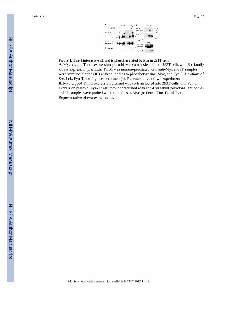
Figure 1. Tim-1 interacts with and is phosphorylated by Fyn in 293T cellsA. Myc-tagged Tim-1 expression plasmid was co-transfected into 293T cells with Src familykinase expression plasmids. Tim-1 was immunoprecitated with anti-Myc and IP sampleswere immuno-blotted (IB) with antibodies to phosphotyrosine, Myc, and Fyn-T. Positions ofSrc, Lck, Fyn-T, and Lyn are indicated (*). Representative of two experiments.B. Myc-tagged Tim-1 expression plasmid was co-transfected into 293T cells with Fyn-Texpression plasmid. Fyn-T was immunoprecitated with anti-Fyn rabbit polyclonal antibodiesand IP samples were probed with antibodies to Myc (to detect Tim-1) and Fyn.Representative of two experiments.
Curtiss et al. Page 12
Mol Immunol. Author manuscript; available in PMC 2012 July 1.
NIH
-PA Author Manuscript
NIH
-PA Author Manuscript
NIH
-PA Author Manuscript
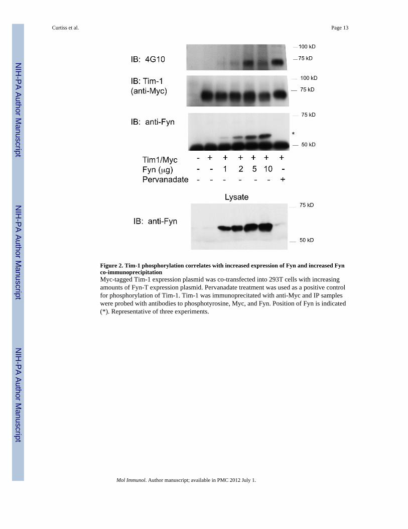
Figure 2. Tim-1 phosphorylation correlates with increased expression of Fyn and increased Fynco-immunoprecipitationMyc-tagged Tim-1 expression plasmid was co-transfected into 293T cells with increasingamounts of Fyn-T expression plasmid. Pervanadate treatment was used as a positive controlfor phosphorylation of Tim-1. Tim-1 was immunoprecitated with anti-Myc and IP sampleswere probed with antibodies to phosphotyrosine, Myc, and Fyn. Position of Fyn is indicated(*). Representative of three experiments.
Curtiss et al. Page 13
Mol Immunol. Author manuscript; available in PMC 2012 July 1.
NIH
-PA Author Manuscript
NIH
-PA Author Manuscript
NIH
-PA Author Manuscript

Figure 3. Fyn binds Tim-1 in the absence of tyrosine 299A. Myc-tagged Tim-1 or a Tim-1 Y299F mutant expression plasmid were co-transfectedinto 293T cells with Fyn-T expression plasmid. Pervanadate treatment was used as apositive control for phosphorylation of Tim-1. Tim-1 was immunoprecitated with anti-Mycand IP samples were probed with antibodies to phosphotyrosine, Myc, and Fyn. Position ofFyn-T is indicated (*). Representative of three experiments.B. Myc-tagged Tim-1 was co-transfected into 293T cells with increasing amounts of a“kinase-dead” K296E mutant Fyn (isoform Fyn-b) expression plasmid. Pervanadatetreatment or co-transfection with wildtype Fyn expression plasmid was used as a positivecontrol for phosphorylation of Tim-1. Tim-1 was immunoprecitated with anti-Myc and IPsamples were probed with antibodies to phosphotyrosine, Myc, and Fyn. Position of Fyn-bis indicated (*). Representative of three experiments.
Curtiss et al. Page 14
Mol Immunol. Author manuscript; available in PMC 2012 July 1.
NIH
-PA Author Manuscript
NIH
-PA Author Manuscript
NIH
-PA Author Manuscript

Figure 4. Tim-1 is phosphorylated in B cells upon receptor engagement with antibody-coatedbeadsM12 cells were incubated with beads conjugated to anti-Tim-1 or isotype control antibodiesfor 5 minutes, then lysed and Tim-1 was immunoprecipitated. M12 cells treated withpervandate and anti-Tim-1 beads for 10 minutes were used as a positive control. IPs wereprobed with antibodies to phosphotyrosine, Tim-1, and Fyn. Position of Fyn is indicated (*).Representative of four experiments.
Curtiss et al. Page 15
Mol Immunol. Author manuscript; available in PMC 2012 July 1.
NIH
-PA Author Manuscript
NIH
-PA Author Manuscript
NIH
-PA Author Manuscript

Figure 5. Tim-1 phosphorylation is reduced in Fyn-deficient B cell linesA. M12 cells were transduced with a retrovirus encoding constructs targeting Fyn or acontrol shRNA to luciferase. Fyn knockdown in stable cell lines was assessed by Westernblot using a FujiImager to quantify signal intensity. Mean values of three experiments areshown; error bars represent standard error of mean.B. M12 cells were incubated with beads conjugated to anti-Tim-1 or isotype control for 5minutes, then lysed and Tim-1 was immunoprecipitated. IPs were probed with antibodies tophosphotyrosine, Tim-1, and Fyn. Phosphorylated Tim-1 (4G10 relative to anti-Tim1 signal)was assessed by western blot using a FujiImager to quantify signal intensity fromimmunoprecipitated Tim-1. Representative of two experiments.
Curtiss et al. Page 16
Mol Immunol. Author manuscript; available in PMC 2012 July 1.
NIH
-PA Author Manuscript
NIH
-PA Author Manuscript
NIH
-PA Author Manuscript