Formation and deformation mechanisms of pyroxene-spinel symplectite in an ascending mantle, the...
15
1 Formation and deformation mechanisms of pyroxene-spinel symplectite in an ascending mantle Journal of Mineralogical and Petrological Sciences, Volume 103, page 1─15, 2008 doi:10.2465/jmps.070222b K. Ozawa, [email protected]-tokyo.ac.jp Corresponding authour T. Morishita, [email protected]-u.ac.jp Formation and deformation mechanisms of pyroxene-spinel symplectite in an ascending mantle, the Horoman peridotite complex, Japan: An EBSD (electron backscatter diffraction) study Norihiro ODASHIMA * , Tomoaki MORISHITA ** , Kazuhito OZAWA * , Hiroko NAGAHARA * , Akira T SUCHIYAMA *** and Ryoko NAGASHIMA **** * Department of Earth and Planetary Science, The University of Tokyo, Tokyo 113-0033, Japan ** Frontier Science Organization, Kanazawa University, Kanazawa 920-1192, Japan *** Department of Earth and Space Science, Graduate School of Science, Osaka University, 1 -1 Machikaneyama, Toyonaka 560-0043, Japan **** Department of Earth Sciences, Faculty of Science, Kanazawa University, Kanazawa 920-1192, Japan Symplectites, fine vermicular intergrowth (< 10 μm) of orthopyroxene, clinopyroxene, and spinel, and the sur- rounding lenticular coarser-grained (100-200 μm) aggregate (seam) with the same mineral assemblage of the symplectites define remarkable foliation and lineation in spinel lherzolites of the Horoman complex, northern Japan. They are inferred to be products of reaction between garnet and olivine during decompression of the host peridotite accompanying deformation. Microstructure of a symplectite was investigated with automated electron backscattered diffraction (EBSD) analysis using a field-emission gun SEM (FE-SEM) in order to clarify reaction and deformation mechanisms and thereby better constraining mechanical interaction between the Earth’s upper mantle and lower crust. The symplectite is composed of two segments with a large misorien- tation angle of ~ 60° only for spinel, and the two spinel crystals are in mirror symmetry with the segment boundary approximately parallel to the mirror plane. The segment boundary is interpreted as spinel law twin formed during phase transition from garnet. Each segment is further subdivided into several sectors with grad- ual lattice distortion smaller than a few degrees/mm and intra-sector misorientation mostly smaller than 25° for all constituent minerals and with misorientation axes nearly perpendicular to the lineation and parallel to the foliation. The sector boundaries are inferred to be subgrain boundaries formed by dislocation creep of py- roxenes and spinel in the spinel stability field. The spinel twin suggests that a garnet was decomposed directly into entangled aggregate of pyroxenes and spinel, which grew from an embryo nucleated on the surface of the reactant garnet. The symplectite minerals in each sector show systematic crystallographic orientations (topo- taxy) with each other. The topotaxial relationship in the fine intergrowth with subgrain structure demonstrates that the systematic crystallographic orientations were acquired when the crystals grew by decomposing the garnet and were later modified by deformation during the consecutive ascent of the complex. Keywords: Symplectite, Mantle peridotite, Crystallographic orientation, Phase transition, Ascent history, Spinel law twin
-
Upload
kanazawa-u -
Category
Documents
-
view
1 -
download
0
Transcript of Formation and deformation mechanisms of pyroxene-spinel symplectite in an ascending mantle, the...

1Formation and deformation mechanisms of pyroxene-spinel symplectite in an ascending mantleJournal of Mineralogical and Petrological Sciences, Volume 103, page 1─15, 2008
doi:10.2465/jmps.070222bK. Ozawa, [email protected]-tokyo.ac.jp Corresponding authourT. Morishita, [email protected]-u.ac.jp
Formation and deformation mechanisms of pyroxene-spinel symplectite in an ascending mantle, the Horoman peridotite
complex, Japan: An EBSD (electron backscatter diffraction) study
Norihiro ODASHIMA*, Tomoaki MORISHITA**, Kazuhito OZAWA*, Hiroko NAGAHARA*, Akira TSUCHIYAMA*** and Ryoko NAGASHIMA****
*Department of Earth and Planetary Science, The University of Tokyo, Tokyo 113-0033, Japan**Frontier Science Organization, Kanazawa University,
Kanazawa 920-1192, Japan***Department of Earth and Space Science, Graduate School of Science, Osaka University,
1-1 Machikaneyama, Toyonaka 560-0043, Japan****Department of Earth Sciences, Faculty of Science, Kanazawa University,
Kanazawa 920-1192, Japan
Symplectites, fine vermicular intergrowth (< 10 μm) of orthopyroxene, clinopyroxene, and spinel, and the sur-rounding lenticular coarser-grained (100-200 μm) aggregate (seam) with the same mineral assemblage of the symplectites define remarkable foliation and lineation in spinel lherzolites of the Horoman complex, northern Japan. They are inferred to be products of reaction between garnet and olivine during decompression of the host peridotite accompanying deformation. Microstructure of a symplectite was investigated with automated electron backscattered diffraction (EBSD) analysis using a field-emission gun SEM (FE-SEM) in order to clarify reaction and deformation mechanisms and thereby better constraining mechanical interaction between the Earth’s upper mantle and lower crust. The symplectite is composed of two segments with a large misorien-tation angle of ~ 60° only for spinel, and the two spinel crystals are in mirror symmetry with the segment boundary approximately parallel to the mirror plane. The segment boundary is interpreted as spinel law twin formed during phase transition from garnet. Each segment is further subdivided into several sectors with grad-ual lattice distortion smaller than a few degrees/mm and intra-sector misorientation mostly smaller than 25° for all constituent minerals and with misorientation axes nearly perpendicular to the lineation and parallel to the foliation. The sector boundaries are inferred to be subgrain boundaries formed by dislocation creep of py-roxenes and spinel in the spinel stability field. The spinel twin suggests that a garnet was decomposed directly into entangled aggregate of pyroxenes and spinel, which grew from an embryo nucleated on the surface of the reactant garnet. The symplectite minerals in each sector show systematic crystallographic orientations (topo-taxy) with each other. The topotaxial relationship in the fine intergrowth with subgrain structure demonstrates that the systematic crystallographic orientations were acquired when the crystals grew by decomposing the garnet and were later modified by deformation during the consecutive ascent of the complex.
Keywords: Symplectite, Mantle peridotite, Crystallographic orientation, Phase transition, Ascent history, Spinel law twin
2 N. Odashima, T. Morishita, K. Ozawa, H. Nagahara, A. Tsuchiyama and R. Nagashima 3Formation and deformation mechanisms of pyroxene-spinel symplectite in an ascending mantle
INTRODUCTION
Symplectite, fine irregular intergrowth of multiple miner-als commonly found in metamorphic rocks records sub-solidus reactions in response to changes in pressure-tem-perature conditions. Corona structures, which is a con-centric symplectic aggregate of minerals mantling a react-ant mineral grain (e.g., kelyphite rim around garnet), have been investigated to reveal kinetics of mass transfer of chemical species through reaction zones (e.g., Nishiyama, 1983; Obata, 1994). There are, however, limited studies on crystallography and morphology of symplectite miner-als, although transformation-induced microstructures have been extensively studied with transmission electron microscopy to constrain pressure-temperature-time paths of rocks or to evaluate effects of microstructures on phys-ical properties of minerals (Putnis, 1992; Nord, 1992).
Occurrence of symplectites consisting of orthopy-roxene, clinopyroxene and spinel (pyroxene-spinel sym-plectite) and being surrounded by lenticular mineral ag-gregates (lenticular seam hereafter) is a peculiar feature of spinel lherzolite of the Horoman peridotite complex, Japan. The symplectite and the surrounding seam have been interpreted as products of reaction between pyropic garnet and surrounding olivine during decompression of the complex (Kushiro and Yoder, 1966; Tazaki et al., 1972; Takahashi and Arai, 1989; Ozawa and Takahashi, 1995; Takazawa et al., 1996; Morishita and Arai, 1997, 2003; Obata et al., 1997; Takahashi, 2001). Obata et al. (1997), on the basis of the observed spatial correlations between spinel and pyroxene interphase boundaries in some symplectites, argued that the transformation from garnet to two-pyroxene spinel symplectite took place through the initial transformation of garnet to metastable aluminous pyroxene and subsequent spinodal decomposi-tion into Ca-rich and Ca-poor pyroxenes followed by preferential nucleation and growth of spinel along inter-phase boundaries of the two pyroxenes. They, however, did not investigate crystallographic orientation of the con-stituent minerals, which may provide key information to constrain the reaction mechanism. Morishita et al. (2003) studied three-dimensional morphology of the symplectite minerals with high-resolution X-ray CT, but they did not determine crystallographic orientations, either.
Crystallographic orientations of symplectite miner-als as well as their morphological relationship are difficult to determine with a conventional universal stage attached to an optical microscope, because the scale of intergrowth is smaller than thickness of thin sections. Backscattered electron (BSE) images obtained by a secondary electron microscope (SEM) show mean atomic number contrasts with high spatial resolution (Dilks and Graham, 1984),
but are not good enough for phase identification because the mean atomic number depends not only on crystal structure but also on the composition of solid solutions.
An electron backscatter diffraction (EBSD) system attached to an SEM provides crystallographic orientation and images of rock textures by orientation contrasts of mineral grains with high spatial resolution in a relatively short period of time (e.g., Prior et al., 1999). The EBSD system identifies phases according to crystal structure ir-respective of chemical composition, which is one of the robust features of the EBSD analyses. Its spatial resolu-tion is much better if it is attached to a field-emission gun SEM by a factor of 2-3 than that using a tungsten-fila-ment gun at the same beam currents (Humphreys et al., 1999). EBSD study is, therefore, expected to provide use-ful information on the relationship between very fine rock textures and crystallographic orientation of the constitu-ent minerals. It is particularly useful for optically isotro-pic minerals, such as spinel. This paper aims to investi-gate crystallographic orientation of symplectite minerals by EBSD attached to a field-emission gun SEM, and to give constraints on formation and deformation processes of a symplectite.
GEOLOGICAL BACKGROUND AND SAMPLE DESCRIPTION
The Horoman peridotite complex is located at the south-ern end of the Main Zone of the Hidaka metamorphic belt (e.g., Komatsu et al., 1982). The complex is 8 × 10 km2 in area and more than 3 km in thickness, and consists of various kinds of peridotite with small amounts of mafic rocks (e.g., Igi, 1953; Komatsu and Nochi, 1966; Niida, 1984; Takahashi, 1991, 1992; Takazawa et al., 1992, 1999, 2000; Yoshikawa and Nakamura, 2000; Sawaguchi 2004). Symplectites occur in spinel lherzolite, which is usually sandwiched between plagioclase lherzolite and harzburgite on various scales (Takahashi, 1991; Takazawa et al., 2000). Morishita and Arai (2003) reported that the size of symplectite minerals increases continuously from
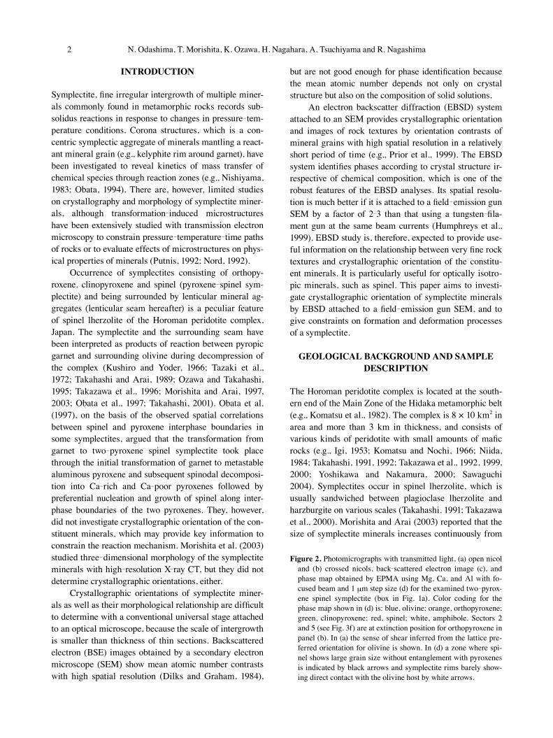
Figure 2. Photomicrographs with transmitted light, (a) open nicol and (b) crossed nicols, back-scattered electron image (c), and phase map obtained by EPMA using Mg, Ca, and Al with fo-cused beam and 1 μm step size (d) for the examined two-pyrox-ene spinel symplectite (box in Fig. 1a). Color coding for the phase map shown in (d) is: blue, olivine; orange, orthopyroxene; green, clinopyroxene; red, spinel; white, amphibole. Sectors 2 and 5 (see Fig. 3f) are at extinction position for orthopyroxene in panel (b). In (a) the sense of shear inferred from the lattice pre-ferred orientation for olivine is shown. In (d) a zone where spi-nel shows large grain size without entanglement with pyroxenes is indicated by black arrows and symplectite rims barely show-ing direct contact with the olivine host by white arrows.

2 N. Odashima, T. Morishita, K. Ozawa, H. Nagahara, A. Tsuchiyama and R. Nagashima 3Formation and deformation mechanisms of pyroxene-spinel symplectite in an ascending mantle
Figure 1. Photographs of the studied thin section of symplectite-bearing spinel lherzolite from the basal part of the Lower Zone of the Horoman complex: (a) open nicol and (b) crossed nicols. Brownish streaks in (a) are lenticular mineral aggregate (seam) including spinel-pyroxene symplectites, indicated by arrows. The aggregates define the foliation and lineation. The arrow with X in-dicates the lineation and that with Z the pole of the foliation. The largest symplectite indicated by a box was examined in detail in this paper. The rock shows porphyro-clastic texture featuring porphyro-clasts of pyroxenes and finer-grained matrix of olivine as shown in (b). The sense of shear deter-mined on the basis of the lattice preferred orientation of olivine is shown in (a).
4 N. Odashima, T. Morishita, K. Ozawa, H. Nagahara, A. Tsuchiyama and R. Nagashima 5Formation and deformation mechanisms of pyroxene-spinel symplectite in an ascending mantle
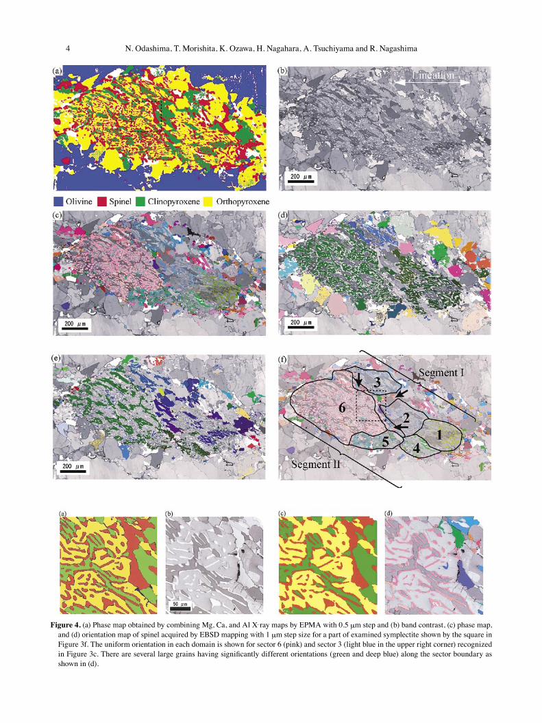
Figure 4. (a) Phase map obtained by combining Mg, Ca, and Al X-ray maps by EPMA with 0.5 μm step and (b) band contrast, (c) phase map, and (d) orientation map of spinel acquired by EBSD mapping with 1 μm step size for a part of examined symplectite shown by the square in Figure 3f. The uniform orientation in each domain is shown for sector 6 (pink) and sector 3 (light blue in the upper right corner) recognized in Figure 3c. There are several large grains having significantly different orientations (green and deep blue) along the sector boundary as shown in (d).

4 N. Odashima, T. Morishita, K. Ozawa, H. Nagahara, A. Tsuchiyama and R. Nagashima 5Formation and deformation mechanisms of pyroxene-spinel symplectite in an ascending mantle
the bottom to the top of the complex.The sample examined in this study (Fig. 1) was col-
lected from an outcrop at the entrance of abandoned quarry of the Toho Olivine Ltd. in the basal part of the complex. It is a porphyroclastic spinel lherzolite belong-ing to the Main Harzburgite Lherzolite (MHL) suite of Takahashi (1991, 1992), and consists of olivine (42 vol%), orthopyroxene (29%), clinopyroxene (15%), and symplec-tite + lenticular mineral aggregate (14%). The peridotite shows strong foliation and lineation defined by elongated and flattened lenticular seams and porphyroclasts of py-roxenes (Fig. 1). Olivine has Fo mol% of 90.8, typical for lherzolite of the MHL and shows a lattice-preferred ori-entation (LPO) featuring a (100) point maximum several degrees away from the lineation and partial girdles of (010) and (001) oriented vertical to the lineation. The sense of shear inferred from the asymmetric LPO relative to the structural elements is shown in Figure 1a, which is top-to-the-north according to Sawaguchi (2004). Spinel occurs in lenticular seams or as isolated grains, and has Cr/(Cr + Al)(Cr#) of ~ 0.3. Spinel in lenticular seams tends to have higher Cr# and more notable heterogeneity characterized by coexistence of Cr and Al maxima along grain rims (Ozawa, 1989) than those in symplectite.
The examined pyroxene-spinel symplectites are em-bedded in a lenticular seam consisting of the same miner-als as those in the symplectites (orthopyroxene, clinopy-roxene, and spinel). The seam minerals have grain size of 100-200 μm, which are smaller than the minerals occur-ring outside of the lenticular seam (matrix hereafter) but are greater than those in the symplectites. The symplec-tites are not in direct contact with olivine in the matrix.
SAMPLE PREPARATION AND ANALYSIS
The sample was sliced perpendicular to the foliation and parallel to the lineation for a thin section. The thin sec-tion was at first polished with diamond powder of 1 μm in size. Then, it was finished with 0.04 μm colloidal silica in order to get an undamaged crystalline lattice with a few nanometers thickness from the specimen surface by chemical-mechanical polishing. Thin section was finally coated with a thin layer of carbon.
Back-scattered electron images of the samples were taken under beam-scan mode of an EPMA (JEOL JXA 8800) at the Cooperative Research Center of Kanazawa University. Analytical conditions are an acceleration volt-age of 20 kV and a probe current of 1 × 10⁻⁸A with beam size of 1 μm. X-ray maps were obtained by an EPMA (JEOL JXA8900) at the Department of Earth and Plane-tary Science, the University of Tokyo (EPS-UT) with fo-cused beam, step sizes of 1 μm or 0.5 μm, 15kV accelera-tion voltage, and 1 × 10⁻⁷A probe current. The intensity maps for Mg, Al, and Ca were combined to construct a phase map. The crystallographic axes for individual points were determined by an SEM JEOL 7000F with a field-emission gun at EPS-UT under the following condi-tions: tilt angle of 70°, a working distance of 15 mm, an acceleration voltage of 15 kV, and a probe current of 9 × 10⁻⁹ A. The diffraction pattern was obtained with the HKL-Channel 5 system (a phosphor screen, a low light CCD camera, and a Hamamatsu image processor). The HKL data processing system (Flamenco) was used to de-tect Kikuchi bands and obtain crystallographic orienta-tion. Crystal orientation was determined by comparing the observed pattern of Kikuchi bands with simulated patterns for known crystallographic structures. Orien-tation measurements below 1.3° in Mean Angular Devi-ation (MAD) were accepted, where MAD is the averaged angular misfit between detected and simulated Kikuchi bands. Measured crystallographic axes are plotted on ste-reographic projection (upper hemisphere) in the structural XZ reference frame (plane vertical to the foliation and parallel to the lineation). The step size in orientation map-ping was 1-3 μm. In orientation mapping, measurements failed to be indexed due to the above mentioned limit of MAD > 1.3 or those clearly misidentified as an incorrect phase were either deleted or replaced by the surrounding data points with MAD < 1.3. Several symplectites chosen from those indicated by arrows in Figure 1a were exam-ined, and internal structure of the largest symplectite in-dicated by the box in Figure 1a was examined in detail.
Figure 3. Results of EBSD analyses of the symplectite studied in detail (box in Figs. 1a and Fig. 2) with the step size of 3 μm. (a) EBSD phase map showing each mineral with specific color cod-ing. (b) Band contrast (the relative averaged intensity of the Ki-kuchi bands). (c), (d), and (e) Orientation maps showing crystal-lographic orientations of spinel, orthopyroxene, and clinopyroxene, respectively, with Euler coloring (coloring scheme that maps the three Euler angles to an RGB color) over-lapped with band contrast. In (f), six sectors with consistent crystallographic orientation (i.e., the same color) for spinel are identified and labeled as 1 to 6. These sectors are grouped into two segments I and II, according to the magnitude of spinel mis-orientation angles between sectors. Sectors 1, 2, and 3 from seg-ment I and sectors 4, 5, and 6 from segment II are also noticed in the orientation map for clinopyroxene. A blow up of squared area in (a) and (f) is shown in Figure 4. White parts are grains (mostly orthopyroxene) that were not properly indexed with MAD < 1.3. Arrows in (f) indicate zones where spine grains are coarse, show Euler colors distinct from the nearby sectors, and occur along interphase boundary of pyroxenes.

6 N. Odashima, T. Morishita, K. Ozawa, H. Nagahara, A. Tsuchiyama and R. Nagashima 7Formation and deformation mechanisms of pyroxene-spinel symplectite in an ascending mantle
RESULTS
Optical photomicrographs of the studied symplectite are shown in Figures 2a and 2b, in which morphology of fine symplectite minerals are not clearly recognized. A BSE image shown in Figure 2c more clearly delineate phase boundaries than optical photomicrographs, but mineral species are not properly distinguished. Figure 2d shows an EPMA phase map combining X-ray maps of Mg, Ca, and Al acquired with 1 μm step size, and Figure 3a is a phase map acquired by EBSD with 3 μm step size. A part of the symplectite was mapped by EMPA with 0.5 μm step size (Fig. 4a) and EBSD with 1 μm step size (Fig. 4c). These phase maps provide superior images of the symplectite texture over optical or BSE images, and are mainly used below to show morphology, grain size, and distribution of minerals in symplectite.
Figures 3b and 4b are images of band contrast (aver-age relative intensity of the Kikuchi band) acquired in EBSD measurement and have information on crystallo-graphic orientations and crystal structure. Combination of the band contrast images and phase maps provides excel-lent information on microstructure of symplectite, al-though the information is limited in two-dimensions. Figures 3c and 4d are orientation maps for spinel ren-dered by Euler coloring, which is a coloring scheme that maps three Euler angles (three rotation angles to match two coordinate systems) to an RGB color, and Figures 3d and 3e for pyroxenes. Combination of the orientation maps for all constituent minerals represents an image of two-dimensional microstructure of symplectite with com-plete information on distribution, morphology, and crys-tallographic orientation of symplectite minerals. The ori-entation maps (Figs. 3c-3e and 4c) are used below to describe crystallographic orientations of symplectite min-erals with additional quantitative information on misori-entation (axis and angle of rotation).
Morphology, grain size, and distribution of minerals
The symplectite consists of ~ 50% orthopyroxene, ~ 25% clinopyroxene, and ~ 25% spinel (Figs. 2d and 3a), which is a typical modal abundance of two-pyroxene spinel symplectites from the Horoman complex (Obata et al., 1997; Morishita and Arai, 2003). Orthopyroxene is appar-ently continuous in the two dimensional section, whereas clinopyroxene is isolated except for the domain indicated by black arrows in Figure 2d. Spinel is mostly finely en-tangled with pyroxenes on the scale of < 5 μm (Fig. 4), but it has apparent size of ~ 50 μm and occurs along in-terphase boundaries of pyroxenes in the domain where clinopyroxene is larger in grain size and exceptionally
continuous (indicated by black arrows in Fig. 2d). Clino-pyroxene and spinel elongate in the direction oblique to the lineation by ~ 30° (Figs. 2c, 2d, 3a, 3c, and 3e), which is also visible in optical photomicrographs as alignment of brownish streaky spinel (Figs. 2a and 2b). The size of spinel decreases from the lower right to the upper left ends of the symplectite, approximately paralleling the elon-gation of spinel and clinopyroxene (Fig. 2d).
The symplectite has an elliptic outline with its long axis running oblique to the lineation by ~ 15° (Figs. 2a and 2b). It is mantled by lenticular seam consisting of coarser-grained (100-200 μm) orthopyroxene with minor clinopyroxene and spinel (Figs. 2d and 3a). The thickness of the lenticular seam surrounding the symplectite tapers off at the lower right and the upper left of the symplectite, barely exposing the symplectite to the host olivine (white arrows in Fig. 2d). A minor amount of amphibole is pres-ent in the seam, white parts in Figure 2d, but not within the symplectite, which suggests that the amphibole forma-tion is not associated with the formation of symplectite but with that of lenticular seam. Olivine is absent in the central part of lenticular seams as well as in the symplec-tite, but invades into the seam as shown in Figure 2d (e.g., near the lower right end or near the lower left side of the symplectite).
Crystallographic orientations of minerals and internal structure
The symplectite is divided into two segments, I and II, ac-cording to the difference in crystallographic orientations of spinel as large as 55° in misorientation angle (angle of rotation around a misorientation axis to match two neigh-boring crystals; Figs. 3f and 5a). By contrast, the misori-entation angles for clinopyroxene and orthopyroxene be-tween the two segments are less than 10° (Fig. 5a). Each segment is further divided into several sectors with differ-ent crystallographic orientations noted by different colors for all constituent minerals (Figs. 3c and 4d for spinel, 3d for orthopyroxene, and 3e for clinopyroxene); 1, 2, and 3 sectors in segment I and 4, 5, and 6 sectors in segment II as shown in Figure 3f. Sectors recognized by a small dif-ference in extinction position of pyroxenes under an opti-cal microscope (Fig. 2b) well correspond to those identi-fied with EBSD (Figs. 3d and 3e). The crystallographic orientations of spinel and pyroxene progressively change within each sector and show notable gaps at the sector boundaries along the elongated direction (Fig. 5b). The misorientation angles between adjacent sectors are 10-20° for spinel and 10 to 30° for pyroxenes between 1-2, 2-3, 4-5, and 5-6 pairs (Fig. 5b), and the misorientation angles for spinel and pyroxenes are comparable. As mentioned

6 N. Odashima, T. Morishita, K. Ozawa, H. Nagahara, A. Tsuchiyama and R. Nagashima 7Formation and deformation mechanisms of pyroxene-spinel symplectite in an ascending mantle
above, the size of spinel decreases from the lower right to the upper left, and correspondingly the width of spinel varies from ~ 10 μm in sectors 1 and 4 to ~ 5 μm in sec-tors 2 and 5, through ~ 3 μm in sectors 3 and 6. The size change is locally abrupt, but it does not necessarily occur along the sector or segment boundaries (e.g., sectors 6 and 3 in Fig. 2d). Smaller symplectites (< 50 μm across) examined in the thin section do not have multiple sectors nor segment structure.
Misorientation axes (rotation axis to match two neighboring crystals) for sector and segment boundaries are shown in Figures 6a and 6b. They are at high angles or nearly perpendicular to the thin section, parallel to the foliation and nearly perpendicular to the lineation. The misorientation axes for the 1-2 and 4-5 boundaries are in-clined by 10-40° towards (100) of pyroxenes and one of {111} of spinel in segment II (Fig. 6c), and those for the 2-3 and 5-6 boundaries 10-40° in the opposite direction. Misorientation axes of all symplectite minerals for the I-
II segment boundaries are also at high angle to the thin section (Fig. 6b). All the minerals show gradual changes in crystallographic orientation due to continuous lattice distortion less than a few degrees/mm [1-15° in total; see open (segment I) and shaded (segment II) areas delin-eated for {111} and {101} in Figs. 6c and 6d, respectively]. This lattice distortion has misorientation axes nearly per-pendicular to the thin section (e.g., contours for orthopy-roxene in Fig. 6a). The misorientation axes more or less coincide with those of sector boundaries irrespective of minerals (Fig. 6a).
The crystallographic orientations of minerals in each sector have systematic relationships with each other (Fig. 6e): the (100) and (010) planes and [001] axis of orthopy-roxene and those of clinopyroxene are identical within < 5°, and the (100) and (010) planes of pyroxenes are nearly parallel to one of the {111} and {101} planes of spi-nel within < 10°, respectively. The degree of deviation from the systematic relationship between crystallographic orientations of spinel and pyroxenes varies sector by sec-tor. Smaller sectors, such as 5 and 4 show the largest de-viation and the largest sector 6 shows negligible deviation (Figs. 6e and 7a). The amount of deviation from the coin-cidence of one of {111} of spinel and (100) of orthopyrox-ene tends to increase from sectors 3 and 6 towards sectors 1 and 4 through sectors 2 and 5, which shows the same trend as the change in size of spinel mentioned above.
Larger spinel grains are present along the boundary of the two segments and 2-3 sector boundary with crys-tallographic orientations deviated from the adjacent sym-plectic grains (arrows in Fig. 3f and upper right corner of Fig. 4d). This coarse-grained domain is similar to the lenticular seam mantling the symplectite in terms of size, morphology, and crystallographic orientations for all the constituent minerals (Figs. 2 and 3).
Lattice preferred orientation (LPO) of orthopyroxene porphyroclasts is characterized by weak (001) maximum near the lineation, exhibiting the same sense of shear esti-mated by the olivine LPO and girdles of (100) and (010) nearly perpendicular to the lineation. Orthopyroxene in the surrounding lenticular seam shows an LPO similar to that of orthopyroxene porphyroclasts, but it has weak (100) maximum nearly perpendicular to the foliation. Crystallographic orientations of symplectite orthopyrox-ene in the studied thin section show a large scatter, but are roughly concordant with LPO of orthopyroxene in the lenticular seam and in the matrix as porphyroclasts.
Figure 5. Misorientation angles between adjacent sectors identi-fied in constituent minerals of the symplectite shown in Figure 2 with EBSD: (a) those for 1-4, 2-5, and 3-6 segment boundaries, which are < 10° for pyroxenes but 50-60° for spinel, and (b) misorientation angles for 6-5, 5-4, 1-2, and 2-3 sector boundar-ies, which are usually 10-30°. There are continuous change in crystallographic orientation in each sector, which is smaller than a few degrees/mm. Insets in (b) show outlines of sectors recog-nized by spinel orientation map (Fig. 3f), and two groups of sec-tors, segments I and II, which are recognized by a large misori-entation angle of spinel. The axes for misorientations are mostly nearly perpendicular to the thin section (see Figs. 6a and 6b).

8 N. Odashima, T. Morishita, K. Ozawa, H. Nagahara, A. Tsuchiyama and R. Nagashima 9Formation and deformation mechanisms of pyroxene-spinel symplectite in an ascending mantle
DISCUSSION
Three-dimensional morphology of symplectite spinel
The three-dimensional morphologies of spinel and pyrox-enes in symplectite constructed by successive sectioning or by X-ray C-T scan (Morishita, 2000; Morishita et al., 2003) show that the spinel grains, which are apparently isolated in a two-dimensional section, forms a three-di-mensionally continuous network, and that one symplectite contains several networks of entangled crystals of clino-pyroxene and orthopyroxene finely intergrown with spi-nel.
Another important three-dimensional morphological feature of symplectite spinel is that spinel elongates in one direction. The X-ray C-T observation shows that spinel shows coherent and strong elongation in a particular di-rection (Morishita et al., 2003). This unidirectional elon-gation of spinel is not rare and has been reported as two-dimensional elongation of symplectite spinel (e.g., Ozawa and Takahashi, 1995; Morishita et al., 1995). Vermicular intergrowth without preferential elongation is also ob-served under an optical microscope in the same thin sec-tion, suggesting that the elongation is not solely due to de-formation. The occurrence of such vermicular intergrowth is understood as morphology in a section highly oblique
to the unidirectional elongation (Morishita et al., 2003). Symplectite consisting of spinel with radial elongation from the symplectite center is rarely observed but always occurs as an inclusion in orthopyroxene porphyroclasts (e.g., Takahashi and Arai, 1989). Symplectite clinopyrox-ene also elongates paralleling the spinel elongation (Figs. 2c and 2d). As described above, the size of spinel in the symplectite decreases from the lower right towards the upper left. This size change paralleling the aforemen-tioned elongation of symplectite spinel is often observed in symplectite-bearing spinel lherzolites (e.g., Ozawa and Takahashi, 1995).
The origin of the segment boundary
The segment boundary at which spinel shows ~ 60° mis-orientation is inferred to be a spinel law twin formed dur-ing phase transition from pyropic garnet. Pyroxenes, finely entangled with spinel, show less than 5-10° misori-entation (Fig. 5a), which is much smaller than ~ 60° mis-orientation for spinel, indicating that the boundary forma-tion must have been concurrent with or predated the garnet decomposition. Any processes conceivable after the symplectite formation, such as deformation, cannot explain the large difference in misorientation angles. The twin relation of spinel is more clearly shown by Figures
Figure 6. (a) Misorientation axes of gradual distortion for orthopyroxene (con-tours) within sectors and those at sector boundary for spinel (solid circle), orthopyroxene (open circle), and clinopyroxene (gray circle). (b) Misorienta-tion axes at the segment boundary for constituent minerals. Symbols are the same as those in (a). (c) and (d) Stereographic projection of poles of all the {111} and {101} planes of spinel for each segment in the orientation map (Fig. 3f), respectively, for segment I (white) and segment II (shaded). (e) Stereo-graphic projection of representative orientations of crystallographic axes and planes of symplectite minerals in the largest sector 6. The (100) and (010) planes and [001] axis of orthopyroxene and those of clinopyroxene are coin-cident, respectively. One of the {111} and {101} planes of spinel (indicated by *) coincides with (100) and (010) planes of pyroxenes, respectively. (f) Ste-reographic projection (upper hemisphere) of representative orientations of the {100} planes of spinel for each sector in the orientation map. Numbers denote those in the orientation map (Fig. 3f); sectors 1, 2, and 3 constitute segment I, and sectors 4, 5, and 6 constitute segment II. The data are taken from adjacent measurement within 2 μm in (e). Dashed curves with arrow heads in (c), (d), and (f) represent 55° rotation (misorientation angle between segments I and II; Fig. 5a) around the axis approximately perpendicular to the thin section [1-4 axis for spinel shown in (b)], matching the clusters of segments I and II. Thin solid lines in (a)-(d) and (f) represent a trace of the segment boundary, which nearly coincides with the trace of the mirror plane in (c), (d), and (f) to bring the spinel crystals to match. The tilt angle of the segment boundary relative to the XZ plane is not clear. Broken lines repre-sent trace of great circle perpendicular to the twin boundary {111} of spinel as well as (100) of pyroxenes, which is thought to be the boundary of the two segments in three dimensions. The misorientation axes of internal strain for spinel and clinopyroxene are essentially the same as those for orthopyroxene shown in (a).
8 N. Odashima, T. Morishita, K. Ozawa, H. Nagahara, A. Tsuchiyama and R. Nagashima 9Formation and deformation mechanisms of pyroxene-spinel symplectite in an ascending mantle
6c, 6d, and 6f. Spinel crystals in the two segments are in mirror symmetry with regard to the trace of one of {111} of spinel, which approximately conincides with the trace of the segment boundary (thin solid line in Figs. 6c and 6d). Figure 7c compares the lattice orientations of spinel in sector 3 with the lattice orientations in a twin relation to the spinel in sector 6. The two sets of orientations
match within ~ 10°.
Processes of symplectite formation
If the sector structure and 5-10° rotation between the two segments, which are attributed to deformation of the sym-plectite as will be shown below, are ignored, the symplec-tite consists of two crystals of spinel in twin relationship with the segment boundary as twin plane and single crys-tals of orthopyroxene and clinopyroxene (at least in two dimensions). The unique crystallographic orientation for pyroxenes in the studied symplectite is contrasting to ke-lyphitic rims developed between garnet and olivine in garnet peridotites from near Mt. Kinabalu, Kalimantan, East Malaysia (Imai and Ozawa, 1991) and in garnet py-roxenites from Ronda (Obata, 1994). In these cases, py-roxenes mantling a reactant garnet have variable crystal-lographic orientations controlled mainly by the orientation of the reaction front or the initial garnet-olivine interface. The fibrous mineral elongation is principally controlled by the garnet morphology, which resulted in the concen-tric reaction rim if the reactant garnet is completely sur-rounded by olivine. The observed symplectite morphol-ogy, particularly the unidirectional elongation of spinel and clinopyroxene, is contrasting to the concentric micro-structures of kelyphite.
The contrasting textural features of the Horoman symplectite and Kalimantan kelyphite suggests that the transformation initiated by a very few or even a single nu-cleation of pyroxenes and spinel on the surface of garnet or that crystals with a specific crystallographic orientation were selectively grown towards interior of reacting gar-net. The single nucleus (at least in two dimensions) sce-nario is more plausible, because it is highly unlikely that all nuclei of spinel formed on one side of the garnet-oliv-ine boundary exclusively chose one of the two twin orien-tations and encountered with spinel nucleated and grown from the opposite boundary forming a planner segment boundary.
The change in spinel size from sectors 1 and 4 to sectors 3 and 6, approximately paralleling the two-di-mensional elongation of spinel and clinopyroxene as well as the orientation of twin boundary of spinel, suggests that symplectite was firstly nucleated on a garnet surface located at a 1 and 4 segment boundary and grew towards sectors 3 and 6. This scenario is in agreement with pre-sumed diffusion-controlled mechanism of the phase tran-sition, because the diffusion distance for the growth of symplectite is much larger that that for grain growth of spinel. The spinel grains formed earlier in sectors 1 and 4 may have chance to have been coarsened during the phase transition in order to minimize interfacial energy.
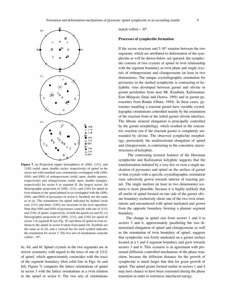
Figure 7. (a) Projection (upper hemisphere) of {100}, {111}, and {110} (solid, open, double circles, respectively) of spinel in the stereo net with standard axes orientations overlapped with (100), (010), and [001] of orthopyroxene (solid, open, double squares, respectively) and clinopyroxene (solid, open, double triangles, respectively) for sector 6 in segment II, the largest sector. (b) Stereographic projection of {100}, {111}, and {110} for spinel in twin relation to the spinel plotted in (a) overlapped with the (100), (010), and [001] of pyroxenes of sector 6. Symbols are the same as in (a). The orientations for spinel indicated by dashed circle (one {111} and three {110}) are invariant in the twin operation. Note that (100) and (010) of pyroxenes coincide with one of {111} and {110} of spinel, respectively, in both the panels (a) and (b). (c) Stereographic projection of {100}, {111}, and {110} for spinel of sector 3 in segment II (see Fig. 3f) and those of spinel in twin re-lation to the spinel in sector 6 taken from panel (b). Symbols are the same as in (b), and a vertical bar for each symbol indicates the orientation for sector 3. The two sets of orientations coincide within ~ 10°.

10 N. Odashima, T. Morishita, K. Ozawa, H. Nagahara, A. Tsuchiyama and R. Nagashima 11Formation and deformation mechanisms of pyroxene-spinel symplectite in an ascending mantle
The inference that the twinning was already present in spinel embryo formed on the garnet surface at a 1-4 sector boundary is in accordance with the lack of defor-mation twinning for spinel (Mitchell, 1999). The infer-ence is also supported by Buerger (1945), who advocated that twin formation is favored in the nucleation stage be-cause the presence of twin boundary requires more en-ergy than without it. Buerger (1945) argued that the most obvious time when the degree of super-saturation is very high is just before nuclei start to form. We, therefore, ar-gue that the nucleation of symplectite on garnet surface was very difficult and required a large degree of super-saturation.
Obata et al. (1997) argued that the transformation from garnet to two-pyroxene spinel symplectite was initi-ated by transformation of garnet to metastable aluminous pyroxene followed by spinodal decomposition into Ca-rich and Ca-poor pyroxenes and further by preferential nucleation and growth of spinel along interphase bound-aries of the two pyroxenes. The presence of spinel law twin dividing the symplectite into two segments is irrec-oncilable with this mechanism. In Obata’s model, spinel
nucleated and grew inward at many intersections of py-roxene boundaries and interface with the surrounding ol-ivine randomly choosing one of the two possible crystal-lographic orientations. Obata et al. (1997) also argued that the presumed spatial correlations between spinel and py-roxene interphase boundaries were gradually lost by grain growth of spinel inside pyroxene grains with increasing sphericity or roundness to minimize the interfacial en-ergy. Considering the observed systematic crystallograph-ic orientation of pyroxenes and spinel, however, it is fairly difficult to imagine that spinel nucleated and grew within or penetrated into pyroxene grains by consuming spinel occurring along interphase boundaries. If the mechanism proposed by Obata et al. (1997) was actually operated, spinel grain stayed along interphase boundaries and grew to rounded grains to minimize the interfacial energy, end-ing up microstructure typically observed as the coarse-grained domain in the middle of the symplectite (the area indicated by the arrows in Figs. 2d and 3f) or that of len-ticular seams surrounding symplectites.
Simultaneous growth of spinel and pyroxenes entan-gling with each other starting from their nucleation at
Figure 8. Schematic illustration of the inferred formation and deformation processes of the studied symplectite and the surrounding lenticular aggregate (seam) in spinel lherzolite of the Horoman peridotite complex. (a) A garnet crystal, surrounded by olivine, was stable in the gar-net stability field; (b) The garnet crystal started to react with olivine to form reaction rim consisting of intergrowth of pyroxenes and spinel when the peridotite went into the spinel stability field; (c) The reaction rim slowly grew associated with recrystallization, coarsening, and deformation during the ascent through the spinel stability field; (d) The surrounding reaction product deformed with the relict rigid garnet in the center to produce thin mantle in the direction vertical to the foliation, which acted as short diffusion path to induce nucleation and rapid growth of symplectite with twinned spinel at the garnet surface under highly super-saturated condition; (e) Rapid growth of symplec-tite with keeping spinel twin and suppressing other nuclei to grow; (f) Deformation of the surrounding mineral aggregate and the symplec-tite forming subgrain boundaries and tilting the elongate spinel concordant with the shear sense estimated by the LPO of the matrix olivine. Probable rotation and antithetic shear of the symplectite (Ishii and Sawaguchi, 2002) are shown by arrows in (f).

10 N. Odashima, T. Morishita, K. Ozawa, H. Nagahara, A. Tsuchiyama and R. Nagashima 11Formation and deformation mechanisms of pyroxene-spinel symplectite in an ascending mantle
limited sites on the garnet surface is a more plausible mechanism for garnet decomposition. A limited number of nuclei on the garnet surface implies higher kinetic bar-rier and suppression of further nucleation after the first nucleus started to grow. Plausible mechanisms of the symplectite formation will be discussed below in rele-vance to the formation of the surrounding lenticular seam.
Topotaxial relationships among symplectite minerals
The EBSD study revealed systematic crystallographic re-lationships (topotaxy) among constituent minerals. The observed coincidence of (100), (010), and [001] of ortho-pyroxene and clinopyroxene and that of one of {111} of spinel and (100) of orthopyroxene and clinopyroxene are understood as the shareable orientation of oxygen stack-ing (Cameron and Papike, 1980). Oxygen atoms are in cu-bic close-packing parallel to {111} in the spinel structure and close-packed parallel to (100) in the pyroxene struc-ture. The crystallographic relationship between orthopy-roxene and clinopyroxene in the symplectites is the same as that of Bushveld type exsolution lamella (Mossman, 1971; Cameron and Papike, 1980). This topotaxial rela-tionship between pyroxenes and spinel is invariant to symmetry operation of the spinel law twin as shown in Figures 7a and 7b. Gruner (1929) noted that “oriented growth” takes place only on the crystallographic planes in which the atomic arrangement and spacing are almost alike. Oxygen layers parallel to (100) of pyroxenes and those parallel to {111} of spinel matches with each other within 1 Å misfit. The matching is extremely good for clinopyroxene and orthopyroxene. Therefore, topotaxial relationships between spinel and pyroxenes satisfy the cri-terion for oriented growth after Gruner (1929).
The crystallographic orientation of symplectite min-erals relative to the precursor garnet is not clear in the Horoman complex because of the complete breakdown of garnet. Katayama et al., (2005) reported an elongated gar-net consisting of subgrains, which are interpreted to be induced by deformation. There is a possibility that garnet grains in the Horoman complex also had subgrains before the phase transition and that the spinel and pyroxenes were in topotaxial relationship with the reactant garnet. If this is the case, the mineral orientations and sector struc-ture could have information on deformation of the perido-tite in the garnet stability field. According to Gruner (1929), however, it is fairly unlikely that garnet and spinel or pyroxenes have topotaxial relationships, because the crystal structure of garnet is very complex and has fairly loose oxygen packing, quite different from that of spinel or pyroxenes. This is in accordance with a kelyphitic re-action zone rimming a garnet in garnet lherzolites from
Kalimantan (Imai and Ozawa, 1991) exhibiting various crystallographic orientations of orthopyroxene on the scale of 0.25 mm where it is in contact with olivine. Twinning of pyropic garnet is uncommon, whereas spinel law twin is fairly common.
Deformation of symplectite
The symplectite was deformed after nucleation and growth of spinel and pyroxenes. The deformation is shown by distortion in each sector represented by gradual change in crystallographic orientation. Their misorienta-tion axes of all constituent minerals coincide with each other and nearly perpendicular to the lineation and paral-lel to the foliation. The elongation of the symplectite, in-clined from the lineation by ~ 15° (Fig. 2), the presence of subgrain boundaries (Fig. 3), and the lattice orientations of pyroxenes and spinel (Fig. 6) suggest that the deforma-tion of the symplectite may be due to the (100)[001] slip systems for pyroxenes, which is the dominant slip system observed in orthopyroxene in lenticular seams and por-phyroclasts, and the {111}<110> slip system for spinel. The coincidence of (100) of pyroxenes and one of {111} and the presence of [001] of pyroxenes in the plane de-fined by three of <110> of spinel (Figs. 6 and 7) may have allowed the homogeneous deformation of symplectite by dislocation creep with keeping entangled relationship be-tween spinel and pyroxenes. The angle of ~ 15° between the long axis of the symplectite and [001] of pyroxenes suggests that the symplectite deformed by antithetic slip accompanying rigid body rotation (Ishii and Sawaguchi, 2002; Fig. 8f).
The sector boundaries are interpreted as subgrain boundaries, which were formed by dislocation creep. They are curved and are close to tilt boundaries with a certain twist component. The tapering thickness in the surrounding lenticular seam (white arrows pointing the upper left and the lower right of the symplectite in Fig. 2d) and 5-10° rotation along the segment boundary sug-gest that the deformation may have been associated with counterclockwise rotation and produced elongate outline of the symplectite.
Coarsening of spinel and clinopyroxene occurring in the symplectite along the segment boundaries between 2 - 5 and 3 - 6 sectors (Figs. 2d and 3f arrowed zone and Fig. 4 upper left corner) may be initiated by the transition of twin boundary for spinel to incoherent boundaries probably due to deformation along the twin boundary, the weakest plane in the symplectite. The misorientation an-gle of ~ 55° for spinel, which is deviated from 60° ex-pected for spinel law twin by ~ 5°, and that of 5-10° for pyroxene are attributed to relative rotation of spinel and

12 N. Odashima, T. Morishita, K. Ozawa, H. Nagahara, A. Tsuchiyama and R. Nagashima 13Formation and deformation mechanisms of pyroxene-spinel symplectite in an ascending mantle
pyroxene as a whole by 5-10° around the axis parallel to the foliation and perpendicular to the lineation due to de-formation along the segment boundary. The deviation from the exact twin relation indicates that the coarse-grained zone cannot be explained by a cross sectional ef-fect due to the irregularity of the outline of symplectite and the surrounding lenticular aggregate. The presence of coarse-grained zone along the modified twin boundary supports deformation induced grain growth.
Ozawa (1989) showed that diffusion creep was oper-ated in deformation of a large spinel grain from a harz-burgite sample of the Horoman complex. Marked Cr-Al heterogeneity as reported by Ozawa (1989) is commonly observed in large spinel grains in the lenticular seam, suggesting diffusion creep was actually operated to de-form seam spinel. Diffusion must have taken place to purge spinel filament from pyroxene host and to allow growth of coarser spinel grains along the segment bound-ary (indicated by the arrow in Figs. 2d and 3f). The small size of symplectite spinel suggests possible operation of diffusion creep in its deformation, although any clear Cr# heterogeneity is observed. The variation in magnitude of mismatch of the topotaxial relationship between spinel and orthopyroxene is attributed to a variation in contribu-tion of diffusion creep in addition to the aforementioned dislocation creep, since diffusion creep does not affect the crystallographic orientation. It is not clear which mineral controlled the deformation of the symplectite because of the ambiguity in relative strength of spinel and pyroxenes.
The mechanism and timing of formation of lenticular seems
The same mineral assemblage of symplectite and the sur-rounding lenticular seam indicates that they are both re-action products between olivine and garnet (Ozawa and Takahashi, 1995). There are two possible formation pro-cesses of the seam. One is coarsening of a symplectic complete breakdown product of garnet from the margin with mostly intact symplectite preserved in the core, and the other is a coarsened or intact reaction product be-tween garnet and olivine with relict garnet in the center, which was reacted with olivine to form symplectite later. In the former case, a garnet was efficiently transformed into symplectite leaving no relict core, but in the latter case a relict garnet crystal was present in the center of a lenticular seam. Documented grain growth after the for-mation of the examined symplectite did actually took place as demonstrated by the occurrence of coarse-grained domain along the sector and segment boundaries. Therefore, grain growth after the symplectite formation could have taken place, but was limited in the latter sce-
nario.The limited number of nucleation on a garnet crystal
and maintenance of high degree of super-saturation, which are required to explain the textural features of the symplectite, are realized by the presence of lenticular seam prohibiting direct contact of relict garnet with oliv-ine (Fig. 8c). This is supported by the observation that garnet crystals in garnet lherzolite do not have the outer-most coarse reaction zone along interphase boundary with pyroxenes, although a thicker reaction zone develops along the boundary with olivine (e.g., garnet lherzolite from Kalimantan; Imai and Ozawa, 1991). The lenticular seam, therefore, must have been present before the forma-tion of symplectite. A relict garnet was present in the cen-ter of lenticular seams during slow phase transition gov-erned by diffusion along interphase boundaries of coarsened mantle of pyroxene-spinel aggregate (Fig. 8c).
The nucleation may have started at the place where lenticular seam became thin due to deformation of the pe-ridotite in the deeper part of the spinel stability field, shortening diffusion distance for garnet-olivine reaction. The thinning was caused by the presence of rigid garnet in the middle of pyroxenes and spinel aggregate. As de-scribed above, olivine tends to have invaded into lenticu-lar seams probably by deformation, which also could have triggered nucleation of the symplectite (Fig. 8d). Suppres-sion of further nucleation can be explained by depletion of olivine component along garnet-pyroxene interphase boundaries present before the nucleation due to preferen-tial diffusion of olivine component towards the growth site of symplectite through huge interphase areas created by symplectite formation itself, further enhancing sym-plectite growth inside the garnet. This is a typical positive feedback or self-focusing often observed in reactive trans-port processes (Ortoleva, 1994).
The notable contrasts of spinel morphology and grain size of pyroxenes and spinel between the symplec-tite and lenticular seam suggest at least two stages of re-action of garnet break down (Fig. 8). Abart et al. (2004) experimentally reproduced a reaction zone (enstatite) be-tween forsterite and quartz, which is zoned in terms of grain size, microstructure, and LPO. They interpreted that the outer zone composed of large enstatite grew from the initial forsterite-quartz interface consuming quartz and that the inner zone composed of much finer enstatite grew consuming forsterite. In the inner zone, the grain size of enstatite increases towards the initial boundary suggesting the concomitant grain growth during forster-ite-quartz reaction. The reported structure of reaction zone mimics the relationship between the inner finer-grained symplectite aggregate and the surrounding coarser-grained lenticular seam observed in our sample.

12 N. Odashima, T. Morishita, K. Ozawa, H. Nagahara, A. Tsuchiyama and R. Nagashima 13Formation and deformation mechanisms of pyroxene-spinel symplectite in an ascending mantle
However, there are critical differences from experimen-tally reproduced reaction zone by Abart et al. (2004). Firstly, the size contrast between the inner and outer zones is almost two orders of magnitude in the Horoman case, indicating that there must be discontinuity in garnet decomposition. Secondary, the size change in symplectite is from one end to another end of the symplectite, which is contrasting to the concentric size change observed in the experimental results. Therefore, olivine-garnet reac-tion in the Horoman complex proceeded through two stages of reaction: slow reaction forming coarser-grained lenticular aggregate under low super-saturation followed by fast reaction forming symplectite under high super-sat-uration.
Consecutive decompression or even its acceleration during the formation of pyroxene-spinel symplectites is more favored to maintain the highly supersaturated con-dition for the contrasting morphologies of constituent min-erals between the symplectite and lenticular seam. Anoth-er preferable factor for the morphological contrasts is iso-thermal decompression or a heating event during the sym-plectite formation to maintain rapid diffusion to supply olivine components to the reaction site from the surround-ings, which is suggested by Ozawa (2004).
CONCLUSIONS
The studied symplectite in a spinel lherzolite from the lowermost horizon of the Horoman peridotite complex is composed of two segments, which is in mirror symmetry in terms of spinel crystal with minor change in crystallo-graphic orientation of pyroxenes. The boundary repre-sents spinel law twin with modification by 5-10° rotation due to later deformation. Each segment is further subdi-vided into three sectors with ~ 20° misorientation for both spinel and pyroxene, which is attributed to plastic deformation of symplectite with keeping fine intergrowth resulting in a subgrain structure. In each sector, spinel and pyroxenes are in systematic crystallographic orienta-tions (topotaxy) with coincidence of (100) and (010) of pyroxenes and one of {111} and {101} of spinel, respec-tively. The topotaxial relationship and the spinel law twin suggest that garnet decomposed directly into entangled aggregate of pyroxenes and spinel, which grew from a twinned nucleus on the surface of reactant garnet. These crystallographic relationships were principally main-tained during deformation, although internal microstruc-tures and overall shape of the symplectite were modified after the completion of phase transition.
The remarkable contrasts in grain size and morphol-ogy between the symplectite and the surrounding lenticu-lar seam further suggest that a garnet had survived the
diffusion-controlled slow breakdown reaction under low super-saturation into coarse-grained equant mineral ag-gregates by enhanced grain growth before the relict gar-net was transformed into the symplectite under highly su-per-saturated conditions due to consecutive decom-pression. The symplectite formation was probably induced by deformation of the surrounding mineral aggregate around rigid relict garnet producing a short circuit for dif-fusion of olivine component in the direction vertical to the foliation.
This study shows that the microstructures of two-py-roxene spinel symplectite record valuable information on decompression processes of the upper mantle. Further studies are needed to quantify mechanism of the phase transition and deformation of symplectic aggregate, which could provide useful information on mechanical interac-tion between the Earth’s upper mantle and lower crust.
ACKNOWLEDGMENTS
We are grateful to Shoji Arai for his guidance and discus-sions on symplectite in the Horoman complex. We thank Masaaki Obata and Takehiko Hiraga for reviewing the manuscript and giving very helpful comments. We wish to acknowledge the supports from the Board of Education of Samani Town and the Managing Office of Apoi-dake. Kazue Tazaki is thanked for EPMA analysis at Kanazawa University and Hideto Yoshida for FE-SEM-EBSD analy-sis at the University of Tokyo. This study is partly sup-ported by a Grant-in-Aid for Scientific Research of the Ministry of Education, Culture, Sports, Science and Technology of Japan (No. 17740349 and No. 44204050).
REFERENCES
Abart, R., Kunze, K., Milke, R., Sperb, R. and Heinrich W. (2004) Silicon and oxygen self diffusion in enstatite polycrystals: the Milke et al. (2001) rim growth experiments revisited. Contributions to Mineralogy and Petrology, 147, 633-646.
Buerger, M.J. (1945) The genesis of twin crystals. American Mineralogist, 30, 469-482.
Cameron, M. and Papike, J.J. (1980) Crystal chemistry of silicate pyroxenes. In Pyroxene, Reviews in Mineralogy, 7 (Prewitt, C.T. Ed.). Mineralogical Society of America, Washington, D.C., 5-92.
Dilks, A. and Graham C. (1984) Quantitative mineralogical char-acterization of sandstones by back-scattered electron image analysis. Journal of Sedimentary Petrology, 55, 347-355.
Grunter, J.W. (1929) Structural reasons for oriented intergrowth in some minerals. American Mineralogist, 14, 227-237.
Humphreys, F.J., Huang, Y., Brough, I. and Harris, C. (1999) Electron backscatter diffraction of grain and subgrain struc-tures -resolution considerations. Journal of Microscopy, 195, 212-216.
Igi, S. (1953) Petrographical studies on the peridotite in the

14 N. Odashima, T. Morishita, K. Ozawa, H. Nagahara, A. Tsuchiyama and R. Nagashima 15Formation and deformation mechanisms of pyroxene-spinel symplectite in an ascending mantle
Horoman region at the southern end of the Hidaka mountain range, Hokkaido. Journal of the Geological Society of Japan, 59, 111-121 (in Japanese with English abstract).
Imai, A. and Ozawa, K. (1991) Tectonic implications of the hy-drated garnet peridotites near Mt Kinabalu, Sabah, East Malaysia, Journal of Southeast Asian Sciences, 6, 431-445.
Ishii, K. and Sawaguchi, T. (2002) Lattice- and shape-preferred orientation of orthopyroxene porphyroclasts in peridotites: and application of two-dimensional numerical modeling. Journal of Structural Geology, 24, 517-530.
Katayama, I., Karato, S. and Brandon, M. (2005) Evidence of high water content in the deep upper mantle inferred from deformation microstructures. Geology, 33, 613-616.
Komatsu, M. and Nochi, M. (1966) Ultrabasic rocks in the Hidaka metamorphic belt, Hokkaido, Japan I -Mode of oc-currence of the Horoman ultrabasic rocks. Earth Science (Chikyuu-kagaku), 20, 21-29 (in Japanese with English ab-stract).
Komatsu, M., Miyashita, S., Maeda, J., Osanai, Y., Toyoshima, T., Motoyoshi, Y. and Arita, K. (1982) Petrological constitution of the continental type crust upthrust in the Hidaka belt, Hokkaido. The Journal of the Japanese Association of Mineralogists, Petrologists and Economic Geologists (Special Issue No. 3), 229-238 (in Japanese with English abstract).
Kushiro, I. and Yoder, H.S.Jr. (1966) Anorthite-forsterite and an-orthite-enstatite reactions and their bearing on the basalt-eclogite transformation. Journal of Petrology, 7, 337-362.
Mercier, J-C.C. and Nicolas, A. (1975) Textures and fabrics of up-per-mantle peridotites as illustrated by xenoliths from ba-salts. Journal of Petrology, 16, 454-487.
Mitchell, T.E. (1999) Dislocations and mechanical properties of MgO-Al2O3 spinel single crystals. Journal of American Ceramic Society, 82, 3305-3316.
Morishita, T. (2000) Three-dimensional microstructure of sym-plectite minerals in the Horoman peridotite: a preliminary analysis. Journal of the Geological Society of Japan, 106, 800-811.
Morishita, T., Arai, S. and Takahashi, N. (1995) Partial melting process in the upper mantle deduced from morphological and chemical variations of the chromian spinel two-pyrox-ene symplectite due to progressive partial melting of the Horoman peridotite, Hokkaido, Japan. Journal of Miner-alogy, Petrology and Economic Geology, 90, 93-102.
Morishita, T. and Arai, S. (1997) Diversity of occurrence of sym-plectite in the Horoman Peridotite Complex of the Hidaka belt, Hokkaido, northern Japan, and its bearing on the P-T history. The Memoirs of the Geological Society of Japan, 47, 149-162 (in Japanese with English abstract).
Morishita, T. and Arai, S. (2003) Evolution of spinel-pyroxene symplectite in spinel-lherzolites from the Horoman Com-plex, Japan. Contributions to Mineralogy and Petrology, 144, 509-522.
Morishita, T., Tsuchiyama, A., Nakano, T. and Uesugi, K. (2003) Observations of three-dimensional microstructures of sym-plectite minerals in the Horoman Peridotite Complex using a high-resolution X-ray CT system at SPring-8. Journal of the Geological Society of Japan, 109, III-IV.
Mosman, D.J. (1971) Composition and orientation of clinopyrox-ene lamellae in Bushveld- type orthopyroxene from the Greenhills ultramafic complex, Bluff peninsula, Southland, New Zealand. Mineralogical Magazine, 38, 160-164.
Niida, K. (1984) Petrology of the Horoman ultramafic rocks.
Journal of the Faculty of Science, Hokkaido University, IV, 21, 61-81.
Nishiyama, T. (1983) Steady diffusion model for olivine-plagio-clase corona growth. Geochimica et Cosmochimica Acta, 47, 283-294.
Nord, Jr.G.L. (1992) Imaging transformation-induced microstruc-tures. In Minerals and reactions at the atomic scale: Trans-mission electron microscopy, Reviews in Mineralogy, 27 (Buseck, P.R. Ed.). Mineralogical Society of America, Wash-ington, DC., 455-508.
Obata, M. (1994) Material transfer and local equilibria in a zoned kelyphite from a garnet pyroxenite, Ronda, Spain. Journal of Petrology, 35, 271-287.
Obata, M., Morishita, R. and Tanaka, K. (1997) The microstruc-ture of pyroxene-symplectite from the Horoman peridotite and its formation processes. The Memoirs of the Geological Society of Japan, 47, 163-171 (in Japanese with English ab-stract).
Ortoleva, P.J. (1994) Geochemical Self-Organization. pp. 411, Oxford University Press, New York.
Ozawa, K. (1989) Stress-induced Al-Cr zoning of spinel in de-formed peridotites. Nature, 338, 141-144.
Ozawa, K. (2004) Thermal history of the Horoman peridotite complex: a record of thermal perturbation in the lithospheric mantle. Journal of Petrology, 45, 253-273.
Ozawa, K. and Takahashi, N. (1995) P-T history of a mantle dia-pir: the Horoman peridotite complex, Hokkaido, northern Japan. Contributions to Mineralogy Petrology, 120, 223-248.
Prior, D.J., Boyle, A.P., Brenker, F., Cheadle, M.C., Day, A., Lopez, G., Peruzzo, L., Potts, G. J., Reddy, S., Spiess, R., Timms, N.E., Trimby, P., Wheeler, J. and Zetterström, L. (1999) The application of electron backscatter diffraction and orientation contrast imaging in the SEM to textural problems in rocks. American Mineralogist, 84, 1741-1759.
Putnis, A. (1992) Introduction to Mineral Sciences, Cambridge University Press, Cambridge.
Sawaguchi, T. (2004) Deformation history and exhumation pro-cess of the Horoman Peridotite Complex, Hokkaido, Japan. Tectonophysics, 379, 109-126.
Takahashi, N. (1991) Origin of three peridotite suites from Horoman peridotite complex, Hokkaido, Japan; Melting, melt segregation and solidification processes in the upper mantle. Journal of Mineralogy, Petrology and Economic Geology, 86, 199-215.
Takahashi, N. (1992) Evidence for melt segregation towards frac-tures in the Horoman mantle peridotite complex. Nature, 359, 52-55.
Takahashi, N. (2001) Origin of plagioclase lherzolite from the Nikanbetsu peridotite complex, Hokkaido, northern Japan: implications for incipient melt migration and segregation in the partially molten upper mantle. Journal of Petrology, 42, 39-54.
Takahashi, N. and Arai, S. (1989) Textural and chemical features of chromian spinel- pyroxene symplecdtite in the Horoman peridotites, Hokkaido, Japan. Science Report of Institute of Geosciences of the University of Tsukuba, Section B 10, 45-55.
Takazawa, E., Frey, F.A., Shimizu, N. and Obata, M. (1992) Geochemical evidence for melt migration and reaction in the upper mantle. Nature, 359, 55-58.
Takazawa, E., Frey F.A., Shimizu, N. and Obata, M. (1996) Evolution of the Horoman peridotite (Hokkaido, Japan): im-

14 N. Odashima, T. Morishita, K. Ozawa, H. Nagahara, A. Tsuchiyama and R. Nagashima 15Formation and deformation mechanisms of pyroxene-spinel symplectite in an ascending mantle
plications from pyroxene compositions. Chemical Geology, 134, 3-26.
Takazawa, E., Frey, F.A., Shimizu, N., Saal, A. and Obata, M. (1999) Polybaric origin of mafic layers in the Horoman Peridotite Complex, Japan. Journal of Petrology, 40, 1827-1851.
Takazawa, E., Frey, F.A., Shimizu, N. and Obata, M. (2000) Whole-rock compositional variations in an upper mantle pe-ridotite (Horoman, Hokkaido, Japan): implications for melt segregation, migration and reaction. Geochimica et Cos-mochimica Acta., 64, 695-716.
Tazaki, K., Ito, E. and Komatsu, M. (1972) Experimental study
on pyroxene-spinel symplectite of high pressures and tem-peratures. Journal of the Geological Society of Japan, 78, 347-354.
Yoshikawa, M. and Nakamura, E. (2000) Geochemical evolution of the Horoman Peridotite Complex: Implications for melt extraction, metasomatism and compositional layering in the mantle. Journal of Geophysical Research, 205, 2879-2901.
Manuscript received February 22, 2007Manuscript accepted August 10, 2007
Manuscript handled by Yasuhiro Kudoh