Factors influencing early and late outcome of the arterial switch operation for transposition of the...
14
Surgery for Congenital Heart Disease Factors influencing early and late outcome of the arterial switch operation for transposition of the great arteries Between January 1983 and January 1992, 470 patients underwent an arterial switch operation at our institution. An intact (or virtually intact) ventricular septum was present in 278 of 470 (59%); a ventricular septal defect was closed in the remaining 192. Survivals at 1 month and 1, 5, and 8 years among the 470 patients were 93%, 92%, 91%, and 91%, respectively. The hazard function for death (at any time) had a rapidly declining single phase that approached zero by one year after the operation. Risk factors for death included coronary artery patterns with a retropulmonary course of the left coronary artery (two types) and a pattern in which the right coronary artery and left anterior descending arose from the anterior sinus with a posterior course of the circumflex coronary. The only procedural risk factor identified was augmentation of the aortic arch; longer duration of circulatory arrest was also a risk factor for death. Earlier date of operation was a risk factor for death, but only in the case of the senior surgeon. Reinterventions were performed to relieve right ventricular and/or pulmonary artery stenoses alone in 28 patients. The hazard function for reintervention for pulmonary artery or valve stenosis revealed an early phase that peaked at 9 months after the operation and a constant phase for the duration of follow-up. Incremental risk factors for the early phase included multiple ventricular septal defects, the rapid two-stage arterial switch, and a coronary pattern with a single ostium supplying the right coronary and left anterior descending, with a retropulmonary course of the circumflex. The need for reintervention has decreased with time. The arterial switch operation can currently be performed early in life with a low mortality risk (<5%) and a low incidence of reintervention (<10%) for supravalvalar pulmonary stenosis. The analyses indicate that both the mortality and reintervention risks are lower in patients with less complex anatomy. (J THORACCARDIOVASC SURG 1995;109:289-302) Gil Wernovsky, MD* (by invitation), John E. Mayer, Jr., MD, Richard A. Jonas, MD, Frank L. Hanley, MD, Eugene H. Blackstone, MD, John W. Kirklin, MD, and Aldo R. Castafieda, MD, PhD, Boston, Mass., and Birmingham, Ala. From the Departments of Cardiology and Cardiac Surgery, Children's Hospital; the Departments of Pediatrics and Sur- gery, Harvard Medical School, Boston, Mass.; and the Divi- sion of Cardiothoracic Surgery, The Universityof Alabama at Birmingham Medical Center, Birmingham, Ala. Supported in part by grant HL41786 from the National Heart, Lung, and Blood Institute, National Institutes of Health, Bethesda, Md. Read at the Seventy-fourth Annual Meeting of The American Association for Thoracic Surgery, New York, N.Y., April 24-27, 1994. In the past decade, the arterial switch operation has become the preferred surgical procedure for transposition of the great arteries (TGA) and trans- *Address for reprints and current address: Gil Wernovsky,MD, Children's Hospital of Philadelphia, 34th St. and Civic Center Blvd., Philadelphia, PA 19104. Copyright © 1995 by Mosby-Year Book, Inc. 0022-5223/95 $3.00 + 0 12/6/60251 289
Transcript of Factors influencing early and late outcome of the arterial switch operation for transposition of the...
Surgery for Congenital Heart Disease
Factors influencing early and late outcome of the arterial switch operation for transposition of the great arteries
Between January 1983 and January 1992, 470 patients underwent an arterial switch operation at our institution. An intact (or virtually intact) ventricular septum was present in 278 of 470 (59%); a ventricular septal defect was closed in the remaining 192. Survivals at 1 month and 1, 5, and 8 years among the 470 patients were 93%, 92%, 91%, and 91%, respectively. The hazard function for death (at any time) had a rapidly declining single phase that approached zero by one year after the operation. Risk factors for death included coronary artery patterns with a retropulmonary course of the left coronary artery (two types) and a pattern in which the right coronary artery and left anterior descending arose from the anterior sinus with a posterior course of the circumflex coronary. The only procedural risk factor identified was augmentation of the aortic arch; longer duration of circulatory arrest was also a risk factor for death. Earlier date of operation was a risk factor for death, but only in the case of the senior surgeon. Reinterventions were performed to relieve right ventricular and/or pulmonary artery stenoses alone in 28 patients. The hazard function for reintervention for pulmonary artery or valve stenosis revealed an early phase that peaked at 9 months after the operation and a constant phase for the duration of follow-up. Incremental risk factors for the early phase included multiple ventricular septal defects, the rapid two-stage arterial switch, and a coronary pattern with a single ostium supplying the right coronary and left anterior descending, with a retropulmonary course of the circumflex. The need for reintervention has decreased with time. The arterial switch operation can currently be performed early in life with a low mortality risk (<5%) and a low incidence of reintervention (<10%) for supravalvalar pulmonary stenosis. The analyses indicate that both the mortality and reintervention risks are lower in patients with less complex anatomy. (J THORAC CARDIOVASC SURG 1995;109:289-302)
Gil Wernovsky, MD* (by invitation), John E. Mayer, Jr., MD, Richard A. Jonas, MD, Frank L. Hanley, MD, Eugene H. Blackstone, MD, John W. Kirklin, MD, and
Aldo R. Castafieda, MD, PhD, Boston, Mass., and Birmingham, Ala.
From the Departments of Cardiology and Cardiac Surgery, Children's Hospital; the Departments of Pediatrics and Sur- gery, Harvard Medical School, Boston, Mass.; and the Divi- sion of Cardiothoracic Surgery, The University of Alabama at Birmingham Medical Center, Birmingham, Ala.
Supported in part by grant HL41786 from the National Heart, Lung, and Blood Institute, National Institutes of Health, Bethesda, Md.
Read at the Seventy-fourth Annual Meeting of The American Association for Thoracic Surgery, New York, N.Y., April 24-27, 1994.
I n the past decade, the arterial switch operation has become the preferred surgical procedure for transposition of the great arteries (TGA) and trans-
*Address for reprints and current address: Gil Wernovsky, MD, Children's Hospital of Philadelphia, 34th St. and Civic Center Blvd., Philadelphia, PA 19104.
Copyright © 1995 by Mosby-Year Book, Inc. 0022-5223/95 $3.00 + 0 12/6/60251
2 8 9
2 9 0 Wemovsky et aL The Journal of Thoracic and
Cardiovascular Surgery
February 1995
° . * ° . . . . . . . . . . . .
go
80
ro TGANSD
~ 60 ~-~ Percenme (n = 2 7 8 ) ( n = 192)
CI Min 0 (day of birth) 2 days 50 10% 3 days 5 days 25% 4 days 9 days
40 50% 6 days 1.9 mos 75% 10 days 5.0 mos
30 90% 1.9 mos 17 mos Max 33 mos 94 mos
20
IO
P<0.0001
Arterial Switch Operation (8CH; 1983 to 1992; n =470)
0 . . . . . . . , , I , , , , - . . . . . . I . . . l t . . . . . . i . . . . . . . . . . . i . . . . . . . . . . . i . . . . . . . . . . . i . . . . . . . . . . . , . . . . . . . . . . . I
0 1 2 2 4 3 6 4 8 6 0 7 2 8 4 96 Age (months) at Operation
Fig. 1. Cumulative frequency distribution of age at arte- rial switch repair in patients with TGA and IVS and in those with a coexisting VSD. BCH, Boston Children's Hospital.
Table I. Categorization of the patients, and the total deaths in each category, according to the presence of a VSD and of other anomalies of ventriculoarterial connection
Total deaths %oy
Morphology n 470 No. % CL (%)
TGA/IVS 278 59 19 7 5-9 TGA/VSD 164 35 18 11 8-14 Taussig-Bing heart 24 5 2 8 3-19 DORV 3 0.6 1 33 4-76 DOLV 1 0.2 0 0 0-85
Total 470 100 40 9 7-10
P0d) 0.3
eL, 70% conf idence limits; TGA, t ransposi t ion of the grea t arteries; 1VS, intact vent r icular septum; VSD, ventr icular septal defect; DORV, double- out le t r ight ventricle; DOLE double-out le t left ventricle.
position-like forms of double-outlet right ventricle. As previous reports from this and other institutions have indicated, 1-7 this approach has rapidly become the standard one in many parts of the world. The theoretical advantages of anatomic correction over the physiologic repairs of Mustard and Senning--a lower incidence of arrhythmia and normal systemic ventricular and atrioventricular valve funct ion-- have been supported in many midterm follow-up studies. 6-17
Few reports have examined objectively and in detail the factors influencing (1) early mortality from the operation, (2) the intermediate-term free- dom from death, and (3) freedom from other unfa- vorable outcome events. This report will describe the total experience with the arterial switch opera-
10o Arterial Switch Operation ~ . . . . . J" (BCH; 1983 to 1992; n = 470) ,f' ., ......
r ,d.o " - j ~
80
~ To ~. tile
60 P(Wilcoxon) = .007 ~.~ ' Time \TGA/IVS~TGA/VSD m ~-~v"-' ( n = 278) (n = 192)
vl4o f jr-' 30 ~*/ , 3~;~ .r e .r ~ 45 55*/° 53'/o
................. ii ??? ii;ii O , . . . . . . . . . i . . . . . . . . . . . . . . i , i . . . . . . . . . . . . J . . . . . . . . . i
l 0 2 0 Z~O 4 0 ~ 0 6 0 7 0 8 0 g O 1 0 0 1 I 0 1 2 0
Total Circulatory Arrest Time
Fig. 2. Cumulative frequency distribution of the duration of circulatory arrest. BCH, Boston Children's Hospital.
tion during a 9-year period, both in hospital and during follow-up, at Children's Hospital in Boston.
Patienis and methods
Background. Between January 1983 and January 1, 1992, 470 patients have undergone an arterial switch. The first arterial switch in this institution was performed on January 20, 1983.1 Previous studies from our institution have reported the results in the first 49 survivors of a primary arterial switch for TGA with intact ventricular septum (IVS), s'9 the first 62 patients undergoing an arterial switch with a coexisting ventricular septal defect (VSD) TM and the first 26 patients undergoing a rapid, two-stage arterial switcha9; these patients are included in this summary report.
Study group. About 60% (278/470) of the patients had an intact (or virtually intact) ventricular septum, with or without other anomalies of ventriculoarterial connection (Table I). Patients with hemodynamically insignificant VSDs (i.e., which were not surgically addressed) are considered in the TGA/IVS group. Thirty-four patients (5 total deaths) had coexisting aortic coarctation, among whom 3 had severe arch hypoplasia; 6 (0 deaths) had interrupted aortic arch; and 1 other (0 deaths) had important isolated hypoplasia of a portion of the aortic arch. Four patients (3 deaths) had dextrocardia with or without situs inversus.
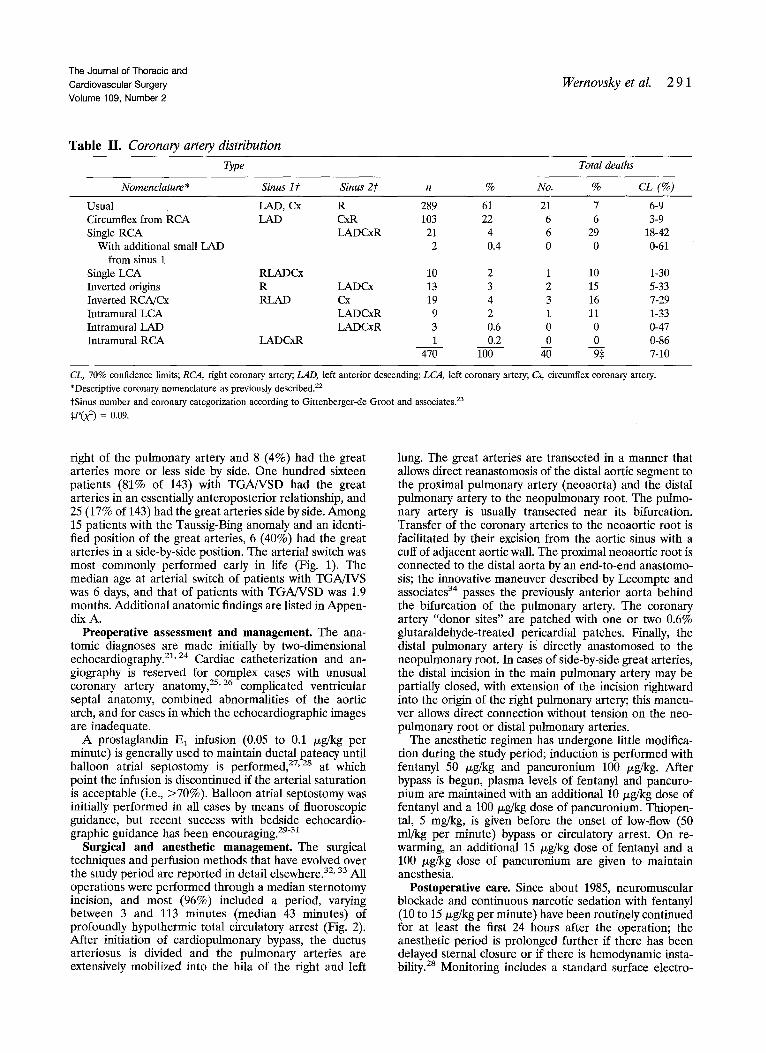
Coronary artery distribution was determined from the operative descriptions; the angiograms were retrospec- tively reviewed when this information was not available (Table II). Coronary nomenclature is as previously de- scribed. 2°-2z The leftward, more anterior "facing" sinus was alternatively designated as sinus I and the rightward, more posterior "facing" sinus as sinus 2 as previously described by Gittenberger-de Groot. 23 The usual coronary distribution for TGA and the most common variant (circumflex coronary from the right coronary artery) were seen in 83% of the patients. Unusual coronary patterns were more common in patients with TGA/VSD. 21
Among the patients in whom it could be determined in the review process, 212 patients (96% of 220) with TGA/ IVS had the aorta directly anterior to or slightly to the
The Journal of Thoracic and Cardiovascular Surgery Volume 109, Number 2
Wernovsky et al. 2 9 1
Table II . Coronary artery d&tribution
Type Total deaths
Nomenclature* Sinus 17. Sinus 27" n % No. % CL (%)
Usual LAD, Cx R 289 61 21 7 6-9 Circumflex from RCA LAD ~ 103 22 6 6 3-9 Single RCA LADC3cR 21 4 6 29 18-42
With additional small LAD 2 0.4 0 0 0-61 from sinus 1
Single LCA RLADCx 10 2 1 10 1-30 Inverted origins R LADCx 13 3 2 15 5-33 Inverted RCA/Cx RLAD Cx 19 4 3 16 7-29 Intramural LCA LADCxR 9 2 1 11 1-33 Intramural LAD LADCxR 3 0.6 0 0 0-47 Intramural RCA LADCxR 1 0.2 0 0 0-86
470 100 40 9:~ 7-10
CL, 70% confidence limits; RCA, right coronary artery; LAD, left anterior descending; LCA, left coronary artery; Cx, circumflex coronary artery.
*Descriptive coronary nomenclature as previously described. 22
tSinus number and coronary categorization according to Gittenberger-de Groot and associates. 23
,e(x ~) = 0.09.
right of the pulmonary artery and 8 (4%) had the great arteries more or less side by side. One hundred sixteen patients (81% of 143) with TGA/VSD had the great arteries in an essentially anteroposterior relationship, and 25 (17% of 143) had the great arteries side by side. Among 15 patients with the Taussig-Bing anomaly and an identi- fied position of the great arteries, 6 (40%) had the great arteries in a side-by-side position. The arterial switch was most commonly performed early in life (Fig. 1). The median age at arterial switch of patients with TGA/IVS was 6 days, and that of patients with TGA/VSD was 1.9 months. Additional anatomic findings are listed in Appen- dix A.
Preoperative assessment and management. The ana- tomic diagnoses are made initially by two-dimensional echocardiography.21, 24 Cardiac catheterization and an- giography is reserved for complex cases with unusual coronary artery anatomy, 2s'26 complicated ventricular septal anatomy, combined abnormalities of the aortic arch, and for cases in which the echocardiographic images are inadequate.
A prostaglandin E 1 infusion (0.05 to 0.1 /xg/kg per minute) is generally used to maintain ductal patency until
27 28 balloon atrial septostomy is performed, ' at which point the infusion is discontinued if the arterial saturation is acceptable (i.e., >70%). Balloon atrial septostomy was initially performed in all cases by means of fluoroscopic guidance, but recent success with bedside echocardio- graphic guidance has been encouraging. 29"3~
Surgical and anesthetic management. The surgical techniques and perfusion methods that have evolved over the study period are reported in detail elsewhere. 32' 33 All operations were performed through a median sternotomy incision, and most (96%) included a period, varying between 3 and 113 minutes (median 43 minutes) of profoundly hypothermic total circulatory arrest (Fig. 2). After initiation of cardiopulmonary bypass, the ductus arteriosus is divided and the pulmonary arteries are extensively mobilized into the hila of the right and left
lung. The great arteries are transected in a manner that allows direct reanastomosis of the distal aortic segment to the proximal pulmonary artery (neoaorta) and the distal pulmonary artery to the neopulmonary root. The pulmo- nary artery is usually transected near its bifurcation. Transfer of the coronary arteries to the neoaortic root is facilitated by their excision from the aortic sinus with a cuff of adjacent aortic wall. The proximal neoaortic root is connected to the distal aorta by an end-to-end anastomo- sis; the innovative maneuver described by Lecompte and associates 34 passes the previously anterior aorta behind the bifurcation of the pulmonary artery. The coronary artery "donor sites" are patched with one or two 0.6% glutaraldehyde-treated Pericardial patches. Finally, the distal pulmonary artery is directly anastomosed to the neopulmonary root. In cases of side-by-side great arteries, the distal incision in the main pulmonary artery may be partially closed, with extension of the incision rightward into the origin of the right pulmonary artery; this maneu- ver allows direct connection without tension on the neo- pulmonary root or distal pulmonary arteries.
The anesthetic regimen has undergone little modifica- tion during the study period; induction is performed with fentanyl 50 /xg/kg and pancuronium 100 /xg/kg. After bypass is begun, plasma levels of fentanyl and pancuro- nium are maintained with an additional 10/~g/kg dose of fentanyl and a 100/xg/kg dose of pancuronium. Thiopen- tal, 5 mg/kg, is given before the onset of low-flow (50 ml/kg per minute) bypass or circulatory arrest. On re- warming, an additional 15 /xg/kg dose of fentanyl and a 100 /xg/kg dose of pancuronium are given to maintain anesthesia.
Postoperative care. Since about 1985, neuromuscular blockade and continuous narcotic sedation with fentanyl (10 to 15/xg/kg per minute) have been routinely continued for at least the first 24 hours after the operation; the anesthetic period is prolonged further if there has been delayed sternal closure or if there is hemodynamic insta- bility. 2s Monitoring includes a standard surface electro-
2 9 2 Wernovsky et aL The J o u r n a l o f T h o r a c i c a n d
C a r d i o v a s c u l a r S u r g e r y
F e b r u a r y 1 9 9 5
| 0 0 . . . . . . . . . . . . . . . . . . . . . . . . . . . . . . . . . . . . . . . . . . ( 4 1 7 ) ( 2 8 1 ) ( 1 4 8 )
80
- - 7O m .~ e~
== 30
20
I 0
0 A o
Interval (Years) % Survival
1/12 9 3 % 1 92% 3 9 1 % 5 9 1 % 8 9 1 %
Arterial Switch Operation (BCH; 1983 t o 1992; n = 470)
. . . . . . . . . = . . . . . . . . . . . i . . . . . . . . . . . , . . . . . . . . . . . , . . . . . . . . . . . i . . . . . . . . . . . = . . . . . . . . . . . i . . . . . . . . . . d 1 2 3 4 5 6 7 8
Interval (Years) After Operation
0 . 0 5 0
0. 045
0.OdO
0. 035
5= ~ 0 . 0 3 0
j ~ 0 . 025
0. 020
0 . 0 1 5
0 . 0 1 0
0. 005
0 . 0 0 0 0
B
Interval Hazard (Years) (x 1000)
1/12 7.8 1 0.31 3 0.07 5 0.04 8 0.02
~ _ A r t e r i a l Switch Operation (BCH; 1983 to 1992; n = 4 7 0 )
• %- . . , . . . . . . . . . . i i i , I i i r i i i i i , r , I I 2 3 4
Interval (Years) After Operation
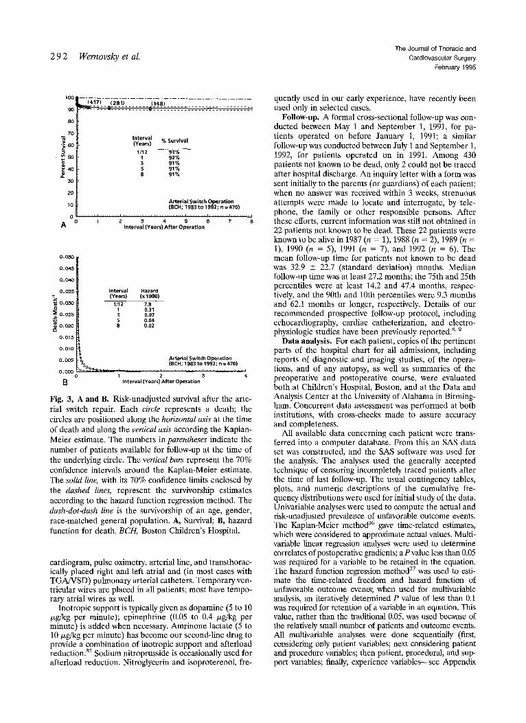
Fig. 3, A and B. Risk-unadjusted survival after the arte- rial switch repair. Each circle represents a death; the circles are positioned along the horizontal axis at the time of death and along the vertical axis according the Kaplan- Meier estimate. The numbers in parentheses indicate the number of patients available for follow-up at the time of the underlying circle. The vertical bars represent the 70% confidence intervals around the Kaplan-Meier estimate. The solid line, with its 70% confidence limits enclosed by the dashed lines, represent the survivorship estimates according to the hazard function regression method. The dash-dot-dash line is the survivorship of an age, gender, race-matched general population. A, Survival; B, hazard function for death. BCII, Boston Children's Hospital.
cardiogram, pulse oximetry, arterial line, and transthorac- ically placed right and left atrial and (in most cases with TGA/VSD) pulmonary arterial catheters. Temporary ven- tricular wires are placed in all patients; most have tempo- rary atrial wires as well.
Inotropic support is typically given as dopamine (5 to 10 /xg/kg per minute); epinephrine (0.05 to 0.4 /xg/kg per minute) is added when necessary. Amrinone lactate (5 to 10 ixg/kg per minute) has become our second-line drug to provide a combination of inotropic support and afterload reduction. 35 Sodium nitroprusside is occasionally used for afterload reduction. Nitroglycerin and isoproterenol, fre-
quently used in our early experience, have recently been used only in selected cases.
Follow-up. A formal cross-sectional follow-up was con- ducted between May 1 and September 1, 1991, for pa- tients operated on before January 1, 1991; a similar follow-up was conducted between July i and September 1, 1992, for patients operated on in 1991. Among 430 patients not known to be dead, only 2 could not be traced after hospital discharge. An inquiry letter with a form was sent initially to the parents (or guardians) of each patient; when no answer was received within 3 weeks, strenuous attempts were made to locate and interrogate, by tele- phone, the family or other responsible persons. After these efforts, current information was still not obtained in 22 patients not known to be dead. These 22 patients were known to be alive in 1987 (n = 1), 1988 (n = 2), 1989 (n = 1), 1990 (n = 5), 1991 (n = 7), and 1992 (n = 6). The mean follow-up time for patients not known to be dead was 32.9 _+ 22.7 (standard deviation) months. Median follow-up time was at least 27.2 months; the 75th and 25th percentiles were at least 14.2 and 47.4 months, respec- tively, and the 90th and 10th percentiles were 9.3 months and 62.1 months or longer, respectively. Details of our recommended prospective follow-up protocol, including echocardiography, cardiac catheterization, and electro- physiologic studies have been previously reported. 8' 9
Data analysis. For each patient, copies of the pertinent parts of the hospital chart for all admissions, including reports of diagnostic and imaging studies, of the opera- tions, and of any autopsy, as well as summaries of the preoperative and postoperative course, were evaluated both at Children's Hospital, Boston, and at the Data and Analysis Center at the University of Alabama in Birming- ham. Concurrent data assessment was performed at both institutions, with cross-checks made to assure accuracy and completeness.
All available data concerning each patient were trans- ferred into a computer database. From this an SAS data set was constructed, and the SAS software was used for the analysis. The analyses used the generally accepted technique of censoring incompletely traced patients after the time of last follow-up. The usual contingency tables, plots, and numeric descriptions of the cumulative fre- quency distributions were used for initial study of the data. Univariable analyses were used to compute the actual and risk-unadjusted prevalence of unfavorable outcome events. The Kaplan-Meier method 36 gave time-related estimates, which were considered to approximate actual values. Multi- variable linear regression analyses were used to determine correlates of postoperative gradients; a P value less than 0.05 was required for a variable to be retained in the equation. The hazard function regression method 37 was used to esti- mate the time-related freedom and hazard function of unfavorable outcome events; when used for multivariable analysis, an iteratively determined P value of less than 0.1 was required for retention of a variable in an equation. This value, rather than the traditional 0.05, was used because of the relatively small number of patients and outcome events. All multivariable analyses were done sequentially (first, considering only patient variables; next considering patient and procedure variables; then patient, procedural, and sup- port variables; finally, experience variables---see Appendix
The Journal of Thoracic and
Cardiovascular Surgery
Volume 109, Number 2
Wernovsky et aL 2 9 3
I00
8o ua ._=
70
60
50.
40
• -- 20
0
Arterial Switch Operation (BCH; 1983 to 1992; n = 470)
Age (Days) % Mortal i ty
1 1% 3 3% 5 5% 7 6%
14 8% 21 9% 28 9%
======================================= 7 14 21 28
Age (Days) at Operation
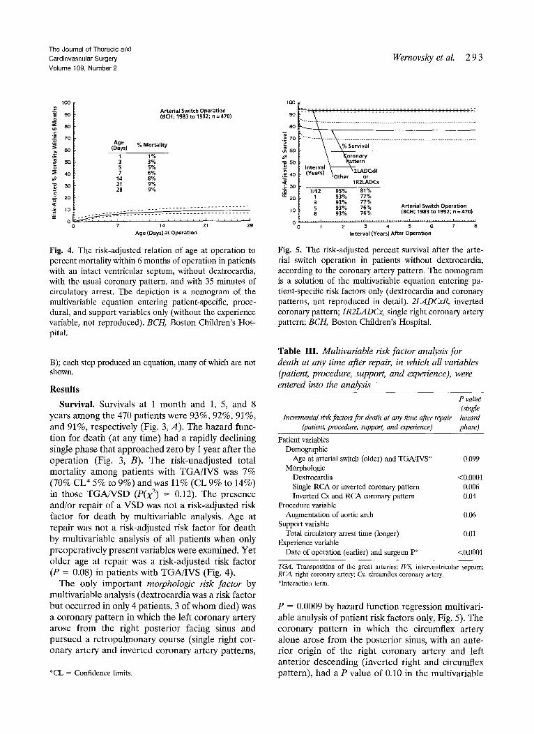
Fig. 4. The risk-adjusted relation of age at operation to percent mortality within 6 months of operation in patients with an intact ventricular septum, without dextrocardia, with the usual coronary pattern, and with 35 minutes of circulatory arrest. The depiction is a nomogram of the multivariable equation entering patient-specific, proce- dural, and support variables only (without the experience variable, not reproduced). BCH, Boston Children's Hos- pital.
B); each step produced an equation, many of which are not shown.
Results
Survival. Survivals at 1 month and 1, 5, and 8 years among the 470 patients were 93%, 92%, 91%, and 91%, respectively (Fig. 3, A). The hazard func- tion for death (at any time) had a rapidly declining single phase that approached zero by 1 year after the operation (Fig. 3, B). The risk-unadjusted total mortality among patients with TGA/IVS was 7% (70% CL* 5% to 9%) and was 11% (CL9% to 14%) in those TGA/VSD (P0(~) = 0.12). The presence and/or repair of a VSD was not a risk-adjusted risk factor for death by multivariable analysis. Age at repair was not a risk-adjusted risk factor for death by multivariable analysis of all patients when only preoperatively present variables were examined. Yet older age at repair was a risk-adjusted risk factor (P = 0.08) in patients with TGA/IVS (Fig. 4).
The only important morphologic risk factor by multivariable analysis (dextrocardia was a risk factor but occurred in only 4 patients, 3 of whom died) was a coronary pattern in which the left coronary artery arose from the right posterior facing sinus and pursued a retropulmonary course (single right cor- onary artery and inverted coronary artery patterns,
*CL = Confidence limits.
100
9O
8O
.~ 70
" o 5 0
<
._~ a~ 20
10
e o
. . . . . . . . . . . . . . . . . . . . . . . . . . . . . . . . . . . . . . . . . . . . . . . . . . . . . . . . . . . . . . . . . . . . . . . . . . . . . . . . . . . . . . . . . . . ~ ' ~ Survival
\ ~oronary Int . . . . I \ R~kttern (Years) \~,~,=. \2LADCxR
O~ner or 1R2LADCx
1/12 95% 81% 1 93% 77% 3 93% 77% 5 93% 76% Arterial Switch Operation 8 93% 76% (8CH; 1983 to 1992; n =470)
. . . . . . . . . ', . . . . . . . . . . . '2 . . . . . . . . . . ; . . . . . . . . . . ' . . . . . . . . . . . 's . . . . . . . . . . ~ . . . . . . . . . . ~ . . . . . . . . . . '~ Interval (Years) After Operation
Fig. 5. The risk-adjusted percent survival after the arte- rial switch operation in patients without dextrocardia, according to the coronary artery pattern. The nomogram is a solution of the multivariable equation entering pa- tient-specific risk factors only (dextrocardia and coronary patterns, not reproduced in detail). 2LADCxR, inverted coronary pattern; 1R2LADCx, single right coronary artery pattern; BCH, Boston Children's Hospital.
Table III. Multivariable risk factor analysis for death at any time after repair, in which all variables (patient, procedure, support, and experience), were entered into the analysis "
Incremental risk factors for death at any time after repair (patien~ procedure, support, and experience)
P value (single hazard phase)
Patient variables Demographic
Age at arterial switch (older) and TGA/IVS* 0.099 Morphologic
Dextrocardia <0.0001 Single RCA or inverted coronary pattern 0.006 Inverted Cx and RCA coronary pattern 0.04
Procedure variable Augmentation of aortic arch 0.06
Support variable Total circulatory arrest time (longer) 0.01
Experience variable Date of operation (earlier) and surgeon P* <0.0001
TGA, Transposition of the great arteries; 1VS, interventricular septum; RCA, right coronary artery; Cx, circumflex coronary artery. *Interaction term.
P = 0.0009 by hazard function regression multivari- able analysis of patient risk factors only, Fig. 5). The coronary pattern in which the circumflex artery alone arose from the posterior sinus, with an ante- rior origin of the right coronary artery and left anterior descending (inverted right and circumflex pattern), had a P value of 0.10 in the multivariable

2 9 4 Wernovsky et aL The Journal of Thoracic and
Cardiovascular Surgery February 1995
IO0
~o ~ o u~
7o
=i40
Arterial Switch Operation (BCH; 1983 to 1992; n = 470)
TCA Time (Minutes) % Mortality
15 2% 30 5% 45 7% 60 9% 75 12% 90 14%
.~ 2O
O 10 20 ~3 40 50 60 70 80 90
Total Circulatory Arrest Time (Minutes)
Fig. 6. Nomogram showing the relation between the du- ration of circulatory arrest and the risk-adjusted percent mortality within 6 months in patients with IVS, usual coronary pattern, without dextrocardia, and 6 days of age at the time of the repair. The depiction is a specific solution of the multivariable equation in Table III. BCH, Boston Children's Hospital. TCA, Total circulatory arrest.
Table IV. Total number of arterial switch operations, and the prevalence of death, in each year of this study
Hospital Total deaths deaths
Year of % of arterial switch n 470 No. % No. %
1983 21 4 3 14 3 14 1984 15 3 4 27 4 27 1985 20 4 4 20 4 20 1986 30 6 1 3 2 7 1987 81 17 4 5 7 9 1988 67 14 3 4 6 9 1989 75 16 3 4 3 4 1990 83 18 4 5 6 7 1991 78 17 4 5 5 6
Total 470 100 30 6 40 9
P(X ~) 0.01
analysis including only morphologic variables, and thus narrowly missed the criterion o f P < 0.1; in the analysis in which all categories of variables were entered, this pattern was a risk-adjusted risk factor for death (Table III).
The procedural risk factors for death identified by multivariable analysis included only arch augmenta- tion, but that was performed in only a small group of patients (n = 6, total deaths = 3). Of interest is that coarctation was a coexisting cardiac anomaly in all 6 patients, and severe hypoplasia of the distal arch was present in 2 of these. No variable describing the
lO0
ul 40
?~o
lO k2
I~ - - S u r g e o n P 'Others
li" ~ ~ -- 1985 93% 1987 94% 95% 1989 94% 95% 1991 95% 95%
Arterial Switch Operation (EEH; 1983 to 1992; n =470)
i O 9 8 ' ' I ' " " t ' ' ' I " " " I ' ' ' I ' ' ' t ' ' ' I ' " ' I ' " ' I
1 I ~ 8 4 1 9 8 5 1986 1 9 8 7 1 9 8 8 1 9 8 9 1 9 9 0 1 9 ~ 1 1 ~ 9 2
Year of Arterial Switch Operation
Fig. 7. The risk-adjusted effect of date of operation on survival for at least 6 months after the arterial switch in a 6-day-old neonate with intact ventricular septum, without dextrocardia or arch augmentation, the usual coronary anatomy, and a circulatory arrest time of 35 minutes. Note that after 1986 the risk-adjusted percent survival was the same for all surgeons. The depiction is a nomogram of the multivariable equation in Table III. BCH, Boston Chip dren's Hospital.
details of the technique of the coronary explanation and transfer was available for inclusion in the mul- tivariable analysis, and thus none could be identified or rejected as risk factors. Importantly, however, the Lecompte maneuver in general and specifically in patients with side-by-side great arteries, and the use of one or two pericardial patches to fill the defect left after removal of the coronary buttons, were not risk-adjusted correlates (risk factors) for death.
Among the support techniques, longer duration of total circulatory arrest was a risk factor for death in both a risk-unadjusted (P[logistic] = 0.0004) and a risk-adjusted sense (Fig. 6 and Table III).
Earlier date of operation was a risk-unadjusted correlate of mortality (Table IV), as well as a strong risk-adjusted risk factor for death in the case of surgeon P (see Table III), but not in the case of any other surgeon (Fig. 7).
Hospital morbidity. The mean length of stay for hospital survivors (n = 440) was 12.7 +_ 8.6 days (median 10 days, range 5 to 77 days). The length of stay was significantly shorter for patients with TGA/ IVS (mean 11.6 _ 7.1 days, range 6 to 72 days) compared with patients with TGAWSD (mean 14.4 _ 10.1 days, range 5 to 77 days, p < 0.01).
Early postoperative complications in the 440 hos- pital survivors included reoperations for bleeding (n = 3), resection of obstructive muscle bundles in
The Journal of Thoracic and Cardiovascular Surgery Volume 109, Number 2
Wernovsky et aL 2 9 5
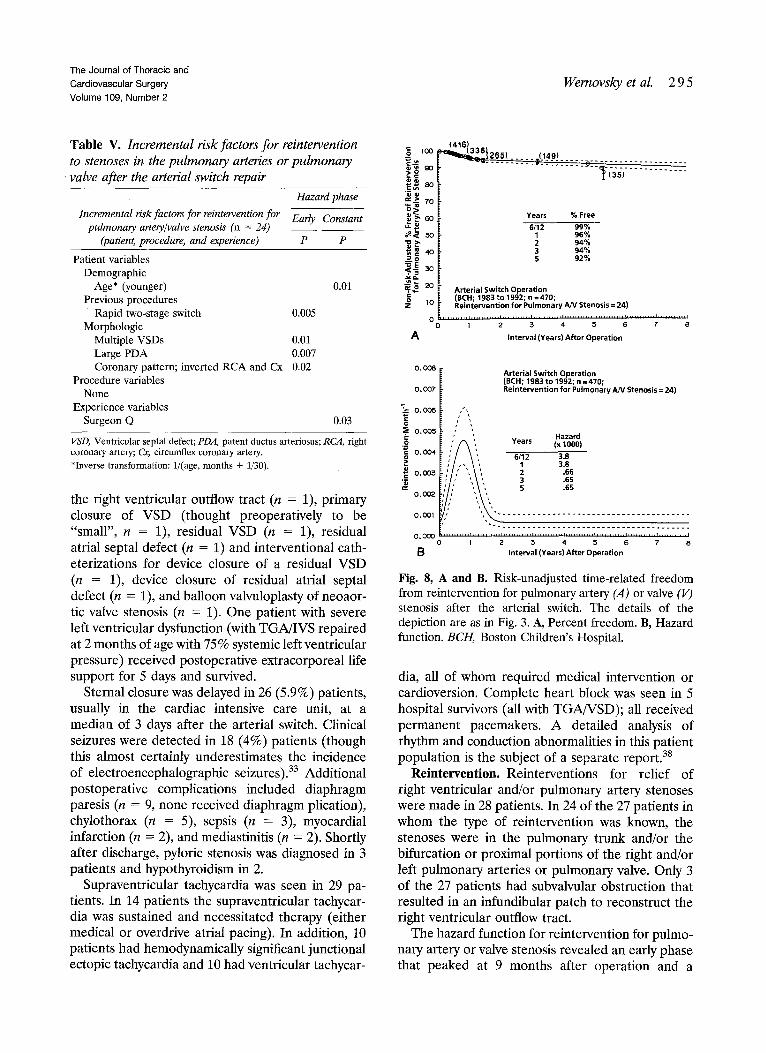
Table V. Incremental risk factors for reintervention to stenoses in the pulmonary arteries or pulmonary valve after the arterial switch repair
Incremental risk factors for reintervention for pulmonary artery~valve stenosis (n = 24)
(patient, procedure, and experience)
Hazard phase
Early Constant
P P
Patient variables Demographic
Age* (younger) 0.01 Previous procedures
Rapid two-stage switch 0.005 Morphologic
Multiple VSDs 0.01 Large PDA 0.007 Coronary pattern; inverted RCA and Cx 0.02
Procedure variables None
Experience variables Surgeon Q 0.03
VSD, Ventricular septal defect; PDA, patent ductus arteriosus; RCA, right coronary artery; Cx, circumflex coronary artery. *Inverse transformation: 1/(age, months + 1/30).
the right ventricular outflow tract (n = 1), primary closure of VSD (thought preoperatively to be "small", n = 1), residual VSD (n = 1), residual atrial septal defect (n = 1) and interventional cath- eterizations for device closure of a residual VSD (n = 1), device closure of residual atrial septal defect (n = 1), and balloon valvuloplasty of neoaor- tic valve stenosis (n = 1). One patient with severe left ventricular dysfunction (with TGA/IVS repaired at 2 months of age with 75% systemic left ventricular pressure) received postoperative extracorporeal life support for 5 days and survived.
Sternal closure was delayed in 26 (5.9%) patients, usually in the cardiac intensive care unit, at a median of 3 days after the arterial switch. Clinical seizures were detected in 18 (4%) patients (though this almost certainly underestimates the incidence of electroencephalographic seizures). 33 Additional postoperative complications included diaphragm paresis (n = 9, none received diaphragm plication), chylothorax (n = 5), sepsis (n = 3), myocardial infarction (n = 2), and mediastinitis (n = 2). Shortly after discharge, pyloric stenosis was diagnosed in 3 patients and hypothyroidism in 2.
Supraventricular tachycardia was seen in 29 pa- tients. In 14 patients the supraventricular tachycar- dia was sustained and necessitated therapy (either medical or overdrive atrial pacing). In addition, 10 patients had hemodynamically significant junctional ectopic tachycardia and 10 had ventricular tachycar-
100
> 0
e ~ 8 o
~ ro
= 5 o
. . ~ o . u n ~
N o 20
( 4 1 6 1 3 3 6 ~ ! ~ _ ~ . . ~ , : : : : _ . ~_~:.~.~ . . . . . . . . . . . . . . . . . . . . . . . . . .
. . . . . . . . . . . ~ - ~ ~ ' i , . . . . . . . . . . . - -
Years % Free 6 /12 9 9 %
1 9 6 % 2 9 4 % 3 9 4 % 5 9 2 %
Arterial Switch Operation {BCH; 1 9 8 3 t o 1992 ; n = 470 ; R e i n t e r v e n t i o n for Pulmonary AN Stenosis = 24 )
0 . . . . . . . . . . i . . . . . . . . . . . i . . . . . . . . . . . i . . . . . . . . . . . i . . . . . . . . . . . i . . . . . . . . . . . i . . . . . . . . . . . i . . . . . . . . . . . i
0 I 2 5 4 5 6 7 8
Interval (Years) After Operation
0 . 0 0 8
0 . (307"
'7 ,~ O. (:X~
0.005
0 . 0 0 4
0 . ( X ? 5
0 . 0 0 2
0 . 0 0 1
0 , 0 0 0
B
Arterial Switch Operation (BCH; 1983 t o 1992 ; n = 4 7 0 ; Reintervention for Pulmonary A N S t e n o s i s = 24 )
" " Years Hazard ,,'/~,,, (x lOOO)
' / " , \ ' 1 3 .8 ~ / , " \ \ ', 2 .66 , 1 : \ \ ' 3 .65
. . . . . . . . . . . i . . . . . . . . . . . i . . . . . . . . . . . I . . . . . . . . . . . i . . . . . . . . . . . , . . . . . . . . . . . i . . . . . . . . . . . , . . . . . . . . . , l l
I 2 3 4 5 e 7 8
Interval (Years) After Operation
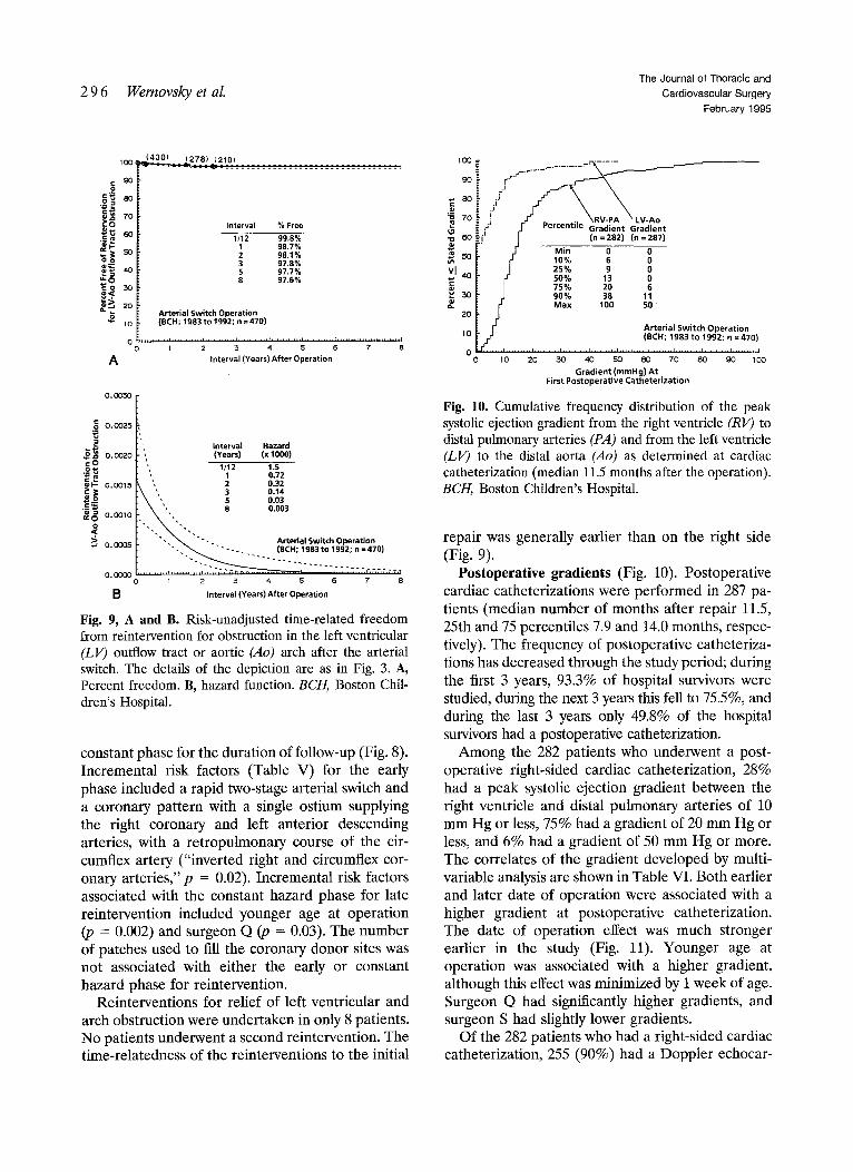
Fig. 8, A and B. Risk-unadjusted time-related freedom from reintervention for pulmonary artery (A) or valve (V) stenosis after the arterial switch. The details of the depiction are as in Fig. 3. A, Percent freedom. B, Hazard function. BCH, Boston Children's Hospital.
dia, all of whom required medical intervention or cardioversion. Complete heart block was seen in 5 hospital survivors (all with TGA/VSD); all received permanent pacemakers. A detailed analysis of rhythm and conduction abnormalities in this patient population is the subject of a separate report. 3s
Reintervent ion. Reinterventions for relief of right ventricular and/or pulmonary artery stenoses were made in 28 patients. In 24 of the 27 patients in whom the type of reintervention was known, the stenoses were in the pulmonary trunk and/or the bifurcation or proximal portions of the right and/or left pulmonary arteries or pulmonary valve. Only 3 of the 27 patients had subvalvular obstruction that resulted in an infundibular patch to reconstruct the right ventricular outflow tract.
The hazard function for reintervention for pulmo- nary artery or valve stenosis revealed an early phase that peaked at 9 months after operation and a
2 9 6 Wernovsky et aL The Journal of Thoracic and
Cardiovascular Surgery February 1995
= = = = = = = = = = = = = = = = = = = = = = = = = = = = = = = = = = = = = = = = = = = = = =
gO
~ 6 o
oo ~ 40 ~ O ~ o 30
~> ~ 2 0 O
O
A
Interval %Free 1/12 99,8%
1 98,7% 2 98.1% 3 97,8% 5 97,7% 8 97,6%
Arterial Switch Operation (BCH; 1983 to 1992; n = 470)
,,r . . . . . . . ' . . . . . . . . . . . * . . . . . . . . . . . ' . . . . . . . . . . . ' . . . . . . . . . . . ' . . . . . . . . . . . ' . . . . . . . . . . . ' . . . . . . . . . . . I I 2 3 4 5 6 7 8
Interval (Years) After Operation
0 , 0 0 3 0 I
i o.oo2s ' Interval Hazard
o O O.0020 ~- ', (Years) (x 1000) o ~ ~ ', 1 / ~ 1.5 "~ ~ [ , 1 0.72 • ~ O.OOt5 ~ ', 2 0.32 ~ > [ \ ' , 3 0.14 ~ ~ \ ' 5 0.03 • ~ ~ = ~_ ~ \ 8 0,003 mO 0 (3010
~' O 0005 r ~ " - - . Arterial Switch Operation
0 0 0 0 0 ' " ' . . . . . . . . . . . . . . . . . . . . . . . . . . . . . . . . . . . . _ . _ _ _ _ . ' ' - T " ' , - ' ~ ' T z z r . r . r . r . v . v . ~ . ~
• o 1 2 ~ 4 S 6 7 8
B Interval (Years) After Operation
Fig. 9, A and B. Risk-unadjusted time-related freedom from reintervention for obstruction in the left ventricular (LV) outflow tract or aortic (Ao) arch after the arterial switch. The details of the depiction are as in Fig. 3. A, Percent freedom. B, hazard function. BCI1, Boston Chip dren's Hospital.
constant phase for the duration of follow-up (Fig. 8). Incremental risk factors (Table V) for the early phase included a rapid two-stage arterial switch and a coronary pattern with a single ostium supplying the right coronary and left anterior descending arteries, with a retropulmonary course of the cir- cumflex artery ("inverted right and circumflex cor- onary arteries,"p = 0.02). Incremental risk factors associated with the constant hazard phase for late reintervention included younger age at operation (p = 0.002) and surgeon Q (p = 0.03). The number of patches used to fill the coronary donor sites was not associated with either the early or constant hazard phase for reintervention.
Reinterventions for relief of left ventricular and arch obstruction were undertaken in only 8 patients. No patients underwent a second reintervention. The time-relatedness of the reinterventions to the initial
so ?
7o 1 ~ ,'; .I- I'ercenzlle Gradient Gradient
¢ 100 s 0 vl 40 ~ J 25*/, 9 0
[ C r 50 % 13 0 c ~ I 75% 20 6
~. 30 ~- rJ 90% 38 11 2 0 [ [ ~ Max 100 50
10 I~ ~ Arterial Switch Operation 0 (BCH; 1983 to 19§2; n =470)
0 10 20 30 40 50 60 70 80 90 1 0 0
Gradient (mmHg) At First Postoperative Catheterization
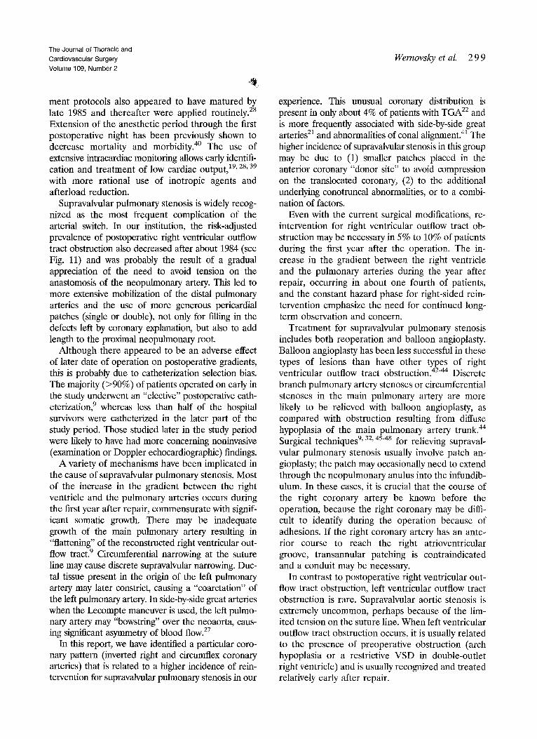
Fig. 10. Cumulative frequency distribution of the peak systolic ejection gradient from the right ventricle (RV) to distal pulmonary arteries (PA) and from the left ventricle (LV) to the distal aorta (Ao) as determined at cardiac catheterization (median 11.5 months after the operation). BCH, Boston Children's Hospital.
repair was generally earlier than on the right side (Fig. 9).
Postoperative gradients (Fig. 10). Postoperative cardiac catheterizations were performed in 287 pa- tients (median number of months after repair 11.5, 25th and 75 percentiles 7.9 and 14.0 months, respec- tively). The frequency of postoperative catheteriza- tions has decreased through the study period; during the first 3 years, 93.3% of hospital survivors were studied, during the next 3 years this fell to 75.5%, and during the last 3 years only 49.8% of the hospital survivors had a postoperative catheterization.
Among the 282 patients who underwent a post- operative right-sided cardiac catheterization, 28% had a peak systolic ejection gradient between the right ventricle and distal pulmonary arteries of 10 mm Hg or less, 75% had a gradient of 20 mm Hg or less, and 6% had a gradient of 50 mm Hg or more. The correlates of the gradient developed by multi- variable analysis are shown in Table VI. Both earlier and later date of operation were associated with a higher gradient at postoperative catheterization. The date of operation effect was much stronger earlier in the study (Fig. 11). Younger age at operation was associated with a higher gradient, although this effect was minimized by 1 week of age. Surgeon Q had significantly higher gradients, and surgeon S had slightly lower gradients.
Of the 282 patients who had a right-sided cardiac catheterization, 255 (90%) had a Doppler echocar-

The Journal o f Thorac ic and
Card iovascu la r Surgery
Vo lume 109, Number 2
Wernovsky et al. 2 9 7
Table VI. Multivariable linear regression analysis of the correlates of the gradient across the RV-PA outflow tract as determined at postoperative cardiac catheterization
Regression coefficient Correlates of RV-PA gradient
(at postoperative catheterization) Coeficient (n = 282) + SD P value
Patient variables Demographic
Age at operation* (younger) 0.027 -+ 0.012 0.02 Preoperative hemodynamics
Pulmonary artery systolic -0.012 -+ 0.0038 0.002 pressure (ram Hg) (lower)
Usual coronary pattern 0.7 -+ 0.27 0.01 Experience variables
Surgeon Q 1.0 -+ 0.29 0.0006 Surgeon S -0.19 -+ 0.092 0.04 Date of operation? (earlier) 0.3 -+ 0.116 0.007 Date of operation:~ (later) 0.49 -+ 0.116 <0.0001
Early postoperative echocardio- graphic study
RV-PA echo gradient (mm Hg) 0.0191 -+ 0.0057 0.0008
Intercept = 2.143
RV-PA, Right ventricular-pulmonary artery; SD, standard deviation. Cath- eterizations were performed at a median of 11.5 months after the operation (see text). Logarithmic transformation of the RV-PA gradient was the dependent variable (values of zero gradient were retained as a zero logarithmic value). These variables account for 20% of the scatter in the data and therefore should be considered weak in predictive capacity. *Inverse transformation: 1/(age, months + 1/30). ?Inverse transformation: 1/(years since 1/1/1983). SLogarithmic transformation: In (years since 1/1/1983).
diogram at hospital discharge. In these 255 patients, 56 (22%) had an increase of the gradient from the right ventricle to pulmonary artery of 20 mm Hg or more. There was considerable scatter in the change in gradient over time (Fig. 12).
Among the 287 who underwent a left-heart cath- eterization, 90% had peak systolic ejection gradients between the left ventricle and distal aorta of 10 mm Hg or less and none had a gradient greater than 50 mm Hg. As a group, patients with anatomic left ventricular outflow tract obstruction before the op- eration had the highest risk-unadjusted left ventricu- lar-aortic gradient at postoperative catheterization (Table VII).
Functional class. Nearly all patients were func- tioning normally compared with their peers. Of the 412 survivors fo r whom functional class could be determined, 402 (97.6%) were in New York Heart Association class I, 9 (2.2%) were in class II, and 1 (0.2%) was in class III.
50 ~ Arterial Switch Operation 45 l (BCH; 1983 to 1992; n = 470)
A 4O
~ 2s . . . . . . . .
I ~ t o
o . . . . . . . . . ' . . . . . . . . . = ' " lg83 Ige't I ~ 1988 108 IT I ~ 1~9 19gCI 1~1 lgg2
Date (Year) of Operation
Fig. 11. Nomogram showing the relation between the date of operation and the predicted peak systolic ejection gradi- ent from the right ventricle (RV) to the distal pulmonary artery (PA) as determined at postoperative catheterization. The depiction is a specific solution of the multivariable equation in Table VI, with 70% confidence limits for an individual patient. BCH, Boston Children's Hospital.
Discussion
A 10-year survival of 93%, including hospital mortality, is predicted for most patients with mor- phologically uncomplicated TGA, with or without VSD, operated on by any surgeon in the institution (see Fig. 7). The analyses indicate, with a reasonable degree of certainty, that the survival will be even better than this for patients with more usual coro- nary artery patterns (see Fig. 5).
The increased risk resulting from earlier date of operation was neutralized by the end of 1985 (see Fig. 7), 3 years and 56 patients into the study period. This "learning curve" suggests that overall institu- tional improvements occurred in the approach to these patients; after 1985 three "new" surgeons began performing the operation without a higher mortality risk than the senior surgeon.
As many institutional changes took place nearly simultaneously from 1984 to 1985, it is difficult to sort out the relative contributions of each, even with a multivariable analysis. The striking improvement that occurred in about 1985 undoubtedly resulted from multiple factors, some of which probably were not available for analysis in this study. We believe that these changes occurred in three general areas: (1) surgical technique (with increasing experience in coronary translocation in particular), (2) patient selection (elective repair at younger age in particu- lar), and (3) perioperative care.

2 9 8 Wernovsky et aL The Journal of Thoracic and
Cardiovascular Surgery
February 1995
140 0
120 2: Arterial Switch Operation
I00 O (BCH; 1983 to 1992; n =470)
so 8 o
~ s o ; ~ o o • a 4o ~o ~ ,e. ° o~,oo o
= _ o
c - 2 o o o 0% o ~' o oo
- 8 o [ .. . . . . . . . . P ........ ' . . . . . . . . . . . . . . . . . . . . . . . . . . . . . . . . . . . . . . . . . . . . . . . . . . . . . . . . . . . . 0 1 2 3 4 5 6 7 B
Interval (Years) Between Studies
Fig. 12. Scattergram showing the change in right ventri- cle (RV) to pulmonary artery (PA) gradient over time, as determined during follow-up by either cardiac catheter- ization or Doppler echocardiography. A time sequence was generated for each patient, with up to four studies per patient. Patients were removed from further analysis if a surgical or catheterization reintervention was performed, although the pre-reintervention study is shown. Circles represent changes from the first to the second study (n -- 168), squares represent changes from the second study to the third study (n = 324), and diamonds represent changes from the third study to the fourth study (n = 12).
The inference was gradually developed during the first few years of the study that the coronary-related mortality could be at least partially neutralized by modifications in surgical technique. These include more extensive epicardial mobilization of the prox- imal coronary arteries, division of small conal branches that may hinder posterior mobilization to the neoaorta, and small modifications of the reim- plantation site if necessary.
The increased risk of death (and neurologic com- plications) from long periods of hypothermic circu- latory arrest were also gradually appreciated, and this also may have contributed to the lower mortality after 1985. A more in-depth assessment of the neurologic 33 and nonneurologic morbidity related to circulatory arrest is reported elsewhere 39 and is beyond the scope of this report.
The preoperative management protocols 27' as de- veloped and matured in the early part of the expe- rience. For example, balloon atrial septostomy was more frequently performed at the bedside, with the elimination of angiographic studies, toward the lat- ter part of the study period. We recommend balloon atrial septostomy in all patients without a large, native atrial septal defect. An adequate atrial communica- tion allows for (1) improved intercirculatory mixing
Table VII. Correlates of the gradient between left ventricle and aorta, obtained at postoperative cardiac catheterization
Correlates o f 1-year LV-aortic gradient (patient variables)
Regression coefficient
Coefficient + S D P
Demograph ic Female sex Age at operat ion* (younger)
Morphology Small VSD L V O T O (anatomic or dynamic) or
aortic arch obstruction Additional increment f rom pre-
operative anatomic L V O T O *
Intercept = 0.3662
0.29 + 0.138 0.04 0.053 -- 0.0181 0.003
0.49 __+ 0.168 0.004 0.56 - 0.178 0.002
1.6 + 0.42 0.0002
LV,, Left ventricular; VSD, ventricular septal defect; LVOTO, left ventric- ular outflow tract obstruction. Logarithmic transformation of the LV- aortic gradient was the dependent variable (values of zero gradient were retained as a zero logarithmic value). P value threshold was 0.5. These variables account for 16% of the scatter in the data. *Inverse transformation: 1/(age, months + 1/30).
and oxygen delivery before the operation, (2) the ability (in most cases) to discontinue prostaglandin infusion, thereby limiting total body edema and maintaining tissue integrity, (3) more "elective" timing of the arterial switch, during which time nutrition may be instituted and other potential neonatal problems (e.g., sepsis) may be ruled out, and (4) adequate decompression of the left atrium during cardiopulmonary bypass via a single venous cannula placed in the right atrium.
Elective repair was performed in the first 1 to 2 weeks of life whenever possible. Although earlier in the experience some patients (usually with TGA/ VSD) underwent elective repair in later infancy, at the current time the arterial switch is recommended during the same admission as the diagnosis is made, even in the presence of a large VSD with adequate systemic oxygen saturation (see Fig. 4). The new- born infant with TGA/IVS should be repaired within the first weeks of life while the left ventricle is still capable of systemic work. Early, neonatal repair is also performed for the patient with TGA/VSD. In TGA/VSD, early repair is recommended to mini- mize the adverse systemic effects of prolonged cya- nosis or congestive heart failure, limit the likelihood of spontaneous closure of VSDs (which would result in a low-pressure left ventricle that may be "unpre- pared" for systemic work), and to decrease the likelihood of pulmonary vascular disease.
The intraoperative and postoperative manage-
The Journal of Thoracic and Cardiovascular Surgery Volume 109, Number 2
ment protocols also appeared to have matured by late 1985 and thereafter were applied routinely. 28 Extension of the anesthetic period through the first postoperative night has been previously shown to decrease mortality and morbidity. 4° The use of extensive intracardiac monitoring allows early identifi- cation and treatment of low cardiac output, 19' 28, 39 with more rational use of inotropic agents and afterload reduction.
Supravalvular pulmonary stenosis is widely recog- nized as the most frequent complication of the arterial switch. In our institution, the risk-adjusted prevalence of postoperative right ventricular outflow tract obstruction also decreased after about 1984 (see Fig. 11) and was probably the result of a gradual appreciation of the need to avoid tension on the anastomosis of the neopulmonary artery. This led to more extensive mobilization of the distal pulmonary arteries and the use of more generous pericardial patches (single or double), not only for filling in the defects left by coronary explanation, but also to add length to the proximal neopulmonary root.
Although there appeared to be an adverse effect of later date of operation on postoperative gradients, this is probably due to catheterization selection bias. The majority (>90%) of patients operated on early in the study underwent an "elective" postoperative cath- eterization, 9 whereas less than half of the hospital survivors were catheterized in the later part of the study period. Those studied later in the study period were likely to have had more concerning noninvasive (examination or Doppler echocardiographic) findings.
A variety of mechanisms have been implicated in the cause of supravalvular pulmonary stenosis. Most of the increase in the gradient between the right ventricle and the pulmonary arteries occurs during the first year after repair, commensurate with signif- icant somatic growth. There may be inadequate growth of the main pulmonary artery resulting in "flattening" of the reconstructed right ventricular out- flow tract. 9 Circumferential narrowing at the suture line may cause discrete supravalvular narrowing. Duc- tal tissue present in the origin of the left pulmonary artery may later constrict, causing a "coarctation" of the left pulmonary artery. In side-by-side great arteries when the I_ecompte maneuver is used, the left pulmo- nary artery may "bowstring" over the neoaorta, caus- ing significant asymmetry of blood flow. 27
In this report, we have identified a particular coro- nary pattern (inverted right and circumflex coronary arteries) that is related to a higher incidence of rein- tervention for supravalvular pulmonary stenosis in our
Wernovsky et aL 2 9 9
experience. This unusual coronary distribution is present in only about 4% of patients with TGA 22 and is more frequently associated with side-by-side great arteries 21 and abnormalities of conal alignment. 41 The higher incidence of supravalvular stenosis in this group may be due to (1) smaller patches placed in the anterior coronary "donor site" to avoid compression on the translocated coronary, (2) to the additional underlying conotruncal abnormalities, or to a combi- nation of factors.
Even with the current surgical modifications, re- intervention for right ventricular outflow tract ob- struction may be necessary in 5% to 10% of patients during the first year after the operation. The in- crease in the gradient between the right ventricle and the pulmonary arteries during the year after repair, occurring in about one fourth of patients, and the constant hazard phase for right-sided rein- tervention emphasize the need for continued long- term observation and concern.
Treatment for supravalvular pulmonary stenosis includes both reoperation and balloon angioplasty. Balloon angioplasty has been less successful in these types of lesions than have other types of right ventricular outflow tract obstructionY -44 Discrete branch pulmonary artery stenoses or circumferential stenoses in the main pulmonary artery are more likely to be relieved with balloon angioplasty, as compared with obstruction resulting from diffuse hypoplasia of the main pulmonary artery trunk. 44 Surgical techniques 9' 32, 45-48 for relieving supraval- vular pulmonary stenosis usually involve patch an- gioplasty; the patch may occasionally need to extend through the neopulmonary anulus into the infundib- ulum. In these cases, it is crucial that the course of the right coronary artery be known before the operation, because the right coronary may be diffi- cult to identify during the operation because of adhesions. If the right coronary artery has an ante- rior course to reach the right atrioventricular groove, transannular patching is contraindicated and a conduit may be necessary.
In contrast to postoperative right ventricular out- flow tract obstruction, left ventricular outflow tract obstruction is rare. Supravalvular aortic stenosis is extremely uncommon, perhaps because of the lim- ited tension on the suture line. When left ventricular outflow tract obstruction occurs, it is usually related to the presence of preoperative obstruction (arch hypoplasia or a restrictive VSD in double-outlet right ventricle) and is usually recognized and treated relatively early after repair.
3 0 0 Wernovsky et aL The Journal of Thoracic and
Cardiovascular Surgery February 1995
Summary A continuation of the application of the arterial
switch operation to patients with transposition of the great arteries with or without VSD, as early in life as is possible, is indicated.
This manuscript represents the combined work of many individuals. We thank the medical and nursing staffs of the Cardiovascular Program at Children's Hospital in Boston for their care of these patients; Drs. Paul R. Hickey, David L. Wessel, Redmond P. Burke, and Steven D. Colan for their comments and suggestions for the final manuscript; Erin Carroll, Amy Itzkovitz, Cheryl King, Barbara Lock, and Amy Walsh, RN, for help with data management at Children's Hospital; Mary Lynn Clark and Rob Brown at University of Alabama, Birmingham, for their tireless efforts during the cross-sectional follow-up and data anal- ysis; and Debbie Nuby, Matthew Martin, and Tannis Bolton for help with data acquisition, management, and manuscript preparation.
R E F E R E N C E S 1. Castafieda AR, Norwood WI, Jonas RA, Colan SD,
Sanders SP, Lang P. Transposition of the great arter- ies and intact ventricular septum: anatomical repair in the neonate. Ann Thorac Surg 1984;38:438-43.
2. Kirklin JW, Blackstone EH, Tchervenkov CI, Castafieda AR, and the Congenital Heart Surgeons Society. Clini- cal outcomes after the arterial switch operation for transposition: patient, support, procedural and institu- tional risk factors. Circulation 1992;86:1501-15.
3. SerrafA, Lacour-Gayet F, Bruniaux J, et al. Anatomic correction of transposition of the great arteries in neonates. J Am Coll Cardiol 1993;22:193-200.
4. Quaegebeur JM, Rohmer J, Ottenkamp J, et al. The arterial switch operation: an eight-year experience. J THORAC CARDIOVASC SURG 1986;92:361-84.
5. Mee RBB. Results of the arterial switch procedure for complete transposition with an intact ventricular sep- turn. Cardiol Young 1991;1:97-8.
6. Yamaguchi M, Hosokawa Y, Imai Y, et al. Early and midterm results of the arterial switch operation for transposition of the great arteries in Japan. J THORAC CARDIOVASC SURG 1990;100:261-9.
7. Lupinetti FM, Bove EL, Minich LL, et al. Intermedi- ate-term survival and functional results after arterial repair for transposition of the great arteries. J THO- RAC CARDIOVASC SURG 1992;103:421-7.
8. Colan SD, Trowitzsch E, Wernovsky G, Sholler GF, Sanders SP, Castafieda AR. Myocardial performance after arterial switch operation for transposition of the great arteries with intact ventricular septum. Circula- tion 1988;78:132-41.
9. Wernovsky G, Hougen TJ, Walsh EP, et al. Midterm results after the arterial switch operation for transposi- tion of the great arteries with intact ventricular septum:
clinical, hemodynamic, echocardiographic and electro- physiologic data. Circulation 1988;77:1333-44.
10. Gleason MM, Chin AJ, Andrews BA, et al. Two- dimensional and Doppler echocardiographic assess- ment of neonatal arterial repair for transposition of the great arteries. J Am Coll Cardiol 1989;13:1320-8.
11. Arensman FW, Sievers H, Lange PE, et al. Assess- ment of coronary and aortic anastomoses after ana- tomic correction of transposition of the great arteries. J THORAC CARD]OVASC SURG 1985;90:597-604.
12. Planche C, Bruniaux J, Lacour-Gayet F, et al. Switch operation for transposition of the great arteries in neonates: a study of 120 patients. J THORA¢ CARDIO- VASe SURO 1988;96:354-63.
13. Day RW, Laks H, Drinkwater DC. The influence of coronary anatomy on the arterial switch operation in neonates. J THORAC CARDIOVASC SURG 1992;104:706-12.
14. Okuda H, Nakazawa M, Imai Y, et al. Comparison of ventricular function after Senning and Jatene proce- dures for complete transposition of the great arteries. Am J Cardiol 1985;55:530-4.
15. Borow KM, Arensman FW, Webb C, Radley-Smith R, Yacoub MH. Assessment of left ventricular con- tractile state after anatomic correction of transposi- tion of the great arteries. Circulation 1984;69:106-12.
16. Menahem S, Ranjit MS, Stewart C, Brawn WJ, Wilkinson JL. Cardiac conduction abnormalities and rhythm changes after neonatal anatomical correction of transposition of the great arteries. Br Heart J 1992;67:246-9.
17. Vetter VL, Tanner CS. Electrophysiologic conse- quences of the arterial switch repair of d-transposition of the great arteries. J Am Coll Cardiol 1988;12:229-37.
18. Di Donato RM, Wernovsky G, Walsh EP, et al. Results of the arterial switch operation for transposi- tion of the great arteries with ventricular septal de- fect: surgical considerations and midterm follow-up data. Circulation 1989;80:1689-705.
19. Wernovsky G, Giglia TM, Jonas RA, Mone SM, Colan SD, Wessel DL. Course in the intensive care unit following "preparatory" pulmonary artery band- ing and aortopulmonary shunt placement for transpo- sition of the great arteries with low left ventricular pressure. Circulation 1992;86(Suppl):II133-9.
20. Mayer JE Jr, Sanders SP, Jonas RA, Castafieda AR, Wernovsky G. Coronary artery pattern and outcome of arterial switch operation for transposition of the great arteries. Circulation 1990;82(Suppl):IV139-45.
21. Pasquini L, Sanders SP, Parness IA, et al. Coronary echocardiography in 406 patients with d-loop trans- position of the great arteries. J Am Coll Cardiol 1994;24:763- 8.
22. Wernovsky G, Sanders SP. Coronary artery anatomy and transposition of the great arteries. Coronary Artery Dis 1993;4:148-57.
23. Gittenberger-de Groot AC, Sauer U, Oppenheimer-

The Journal of Thoracic and Cardiovascular Surgery Volume 109, Number 2
Wernovsky et aL 3 0 1
Dekker A, Quaegebeur JM. Coronary arterial anat- omy in transposition of the great arteries: a morpho- logic study. Pediatr Cardiol 1983;4(Suppl):I15-24.
24. Sanders SP, Mayer JE Jr, Wernovsky G, Parness IA, Colan SD. The role of two-dimensional and Doppler echocardiography in the preoperative evaluation of d-transposition of the great arteries. Cardiovasc Imag 1989;1:71-7.
25. Mandell VS, Lock JE, Mayer JE Jr, Parness IA, Kulik TJ. The "laid-back" aortogram: an improved anglo- graphic view for demonstration of coronary arteries in transposition of the great arteries. Am J Cardiol 1990;65:1379-83.
26. Day RW, Isabel-Jones JB, Wetzel GT, Oku GS, Jarmakani JM. Description of a venous technique for selective coronary arteriography in newborns with d-transposition of the great arteries. J Am Coll Car- diol 1989;14:1308-11.
27. Paul MH, Wernovsky G. Transposition of the great arteries. In: Emmanouilides GC, Riemenschneider TA, Allen HD, Gutgesell HP, eds. Moss and Adams' heart disease in infants, children, and adolescents, including the fetus and young adult. 5th ed. chap. 75. Baltimore: Williams & Wilkins, 1995:1154-225.
28. Wernovsky G, Chang AC, Wessel DL. Intensive care. In: Emmanouilides GC, Riemenschneider TA, Allen HD, Gutgesell HP, eds. Moss and Adams' heart disease in infants, children, and adolescents, including the fetus and young adult. 5th ed. chap. 27. Baltimore: Williams & Wilkins, 1995:398-439.
29. Kakadekar AP, Hayes A, Rosenthal E, et al. Balloon atrial septostomy in the intensive care unit under echocardiographic control--nine years experience. Cardiol Young 1992;2:175-8.
30. Boutin C, Dyck J, Benson LN, Houde C, Freedom RM. Balloon atrial septostomy under transesophageal echo- cardiographic guidance. Pediatr Cardiol 1992;13:176-7.
31. Ward CJB, Hawker RE, Cooper SG, et al. Minimally invasive management of transposition of the great arter- ies in the newborn period. Am J Cardio11992;69:1321-3.
32. Castafieda AR, Jonas RA, Mayer JE Jr, Hanley FL. D-Transposition of the great arteries. In: Cardiac surgery of the neonate and infant. Philadelphia: WB Saunders, 1994:409-38.
33. Newburger JW, Jonas RA, Wernovsky G, et al. A comparison of the perioperative neurologic effects of hypothermic circulatory arrest versus low-flow cardio- pulmonary bypass in infant heart surgery. N Engl J Med 1993;329:1057-64.
34. Lecompte Y, Zannini L, Hazan E, et al. Anatomic correction of transposition of the great arteries: new technique without use of a prosthetic conduit. J THORAC CARDIOVASC SURG 1981;82:629-31.
35. Lang P, Wessel DL, Wernovsky G, Jonas RA, Mayer JE Jr, Castafieda AR. Hemodynamic effects of amri- none in infants after cardiac surgery. In: Crupi G,
Parenzan L, Anderson RH, eds. Perspectives in pedi- atric cardiology. Vol 2, Pediatric cardiac surgery, part 2. 1st ed. Mount Kisco: Futura, 1989:292-5.
36. Kaplan EL, Meier P. Nonparametric estimation from incomplete observations. J Am Stat Assoc 1958;53: 457-81.
37. Blackstone EH, Naftel DC, Turner ME Jr. The decomposition of time-varying hazard into phases, each incorporating a separate stream of concomitant information. J Am Stat Assoc 1986;81:615-24.
38. Rhodes LA, Wernovsky G, Keane JF, et al. Arrhythmias and intracardiac conduction after the arterial switch operation. J THORAC CAP, DIOVASC StrRG 1995;109:303-10.
39. Wernovsky G, Jonas RA, Newburger JW, et al. The Boston circulatory arrest study: hemodynamics and hospital course after the arterial switch operation [Abstract]. Circulation 1992;86(Suppl):I237A.
40. Anand KJS, Hickey PR. Halothane-morphine com- pared with high-dose sufentanil for anesthesia and postoperative analgesia in neonatal cardiac surgery. N Engl J Med 1992;326:1-9.
41. Kurosawa H, Imai Y, Takanashi Y, et al. Infundibular septum and coronary anatomy in Jatene operation. J THORAC CARDIOVASC SURG 1986;91:572-83.
42. Zeevi B, Keane JF, Perry SB, Lock JE. Balloon dilation of postoperative right ventricular outflow obstructions. J Am Coll Cardiol 1989;14:401-8.
43. Saxena A, Fong LV, Ogilvie BC, Keeton BR. Use of balloon dilatation to treat supravalvar pulmonary stenosis developing after anatomical correction for complete transposition. Br Heart J 1990;64:151-5.
44. Smoot LB, Wernovsky G, Mayer JE Jr, Hanley FL, Keane JF. Results of balloon angioplasty in patients with right ventricular outflow tract obstruction follow- ing the arterial switch operation [Abstract]. Circula- tion 1992;86(Suppl):I42A.
45. Paillole C, Sidi D, Karchaner J, et al. Fate of pulmo- nary artery after anatomic correction of simple trans- position of the great arteries in newborn infants. Circulation 1988;78:870-6.
46. Yacoub MH, Bernhard A, Radley-Smith R, Lange PE, Sievers H, Heintzen PH. Supravalvular pulmo- nary stenosis after anatomic correction of transposi- tion of the great arteries: causes and prevention. Circulation 1982;66(Suppl):1193-7.
47. Castafieda All. Arterial switch operation for simple and complex TGA--indication criteria and limita- tions relevant to surgery. Thorac Cardiovasc Surg 1991;39:151-5.
48. Mavroudis C, Backer CL, Idriss FS. Special consider- ations for and reoperations after the arterial switch operation. Cardiac Surg 1991;5:119-40.
Discuss ion
Dr. Jan M. Quaegebeur (New York, N.Y). I agree that the arterial switch operation can be applied to different

3 0 2 Wernovsky et al. The Journal of Thoracic and
Cardiovascular Surgery February 1995
subsets of TGA very early in life and that the risk of death is very low. Since my arrival at Columbia Presbyterian Hospital, we have performed 100 consecutive arterial switch operations with a mortality rate of 1%. In many ways, our experience with this operation parallels the Boston experience. On several occasions did we conduct the same type of analysis that Dr. Wernovsky presented here, and several risk factors are similar--the older age at operation and the early date of operation, for example. However, in our experience the coronary arterial distri- bution pattern, especially the retropulmonary course of the left coronary artery, never was an incremental risk factor. Instead, intramural coronary artery was the only risk factor for death after the arterial switch operation, and the only patient dying in our recent experience had a single coronary ostium in sinus 2, with an intramural course of the right and left coronary arteries. I would like to ask you by which mechanism retropulmonary course of the left coronary artery increases the risk of the operation, because the reimplantation technique for this particular coronary anatomy is quite standardized. I have never understood this difference between our experiences.
Dr. Wernovsky. Thank you, Dr. Quaegebeur. I do not understand the difference either. As you know, this is a rare type of coronary variant, occurring in only 3% to 5% of the total experience. I suspect that the increased risk has to do with angulation of the retropulmonary course of the left coronary artery, especially in patients with a single coronary ostium. The left coronary artery must make a 180-degree turn and tension on the right coronary may be a problem. Exactly why in any individual experience certain patterns fall out as risk factors is unclear to me as well. Your study, as well as ours, emphasizes the impor- tance of a critical analysis of one's results, to identify risk factors and ideally modify their effects.
Dr. Carl Lewis Backer (Chicago, Ill.). These are great results, Dr. Wernovsky. Was there a change in the tech- nique of right ventricular outflow tract reconstruction during the reported time period? It sounds as i f your results with this aspect of the arterial switch have im- proved considerably since the original series of 11 infants.
Dr. Wernovsky. Yes, I think that the most important
Appendix A. Addit ional morphologic findings
No. %
Abnormal pulmonary (neoaortic) valve 19 4 Left ventricular outflow tract obstruction 50 11
Dynamic 35 Anatomic 15
Right ventricular outflow tract obstruction 11 2 Cleft mitral valve 6 1 Coarctation 34 7 Hypoplastic aortic arch 4 0.9 Interrupted aortic arch 6 1 Right aortic arch 3 0.6 Left superior vena cava 11 2 Juxtaposition of atrial appendages 17 4 Multiple VSDs (in VSD group) 33 7 Neuroblastoma 2 0.5 Tracheoesophageal fistula 1 0.2
technical change after the early experience was the rec- ognition of the importance of avoiding tension on the pulmonary anastomosis. My surgical colleagues tell me that extensive dissection and mobilization of the distal pulmonary arteries well out into the hilum, as well as placement of a generous pericardial patch to add length to the proximal neopulmonary artery, were the two major changes that were done after the early experience. At the current time, it appears that patient-related anatomic factors are responsible for the small but continued risk of late supravalvular pulmonary stenosis.
Appendix B Patient variables Demographic. Age at operation, gender, birth weight, height,
weight, and body surface area at arterial switch. Previous procedures. Previous balloon septostomy, use of
prostaglandin El, pulmonary artery banding, "rapid" pul- monary artery banding, "one-stage" repair (actual or with short banding), duration of banding, previous repair of aortic arch abnormality.
Morphologic variables (other than coronary pattern). Situs, iso- lated dextrocardia, mesocardia, TGA/IVS, TGA/VSD (in- cluding double-outlet left ventricle, double-outlet right ventricle, and the Taussig-Bing anomaly), number of VSDs, interruption of the aortic arch, coarctation of the aorta, hypoplasia (moderate or severe) of the aortic arch, left ventricular outflow tract obstruction (dynamic, "flow- related," anatomic [pulmonary valve stenosis, tricuspid valve excrescences, other]), severity of left ventricular out- flow tract obstruction, aortic valve stenosis, right ventricu- lar outflow tract obstruction, juxtaposition of the atrial ap- pendages, patent ductus arteriosus (and its size), position of the great arteries.
Coronary artery pattern. Individual patterns (as previously re- ported) 22 and grouped patterns: (1) all coronary arteries arising from a single sinus, (2) all variations of intramural coronary arteries, (3) patterns with a retropuhnonary course of the entire left coronary system (1-;2LadCxR* and 1R;2LadCx), (4) patterns with a retropuhnonary course of the circumflex only (1Lad;2CxR and 1RLad;2Cx), (5) any left coronary supply from the posterior facing sinus.
Procedural variables. Atrial septal defect repair, VSD repair, revision of coronary anastomosis, left ventricular outflow procedure, right ventricular outflow procedure, augmenta- tion of the aortic arch, Lecompte maneuver, Lecompte maneuver in side-by-side great arteries, coronary donor site repair technique (one versus two patches [or one patch in cases of single coronary artery]), coarctation repair.
Support technique variables. Use and duration of circulatory arrest and myocardial ischemic time (total elapsed time of cardiopulmonary bypass and total support time [bypass time plus circulatory arrest time] were analyzed but not permitted to enter multivariable analyses of death).
Experience variables. Date of operation, sequence number of arterial switch, surgeon, temporal experience, and se- quence number for each surgeon.
Postoperative variables. Use of neuromuscular blockade/ continuous narcotic sedation.
*R, Right coronary artery; Lad, left anterior descending; Cx, circumflex.