Tricyclic quinoxalines as potent kinase inhibitors of PDGFR kinase, Flt3 and Kit
Upload
independentCategory
view
3download
0
BioMed CentralBMC Developmental Biology
ss
Open AcceResearch articleExpression patterns of protein kinase D 3 during mouse developmentKornelia Ellwanger, Klaus Pfizenmaier, Sylke Lutz and Angelika Hausser*Address: Institute of Cell Biology and Immunology, University of Stuttgart, Allmandring 31, 70569 Stuttgart, Germany
Email: Kornelia Ellwanger - [email protected]; Klaus Pfizenmaier - [email protected]; Sylke Lutz - [email protected]; Angelika Hausser* - [email protected]
* Corresponding author
AbstractBackground: The PKD family of serine/threonine kinases comprises a single member in Drosophila(dPKD), two isoforms in C. elegans (DKF-1 and 2) and three members, PKD1, PKD2 and PKD3 inmammals. PKD1 and PKD2 have been the focus of most studies up to date, which implicate theseenzymes in very diverse cellular functions, including Golgi organization and plasma membranedirected transport, immune responses, apoptosis and cell proliferation. Concerning PKD3, a rolein the formation of vesicular transport carriers at the trans-Golgi network (TGN) and in basalglucose transport has been inferred from in vitro studies. So far, however, the physiologicalfunctions of the kinase during development remain unknown.
Results: We have examined the expression pattern of PKD3 during the development of mouseembryos by immunohistochemistry. Using a PKD3 specific antibody we demonstrate that thekinase is differentially expressed during organogenesis. In the developing heart a strong PKD3expression is constantly detected from E10 to E16.5. From E12.5 on PKD3 is increasinglyexpressed in neuronal as well as in the supporting connective tissue and in skeletal muscles.
Conclusion: The data presented support an important role for PKD3 during development ofthese tissues.
BackgroundThe protein kinase D (PKD) family of serine/threoninekinases comprises a single member in Drosophila [1,2],two isoforms in C. elegans [3,4] and three members, PKD1(PKCμ), PKD2 and PKD3 (PKCν) in mammals. The threemammalian isoforms share similar structural modules.They consist of an N-terminal regulatory domain and a C-terminal kinase domain. While PKD1 and PKD2 possesan alanine/proline-rich region at their N-terminus, inPKD3 this hydrophobic domain is absent. All isoformscontain two cysteine-rich domains (CRD) separated by along linker region, an acidic region consisting of nega-
tively charged amino acids and a pleckstrin homologydomain (PH). These characteristic motifs are also impor-tant for the regulation of enzyme activity and localizationwithin cells. The PKD enzymes have recently been impli-cated in very diverse cellular functions, including Golgiorganization and plasma membrane directed transport,metastasis, immune responses, apoptosis and cell prolif-eration (for an overview see [5]). PKD3 was originallyidentified in 1999 [6]. Northern blot analysis revealed aubiquitous expression of the protein in a wide variety ofhuman tissues suggesting a basic housekeeping function[6]. In vitro studies propose a potential role of the kinase
Published: 25 April 2008
BMC Developmental Biology 2008, 8:47 doi:10.1186/1471-213X-8-47
Received: 11 January 2008Accepted: 25 April 2008
This article is available from: http://www.biomedcentral.com/1471-213X/8/47
© 2008 Ellwanger et al; licensee BioMed Central Ltd. This is an Open Access article distributed under the terms of the Creative Commons Attribution License (http://creativecommons.org/licenses/by/2.0), which permits unrestricted use, distribution, and reproduction in any medium, provided the original work is properly cited.
Page 1 of 10(page number not for citation purposes)
BMC Developmental Biology 2008, 8:47 http://www.biomedcentral.com/1471-213X/8/47
in signaling events of GPCR agonists which induced arapid activation of PKD3 by a protein kinase C (PKC)-dependent pathway [7]. PKD3 can also be activated bybombesin in a Rac and Gα dependent mechanism [8,9].Moreover, PKD3 was implicated to play a role in B cellantigen receptor signaling by phosphorylating class IIHDAC5 and 7 thereby promoting nuclear export of theseproteins [10,11]. Activation of PKD3 in these cellsinvolves the phosphorylation of the activation loop ser-ines which is mediated by a DAG-PLC-PKC-dependentpathway. Putative upstream kinases for PKD3 are PKCε,PKCη or PKCθ [10]. According to the localization of thekinase at the trans-Golgi network (TGN) [12], a functionin the formation of exocytotic transport carriers has beendescribed [13]. Recently, it could be demonstrated thatPKD3 and PKD2 dimerize at the TGN and regulate mem-brane fission [14]. However, there is also substantial evi-dence that PKD3 has a distinct and non-redundantfunction within the PKD family. Compared to PKD1 and2, PKD3-specific direct substrates at the TGN have notbeen identified yet [12,15]. A PDZ domain identified inPKD1 and 2 is lacking in PKD3 [16]. Moreover, PKD1 andPKD2, but not PKD3, are targets for PKCδ in response tooxidative stress, because PKD3 lacks the relevant tyrosineresidue that generates a PKCδ interaction motif [17].PKD3 also localizes to vesicular structures that are part ofthe endocytic compartment. This localization may bemediated by the interaction of PKD3 with the vesicle-associated membrane protein VAMP2 [18]. In L6 skeletalmuscle cells, a specific role for PKD3 in basal glucoseuptake could be demonstrated [19].
The functional activities of PKD3 described so far arederived from in vitro studies performed with establishedcell lines. Transgenic mouse models, which allow interfer-ence with endogenous PKD3, are not available. Conse-quently, the in vivo functions of PKD3 remain unknownso far. In a recent report, Oster and colleagues describedthe expression of PKD isoforms during mouse embryo-genesis using in situ hybridization techniques [20]. Inearly stages of development, PKD3 mRNA was clearlydetected in the heart, nasal processes and limb buds. Dur-ing later stages of development, PKD3 transcript was moreor less ubiquitously present. In an independent study, wehave investigated the expression of PKD3 protein byimmunohistochemistry on sagittal sections of mouseembryos using a PKD3 specific antibody. We heredescribe PKD3 protein expression in developmental stageE10 through E16.5 of the mouse.
Results and DiscussionSpecificity of the PKD3 antibodyThe affinity purified polyclonal PKD3 specific antibodyused in this study was generated by immunization of rab-bits with a peptide corresponding to the C-terminal
epitope of human PKD3 (amino acids 875 to 890). Thisepitope is conserved in the mouse orthologous gene, butnot present in PKD1 or PKD2 isoforms (Fig. 1A). To dem-onstrate that the antibody is specific for PKD3 and notcross-reactive with PKD1 and PKD2, we performed West-ern blot analysis. Total cell lysates of HEK293T cells tran-siently transfected with plasmids encoding GFP-taggedhuman PKD1, GFP-tagged human PKD2, and humanFlag-tagged PKD3 were analyzed (Fig. 1B). The PKD3-spe-cific antibody detected Flag-PKD3, migrating at about 120kDa, and endogenous PKD3, migrating at about 105 kDa,but not PKD1 or PKD2, thus demonstrating the specificityof the reagent. Since no PKD3 mutants are available, the
Specificity of the PKD3 antibodyFigure 1Specificity of the PKD3 antibody. A: Sequence alignment of mouse PKD3 with human PKD3, mouse PKD1 and PKD2. The sequence of PKD3 used for peptide synthesis and immu-nisation of mice is marked in red. B: HEK293T cells were transfected with the indicated plasmids, whole cell lysates were prepared and Western blot analysis was performed with the indicated antibodies. C: Immunohistochemistry on sagittal sections (6 μm) of E14.5 mouse embryos. Sections incubated with mouse Flag- and rabbit GFP-specific antibod-ies (both 0.5 μg/ml), respectively or without primary anti-body (-) were negative and used as control. Section incubated with rabbit anti-PKD3 antibody (1:2000) displayed specific staining.
Page 2 of 10(page number not for citation purposes)

BMC Developmental Biology 2008, 8:47 http://www.biomedcentral.com/1471-213X/8/47
antibody could not be tested in a PKD3 deficient back-ground. We have therefore applied several approaches toconfirm specificity of the PKD3 antibody used here; con-trol staining procedures performed without the primaryantibody or with rabbit preimmune-serum were all nega-tive. Various antibody dilutions (1:500 – 1:2000) had noeffect on the staining pattern (data not shown). In addi-tion, we performed staining with either Flag-specificmouse monoclonal or GFP-specific rabbit polyclonalantibodies that was negative, too (Fig. 1C).
PKD3 expression in early and later stages of developmentPerforming immunohistochemistry on paraffin sectionsof mouse embryos at developmental stage E10 – E16.5 weobtained histological details of PKD3 expression.
In E10.0 the most prominent expression was evident inthe developing heart (Fig 2C, D and 2E). Cells forming the
ventricular and atrial chambers showed a strong staining(Fig. 2E and 2I). This strong expression in the heart wasvisible from early to late stages of embryonic development(Fig. 2, 3, 4, 5, 6, 7). Recent work suggests an importantrole for the PKD kinase family in cardiac myocytes. Theexpression of PKD1 and PKD2 or PKD3 was demon-strated in neonatal and adult rat ventricular myocytes aswell as adult mouse, rat, rabbit, and human myocardium(for an overview see [21]). Of note, the PKD expressionseems to be subject to developmental regulation anddeclines significantly in adulthood [22]. Furthermore,studies with transgenic mice revealed a role for PKD1 inpathological cardiac remodeling. Cardiac-specific expres-sion of a constitutive active PKD1 in vivo caused hypertro-phy, chamber dilation, and impaired systolic function[23]. Conversely, mice with a cardiac specific PKD1knock-out demonstrated an impaired response to stresssignals that normally lead to cardiac hypertrophy, fibrosis
PKD3 expression at embryonic stage E10Figure 2PKD3 expression at embryonic stage E10. Immunohistochemistry was performed on sagittal sections (6 μm) of E10 mouse embryos. (A) – (B) Control sections incubated without primary antibody. (C) – (I) Sections incubated with anti-PKD3 antibody (1:2000). ac: atrial chamber, ba: branchial arch, da: dorsal aorta, dm: dermomyotome, flb: forelimb bud, hlb: hindlimb bud, nc: notochord, np: nasal process, nt: neural tube, op: olfactory pit, som: somites, vh: ventricular chamber of heart. Scale bars: 100 μm.
Page 3 of 10(page number not for citation purposes)
BMC Developmental Biology 2008, 8:47 http://www.biomedcentral.com/1471-213X/8/47
and fetal gene activation [24]. There is substantial evi-dence that this phenotype is associated with the PKD-mediated phosphorylation of class II HDAC5. However,each PKD isoform is capable of phosphorylating class IIHDACs on the serines that mediate nuclear export viainteraction with 14-3-3 proteins [11,25], suggesting thatPKD family members could act redundant. Although thediminished hypertrophic response of PKD1 cardiacknock-out mice indicates that PKD2 and 3 cannot fullycompensate for the loss of PKD1 it is likely that the resid-ual hypertrophy and fetal gene activation in these animalsreflects redundant functions of cardiac PKD2 and 3.
In addition, PKD3 was expressed in the nasal processes(Fig. 2D, E and 2H) and forelimb buds (Fig. 2I). These
observations are in line with previously published data onmRNA distribution at this stage [20]. On top of this, anobvious PKD3 expression was also visible in the embry-onic mesoderm. Especially somite derived structuresforming the dermomyotome (Fig. 2F, G and 2I) and thenotochord (Fig. 2G, H) were PKD3 positive. Of note,PKD3 was not expressed in erythrocytes present in theatrium and the dorsal aorta (Fig. 2F and 2I). In contrast toin situ hybridization studies [20] PKD3 protein could notbe detected in the forebrain or midbrain region at thisstage. This might be due to low levels of PKD3 mRNA atthis stage [20], which might result in low protein levelsthat are difficult to detect and/or additional regulation ofPKD3 expression at the posttranscriptional level in thistissue.
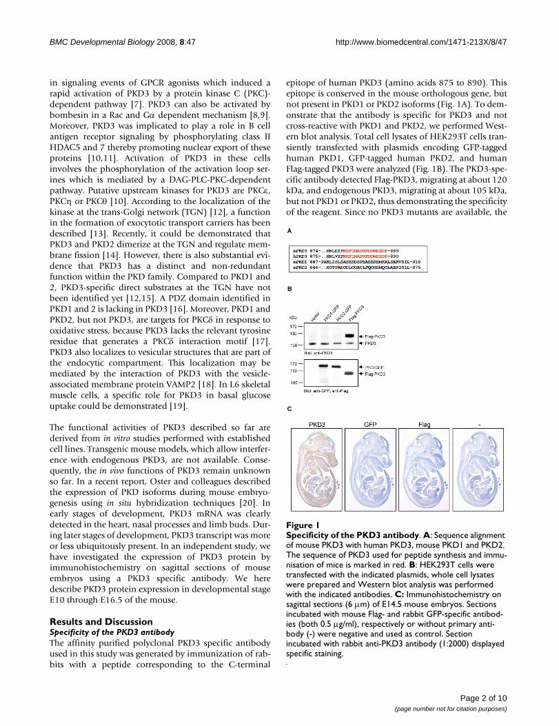
PKD3 expression at embryonic stage E11.5Figure 3PKD3 expression at embryonic stage E11.5. Immunohistochemistry was performed on sagittal sections (6 μm) of E11.5 mouse embryos. (A) – (B) Control sections incubated without primary antibody. (C) – (J) Sections incubated with anti-PKD3 antibody (1:2000). ac: atrial chamber, bs: cartilage primordium of base of the skull, C1: condensation of sclerotomic material forming centrum of atlas, cm: cephalic mesenchyme, da: dorsal aorta, ec: endocardial cushion tissue lining the atrio-ventricular canal, lb: lung bud, mb: main bronchus, nc: notochord, np: nasal process, op: olfactory pit, rh: roof of hindbrain, vh: ventricular chamber of heart. Scale bars: 100 μm.
Page 4 of 10(page number not for citation purposes)

BMC Developmental Biology 2008, 8:47 http://www.biomedcentral.com/1471-213X/8/47
In embryonic stage E11.5 PKD3 expression was detectablein additional tissues (Fig. 3C and 3D). Parts of the headregion near the developing base of the scull and nasalprocess (Fig. 3E) were positive for PKD3 protein. PKD3expression was visible in and along the notochord, wheresclerotomic material is condensed to form the centrum ofthe axis (Fig. 3H and 3J). A strong PKD3 expression wasdetected in the bronchus of the lung bud (Fig. 3J)restricted to the cytoplasm of the epithelium. PKD3expression in the cardiac muscle cells was still obvious(Fig. 3F, G). Moreover, PKD3 was expressed in the wall ofthe dorsal aorta at this stage (Fig. 3H and 3I).
In embryonic stage E12.5 more cytological details ofPKD3 expression were visible (Fig. 4C, D). PKD3 wasdetected in the cartilage primordium of nasal bones (Fig.4E), the temporal bones (Fig. 4J) and the vertebra (Fig.4F). PKD3 expression was still detectable in the noto-chord (Fig. 4F and 4F") and in cardiomyocytes formingthe ventricular and atrial chambers but was absent fromthe atrio-ventricular bulbar cushion tissue (Fig. 4G). Fur-ther, the membrane of the oesophagus showed a strongPKD3 expression (Fig. 4F and 4F'). Within the lung strong
PKD3 expression was found in epithelial cells of segmen-tal bronchii (Fig. 4H). PKD3 expression is also observedin the urogenital ridge surrounding the lumen of the uro-genital sinus (Fig. 4I). Moreover, PKD3 expression nowbecame detectable in the developing brain, with a promi-nent staining of the outer layer of the choroid plexuswithin the fourth ventricle (Fig. 4K). In the stomach, themucous membrane was PKD3 positive (Fig. 4L).
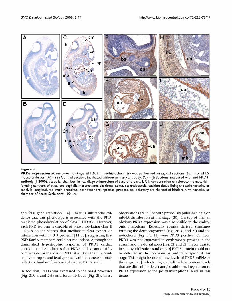
In embryonic stage E13.5 PKD3 was found to be more orless ubiquitously expressed (Fig. 5C and 5D). In additionto PKD3 positive structures like cardiomyocytes (Fig. 5E),the protein could also be detected in further muscle struc-tures: Skeletal muscle cells of the neck region (Fig. 5F) aswell as muscles of the tongue (Fig. 5G and 5G') showedstrong PKD3 expression. In the lung, the epithelial layer ofthe segmental but not the main bronchi was positive (Fig.5H, H'). A prominent PKD3 expression could be detectedin the middle layer (submucosa) of the stomach, but notthe inner layer (mucosa) (Fig. 5I and 5I'). Prominent sitesof PKD3 expression were also detected in the marginallayer of the spinal cord (Fig. 5J) and the choroid plexuswithin the fourth ventricle (Fig 5K and 5K'). Expression of
PKD3 expression at embryonic stage E12.5Figure 4PKD3 expression at embryonic stage E12.5. Immunohistochemistry was performed on sagittal sections (6 μm) of E12.5 mouse embryos. (A) – (B) Control sections incubated without primary antibody. (C) – (L) Sections incubated with anti-PKD3 antibody (1:2000). bc: atrio-ventricular bulbar cushion tissue, cp: origin of choroid plexus, cv: cartilage primordium of vertebra, da: descending (thoracic) aorta, dt: dorsum of tongue, fv: fourth ventricle, ll: left lung, ls: lumen of stomach, lv: lateral ventricle, ml: marginal layer of spinal cord, mm: mucous membrane, nc: nasal cavity, not: notochord, np: nasopharynx, oe: olfactory epi-thelium, op: oesophagus, or: optic recess of diencephalon, pc: pericardial cavity, pl: pleural cavity, rg: root ganglia, sb: segmental bronchus, sc: spinalcord, tb: cartilage primordium of temporal bone, ul: upper lip, us: urogenital sinus, vo: vomeronasal organ. Scale bars: 100 μm.
Page 5 of 10(page number not for citation purposes)
BMC Developmental Biology 2008, 8:47 http://www.biomedcentral.com/1471-213X/8/47
PKD3 was also detected in the developing bones. Espe-cially the cartilage primordium of turbinate bones (Fig.5L) and osteoblasts in the vertebrae which secrete bonematerial into previously existing cartilage matrix wereintensively stained (Fig. 5M and 5M'). The membranouslayer surrounding cartilage during ossification, the peri-chondrium, was also positive for PKD3 expression (Fig.5M'). Of note, the ventricular zone of the neocortex aswell as the liver was negative (Fig 5C and 5D).
PKD3 expression in osteoblasts was even more prominentin stage E14.5 in developing bones of the spinal column(Fig. 6G). The nucleus pulposus in the middle of the spi-nal disc was also intensively stained (Fig. 6H, H'). Skeletalmuscle cells of the diaphragm (Fig. 6I, I') also showed astrong PKD3 expression. Oster and colleagues failed todetect elevated levels of PKD3 mRNA in skeletal muscleby in situ hybridization [20]. The reason for this seemingdiscrepancy is unclear and might be of technical nature orreflect the fact that the level of mRNA does not necessarilycorrelate with the protein level.
In this stage we found a strong expression in the kidneycapsule (Fig. 6J, J'), in terminal bronchioles of the lung(Fig. 6K), in the nuclei of distinct cells of the liver (Fig. 6L)as well as in the middle layer of the duodenum (Fig. 6M).In the brain, PKD3 expression was ubiquitously detectedwith exception of the neocortex, which was negative (Fig.6D, E, F). Interestingly, in the medulla oblongata distinctnerve tracts were intensively stained for PKD3 (Fig. 6N);moreover, the choroid plexus within the fourth and lateralventricle was also positive (Fig. 6E, O). In accordance withpublished data on PKD3 mRNA expression [20] the innerlayer of the retina shows a weak PKD3 specific staining,whereas the extrinsic ocular muscle displays a strongexpression of PKD3 (Fig. 6P). Interestingly, cells withinthe olfactory epithelium displayed a strong PKD3 signalwithin the nuclei (Fig. 6Q). The root sheath of theWhisker follicles showed a steady PKD3 expression inE14.5 and E16.5 (Fig. 6R, R', and 7H). Furthermore, thesuprabasal layer of the epidermis as well as underlyingconnective tissue (Fig. 6S) are positive for PKD3. The epi-dermal staining was impressively evident in the mouth
PKD3 expression at embryonic stage E13.5Figure 5PKD3 expression at embryonic stage E13.5. Immunohistochemistry was performed on sagittal sections (6 μm) of E13.5 mouse embryos. (A) – (B) Control sections incubated without primary antibody. (C) – (N') Sections incubated with anti-PKD3 antibody (1:2000). bb: cartilage primordium of basioccipital bone (clivus), cp:choroid plexus, cr: cartilage primordium of ribs, dt: dorsum of tongue, fv: fourth ventricle, hb: cartilage primordium of body of hyoid bone, li: liver, ll: lower lip, ls: lumen of stom-ach, lu: lung, mb: main bronchus, mc: mucosal lining, ml: marginal layer of spinal cord, mt: muscle mass of the tongue, nc: neo-cortex, oe: olfactory epithelium, or: optic recess of diencephalon, os: osteoblasts, pc: pericardial cavity, pe: perichondrium, pl: pleural cavity, pm: pons-midbrain junction, rg: root ganglion, sb: segmental bronchus, sc: spinal cord, sm: submucosa, st: stom-ach, tb: cartilage primordium of turbinate bones, tp: tooth primordium, ul: upper lip, vc: vena cava. Scale bars: 100 μm.
Page 6 of 10(page number not for citation purposes)

BMC Developmental Biology 2008, 8:47 http://www.biomedcentral.com/1471-213X/8/47
region where PKD3 negative endoderm and PKD3 posi-tive ectoderm derived epidermal layers came into directcontact (Fig. 6T). PKD3 is also expressed in the first pri-mordium of the upper molar tooth (Fig. 6U), which isformed by an incorporation of dental epithelium.
PKD3 was found to be more ore less ubiquitouslyexpressed in embryonic stage E16.5 (Fig. 7C and 7D). Par-ticularly strong PKD3 expression was detected in nervetracts in the pons (Fig. 7E) and the choroid plexus of thelateral and fourth ventricle (Fig. 7F, G). The middle layer(submucosa) but not the mucosa or the muscle layer (Fig.7I) demonstrated PKD3 expression. In contrast to E13.5,the submucosa of the stomach showed only a weak PKD3expression, however, smooth muscle cells in the muscula-ris layer of the stomach were still PKD3 negative (Fig. 7J,
J'). Within the liver PKD3 expression seemed to be furtherincreased (Fig. 7C, D), which is in accordance with highmRNA levels detected by in situ hybridization in E18.5[20]. The strong PKD3 expression in the kidney capsule(Fig. 7K) and in the terminal bronchi of the lung (Fig. 7L)detected in E14.5 was even more evident. In the region ofthe lower lip, the intensive staining of the suprabasal layerof the epidermis was obvious (Fig. 7M). All skeletal mus-cle cells within the embryo showed a strong PKD3 expres-sion (Fig 7N–R) e.g. intercostal muscle cells (Fig. 7N) andthe transverse muscle fibers of the tongue (Fig. 7O). Inter-estingly, PKD3 distribution in skeletal muscle cells of thediaphragm (Fig. 7P) and the neck region (Fig. 7Q) ispolarized, especially visible in cross-sections (Fig. 7Q). Ofnote, PKD3 was not detected in smooth muscle cells andin the thymus at this stage. Interestingly, Oster and col-
PKD3 expression at embryonic stage E14.5Figure 6PKD3 expression at embryonic stage E14.5. Immunohistochemistry was performed on sagittal sections (6 μm) of E14.5 mouse embryos. (A) – (C) Control sections incubated without primary antibody. (D) – (U) Sections incubated with anti-PKD3 antibody (1:2000). bl: basal layer of epidermis, ce: cerebellum, cp: choroid plexus, du: duodenum, il: inner layer of retina, kc: kidney capsule, ki: kidney, ld: lumen of duodenum, le: lens, li: liver, lv: lateral ventricle, mb: main bronchus, md: muscle of dia-phragm, mf: muscle fibers, ml: marginal layer of spinal cord, mo: medulla oblongata, mr: medulla renalis, mt: primordium of upper molar tooth, mu: muscularis layer of duodenum, nb: cartilage primordium of the nasal bone, nc: nasal cavity, nl: neural layer of retina, np: nucleus pulposus in the central region of future invertebral disc, oe: olfactory epithelium, op: oesophagus, or: oropharynx, rg: root ganglia, rp: renal pelvis, sc: spinal cord, scl: sclera, sl: suprabasal layer of epidermis, sm: submucosal layer of duodenum, sp: cartilage primordium of spinal column, st: stomach, tb: terminal bronchus, th: thymus, tt: tip of the tongue, ul: upper lip, vi: primordia of follicles of vibrissae. Scale bars: 100 μm.
Page 7 of 10(page number not for citation purposes)

BMC Developmental Biology 2008, 8:47 http://www.biomedcentral.com/1471-213X/8/47
leagues could detect PKD3 mRNA in the thymus at E18.5[20]. In line with this, Western blot analysis revealed astrong PKD3 expression in the thymus of adult animals(unpublished observation K. Ellwanger), suggesting anincrease in the expression level of PKD3 protein in neona-tal mice.
ConclusionThe expression pattern of PKD3 reveals a tissue selectiveexpression at stage E10, which became more abundantand distributed later on during embryonic development.Our data are in accordance with previously publishedresults on PKD3 mRNA levels using in situ hybridization
analysis [20]. On top of that, we provide a comprehensivestudy on the expression pattern of PKD3 during organo-genesis discovering additional histological details. Thestrong expression of PKD3 in specific tissues, e.g. cardiacand skeletal muscle, points to an important role for thiskinase in the development of these tissues. PKD3 has beenimplicated in the regulation of secretory transport proc-esses at the Golgi compartment [13,14] as well as regula-tion of basal glucose uptake in skeletal muscle cells [19],both of which are important processes during organogen-esis. Moreover, PKD3 has been shown to regulate thenuclear localization and thus activity of its physiologicalsubstrates class II HDAC5 and 7 [11]. Interestingly, class
PKD3 expression at embryonic stage E16.5Figure 7PKD3 expression at embryonic stage E16.5. Immunohistochemistry was performed on sagittal sections (6 μm) of E16.5 mouse embryos. (A) – (B) Control sections incubated without primary antibody. (C) – (Q) Sections incubated with anti-PKD3 antibody (1:2000). bl: basal layer of epidermis, cc: costal cartilage, cp: choroid plexus, dt: dorsum of tongue, du: duodenum, fu: fundus region of stomach, fv: fourth ventricle, im: intercostal muscle, kc: kidney capsule, ki: kidney, ld: lumen of duodenum, li: liver, ll: lower lip, lu: lung, lv: lateral ventricle, mb: main bronchus, md: muscle fibers of diaphragm, mf: muscle fibers, mr: medulla renalis, mt: muscle mass of the tongue, mu: muscularis layer, nr: neck region, po: pons, sl: suprabasal layer of epidermis, sm: submucosal layer, tb: terminal bronchus, vi: primordia of follicles of vibrissae. Scale bars: 100 μm (E) – (Q), 10 μm (R).
Page 8 of 10(page number not for citation purposes)

BMC Developmental Biology 2008, 8:47 http://www.biomedcentral.com/1471-213X/8/47
II HDAC proteins play an important role in heart develop-ment and function [26,27]. It will now be exciting toinvestigate the potential function of PKD3 in these tissuesusing transgenic mouse models, interfering with endog-enous PKD3 function by overexpression of a dominant-negative protein or deletion of the PKD3 gene (knock-out).
MethodsAntibodiesThe primary antibody used in this study was an affinity-purified PKD3 specific polyclonal antibody raised in rab-bits against a C-terminal epitope of human PKD3 (aminoacids 875-890). The mouse monoclonal GFP- and Flag-specific antibodies were obtained from Roche Biosciencesand Sigma-Aldrich, respectively. The rabbit polyclonalGFP-specific antibody was from Santa Cruz Biotechnol-ogy. The secondary IRdye-conjugated antibodies werefrom Li-COR Biosciences. Transfection of HEK293T cellswas performed as described in [12].
AnimalsC57BL/6 mice were time mated and pregnant femaleswere sacrificed to collect the embryos at different stages(E10 – E16.5). The finding of a vaginal plug at noon wasconsidered as E0. The fetuses were isolated from theuterus and dissected free of embryonic membranes in ice-cold PBS. All animal experiments carried out in this studywere approved by the ethical committee at the Universityof Stuttgart.
Western BlotCells were harvested and lysed in lysis buffer (20 mM Tris(pH7.4), 150 mM NaCl, 1 mM EDTA, 1 mM EGTA, and1% Triton X-100, plus protease and phosphatase inhibi-tors). Equal amounts of protein were subjected to a 4–12% NuPAGE Bis-Tris-Gel (Invitrogen, Germany), blottedonto a nitrocellulose membrane and blocked with 0.5%blocking buffer (Roche Biosciences, Germany). Incuba-tion with primary antibodies was performed in blockingbuffer at 4°C overnight. After washing with PBS, sampleswere incubated with secondary IRdye680-conjugatedanti-mouse or IRdye800-conjugated anti-rabbit IgG anti-bodies in blocking buffer for 1 h at room temperature. Thedetection was performed on an Odyssey Infrared ImagingSystem (Li-COR Biosciences, Germany).
ImmunohistochemistryAfter fixation with paraformaldehyde embryos were dehy-drated through a graded ethanol series and into a 50:50mixture of ethanol:Histoclear (Carl Roth GmbH, Karl-sruhe, Germany). Tissue was then incubated in Histoclearfor 1–3 hours and transferred into a 50:50 mixture ofHistoclear:Paraplast (Carl Roth GmbH, Karlsruhe, Ger-many) at 42°C, where it was incubated over night.
Embryos were transferred into pure Paraplast preheated to60°C and incubated for 1–3 days. Serial sagittal sectionswere cut at a thickness of 10 μm on a microtome andfloated on a 40°C water bath. Sections were dewaxed in 3changes of Roticlear for 10 minutes each and incubated inisopropanol and ethanol 15 minutes each. Slides wererehydrated through a reverse series of ethanol dilutions inPBS and finally washed in PBS. Endogenous peroxidaseactivity was quenched by incubation in PBS containing0.3% H2O2 for 30 minutes. Slides were rinsed with PBS for10 minutes and then incubated with 5% normal goatserum in a humidified chamber to block unspecific bind-ing sites. Primary antiserum diluted in 1.5% normal goatserum was applied. Slides were incubated in the humidi-fied chamber at 4°C over night.
Immunohistochemical staining was performed using theVectastain Elite ABC Kit (rabbit IgG) (Vector Laboratories,Burlingame, CA, USA). To remove non-specifically boundantibody, slides were washed three times in PBS for 10minutes each time. Subsequently, sections were incubatedwith biotinylated goat anti-rabbit IgG diluted 1:200 in1.5% normal goat serum for 1 hour at room temperature.In case of the mouse monoclonal Flag-specific antibody, abiotin-SP-conjugated goat anti-mouse IgG (JacksonImmunoresearch, West Grove, PA, USA) diluted 1:500 in1.5% normal goat serum was used as secondary antibody.After the slides had been rinsed in PBS as before, immu-noperoxidase staining was performed. Sections were incu-bated with the Vectastain Elite ABC reagent (prepared 30–60 minutes before use) for 30 minutes and washed threetimes with PBS for 5 minutes each time. For peroxidasevisualization the DAB Substrate Kit for peroxidase (VectorLaboratories, Burlingame, CA, USA) was used. Colordevelopment was stopped by rinsing the sections in waterfor 5 minutes. Sections were counterstained with hema-toxylin for 1 minute and coverslipped with Mowiol (Poly-sciences, Warrington, PA, USA) or Eukitt (EMS, FortWashington, PA, USA).
Microscopy and image processingStained sections were analyzed using a widefield micro-scope (Zeiss Axiovert 200 M) equipped with the AxioCamHRC (Zeiss, Germany) and an Achroplan 10×/0,25 Ph1 ora LD Achroplan 40×/0,60 Korr Ph2 (DICIII) objective.Images were further processed with Axiovision softwareversion 4.5 (Zeiss, Germany).
Authors' contributionsKE carried out the immunohistochemistry, analyzed thedata and helped to draft the manuscript. SL purified theanti-PKD3 polyclonal immune serum. KP was involved inconception of study, data interpretation and manuscriptwriting. AH designed the study, analyzed and interpreted
Page 9 of 10(page number not for citation purposes)

BMC Developmental Biology 2008, 8:47 http://www.biomedcentral.com/1471-213X/8/47
Publish with BioMed Central and every scientist can read your work free of charge
"BioMed Central will be the most significant development for disseminating the results of biomedical research in our lifetime."
Sir Paul Nurse, Cancer Research UK
Your research papers will be:
available free of charge to the entire biomedical community
peer reviewed and published immediately upon acceptance
cited in PubMed and archived on PubMed Central
yours — you keep the copyright
Submit your manuscript here:http://www.biomedcentral.com/info/publishing_adv.asp
BioMedcentral
the data, and drafted the manuscript. All authors read andapproved the final manuscript.
AcknowledgementsWe would like to thank Vivek Malhotra for providing us with the anti-PKD3 polyclonal serum, Katalin Schlett for the biotinylated goat anti-mouse IgG, Olaf Selchow, Franz Brümmer and Martin Pfannkuchen for support with microscopy, Gisela Link for excellence technical assistance and Heinz Stre-ble for insightful discussions. This work was supported by grants from the Landesstiftung Baden-Württemberg and the Deutsche Forschungsgemein-schaft (HA3557/2-1) to A. Hausser.
References1. Maier D, Hausser A, Nagel AC, Link G, Kugler SJ, Wech I, Pfizenmaier
K, Preiss A: Drosophila protein kinase D is broadly expressedand a fraction localizes to the Golgi compartment. Gene ExprPatterns 2006, 6:849-856.
2. Maier D, Nagel AC, Gloc H, Hausser A, Kugler SJ, Wech I, Preiss A:Protein kinase D regulates several aspects of development inDrosophila melanogaster. BMC Dev Biol 2007, 7:74.
3. Feng H, Ren M, Rubin CS: Conserved domains subserve novelmechanisms and functions in DKF-1, a C. elegans proteinkinase D. J Biol Chem 2006, 281:17815-26.
4. Feng H, Ren M, Wu SL, Hall DH, Rubin CS: Characterization of anovel protein kinase D: C. elegans DKF-1 is activated bytranslocation-phosphorylation and regulates movement andgrowth in vivo. J Biol Chem 2006, 281(26):17801-14.
5. Wang QJ: PKD at the crossroads of DAG and PKC signaling.Trends Pharmacol Sci 2006, 27:317-323.
6. Hayashi A, Seki N, Hattori A, Kozuma S, Saito T: PKCnu, a newmember of the protein kinase C family, composes a fourthsubfamily with PKCmu. Biochim Biophys Acta 1999, 1450:99-106.
7. Rey O, Yuan J, Young SH, Rozengurt E: Protein kinase C nu/pro-tein kinase D3 nuclear localization, catalytic activation, andintracellular redistribution in response to G protein-coupledreceptor agonists. J Biol Chem 2003, 278:23773-23785.
8. Yuan J, Rey O, Rozengurt E: Activation of protein kinase D3 bysignaling through Rac and the alpha subunits of the heterot-rimeric G proteins G(12) and G(13). Cell Signal 2006,18:1051-1062.
9. Yuan J, Rey O, Rozengurt E: Protein kinase D3 activation andphosphorylation by signaling through G alpha q. Biochem Bio-phys Res Commun 2005, 335:270-276.
10. Matthews SA, Dayalu R, Thompson LJ, Scharenberg AM: Regulationof protein kinase Cnu by the B-cell antigen receptor. J BiolChem 2003, 278:9086-9091.
11. Matthews SA, Liu P, Spitaler M, Olson EN, McKinsey TA, Cantrell DA,Scharenberg AM: Essential role for protein kinase D familykinases in the regulation of class II histone deacetylases in Blymphocytes. Mol Cell Biol 2006, 26:1569-1577.
12. Hausser A, Storz P, Martens S, Link G, Toker A, Pfizenmaier K: Pro-tein kinase D regulates vesicular transport by phosphorylat-ing and activating phosphatidylinositol-4 kinase IIIbeta at theGolgi complex. Nat Cell Biol 2005, 7:880-886.
13. Yeaman C, Ayala MI, Wright JR, Bard F, Bossard C, Ang A, Maeda Y,Seufferlein T, Mellman I, Nelson WJ, Malhotra V: Protein kinase Dregulates basolateral membrane protein exit from trans-Golgi network. Nat Cell Biol 2004, 6:106-112.
14. Bossard C, Bresson D, Polishchuk RS, Malhotra V: Dimeric PKDregulates membrane fission to form transport carriers at theTGN. J Cell Biol 2007, 179:1123-1131.
15. Fugmann T, Hausser A, Schoffler P, Schmid S, Pfizenmaier K, OlayioyeMA: Regulation of secretory transport by protein kinase D-mediated phosphorylation of the ceramide transfer protein.J Cell Biol 2007, 178:15-22.
16. Sanchez-Ruiloba L, Cabrera-Poch N, Rodriguez-Martinez M, Lopez-Menendez C, Jean-Mairet RM, Higuero AM, Iglesias T: Proteinkinase D intracellular localization and activity control kinaseD-interacting substrate of 220-kDa traffic through a postsy-naptic density-95/discs large/zonula occludens-1-bindingmotif. J Biol Chem 2006, 281:18888-18900.
17. Doppler H, Storz P: A novel tyrosine phosphorylation site inprotein kinase D contributes to oxidative stress-mediatedactivation. J Biol Chem 2007, 282:31873-31881.
18. Lu G, Chen J, Espinoza LA, Garfield S, Toshiyuki S, Akiko H, HupplerA, Wang QJ: Protein kinase D 3 is localized in vesicular struc-tures and interacts with vesicle-associated membrane pro-tein 2. Cell Signal 2007, 19:867-879.
19. Chen J, Lu G, Wang QJ: Protein kinase C-independent effects ofprotein kinase D3 in glucose transport in L6 myotubes. MolPharmacol 2005, 67:152-162.
20. Oster H, Abraham D, Leitges M: Expression of the protein kinaseD (PKD) family during mouse embryogenesis. Gene Expr Pat-terns 2006, 6:400-408.
21. Avkiran M, Rowland AJ, Cuello F, Haworth RS: Protein kinase d inthe cardiovascular system: emerging roles in health and dis-ease. Circ Res 2008, 102:157-163.
22. Haworth RS, Goss MW, Rozengurt E, Avkiran M: Expression andactivity of protein kinase D/protein kinase C mu in myocar-dium: evidence for alpha1-adrenergic receptor- and proteinkinase C-mediated regulation. J Mol Cell Cardiol 2000,32:1013-1023.
23. Harrison BC, Kim MS, van Rooij E, Plato CF, Papst PJ, Vega RB, McA-nally JA, Richardson JA, Bassel-Duby R, Olson EN, McKinsey TA:Regulation of cardiac stress signaling by protein kinase d1.Mol Cell Biol 2006, 26:3875-3888.
24. Fielitz J, Kim MS, Shelton JM, Qi X, Hill JA, Richardson JA, Bassel-DubyR, Olson EN: Requirement of protein kinase D1 for patholog-ical cardiac remodeling. Proc Natl Acad Sci U S A 2008,105:3059-3063.
25. Huynh QK, McKinsey TA: Protein kinase D directly phosphor-ylates histone deacetylase 5 via a random sequential kineticmechanism. Arch Biochem Biophys 2006, 450:141-8.
26. Chang S, McKinsey TA, Zhang CL, Richardson JA, Hill JA, Olson EN:Histone deacetylases 5 and 9 govern responsiveness of theheart to a subset of stress signals and play redundant roles inheart development. Mol Cell Biol 2004, 24:8467-8476.
27. Zhang CL, McKinsey TA, Chang S, Antos CL, Hill JA, Olson EN: ClassII histone deacetylases act as signal-responsive repressors ofcardiac hypertrophy. Cell 2002, 110:479-488.
Page 10 of 10(page number not for citation purposes)
Copyright © 2022 FDOKUMEN