Shortcomings of short hairpin RNA-based transgenic RNA interference in mouse oocytes
Age-associated alteration of gene expressionpatterns in mouse oocytes
Toshio Hamatani{, Geppino Falco{, Mark G. Carter, Hidenori Akutsu, Carole A. Stagg,
Alexei A. Sharov, Dawood B. Dudekula, Vincent VanBuren and Minoru S.H. Ko*
Developmental Genomics and Aging Section, Laboratory of Genetics, National Institute on Aging,
National Institutes of Health, 333 Cassell Drive, Suite 3000, Baltimore, MD 21224, USA
Received May 9, 2004; Revised and Accepted July 20, 2004
Decreasing oocyte competence with maternal aging is a major factor in human infertility. To investigate theage-dependent molecular changes in a mouse model, we compared the expression profiles of metaphase IIoocytes collected from 5- to 6-week-old mice with those collected from 42- to 45-week-old mice using the NIA22K 60-mer oligo microarray. Among approximately 11 000 genes whose transcripts were detected inoocytes, about 5% (530) showed statistically significant expression changes, excluding the possibility ofglobal decline in transcript abundance. Consistent with the generally accepted view of aging, the differen-tially expressed genes included ones involved in mitochondrial function and oxidative stress. However,the expression of other genes involved in chromatin structure, DNA methylation, genome stability andRNA helicases was also altered, suggesting the existence of additional mechanisms for aging. Among thetranscripts decreased with aging, we identified and characterized a group of new oocyte-specific genes,members of the human NACHT, leucine-rich repeat and PYD-containing (NALP) gene family. These resultshave implications for aging research as well as for clinical ooplasmic donation to rejuvenate aging oocytes.
INTRODUCTION
Reproductive capacity in women declines dramatically beyondthe mid-30s (1–4). Although the primary cause for this declineis the gradual depletion of oocytes, the decline of oocyte qualityis also suggested to play an important role. For example, it hasbeen shown that young women undergoing standard in vitrofertilization (IVF) with their own eggs show a success ratecomparable with older women (.40 years) undergoing IVFwith eggs donated by this younger subset of women (5). Age-related decline in oocyte quality has been associated with aneu-ploidy because of the age-associated increase of spontaneousabortions and numerical meiotic division errors (6), but theprimary cause of such anomalies and other factors determiningoocyte quality remains to be elucidated.
The degree to which oocytes age is also relevant to the clini-cal application of ooplasmic donation (1,7). Ooplasmicdonation has been performed by injecting ooplasm from ayoung healthy donor oocyte into a patient oocyte at metaphaseII (MII) stage to improve the outcome of assisted reproductionmethods (8,9). Some mouse studies suggested that ooplasmicdonation by cytoplasmic injection or by nuclear transfer from
an oocyte to another enucleated oocyte has no detrimentaleffect on development at the MII or germinal vesicle stage(8,10,11) (reviewed in 12), but no direct supporting molecularbiological evidence has been reported (13,14). In fact, the pre-mature rupture of membranes and intrauterine fetal death fol-lowing ooplasmic donation by nuclear transfer from a patientoocyte to an enucleated young donor oocyte (15), and mito-chondrial heteroplasmy after birth following ooplasmic injec-tion have been reported very recently (16,17). Thus, studies ofmolecular mechanisms involved in the decline of oocytequality with maternal age could have important implicationsfor the efficacy and safety of clinical ooplasmic donation.
Whether oocytes, which are germline-derived and in somesense immortal, age in ways similar to somatic cells is a fun-damental question in aging research (18–21). The alteration ofgene expression patterns in aging cells and organs has beenstudied (22,23) and global expression profiling of agingorgans has also been performed (24,25). However, oocytesprovide unique and challenging material for microarray-based studies of aging.
First, in contrast to organs and continuously culturedfibroblast cells (26), in oocytes all the aging-associated
Human Molecular Genetics, Vol. 13, No. 19 # Oxford University Press 2004; all rights reserved
{The authors wish it to be known that, in their opinion, the first two authors should be regarded as joint First Authors.
*To whom correspondence should be addressed. Tel: þ1 4105588359; Fax: þ1 4105588331; Email: [email protected]
Human Molecular Genetics, 2004, Vol. 13, No. 19 2263–2278doi:10.1093/hmg/ddh241Advance Access published on August 18, 2004
by guest on January 8, 2015http://hm
g.oxfordjournals.org/D
ownloaded from

complications will be imposed on the cell cycle-synchronizedand -arrested cells. Most of the primordial follicles are formedduring fetal development, though a recent study suggestedmeiotic entry of germline stem cells in post-natal mammalianovary (27). Oocytes are arrested and stay in the middle of pro-metaphase II for more than 40 years in humans and more thana year in mice. At various time points of their adult life, theseprimordial oocytes with a diameter of 15–20 mm grow tobecome full-grown oocytes of 70–150 mm (28). The numberof mtDNA copies also increases about 100 times (29). Thus,it is reasonable to assume that the microarray analysis willmonitor mainly the cumulative results of the transcriptionalactivity during follicular growth in adult, which takes only afew weeks and the same amount of time for both young andold oocytes.
Second, the analysis of germline aging on a global scale hasbeen hampered by the lack of an expression profiling platformincluding genes unique to oocytes and by the difficulties ofcollecting significant numbers of oocytes. With the recentdevelopment of a suitable microarray platform (30), we haveovercome these problems and have performed expression pro-filing of oocytes.
RESULTS AND DISCUSSION
Global characteristics of oocyte aging
We collected three sets of 500 MII oocytes from young (5–6weeks) and those from aged (42–45 weeks) mice—near theend of their reproductive lifespan (Fig. 1A). For brevity,these oocytes are called ‘young oocytes’ and ‘old oocytes’here. As would be the case in a human IVF clinic, onlyoocytes with good morphology were harvested from super-ovulated C57BL/6 mice. These oocytes were free ofcumulus cell contamination (Materials and Methods). Thelabeling of these RNAs provided nearly equal amount of flu-orescence probes, indicating the initial amount of RNAs wassimilar. Therefore, a possibility of general decline in transcriptabundance can be excluded.
To control sample-to-sample variation, we analyzed threeindependent sets of pooled oocytes by microarrays. Toassess the variations detected by both technical and biologicalreplications, we also carried out two sets of dye-swap exper-iments. The expression profiling was performed on the NIA22K 60-mer oligo microarray, which is enriched for genesexpressed in stem cells and early embryos, includingoocytes (30).
A total of 131 634 experimental data points were generatedand analyzed. Using a cut-off for signal intensities (logintensity � 2.3), transcripts of 10 977 genes were scored aspresent in oocytes (data available at http://lgsun.grc.nia.nih.gov/microarray/data.html). Along with previous reports(31,32), this list provides a repertoire of genes whose tran-scripts are accumulated through oogenesis and are present inMII oocytes.
Young and old oocytes had very similar gene expressionpatterns. Only 530 genes (�5%) exhibited statistically signifi-cant differences (449 genes for young oocytes and 81 genesfor old oocytes) (Fig. 1B and Supplementary Material,Table S1). Furthermore, the fold difference, i.e. log-ratio of
these 530 genes, was relatively small (99 genes, . 2-fold;431 genes, .1.5-fold). Consistent with the identification ofbona fide genes, we found that 425 (80.2%) out of the 530 dif-ferentially expressed genes were represented as expressedsequence tags (ESTs) in cDNA libraries from unfertilizedeggs, fertilized eggs and two-cell embryos (31).
To validate the microarray data further, we also performedreal-time quantitative RT–PCR (Q-PCR) analysis in triplicatefor 50 selected genes (Figs 1C and D). Poly(A) length, whichcan be regulated by cytoplasmic polyadenylation element andis involved in mRNA stability in oocyte and early preimplan-tation embryos, might affect the efficiency of cRNA linearamplification during probe synthesis. To exclude suchpoly(A) length-dependent bias, we used random hexamerrather than oligo(dT) primers in cDNA synthesis for Q-PCR.Results from the microarrays and Q-PCR were well correlated:the correlation coefficient was 0.80, with a slope of 0.78(Fig. 1C). The microarrays thus provide reliable geneexpression comparisons between young and old oocytes.
Of the 449 genes showing decreased transcript levels withmaternal aging, 284 (63.3%) genes were known, whereas 47(58.0%) of 81 genes showing the increased transcript levelswith maternal aging were known. Functional categories ofthese genes were assigned by Gene Ontology (GO) terms(33), using the MAPPFinder (34,35) (Fig. 2 and Supplemen-tary Material, Fig. S1). We discuss four functional groups.
Genes involved in mitochondrial function, oxidativedamages and stress-responses
Most prominent in the list were ‘mitochondrial function’, ‘oxi-dative damages’ and ‘stress-responses’ (Fig. 2). Genes such asmt-Nd3, mt-Atp6 and mt-Co1-3, encoded in the mitochondrialgenome and involved in mitochondrial electron transportchain, were more highly expressed in old oocytes. In contrast,genes encoded in the nuclear genome but related to ‘energypathways’ and mitochondrial function (such as Sdha, knownto be an index for mitochondrial activity) were more highlyexpressed in young oocytes (Fig. 2). These changes suggesta relative decline in ATP production in aging oocytes. Infact, it has been shown that the aging oocytes contain lessATP and have a lower electrical potential at the innermitochondrial membrane (36). Interestingly, genes cate-gorized in ‘nucleotides and ATP metabolism’ and ‘ATPbinding’ also showed decreased expression.
A group of genes that provide protection against stress-responses and damages were also downregulated. Notablewere oxidative stress/damage-related genes, such as Sod1 andthe thioredoxin family (Txn1 and Apacd ) (Fig. 2). Sod1 isreported to be highly expressed in human oocytes (37) and theaddition of Sod1 protein improves preimplantation developmentin mouse (38). Thioredoxins are involved in reduction and pro-tection against oxidative stress-induced apoptosis, and the tar-geted mutation of Txn1 caused embryonic lethality shortlyafter implantation (39). Other ‘anti-apoptosis’ genes, includingBcl2l10/Diva, Nckap1, Psip2, Ppid, Rnf34, Bag4 and Pdcd6ip,were also downregulated in old oocytes (Fig. 2).
Second, ‘chaperones’ such as the heat shock 70 kDa pro-teins (Hspa4, Hspa8 and Hsp70-4 ) and Cct genes (Cct1-3and Cct5 ), as well as many other genes involved in the
2264 Human Molecular Genetics, 2004, Vol. 13, No. 19
by guest on January 8, 2015http://hm
g.oxfordjournals.org/D
ownloaded from
Figure 1. Microarray gene expression profiles from oocytes of old (old oocytes) and young (young oocytes) mice. (A) RNAs were extracted from three sub-pooled lots of approximately 500 oocytes at each age, and samples corresponding to 18 oocytes were separately labeled by two rounds of linear amplification,then hybridized to arrays in duplicate dye-swap pairs. The microarray results for selected genes in the same RNA preparations were validated by quantitativereal-time RT–PCR (Q-PCR) in triplicate. (B) A scatter plot of all 21 393 features on NIA 22K 60-mer oligo microarray, comparing gene expression of young andaged oocytes. Microarray data for 21 939 probes were analyzed by ANOVA-FDR statistics (30). The combined results of 12 hybridizations identified 530 differ-entially expressed genes (red circles). (C) Comparison of log-ratio results from microarray with those from Q-PCR. Using the same RNA preparations 50selected genes were validated by Q-PCR in triplicate. The correlation coefficient was 0.80 and the slope was 0.78. (D) The fold changes between old andyoung oocytes by the microarray and Q-PCR analysis about the 50 genes.
Human Molecular Genetics, 2004, Vol. 13, No. 19 2265
by guest on January 8, 2015http://hm
g.oxfordjournals.org/D
ownloaded from
Figure 2. Functional characterization of decreased and increased transcripts in oocytes with maternal aging. The most frequent GO terms (33) for differentiallyexpressed genes in young and old oocytes were identified by FDR statistics (FDR � 15%) using MAPPFinder (34,35) (Supplementary Material, Fig. S1). OnlyGO terms that had more than three genes (‘number changed in hierarchy’ � 3) for young oocytes are shown in (A). Only GO terms that had more than threegenes (‘number changed in hierarchy’ � 2) for old oocytes are shown in (B). We further recategorized these frequent GO terms and showed genes that representeach annotation. If NCBI had no gene symbol corresponding to an NIA 60-mer oligo, an U-cluster ID containing the oligo sequence was shown in these panels.‘Oocyte-specific genes’ were selected on the basis of the frequencies of corresponding ESTs (31).
2266 Human Molecular Genetics, 2004, Vol. 13, No. 19
by guest on January 8, 2015http://hm
g.oxfordjournals.org/D
ownloaded from
‘ubiquitin–proteasome pathway’, such as Hip2, Ubc, Ube1c,Ube2a, Ube2e3, Ube2g1, Pama6, Pamb1, Psmb4, Psmc2,Psmc3, Psmd12, Siah2 and Anapc4, showed decreasedexpression in old oocytes (Fig. 2). Oxidative stress duringaging is known to be associated with the decline of heatshock response, lysosomal activity and ubiquitin–proteasomepathway in somatic cells (25,40). Age-related deterioration ofthe capacity to produce active heat shock proteins could leadto the accumulation of damaged proteins, primarily oxidizedand glycated proteins (41). The production of ubiquitin ismodified during aging, often leading to less free ubiquitinand more ubiquitin–protein conjugates (40). A failure of ubi-quitination could contribute to the accumulation of highlydamaged proteins, which in turn are effective inhibitors ofthe proteasome (42).
Third, I kappa B alpha (Nfkbia/IkBa ), which is a suppressorof NF-kappa B, was upregulated. NF-kappa B and I kappa Balpha are found in the mitochondria and have been suggestedto regulate expression of mitochondrial genes (43). The sup-pression of NF-kappa B by I kappa B alpha induced theexpression of both cytochrome c oxidase III and cytochromeb mRNA (43). This may explain the up-regulation of Ikappa B alpha and cytochrome c oxidases (mtCo1-3 ) inoocytes with maternal aging.
Genes involved in cell cycles, DNA stability andchromosome stability
It is known that cellular ATP deprivation caused by inhibitorsof energy metabolism in turn induces a gradual breakdown ofactin-containing microfilament bundles (44). Consistent withthis notion, transcripts related to ‘microtubule cytoskelton’and ‘chromosome segregation’, including Hook1, Tuba1,Tubd1, Dncic2, Kif3b, Rnf19/Dorfin, Pcnt2, Nin and Smc4l1,all decreased with maternal aging (Fig. 2). It has beenshown that cytoskeletal alteration could cause the increasedfrequency of aneuploidy, inhibition of extrusion of the firstpolar body and increased incidence of cellular fragmentation(reviewed in 45,46). Therefore, the alteration of transcriptlevels of these genes may be associated with chromosomeabnormalities.
Although both young and old oocytes were arrested andsynchronized at MII, ‘cell cycle’ was one of the most conspic-uous differences between two cell types (Fig. 2). This suggeststhat oocytes, normally well equipped for the resumption of thecell cycle after fertilization, lost capacity during maternalaging. Another prominent category was ‘genome stability’,which includes CGG triplet repeat binding protein-1(Cggbp1 ), fragile X mental retardation-related protein 2(Fxr2h ) and homologs of bacterial MutH/L/S mismatch-repair-related proteins such as Msh3, Exo1 and Pms1(Figs 1D and 2). These genes, which are often implicated inaccelerated aging syndromes and carcinogenesis in human(47–50), decreased in expressions with maternal aging.
We note that the level of telomerase reverse transcriptase(Tert ), whose expression decreases during general aging andcellular senescence, decreased with maternal aging (Fig. 2).This is consistent with the notion that short telomeres in thechromosomes of human eggs predict poor prognosis followingIVF/embryo transfer (51).
Taken together, these expression changes suggest that oldoocytes may be less able to maintain intact chromosomes.
Genes involved in oogenesis, fertilization andpreimplantation development
Genes involved in the ‘Tgf beta receptor signaling pathway’,including Madh1/Smad1 and Bmpr2, were upregulated in oldoocytes (Fig. 2). It is known that in granulosa cells, Bmpr2protein functions as a receptor for Gdf9 protein, a glycoproteinsecreted by the oocyte and capable of stimulating granulosacell proliferation and inhibiting differentiation (52). Itsexpression is also observed in some oocytes of primary andearly secondary follicles in rat (53). Smad1 signaling isdemonstrated to play a critical function in the initial commit-ment of the germ cell lineage in mice (54), and its mRNA isrecruited to polysomes during oocyte maturation inXenopus, suggesting a role in oocyte maturation (55). In con-trast, genes involved in ‘intracellular protein transport andvesicle trafficking’, were downregulated in old oocytes(Fig. 2), suggesting the dysregulation of oocyte maturation.Indeed, it has been shown that the presence of brefeldin A,a drug that inhibits protein secretion by blocking membranetrafficking from endoplasmic reticulum to Golgi apparatus(56), blocks oocyte maturation before the assembly of themetaphase I spindle maturation (57). Also, Stx7 and Snap23were downregulated in old oocytes. Their encoded proteinsare a part of an integral membrane protein complex termedSNAREs (soluble NSF attachment protein receptors),suggested to be involved in vesicular trafficking and exocyto-sis of cortical granules derived from the Golgi apparatus inmouse oocytes (58).
A number of genes involved in ‘reproduction’ and ‘oocyte-specific genes’ also decreased their expression with aging(Fig. 2). For example, Vasa, a DEAD helicase expressedspecifically in germ cell lineage and development, alongwith other ‘RNA helicases’ including Ddx32, Dhx36, Ddx46,Ddx48 and Supv3l1, were downregulated. Similarly, Rnf35and ePAD, implicated in cytoskeletal organization in eggs,were also downregulated. Furthermore, all three zona pellu-cida glycoprotein genes, Zp1, Zp2 and Zp3, which playimportant roles in oogenesis (59), fertilization and subsequentpreimplantation development (60–62), were downregulated(Figs 1D and 2). Similarly, genes involved in gametogenesis,such as Kit, NIMA (never in mitosis gene a)-related expressedkinase 2 (Nek2 ) and Zfp38, were downregulated (Fig. 2).
Genes involved in transcriptional regulation
Decreased expression of many transcription factors wasobserved in oocytes during maternal aging (Fig. 2). This cat-egory includes many zinc finger proteins (Zfps ), whose zinc-coordinated cysteines are susceptible to attack by reactiveoxygen species (ROS), and whose DNA binding activity canbe attenuated with age (63,64), and polycomb group genessuch as Bmi1, Ezh2, Sfmbt1 and Rybp1. Polycomb group pro-teins have been known to play crucial roles in cell fate deter-mination via the assembly of specialized forms of repressivechromatin. Because transcription is globally reduced inmature oocytes and dramatic chromatin remodeling occurs
Human Molecular Genetics, 2004, Vol. 13, No. 19 2267
by guest on January 8, 2015http://hm
g.oxfordjournals.org/D
ownloaded from

in fertilized eggs, polycomb group genes may be involved inthe control of global transcription in oocytes through nucleo-some modification, chromatin remodeling and interactionwith general transcription factors. Indeed, the expression ofBmi1 and Ezh2, a polycomb-group transcription repressor,was downregulated. Interestingly, Bmi1 is essential for theself-renewal of adult murine hematopoietic stem cells andneuronal stem cells partly via repression of genes involvedin cellular senescence and cell death (65). A null mutationin Ezh2 (which is upregulated upon fertilization and kepthighly expressed during the mouse preimplantation stages)results in early embryonic lethality (66).
Transcripts for both oocyte-specific and somatic forms ofmaintenance DNA methyltransferase (Dnmt1o and Dnmt1s,respectively), Dnmt-associated protein-1 (Dmap1) andDnmt3L, were also downregulated in old oocytes (Figs 1D,2 and 3), whereas the level of de novo methyltransferaseDnmt3b transcript was upregulated (Figs 1D and 2). Thesespecific expression patterns were also observed in WI-38 cul-tured fibroblast cells undergoing cellular senescence (67),suggesting that oocytes and somatic cells may sharecommon features in aging.
Some genes related to ‘chromatin organization and biogen-esis’ were downregulated in old oocytes (Fig. 2). For example,the expression of histone deacetylase 2 (Hdac2 ), whichinteracts with Dnmt1 to represses transcription, was alsodownregulated, as were histone acetyltransferases, Myst1and Mrgx, which are related to Morf, involved in cellularsenescence (68). Changes in these and other chromatin remo-deling factors, combined with the specific changes in DNAmethyltransferase expression, suggest that chromatin structureand epigenetic mechanisms may be appreciably dysregulatedas oocytes age.
Examples of unknown genes: the mouseNalp gene family
Among the unknown genes downregulated with maternalaging, we found several genes homologous to the mousematernal effect gene (Mater ) that was also downregulatedduring aging. Mater expression is detected in the cytoplasmof growing oocytes and remains present through the lateblastocyst stage (69). It has been shown that Mater is essen-tial for the preimplantation development, because the devel-opment of Mater2/2 embryos is arrested at two-cell stage(69). A human homolog of mouse Mater has also beenidentified (70) and categorized as NACHT, leucine-richrepeat and PYD containing 5 (NALP5 ) (71) on the basisof their protein domain structure. Three unknown genesthat showed decreased expression in oocytes duringmaternal aging were found to fall in the Nalp family (row7 in Fig. 2A). Their chromosomal loci have been representedby LOC233001, 4921520L01Rik and E330024M09/AU022726 in National Center for Biotechnology Information(NCBI) LocusLink, respectively. Searching for similar genesin the NIA Mouse Gene Index (31), we identified at leastfive more genes whose ESTs were exclusively from cDNAlibraries of unfertilized eggs, fertilized eggs (one-cellembryos) and two-cell embryos as well as Mater. These
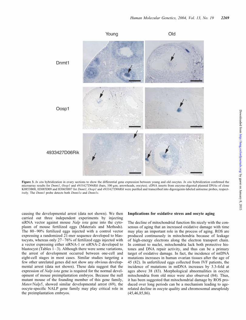
genes were named Nalp alpha–Nalp kappa after the humanNALP gene family (GenBank accession numbers: Nalpalpha, AY596194; Nalp beta, AY596195; Nalp gamma,AY596198; Nalp delta, AY596196; Nalp epsilon,AY596197; Nalp zeta, AY596203; Nalp eta, AY596200;Nalp theta, AY596202; Nalp iota, AY596199; Nalp kappa,AY596201). Interestingly, Nalp alpha–theta and Mater areclustered on Chromosome 7A. Nalp iota is located onChromosome 7F and Nalp kappa is located on Chromosome13. The complete cDNA sequences of Nalp gamma–kappawere obtained by fully sequencing either existing NIAcDNA clones (31) or cDNAs amplified by RT–PCR. Theopen reading frames (ORFs) of these genes were searchedagainst the Pfam HMM database (72). Except for Nalp iotathat lacks a PYD domain, all the genes of the mouse NALPfamily contain NACHT, PYD and leucine-rich repeatdomains like the human NALP family genes (Fig. 4A).
The sequences of these genes were very similar to eachother and it was thus difficult to assign unambiguously ortho-logous relationships to human NALP genes. To help to clarifythe relationships, we also identified rat genes by searching thehuman and mouse gene sequences against Ensembl Ratgenome sequences by WU-BLAST (73) with additional gui-dance by synteny and 1st exon search tool (74). Amino acidsequence similarities were analyzed among all the genes inhuman, mouse and rat, and a phylogenetic tree and dot plotswere produced based on a sequence distance method and theNeighbor Joining (NJ) algorithm (Fig. 4B and SupplementaryMaterial, Fig. S2) (75). Searches for the conserved regions ofthe genomic sequences by PipMaker (http://bio.cse.psu.edu)(76) found no significant similarity in the genomic sequences,other than exons (data not shown). All human gene loci wereidentified to correspond to the mouse Nalp alpha locus onmouse chromosome 7A1 (Fig. 4C). Genes on mouse Chromo-some 13 found no corresponding human genomic regions,though the surrounding genes showed clear correspondenceto human syntenic regions.
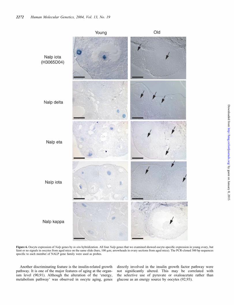
Northern blot analyses with �300 bp gene-specific probesshowed that all genes were specifically expressed in oocytes,with additional testis expression for Nalp iota (Fig. 5A).In situ hybridization with four representative genes revealedoocyte-specific expression in young ovary (Fig. 6). Thegenes were expressed in primary oocytes at various follicularstages. Furthermore, the microarray expression profilingduring mouse preimplantation stages showed that all thegenes are expressed in oocytes but are immediately downregu-lated after fertilization (32) (Fig. 5B). Mouse NALP familygenes are thus rather exclusively expressed in oocytes atall follicle stages, with a presumed role in oogenesis and pre-implantation embryos.
We used the RNA interference approach as a first step toanalyze a function of mouse NALP gene families. Theapproach has been successfully used to inhibit the functionof specific genes in mouse oocytes (77–80). We decided touse the proven method of injecting plasmid vectors expressingsiRNA under the mouse U6 promoter into fertilized eggs (81).We first optimized the condition by injecting various concen-tration of the control vector into the cytoplasm of fertilizedeggs and found that the injection of 1–2 pl of 100 ng/mlvector could achieve the maximum GFP expression without
2268 Human Molecular Genetics, 2004, Vol. 13, No. 19
by guest on January 8, 2015http://hm
g.oxfordjournals.org/D
ownloaded from
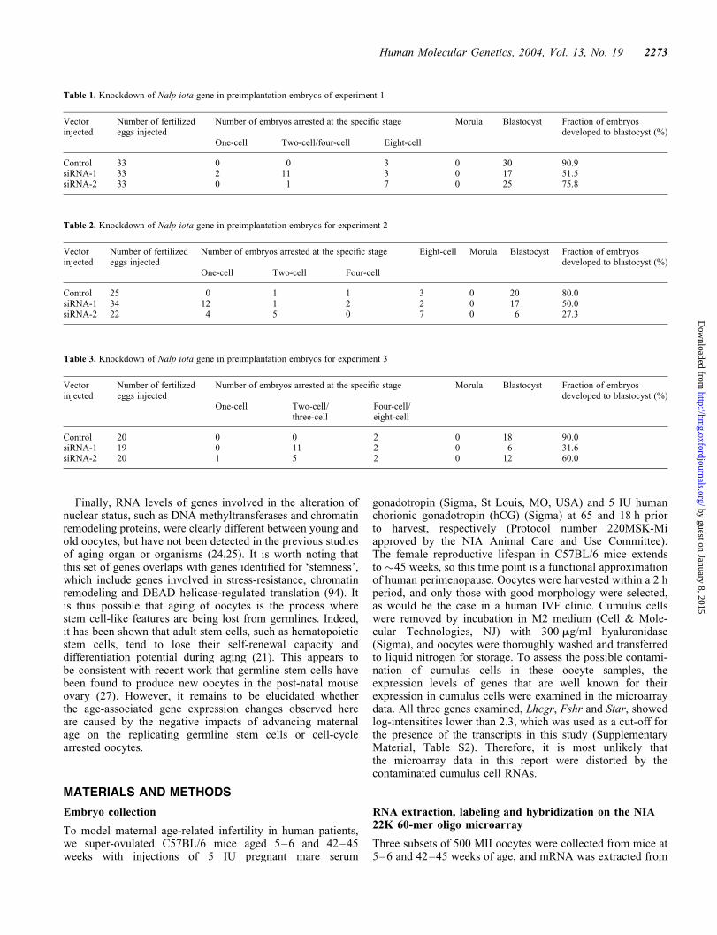
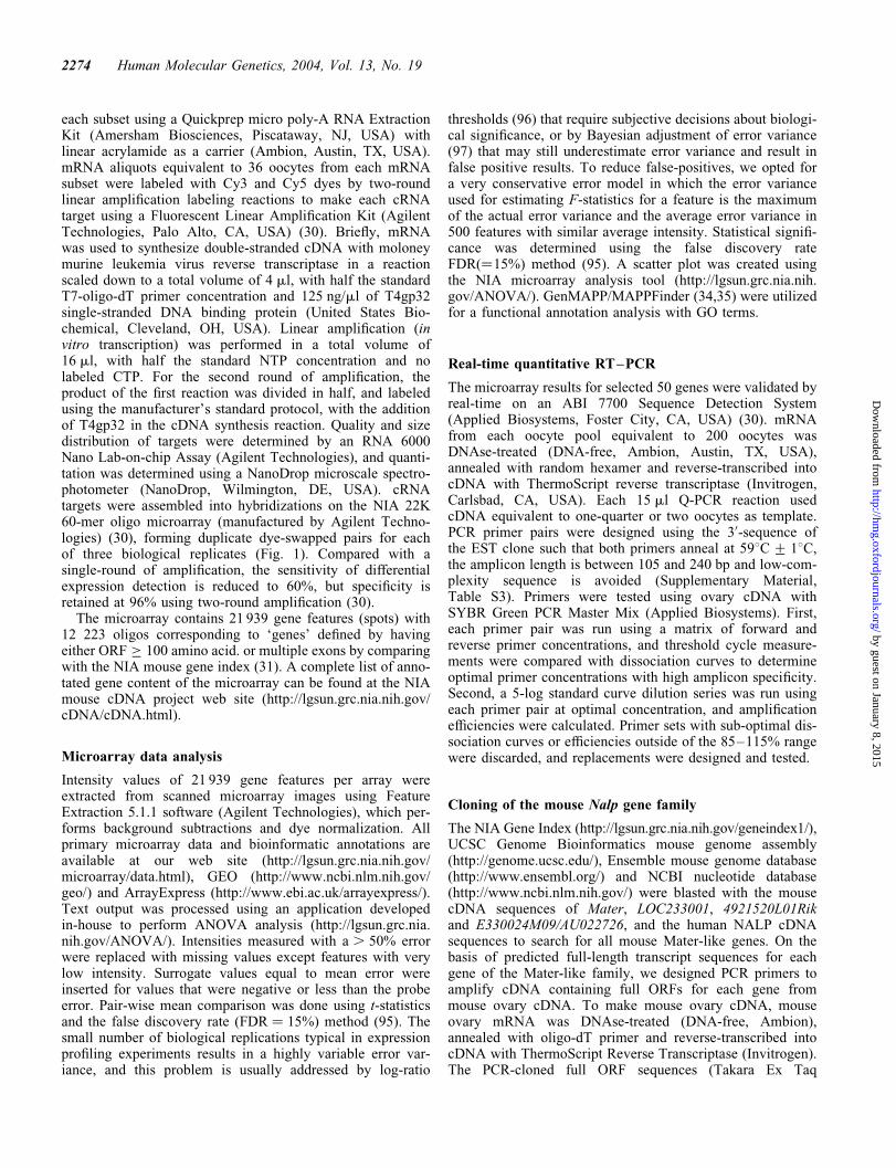
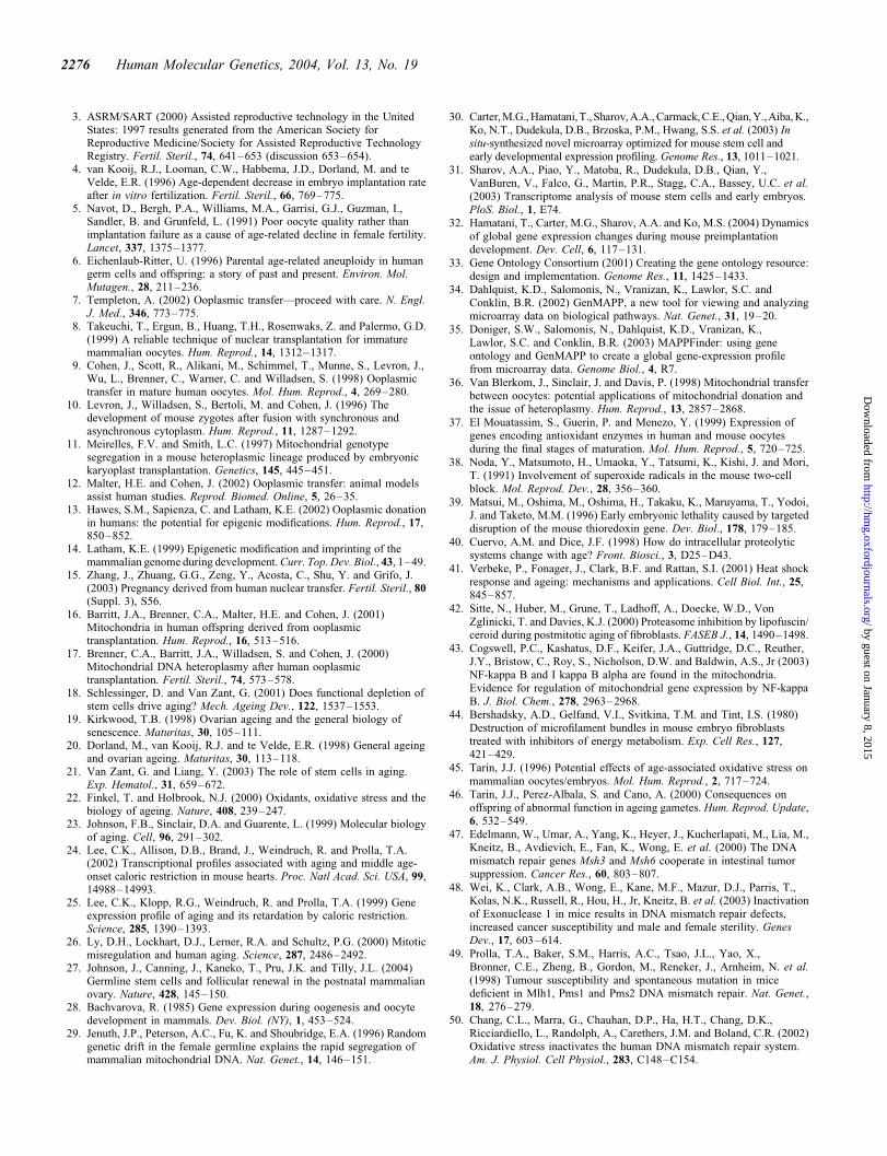
causing the developmental arrest (data not shown). We thencarried out three independent experiments by injectingsiRNA vector against mouse Nalp iota gene into the cyto-plasm of mouse fertilized eggs (Materials and Methods).The 80–90% fertilized eggs injected with a control vectorexpressing a randomized 21-mer sequence developed to blas-tocysts, whereas only 27–76% of fertilized eggs injected witha vector expressing either siRNA-1 or siRNA-2 developed toblastocyst (Tables 1–3). Although there were some variations,the arrest of development occurred between one-cell andeight-cell stages in most cases. Similar studies targeting afew other unrelated genes did not show any obvious develop-mental arrest (data not shown). These data suggest that theexpression of Nalp iota gene is required for the normal devel-opment of mouse preimplantation embryos. Because the nullmutant mouse of the founding member of this gene family,Mater/Nalp5, showed similar developmental arrest (69), theoocyte-specific NALP gene family may play critical role inthe preimplantation embryos.
Implications for oxidative stress and oocyte aging
The decline of mitochondrial function fits nicely with the con-sensus of aging that an increased oxidative damage with timemay play an important role in the process of aging. ROS areproduced continuously in mitochondria because of leakageof high-energy electrons along the electron transport chain.In contrast to nuclei, mitochondria lack both protective his-tones and DNA repair activity, and thus can be a primarytarget of oxidative damage. In fact, the incidence of mtDNAmutations increases in human ovarian tissues after the age of45 (82). In unfertilized eggs collected from IVF patients, theincidence of mutations in mtDNA increases by 3.3-fold atages above 38 (83). Morphological abnormalities in oocytemitochondria from old mice were also observed (84). Thus,it has been suggested that mitochondrial damage by ROS pro-duced over long periods can be a mechanism leading to age-related decline in oocyte quality and chromosomal aneuploidy(45,46,85,86).
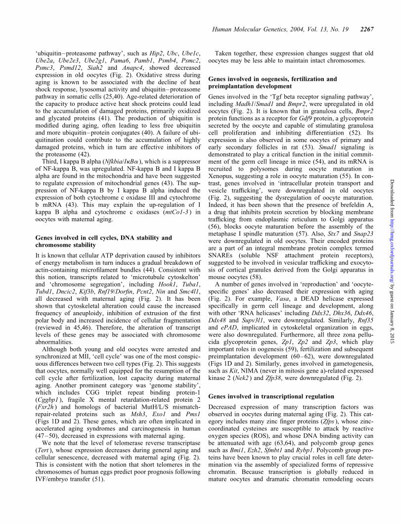
Figure 3. In situ hybridization in ovary sections to show the differential gene expression between young and old oocytes. In situ hybridization confirmed themicroarray results for Dnmt1, Oosp1 and 4933427D06Rik (bars, 100 mm; arrowheads, oocytes). cDNA inserts from enzyme-digested plasmid DNAs of cloneK0853B08, H3085D09 and H3065H07 for Dnmt1, Oosp1 and 4933427D06Rik were purified and transcribed into digoxigenin-labeled antisense probes, respect-ively. The Dnmt1 probe detects both Dnmt1o and Dnmt1s.
Human Molecular Genetics, 2004, Vol. 13, No. 19 2269
by guest on January 8, 2015http://hm
g.oxfordjournals.org/D
ownloaded from
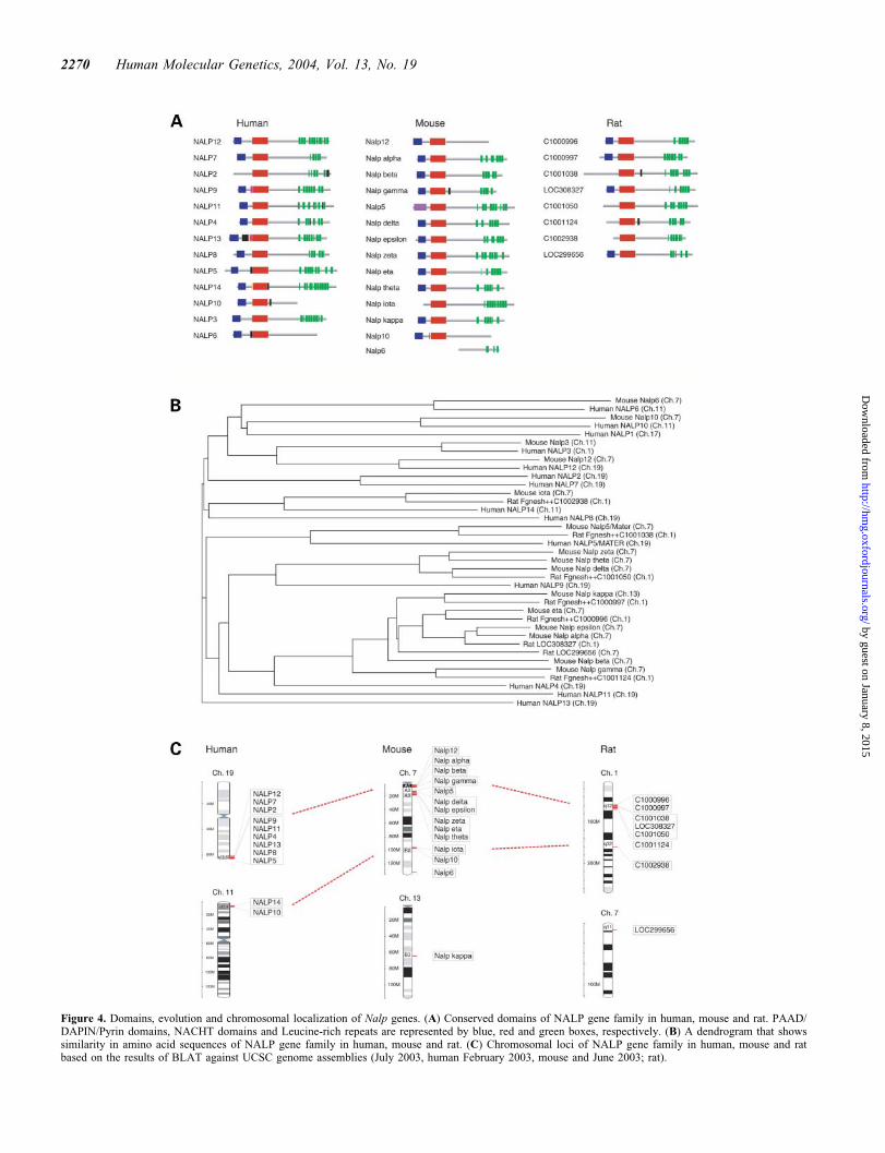
Figure 4. Domains, evolution and chromosomal localization of Nalp genes. (A) Conserved domains of NALP gene family in human, mouse and rat. PAAD/DAPIN/Pyrin domains, NACHT domains and Leucine-rich repeats are represented by blue, red and green boxes, respectively. (B) A dendrogram that showssimilarity in amino acid sequences of NALP gene family in human, mouse and rat. (C) Chromosomal loci of NALP gene family in human, mouse and ratbased on the results of BLAT against UCSC genome assemblies (July 2003, human February 2003, mouse and June 2003; rat).
2270 Human Molecular Genetics, 2004, Vol. 13, No. 19
by guest on January 8, 2015http://hm
g.oxfordjournals.org/D
ownloaded from
In this study we also observed an age-related decrease intranscription levels of ATP-related genes that are representedby GO terms such as ‘nucleotides and ATP metabolism’ and‘ATP binding’. It is known that the depletion of ATP by thedecrease of mitochondrial function can decrease the functionof proteins that require ATP for their proper function. Assuch, the microtubule and cytoskeleton proteins seem to beimportant, because malfunction of these proteins is known tocause failures of chromosome segregation and an accompany-ing increase of aneuploidy—a major pathological phenotypeof old oocytes in humans.
Implications for ooplasmic donation
Discussions about the efficacy and safety of ooplasmicdonation have centered on the mitochondria (7). If loweredATP were the only defect in old oocytes, the injection ofATP could revitalize the oocytes. If the lack of both functionalmitochondria and ATP is the problem, ooplasmic donationindeed helps to increase the fertility rate of old oocytes. Ourresults seem to support this idea, because mitochondria-related genes and ATP-related genes were indeed downregu-lated in old oocytes. However, our results also suggest thatthe nucleus of old oocytes may not be competent to producespecific transcripts that are required for normal oocytes,because the microarray analysis primarily revealed the
differential RNA levels transcribed from young versusold nuclei during oocyte maturation (Introduction). If thestatus of the nucleus in old oocytes is already different fromthat in young oocytes, ooplasmic donation cannot directlycorrect the problem of old oocytes. Even if nuclei in oldoocytes can be reprogramed, such reprograming events areerror-prone, as has been demonstrated in mouse and othermodel systems (87–89).
Implications for germline aging
Although microarray analyses of aging organs and tissueshave been reported (24–26), oocyte aging seems to have dis-tinctive features. For example, one of the most conspicuousfeatures of aging in organs such as brain and liver is theinflammatory response, indicated by the upregulation of anumber of genes involved in the inflammatory process(24,25). However, such trends were not observed in agingoocytes. One possibility is that these genes are not normallyexpressed in oocytes, and thus cannot be detected by micro-array analysis. Alternatively, the upregulation of inflammatorygene expression in organs merely represents secondarychanges during aging, probably due to infiltration of immunecells into organs, but are not seen in single oocytes, especiallyin the immune privileged environment.
Figure 5. The expression profiles of mouse Nalp genes. (A) Northern blot analysis using the 300 bp sequences specific to each member of NALP gene family asprobes with mouse tissue blot (FirstChoice Mouse Blot 1, Ambion). (B) The expression patterns of mouse Nalp genes during mouse preimplantation embryostages based on our previous microarray experiment (32). U, F, 2, 4, 8, M and B denote unfertilized egg, fertilized egg, two-cell embryo, four-cell embryo, eight-cell embryo, morula and blastocyst, respectively.
Human Molecular Genetics, 2004, Vol. 13, No. 19 2271
by guest on January 8, 2015http://hm
g.oxfordjournals.org/D
ownloaded from
Another discriminating feature is the insulin-related growthpathway. It is one of the major features of aging at the organ-ism level (90,91). Although the alteration of the ‘energy,metabolism pathway’ was observed in oocyte aging, genes
directly involved in the insulin growth factor pathway werenot significantly altered. This may be correlated withthe selective use of pyruvate or oxaloacetate rather thanglucose as an energy source by oocytes (92,93).
Figure 6. Oocyte expression of Nalp genes by in situ hybridization. All four Nalp genes that we examined showed oocyte-specific expression in young ovary, butfaint or no signals in oocytes from aged mice on the same slide (bars, 100 mm; arrowheads in ovary sections from aged mice). The PCR-cloned 300 bp sequencesspecific to each member of NALP gene family were used as probes.
2272 Human Molecular Genetics, 2004, Vol. 13, No. 19
by guest on January 8, 2015http://hm
g.oxfordjournals.org/D
ownloaded from
Finally, RNA levels of genes involved in the alteration ofnuclear status, such as DNA methyltransferases and chromatinremodeling proteins, were clearly different between young andold oocytes, but have not been detected in the previous studiesof aging organ or organisms (24,25). It is worth noting thatthis set of genes overlaps with genes identified for ‘stemness’,which include genes involved in stress-resistance, chromatinremodeling and DEAD helicase-regulated translation (94). Itis thus possible that aging of oocytes is the process wherestem cell-like features are being lost from germlines. Indeed,it has been shown that adult stem cells, such as hematopoieticstem cells, tend to lose their self-renewal capacity anddifferentiation potential during aging (21). This appears tobe consistent with recent work that germline stem cells havebeen found to produce new oocytes in the post-natal mouseovary (27). However, it remains to be elucidated whetherthe age-associated gene expression changes observed hereare caused by the negative impacts of advancing maternalage on the replicating germline stem cells or cell-cyclearrested oocytes.
MATERIALS AND METHODS
Embryo collection
To model maternal age-related infertility in human patients,we super-ovulated C57BL/6 mice aged 5–6 and 42–45weeks with injections of 5 IU pregnant mare serum
gonadotropin (Sigma, St Louis, MO, USA) and 5 IU humanchorionic gonadotropin (hCG) (Sigma) at 65 and 18 h priorto harvest, respectively (Protocol number 220MSK-Miapproved by the NIA Animal Care and Use Committee).The female reproductive lifespan in C57BL/6 mice extendsto �45 weeks, so this time point is a functional approximationof human perimenopause. Oocytes were harvested within a 2 hperiod, and only those with good morphology were selected,as would be the case in a human IVF clinic. Cumulus cellswere removed by incubation in M2 medium (Cell & Mole-cular Technologies, NJ) with 300 mg/ml hyaluronidase(Sigma), and oocytes were thoroughly washed and transferredto liquid nitrogen for storage. To assess the possible contami-nation of cumulus cells in these oocyte samples, theexpression levels of genes that are well known for theirexpression in cumulus cells were examined in the microarraydata. All three genes examined, Lhcgr, Fshr and Star, showedlog-intensitites lower than 2.3, which was used as a cut-off forthe presence of the transcripts in this study (SupplementaryMaterial, Table S2). Therefore, it is most unlikely thatthe microarray data in this report were distorted by thecontaminated cumulus cell RNAs.
RNA extraction, labeling and hybridization on the NIA22K 60-mer oligo microarray
Three subsets of 500 MII oocytes were collected from mice at5–6 and 42–45 weeks of age, and mRNA was extracted from
Table 2. Knockdown of Nalp iota gene in preimplantation embryos for experiment 2
Vectorinjected
Number of fertilizedeggs injected
Number of embryos arrested at the specific stage Eight-cell Morula Blastocyst Fraction of embryosdeveloped to blastocyst (%)
One-cell Two-cell Four-cell
Control 25 0 1 1 3 0 20 80.0siRNA-1 34 12 1 2 2 0 17 50.0siRNA-2 22 4 5 0 7 0 6 27.3
Table 3. Knockdown of Nalp iota gene in preimplantation embryos for experiment 3
Vectorinjected
Number of fertilizedeggs injected
Number of embryos arrested at the specific stage Morula Blastocyst Fraction of embryosdeveloped to blastocyst (%)
One-cell Two-cell/three-cell
Four-cell/eight-cell
Control 20 0 0 2 0 18 90.0siRNA-1 19 0 11 2 0 6 31.6siRNA-2 20 1 5 2 0 12 60.0
Table 1. Knockdown of Nalp iota gene in preimplantation embryos of experiment 1
Vectorinjected
Number of fertilizedeggs injected
Number of embryos arrested at the specific stage Morula Blastocyst Fraction of embryosdeveloped to blastocyst (%)
One-cell Two-cell/four-cell Eight-cell
Control 33 0 0 3 0 30 90.9siRNA-1 33 2 11 3 0 17 51.5siRNA-2 33 0 1 7 0 25 75.8
Human Molecular Genetics, 2004, Vol. 13, No. 19 2273
by guest on January 8, 2015http://hm
g.oxfordjournals.org/D
ownloaded from
each subset using a Quickprep micro poly-A RNA ExtractionKit (Amersham Biosciences, Piscataway, NJ, USA) withlinear acrylamide as a carrier (Ambion, Austin, TX, USA).mRNA aliquots equivalent to 36 oocytes from each mRNAsubset were labeled with Cy3 and Cy5 dyes by two-roundlinear amplification labeling reactions to make each cRNAtarget using a Fluorescent Linear Amplification Kit (AgilentTechnologies, Palo Alto, CA, USA) (30). Briefly, mRNAwas used to synthesize double-stranded cDNA with moloneymurine leukemia virus reverse transcriptase in a reactionscaled down to a total volume of 4 ml, with half the standardT7-oligo-dT primer concentration and 125 ng/ml of T4gp32single-stranded DNA binding protein (United States Bio-chemical, Cleveland, OH, USA). Linear amplification (invitro transcription) was performed in a total volume of16 ml, with half the standard NTP concentration and nolabeled CTP. For the second round of amplification, theproduct of the first reaction was divided in half, and labeledusing the manufacturer’s standard protocol, with the additionof T4gp32 in the cDNA synthesis reaction. Quality and sizedistribution of targets were determined by an RNA 6000Nano Lab-on-chip Assay (Agilent Technologies), and quanti-tation was determined using a NanoDrop microscale spectro-photometer (NanoDrop, Wilmington, DE, USA). cRNAtargets were assembled into hybridizations on the NIA 22K60-mer oligo microarray (manufactured by Agilent Techno-logies) (30), forming duplicate dye-swapped pairs for eachof three biological replicates (Fig. 1). Compared with asingle-round of amplification, the sensitivity of differentialexpression detection is reduced to 60%, but specificity isretained at 96% using two-round amplification (30).
The microarray contains 21 939 gene features (spots) with12 223 oligos corresponding to ‘genes’ defined by havingeither ORF � 100 amino acid. or multiple exons by comparingwith the NIA mouse gene index (31). A complete list of anno-tated gene content of the microarray can be found at the NIAmouse cDNA project web site (http://lgsun.grc.nia.nih.gov/cDNA/cDNA.html).
Microarray data analysis
Intensity values of 21 939 gene features per array wereextracted from scanned microarray images using FeatureExtraction 5.1.1 software (Agilent Technologies), which per-forms background subtractions and dye normalization. Allprimary microarray data and bioinformatic annotations areavailable at our web site (http://lgsun.grc.nia.nih.gov/microarray/data.html), GEO (http://www.ncbi.nlm.nih.gov/geo/) and ArrayExpress (http://www.ebi.ac.uk/arrayexpress/).Text output was processed using an application developedin-house to perform ANOVA analysis (http://lgsun.grc.nia.nih.gov/ANOVA/). Intensities measured with a . 50% errorwere replaced with missing values except features with verylow intensity. Surrogate values equal to mean error wereinserted for values that were negative or less than the probeerror. Pair-wise mean comparison was done using t-statisticsand the false discovery rate (FDR ¼ 15%) method (95). Thesmall number of biological replications typical in expressionprofiling experiments results in a highly variable error var-iance, and this problem is usually addressed by log-ratio
thresholds (96) that require subjective decisions about biologi-cal significance, or by Bayesian adjustment of error variance(97) that may still underestimate error variance and result infalse positive results. To reduce false-positives, we opted fora very conservative error model in which the error varianceused for estimating F-statistics for a feature is the maximumof the actual error variance and the average error variance in500 features with similar average intensity. Statistical signifi-cance was determined using the false discovery rateFDR(¼15%) method (95). A scatter plot was created usingthe NIA microarray analysis tool (http://lgsun.grc.nia.nih.gov/ANOVA/). GenMAPP/MAPPFinder (34,35) were utilizedfor a functional annotation analysis with GO terms.
Real-time quantitative RT–PCR
The microarray results for selected 50 genes were validated byreal-time on an ABI 7700 Sequence Detection System(Applied Biosystems, Foster City, CA, USA) (30). mRNAfrom each oocyte pool equivalent to 200 oocytes wasDNAse-treated (DNA-free, Ambion, Austin, TX, USA),annealed with random hexamer and reverse-transcribed intocDNA with ThermoScript reverse transcriptase (Invitrogen,Carlsbad, CA, USA). Each 15 ml Q-PCR reaction usedcDNA equivalent to one-quarter or two oocytes as template.PCR primer pairs were designed using the 30-sequence ofthe EST clone such that both primers anneal at 598C + 18C,the amplicon length is between 105 and 240 bp and low-com-plexity sequence is avoided (Supplementary Material,Table S3). Primers were tested using ovary cDNA withSYBR Green PCR Master Mix (Applied Biosystems). First,each primer pair was run using a matrix of forward andreverse primer concentrations, and threshold cycle measure-ments were compared with dissociation curves to determineoptimal primer concentrations with high amplicon specificity.Second, a 5-log standard curve dilution series was run usingeach primer pair at optimal concentration, and amplificationefficiencies were calculated. Primer sets with sub-optimal dis-sociation curves or efficiencies outside of the 85–115% rangewere discarded, and replacements were designed and tested.
Cloning of the mouse Nalp gene family
The NIA Gene Index (http://lgsun.grc.nia.nih.gov/geneindex1/),UCSC Genome Bioinformatics mouse genome assembly(http://genome.ucsc.edu/), Ensemble mouse genome database(http://www.ensembl.org/) and NCBI nucleotide database(http://www.ncbi.nlm.nih.gov/) were blasted with the mousecDNA sequences of Mater, LOC233001, 4921520L01Rikand E330024M09/AU022726, and the human NALP cDNAsequences to search for all mouse Mater-like genes. On thebasis of predicted full-length transcript sequences for eachgene of the Mater-like family, we designed PCR primers toamplify cDNA containing full ORFs for each gene frommouse ovary cDNA. To make mouse ovary cDNA, mouseovary mRNA was DNAse-treated (DNA-free, Ambion),annealed with oligo-dT primer and reverse-transcribed intocDNA with ThermoScript Reverse Transcriptase (Invitrogen).The PCR-cloned full ORF sequences (Takara Ex Taq
2274 Human Molecular Genetics, 2004, Vol. 13, No. 19
by guest on January 8, 2015http://hm
g.oxfordjournals.org/D
ownloaded from

Polymerase, Takara Mirus Bio, Madison, WI, USA; WizardSV Gel and PCR Clean-Up System, Promega Biosciences,San Luis Obispo, CA, USA; pENTR Directional TOPOCloning Kit and TOPO XL PCR Cloning Kit, Invitrogen)were sequenced using a BigDye Terminator kit (PE AppliedBiosystems, Foster City, CA, USA), DyeEX 96 Kit (QiagenValencia, CA, USA) and ABI 3100 Genetic Analyzer (PEApplied Biosystems). To discriminate each gene from otherfamily members in tissue expression analysis, 300 bpsequences unique to each cDNA were selected using ProbeHunter, a software developed in-house (VanBuren et al.,manuscript in preparation). We designed primers withinthese unique 300 bp sequences for each gene (SupplementaryMaterial, Table S4) to amplify the sequences. The PCR-ampli-fied sequences were cloned using a pCR4-TOPO Kit (Invitro-gen) and sequenced.
Computational analysis of the mouse Mater-like genecDNA and human NALP gene cDNA sequences
The ORF finder (http://www.ncbi.nlm.nih.gov/gorf/gorf.html)and Pfam HMM databases (http://pfam.wustl.edu/hmmsearch.shtml) were used to analyze the mouse Nalp cDNA sequences,identifying ORF and protein domains, respectively. To obtainorthologs and analyze chromosome synteny and nucleotidehomology among mouse, rat and human NALPs, Ensemblesynteny view (http://www.ensembl.org/Mus_musculus/syntenyview), WU-BALST (http://www.ensembl.org/Multi/blastview) and comparative genomics information in theUCSC genome browser were used. Amino acid sequencesimilarities were analyzed among all the NALP genes inhuman, rat and mouse, and a phylogenetic tree was postulatedon the basis of a sequence distance method and the NJ algo-rithm of Saitou and Ne (75), using Vector NTI software(Informax, Bethesda, MD, USA).
Northern blot
Northern blot membranes (FirstChoice Mouse Blot 1,Ambion) containing 2 mg of poly(A) RNA per lane isolatedfrom mouse tissues were purchased to detect the expressionof Nalps. The 300 bp sequences specific to each member ofNalps were excised from pCR4 vectors and labeled with[a-32P]dCTP as probes. ExpressHyb solution (Clontech) wasused for both prehybridization and hybridization. Afterhybridization at 658C for 20 h, both membranes werewashed twice with 2� SSC at room temperature for 30 min,twice with 2� SSC at 658C for 30 min and then with 0.1�SSC at 658C for 30 min, followed by exposure to aPhosphorScreen.
In situ hybridization
In situ hybridization was performed in ovaries from young (6week) and aged (42 week) C57BL/6 mice. Ovaries were fixedin 4% paraformaldehyde and sectioned (5 mm). PlasmidDNAs were purified from NIA cDNA library clones(H3065D04 for Nalp iota, K0853B08 for Dnmt1s andDnmt1o, H3085D09 for Oosp1 and H3065D07 for4933427D06Rik) and pCR4 clones containing Nalp unique
300 bp sequences. The enzyme-digested plasmid DNAs weretranscribed into digoxigenin-labeled sense probe controlsand antisense probes. The probes were prepared by alkalinehydrolysis and hybridized (608C, 24 h) to the ovarian sectionsfrom young and aged mice, placed on the same slides.
Knockdown of Nalp iota gene inpreimplantation embryos
Plasmid vectors expressing an siRNA against mouse Nalp iotaconstitutively were constructed by inserting the following 21-mer target sequences (shown in bold) in pRNAT-U6.1/Neo(GeneScript Corp., Scotch Plains, NJ, USA): siRNA-1,GGATCCCAGCTGCAGATATAGTATGTGACTTCCTGTCATCACATACTATATCTGCAGCTTTTTTTCCAAAAGCTT; siRNA-2, GGATCCCGTTACATCGACCAGAAACTTCTCTTCCTGTCAAGAAGTTTCTGGTCGATGTAATTTTTTCCAAAAGCTT, and control (randomized 21-mer),GGATCCCAGAGACATAGAATCGCACGCACTTCCTGTCATGCGTGCGATTCTATGTCTCTTTTTTTCCAAAAGCTT. This vector contains GFP marker under CMV promo-ter. Fertilized eggs were harvested at 21 h post-hCG frommated superovulated C57BL/6J mice (4–5-week-old) by thestandard method (98). After removing cumulus cells by300 mg/ml hyaluronidase (Sigma) in HEPES-buffered modi-fied Chatot, Ziomek, Bavister (CZB) medium, followed bythorough washing by HEPES-buffered CZB medium, fertilizedeggs with good morphology were selected. We injected 1pl of100 ng/ml vector into the cytoplasm of fertilized eggs. Allinjections were performed using a manipulator (MM-89, Nar-ishige, Tokyo, Japan) with a piezo-electric actuator (PMMController, model PMAS-CT150; Prime Tech, Tsukuba,Japan) in HEPES-buffered CZB medium (99) under an invertedmicroscope (IX-71; Olympus, Nagano, Japan). After injection,embryos were cultured in 50 ml droplets of CZB supplementedwith 5.56 mM D-glucose at 378C with a humidified atmosphereof 5% CO2 in air (100). The developmental stage of eachembryo was observed and scored until 4 days in culture.
SUPPLEMENTARY MATERIAL
Supplementary Material is available at HMG Online.
ACKNOWLEDGEMENTS
We would like to thank Drs Chris Ottolenghi and DavidSchlessinger for discussion, Mr Yong Qian for helpingthe data analysis and Drs Kazuhiro Aiba, Ryo Matobaand Wendy Kimber for technical advices. T.H. andM.G.C. acknowledge post-doctoral fellowships from TheSerono Foundation and The NIGMS PRAT program,respectively.
REFERENCES
1. Klein, J. and Sauer, M.V. (2001) Assessing fertility in women ofadvanced reproductive age. Am. J. Obstet. Gynecol., 185, 758–770.
2. Armstrong, D.T. (2001) Effects of maternal age on oocytedevelopmental competence. Theriogenology, 55, 1303–1322.
Human Molecular Genetics, 2004, Vol. 13, No. 19 2275
by guest on January 8, 2015http://hm
g.oxfordjournals.org/D
ownloaded from
3. ASRM/SART (2000) Assisted reproductive technology in the UnitedStates: 1997 results generated from the American Society forReproductive Medicine/Society for Assisted Reproductive TechnologyRegistry. Fertil. Steril., 74, 641–653 (discussion 653–654).
4. van Kooij, R.J., Looman, C.W., Habbema, J.D., Dorland, M. and teVelde, E.R. (1996) Age-dependent decrease in embryo implantation rateafter in vitro fertilization. Fertil. Steril., 66, 769–775.
5. Navot, D., Bergh, P.A., Williams, M.A., Garrisi, G.J., Guzman, I.,Sandler, B. and Grunfeld, L. (1991) Poor oocyte quality rather thanimplantation failure as a cause of age-related decline in female fertility.Lancet, 337, 1375–1377.
6. Eichenlaub-Ritter, U. (1996) Parental age-related aneuploidy in humangerm cells and offspring: a story of past and present. Environ. Mol.Mutagen., 28, 211–236.
7. Templeton, A. (2002) Ooplasmic transfer—proceed with care. N. Engl.J. Med., 346, 773–775.
8. Takeuchi, T., Ergun, B., Huang, T.H., Rosenwaks, Z. and Palermo, G.D.(1999) A reliable technique of nuclear transplantation for immaturemammalian oocytes. Hum. Reprod., 14, 1312–1317.
9. Cohen, J., Scott, R., Alikani, M., Schimmel, T., Munne, S., Levron, J.,Wu, L., Brenner, C., Warner, C. and Willadsen, S. (1998) Ooplasmictransfer in mature human oocytes. Mol. Hum. Reprod., 4, 269–280.
10. Levron, J., Willadsen, S., Bertoli, M. and Cohen, J. (1996) Thedevelopment of mouse zygotes after fusion with synchronous andasynchronous cytoplasm. Hum. Reprod., 11, 1287–1292.
11. Meirelles, F.V. and Smith, L.C. (1997) Mitochondrial genotypesegregation in a mouse heteroplasmic lineage produced by embryonickaryoplast transplantation. Genetics, 145, 445–451.
12. Malter, H.E. and Cohen, J. (2002) Ooplasmic transfer: animal modelsassist human studies. Reprod. Biomed. Online, 5, 26–35.
13. Hawes, S.M., Sapienza, C. and Latham, K.E. (2002) Ooplasmic donationin humans: the potential for epigenic modifications. Hum. Reprod., 17,850–852.
14. Latham, K.E. (1999) Epigenetic modification and imprinting of themammalian genome during development.Curr. Top. Dev. Biol., 43, 1–49.
15. Zhang, J., Zhuang, G.G., Zeng, Y., Acosta, C., Shu, Y. and Grifo, J.(2003) Pregnancy derived from human nuclear transfer. Fertil. Steril., 80(Suppl. 3), S56.
16. Barritt, J.A., Brenner, C.A., Malter, H.E. and Cohen, J. (2001)Mitochondria in human offspring derived from ooplasmictransplantation. Hum. Reprod., 16, 513–516.
17. Brenner, C.A., Barritt, J.A., Willadsen, S. and Cohen, J. (2000)Mitochondrial DNA heteroplasmy after human ooplasmictransplantation. Fertil. Steril., 74, 573–578.
18. Schlessinger, D. and Van Zant, G. (2001) Does functional depletion ofstem cells drive aging? Mech. Ageing Dev., 122, 1537–1553.
19. Kirkwood, T.B. (1998) Ovarian ageing and the general biology ofsenescence. Maturitas, 30, 105–111.
20. Dorland, M., van Kooij, R.J. and te Velde, E.R. (1998) General ageingand ovarian ageing. Maturitas, 30, 113–118.
21. Van Zant, G. and Liang, Y. (2003) The role of stem cells in aging.Exp. Hematol., 31, 659–672.
22. Finkel, T. and Holbrook, N.J. (2000) Oxidants, oxidative stress and thebiology of ageing. Nature, 408, 239–247.
23. Johnson, F.B., Sinclair, D.A. and Guarente, L. (1999) Molecular biologyof aging. Cell, 96, 291–302.
24. Lee, C.K., Allison, D.B., Brand, J., Weindruch, R. and Prolla, T.A.(2002) Transcriptional profiles associated with aging and middle age-onset caloric restriction in mouse hearts. Proc. Natl Acad. Sci. USA, 99,14988–14993.
25. Lee, C.K., Klopp, R.G., Weindruch, R. and Prolla, T.A. (1999) Geneexpression profile of aging and its retardation by caloric restriction.Science, 285, 1390–1393.
26. Ly, D.H., Lockhart, D.J., Lerner, R.A. and Schultz, P.G. (2000) Mitoticmisregulation and human aging. Science, 287, 2486–2492.
27. Johnson, J., Canning, J., Kaneko, T., Pru, J.K. and Tilly, J.L. (2004)Germline stem cells and follicular renewal in the postnatal mammalianovary. Nature, 428, 145–150.
28. Bachvarova, R. (1985) Gene expression during oogenesis and oocytedevelopment in mammals. Dev. Biol. (NY), 1, 453–524.
29. Jenuth, J.P., Peterson, A.C., Fu, K. and Shoubridge, E.A. (1996) Randomgenetic drift in the female germline explains the rapid segregation ofmammalian mitochondrial DNA. Nat. Genet., 14, 146–151.
30. Carter, M.G., Hamatani, T., Sharov, A.A., Carmack, C.E., Qian, Y.,Aiba, K.,Ko, N.T., Dudekula, D.B., Brzoska, P.M., Hwang, S.S. et al. (2003) Insitu-synthesized novel microarray optimized for mouse stem cell andearly developmental expression profiling. Genome Res., 13, 1011–1021.
31. Sharov, A.A., Piao, Y., Matoba, R., Dudekula, D.B., Qian, Y.,VanBuren, V., Falco, G., Martin, P.R., Stagg, C.A., Bassey, U.C. et al.(2003) Transcriptome analysis of mouse stem cells and early embryos.PloS. Biol., 1, E74.
32. Hamatani, T., Carter, M.G., Sharov, A.A. and Ko, M.S. (2004) Dynamicsof global gene expression changes during mouse preimplantationdevelopment. Dev. Cell, 6, 117–131.
33. Gene Ontology Consortium (2001) Creating the gene ontology resource:design and implementation. Genome Res., 11, 1425–1433.
34. Dahlquist, K.D., Salomonis, N., Vranizan, K., Lawlor, S.C. andConklin, B.R. (2002) GenMAPP, a new tool for viewing and analyzingmicroarray data on biological pathways. Nat. Genet., 31, 19–20.
35. Doniger, S.W., Salomonis, N., Dahlquist, K.D., Vranizan, K.,Lawlor, S.C. and Conklin, B.R. (2003) MAPPFinder: using geneontology and GenMAPP to create a global gene-expression profilefrom microarray data. Genome Biol., 4, R7.
36. Van Blerkom, J., Sinclair, J. and Davis, P. (1998) Mitochondrial transferbetween oocytes: potential applications of mitochondrial donation andthe issue of heteroplasmy. Hum. Reprod., 13, 2857–2868.
37. El Mouatassim, S., Guerin, P. and Menezo, Y. (1999) Expression ofgenes encoding antioxidant enzymes in human and mouse oocytesduring the final stages of maturation. Mol. Hum. Reprod., 5, 720–725.
38. Noda, Y., Matsumoto, H., Umaoka, Y., Tatsumi, K., Kishi, J. and Mori,T. (1991) Involvement of superoxide radicals in the mouse two-cellblock. Mol. Reprod. Dev., 28, 356–360.
39. Matsui, M., Oshima, M., Oshima, H., Takaku, K., Maruyama, T., Yodoi,J. and Taketo, M.M. (1996) Early embryonic lethality caused by targeteddisruption of the mouse thioredoxin gene. Dev. Biol., 178, 179–185.
40. Cuervo, A.M. and Dice, J.F. (1998) How do intracellular proteolyticsystems change with age? Front. Biosci., 3, D25–D43.
41. Verbeke, P., Fonager, J., Clark, B.F. and Rattan, S.I. (2001) Heat shockresponse and ageing: mechanisms and applications. Cell Biol. Int., 25,845–857.
42. Sitte, N., Huber, M., Grune, T., Ladhoff, A., Doecke, W.D., VonZglinicki, T. and Davies, K.J. (2000) Proteasome inhibition by lipofuscin/ceroid during postmitotic aging of fibroblasts. FASEB J., 14, 1490–1498.
43. Cogswell, P.C., Kashatus, D.F., Keifer, J.A., Guttridge, D.C., Reuther,J.Y., Bristow, C., Roy, S., Nicholson, D.W. and Baldwin, A.S., Jr (2003)NF-kappa B and I kappa B alpha are found in the mitochondria.Evidence for regulation of mitochondrial gene expression by NF-kappaB. J. Biol. Chem., 278, 2963–2968.
44. Bershadsky, A.D., Gelfand, V.I., Svitkina, T.M. and Tint, I.S. (1980)Destruction of microfilament bundles in mouse embryo fibroblaststreated with inhibitors of energy metabolism. Exp. Cell Res., 127,421–429.
45. Tarin, J.J. (1996) Potential effects of age-associated oxidative stress onmammalian oocytes/embryos. Mol. Hum. Reprod., 2, 717–724.
46. Tarin, J.J., Perez-Albala, S. and Cano, A. (2000) Consequences onoffspring of abnormal function in ageing gametes. Hum. Reprod. Update,6, 532–549.
47. Edelmann, W., Umar, A., Yang, K., Heyer, J., Kucherlapati, M., Lia, M.,Kneitz, B., Avdievich, E., Fan, K., Wong, E. et al. (2000) The DNAmismatch repair genes Msh3 and Msh6 cooperate in intestinal tumorsuppression. Cancer Res., 60, 803–807.
48. Wei, K., Clark, A.B., Wong, E., Kane, M.F., Mazur, D.J., Parris, T.,Kolas, N.K., Russell, R., Hou, H., Jr, Kneitz, B. et al. (2003) Inactivationof Exonuclease 1 in mice results in DNA mismatch repair defects,increased cancer susceptibility and male and female sterility. GenesDev., 17, 603–614.
49. Prolla, T.A., Baker, S.M., Harris, A.C., Tsao, J.L., Yao, X.,Bronner, C.E., Zheng, B., Gordon, M., Reneker, J., Arnheim, N. et al.(1998) Tumour susceptibility and spontaneous mutation in micedeficient in Mlh1, Pms1 and Pms2 DNA mismatch repair. Nat. Genet.,18, 276–279.
50. Chang, C.L., Marra, G., Chauhan, D.P., Ha, H.T., Chang, D.K.,Ricciardiello, L., Randolph, A., Carethers, J.M. and Boland, C.R. (2002)Oxidative stress inactivates the human DNA mismatch repair system.Am. J. Physiol. Cell Physiol., 283, C148–C154.
2276 Human Molecular Genetics, 2004, Vol. 13, No. 19
by guest on January 8, 2015http://hm
g.oxfordjournals.org/D
ownloaded from

51. Keefe, D., Franco, S., Liu, L., Trimarchi, J., Blasco, M. and Weitzen, S.(2003) Short telomeres in the chromosomes of spare eggs predict poorprognosis following in vitro fertilization/embryo transfer—toward atelomere theory of reproductive aging in women. Fertil. Steril., 80(Suppl. 3), S1.
52. Vitt, U.A., Mazerbourg, S., Klein, C. and Hsueh, A.J. (2002) Bonemorphogenetic protein receptor type II is a receptor for growthdifferentiation factor-9. Biol. Reprod., 67, 473–480.
53. Erickson, G.F. and Shimasaki, S. (2003) The spatiotemporal expressionpattern of the bone morphogenetic protein family in rat ovary cell typesduring the estrous cycle. Reprod. Biol. Endocrinol., 1, 9.
54. Hayashi, K., Kobayashi, T., Umino, T., Goitsuka, R., Matsui, Y. andKitamura, D. (2002) SMAD1 signaling is critical for initial commitmentof germ cell lineage from mouse epiblast. Mech. Dev., 118, 99–109.
55. Fritz, B.R. and Sheets, M.D. (2001) Regulation of the mRNAs encodingproteins of the BMP signaling pathway during the maternal stages ofXenopus development. Dev. Biol., 236, 230–243.
56. Lippincott-Schwartz, J., Yuan, L.C., Bonifacino, J.S. and Klausner, R.D.(1989) Rapid redistribution of Golgi proteins into the ER in cells treatedwith brefeldin A: evidence for membrane cycling from Golgi to ER.Cell, 56, 801–813.
57. Moreno, R.D., Schatten, G. and Ramalho-Santos, J. (2002) Golgi apparatusdynamics during mouse oocyte in vitro maturation: effect of the membranetrafficking inhibitor brefeldin A. Biol. Reprod., 66, 1259–1266.
58. Iwahashi, K., Kuji, N., Fujiwara, T., Tanaka, H., Takahashi, J.,Inagaki, N., Komatsu, S., Yamamoto, A., Yoshimura, Y. andAkagawa, K. (2003) Expression of the exocytotic protein syntaxin inmouse oocytes. Reproduction, 126, 73–81.
59. Rankin, T., Talbot, P., Lee, E. and Dean, J. (1999) Abnormal zonaepellucidae in mice lacking ZP1 result in early embryonic loss.Development, 126, 3847–3855.
60. Rankin, T.L., O’Brien, M., Lee, E., Wigglesworth, K., Eppig, J. andDean, J. (2001) Defective zonae pellucidae in Zp2-null mice disruptfolliculogenesis, fertility and development. Development, 128, 1119–1126.
61. Liu, C., Litscher, E.S., Mortillo, S., Sakai, Y., Kinloch, R.A.,Stewart, C.L. and Wassarman, P.M. (1996) Targeted disruption of themZP3 gene results in production of eggs lacking a zona pellucida andinfertility in female mice. Proc. Natl Acad. Sci. USA, 93, 5431–5436.
62. Rankin, T., Familari, M., Lee, E., Ginsberg, A., Dwyer, N., Blanchette-Mackie, J., Drago, J., Westphal, H. and Dean, J. (1996) Micehomozygous for an insertional mutation in the Zp3 gene lack a zonapellucida and are infertile. Development, 122, 2903–2910.
63. Webster, K.A., Prentice, H. and Bishopric, N.H. (2001) Oxidation ofzinc finger transcription factors: physiological consequences. Antioxid.Redox Signal., 3, 535–548.
64. Allen, R.G. and Tresini, M. (2000) Oxidative stress and gene regulation.Free Radic. Biol. Med., 28, 463–499.
65. Park, I.K., Morrison, S.J. and Clarke, M.F. (2004) Bmi1, stem cells andsenescence regulation. J. Clin. Invest., 113, 175–179.
66. O’Carroll, D., Erhardt, S., Pagani, M., Barton, S.C., Surani, M.A. andJenuwein, T. (2001) The polycomb-group gene Ezh2 is required for earlymouse development. Mol. Cell. Biol., 21, 4330–4336.
67. Lopatina, N., Haskell, J.F., Andrews, L.G., Poole, J.C., Saldanha, S. andTollefsbol, T. (2002) Differential maintenance and de novo methylatingactivity by three DNA methyltransferases in aging and immortalizedfibroblasts. J. Cell. Biochem., 84, 324–334.
68. Bertram, M.J., Berube, N.G., Hang-Swanson, X., Ran, Q., Leung, J.K.,Bryce, S., Spurgers, K., Bick, R.J., Baldini, A., Ning, Y. et al. (1999)Identification of a gene that reverses the immortal phenotype of a subsetof cells and is a member of a novel family of transcription factor-likegenes. Mol. Cell. Biol., 19, 1479–1485.
69. Tong, Z.B., Gold, L., Pfeifer, K.E., Dorward, H., Lee, E., Bondy, C.A.,Dean, J. and Nelson, L.M. (2000) Mater, a maternal effect gene requiredfor early embryonic development in mice. Nat. Genet., 26, 267–268.
70. Tong, Z.B., Bondy, C.A., Zhou, J. and Nelson, L.M. (2002) A humanhomologue of mouse Mater, a maternal effect gene essential for earlyembryonic development. Hum. Reprod., 17, 903–911.
71. Tschopp, J., Martinon, F. and Burns, K. (2003) NALPs: a novelprotein family involved in inflammation. Nat. Rev. Mol. Cell. Biol., 4,95–104.
72. Bateman, A., Birney, E., Cerruti, L., Durbin, R., Etwiller, L., Eddy, S.R.,Griffiths-Jones, S., Howe, K.L., Marshall, M. and Sonnhammer, E.L. (2002)The Pfam protein families database. Nucl. Acids Res., 30, 276–280.
73. Altschul, S.F. and Gish, W. (1996) Local alignment statistics. Methods
Enzymol., 266, 460–480.
74. Davuluri, R.V., Grosse, I. and Zhang, M.Q. (2001) Computationalidentification of promoters and first exons in the human genome.Nat. Genet., 29, 412–417.
75. Saitou, N. and Nei, M. (1987) The neighbor-joining method: a newmethod for reconstructing phylogenetic trees. Mol. Biol. Evol., 4,406–425.
76. Schwartz, S., Zhang, Z., Frazer, K.A., Smit, A., Riemer, C., Bouck, J.,Gibbs, R., Hardison, R. and Miller, W. (2000) PipMaker—a webserver for aligning two genomic DNA sequences. Genome Res., 10,577–586.
77. Svoboda, P., Stein, P., Hayashi, H. and Schultz, R.M. (2000) Selectivereduction of dormant maternal mRNAs in mouse oocytes by RNAinterference. Development, 127, 4147–4156.
78. Kim, M.H., Yuan, X., Okumura, S. and Ishikawa, F. (2002) Successfulinactivation of endogenous Oct-3/4 and c-mos genes in mousepreimplantation embryos and oocytes using short interfering RNAs.Biochem. Biophys. Res. Commun., 296, 1372–1377.
79. Wianny, F. and Zernicka-Goetz, M. (2000) Specific interference withgene function by double-stranded RNA in early mouse development.Nat. Cell. Biol., 2, 70–75.
80. Stein, P., Svoboda, P. and Schultz, R.M. (2003) Transgenic RNAi inmouse oocytes: a simple and fast approach to study gene function.Dev. Biol., 256, 187–193.
81. Haraguchi, S., Saga, Y., Naito, K., Inoue, H. and Seto, A. (2004) Specificgene silencing in the pre-implantation stage mouse embryo by an siRNAexpression vector system. Mol. Reprod. Dev., 68, 17–24.
82. Kitagawa, T., Suganuma, N., Nawa, A., Kikkawa, F., Tanaka, M.,
Ozawa, T. and Tomoda, Y. (1993) Rapid accumulation of deletedmitochondrial deoxyribonucleic acid in postmenopausal ovaries. Biol.Reprod., 49, 730–736.
83. Keefe, D.L., Niven-Fairchild, T., Powell, S. and Buradagunta, S. (1995)Mitochondrial deoxyribonucleic acid deletions in oocytes andreproductive aging in women. Fertil. Steril., 64, 577–583.
84. Tarin, J.J., Perez-Albala, S. and Cano, A. (2001) Cellular and
morphological traits of oocytes retrieved from aging mice afterexogenous ovarian stimulation. Biol. Reprod., 65, 141–150.
85. Tarin, J.J., Vendrell, F.J., Ten, J. and Cano, A. (1998) Antioxidanttherapy counteracts the disturbing effects of diamide and maternalageing on meiotic division and chromosomal segregation in mouseoocytes. Mol. Hum. Reprod., 4, 281–288.
86. Schon, E.A., Kim, S.H., Ferreira, J.C., Magalhaes, P., Grace, M.,
Warburton, D. and Gross, S.J. (2000) Chromosomal non-disjunction inhuman oocytes: is there a mitochondrial connection? Hum. Reprod., 15(Suppl. 2), 160–172.
87. Wakayama, T., Perry, A.C., Zuccotti, M., Johnson, K.R. andYanagimachi, R. (1998) Full-term development of mice fromenucleated oocytes injected with cumulus cell nuclei. Nature, 394,369–374.
88. Humpherys, D., Eggan, K., Akutsu, H., Hochedlinger, K., Rideout,W.M., III, Biniszkiewicz, D., Yanagimachi, R. and Jaenisch, R. (2001)Epigenetic instability in ES cells and cloned mice. Science, 293, 95–97.
89. Wilmut, I., Beaujean, N., De Sousa, P.A., Dinnyes, A., King, T.J.,Paterson, L.A., Wells, D.N. and Young, L.E. (2002) Somatic cell nucleartransfer. Nature, 419, 583–587.
90. McCarroll, S.A., Murphy, C.T., Zou, S., Pletcher, S.D., Chin, C.S.,
Jan, Y.N., Kenyon, C., Bargmann, C.I. and Li, H. (2004) Comparinggenomic expression patterns across species identifies sharedtranscriptional profile in aging. Nat. Genet., 36, 197–204.
91. Tatar, M., Bartke, A. and Antebi, A. (2003) The endocrine regulation ofaging by insulin-like signals. Science, 299, 1346–1351.
92. Downs, S.M., Humpherson, P.G. and Leese, H.J. (2002) Pyruvateutilization by mouse oocytes is influenced by meiotic status and thecumulus oophorus. Mol. Reprod. Dev., 62, 113–123.
93. Biggers, J.D., Whittingham, D.G. and Donahue, R.P. (1967) Thepattern of energy metabolism in the mouse oocyte and zygote.Proc. Natl Acad. Sci. USA, 58, 560–567.
94. Ramalho-Santos, M., Yoon, S., Matsuzaki, Y., Mulligan, R.C. andMelton, D.A. (2002) ‘Stemness’: transcriptional profiling of embryonicand adult stem cells. Science, 298, 597–600.
Human Molecular Genetics, 2004, Vol. 13, No. 19 2277
by guest on January 8, 2015http://hm
g.oxfordjournals.org/D
ownloaded from
95. Benjamini, Y. and Hochberg, Y. (1995) Controlling the false discoveryrate—a practical and powerful approach to multiple testing. J. R. Stat.Soc. B, 67, 289–300.
96. Schena, M., Shalon, D., Davis, R.W. and Brown, P.O. (1995)Quantitative monitoring of gene expression patterns with acomplementary DNA microarray. Science, 270, 467–470.
97. Baldi, P. and Long, A.D. (2001) A Bayesian framework forthe analysis of microarray expression data: regularized t-testand statistical inferences of gene changes. Bioinformatics, 17,509–519.
98. Hogan, B.L.M., Beddington, R., Costantini, F. and Elizabeth, L. (1994)Manipulating the Mouse Embryo: A Laboratory Manual, 2nd edn.Cold Spring Harbor Laboratory.
99. Kimura, Y. and Yanagimachi, R. (1995) Intracytoplasmic sperminjection in the mouse. Biol. Reprod., 52, 709–720.
100. Akutsu, H., Tres, L.L., Tateno, H., Yanagimachi, R. andKierszenbaum, A.L. (2001) Offspring from normal mouse oocytesinjected with sperm heads from the azh/azh mouse display more severesperm tail abnormalities than the original mutant. Biol. Reprod., 64,249–256.
2278 Human Molecular Genetics, 2004, Vol. 13, No. 19
by guest on January 8, 2015http://hm
g.oxfordjournals.org/D
ownloaded from
Copyright © 2022 FDOKUMEN