Magnetic resonance imaging of abnormal ventricular septal ...
Upload
istituto-bestaCategory
view
2download
0
Neuron
Article
Mutant Prion Protein Expression Causes Motor andMemory Deficits and Abnormal Sleep Patternsin a Transgenic Mouse ModelSara Dossena,1,2,8 Luca Imeri,4,8 Michela Mangieri,5 Anna Garofoli,1,2 Loris Ferrari,1,4 Assunta Senatore,1,2
Elena Restelli,1,2 Claudia Balducci,2 Fabio Fiordaliso,3 Monica Salio,3 Susanna Bianchi,4 Luana Fioriti,1,2,9
Michela Morbin,5 Alessandro Pincherle,6 Gabriella Marcon,5,7 Flavio Villani,6 Mirjana Carli,2 Fabrizio Tagliavini,5
Gianluigi Forloni,2 and Roberto Chiesa1,2,*1Dulbecco Telethon Institute, 20156 Milan, Italy2Department of Neuroscience3Bio-imaging Unit, Department of Cardiovascular Research‘‘Mario Negri’’ Institute for Pharmacological Research, 20156 Milan, Italy4Department of Human Physiology, University of Milan Medical School, 20133 Milan, Italy5Division of Neuropathology and Neurology6Division of Clinical Epileptology and Experimental Neurophysiology‘‘Carlo Besta’’ National Neurological Institute, 20133 Milan, Italy7Department of Pathology and Experimental Medicine, University of Udine, 33100 Udine, Italy8These authors contributed equally to this work9Present address: Department of Neuroscience, Columbia University, New York, NY 10032, USA*Correspondence: [email protected] 10.1016/j.neuron.2008.09.008
SUMMARY
A familial form of Creutzfeldt-Jakob disease (CJD) islinked to theD178N/V129prionprotein (PrP)mutation.Tg(CJD) mice expressing the mouse homolog of thismutant PrP synthesize amisfolded form of themutantprotein, which is aggregated and protease resistant.Thesemice develop clinical and pathological featuresreminiscent of CJD, including motor dysfunction,memory impairment, cerebral PrP deposition, andgliosis. Tg(CJD) mice also display electroencephalo-graphic abnormalities and severe alterations ofsleep-wake patterns strikingly similar to those seenin a humanpatient carrying theD178N/V129mutation.Neurons in thesemice show swelling of the endoplas-mic reticulum (ER) with intracellular retention ofmutant PrP, suggesting that ER dysfunction couldcontribute to the pathology. These results establisha transgenic animal model of a genetic prion diseaserecapitulating cognitive, motor, and neurophysiologi-cal abnormalities of the human disorder. Tg(CJD)mice have the potential for giving greater insightinto the spectrum of neuronal dysfunction in priondiseases.
INTRODUCTION
Clinical signs in the diagnosis of prion diseases in animal modelsare essentially confined to late motor deficits. In humans, priondiseases have a more complex presentation, with dementiaand nonmotor as well as motor disturbances, often following
a long prodromal phase. A broader spectrum of clinical signsis needed in experimental models for insight into the mecha-nisms of neuronal dysfunction and its evolution, and to identifyearlier markers of clinical disease when therapeutic interventionmay be effective. Here we report the emergence of behavioral,electrophysiological, and motor deficits in a mouse model ofinherited prion disease that closely mirror those seen in a newlydiagnosed human patient with the same mutation.Approximately 15% of human prion diseases display autoso-
mal dominant inheritance and are linked to point or insertionalmutations in the gene encoding PrPC (PRNP) (Young et al.,1999). The mechanism of neurotoxicity of mutant PrP moleculesis not clear (Chiesa and Harris, 2001), but structural changes in-volving increased b sheet structure, aggregation, and resistanceto protease digestion may contribute to the pathogenicity of themutant protein (Prusiner, 1998). PRNP mutations have beenassociated with defined clinical and neuropathological pheno-types—Creutzfeldt-Jakob disease (CJD), Gerstmann-Strauss-ler-Scheinker syndrome (GSS), and fatal familial insomnia(FFI)—but there is extensive variability in disease presentationfor individual mutations and even within the same family (Younget al., 1999).An important source of phenotypic variation is the polymor-
phism at codon 129 of PRNP, where either methionine (M) or va-line (V) can be encoded. The prion disease linked to the substitu-tion of aspartic acid (D) to asparagine (N) at codon 178 is a typicalexample. The D178N/V129 haplotype segregates with a subtypeof Creutzfeldt-Jakob disease (CJD178) recognized clinically byglobal cortical dementia, motor abnormalities, and myoclonus,whereas the D178N/M129 allele is associated with FFI, primarilycharacterized by severe sleep alterations, with total disorganiza-tion of normal sleep structure and endocrine dysfunction (Gold-farb et al., 1992). Electroencephalographic (EEG) changes
598 Neuron 60, 598–609, November 26, 2008 ª2008 Elsevier Inc.
characterize CJD (Wieser et al., 2006). However, sleep alter-ations are also increasingly recognized in sporadic and inheritedCJD (Calleja et al., 1985; Chapman et al., 1996; Kazukawa et al.,1987; Landolt et al., 2006; Taratuto et al., 2002; Terzano et al.,1995).Investigation of inherited prion disease biology requires animal
models with the essential features of the human disorders. Todate, the existing mouse models of inherited human prion dis-ease, Tg(P101L) (Hsiao et al., 1990; Nazor et al., 2005; Tellinget al., 1996) and Tg(PG14) (Chiesa et al., 1998), develop motordeficits, but models showing the cognitive and neurophysiolog-ical abnormalities typical of CJD have not been reported.Here we describe a transgenic mouse model of inherited CJD
expressing the mouse homolog of the D178N/V129 mutation, inwhich we found EEG and sleep abnormalities as well as memoryimpairment and motor dysfunction. Striking morphological alter-ations of the neuronal endoplasmic reticulum (ER) associatedwith ER retention of mutant PrP were found in these animals,suggesting that perturbation of ER homeostasis may be involvedin the pathogenesis. These mice increase the spectrum of clini-cal signs and other functional abnormalities in experimentalmodels of prion disease. They provide a platform for greater in-sight into mechanisms of disease pathogenesis and for potentialapproaches to intervention.
RESULTS
Generation of Transgenic Mice and Characterizationof Mutant PrPWe produced transgenic mice that express a mouse PrP(moPrP) homolog of the D178N/V129 mutation associated withCJD178 (moPrP D177N/V128). We identified five founders (A21,G1, G5, H, and I). Transgene copy number and mutant PrP ex-pression are shown in Table S1 and Figure S1 in the Supplemen-tal Data available online. To generate transgenic lines, referred toas Tg(CJD), founders were bred with Prnp0/0 mice (Bueler et al.,1992), so that the progeny expressed only mutant PrP. The pat-tern of transgenic PrP expression in the brain was similar to thatof endogenous PrP in nontransgenic mice (Figure S2), althoughsubtle differences in cellular distribution cannot be excluded.Unglycosylated PrP was underrepresented (Figures S1 andS2A), consistent with observations in humans carrying theD178N mutation (Petersen et al., 1996).Approximately 50% of mutant PrP in the mouse brains was in-
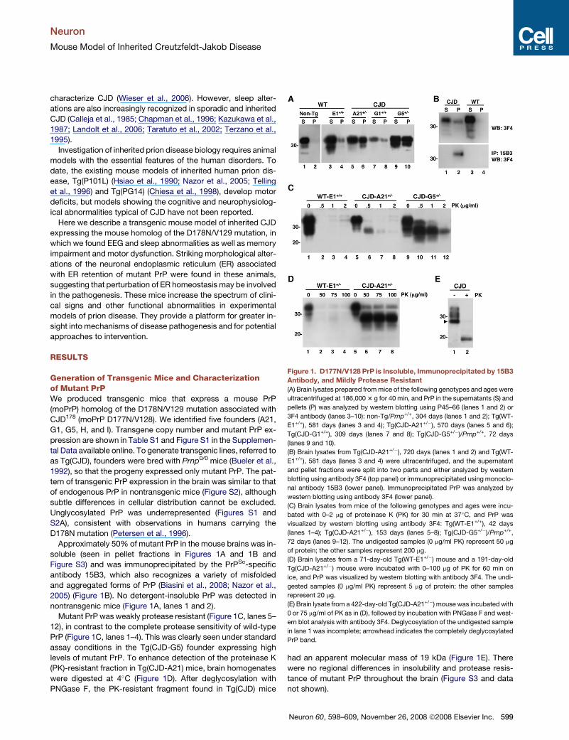
soluble (seen in pellet fractions in Figures 1A and 1B andFigure S3) and was immunoprecipitated by the PrPSc-specificantibody 15B3, which also recognizes a variety of misfoldedand aggregated forms of PrP (Biasini et al., 2008; Nazor et al.,2005) (Figure 1B). No detergent-insoluble PrP was detected innontransgenic mice (Figure 1A, lanes 1 and 2).Mutant PrP was weakly protease resistant (Figure 1C, lanes 5–
12), in contrast to the complete protease sensitivity of wild-typePrP (Figure 1C, lanes 1–4). This was clearly seen under standardassay conditions in the Tg(CJD-G5) founder expressing highlevels of mutant PrP. To enhance detection of the proteinase K(PK)-resistant fraction in Tg(CJD-A21) mice, brain homogenateswere digested at 4!C (Figure 1D). After deglycosylation withPNGase F, the PK-resistant fragment found in Tg(CJD) mice
had an apparent molecular mass of 19 kDa (Figure 1E). Therewere no regional differences in insolubility and protease resis-tance of mutant PrP throughout the brain (Figure S3 and datanot shown).
Figure 1. D177N/V128 PrP is Insoluble, Immunoprecipitated by 15B3Antibody, and Mildly Protease Resistant(A) Brain lysates prepared frommice of the following genotypes and ages were
ultracentrifuged at 186,0003 g for 40 min, and PrP in the supernatants (S) and
pellets (P) was analyzed by western blotting using P45–66 (lanes 1 and 2) or
3F4 antibody (lanes 3–10): non-Tg/Prnp+/+, 304 days (lanes 1 and 2); Tg(WT-
E1+/+), 581 days (lanes 3 and 4); Tg(CJD-A21+/"), 570 days (lanes 5 and 6);
Tg(CJD-G1+/+), 309 days (lanes 7 and 8); Tg(CJD-G5+/")/Prnp+/+, 72 days
(lanes 9 and 10).
(B) Brain lysates from Tg(CJD-A21+/"), 720 days (lanes 1 and 2) and Tg(WT-
E1+/+), 581 days (lanes 3 and 4) were ultracentrifuged, and the supernatant
and pellet fractions were split into two parts and either analyzed by western
blotting using antibody 3F4 (top panel) or immunoprecipitated using monoclo-
nal antibody 15B3 (lower panel). Immunoprecipitated PrP was analyzed by
western blotting using antibody 3F4 (lower panel).
(C) Brain lysates from mice of the following genotypes and ages were incu-
bated with 0–2 mg of proteinase K (PK) for 30 min at 37!C, and PrP was
visualized by western blotting using antibody 3F4: Tg(WT-E1+/+), 42 days
(lanes 1–4); Tg(CJD-A21+/"), 153 days (lanes 5–8); Tg(CJD-G5+/")/Prnp+/+,
72 days (lanes 9–12). The undigested samples (0 mg/ml PK) represent 50 mg
of protein; the other samples represent 200 mg.
(D) Brain lysates from a 71-day-old Tg(WT-E1+/") mouse and a 191-day-old
Tg(CJD-A21+/") mouse were incubated with 0–100 mg of PK for 60 min on
ice, and PrP was visualized by western blotting with antibody 3F4. The undi-
gested samples (0 mg/ml PK) represent 5 mg of protein; the other samples
represent 20 mg.
(E) Brain lysate from a 422-day-old Tg(CJD-A21+/") mouse was incubated with
0 or 75 mg/ml of PK as in (D), followed by incubation with PNGase F and west-
ern blot analysis with antibody 3F4. Deglycosylation of the undigested sample
in lane 1 was incomplete; arrowhead indicates the completely deglycosylated
PrP band.
Neuron
Mouse Model of Inherited Creutzfeldt-Jakob Disease
Neuron 60, 598–609, November 26, 2008 ª2008 Elsevier Inc. 599
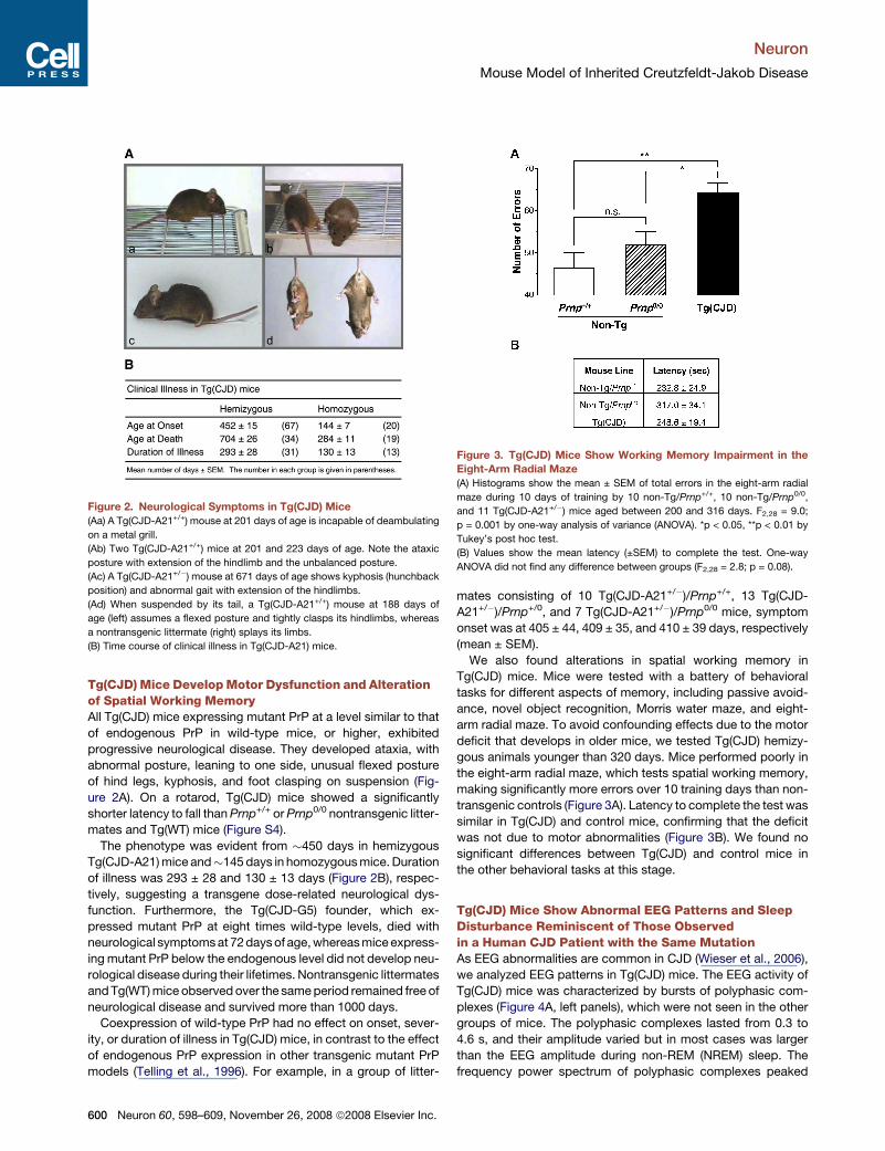
Tg(CJD) Mice DevelopMotor Dysfunction and Alterationof Spatial Working MemoryAll Tg(CJD) mice expressing mutant PrP at a level similar to thatof endogenous PrP in wild-type mice, or higher, exhibitedprogressive neurological disease. They developed ataxia, withabnormal posture, leaning to one side, unusual flexed postureof hind legs, kyphosis, and foot clasping on suspension (Fig-ure 2A). On a rotarod, Tg(CJD) mice showed a significantlyshorter latency to fall thanPrnp+/+ orPrnp0/0 nontransgenic litter-mates and Tg(WT) mice (Figure S4).
The phenotype was evident from #450 days in hemizygousTg(CJD-A21)mice and#145 days in homozygousmice. Durationof illness was 293 ± 28 and 130 ± 13 days (Figure 2B), respec-tively, suggesting a transgene dose-related neurological dys-function. Furthermore, the Tg(CJD-G5) founder, which ex-pressed mutant PrP at eight times wild-type levels, died withneurological symptomsat 72daysof age,whereasmiceexpress-ing mutant PrP below the endogenous level did not develop neu-rological disease during their lifetimes. Nontransgenic littermatesandTg(WT)mice observedover the sameperiod remained free ofneurological disease and survived more than 1000 days.
Coexpression of wild-type PrP had no effect on onset, sever-ity, or duration of illness in Tg(CJD) mice, in contrast to the effectof endogenous PrP expression in other transgenic mutant PrPmodels (Telling et al., 1996). For example, in a group of litter-
mates consisting of 10 Tg(CJD-A21+/")/Prnp+/+, 13 Tg(CJD-A21+/")/Prnp+/0, and 7 Tg(CJD-A21+/")/Prnp0/0 mice, symptomonset was at 405 ± 44, 409 ± 35, and 410 ± 39 days, respectively(mean ± SEM).We also found alterations in spatial working memory in
Tg(CJD) mice. Mice were tested with a battery of behavioraltasks for different aspects of memory, including passive avoid-ance, novel object recognition, Morris water maze, and eight-arm radial maze. To avoid confounding effects due to the motordeficit that develops in older mice, we tested Tg(CJD) hemizy-gous animals younger than 320 days. Mice performed poorly inthe eight-arm radial maze, which tests spatial working memory,making significantly more errors over 10 training days than non-transgenic controls (Figure 3A). Latency to complete the test wassimilar in Tg(CJD) and control mice, confirming that the deficitwas not due to motor abnormalities (Figure 3B). We found nosignificant differences between Tg(CJD) and control mice inthe other behavioral tasks at this stage.
Tg(CJD) Mice Show Abnormal EEG Patterns and SleepDisturbance Reminiscent of Those Observedin a Human CJD Patient with the Same MutationAs EEG abnormalities are common in CJD (Wieser et al., 2006),we analyzed EEG patterns in Tg(CJD) mice. The EEG activity ofTg(CJD) mice was characterized by bursts of polyphasic com-plexes (Figure 4A, left panels), which were not seen in the othergroups of mice. The polyphasic complexes lasted from 0.3 to4.6 s, and their amplitude varied but in most cases was largerthan the EEG amplitude during non-REM (NREM) sleep. Thefrequency power spectrum of polyphasic complexes peaked
Figure 2. Neurological Symptoms in Tg(CJD) Mice(Aa) A Tg(CJD-A21+/+) mouse at 201 days of age is incapable of deambulating
on a metal grill.
(Ab) Two Tg(CJD-A21+/+) mice at 201 and 223 days of age. Note the ataxic
posture with extension of the hindlimb and the unbalanced posture.
(Ac) A Tg(CJD-A21+/") mouse at 671 days of age shows kyphosis (hunchback
position) and abnormal gait with extension of the hindlimbs.
(Ad) When suspended by its tail, a Tg(CJD-A21+/+) mouse at 188 days of
age (left) assumes a flexed posture and tightly clasps its hindlimbs, whereas
a nontransgenic littermate (right) splays its limbs.
(B) Time course of clinical illness in Tg(CJD-A21) mice.
Figure 3. Tg(CJD) Mice Show Working Memory Impairment in theEight-Arm Radial Maze(A) Histograms show the mean ± SEM of total errors in the eight-arm radial
maze during 10 days of training by 10 non-Tg/Prnp+/+, 10 non-Tg/Prnp0/0,
and 11 Tg(CJD-A21+/") mice aged between 200 and 316 days. F2,28 = 9.0;
p = 0.001 by one-way analysis of variance (ANOVA). *p < 0.05, **p < 0.01 by
Tukey’s post hoc test.
(B) Values show the mean latency (±SEM) to complete the test. One-way
ANOVA did not find any difference between groups (F2,28 = 2.8; p = 0.08).
Neuron
Mouse Model of Inherited Creutzfeldt-Jakob Disease
600 Neuron 60, 598–609, November 26, 2008 ª2008 Elsevier Inc.
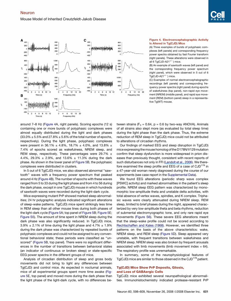
around 7–8 Hz (Figure 4A, right panels). Scoring epochs (12 s)containing one or more bursts of polyphasic complexes werealmost equally distributed during the light and dark phases(33.0%± 5.5%and 27.8%± 5.6%of the total number of epochs,respectively). During the light phase, polyphasic complexeswere present in 36.1% ± 4.8%, 18.7% ± 4.0%, and 13.8% ±7.4% of epochs scored as wakefulness, NREM sleep, andREM sleep, respectively. These percentages were 29.7% ±4.4%, 29.3% ± 2.9%, and 13.6% ± 11.3% during the darkphase. As shown in the lower panel of Figure 5B, the polyphasiccomplexes were distributed in clusters.In 5 out of 6 Tg(CJD) mice, we also observed abnormal ‘‘saw-
tooth’’ waves with a frequency power spectrum that peakedaround 4Hz (Figure 4B). The number of epochs with thesewavesranged from 5 to 33 during the light phase and from 4 to 56 duringthe dark phase, except in one Tg(CJD) mouse in which hundredsof sawtooth waves were recorded during the light-dark cycle.Mice expressing mutant PrP showed marked sleep abnormal-
ities; 24 hr polygraphic analysis indicated significant alterationsof sleep-wake patterns. Tg(CJD) mice spent strikingly less timein REM sleep than all other mouse lines during both phases ofthe light-dark cycle (Figure 5A; top panel of Figure 5B; Figure 5E;Figure S5). The amount of time spent in NREM sleep during thedark phase was also significantly reduced. In Tg(CJD) mice,3.9% ± 2.1% of time during the light phase and 4.7% ± 1.9%during the dark phase was characterized by repeated bursts ofpolyphasic complexes and could not be assigned to any conven-tional behavioral state; these periods were classified as ‘‘un-scored’’ (Figure 5B, top panel). There were no significant differ-ences in the number of transitions between behavioral states(an indicator of continuous or broken sleep) or state-specificEEG power spectra in the different groups of mice.Analysis of circadian distribution of sleep and gross body
movements did not bring to light any differences betweenTg(CJD) and control mice. As expected in nocturnal animals,mice of all experimental groups spent more time awake (Fig-ure 5E, top panel) and moved more during the dark phase thanthe light phase of the light-dark cycle, with no differences be-
tween strains (F3 = 0.64, p = 0.6 by two-way ANOVA). Animalsof all strains also slept more (as evaluated by total sleep time)during the light phase than the dark phase. Thus, the extremereduction of REM sleep in Tg(CJD) mice could not be attributedto alterations of circadian rhythms.Our findings of marked EEG and sleep disruption in Tg(CJD)
miceexpressing themousehomologof theD178N/V129mutationconfirm that sleep dysfunction is more widespread in prion dis-eases than previously thought, consistent with recent reports ofsuch disturbances not only in FFI (Landolt et al., 2006). We there-fore examined the sleep profile and EEG of a case of CJD178 ina 47-year-old woman newly diagnosed during the course of ourexperiments (see case report in the Supplemental Data).We found EEG alterations (periodic sharp wave complex
[PSWC] activity) and marked abnormalities in the patient’s sleepprofile. NREM sleep EEG pattern was characterized by mono-morphic low-amplitude theta and unstable delta activities, withtotal absence of vertex waves, spindles, and K complex. Tripha-sic waves were clearly attenuated during NREM sleep. REMsleep, limited to brief phases during the night, appeared charac-terized by very low-amplitude theta and beta rhythms, reductionof submental electromyographic tone, and only rare rapid eyemovements (Figure S6). These severe EEG alterations meantthat the sleep-wake profile could not be scored according toRechtschaffen and Kales (1968). However, we identified threepatterns on the basis of the above characteristics: wake,NREM sleep, and REM sleep (Figure 5D). Sleep appeared veryunstable, with frequent transitions between wakefulness andNREM sleep. NREM sleep was also broken by frequent arousalsassociated with limb movements (limb movement index = 64).The respiratory profile was unremarkable.In summary, some of the neurophysiological features of
Tg(CJD)mice are similar to those observed in the CJD178 patient.
Tg(CJD) Mice Show PrP Deposits, Gliosis,and Loss of GABAergic CellsTg(CJD) mice exhibited several neuropathological abnormali-ties. Immunohistochemistry indicated protease-resistant PrP
Figure 4. Electroencephalographic ActivityIs Altered in Tg(CJD) Mice(A) Three examples of bursts of polyphasic com-
plexes (left panels) and corresponding frequency
power spectra obtained by fast Fourier transform
(right panels). These alterations were observed in
all 6 Tg(CJD-A21+/") mice.
(B) An example of sawtooth waves (left panel) and
the corresponding frequency power spectrum
(right panel), which were observed in 5 out of 6
Tg(CJD-A21+/") mice.
(C) Examples of normal electroencephalographic
recordings (left panels) and corresponding fre-
quency power spectra (right panel) during epochs
of wakefulness (top panel), non-rapid eye move-
ment (NREM) (middle panel), and rapid eye move-
ment (REM) (bottom panel) sleep in a representa-
tive Tg(WT) mouse.
Neuron
Mouse Model of Inherited Creutzfeldt-Jakob Disease
Neuron 60, 598–609, November 26, 2008 ª2008 Elsevier Inc. 601
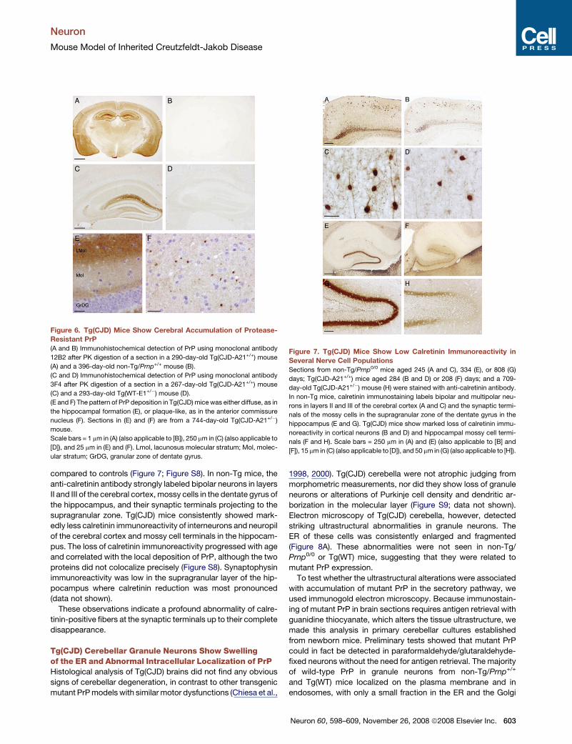
deposits in many brain regions of Tg(CJD) mice that were absentin nontransgenic and Tg(WT)mice (Figures 6A–6D). Diffuse ‘‘syn-aptic-type’’ PrP deposition was prominent in the hippocampalformation, particularly in the stratum moleculare (Figure 6E),and in the amygdala and olfactory bulb. Definite PrP immunos-taining was also detected in the neocortex and striatum and,less intensely, in the thalamus, hypothalamus, brainstem, andmolecular layer of the cerebellum. In addition, small PrP ‘‘pla-que-like’’ deposits were found in several brain regions, includingthe fimbria of the hippocampus, the reticular thalamic nucleus,the corpus callosum, the external and internal capsule, the cin-gulate cortex, and the anterior commissure nuclei (Figure 6F).
These deposits were not fluorescent after thioflavine S staining,indicating that they did not contain amyloid fibrils.PrP deposition was associated with hypertrophy and prolifer-
ation of astrocytes, prominent in the hippocampus, amygdala,and olfactory bulb as revealed by immunostaining with anti-glialfibrillary acidic protein (GFAP) antibody (Figure S7). No spongi-form-like changes were observed.There is evidence that parvalbumin-, calbindin-, and calreti-
nin-positive subpopulations of GABAergic neurons are primarilyaffected in CJD (Belichenko et al., 1999; Guentchev et al., 1997).Immunohistochemistry for calretinin showed striking differencesin the hippocampus and cerebral cortex of Tg(CJD) mice
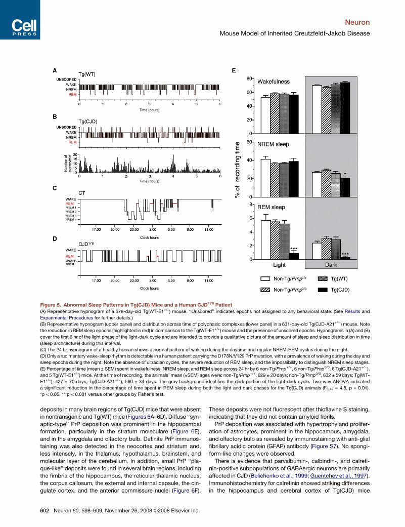
Figure 5. Abnormal Sleep Patterns in Tg(CJD) Mice and a Human CJD178 Patient(A) Representative hypnogram of a 578-day-old Tg(WT-E1+/+) mouse. ‘‘Unscored’’ indicates epochs not assigned to any behavioral state. (See Results and
Experimental Procedures for further details.)
(B) Representative hypnogram (upper panel) and distribution across time of polyphasic complexes (lower panel) in a 631-day-old Tg(CJD-A21+/") mouse. Note
the reduction in REMsleep epochs (highlighted in red) in comparison to the Tg(WT-E1+/+) mouse and the presence of unscored epochs. Hypnograms in (A) and (B)
cover the first 6 hr of the light phase of the light-dark cycle and are intended to provide a qualitative picture of the amount of sleep and sleep distribution in time
(sleep architecture) during this interval.
(C) The 24 hr hypnogram of a healthy human shows a normal pattern of waking during the daytime and regular NREM-REM cycles during the night.
(D) Only a rudimentary wake-sleep rhythm is detectable in a human patient carrying theD178N/V129 PrPmutation, with a prevalence of waking during the day and
sleep epochs during the night. Note the absence of ultradian cycles, the severe reduction of REM sleep, and the impossibility to distinguish NREM sleep stages.
(E) Percentage of time (mean ± SEM) spent in wakefulness, NREM sleep, and REM sleep across 24 hr by 6 non-Tg/Prnp+/+, 6 non-Tg/Prnp0/0, 6 Tg(CJD-A21+/"),
and 5 Tg(WT-E1+/+) mice. At the time of recording, the animals’ mean (±SEM) ages were: non-Tg/Prnp+/+, 629 ± 20 days; non-Tg/Prnp0/0, 632 ± 59 days; Tg(WT-
E1+/+), 427 ± 70 days; Tg(CJD-A21+/"), 560 ± 34 days. The gray background identifies the dark portion of the light-dark cycle. Two-way ANOVA indicated
a significant reduction in the percentage of time spent in REM sleep during both the light and dark phases for the Tg(CJD) animals (F3,42 = 4.8, p = 0.01).
*p < 0.05, ***p < 0.001 versus other groups by Fisher’s test.
Neuron
Mouse Model of Inherited Creutzfeldt-Jakob Disease
602 Neuron 60, 598–609, November 26, 2008 ª2008 Elsevier Inc.
compared to controls (Figure 7; Figure S8). In non-Tg mice, theanti-calretinin antibody strongly labeled bipolar neurons in layersII and III of the cerebral cortex, mossy cells in the dentate gyrus ofthe hippocampus, and their synaptic terminals projecting to thesupragranular zone. Tg(CJD) mice consistently showed mark-edly less calretinin immunoreactivity of interneurons and neuropilof the cerebral cortex and mossy cell terminals in the hippocam-pus. The loss of calretinin immunoreactivity progressed with ageand correlated with the local deposition of PrP, although the twoproteins did not colocalize precisely (Figure S8). Synaptophysinimmunoreactivity was low in the supragranular layer of the hip-pocampus where calretinin reduction was most pronounced(data not shown).These observations indicate a profound abnormality of calre-
tinin-positive fibers at the synaptic terminals up to their completedisappearance.
Tg(CJD) Cerebellar Granule Neurons Show Swellingof the ER and Abnormal Intracellular Localization of PrPHistological analysis of Tg(CJD) brains did not find any obvioussigns of cerebellar degeneration, in contrast to other transgenicmutant PrPmodels with similarmotor dysfunctions (Chiesa et al.,
1998, 2000). Tg(CJD) cerebella were not atrophic judging frommorphometric measurements, nor did they show loss of granuleneurons or alterations of Purkinje cell density and dendritic ar-borization in the molecular layer (Figure S9; data not shown).Electron microscopy of Tg(CJD) cerebella, however, detectedstriking ultrastructural abnormalities in granule neurons. TheER of these cells was consistently enlarged and fragmented(Figure 8A). These abnormalities were not seen in non-Tg/Prnp0/0 or Tg(WT) mice, suggesting that they were related tomutant PrP expression.To test whether the ultrastructural alterations were associated
with accumulation of mutant PrP in the secretory pathway, weused immunogold electron microscopy. Because immunostain-ing of mutant PrP in brain sections requires antigen retrieval withguanidine thiocyanate, which alters the tissue ultrastructure, wemade this analysis in primary cerebellar cultures establishedfrom newborn mice. Preliminary tests showed that mutant PrPcould in fact be detected in paraformaldehyde/glutaraldehyde-fixed neurons without the need for antigen retrieval. The majorityof wild-type PrP in granule neurons from non-Tg/Prnp+/+
and Tg(WT) mice localized on the plasma membrane and inendosomes, with only a small fraction in the ER and the Golgi
Figure 6. Tg(CJD) Mice Show Cerebral Accumulation of Protease-Resistant PrP(A and B) Immunohistochemical detection of PrP using monoclonal antibody
12B2 after PK digestion of a section in a 290-day-old Tg(CJD-A21+/+) mouse
(A) and a 396-day-old non-Tg/Prnp+/+ mouse (B).
(C and D) Immunohistochemical detection of PrP using monoclonal antibody
3F4 after PK digestion of a section in a 267-day-old Tg(CJD-A21+/+) mouse
(C) and a 293-day-old Tg(WT-E1+/") mouse (D).
(E and F) The pattern of PrP deposition in Tg(CJD) mice was either diffuse, as in
the hippocampal formation (E), or plaque-like, as in the anterior commissure
nucleus (F). Sections in (E) and (F) are from a 744-day-old Tg(CJD-A21+/")
mouse.
Scale bars = 1 mm in (A) (also applicable to [B]), 250 mm in (C) (also applicable to
[D]), and 25 mm in (E) and (F). Lmol, lacunosus molecular stratum; Mol, molec-
ular stratum; GrDG, granular zone of dentate gyrus.
Figure 7. Tg(CJD) Mice Show Low Calretinin Immunoreactivity inSeveral Nerve Cell PopulationsSections from non-Tg/Prnp0/0 mice aged 245 (A and C), 334 (E), or 808 (G)
days; Tg(CJD-A21+/+) mice aged 284 (B and D) or 208 (F) days; and a 709-
day-old Tg(CJD-A21+/") mouse (H) were stained with anti-calretinin antibody.
In non-Tg mice, calretinin immunostaining labels bipolar and multipolar neu-
rons in layers II and III of the cerebral cortex (A and C) and the synaptic termi-
nals of the mossy cells in the supragranular zone of the dentate gyrus in the
hippocampus (E and G). Tg(CJD) mice show marked loss of calretinin immu-
noreactivity in cortical neurons (B and D) and hippocampal mossy cell termi-
nals (F and H). Scale bars = 250 mm in (A) and (E) (also applicable to [B] and
[F]), 15 mm in (C) (also applicable to [D]), and 50 mm in (G) (also applicable to [H]).
Neuron
Mouse Model of Inherited Creutzfeldt-Jakob Disease
Neuron 60, 598–609, November 26, 2008 ª2008 Elsevier Inc. 603
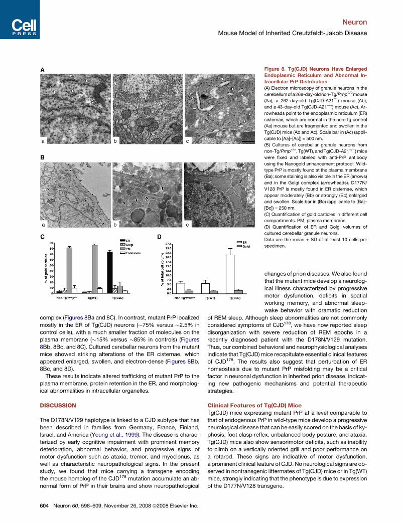
complex (Figures 8Ba and 8C). In contrast, mutant PrP localizedmostly in the ER of Tg(CJD) neurons (#75% versus #2.5% incontrol cells), with a much smaller fraction of molecules on theplasma membrane (#15% versus #85% in controls) (Figures8Bb, 8Bc, and 8C). Cultured cerebellar neurons from the mutantmice showed striking alterations of the ER cisternae, whichappeared enlarged, swollen, and electron-dense (Figures 8Bb,8Bc, and 8D).
These results indicate altered trafficking of mutant PrP to theplasma membrane, protein retention in the ER, and morpholog-ical abnormalities in intracellular organelles.
DISCUSSION
The D178N/V129 haplotype is linked to a CJD subtype that hasbeen described in families from Germany, France, Finland,Israel, and America (Young et al., 1999). The disease is charac-terized by early cognitive impairment with prominent memorydeterioration, abnormal behavior, and progressive signs ofmotor dysfunction such as ataxia, tremor, and myoclonus, aswell as characteristic neuropathological signs. In the presentstudy, we found that mice carrying a transgene encodingthe mouse homolog of the CJD178 mutation accumulate an ab-normal form of PrP in their brains and show neuropathological
Figure 8. Tg(CJD) Neurons Have EnlargedEndoplasmic Reticulum and Abnormal In-tracellular PrP Distribution(A) Electron microscopy of granule neurons in the
cerebellumofa268-day-oldnon-Tg/Prnp0/0mouse
(Aa), a 262-day-old Tg(CJD-A21+/") mouse (Ab),
and a 43-day-old Tg(CJD-A21+/+) mouse (Ac). Ar-
rowheads point to the endoplasmic reticulum (ER)
cisternae, which are normal in the non-Tg control
(Aa) mouse but are fragmented and swollen in the
Tg(CJD) mice (Ab and Ac). Scale bar in (Ac) (appli-
cable to [Aa]–[Ac]) = 500 nm.
(B) Cultures of cerebellar granule neurons from
non-Tg/Prnp+/+, Tg(WT), andTg(CJD-A21+/")mice
were fixed and labeled with anti-PrP antibody
using the Nanogold enhancement protocol. Wild-
type PrP is mostly found at the plasma membrane
(Ba); some staining is also visible in the ER (arrows)
and in the Golgi complex (arrowheads). D177N/
V128 PrP is mostly found in ER cisternae, which
appear moderately (Bb) or strongly (Bc) enlarged
and swollen. Scale bar in (Bc) (applicable to [Ba]–
[Bc]) = 250 nm.
(C) Quantification of gold particles in different cell
compartments. PM, plasma membrane.
(D) Quantification of ER and Golgi volumes of
cultured cerebellar granule neurons.
Data are the mean ± SD of at least 10 cells per
specimen.
changes of prion diseases.We also foundthat the mutant mice develop a neurolog-ical illness characterized by progressivemotor dysfunction, deficits in spatialworking memory, and abnormal sleep-wake behavior with dramatic reduction
of REM sleep. Although sleep abnormalities are not commonlyconsidered symptoms of CJD178, we have now reported sleepdisorganization with severe reduction of REM epochs in arecently diagnosed patient with the D178N/V129 mutation.Thus, our combined behavioral and neurophysiological analysesindicate that Tg(CJD) mice recapitulate essential clinical featuresof CJD178. The results also suggest that perturbation of ERhomeostasis due to mutant PrP misfolding may be a criticalfactor in neuronal dysfunction in inherited prion disease, indicat-ing new pathogenic mechanisms and potential therapeuticstrategies.
Clinical Features of Tg(CJD) MiceTg(CJD) mice expressing mutant PrP at a level comparable tothat of endogenous PrP in wild-type mice develop a progressiveneurological disease that can be easily scored on the basis of ky-phosis, foot clasp reflex, unbalanced body posture, and ataxia.Tg(CJD) mice also show sensorimotor deficits, such as inabilityto climb on a vertically oriented grill and poor performance ona rotarod. These signs are indicative of motor dysfunction,a prominent clinical feature of CJD. No neurological signs are ob-served in nontransgenic littermates of Tg(CJD) mice or in Tg(WT)mice, strongly indicating that the phenotype is due to expressionof the D177N/V128 transgene.
Neuron
Mouse Model of Inherited Creutzfeldt-Jakob Disease
604 Neuron 60, 598–609, November 26, 2008 ª2008 Elsevier Inc.
We found that expression of mutant PrP above the endoge-nous level dramatically exacerbates the Tg(CJD) phenotype. Ho-mozygous mice of the A21 line develop neurological signs muchearlier than hemizygous mice (at#145 versus#450 days of age)and show much faster disease progression, reaching the termi-nal stage in #130 days (Figure 2B). Moreover, the G5 founder,which expressed eight times the endogenous PrP level, diedwith neurological disease at only 72 days of age, further support-ing a direct correlation between D177N/V128 level and diseaseseverity. In contrast, Tg(CJD) lines with PrP expression belowthe endogenous level remain healthy during their lifetime. Thisdose-dependent effect of mutant PrP expression has been de-scribed in other PrP mutant mice (Chiesa et al., 2000; Nazoret al., 2005; Telling et al., 1996) and supports the concept thathigher levels of mutant PrP molecules result in earlier onsetand a faster course of disease.Consistent with the dominant mode of inheritance of CJD178
(Goldfarb et al., 1992), expression of wild-type PrP does not res-cue the neurological illness of Tg(CJD) mice. Tg(CJD) mice car-rying either one or two copies of the endogenous wild-type Prnpallele develop neurological signs at an age similar to Tg(CJD)/Prnp0/0 littermates and invariably progress to terminal disease.Further experiments, however, are necessary to determinewhether coexpression of wild-type PrP modifies any other as-pects of the Tg(CJD) phenotype, such as the deficits in workingmemory or the alteration of sleep-wake patterns.Memory loss is a distinctive early sign of CJD178 (Brown et al.,
1992). Tg(CJD) mice have a significant deficit in working memorydetectable in the eight-arm radial maze well before onset of neu-rological signs. Tg(CJD) mice appear to be affected specificallyin this function since they have no significant alterations inaspects of referencememory asmeasured by the passive avoid-ance, object recognition, or Morris water maze tests. Interest-ingly, Tg(CJD) mice show alterations in calretinin-positive mossycell terminals of the hippocampus and bipolar interneurons of thecerebral cortex, which are thought to play important roles in spa-tial orientation and working memory (Crusio and Schwegler,2005; Wang et al., 2004).
EEGAbnormalities andSleepDisruption in Tg(CJD)Miceand a CJD178 PatientEEG alterations have been described in CJD since the early1980s. The alterations depend on the stage of the disease, rang-ing from nonspecific findings such as diffuse slowing and frontalrhythmic delta activity in the early stage of disease to typicalPSWC in the middle and late stages. PSWC has been reportedin a carrier of the D178N/V128 mutation (Laplanche et al.,1992) and were also detected in the patient analyzed here.Tg(CJD) mice develop striking pathological EEG alterations
mimicking the human EEG pattern. Bursts of polyphasic com-plexes and, to a lesser extent, ‘‘sawtooth’’ waves resemble theEEG patterns observed in a feline model of CJD (Gourmelonet al., 1987) and can be regarded as the equivalent of humanPSWC (Wieser et al., 2006).Tg(CJD) mice also exhibit the sleep alterations seen in
sporadic CJD (Landolt et al., 2006) and in the CJD178 patientdescribed here. The most striking feature is a highly significantreduction of REM sleep during both the light and dark phases
of the light-dark cycle in thesemice. A second finding is the pres-ence of epochs that cannot be assigned to any normal behav-ioral state based on standard criteria used for the polygraphicscoring of mouse sleep-wake behavior (‘‘unscored’’ epochs).The pattern was similar in the CJD178 patient, who spent a signif-icant amount of time in a condition that could not be assigned toany normal vigilance or sleep state. A ‘‘nonwake-nonsleep’’ sub-wakefulness state has been recognized in FFI (Montagna et al.,2003) and in sporadic CJD (Landolt et al., 2006), and we founda severe reduction of sleep efficiency, virtual absence of REMsleep, and absence of usual ultradian modulation in our CJD178
patient.Longitudinal 24 hr monitoring and spectral EEG analysis in FFI
patients show that an early and progressive reduction in thalamicsleep spindles and K complexes is an early marker in the naturalhistory of the disease (Cortelli et al., 2006;Montagna et al., 2003);spindling activity was completely absent in the whole 24 hrrecording of the CJD178 patient described here. Thus, sleep pro-motion and organization appear to be altered in prion diseasesregardless of the specific etiology, suggesting a ‘‘continuum’’between FFI, CJD178, and sporadic CJD.The sleep-wake alterations observed in Tg(CJD) mice are not
seen in non-Tg littermates and Tg(WT) mice, consistent withthese alterations being caused by mutant PrP expression. PrPmay regulate circadian rhythm and promote sleep continuity(Tobler et al., 1996), but no significant changes are seen inthe percentage of time spent in wakefulness, REM sleep, andNREM sleep in Prnp0/0 mice. Therefore, the altered sleep-wakebehavior of Tg(CJD) mice seen here is unlikely to be due toloss of any putative PrP function in sleep regulation.Interleukin-1 and tumor necrosis factor are potent inhibitors of
REM sleep (Obal and Krueger, 2003). These cytokines areelevated in sporadic and variant CJD and in CJD-infected mice(Kordek et al., 1996; Sharief et al., 1999), suggesting theirinvolvement in the severe REM sleep reduction in Tg(CJD)mice and the CJD178 patient.
Neuropathological Features and BiochemicalProperties of Mutant PrP in Tg(CJD) MicePrP deposition inmost forms of prion disease is extracellular, oc-curring either in typical amyloid plaques or as more diffuse ‘‘syn-aptic-like’’ deposits in perineuronal and perivacuolar structuresthroughout the neuropil. In familial CJD linked to the D178N/V129 mutation, weak ‘‘synaptic-like’’ and small focal depositsof PrP are observed (Parchi et al., 2003), and Tg(CJD) micehave the same types of PrP deposits.A striking pathological alteration in Tg(CJD) mice that appears
to be related to PrP deposition is the decrease of calretinin-pos-itive GABAergic neurons in the dentate gyrus of the hippocam-pus and in layers II and III of the neocortex. In the hippocampus,loss of calretinin is accompanied by reduced synaptophysinstaining, suggesting that presynaptic terminals are affected. Inthe neocortex, the decrease in calretinin is most evident in theneuropil, whereas the cell bodies retain a certain degree of im-munoreactivity. These observations suggest that nerve terminalsare primarily affected, consistent with the idea that synapses arethe main targets of abnormal PrP (Bouzamondo-Bernstein et al.,2004; Jeffrey et al., 2000; Kitamoto et al., 1992).
Neuron
Mouse Model of Inherited Creutzfeldt-Jakob Disease
Neuron 60, 598–609, November 26, 2008 ª2008 Elsevier Inc. 605
Specific subpopulations of GABAergic neurons in the hippo-campus and cerebral cortex are selectively affected in sporadicCJD and in experimental prion diseases (Guentchev et al., 1997,1998). In sporadic CJD, parvalbumin-positive interneurons in thefrontal cortex are mostly involved, although there is also degen-eration of calbindin- and calretinin-positive cells (Belichenkoet al., 1999).
InCJD-infectedmice, lossofGABAergic inhibitory neuronsoc-curs very early after inoculation, suggesting that it may be an im-portant step during disease development representing the basisof excitatory symptoms (Guentchev et al., 1997, 1998). Whetheralterations of calretinin-positive GABAergic neurons in Tg(CJD)mice contribute to the EEG abnormalities observed in this modelremains to be established.
Mutant PrP in the brains of Tg(CJD) mice can be distinguishedfrom PrPC by several biochemical properties, including propen-sity to formdetergent-insoluble aggregates, protease resistance,and reactivitywith the15B3antibody.D177N/V128PrP,however,differs frommutant PrP in the brains of CJD178 patients because itis much less protease resistant and has a smaller protease-resis-tant core, suggesting possible structural differences. Experi-ments to test whether mutant PrP in Tg(CJD) brains is infectiouslike that in CJD178 patients (Brown et al., 1992) are underway.
Impaired Trafficking ofMutant PrP and Its Potential Rolein Disease PathogenesisWe observed that the ER of Tg(CJD) cerebellar granule neuronswas dramatically swollen and electron-dense and containedabnormal amounts of mutant PrP. These data suggest that ERretention of mutant PrP may be an instigating pathogenic mech-anism in cerebellar granule neurons that could also be active inother neuronal populations.
A number of inherited human diseases are attributable todefects in export of a mutant protein out of the ER (Aridor andHannan, 2002). In some cases themisfoldedmutant protein is tri-aged by the ER quality control and degraded by the proteasome;in other cases it is retained in the ER lumen and stimulates ERstress response pathways, such as the unfolded protein re-sponse (UPR), eventually leading to apoptosis (Kaufman,1999). We have reported that PrP molecules carrying theD177N mutation are delayed in their biosynthetic maturation inthe ER (Drisaldi et al., 2003) but are not subject to retrotransloca-tion and proteasomal degradation in cerebellar granule neurons(Fioriti et al., 2005). No signs of UPR were detected in the brainsof carriers of the D178N/V129 mutation (Unterberger et al.,2006), and we did not find splicing of the mRNA encoding Xbox-binding protein 1 or increased expression of UPR-regulatedgenes such as Grp78/Bip and CHOP/GADD153 in the brains ofTg(CJD) mice (data not shown), suggesting that some otherpathogenic mechanism may be triggered by this mutation. ERoverload with mutant PrP may lead to activation of NF-kB(Pahl and Baeuerle, 1997), with possible consequences for neu-ronal function and synaptic plasticity (O’Mahony et al., 2006). Al-ternatively, buildup of the mutant protein in the ER may perturbthe ER calcium signaling essential for normal neuronal function(Mattson et al., 2000). Finally, aggregation of mutant PrP in thesecretory pathway may interfere with folding, assembly, andtransport of other membrane proteins, such as multimeric ion
channels or receptors (Schwappach, 2008), leading to defectivesynaptic transmission.The fact that Tg(CJD) mice accumulate a misfolded form of
mutant PrP in their brains and develop clinical features of CJD ar-gues that essential aspects of pathogenesis are modeled inthese animals. Tg(CJD) mice may be suitable for investigatingdisease mechanisms and testing potential therapies in inheritedprion diseases. Since the mutant mice spontaneously developprofound alterations of sleep-wake behavior, they offer a uniquemodel for investigating the pathophysiology of sleep in prion dis-orders. Finally, comparative studies of Tg(CJD) and transgenicmice expressing other mutant PrPs may provide important infor-mation on the cellular and molecular mechanisms responsiblefor the phenotypic heterogeneity of inherited prion diseases.
EXPERIMENTAL PROCEDURES
Generation of Transgenic MiceProduction of transgenic Tg(WT)mice expressingwild-type PrP taggedwith an
epitope for the monoclonal antibody 3F4 has been reported previously (Chiesa
et al., 1998). In this study, we used hemizygous or homozygous mice of the E1
line, which express two and four times the endogenous PrP level, respectively.
The cDNA encoding mouse PrP derived from the Prnpa allele and containing
the 3F4 epitope tag and the D177N and M128V substitutions has been de-
scribed previously (Fioriti et al., 2005). The coding region of this cDNAwas am-
plified by PCR using Vent polymerase (New England Biolabs) and ligated into
the MoPrP.Xho vector, which contains a 12 kb fragment of Prnp, including the
promoter and intron 1, and drives expression of transgenic PrP in a tissue pat-
tern similar to that of the endogenous protein (Borchelt et al., 1996). Recombi-
nant plasmids were selected by PCR screening and restriction analysis, and
their identity was confirmed by sequencing the entire coding region (Chiesa
et al., 1998). The transgene was excised by NotI digestion and injected into
the pronuclei of fertilized eggs from an F2 cross of C57BL/6J3 CBA/J F1 pa-
rental mice. Transgenic founders were bred with an inbred colony of Zurich I
Prnp0/0 mice (C57BL/6J 3 129 background) (Bueler et al., 1992; Chiesa
et al., 2000). The status of the Prnp gene and the presence of the transgene
were determined by PCR, and the zygosity of the transgene was determined
by Southern blot analysis (Chiesa et al., 1998). The transgenic lines were
maintained on the Prnp0/0 genotype. For some experiments, the Prnp allele
was reintroduced by breeding transgenic mice to C57BL/6J 3 CBA/J mice;
nontransgenic Prnp+/+ and Prnp0/0 littermates were used as controls.
All procedures involving animals and their care were conducted according
to European Union (EEC Council Directive 86/609, OJ L 358,1; December
12, 1987) and Italian (D.L. n.116, G.U., suppl. 40; February 18, 1992) laws
and policies and in accordance with the United States Department of Agricul-
ture Animal Welfare Act and National Institutes of Health Policy on Humane
Care and Use of Laboratory Animals.
Biochemical AnalysesTissue homogenates were prepared in phosphate-buffered saline (PBS) con-
taining 0.5% NP-40 and 0.5% sodium deoxycholate using a glass/Teflon tis-
sue homogenizer. In some experiments, protease inhibitor cocktail (1 mg/ml
pepstatin and leupeptin; 0.5 mM phenylmethylsulfonyl fluoride; 2 mM EDTA)
was added to the homogenization buffer. Assays of detergent insolubility, pro-
teinase K (PK) resistance, and immunoprecipitation with antibody 15B3 were
carried out as described previously (Biasini et al., 2008; Chiesa et al., 1998;
Tremblay et al., 2004). Western blots were developed with monoclonal anti-
body 3F4 (Kascsak et al., 1987), which selectively recognizes transgenic
PrP, or polyclonal antibody P45–66 raised against a synthetic peptide encom-
passing residues 45–66 of mouse PrP.
Clinical Analysis of MiceMice were observed weekly for signs of neurological dysfunction according to
a set of objective criteria (Chiesa et al., 1998). Onset of disease was scored as
Neuron
Mouse Model of Inherited Creutzfeldt-Jakob Disease
606 Neuron 60, 598–609, November 26, 2008 ª2008 Elsevier Inc.
the time at which at least two of the following neurological signs were ob-
served: foot-clasp reflex, kyphosis, unbalanced body posture, inability to
walk on a horizontal metal grid, and inability to remain on a vertical grid for
at least 30 s. An accelerating Rota-Rod 7650 (Ugo Basile) was used; mice
were first trained twice the week before official testing. They were positioned
on the rotating bar and allowed to become acquaintedwith the environment for
30 s. The rod motor was started at an initial setting of 3 rpm and accelerated to
30 rpm at a constant rate of 0.3 rpm/s for a maximum of 300 s. The perfor-
mance was scored as latency to fall, in seconds. Animals were given three tri-
als, and the average was used for statistical analysis.
Radial MazeSpatial working memory was measured using an eight-arm radial maze made
of gray plastic with a Plexiglas lid. The arms, which radiated from an octagonal
central arena with a diameter of 12 cm, were 30 cm long, 5 cm wide, and 4 cm
high. Several external visual cues surrounded the apparatus. Starting one
week before testing, the mice were water deprived by being given access to
water for only 1 hr per day. One day before starting the task schedule, a habit-
uation trial was run. Themice were placed in the center of themaze and let free
to explore the environment for 10 min. The next day, the arms of the radial
maze were baited with 50 ml of water. Animals were placed in the center of
the maze, and the arm entry sequence was recorded. The task ended once
all eight arms of the maze had been visited or after a maximum of 16 trials,
whichever came first. Repeated entry into an arm that had already been visited
constituted an error. The number of errors and the latency to complete the test
were recorded manually by an operator (A.G.) blinded to the experimental
groups. Animals were tested for 10 consecutive days.
EEG and Sleep-Wake BehaviorEEG and sleep patterns were investigated in six non-Tg/Prnp+/+, six non-Tg/
Prnp0/0, sixTg(CJD-A21+/"), andfiveTg(WT-E1+/+)mice.Micewereanesthetized
and instrumented for chronic EEG recording according to standard techniques
(Baracchi and Opp, 2008) and then allowed at least 10 days to recover from sur-
gery and adapt to the recording conditions. Mice were individually housed in
standard cageswith food andwater ad libitum. Cageswere kept in sound-atten-
uated boxes at a constant temperature between 24.5!C and 26.0!C, with a 12/
12 hr light-dark cycle. Gross body activity was detected using an infrared sensor
housed in anobservation unit that also contained a camera (BIOBSERVEGmbH)
allowing continuous recording of the animals’ behavior.Movements detected by
the infrared sensor were converted to a voltage output. The conditioned EEG
signal and the voltage output from the infrared sensor were digitized and col-
lected using custom software (M.R. Opp, University of Michigan). EEG signals
and gross body activity were recorded for 24 hr (starting at first light) in undis-
turbed conditions and used for polygraphic determination of vigilance state.
The animals were not handled starting from 48 hr before the recording session.
Twomice of different genotypes were randomlymatched and recorded simulta-
neously.Theorderof recordingofmiceof thedifferent lineswasalso randomized.
Postacquisition determination of vigilance statewas performed according to
standard criteria (Baracchi and Opp, 2008). Visual scoring of 12 s epochs was
performed by an investigator (L. Ferrari or S.B.) blinded to the strain. As de-
scribed in Results, it was sometimes impossible to assign certain epochs to
any behavioral state in Tg(CJD) mice. In these cases, epochs were classified
as ‘‘unscored.’’ EEG power density values were obtained for each animal
and each behavioral state by Fourier transform for each artifact-free 12 s scor-
ing epoch for the frequency range 0.5–20 Hz.
HistologyBrains were fixed in Carnoy’s fixative (6:3:1 ethanol:chloroform:acetic acid),
dehydrated in graded ethanol solutions, cleared in xylene, and embedded in
paraffin. Serial sections (5 mm thick) were cut and stained with hematoxylin
and eosin, Nissl, or thioflavin S. Some sections were immunostained with
rabbit polyclonal anti-glial fibrillary acidic protein (GFAP) (Dako, 1:1000),
anti-calretinin (Swant, 1:2000), anti-calbindin (Chemicon, 1:1000), or anti-
synaptophysin (Dako, 1:200), followed by visualization with a Vectastain
ABC kit (Vector) using 3,30-diaminobenzidine as chromogen. Alexa 488- or
Alexa 546-conjugated secondary antibodies (Molecular Probes Inc.) were
used for immunofluorescence.
For PrP immunohistochemistry, sections were incubated with PK (2 mg/ml in
0.1% Brij-35, 50 mM NaCl, 50 mM Tris-HCl [pH 7.8]) for 30 min at room tem-
perature and exposed to guanidine thiocyanate (3 M in H2O) for 30 min (Giac-
cone et al., 2000). PK-resistant PrP was detected with monoclonal antibody
3F4 (1:200) (Kascsak et al., 1987) or 12B2 (1:1000) (Langeveld et al., 2006).
Results were similar with the two antibodies.
Electron MicroscopyOne 269-day-old Tg(WT) mouse, two non-Tg/Prnp0/0 mice aged 268 and
392 days, two Tg(CJD-A21+/")/Prnp0/0 mice aged 262 and 488 days, and
one 43-day-old Tg(CJD-A21+/+)/Prnp0/0 mouse were deeply anesthetized
and perfused through the ascending aorta with PBS (0.1 M; pH 7.4) followed
by 4%paraformaldehyde (PFA) and 2.5% glutaraldehyde in PBS. The cerebel-
lum was excised and cut along the sagittal plane with a razor blade, postfixed
in 3% glutaraldehyde in PBS, and immersed for 2 hr in OsO4. After dehydration
in a graded series of ethanol, tissue samples were cleared in propylene oxide,
embedded in Epon 812 epoxy medium (Fluka), and polymerized at 60!C for
72 hr. From each sample, one semithin section (1 mm) was cut with a Leica
EM UC6 ultramicrotome and mounted on glass slides for light microscopic
inspection to identify the Purkinje and granular cell layers. Ultrathin sections
(70 nm thick) of the area of interest were obtained, counterstained with uranyl
acetate and lead citrate, and examined with an energy filter transmission elec-
tron microscope (Zeiss LIBRA 120 EFTEM) equipped with a YAG scintillator
slow-scan CCD camera.
Immunoelectron MicroscopyCerebellar granule neurons were prepared from 6-day-old mice as described
previously (Fioriti et al., 2005) and cultured for 7 days before immunoelectron
microscopy. Cells grown on poly-L-lysine-coated glass coverslips were
washed with PBS and fixed in a solution of 4% PFA and 0.1% glutaraldehyde
in 0.2 M HEPES buffer (pH 7.4) for 15 min at room temperature. After washing
with PBS, cells were incubated for 30 min in blocking solution (50 mM NH4Cl,
0.1% saponin, 1% BSA in HEPES buffer) and then overnight at 4!C with anti-
PrPmonoclonal antibody SA65 (Matucci et al., 2005) or 12B2 (Langeveld et al.,
2006) diluted 1:250 in blocking solution. Cells were washed and incubated for
1 hr at room temperature with Nanogold-conjugated anti-mouse IgG Fab0 frag-
ment diluted 1:100 in blocking solution and processed according to the Nano-
gold enhancement protocol (Nanoprobes). Stained cells were embedded in
Epon 812 and cut as described previously (Polishchuk et al., 2003). EM images
were acquired from thin sections using a Philips Tecnai 12 electron micro-
scope equipped with an ULTRA VIEW CCD digital camera (Philips). Gold par-
ticles were quantified in the different compartments of the secretory pathway,
and total cell, ER, and Golgi volumes were analyzed using analySIS software
(Soft Imaging Systems GmbH). The SA65 and 12B2 antibodies yielded similar
results.
PolysomnographyThe 24 hr EEG recordings, including complete polysomnography, were taken
according to guidelines approved by the Standards of Practice Committee of
the American Academy of Sleep Medicine (Kushida et al., 2005). The EEG was
acquired with Ag/AgCl electrodes positioned over the vertex and the frontal,
central, and occipital regions bilaterally, according to the International 10-20
Electrode Placement System. Two electrooculographic channels, respiratory
channels, electrocardiography, bilateral tibialis anterior, and chin electromy-
ography were also recorded. All signals were digitalized at a sampling rate
of 256 Hz. The record was visually scored according to standard criteria (Re-
chtschaffen and Kales, 1968), and the hypnogram was stored digitally, linked
to the recording. Arousals, cardiorespiratory events, and EMG activities were
recognized by dedicated software and reviewed by a clinical neurophysiolo-
gist trained in sleep medicine. A control 24 hr recording was taken in an
age-matched healthy subject in the same sleep laboratory.
SUPPLEMENTAL DATA
SupplementalDatacanbe foundonlineathttp://www.neuron.org/supplemental/
S0896-6273(08)00755-1.
Neuron
Mouse Model of Inherited Creutzfeldt-Jakob Disease
Neuron 60, 598–609, November 26, 2008 ª2008 Elsevier Inc. 607
ACKNOWLEDGMENTS
We thank David Harris for making the transgenic facility of Washington Univer-
sity in St. Louis available to us, for the P45–66 antibody, and for comments on
the manuscript. We also thank David Borchelt for the MoPrP.Xho vector,
Charles Weissmann for the Prnp0/0 mice, Richard Kasksak for the 3F4 anti-
body, Jan P. Langeveld for the 12B2 antibody, Gianluigi Zanusso for the
SA65 antibody, and Alex Raeber and Bruno Oesch from Prionics (Zurich) for
providing the 15B3 antibody. We are grateful to Roman S. Polishchuk of the
Telethon Microscopy and Bio-Imaging Facility (Consorzio Mario Negri Sud)
for immunogold staining of cerebellar granule neurons and for useful discus-
sion, to Susanna Mantovani for assistance with animal perfusion and RNA ex-
traction, and to Simona Airaghi for participating in the initial phase of this pro-
ject. This work was supported by grants from Telethon Italy (TCR05006),
‘‘Fondazione Cariplo’’ and ‘‘Compagnia di San Paolo’’ to R.C., the European
Community (QLG-CT-2001-2353 to R.C. and Network of Excellence Neuro-
Prion to F.T. and R.C.), and the Italian Ministry of Health to F.T. L. Fioriti was
supported by a fellowship from the Fondazione Monzino. R.C. is an Assistant
Telethon Scientist (Dulbecco Telethon Institute, Fondazione Telethon).
Accepted: September 4, 2008
Published: November 25, 2008
REFERENCES
Aridor, M., and Hannan, L.A. (2002). Traffic jams II: an update of diseases of
intracellular transport. Traffic 3, 781–790.
Baracchi, F., and Opp, M.R. (2008). Sleep-wake behavior and responses to
sleep deprivation of mice lacking both interleukin-1 beta receptor 1 and tumor
necrosis factor-alpha receptor 1. Brain Behav. Immun. 22, 982–993.
Belichenko, P.V., Miklossy, J., Belser, B., Budka, H., and Celio, M.R. (1999).
Early destruction of the extracellular matrix around parvalbumin-immunoreac-
tive interneurons in Creutzfeldt-Jakob disease. Neurobiol. Dis. 6, 269–279.
Biasini, E., Seegulam, M.E., Patti, B.N., Solforosi, L., Medrano, A.Z., Christen-
sen, H.M., Senatore, A., Chiesa, R., Williamson, R.A., and Harris, D.A. (2008).
Non-infectious aggregates of the prion protein react with several PrPSc-
directed antibodies. J. Neurochem. 105, 2190–2204.
Borchelt, D.R., Davis, J., Fischer, M., Lee, M.K., Slunt, H.H., Ratovitsky, T., Re-
gard, J., Copeland, N.G., Jenkins, N.A., Sisodia, S.S., and Price, D.L. (1996). A
vector for expressing foreign genes in the brains and hearts of transgenic mice.
Genet. Anal. 13, 159–163.
Bouzamondo-Bernstein, E., Hopkins, S.D., Spilman, P., Uyehara-Lock, J.,
Deering, C., Safar, J., Prusiner, S.B., Ralston, H.J., 3rd, and DeArmond, S.J.
(2004). The neurodegeneration sequence in prion diseases: evidence from
functional, morphological and ultrastructural studies of theGABAergic system.
J. Neuropathol. Exp. Neurol. 63, 882–899.
Brown, P., Goldfarb, L.G., Kovanen, J., Haltia, M., Cathala, F., Sulima, M.,
Gibbs, C.J., Jr., and Gajdusek, D.C. (1992). Phenotypic characteristics of
familial Creutzfeldt-Jakob disease associated with the codon 178Asn PRNP
mutation. Ann. Neurol. 31, 282–285.
Bueler, H., Fischer, M., Lang, Y., Bluethmann, H., Lipp, H.P., DeArmond, S.J.,
Prusiner, S.B., Aguet, M., andWeissmann, C. (1992). Normal development and
behaviour of mice lacking the neuronal cell-surface PrP protein. Nature 356,
577–582.
Calleja, J., Carpizo, R., Berciano, J., Quintial, C., and Polo, J. (1985). Serial
waking-sleep EEGs and evolution of somatosensory potentials in Creutz-
feldt-Jakob disease. Electroencephalogr. Clin. Neurophysiol. 60, 504–508.
Chapman, J., Arlazoroff, A., Goldfarb, L.G., Cervenakova, L., Neufeld, M.Y.,
Werber, E., Herbert, M., Brown, P., Gajdusek, D.C., and Korczyn, A.D.
(1996). Fatal insomnia in a case of familial Creutzfeldt-Jakob disease with
the codon 200(Lys) mutation. Neurology 46, 758–761.
Chiesa, R., and Harris, D.A. (2001). Prion diseases: what is the neurotoxic mol-
ecule? Neurobiol. Dis. 8, 743–763.
Chiesa, R., Piccardo, P., Ghetti, B., and Harris, D.A. (1998). Neurological illness
in transgenic mice expressing a prion protein with an insertional mutation.
Neuron 21, 1339–1351.
Chiesa, R., Drisaldi, B., Quaglio, E., Migheli, A., Piccardo, P., Ghetti, B., and
Harris, D.A. (2000). Accumulation of protease-resistant prion protein (PrP)
and apoptosis of cerebellar granule cells in transgenic mice expressing
a PrP insertional mutation. Proc. Natl. Acad. Sci. USA 97, 5574–5579.
Cortelli, P., Perani, D., Montagna, P., Gallassi, R., Tinuper, P., Federica, P.,
Avoni, P., Ferrillo, F., Anchisi, D., Moresco, R.M., et al. (2006). Pre-symptom-
atic diagnosis in fatal familial insomnia: serial neurophysiological and 18FDG-
PET studies. Brain 129, 668–675.
Crusio, W.E., and Schwegler, H. (2005). Learning spatial orientation tasks in
the radial-maze and structural variation in the hippocampus in inbred mice.
Behav. Brain Funct. 1, 3.
Drisaldi, B., Stewart, R.S., Adles, C., Stewart, L.R., Quaglio, E., Biasini, E., Fior-
iti, L., Chiesa, R., and Harris, D.A. (2003). Mutant PrP is delayed in its exit from
the endoplasmic reticulum, but neither wild-type nor mutant PrP undergoes
retrotranslocation prior to proteasomal degradation. J. Biol. Chem. 278,
21732–21743.
Fioriti, L., Dossena, S., Stewart, L.R., Stewart, R.S., Harris, D.A., Forloni, G.,
and Chiesa, R. (2005). Cytosolic prion protein (PrP) is not toxic in N2a cells
and primary neurons expressing pathogenic PrP mutations. J. Biol. Chem.
280, 11320–11328.
Giaccone, G., Canciani, B., Puoti, G., Rossi, G., Goffredo, D., Iussich, S., Fo-
ciani, P., Tagliavini, F., and Bugiani, O. (2000). Creutzfeldt-Jakob disease:
Carnoy’s fixative improves the immunohistochemistry of the proteinase K-re-
sistant prion protein. Brain Pathol. 10, 31–37.
Goldfarb, L.G., Petersen, R.B., Tabaton, M., Brown, P., LeBlanc, A.C., Monta-
gna, P., Cortelli, P., Julien, J., Vital, C., Pendelbury, W.W., et al. (1992). Fatal
familial insomnia and familial Creutzfeldt-Jakob disease: disease phenotype
determined by a DNA polymorphism. Science 258, 806–808.
Gourmelon, P., Amyx, H.L., Baron, H., Lemercier, G., Court, L., and Gibbs,
C.J., Jr. (1987). Sleep abnormalities with REM disorder in experimental
Creutzfeldt-Jakob disease in cats: a new pathological feature. Brain Res.
411, 391–396.
Guentchev, M., Hainfellner, J.A., Trabattoni, G.R., and Budka, H. (1997).
Distribution of parvalbumin-immunoreactive neurons in brain correlates with
hippocampal and temporal cortical pathology in Creutzfeldt-Jakob disease.
J. Neuropathol. Exp. Neurol. 56, 1119–1124.
Guentchev, M., Groschup, M.H., Kordek, R., Liberski, P.P., and Budka, H.
(1998). Severe, early and selective loss of a subpopulation of GABAergic inhib-
itory neurons in experimental transmissible spongiform encephalopathies.
Brain Pathol. 8, 615–623.
Hsiao, K.K., Scott, M., Foster, D., Groth, D.F., DeArmond, S.J., and Prusiner,
S.B. (1990). Spontaneous neurodegeneration in transgenic mice with mutant
prion protein. Science 250, 1587–1590.
Jeffrey, M., Halliday, W.G., Bell, J., Johnston, A.R., MacLeod, N.K., Ingham,
C., Sayers, A.R., Brown, D.A., and Fraser, J.R. (2000). Synapse loss associ-
ated with abnormal PrP precedes neuronal degeneration in the scrapie-
infected murine hippocampus. Neuropathol. Appl. Neurobiol. 26, 41–54.
Kascsak, R.J., Rubenstein, R., Merz, P.A., Tonna-DeMasi, M., Fersko, R.,
Carp, R.I., Wisniewski, H.M., and Diringer, H. (1987). Mouse polyclonal
and monoclonal antibody to scrapie-associated fibril proteins. J. Virol. 61,
3688–3693.
Kaufman, R.J. (1999). Stress signaling from the lumen of the endoplasmic
reticulum: coordination of gene transcriptional and translational controls.
Genes Dev. 13, 1211–1233.
Kazukawa, S., Nakamura, I., Endo, M., Hori, A., and Inao, G. (1987). Serial
polysomnograms in Creutzfeldt-Jakob disease. Jpn. J. Psychiatry Neurol.
41, 651–661.
Kitamoto, T., Shin, R.W., Doh-ura, K., Tomokane, N., Miyazono, M., Mura-
moto, T., and Tateishi, J. (1992). Abnormal isoform of prion proteins
Neuron
Mouse Model of Inherited Creutzfeldt-Jakob Disease
608 Neuron 60, 598–609, November 26, 2008 ª2008 Elsevier Inc.
accumulates in the synaptic structures of the central nervous system in pa-
tients with Creutzfeldt-Jakob disease. Am. J. Pathol. 140, 1285–1294.
Kordek, R., Nerurkar, V.R., Liberski, P.P., Isaacson, S., Yanagihara, R., and
Gajdusek, D.C. (1996). Heightened expression of tumor necrosis factor alpha,
interleukin 1-alpha, and glial fibrillary acidic protein in experimental Creutz-
feldt-Jakob disease in mice. Proc. Natl. Acad. Sci. USA 93, 9754–9758.
Kushida, C.A., Littner, M.R., Morgenthaler, T., Alessi, C.A., Bailey, D., Cole-
man, J., Jr., Friedman, L., Hirshkowitz, M., Kapen, S., Kramer, M., et al.
(2005). Practice parameters for the indications for polysomnography and
related procedures: an update for 2005. Sleep 28, 499–521.
Landolt, H.P., Glatzel, M., Blattler, T., Achermann, P., Roth, C., Mathis, J.,
Weis, J., Tobler, I., Aguzzi, A., and Bassetti, C.L. (2006). Sleep-wake distur-
bances in sporadic Creutzfeldt-Jakob disease. Neurology 66, 1418–1424.
Langeveld, J.P., Jacobs, J.G., Erkens, J.H., Bossers, A., van Zijderveld, F.G.,
and van Keulen, L.J. (2006). Rapid and discriminatory diagnosis of scrapie and
BSE in retro-pharyngeal lymph nodes of sheep. BMC Vet. Res. 2, 19.
Laplanche, J.L., Chatelain, J., Thomas, S., Launay, J.M., Gaultier, C., and
Derouesne, C. (1992). Uncommon phenotype for a codon 178 mutation of
the human PrP gene. Ann. Neurol. 31, 345.
Mattson, M.P., LaFerla, F.M., Chan, S.L., Leissring, M.A., Shepel, P.N., and
Geiger, J.D. (2000). Calcium signaling in the ER: its role in neuronal plasticity
and neurodegenerative disorders. Trends Neurosci. 23, 222–229.
Matucci, A., Zanusso, G., Gelati, M., Farinazzo, A., Fiorini, M., Ferrari, S., An-
drighetto, G., Cestari, T., Caramelli, M., Negro, A., et al. (2005). Analysis of
mammalian scrapie protein by novel monoclonal antibodies recognizing dis-
tinct prion protein glycoforms: an immunoblot and immunohistochemical
study at the light and electronmicroscopic levels. Brain Res. Bull. 65, 155–162.
Montagna, P., Gambetti, P., Cortelli, P., and Lugaresi, E. (2003). Familial and
sporadic fatal insomnia. Lancet Neurol. 2, 167–176.
Nazor, K.E., Kuhn, F., Seward, T., Green, M., Zwald, D., Purro, M., Schmid, J.,
Biffiger, K., Power, A.M., Oesch, B., et al. (2005). Immunodetection of disease-
associated mutant PrP, which accelerates disease in GSS transgenic mice.
EMBO J. 24, 2472–2480.
Obal, F., Jr., and Krueger, J.M. (2003). Biochemical regulation of non-rapid-
eye-movement sleep. Front. Biosci. 8, d520–d550.
O’Mahony, A., Raber, J., Montano, M., Foehr, E., Han, V., Lu, S.M., Kwon, H.,
LeFevour, A., Chakraborty-Sett, S., and Greene, W.C. (2006). NF-kappaB/Rel
regulates inhibitory and excitatory neuronal function and synaptic plasticity.
Mol. Cell. Biol. 26, 7283–7298.
Pahl, H.L., and Baeuerle, P.A. (1997). The ER-overload response: activation of
NF-kappa B. Trends Biochem. Sci. 22, 63–67.
Parchi, P., Capellari, S., Chen, S.G., and Gambetti, P. (2003). Familial Creutz-
feldt-Jakob disease. In Neurodegeneration: The Molecular Pathology of De-
mentia and Movement Disorders, D.W. Dickson, ed. (Basel, Switzerland: ISN
Neuropath Press), pp. 298–306.
Petersen, R.B., Parchi, P., Richardson, S.L., Urig, C.B., and Gambetti, P.
(1996). Effect of the D178N mutation and the codon 129 polymorphism on
the metabolism of the prion protein. J. Biol. Chem. 271, 12661–12668.
Polishchuk, E.V., Di Pentima, A., Luini, A., and Polishchuk, R.S. (2003). Mech-
anism of constitutive export from the golgi: bulk flow via the formation, protru-
sion, and en bloc cleavage of large trans-golgi network tubular domains. Mol.
Biol. Cell 14, 4470–4485.
Prusiner, S.B. (1998). Prions. Proc. Natl. Acad. Sci. USA 95, 13363–13383.
Rechtschaffen, A., and Kales, A. (1968). A Manual of Standardized Terminol-
ogy, Techniques and Scoring System for Sleep Stages in Human Subjects,
National Institutes of Health Publication 204 (Washington, DC: USGovernment
Printing Office).
Schwappach, B. (2008). An overview of trafficking and assembly of neuro-
transmitter receptors and ion channels. Mol. Membr. Biol. 25, 270–278.
Sharief, M.K., Green, A., Dick, J.P., Gawler, J., and Thompson, E.J. (1999).
Heightened intrathecal release of proinflammatory cytokines in Creutzfeldt-
Jakob disease. Neurology 52, 1289–1291.
Taratuto, A.L., Piccardo, P., Reich, E.G., Chen, S.G., Sevlever, G., Schultz, M.,
Luzzi, A.A., Rugiero, M., Abecasis, G., Endelman, M., et al. (2002). Insomnia
associated with thalamic involvement in E200K Creutzfeldt-Jakob disease.
Neurology 58, 362–367.
Telling, G.C., Haga, T., Torchia, M., Tremblay, P., Dearmond, S.J., and
Prusiner, S.B. (1996). Interactions between wild-type and mutant prion
proteins modulate neurodegeneration in transgenic mice. Genes Dev. 10,
1736–1750.
Terzano, M.G., Parrino, L., Pietrini, V., Mancia, D., Spaggiari, M.C., Rossi, G.,
and Tagliavini, F. (1995). Precocious loss of physiological sleep in a case of
Creutzfeldt Jakob disease: a serial polygraphic study. Sleep 18, 849–858.
Tobler, I., Gaus, S.E., Deboer, T., Achermann, P., Fischer, M., Rulicke, T.,
Moser, M., Oesch, B., McBride, P.A., and Manson, J.C. (1996). Altered circa-
dian activity rhythms and sleep in mice devoid of prion protein. Nature 380,
639–642.
Tremblay, P., Ball, H.L., Kaneko, K., Groth, D., Hegde, R.S., Cohen, F.E., DeAr-
mond, S.J., Prusiner, S.B., and Safar, J.G. (2004). Mutant PrPSc conformers in-
duced by a synthetic peptide and several prion strains. J. Virol. 78, 2088–2099.
Unterberger, U., Hoftberger, R., Gelpi, E., Flicker, H., Budka, H., and Voigt-
lander, T. (2006). Endoplasmic reticulum stress features are prominent in Alz-
heimer disease but not in prion diseases in vivo. J. Neuropathol. Exp. Neurol.
65, 348–357.
Wang, X.J., Tegner, J., Constantinidis, C., and Goldman-Rakic, P.S. (2004).
Division of labor among distinct subtypes of inhibitory neurons in a cortical
microcircuit of working memory. Proc. Natl. Acad. Sci. USA 101, 1368–1373.
Wieser, H.G., Schindler, K., and Zumsteg, D. (2006). EEG in Creutzfeldt-Jakob
disease. Clin. Neurophysiol. 117, 935–951.
Young, K., Piccardo, P., Dlouhy, S., Bugiani, O., Tagliavini, F., and Ghetti, B.
(1999). The human genetic prion diseases. In Prions: Molecular and Cellular
Biology, D.A. Harris, ed. (Wymondham, UK: Horizon Scientific Press),
pp. 139–175.
Neuron
Mouse Model of Inherited Creutzfeldt-Jakob Disease
Neuron 60, 598–609, November 26, 2008 ª2008 Elsevier Inc. 609
Neuron, Volume 60
Supplemental Data
Article
Mutant Prion Protein Expression Causes Motor and
Memory Deficits and Abnormal Sleep Patterns
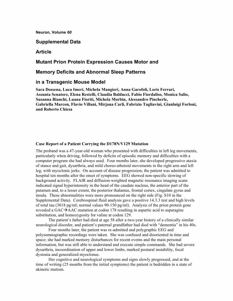
in a Transgenic Mouse Model Sara Dossena, Luca Imeri, Michela Mangieri, Anna Garofoli, Loris Ferrari, Assunta Senatore, Elena Restelli, Claudia Balducci, Fabio Fiordaliso, Monica Salio, Susanna Bianchi, Luana Fioriti, Michela Morbin, Alessandro Pincherle, Gabriella Marcon, Flavio Villani, Mirjana Carli, Fabrizio Tagliavini, Gianluigi Forloni, and Roberto Chiesa Case Report of a Patient Carrying the D178N/V129 Mutation The proband was a 47-year-old woman who presented with difficulties in left leg movements, particularly when driving, followed by deficits of episodic memory and difficulties with a computer program she had always used. Four months later, she developed progressive ataxia of stance and gait, dysarthria, and mild choreo-athetoid movements in the right arm and left leg, with myoclonic jerks. On account of disease progression, the patient was admitted to hospital ten months after the onset of symptoms. EEG showed non-specific slowing of background activity. FLAIR and diffusion-weighted magnetic resonance imaging scans indicated signal hyperintensity in the head of the caudate nucleus, the anterior part of the putamen and, to a lesser extent, the posterior thalamus, frontal cortex, cingulate gyrus and insula. These abnormalities were more pronounced on the right side (Fig. S10 in the Supplemental Data). Cerebrospinal fluid analysis gave a positive 14.3.3 test and high levels of total tau (3018 pg/ml; normal values 90-150 pg/ml). Analysis of the prion protein gene revealed a GAC!AAC mutation at codon 178 resulting in aspartic acid to asparagine substitution, and homozygosity for valine at codon 129.
The patient’s father had died at age 58 after a two-year history of a clinically similar neurological disorder, and patient’s paternal grandfather had died with “dementia” in his 40s.
Four months later, the patient was re-admitted and polygraphic EEG and polysomnographic recordings were taken. She was confused and disoriented in time and space; she had marked memory disturbances for recent events and the main personal information, but was still able to understand and execute simple commands. She had severe dysarthria, incoordination of upper and lower limbs, marked postural instability, focal dystonia and generalized myoclonus.
Her cognitive and neurological symptoms and signs slowly progressed, and at the time of writing (25 months from the initial symptoms) the patient is bedridden in a state of akinetic mutism.
FounderTransgene copy numbera
Tg PrP proteinb
A21 22 1XG1 6 0.15X
G5c 37 8X
Hd 1 ND
Id 1 0.03X
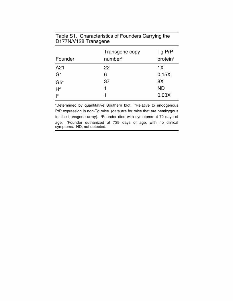
Table S1. Characteristics of Founders Carrying the D177N/V128 Transgene
aDetermined by quantitative Southern blot. bRelative to endogenousPrP expression in non-Tg mice c(data are for mice that are hemizygousfor the transgene array). cFounder died with symptoms at 72 days ofage. dFounder euthanized at 739 days of age, with no clinicalsymptoms. ND, not detected.
A
C
B
Non-Tg CJD-A21+/-
30-
1 2 3 4 5 6 7 8
10 5 2.5 1.25 10 5 2.5 1.25
CJD-G5+/-
10 5 2.5 10 5 2.5 1.25 10 5 2.5
1 2 3 4 5 6 7 8 9 10
30-
WT-E1+/+
30-
1 2 3 4 5 6 7 8
20 10 5 2.5 1.25 20 10 5
!g protein
!g protein
!g protein
CJD-A21+/-
WT-E1+/+ CJD-G1+/+
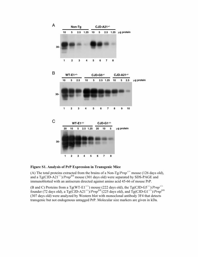
Figure S1. Analysis of PrP Expression in Transgenic Mice (A) The total proteins extracted from the brains of a Non-Tg/Prnp+/+ mouse (126 days old), and a Tg(CJD-A21+/-)/Prnp0/0 mouse (301 days old) were separated by SDS-PAGE and immunoblotted with an antiserum directed against amino acid 45-66 of mouse PrP.
(B and C) Proteins from a Tg(WT-E1+/+) mouse (222 days old), the Tg(CJD-G5+/-)/Prnp+/+ founder (72 days old), a Tg(CJD-A21+/-)/Prnp0/0 (225 days old), and Tg(CJD-G1+/+)/Prnp0/0 (307 days old) were analyzed by Western blot with monoclonal antibody 3F4 that detects transgenic but not endogenous untagged PrP. Molecular size markers are given in kDa.
A
Non-Tg/Prnp+/+
Tg(CJD)/Prnp0/0
1 2 3 4 5 6 7 8 9 10
OB NAcc Hyp FCx CP Hipp Ctx Thal Mes PonsM Cb
30-
30-
11
BNon-Tg/Prnp+/+
Non-Tg/Prnp0/0
Tg(CJD)/Prnp0/0
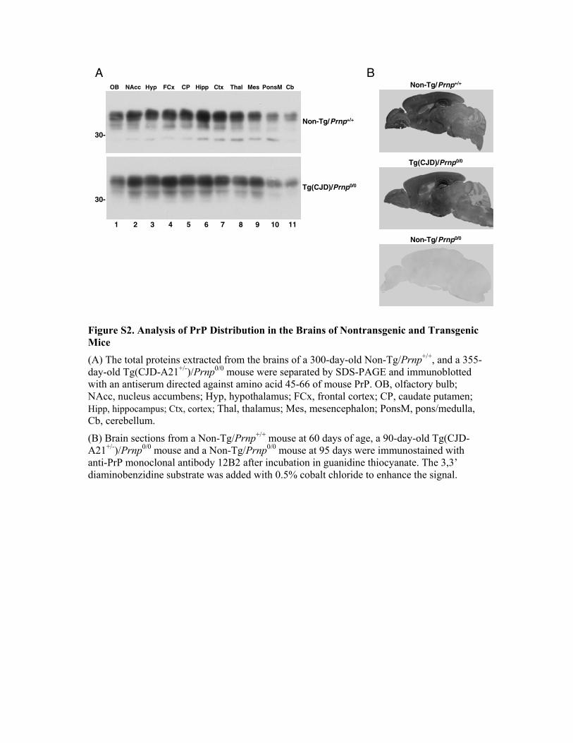
Figure S2. Analysis of PrP Distribution in the Brains of Nontransgenic and Transgenic Mice (A) The total proteins extracted from the brains of a 300-day-old Non-Tg/Prnp+/+, and a 355-day-old Tg(CJD-A21+/-)/Prnp0/0 mouse were separated by SDS-PAGE and immunoblotted with an antiserum directed against amino acid 45-66 of mouse PrP. OB, olfactory bulb; NAcc, nucleus accumbens; Hyp, hypothalamus; FCx, frontal cortex; CP, caudate putamen; Hipp, hippocampus; Ctx, cortex; Thal, thalamus; Mes, mesencephalon; PonsM, pons/medulla, Cb, cerebellum.
(B) Brain sections from a Non-Tg/Prnp+/+ mouse at 60 days of age, a 90-day-old Tg(CJD-A21+/-)/Prnp0/0 mouse and a Non-Tg/Prnp0/0 mouse at 95 days were immunostained with anti-PrP monoclonal antibody 12B2 after incubation in guanidine thiocyanate. The 3,3’ diaminobenzidine substrate was added with 0.5% cobalt chloride to enhance the signal.
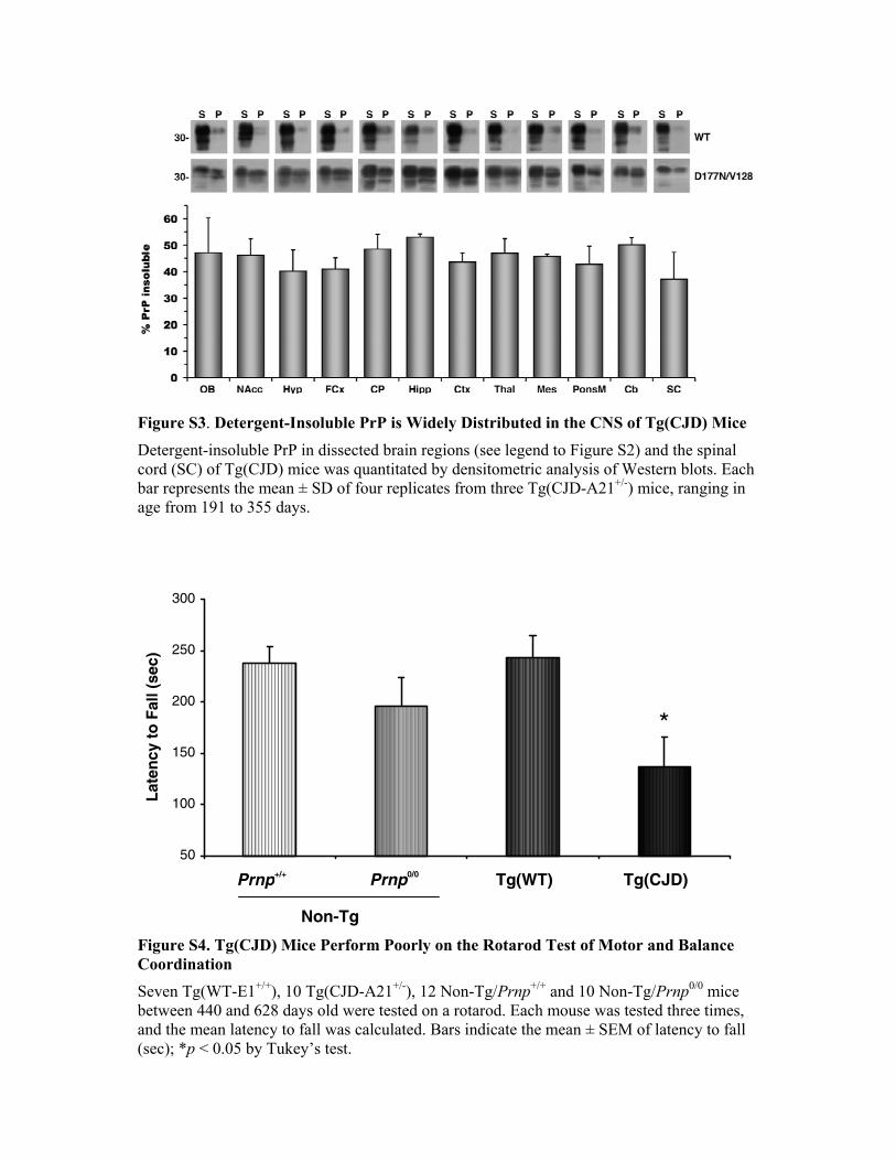
Figure S3. Detergent-Insoluble PrP is Widely Distributed in the CNS of Tg(CJD) Mice Detergent-insoluble PrP in dissected brain regions (see legend to Figure S2) and the spinal cord (SC) of Tg(CJD) mice was quantitated by densitometric analysis of Western blots. Each bar represents the mean ± SD of four replicates from three Tg(CJD-A21+/-) mice, ranging in age from 191 to 355 days. Figure S4. Tg(CJD) Mice Perform Poorly on the Rotarod Test of Motor and Balance Coordination Seven Tg(WT-E1+/+), 10 Tg(CJD-A21+/-), 12 Non-Tg/Prnp+/+ and 10 Non-Tg/Prnp0/0 mice between 440 and 628 days old were tested on a rotarod. Each mouse was tested three times, and the mean latency to fall was calculated. Bars indicate the mean ± SEM of latency to fall (sec); *p < 0.05 by Tukey’s test.
50
100
150
200
250
300
Late
ncy
to F
all (
sec)
*
Non-Tg
Tg(CJD) Tg(WT) Prnp+/+ Prnp0/0
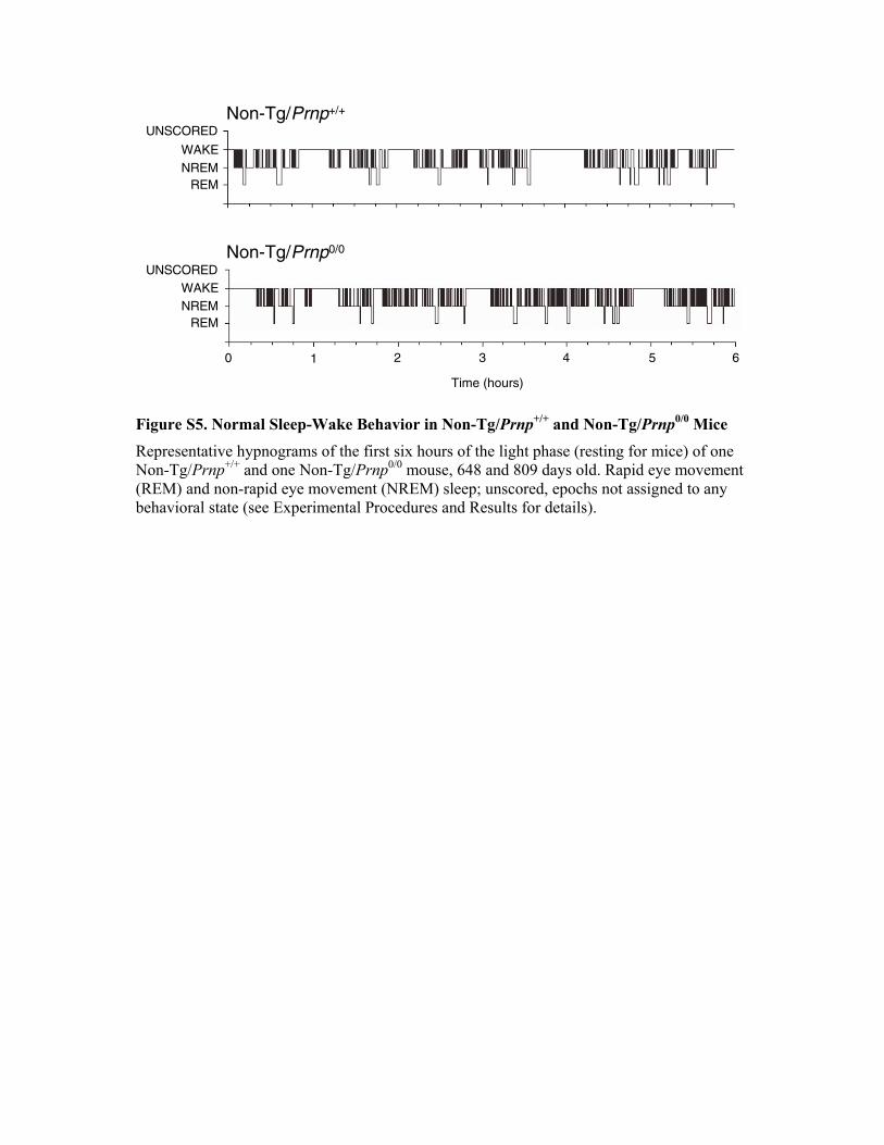
Non-Tg/Prnp+/+
UNSCOREDWAKENREM
REM
Non-Tg/Prnp0/0
UNSCOREDWAKENREM
REM
0 1 2 3 4 5 6
Time (hours) Figure S5. Normal Sleep-Wake Behavior in Non-Tg/Prnp+/+ and Non-Tg/Prnp0/0 Mice Representative hypnograms of the first six hours of the light phase (resting for mice) of one Non-Tg/Prnp+/+ and one Non-Tg/Prnp0/0 mouse, 648 and 809 days old. Rapid eye movement (REM) and non-rapid eye movement (NREM) sleep; unscored, epochs not assigned to any behavioral state (see Experimental Procedures and Results for details).
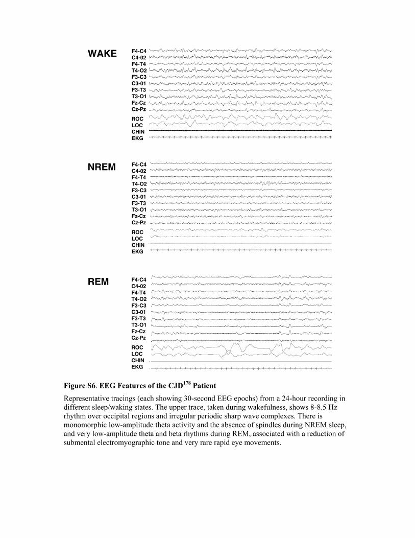
WAKE
NREM
REM F4-C4C4-02F4-T4T4-O2F3-C3C3-01F3-T3T3-O1Fz-CzCz-Pz
ROCLOCCHINEKG
F4-C4C4-02F4-T4T4-O2F3-C3C3-01F3-T3T3-O1Fz-CzCz-Pz
ROCLOCCHINEKG
F4-C4C4-02F4-T4T4-O2F3-C3C3-01F3-T3T3-O1Fz-CzCz-Pz
ROCLOCCHINEKG
Figure S6. EEG Features of the CJD178 Patient Representative tracings (each showing 30-second EEG epochs) from a 24-hour recording in different sleep/waking states. The upper trace, taken during wakefulness, shows 8-8.5 Hz rhythm over occipital regions and irregular periodic sharp wave complexes. There is monomorphic low-amplitude theta activity and the absence of spindles during NREM sleep, and very low-amplitude theta and beta rhythms during REM, associated with a reduction of submental electromyographic tone and very rare rapid eye movements.
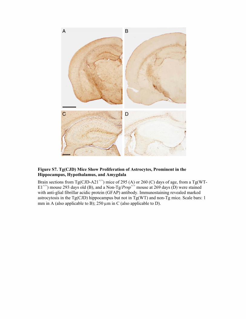
A B
C D
Figure S7. Tg(CJD) Mice Show Proliferation of Astrocytes, Prominent in the Hippocampus, Hypothalamus, and Amygdala Brain sections from Tg(CJD-A21+/+) mice of 295 (A) or 260 (C) days of age, from a Tg(WT-E1+/+) mouse 293 days old (B), and a Non-Tg/Prnp+/+ mouse at 269 days (D) were stained with anti-glial fibrillar acidic protein (GFAP) antibody. Immunostaining revealed marked astrocytosis in the Tg(CJD) hippocampus but not in Tg(WT) and non-Tg mice. Scale bars: 1 mm in A (also applicable to B); 250 !m in C (also applicable to D).
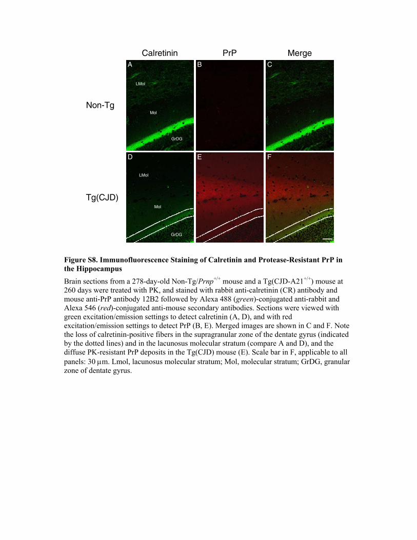
PrPCalretinin Merge
Non-Tg
Tg(CJD)
LMol
GrDG
Mol
GrDG
LMol
Mol
A B C
FED
Figure S8. Immunofluorescence Staining of Calretinin and Protease-Resistant PrP in the Hippocampus Brain sections from a 278-day-old Non-Tg/Prnp+/+ mouse and a Tg(CJD-A21+/+) mouse at 260 days were treated with PK, and stained with rabbit anti-calretinin (CR) antibody and mouse anti-PrP antibody 12B2 followed by Alexa 488 (green)-conjugated anti-rabbit and Alexa 546 (red)-conjugated anti-mouse secondary antibodies. Sections were viewed with green excitation/emission settings to detect calretinin (A, D), and with red excitation/emission settings to detect PrP (B, E). Merged images are shown in C and F. Note the loss of calretinin-positive fibers in the supragranular zone of the dentate gyrus (indicated by the dotted lines) and in the lacunosus molecular stratum (compare A and D), and the diffuse PK-resistant PrP deposits in the Tg(CJD) mouse (E). Scale bar in F, applicable to all panels: 30 !m. Lmol, lacunosus molecular stratum; Mol, molecular stratum; GrDG, granular zone of dentate gyrus.
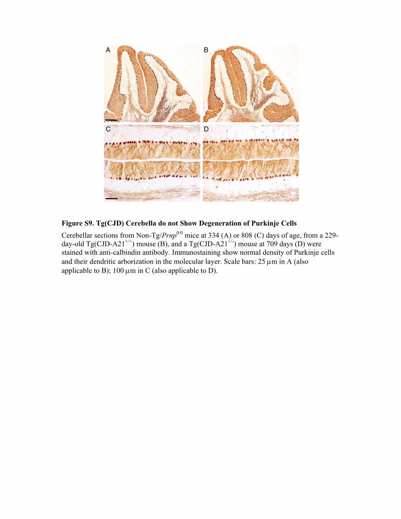
A B
C D
Figure S9. Tg(CJD) Cerebella do not Show Degeneration of Purkinje Cells Cerebellar sections from Non-Tg/Prnp0/0 mice at 334 (A) or 808 (C) days of age, from a 229-day-old Tg(CJD-A21+/+) mouse (B), and a Tg(CJD-A21+/-) mouse at 709 days (D) were stained with anti-calbindin antibody. Immunostaining show normal density of Purkinje cells and their dendritic arborization in the molecular layer. Scale bars: 25 !m in A (also applicable to B); 100 !m in C (also applicable to D).
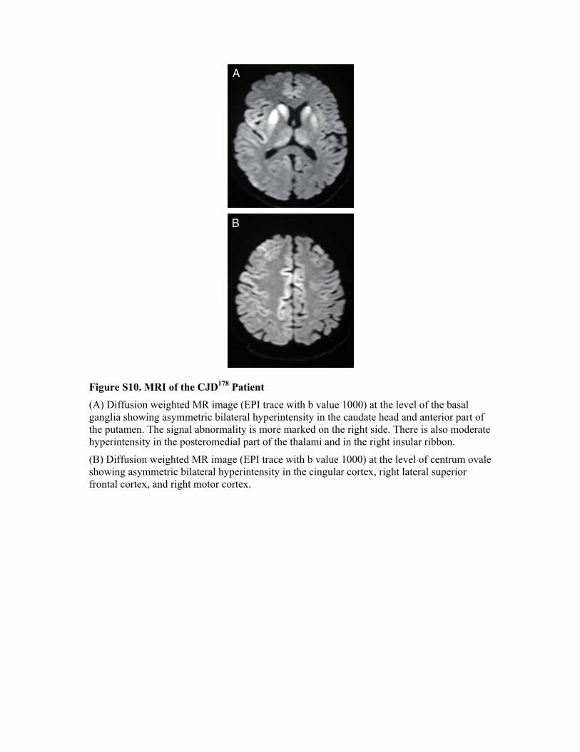
A
B
Figure S10. MRI of the CJD178 Patient (A) Diffusion weighted MR image (EPI trace with b value 1000) at the level of the basal ganglia showing asymmetric bilateral hyperintensity in the caudate head and anterior part of the putamen. The signal abnormality is more marked on the right side. There is also moderate hyperintensity in the posteromedial part of the thalami and in the right insular ribbon.
(B) Diffusion weighted MR image (EPI trace with b value 1000) at the level of centrum ovale showing asymmetric bilateral hyperintensity in the cingular cortex, right lateral superior frontal cortex, and right motor cortex.
Copyright © 2022 FDOKUMEN