IGF-I-induced Differentiation of L6 Myogenic Cells Requires the Activity of cAMP-Phosphodiesterase
Upload
independentCategory
view
1download
0
Changes in the expression of extracellular regulated kinase (ERK 1/2) in the R6/2mouse model of Huntington's disease after phosphodiesterase IV inhibition
Francesca R. Fusco a,1,!, Serenella Anzilotti a,1, Carmela Giampà a, Clemente Dato b, Daunia Laurenti a,Alessandro Leuti a, Luca Colucci D'Amato e, Lorena Perrone c, Giorgio Bernardi a,d, Mariarosa A. B. Melone b
a Laboratory of Neuroanatomy, Santa Lucia Foundation IRCCS Hospital at the European Center for Brain Research, Rome, Italyb Department of Clinical and Experimental Medicine and Surgery, First Neurological Clinic, Second University of Naples, Naples, Italyc Laboratoire des Neurobiologie des Interactions Cellulaires et Neurophysiopathologie, Université de la Méditerranée Aix-Marseille 2, Marseille, Franced Department of Neuroscience, University of Rome Tor Vergata, Rome, Italye Department of Life Science, II University of Naples and Institute of Genetics and Biophysics "Adriano Buzzati Traverso", CNR, Naples, Italy
a b s t r a c ta r t i c l e i n f o
Article history:Received 10 October 2011Revised 9 January 2012Accepted 21 January 2012Available online xxxx
Keywords:immunohistochemistryERKrolipramHuntington's diseaseR6/2
The mitogen-activated protein kinases (MAPKs) superfamily comprises three major signaling pathways: theextracellular signal-regulated protein kinases (ERKs), the c-Jun N-terminal kinases or stress-activated proteinkinases (JNKs/SAPKs) and the p38 family of kinases.ERK 1/2 signaling has been implicated in a number of neurodegenerative disorders, including Huntington'sdisease (HD). Phosphorylation patterns of ERK 1/2 and JNK are altered in cell models of HD. In this study,we aimed at studying the correlations between ERK 1/2 and the neuronal vulnerability to HD degenerationin the R6/2 transgenic mouse model of HD. Single and double-label immuno!uorescence for phospho-ERK(pERK, the activated form of ERK) and for each of the striatal neuronal markers were employed onperfusion-"xed brain sections from R6/2 and wild-type mice. Moreover, Phosphodiesterase 4 inhibitionthrough rolipram was used to study the effects on pERK expression in the different types of striatal neurons.We completed our study with western blot analysis. Our study shows that pERK levels increase with age inthe medium spiny striatal neurons and in the parvalbumin interneurons, and that rolipram counteractssuch increase in pERK. Conversely, cholinergic and somatostatinergic interneurons of the striatum containhigher levels of pERK in the R6/2 mice compared to the controls. Rolipram induces an increase in pERK ex-pression in these interneurons. Thus, our study con"rms and extends the concept that the expression ofphosphorylated ERK 1/2 is related to neuronal vulnerability and is implicated in the pathophysiology ofcell death in HD.
© 2012 Elsevier Inc. All rights reserved.
Introduction
Huntington's disease (HD) is a devastating neurodegenerative dis-order characterized by progressive and severe cognitive, psychiatric,and motor dysfunction (Walker, 2007). The disease is caused by ex-pansion of a CAG repeat in exon 1 of IT15, which encodes for the pro-tein huntingtin (The Huntington's disease collaborative researchgroup, 1993). The expanded n-terminal poly-glutamine tract in mu-tant huntingtin is toxic (Mangiarini et al., 1996), with the length ofthe expansion correlating with age of onset and disease course(Andrew et al., 1993; Melone et al., 2005; Perrone and Melone, 2008).
Although huntingtin is widely expressed in the brain (Fusco et al.,1999), HD is associated with a speci"c pattern of neurodegeneration.
The striatal medium spiny projection neurons are most susceptible tothe degeneration and loss of striatal volume is the prominent patho-logical "nding (de la Monte et al., 1988; Vonsattel et al., 1985), whilecholinergic and somatostatinergic interneurons are more resistant.
The striatum is highly innervated by Brain Derived NeurotrophicFactor (BDNF)-releasing synapses. BDNF is delivered to the striatumvia activity-dependent anterograde release from excitatory corticos-triatal axons (Canals et al., 2001; Zuccato and Cattaneo, 2007). BDNFis essential for striatal function and survival (Gratacòs et al., 2001;Zuccato et al., 2001). Interestingly, in HD, because mutated huntingtinloses its physiological role, a dramatic loss of BDNF occurs, contribut-ing prominently to the striatal degeneration.
BDNF signaling is mediated primarily by its high af"nity receptor,the receptor tyrosine kinase tropomyosin-related kinase B (TrkB).TrkB activation initiates three major intracellular signaling cascades:the shc/Frs2 and ras/raf-mediated activation of a MAPK phosphoryla-tion cascade involving mitogen-activated ERK kinase (MEK) andextracellular-signal-regulated kinase (ERK 1/2), the shc/Frs2 andGAB1-mediated activation of a phoshphotidylinositol-3-kinase PI3K
Neurobiology of Disease xxx (2012) xxx–xxx
! Corresponding author at: Laboratory of Neuroanatomy, Santa Lucia FoundationIRCCS Hospital, Via del Fosso Fiorano 64, 00179 Rome, Italy. Fax: +39 06 501703325.
E-mail addresses: [email protected], [email protected] (F.R. Fusco).1 These authors equally contributed to the manuscript.
Available online on ScienceDirect (www.sciencedirect.com).
YNBDI-02638; No. of pages: 9; 4C:
0969-9961/$ – see front matter © 2012 Elsevier Inc. All rights reserved.doi:10.1016/j.nbd.2012.01.011
Contents lists available at SciVerse ScienceDirect
Neurobiology of Disease
j ourna l homepage: www.e lsev ie r .com/ locate /ynbd i
Please cite this article as: Fusco, F.R., et al., Changes in the expression of extracellular regulated kinase (ERK 1/2) in the R6/2 mouse model ofHuntington's disease after phosphodiesterase IV inhibition, Neurobiol. Dis. (2012), doi:10.1016/j.nbd.2012.01.011

pathway involving Akt1, and the direct TrkB-mediated activation ofphospholipase C gamma (PLCc) which produces inositol triphosphate(IP3) and diacylglycerol (DAG) and increases intracellular free calci-um (Corominas et al., 2007; Numakawa et al., 2010).
ERK 1/2 is a strong anti-apoptotic and pro-survival mediator (Kimet al., 2009). ERK 1/2 is also involved in the phosphorylation of synap-sin I (Jovanovic et al., 1996, 2008; Matsubara et al., 1996), which isimplicated in the regulation of neurotransmitter release and synapseformation, and whose function is altered in HD models (Liévens et al.,2002). Moreover, ERK 1/2 downregulation is linked to neurodegener-ative conditions such as ischemia (Liebelt et al., 2010) and traumaticspinal cord injury (Yu et al., 2010).
The MAP kinase/ERK 1/2 signaling pathway has been shown toplay a key role in this transcriptional regulation in the striatum(Roze et al., 2008), and is activated by the binding of BDNF to its re-ceptor TrkB, which leads to the phosphorylation of cAMP response el-ement binding protein (CREB) (Kingsbury and Krueger, 2007).
CREB is involved in the neuronal vulnerability to HD degeneration,and neuroprotection achieved by the Phosphodiesterase IV inhibitorrolipram is mediated by increased CREB and BDNF levels in the R6/2model of HD (DeMarch et al., 2008; Giampà et al., 2009). Neuropro-tection achieved by the increase CREB and BDNF should involve theactivation of ERK 1/2 pathway (Yao et al., 2009). Thus, we aimed atinvestigating if neuroprotection achieved by PDEIV inhibition by roli-pram involved changes in the expression of pERK in R6/2 mice.
Changes in pERK expression in the R6/2 mice were described(Liévens et al., 2002). However, a precise description of possiblechanges in pERK expression in the different neuronal subtypes wasstill lacking. Thus, the analysis of ERK 1/2 expression was performedin the different neuronal subtypes of the striatum.
Materials and methods
Heterozygous transgenic R6/2 males of the CBAXC57BL/6 strainwere obtained from Jackson Laboratories (Bar Harbor, ME, USA) andmaintained by backcrossing carrier males with CBAXC57BL/6_F1 fe-males. The offspring were genotyped by polymerase chain reactionassay of DNA obtained from tail tissue. Mice were weaned and, aftergenotyping, treatments began at 4 weeks of age (n=24/group) andlasted until the animals were killed at 7and 13 weeks of age. Thestudy groups included: R6/2 mice that were given saline by intraper-itoneal injection (0.9%), R6/2 mice with rolipram given by intraperi-toneal injection [dissolved in saline (1.5 mg⁄kg⁄day) and madefresh daily], rolipram-treated and saline-treated wild-type mice.
Mice were handled under the same conditions by one investigatoron the same day and at the same time. Mice were identi"ed by a ran-domly assigned code so that the studies were performed blind as tothe genetic identity. The mice were housed "ve in each cage understandard conditions with ad libitum access to food and water. Allstudies were conducted in accordance with the European Communi-ties Council Directive of 24 November 1986 (86/609/EEC) and ap-proved by the Santa Lucia Foundation Animal Care and Usecommittee. All the histological data were collected by observerswho were blinded to treatment.
Immunohistochemical studies
Tissue processing. For the histological examination, animals at 7and 13 weeks of age were transcardially perfused under deep (chloralhydrate) anesthesia with saline solution containing 0.01 ml heparin,followed by 60 ml of 4% paraformaldehyde in saline solution. Thebrains were removed and post"xed overnight at +4C° and cryopro-tected in 10% sucrose and 20% glycerol in 0.1 M phosphate buffer(PB) with sodium azide 0.02% for 48 h at 4C°. Brains were sectionedfrozen on a sliding microtome at 40 !m thickness (Fusco et al., 2001).
Single and dual-label immuno!uorescence
Primary omission controls, normal serum controls and preimmuneserum controls using immunizing peptide were used to con"rm thespeci"city of our immunohistochemical labeling (Fusco et al., 2003).Sections were incubated with a cocktail of rabbit anti-pERK (Immuno-logical Sciences) and one of the following striatal neuronal markers:mouse anti-calbindin 28 kDa (Sigma) to evaluate the expression, at aprotein level, of pERK in the striatal matrix spiny projection neurons;mouse anti-choline acetyl transferase (ChAT; Chemicon) to study theexpression of pERK in cholinergic interneurons; mouse anti-nitricoxide synthase (NOS; Sigma, St-Louis, MO) to study the distribution ofpERK in the somatostatin neuropeptide Y-NOS containing interneuronsubset; mouse anti-parvalbumin (PARV; Chemicon Temecula CA), forthe labeling of pERK into parvalbuminergic interneurons. All primaryantisera were used at a 1:200 concentration (except for anti-pERKwhich was used at a 1:300), in 0.1 m PB containing Triton X-100, 0.3%and sodium azide, 0.02% for 72 h at 4 °C. Sections were then rinsedthree times for 15 min at room temperature, and subsequently incubat-ed with a cocktail of goat anti rabbit Cy2-conjugated secondary anti-body and donkey anti-mouse Cy3-conjugated secondary antibody(both Jackson Immunoresearch, West Grove, PA, USA) for 2 h at roomtemperature. Tissue was mounted on gelatin-coated slides, cover-slipped with GEL-MOUNT and examined under an epi-illumination!uorescence microscope (Zeiss Axioskop 2), and a CLSM (Zeiss LSM700) was used to acquire all the images.
Striatal tissue protein extraction—Western blotting
Western blot analysis of phosphorylation state of ERK 1/2 afterrolipram inhibition of PDE4 was performed. The animals (n=4 pergroup) were deeply anesthetized and killed by decapitation at theage of 7 and 13 weeks (wild-type and R6/2). The brain was quicklyremoved and the striatum was dissected out and homogenized inlysis buffer containing 1% Triton X-100, 10 mM Tris–HCl, 150 mMNaCl, 1 mM EDTA, 5 mM EDTA, 0.1 mM phenylmethylsulphonyl !uo-ride (PMSF), 1 mM "-glycerolphosphate, phosphatase inhibitors(1 mM Na3VO4, and 10 mM NaF) and protease inhibitor mixture(Sigma-Aldrich). Samples were left for 30 min on ice and then centri-fuged at 1200 !g for 20 min at 4 °C, the supernatants were collectedand stored at !80 °C. The protein concentration was determinedusing the Bradford colorimetric assay (Bio-Rad, Milano, Italy).
Thirty µg of the extracted proteins were resolved by 10% SDS-PAGE gels and transferred to PVDF membranes (Millipore, Billerica,USA). Nonspeci"c sites were blocked by 0.1% Tween 20 and 5% milkpowder in TBS for 1 h at room temperature. Membranes were thenincubated overnight at 4 °C with the speci"c anti-pERK rabbit anti-body (1:1000, Immunological Science, Italy) in TBS/1% Tween-20with 1% of milk powder. After primary antibody incubation, the mem-branes were incubated for 1 h at room temperature with the appro-priate horseradish peroxidase-conjugated secondary antibody(1:5000, Immunological Science) and the reaction was visualized byusing an enhanced chemiluminescent detection system (Millipore,Bilerica, MA). Incubation with a rabbit total ERK 1/2 (1:1000; Cell Sig-naling, Beverly, MA) was performed to obtain loading controls. Densi-tometric quanti"cation of the immunoblots was performed usingImage J software.
Statistical analysis
To obtain an indirect measure of the amount of pERK immunos-taining in spiny projection neurons, and interneurons, image analysisof calbindin positive, cholinergic, somatostatinergic, parvalbuminer-gic interneurons was performed by means of image J software. Brie!y,the background was removed and the image was converted to grey-scale. The double labeled neurons were identi"ed through the use
2 F.R. Fusco et al. / Neurobiology of Disease xxx (2012) xxx–xxx
Please cite this article as: Fusco, F.R., et al., Changes in the expression of extracellular regulated kinase (ERK 1/2) in the R6/2 mouse model ofHuntington's disease after phosphodiesterase IV inhibition, Neurobiol. Dis. (2012), doi:10.1016/j.nbd.2012.01.011

of the function “region of interest (ROI) manager” of the program. Theintensity of !uorescent pERK immunolabeling was calculated in eachof three separate "elds (one dorsolateral, one central and one medial,each 1 mm in diameter) on each hemisphere in each of three rostro-caudally spaced sections in each of saline, rolipram-treated R6/2 miceand wild-type littermates.
The data obtained by image analysis of pERK immunolabeling inall subtypes of striatal neurons were expressed as mean+/!SD. Dif-ferences in the time course experiments were analyzed by two-wayANOVA followed by Tukey's posthoc analysis, and the signi"cancelevel was set to Pb0.05.
For Western blot analysis, the density of pERK protein band wasmeasured using a Kodak Image Station and the ratio between pERK/total ERK was calculated for each group. Statistical comparisons be-tween groups were made by ANOVA including the factors of geno-type, age and treatment as warranted. All posthoc analysis usedHSD Tukey test and the signi"cance level was set to Pb0.05.
Results
Immunohistochemistry
The expression of pERK in the medium spiny neurons increases with ageand is decreased by rolipram
The protein expression of pERK was studied in the immunohisto-chemically labeled striatal medium spiny projection neurons of R6/2mice and wild-type littermates. The analysis was performed at8 and 13 weeks of age, when R6/2 are fully symptomatic and closeto the end stage. pERK levels were lower in the spiny neurons ofR6/2 at 8 weeks compared to wild-types. At the second time pointof 13 weeks, pERK levels increased both in the wild-types and in theR6/2 mice (F(1,1198)=20.22; Pb0.000. At this time point, pERKlevels were comparable between R6/2 and their wild-type littermates(Pb0.9). (Figs. 1, 5). Moreover, the intensity of pERK immunolabelingincreased signi"cantly in both wild-types and R6/2 at 13 weeks,
Fig. 1. Confocal laser scanning microscopy (CLSM) images of double-label immuno!uorescence for pERK and projections neurons marker calbindin (CALB). (A–C–E–G) pERK immu-nolabeling (visualized in red-cy3 immuno!uorescence) and CALB immunolabeling (visualized in green-cy2 immuno!uorescence) in wild-type and R6/2 HD mice striatum at 8 and13 weeks of age treated with vehicle. In (B–D–F–H) pERK and CALB immunolabeling in Wt and R6/2 HD mice striatum at 8–13 weeks of age treated with Rolipram. (For interpre-tation of the references to color in this "gure legend, the reader is referred to the web version of this article.)
3F.R. Fusco et al. / Neurobiology of Disease xxx (2012) xxx–xxx
Please cite this article as: Fusco, F.R., et al., Changes in the expression of extracellular regulated kinase (ERK 1/2) in the R6/2 mouse model ofHuntington's disease after phosphodiesterase IV inhibition, Neurobiol. Dis. (2012), doi:10.1016/j.nbd.2012.01.011
although there were no differences between genotypes. As shown inFig. 5A, the administration of PDE4 inhibitor rolipram decreased dra-matically pERK protein expression both in the wild-type and in theR6/2 animals. Thus, we observed that pERK levels in the spiny projec-tion neurons were comparable betweenwild-type and R6/2 mice, andthat rolipram had a negative effect on pERK expression in those neu-rons, regardless of the genotype (F(1,1198)=349.20; Pb0.000).
The expression of pERK in cholinergic neurons decreases with age in theR6/2 mice and is increased by rolipram
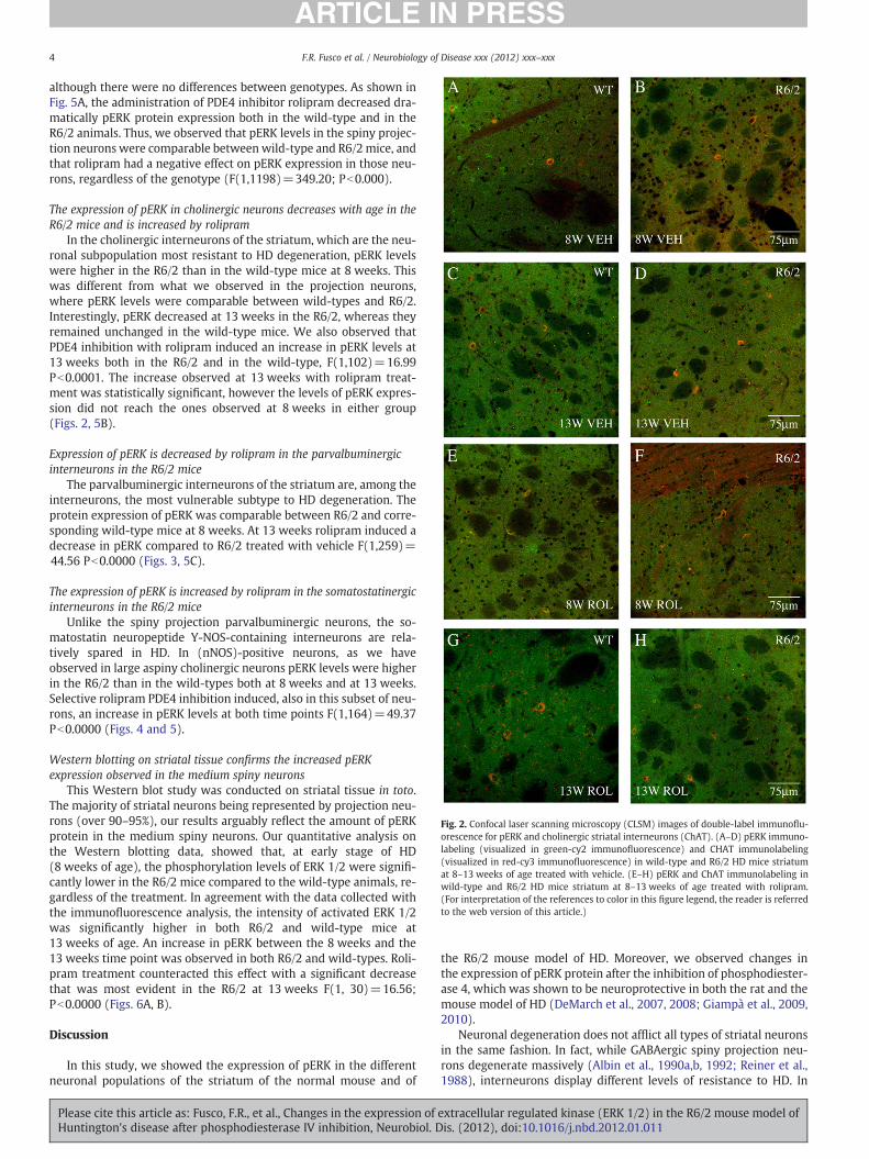
In the cholinergic interneurons of the striatum, which are the neu-ronal subpopulation most resistant to HD degeneration, pERK levelswere higher in the R6/2 than in the wild-type mice at 8 weeks. Thiswas different from what we observed in the projection neurons,where pERK levels were comparable between wild-types and R6/2.Interestingly, pERK decreased at 13 weeks in the R6/2, whereas theyremained unchanged in the wild-type mice. We also observed thatPDE4 inhibition with rolipram induced an increase in pERK levels at13 weeks both in the R6/2 and in the wild-type, F(1,102)=16.99Pb0.0001. The increase observed at 13 weeks with rolipram treat-ment was statistically signi"cant, however the levels of pERK expres-sion did not reach the ones observed at 8 weeks in either group(Figs. 2, 5B).
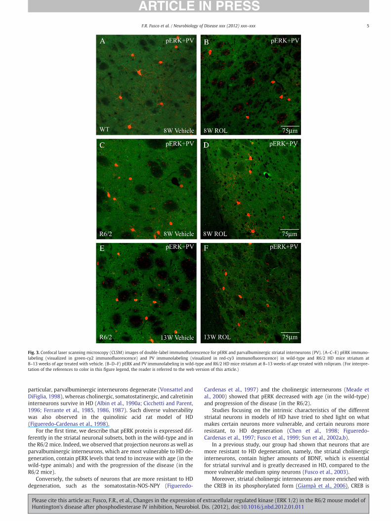
Expression of pERK is decreased by rolipram in the parvalbuminergicinterneurons in the R6/2 mice
The parvalbuminergic interneurons of the striatum are, among theinterneurons, the most vulnerable subtype to HD degeneration. Theprotein expression of pERK was comparable between R6/2 and corre-sponding wild-type mice at 8 weeks. At 13 weeks rolipram induced adecrease in pERK compared to R6/2 treated with vehicle F(1,259)=44.56 Pb0.0000 (Figs. 3, 5C).
The expression of pERK is increased by rolipram in the somatostatinergicinterneurons in the R6/2 mice
Unlike the spiny projection parvalbuminergic neurons, the so-matostatin neuropeptide Y-NOS-containing interneurons are rela-tively spared in HD. In (nNOS)-positive neurons, as we haveobserved in large aspiny cholinergic neurons pERK levels were higherin the R6/2 than in the wild-types both at 8 weeks and at 13 weeks.Selective rolipram PDE4 inhibition induced, also in this subset of neu-rons, an increase in pERK levels at both time points F(1,164)=49.37Pb0.0000 (Figs. 4 and 5).
Western blotting on striatal tissue con"rms the increased pERKexpression observed in the medium spiny neurons
This Western blot study was conducted on striatal tissue in toto.The majority of striatal neurons being represented by projection neu-rons (over 90–95%), our results arguably re!ect the amount of pERKprotein in the medium spiny neurons. Our quantitative analysis onthe Western blotting data, showed that, at early stage of HD(8 weeks of age), the phosphorylation levels of ERK 1/2 were signi"-cantly lower in the R6/2 mice compared to the wild-type animals, re-gardless of the treatment. In agreement with the data collected withthe immuno!uorescence analysis, the intensity of activated ERK 1/2was signi"cantly higher in both R6/2 and wild-type mice at13 weeks of age. An increase in pERK between the 8 weeks and the13 weeks time point was observed in both R6/2 and wild-types. Roli-pram treatment counteracted this effect with a signi"cant decreasethat was most evident in the R6/2 at 13 weeks F(1, 30)=16.56;Pb0.0000 (Figs. 6A, B).
Discussion
In this study, we showed the expression of pERK in the differentneuronal populations of the striatum of the normal mouse and of
the R6/2 mouse model of HD. Moreover, we observed changes inthe expression of pERK protein after the inhibition of phosphodiester-ase 4, which was shown to be neuroprotective in both the rat and themouse model of HD (DeMarch et al., 2007, 2008; Giampà et al., 2009,2010).
Neuronal degeneration does not af!ict all types of striatal neuronsin the same fashion. In fact, while GABAergic spiny projection neu-rons degenerate massively (Albin et al., 1990a,b, 1992; Reiner et al.,1988), interneurons display different levels of resistance to HD. In
Fig. 2. Confocal laser scanning microscopy (CLSM) images of double-label immuno!u-orescence for pERK and cholinergic striatal interneurons (ChAT). (A–D) pERK immuno-labeling (visualized in green-cy2 immuno!uorescence) and CHAT immunolabeling(visualized in red-cy3 immuno!uorescence) in wild-type and R6/2 HD mice striatumat 8–13 weeks of age treated with vehicle. (E–H) pERK and ChAT immunolabeling inwild-type and R6/2 HD mice striatum at 8–13 weeks of age treated with rolipram.(For interpretation of the references to color in this "gure legend, the reader is referredto the web version of this article.)
4 F.R. Fusco et al. / Neurobiology of Disease xxx (2012) xxx–xxx
Please cite this article as: Fusco, F.R., et al., Changes in the expression of extracellular regulated kinase (ERK 1/2) in the R6/2 mouse model ofHuntington's disease after phosphodiesterase IV inhibition, Neurobiol. Dis. (2012), doi:10.1016/j.nbd.2012.01.011
particular, parvalbuminergic interneurons degenerate (Vonsattel andDiFiglia, 1998), whereas cholinergic, somatostatinergic, and calretinininterneurons survive in HD (Albin et al., 1990a; Cicchetti and Parent,1996; Ferrante et al., 1985, 1986, 1987). Such diverse vulnerabilitywas also observed in the quinolinic acid rat model of HD(Figueredo-Cardenas et al., 1998).
For the "rst time, we describe that pERK protein is expressed dif-ferently in the striatal neuronal subsets, both in the wild-type and inthe R6/2 mice. Indeed, we observed that projection neurons as well asparvalbuminergic interneurons, which are most vulnerable to HD de-generation, contain pERK levels that tend to increase with age (in thewild-type animals) and with the progression of the disease (in theR6/2 mice).
Conversely, the subsets of neurons that are more resistant to HDdegeneration, such as the somatostatin-NOS-NPY (Figueredo-
Cardenas et al., 1997) and the cholinergic interneurons (Meade etal., 2000) showed that pERK decreased with age (in the wild-type)and progression of the disease (in the R6/2).
Studies focusing on the intrinsic characteristics of the differentstriatal neurons in models of HD have tried to shed light on whatmakes certain neurons more vulnerable, and certain neurons moreresistant, to HD degeneration (Chen et al., 1998; Figueredo-Cardenas et al., 1997; Fusco et al., 1999; Sun et al., 2002a,b).
In a previous study, our group had shown that neurons that aremore resistant to HD degeneration, namely, the striatal cholinergicinterneurons, contain higher amounts of BDNF, which is essentialfor striatal survival and is greatly decreased in HD, compared to themore vulnerable medium spiny neurons (Fusco et al., 2003).
Moreover, striatal cholinergic interneurons are more enriched withthe CREB in its phosphorylated form (Giampà et al., 2006). CREB is
Fig. 3. Confocal laser scanning microscopy (CLSM) images of double-label immuno!uorescence for pERK and parvalbuminergic striatal interneurons (PV). (A–C–E) pERK immuno-labeling (visualized in green-cy2 immuno!uorescence) and PV immunolabeling (visualized in red-cy3 immuno!uorescence) in wild-type and R6/2 HD mice striatum at8–13 weeks of age treated with vehicle. (B–D–F) pERK and PV immunolabeling in wild-type and R6/2 HD mice striatum at 8–13 weeks of age treated with rolipram. (For interpre-tation of the references to color in this "gure legend, the reader is referred to the web version of this article.)
5F.R. Fusco et al. / Neurobiology of Disease xxx (2012) xxx–xxx
Please cite this article as: Fusco, F.R., et al., Changes in the expression of extracellular regulated kinase (ERK 1/2) in the R6/2 mouse model ofHuntington's disease after phosphodiesterase IV inhibition, Neurobiol. Dis. (2012), doi:10.1016/j.nbd.2012.01.011

required for the survival of adult CNS neurons (Mantamadiotis et al.,2002), and mediates nuclear calcium-regulated gene transcription fol-lowing neuronal cell membrane depolarization (Hardingham et al.,1998). Moreover, a neuroprotective role of CREB was clearly demon-strated in a hypoxic–ischemic brain damage model of programmedcell death (Walton et al., 1996, 1999). In particular, inhibition of cAMPresponse element (CRE)-mediated gene transcription has been hypoth-esized to contribute to HD pathology (Jiang et al., 2003; Kazantsev et al.,1999; Nucifora et al., 2001; Shimohata et al., 2000; Steffan et al., 2000,2001).
Of note, several different agents can induce the phosphorylation ofboth ERK 1/2 and CREB, as the two factors seem to be interlinked inone or more pathways. In fact, in the striatum, glutamate-inducedphosphorylation of CREB appears to be mediated via activation ofERK 1/2 (Sgambato et al., 1998; Vanhoutte et al., 1999). Moreover,
there is evidence indicating that the functioning of cAMP pathway isinextricably linked with that of the extracellular signal-regulated ki-nase (ERK 1/2/mitogen-activated protein kinase (MAPK) pathway(Baillie et al., 2000).
Our results show, however, that the bene"cial effect of rolipramacts differently in the different cell types of the striatum. Indeed,rolipram showed to decrease pERK levels in the neurons that aremore vulnerable to HD degeneration, whereas it induced an upre-gulation of ERK 1/2 in the more resistant neurons, such as the cho-linergic and the SS/NOS/NPY interneurons. In particular, the effectof rolipram was not detectable at 8 weeks, when pERK levelswere comparable between groups. Rolipram effect was, in fact,more evident at 13 weeks of age in the R6/2 mice, showing thatthe compound was able to maintain the levels of pERK necessaryfor cell functions.
Fig. 4. Confocal laser scanning microscopy (CLSM) images of double-label immuno!uorescence for pERK and somatostatinergic striatal interneurons (NOS). (A–C–E) pERK immu-nolabeling (visualized in green-cy2 immuno!uorescence) and NOS immunolabeling (visualized in red-cy3 immuno!uorescence) in wild-type and R6/2 HD mice striatum at8–13 weeks of age treated with vehicle. (B–D–F) pERK and NOS immunolabeling in rolipram-treated wild-type and R6/2 HDmice striatum at 8–13 weeks of age. (For interpretationof the references to color in this "gure legend, the reader is referred to the web version of this article.)
6 F.R. Fusco et al. / Neurobiology of Disease xxx (2012) xxx–xxx
Please cite this article as: Fusco, F.R., et al., Changes in the expression of extracellular regulated kinase (ERK 1/2) in the R6/2 mouse model ofHuntington's disease after phosphodiesterase IV inhibition, Neurobiol. Dis. (2012), doi:10.1016/j.nbd.2012.01.011

Our results were corroborated by the western blot study, whichshowed, in whole striatal samples, changes in pERK that were compa-rable to the ones observed in the immuno!uorescence analysis.
In a previous study, an increase in pERK was observed in R6/2mice at 12 weeks of age (Liévens et al., 2002). Our data expandthese "ndings and describe how rolipram-induced PDE4 inhibitiondecreases such as high levels of pERK in the medium spiny neurons,which account for the largest part of striatal neuronal population.
The observation that changes in ERK 1/2 phosphorylation did notparallel the changes in CREB described previously (Giampà et al.,2006) seems to be con!icting with the concept that CREB and ERK 1/2 pathways are interlinked, particularly because rolipram induces anincrease in CREB both in the mouse and rat models of HD (DeMarchet al., 2007, 2008; Giampà et al., 2009). Indeed, the inhibition of anoth-er kind of PDE, namely, PDE10, induces the activation of CREB and alsoof ERK 1/2 (Kleiman et al., 2011). Thus, one could expect that the
Fig. 5. Graph summarizing the different levels of pERK (expressed in arbitrary units) in the CALB immunoreactive projection neurons (A) and in the interneurons subtypes (B–D) ofstriata following rolipram and vehicle treatment and sacri"ced at the different time points. Please note that the levels of pERK decrease dramatically at 13 weeks of age after roli-pram treatment.
Fig. 6. Regulation of ERK 1/2 activation after rolipram treatment in wild-type and R6/2 mice. A–B: pERK levels were analyzed by Western blotting of protein extracts obtained fromthe striatum of WT and R6/2 mice at different time points (8 and 13 weeks).
7F.R. Fusco et al. / Neurobiology of Disease xxx (2012) xxx–xxx
Please cite this article as: Fusco, F.R., et al., Changes in the expression of extracellular regulated kinase (ERK 1/2) in the R6/2 mouse model ofHuntington's disease after phosphodiesterase IV inhibition, Neurobiol. Dis. (2012), doi:10.1016/j.nbd.2012.01.011

neuroprotective effect of rolipram induced an increase in pERK becauseof the increased pCREB that was observed previously.
This, however, seems to be oversimpli"ed when taking into ac-count the complexity of the cross-talk between cAMP-PKA and ERK1/2 signaling and that ERK 1/2 can play different, and even opposite,roles in the cell.
The role of ERK 1/2 in a variety of cellular functions including celldeath depends on the cell type as well as on the signal that triggerscell death (Bioessays, 2003; Colucci-D'Amato et al., 2003; Stanciu etal., 2000). Indeed, ERK 1/2 facilitates apoptosis in several paradigms(Aoki et al., 2011; Bhat and Zhang, 1999; Fukunaga and Miyamoto,1998) including MPP+-mediated apoptosis in SH-SY5Y neuroblasto-ma cells (Gómez-Santos et al., 2002). Yet MAPK/ERK 1/2 is associatedwith survival (Hetman et al., 1999) and is upregulated in the penum-bra of the ischemic lesion in the focal cerebral ischemia model (Ferreret al., 2003).
Furthermore, a bulk of data shows that the duration of ERK 1/2 ac-tivation, caused by different stimuli in a given cell type, determines itssubcellular compartmentalization and/or traf"cking. The latter, inturn, dictates whether ERK 1/2-expressing cells would enter a pro-gram of cell death or survival (Chu et al., 2004; Colucci-D'Amato etal., 2003; Stanciu and DeFranco, 2002).
In neurons, the high number of different stimuli, such as neurotro-phins, neurotransmitters, growth factors, depolarization, leading toERK 1/2 activation, renders ERK 1/2 an essential signal cross-roadswithin the cell. Thus, the bene"cial effect of rolipram on a numberof different types of neurons could result from its "ne tuning of ERK1/2 phosphorylation. In this context, it is worth noting that also thecAMP-PKA pathway, affected by rolipram treatment, can exert eitheran inhibitory or a facilitating stimulation on MAPK-ERK 1/2 signaling,according to the cell types (Frödin et al., 1994; Sevetson et al., 1993).
In addition, to add complexity to the PKA-ERK 1/2 cross-talk, it isworth mentioning that phosphorylated ERK 1/22 is able to directly in-hibit, a commonly expressed isoenzyme of PDE4, PDE4D3, thus in-creasing cAMP levels (Hoffmann et al., 1999).
In light of this, we did not correlate the upregulation of CREB ob-served after the administration of the PDE4 inhibitor rolipram(Giampà et al., 2009) to the changes in the expression of ERK 1/2 de-scribed in the present study. However, we can speculate that PDE4 in-hibition can be bene"cial by lowering pERK in those cells where anexcessive amount is expressed (such as the medium spiny neurons),and at the same time by increasing pERK in those neurons that havelower levels such, as the cholinergic and SS/NOS/NPY interneurons,so that the anti-apoptotic effect can take place.
The situation that we have uncovered here offers the opportunityfor the rolipram-induced increase in CREB to lead to either an in-crease or a decrease in ERK 1/2 phosphorylation. This could be dueto the possibility of different ERK 1/2 pathways than can be activatedaccording to the neuronal subtype and its characteristic vulnerabilityto HD degeneration.
Appendix A. Supplementary data
Supplementary data to this article can be found online at doi:10.1016/j.nbd.2012.01.011.
References
Albin, R.L., Reiner, A., Anderson, K.D., Penney, J.B., Young, A.B., 1990a. Striatal and nigralneuron subpopulations in rigid Huntington's disease: implications for the func-tional anatomy of chorea and rigidity-akinesia. Ann. Neurol. 27 (4), 357–365.
Albin, R.L., Young, A.B., Penney, J.B., Handelin, B., Balfour, R., Anderson, K.D., et al., 1990b.Abnormalities of striatal projection neurons and N-methyl-D-aspartate receptors inpresymptomatic Huntington's disease. N. Engl. J. Med. 322 (18), 1293–1298.
Albin, R.L., Reiner, A., Anderson, K.D., Dure IV, L.S., Handelin, B., Balfour, R., et al., 1992.Preferential loss of striato-external pallidal projection neurons in presymptomaticHuntington's disease. Ann. Neurol. 31 (4), 425–430.
Andrew, S.E., Goldberg, Y.P., Kremer, B., Telenius, H., Theilmann, J., Adam, S., et al.,1993. The relationship between trinucleotide (CAG) repeat length and clinical fea-tures of Huntington's disease. Nat. Genet. 4 (4), 398–403.
Aoki, K., Yamada, M., Kunida, K., Yasuda, S., Matsuda, M., 2011. Processive phosphory-lation of ERK MAP kinase in mammalian cells. Proc. Natl. Acad. Sci. U. S. A. 108 (31),12675–12680.
Baillie, G.S., MacKenzie, S.J., McPhee, I., 2000. Houslay MD Sub-family selective ac-tions in the ability of Erk2 MAP kinase to phosphorylate and regulate the activ-ity of PDE4 cyclic AMP-speci"c phosphodiesterases. Br. J. Pharmacol. 131 (4),811–819.
Bhat, N.R., Zhang, P., 1999. Hydrogen peroxide activation of multiple mitogen-activatedprotein kinases in an oligodendrocyte cell line: role of extracellular signal-regulatedkinase in hydrogen peroxide-induced cell death. J. Neurochem. 72 (1), 112–119.
Canals, J.M., Checa, N., Marco, S., Akerud, P., Michels, A., Pérez-Navarro, E., et al., 2001.Expression of brain-derived neurotrophic factor in cortical neurons is regulated bystriataltarget area. J. Neurosci. 21 (1), 117–124.
Chen, Q., Veenman, L., Knopp, K., Yan, Z., Medina, L., Song, W.J., et al., 1998. Evidence forthe preferential localization of glutamate receptor-1 subunits of AMPA receptors tothe dendritic spines of medium spiny neurons in rat striatum. Neuroscience 83 (3),749–761.
Chu, C.T., Levinthal, D.J., Kulich, S.M., Chalovich, E.M., DeFranco, D.B., 2004. Oxidativeneuronal injury. The dark side of ERK1/2. Eur. J. Biochem. 271 (11), 2060–2066.
Cicchetti, F., Parent, A., 1996. Striatal interneurons in Huntington's disease: selective in-crease in the density of calretinin-immunoreactive medium-sized neurons. Mov.Disord. 11, 619–626.
Colucci-D'Amato, L., Perrone-Capano, C., di Porzio, U., 2003. Chronic activation of ERKand neurodegenerative diseases. Bioessays 25, 1085–1095.
Corominas, M., Roncero, C., Ribases, M., Castells, X., Casas, M., 2007. Brain-derived neu-rotrophic factor and its intracellular signaling pathways in cocaine addiction. Neu-ropsychobiology 55 (1), 2–13.
de la Monte, S.M., Vonsattel, J.P., Richardson Jr., E.P., 1988. Morphometric demonstra-tion of atrophic changes in the cerebral cortex, white matter, and neostriatum inHuntington's disease. J. Neuropathol. Exp. Neurol. 47, 516–525.
DeMarch, Z., Giampà, C., Patassini, S., Martorana, A., Bernardi, G., Fusco, F.R., 2007. Ben-e"cial effects of rolipram in a quinolinic acid model of striatal excitotoxicity. Neu-robiol. Dis. 25 (2), 266–267.
DeMarch, Z., Giampà, C., Patassini, S., Bernardi, G., Fusco, F.R., 2008. Bene"cial effects ofrolipram in the R6/2 mouse model of Huntington's disease. Neurobiol. Dis. 30 (3),375–387.
Ferrante, R.J., Kowall, N.W., Beal, M.F., Richardson Jr., E.P., Bird, E.D., Martin, J.B., 1985.Selective sparing of a class of striatal neurons in Huntington's disease. Science230 (4725), 561–563.
Ferrante, R.J., Kowall, N.W., Richardson Jr., E.P., Bird, E.D., Martin, J.B., 1986. Topographyof enkephalin, substance P and acetylcholinesterase staining in Huntington's dis-ease striatum. Neurosci. Lett. 71 (3), 283–288.
Ferrante, R.J., Kowall, N.W., Beal, M.F., Martin, J.B., Bird, E.D., Richardson Jr., E.P., 1987.Morphologic and histochemical characteristics of a spared subset of striatal neu-rons in Huntington's disease. J. Neuropathol. Exp. Neurol. 46 (1), 12–27.
Ferrer, I., Friguls, B., Dalfó, E., 2003. Planas AM Early modi"cations in the expression ofmitogen activated protein kinase (MAPK/ERK), stress-activated kinases SAPK/JNKand p38, and their phosphorylated substrates following focal cerebral ischemia.Acta Neuropathol. 105 (5), 425–437.
Figueredo-Cardenas, G., Chen, Q., Reiner, A., 1997. Age-dependent differences in sur-vival of striatal somatostatin-NPY-NADPH-diaphorase-containing interneuronsversus striatal projection neurons after intrastriatal injection of quinolinic acid inrats. Exp. Neurol. 146 (2), 444–457.
Figueredo-Cardenas, G., Harris, C.L., Anderson, K.D., Reiner, A., 1998. Relative resistanceof striatal neurons containing calbindin or parvalbumin to quinolinic acid-mediated excitotoxicity compared to other striatal neuron types. Exp. Neurol.149 (2), 356–372.
Frödin, M., Peraldi, P., Van Obberghen, E., 1994. Cyclic AMP activates the mitogen-activated protein kinase cascade in PC12 cells. J. Biol. Chem. 269 (8), 6207–6214.
Fukunaga, K., Miyamoto, E., 1998. Role of MAP kinase in neurons. Mol. Neurobiol. 16(1), 79–95.
Fusco, F.R., Chen, Q., Lamoreaux, W.J., Figueredo-Cardenas, G., Jiao, Y., Coffman, J.A., etal., 1999. Cellular localization of huntingtin in striatal and cortical neurons inrats: lack of correlation with neuronal vulnerability in Huntington's disease. J. Neu-rosci. 19 (4), 1189–1202.
Fusco, F.R., Viscomi, M.T., Bernardi, G., Molinari, M., 2001. Localization of ataxin-2 with-in the cerebellar cortex of the rat. Brain Res. Bull. 56 (3–4), 343–347.
Fusco, F.R., Zuccato, C., Tartari, M., Martorana, A., De March, Z., Giampà, C., et al., 2003.Co-localization of brain-derived neurotrophic factor (BDNF) and wild-type hun-tingtin in normal and quinolinic acid-lesioned rat brain. Eur. J. Neurosci. 18 (5),1093–1102.
Giampà, C., DeMarch, Z., D'Angelo, V., Morello, M., Martorana, A., Sancesario, G., et al.,2006. Striatal modulation of cAMP-response-element-binding protein (CREB)after excitotoxic lesions: implications with neuronal vulnerability in Huntington'sdisease. Eur. J. Neurosci. 23 (1), 11–20.
Giampà, C., Middei, S., Patassini, S., Borreca, A., Marullo, F., Laurenti, D., et al., 2009.Phosphodiesterase type IV inhibition prevents sequestration of CREB binding pro-tein, protects striatal parvalbumin interneurons and rescues motor de"cits in theR6/2 mouse model of Huntington's disease. Eur. J. Neurosci. 29 (5), 902–910.
Giampà, C., Laurenti, D., Anzilotti, S., Bernardi, G., Menniti, F.S., Fusco, F.R., 2010. Inhibi-tion of the striatal speci"c phosphodiesterase PDE10A ameliorates striatal and cor-tical pathology in R6/2 mouse model of Huntington's disease. PLoS One 5 (10),e13417.
8 F.R. Fusco et al. / Neurobiology of Disease xxx (2012) xxx–xxx
Please cite this article as: Fusco, F.R., et al., Changes in the expression of extracellular regulated kinase (ERK 1/2) in the R6/2 mouse model ofHuntington's disease after phosphodiesterase IV inhibition, Neurobiol. Dis. (2012), doi:10.1016/j.nbd.2012.01.011

Gómez-Santos, C., Ferrer, I., Reiriz, J., Viñals, F., Barrachina, M., Ambrosio, S., 2002. MPP+ increases alpha-synuclein expression and ERK/MAP-kinase phosphorylation inhuman neuroblastoma SHSY5Y cells. Brain Res. 935 (1–2), 32–39.
Gratacòs, E., Pérez-Navarro, E., Tolosa, E., Arenas, E., Alberch, J., 2001. Neuroprotectionof striatal neurons against kainate excitotoxicity by neurotrophins and GDNF fam-ily members. J. Neurochem. 78 (6), 1287–1296.
Hardingham, G.E., Cruzalegui, F.H., Chawla, S., Bading, H., 1998. Mechanisms control-ling gene expression by nuclear calcium signals. Cell Calcium 23 (2–3), 131–134.
Hetman, M., Kanning, K., Cavanaugh, J.E., Xia, Z., 1999. Neuroprotection by brain-derived neurotrophic factor is mediated by extracellular signal-regulated kinaseand phosphatidylinositol 3-kinase. J. Biol. Chem. 274 (32), 22569–22580.
Hoffmann, R., Baillie, G.S., MacKenzie, Yarwood, S., Houslay, M.D., 1999. The MAP ki-nase ERK2 inhibits the cyclic AMP-speci"c phosphodiesterase HSPDE4D3 by phos-phorylating it at Ser579. EMBO J. 18 (4), 893–903.
Jiang, H., Nucifora Jr., F.C., Ross, C.A., DeFranco, D.B., 2003. Cell death triggered bypolyglutamine-expanded huntingtin in a neuronal cell line is associ ated with deg-radation of CREB-binding protein. Hum. Mol. Genet. 12, 1–12.
Jovanovic, J.N., Benfenati, F., Siow, Y.L., Sihra, T.S., Sanghera, J.S., Pelech, S.L., et al., 1996.Neurotrophins stimulate phosphorylation of synapsin I by MAP kinase and regu-late synapsin I-actin interactions. Proc. Natl. Acad. Sci. U. S. A. 93 (8), 3679–3683.
Kazantsev, A., Preisinger, E., Dranovsky, A., Goldgaber, D., Housman, D., 1999. Insolubledetergent-resistant aggregates form between pathological and nonpathologicallengths of polyglutamine in mammalian cells. Proc. Natl. Acad. Sci. U. S. A. 96(20), 11404–11409.
Kim, J.Y., Kim, Y.J., Lee, S., Park, J.H., 2009. The critical role of ERK in death resistanceand invasiveness of hypoxia-selected glioblastoma cells. BMC Cancer 9, 27.
Kingsbury, T.J., Krueger, B.K., 2007. Ca2+, CREB and krüppel: a novel KLF7-binding el-ement conserved in mouse and human TRKB promoters is required for CREB-dependent transcription. Mol. Cell. Neurosci. 35 (3), 447–455.
Kleiman, R.J., Kimmel, L.H., Bove, S.E., Lanz, T.A., Harms, J.F., Romegialli, A., et al., 2011.Chronic suppression of phosphodiesterase 10A alters striatal expression of genes re-sponsible for neurotransmitter synthesis, neurotransmission, and signalling path-ways implicated in Huntington's disease. J. Pharmacol. Exp. Ther. 336 (1), 64–76.
Liebelt, B., Papapetrou, P., Ali, A., Guo, M., Ji, X., Peng, C., et al., 2010. Exercise precondi-tioning reduces neuronal apoptosis in stroke by up-regulating heat shock protein-70 (heat shock protein-72) and extracellular-signal-regulated-kinase. Neurosci-ence 166 (4), 1091–1100.
Liévens, J.C., Woodman, B., Mahal, A., Bates, G.P., 2002. Abnormal phosphorylation ofsynapsin I predicts a neuronal transmission impairment in the R6/2 Huntington'sdisease transgenic mice. Mol. Cell. Neurosci. 20 (4), 638–648.
Mangiarini, L., Sathasivam, K., Seller, M., Cozens, B., Harper, A., Hetherington, C., et al.,1996. Exon 1 of the HD gene with an expanded CAG repeat is suf"cient to causea progressive neurological phenotype in transgenic mice. Cell 87 (3), 493–506.
Mantamadiotis, T., Lemberger, T., Bleckmann, S.C., Kern, H., Kretz, O., Martin Villalba, A.,et al., 2002. Disruption of CREB function in brain leads to neurodegeneration. Nat.Genet. 31 (1), 47–54.
Matsubara,M., Kusubata,M., Ishiguro, K., Uchida, T., Titani, K., Taniguchi, H., 1996. Site-spe-ci"c phosphorylation of synapsin I by mitogen-activated protein kinase and Cdk5 andits effects on physiological functions. J. Biol. Chem. 271 (35), 21108–21113.
Meade, C.A., Figueredo-Cardenas, G., Fusco, F., Nowak Jr., T.S., Pulsinelli, W.A., Reiner,A., 2000. Transient global ischemia in rats yields striatal projection neuron and in-terneuron loss resembling that in Huntington's disease. Exp. Neurol. 166 (2),307–323.
Melone, M.A., Jori, F.P., Peluso, G., 2005. Huntington's disease: new frontiers for molec-ular and cell therapy. Curr. Drug Targets 6 (1), 43–56.
Nucifora Jr., F.C., Sasaki, M., Peters, M.F., Huang, H., Cooper, J.K., Yamada, M., et al., 2001.Interference by huntingtin and atrophin-1 with cbp-mediated transcription lead-ing to cellular toxicity. Science 291 (5512), 2423–2428.
Numakawa, T., Suzuki, S., Kumamaru, E., Adachi, N., Richards, M., Kunugi, H., 2010.BDNF function and intracellular signaling in neurons. Histol. Histopathol. 25 (2),237–258.
Perrone, L., Melone, M., 2008. New targets for therapy in polyglutamine (polyQ) expan-sion diseases. Curr. Drug Ther. 3, 177–189.
Reiner, A., Albin, R.L., Anderson, K.D., D'Amato, C.J., Penney, J.B., Young, A.B., 1988. Proc.Natl. Acad. Sci. U. S. A. 85 (15), 5733–5737.
Roze, E., Betuing, S., Deyts, C., Marcon, E., Brami-Cherrier, K., Pagès, C., et al., 2008. Mi-togen- and stress-activated protein kinase-1 de"ciency is involved in expanded-huntingtin-induced transcriptional dysregulation and striatal death. FASEB J. 22(4), 1083–1093.
Sevetson, B.R., Kong, X., Lawrence Jr., J.C., 1993. Increasing cAMP attenuates activationof mitogen-activated protein kinase. Proc. Natl. Acad. Sci. U. S. A. 90, 10305–10309.
Sgambato, V., Pagès, C., Rogard, M., Besson, M.J., Caboche, J., 1998. Extracellular signal-regulated kinase (ERK) controls immediate early gene induction on corticostriatalstimulation. J. Neurosci. 18 (21), 8814–8825.
Shimohata, T., Nakajima, T., Yamada, M., Uchida, C., Onodera, O., Naruse, S., et al., 2000.Expanded polyglutamine stretches interact with TAFII130, interfering with CREB-dependent transcription. Nat. Genet. 26 (1), 29–36.
Stanciu, M., DeFranco, D.B., 2002. Prolonged nuclear retention of activated extracellularsignalregulated protein kinase promotes cell death generated by oxidative toxicityor proteasome inhibition in a neuronal cell line. J. Biol. Chem. 277 (6), 4010–4017.
Stanciu, M., Wang, Y., Kentor, R., Burke, N., Watkins, S., Kress, G., Reynolds, I., Klann, E.,Angiolieri, M.R., Johnson, J.W., DeFranco, D.B., 2000. Persistent activation of ERKcontributes to glutamate-induced oxidative toxicity in a neuronal cell line and pri-mary cortical neuron cultures. J. Biol. Chem. 275 (16), 12200–12206.
Steffan, J.S., Kazantsev, A., Spasic-Boskovic, O., Greenwald, M., Zhu, Y.Z., Gohler, H., etal., 2000. The Huntington's disease protein interacts with p53 and CREB-bindingprotein and represses transcription. Proc. Natl. Acad. Sci. U. S. A. 97 (12),6763–6768.
Steffan, J.S., Bodai, L., Pallos, J., Poelman, M., McCampbell, A., Apostol, B.L., et al., 2001.Histone deacetylase inhibitors arrest polyglutamine-dependent neurodegenera-tion in Drosophila. Nature 413 (6857), 739–743.
Sun, Z., Xie, J., Reiner, A., 2002a. The differential vulnerability of striatal projection neu-rons in 3-nitropropionic acid-treated rats does not match that typical of adult-onset Huntington's disease. Exp. Neurol. 176 (1), 55–65.
Sun, Z., Del Mar, N., Meade, C., Goldowitz, D., Reiner, A., 2002b. Differential changes instriatal projection neurons in R6/2 transgenic mice for Huntington's disease. Neu-robiol. Dis. 11 (3), 369–385.
The Huntington's Disease Collaborative Research Group, 1993. A novel gene containinga trinucleotide repeat is expanded and unstable on Huntington's disease chromo-somes. Cell 72, 971–983.
Vanhoutte, P., Barnier, J.V., Guibert, B., Pagès, C., Besson, M.J., Hipskind, R.A., Caboche, J.,1999. Glutamate induces phosphorylation of Elk-1 and CREB, along with c-fos ac-tivation, via an extracellular signal-regulated kinase-dependent pathway in brainslices. J. Mol. Cell Biol. 19 (1), 136–146.
Vonsattel, J.P., DiFiglia, M., 1998. Huntington disease. J. Neuropathol. Exp. Neurol. 57(5), 369–384.
Vonsattel, J.P., Myers, R.H., Stevens, T.J., Ferrante, R.J., Bird, E.D., Richardson Jr., E.P.,1985. Neuropathological classi"cation of Huntington's disease. J. Neuropathol.Exp. Neurol. 44 (6), 559–577.
Walker, F.O., 2007. Huntington's disease. Lancet 369, 218–228.Walton, M., Young, D., Sirimanne, E., Dodd, J., Christie, D., Williams, C., et al., 1996. The
role of the cyclic AMP-responsive element binding protein (CREB) in hypoxic-ischemic brain damage and repair. Brain Res. Mol. Brain Res. 43, 21–29.
Walton, M., Connor, B., Lawlor, P., Young, D., Sirimanne, E., Gluckman, P., et al., 1999.Neuronal death and survival in two models of hypoxic-ischemic brain damage.Brain Res. Brain Res. Rev. 29 (2–3), 137–168.
Yao, H., Peng, F., Fan, Y., Zhu, X., Hu, G., Buch, S.J., 2009. TRPC channel-mediated neuro-protection by PDGF involves Pyk2/ERK/CREB pathway. Cell Death Differ. 16,1681–1693.
Yu, C., Yezierski, R., Joshi, A., Raza, K., Geddes, J., 2010. Involvement of ERK2 in traumat-ic spinal cord injury. J. Neurochem. 113, 131–142.
Zuccato, C., Cattaneo, E., 2007. Role of brain-derived neurotrophic factor in Hunting-ton's disease. Prog. Neurobiol. 81 (5–6), 294–330.
Zuccato, C., Ciammola, A., Rigamonti, D., Leavitt, B.R., Goffredo, D., Conti, L., et al., 2001.Loss of huntingtin-mediated BDNF gene transcription in Huntington's disease. Sci-ence 293 (5529), 493–498.
9F.R. Fusco et al. / Neurobiology of Disease xxx (2012) xxx–xxx
Please cite this article as: Fusco, F.R., et al., Changes in the expression of extracellular regulated kinase (ERK 1/2) in the R6/2 mouse model ofHuntington's disease after phosphodiesterase IV inhibition, Neurobiol. Dis. (2012), doi:10.1016/j.nbd.2012.01.011
Copyright © 2022 FDOKUMEN