Immune-Mediated Mechanisms of Parasite Tissue Sequestration during Experimental Cerebral Malaria
Upload
khangminh22Category
view
3download
0
UMEÅ UNIVERSITY MEDICAL DISSERTATIONSNEW SERIES NO. 910 ISSN 0346-6612 - ISBN 91-7305-703-7
Experimental studies of spinal mechanismsassociated with muscle fatigue
From the Department of Surgical and Perioperative Sciences, Sports Medicine Unit,Umeå University, the Center for Musculoskeletal Researach, University of Gävle,
Umeå, Sweden
Department of Surgical and Perioperative Sciences, Sports Medicine UnitUmeå University, SE-901 87 Umeå Sweden
Copyright © 2004 by Ivana Kalezic
ISSN 0346-6612
ISBN 91-7305-703-7
Printed by Arbetslivsinstitutets tryckeri, Umeå, 2004

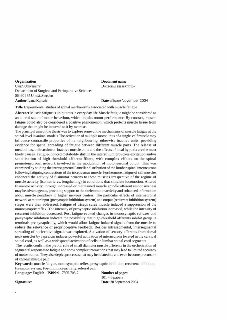
Organization Document nameUMEÅ UNIVERSITY DOCTORAL DISSERTATION
Department of Surgical and Perioperative SciencesSE-901 87 Umeå, SwedenAuthor Ivana Kalezic Date of issue November 2004
Title Experimental studies of spinal mechanisms associated with muscle fatigueAbstract Muscle fatigue is ubiquitous in every day life.Muscle fatigue might be considered asan altered state of motor behaviour, which impairs motor performance. By contrast, musclefatigue could also be considered a positive phenomenon, which protects muscle tissue fromdamage that might be incurred to it by overuse.The principal aim of the thesis was to explore some of the mechanisms of muscle fatigue at thespinal level in animal models.The activation of multiple motor units of a single calf muscle mayinfluence contractile properties of its neighbouring, otherwise inactive units, providingevidence for spatial spreading of fatigue between different muscle parts. The release ofmetabolites, their action on inactive muscle units and the effects of local hypoxia are the mostlikely causes. Fatigue-induced metabolite shift in the interstitium provokes excitation and/orsensitisation of high-threshold afferent fibers, with complex effects on the spinalpremotoneuronal network involved in the modulation of motoneuronal output. This wasexamined by studing the intrasegmental lamellar distribution of the lumbar spinal interneuronsfollowing fatiguing contractions of the triceps surae muscle. Furthermore, fatigue of calf musclesenhanced the activity of fusimotor neurons to these muscles irrespective of the regime ofmuscle activity (isometric vs. lengthening) in conditions that simulate locomotion. Alteredfusimotor activity, through increased or maintained muscle spindle afferent responsivenessmay be advantageous, providing support to the skeletomotor activity and enhanced informationabout muscle periphery to higher nervous centres. The particular effects of interneuronalnetwork at motor input (presynaptic inhibition system) and output (recurrent inhibition system)stages were then addressed. Fatigue of triceps surae muscle induced a suppression of themonosynaptic reflex. The intensity of presynaptic inhibition increased, while the intensity ofrecurrent inhibition decreased. Post fatigue-evoked changes in monosynaptic reflexes andpresynaptic inhibition indicate the possibility that high-threshold afferents inhibit group Iaterminals pre-synaptically, which would allow fatigue-induced signals from the muscle toreduce the relevance of proprioceptive feedback. Besides intrasegmental, intersegmentalspreading of nociceptive signals was explored. Activation of sensory afferents from dorsalneck muscles by capsaicin induces powerful activation of interneurons located in the cervicalspinal cord, as well as a widespread activation of cells in lumbar spinal cord segments. The results confirm the pivotal role of small diameter muscle afferents in the orchestration ofsegmental responses to fatigue and show complex interactions that may lead to limited accuracyof motor output. They also depict processes that may be related to, and even become precursorsof chronic muscle pain.Key words: muscle fatigue, monosynaptic reflex, presynaptic inhibition, recurrent inhibition,fusimotor system, Fos-immunoreactivity, referral painLanguage: English ISBN: 91-7305-703-7 Number of pages:
101 + 6 papersSignature: Date: 30 September 2004
.

1
CONTENTSORIGINAL PAPERS ..................................................................................... 3ABBREVIATIONS ........................................................................................ 41. INTRODUCTION .................................................................................... 51.1 Peripheral effects of muscle fatigue .......................................................... 5
1.1.1 Spreading of fatigue-related effects within the muscle tissue ................... 51.2 Central processing of fatigue effects ....................................................... 6
1.2.1 Muscle wisdom hypothesis ....................................................................... 61.2.2 Possible role of the intrinsic motoneuronal properties ............................... 71.2.3 Reflex action exerted by small-diameter muscle afferent fibres .............. 81.2.4 Muscle spindle afferent fibres input ........................................................ 101.2.5 Presynaptic inhibition. ............................................................................. 151.2.6 Recurrent inhibition .................................................................................. 19
1.3 The case of spinal mechanisms of muscle fatigue at cellular level ............. 201.3.1 c-fos expression in the spinal cord .......................................................... 211.3.2 Activation of nitric oxide synthase (NOS) - containing neurons .............. 23
1.4 Vicious circles related to muscle cramps and chronic myalgia.................. 241.4.1 Spreading of nociceptive signals from cervical to lumbar regions and vice
versa ............................................................................................................ 252. AIMS ....................................................................................................... 273. METHODS ............................................................................................. 293.1General surgical procedures and nerve-muscle preparations .................... 293.2 Stimulation and recording ...................................................................... 313.3 Data acquisition and analysis ................................................................. 373.4 Fos immunohistochemistry and NADPH-d histochemistry ...................... 38
4. RESULTS ................................................................................................. 404.1 Paper I ................................................................................................. 404.2.Paper II ............................................................................................... 454.3 Paper III ............................................................................................... 504.4 Paper IV .............................................................................................. 524.5 Paper V................................................................................................ 564.6 Paper VI .............................................................................................. 62
5. DISCUSSION .......................................................................................... 655.1 Experimental evidence of spreading of fatigue effects between different
muscle parts ......................................................................................... 655.2 Experimental evidence of fatigue-induced activation of small - diameter
afferent fibres ...................................................................................... 67
2
5.3 Could neuromuscular propagation failure (NPF) contribute to fatiguingeffects? ............................................................................................... 68
5.4 Does the gamma-spindle system act as a premotoneuronal integrativesystem in fatiguing conditions related to different regimes of muscle activity?
.......................................................................................................... 685.5 Muscle fatigue-induced alterations at the motoneuron input stage - the
presynaptic inhibitory system ................................................................ 725.6 Muscle fatigue induced alterations at the motoneuron output stage - the
recurrent inhibition system ..................................................................... 745.7. c-fos expression in spinal motoneurons following muscle fatigue and acute
inflammation ........................................................................................ 755.8 NADPH-d reactivity in spinal neurons following muscle fatigue............... 765.9 Potential effects of the activation of preganglionic sympathetic neurons
following long-lasting muscle contraction and fatigue .............................. 775.10 Spreading of nociceptive signals from cervical to lumbar region and vice-
versa .................................................................................................... 785.11 General conclusion ............................................................................. 79
5. ACKNOWLEDGEMENTS..................................................................... 816. REFERENCES ......................................................................................... 82
3
ORIGINAL PAPERSThis thesis is based on the following papers, referred to by their Roman numerals in thetext
AI Kostyukov, I Kalezic, SG Serenko, M Ljubisavljevic, U Windhorst, H Johans-son (2002): Spreading of fatigue-related effects from active to inactive parts inthe medial gastrocnemius muscle of the cat. European Journal of AppliedPhysiology, 86: 295-307
AI Pilyavskii, VA Maisky, I Kalezic, M Ljubisavljevic, AI Kostyukov, UWindhorst, H Johansson (2001): c-fos expression and NADPH-d reactivity inspinal neurons after fatiguing stimulation of hindlimb muscles in the rat. BrainResearch, 923: 91-102
M Ljubisavljevic, I Kalezic, S Radovanovic, S Milanovic, S Blesic and RAnastasijevic (1998): Changes in fusimotor activity during repetitive lengtheningmuscle contractions in decerebrate cats. Neuroscience, 86: 1337 - 1342
M Ljubisavljevic, I Vukcevic*, S Radovanovic, S Milanovic and R Anastasijevic(1997): Effects of cutaneous afferent input on fatigue-induced changes in fusimotoractivity of decerebrate cats. Neuroscience, 79: 935 - 942
I Kalezic, LA Bugaychenko, AI Kostyukov, AI Pilyavskii, M Ljubisavljevic, UWindhorst and H Johansson (2004): Modulation of the monosynaptic reflexes ofthe gastrocnemius-soleus muscle after their fatiguing stimulation in decerebratecats. Journal of Physiogy, 556.1: 293-296
I Kalezic, AI Pilyavskii, VA Maisky, AI Kostyukov, U Windhorst and H Johansson(2004): Distinctive pattern of c-fos expression in the feline cervico-lumbar spinalcord after stimulation of vanilloid receptors in dorsal neck muscles. NeuroscienceLetters, 364:94-97
I Vukcevic* -maiden name
I
II
III
IV
V
IV

4
ABBREVIATIONSABC Avidin-biotin-peroxidase complexAHP AfterhyperpolarizationCDP Cord dorsum potentialCFS Conditioning fatiguing stimulationDAB 3,3’ Diamino-benzidine tetrahydrochlorideDR Dorsal rootDRP Dorsal root potentialDST Distributed stimulationEMG ElectromyogramFST Fatiguing stimulationFos-ir neurons Fos-immunoreactive neuronsGABA γ-Aminobutyric acidGLS Gastrocnemius lateralis and soleusGM Gastrocnemius medialisGS Gastrocnemius and soleusGTO Golgi tendon organH-reflex Hoffman reflexIML Intermediolateral cell columnLLR Long latency reflexL-NAME NG-nitro-L-arginine methyl esterLNOArg L-NG-nitro arginineMSA Muscle spindle afferentMSR Monosynaptic reflexMT Motor thresholdMVC Maximal voluntary contractionNADPH-d Nicotinamide adenine dinucleotide phosphate-
diaphoraseNGS Normal goat serumNMDA N-Methyl-D-aspartic acidNO Nitric oxideNOS Nitric oxide synthaseNPF Neuromuscular propagation failurePAD Primary afferent depolarizationPBS Phosphate buffered salinePBSt Posterior biceps and semitendinosusSA1 Slow-adapting type of of cutaneous receptorsTCT Test-conditioning-test contractionsTTX TetrodotoxinVR Ventral rootWDR STT Wide dynamic range spinothalamic tract neurons
5
1. INTRODUCTIONThis work has as one of its principal aims the study of peripheral and central mechanismspertinent to the neural control of the fatigued muscle. In the introductory part, a gene-ral overview of multiple spinal mechanisms relevant for the optimisation of α-moto-neuron output according to the demands of the fatiguing motor task will be presented.Particular attention will be paid to the spinal action of small-diameter afferent fibres,activated or sensitised by the release of metabolites into the interstitium during sustainedmuscle contractions. Finally, a short review of the current knowledge about the inter-segmental spread of neural input emerging from small-diameter afferent fibres will bepresented. This spinal inter-segmental spread might represent one of the underlyingmechanisms behind referred muscle pain.
1.1 Peripheral effects of muscle fatigue
1.1.1 Spreading of fatigue-related effects within the muscle tissueMuscle tension can be graded by varying the number of active motor units (recruitment),by varying the firing rate and pattern of each motor unit (rate coding), or by bothmechanisms. In both small and large muscles the recruitment mechanism plays a ma-jor role at low force levels (Kukulka and Clamann 1981). Type S motor units have thelongest duty periods throughout the day and are active in most muscle activities ofmoderate strength and speed. This activity pattern corresponds to their discharge thatacts longer during sustained depolarization (Kernell and Monster 1982a, b; Spielmannet al. 1993). At high force levels, regulation is achieved mainly by variation in the firingrate and motor unit rotation. Faster contracting, more fatigable motor units come intoplay during stronger and faster contractions.
Since muscle pain can develop in muscles engaged mainly in low-forcecontractions, e.g., at less than 5% of the maximal force (Viersted et al. 1993), painfulsymptoms do not require the activation of large numbers of motor units. The cause ofpain could be related to the sustained or repeated activation of a few motor units only.Cinderella muscle fibres is the common term for overloaded muscle fibres, whichbelong to the earliest recruited motor units, activated even during low-force contractions(Fallentin 2003; Hagg 2003) and performing most of the motor task. They are prone tothe loss of Ca2+ homeostasis, which might lead to self-destructive processes within themuscle tissue (Armstrong 1990). The activation of these fibres is taken to be one ofthe causes of work-related myalgia.
However, muscle units should not be considered as isolated entities, not even atthe peripheral level. When studying fatigue in the cat gastrocnemius muscle usingdistributed stimulation of isolated ventral root filaments, Kostyukov et al. (2000a, b)noticed that this pattern of activation usually leads to a fast development of high-frequency fatigue (at rates of 40 s-1 per each of the five stimulated VR filaments) anda strong suppression of the evoked electromyographic (EMG) activity. However, there
6
is no experimental evidence so far as to whether the activation and eventual fatigue ofa few motor units might influence the contractile efficiency of other units. This is a realpossibility since fatigue-related ionic and metabolic changes in the muscle part occupiedby a muscle unit may spread through the interstitium to neighbouring muscle fibres. Itis by now well established that the interstitial fluid plays an important role, both participa-ting directly in the excitation-contraction processes and in ensuring the trophic function.Thus, in one work presented here experimental evidence of spreading of fatigue effectswithin the muscle tissue were explored.
1.2 Central processing of fatigue effects
1.2.1 Muscle wisdom hypothesisWhen humans perform a sustained maximal voluntary contraction (MVC), isometricskeletal muscle force declines rapidly, attesting to the ongoing fatigue process. Twitchcontractile properties undergo changes, especially the relaxation, which slows down atthe same time in the whole muscle, motor units and in muscle fibres (for a review seeBigland-Ritchie 1993). This leads to a reduction of the activation rates required fortwitch fusion and, thereby, alleviates the need for high activation rates which areparticularly fatiguing (review in Marsden et al. 1983; Gandevia 1993). In fact, thechange in motor unit twitch properties is accompanied by a marked decrease in motorunit discharge rate. This decrease from initially high values develops over several tensof seconds (Grimby et al. 1981; Bigland-Ritchie et al. 1983; Peters and Fuglevand1999). It has been hypothesised that the decline in motor unit activity might serve tocounteract the effects of fatigue by optimising the force output of motor units as theircontractile speed slows down (Bigland-Ritchie et al. 1983; Marsden et al. 1983) and toprotect against neuromuscular transmission failure related to sustained, high motor-unit discharge rates (Jones et al. 1979; Marsden et al. 1983).
The suggestion that motoneuron firing rates decline in order to match the motorunit’s contractile speed was referred to as the ”muscle wisdom” (Marsden et al. 1983).Although the morphological, biochemical, biophysical and physiological properties ofspinal motoneurons and muscle fibres they innervate are, per se, very well matched(Kernell 1992), it becomes apparent that the moment-to-moment adjustment can hardlybe achieved on the ground of intrinsic properties of motoneurons, because contractileand fatigue properties of motor units also depend on peripheral circumstances, e.g.,muscle length (review in Gandevia 2001). Adjustments to these peripheral circumstancescould be performed using sensory feedback. Thus, the ”sensory-feedback hypothesis”was proposed (Bigland-Ritchie et al. 1986), suggesting that motoneuron firing ratesduring fatigue might be additionally regulated by a reflex originating in response tofatigue-induced changes in the muscle.
Indeed, there are numerous possible processes at the spinal level, which couldcontribute to the decline in motor unit firing rate during MVC. They include intrinsic
7
properties of motoneurons, reflex effects exerted by large- and small-diameter afferentfibres on α- and γ-motoneurons and their presynaptic modulation, recurrent inhibition,as well as other neuromodulatory influences acting on motoneurons and on theinterneuronal spinal network. The supraspinal level is also relevant because it givesrise to descending pathways (including the corticospinal pathways) that influencemotoneurons and the processes that possibly control their output.
The temporal dynamic of these multiple processes may vary during fatigue. Itwas suggested by Gandevia (2001) that, in the first few seconds of a strong sustainedcontraction, the intrinsic properties of motoneurons will drive down their firing rate,possibly supported by inhibitory reflex effects exerted by Ib afferents from the Golgitendon organs in the contracting muscle, recurrent inhibition exerted by the firingmotoneurons onto themselves and ”muscle spindle disfacilitation” resulting frommechanical unloading of the muscle spindles during muscle contractions. Later, dependingupon the metabolic activity of muscle fibres and upon the extent to which the contractionis ischaemic, reflex input from small-diameter afferent fibres might become moreimportant. The potential role of each of these mechanisms in skeleto-motoneuronaladaptation will be briefly described below, in such a manner that particular attentionwill be paid to those spinal mechanisms that were investigated in this work.
1.2.2 Possible role of the intrinsic motoneuronal propertiesThe tendency to ascribe the decline in motoneuron firing rate during a sustained MVCto intrinsic properties of motoneurons arises because an injection of a depolarisingcurrent into the soma of a motoneuron generates an initially high firing rate that, in thecourse of the following 30s, declines through three different phases, known as initial,early and late adaptation (Kernell and Monster 1982a, b; Sawczuk et al. 1995). Similaralterations in motoneuronal activity occur with constant-current stimulation deliveredextracellularly or juxtacellularly (Spielmann et al. 1993). These adaptations seem to beless pronounced in type S motoneurons than in type F and are governed by differentmechanisms. The initial and early adaptations occur rapidly (providing high firing ratesduring intermittent repeated contractions), while the late adaptation takes more time.Although the firing rate of the motoneurons increases with net driving current, the gaini.e. the steepness of the frequency-current curve (f-I) of motoneurons decreases withtime (Sawczuk et al. 1995). The time course of the late adaptation was assumed(Gandevia 2001) to be appropriate for matching motor unit firing rate with the decreasein required motor unit fusion rate due to the slowing of muscle fibre relaxation duringhigh-force contractions. The late adaptation might provide the mechanism behind thedecline in motoneuron firing rates when they are driven to maximal or near-maximalfiring rates during maximal voluntary efforts (Gandevia 2001). However, this processis less likely to occur in intermittent or submaximal contractions, because the motoneuronsare not driven by a constant excitatory input (Garland and Kaufman 1995).
8
1.2.3 Reflex action exerted by small-diameter muscle afferent fibresPotential contributions of differently sized muscle afferents on the reflex inhibitionunderlying the decline in motoneurone firing rates during fatigue have still not beencompletely unravelled, in particular when it comes to their interactions.
A particular role has been ascribed to the spinal effects arising from group IIIand IV afferents (Kaufman et al. 1983; Bigland-Ritchie et al. 1986; Woods et al. 1987;Garland et al. 1988). The first piece of evidence in favour of causative inhibitory actionof group III and IV muscle afferents was the fact that mean firing rates of populationsof motor units remained depressed during MVCs of the biceps brachii and did notrecover during three minutes of rest while the arm was maintained ischaemic. However,their firing rate did recover within three minutes after the blood flow had been re-established (Bigland-Ritchie et al. 1986). The possibility that the loss of excitability atthe neuromuscular junction or sarcolemma was the main mechanism behind the observeddepression of motor units was excluded since the maximal M-wave of the adductorpollicis (evoked by electrical stimulation of the ulnear nerve) remained constant whilethe voluntary EMG was depressed (Woods et al. 1987).
Subsequent studies favoured the concept of reflex inhibition of α-motoneuronsduring fatigue. When the excitability of the homonymous motoneurons was assessedusing the H (Hoffmann) reflex during fatigue of the soleus muscle under ischaemicconditions, the H-reflex became reduced significantly. By contrast, ischaemia alone,and electrical stimulation without ischaemia failed to induce a reduction in the H-reflex(Garland and McComas 1990). In another study, compression of the sciatic nerve wasused to preferentially block large fibres, as estimated by the absence of the soleus H-reflex (Garland 1991). In subjects in whom the block remained stable (and the H-reflex vanished) the EMG during MVC declined. On the other hand, Bigland-Ritchieet al. (1992) found no difference in motoneuron discharge rate when the twitchcontractions were slowed by a change in muscle length.
1.2.3.1 Properties of small-diameter muscle afferent fibres
The properties of small-diameter muscle afferent fibres are well suited for monitoringof the peripheral effects of muscle fatigue. The free nerve endings of group III and IVmuscle afferents are abundant and widely distributed throughout the muscle. Theseafferents are normally silent or maintain low background discharge rates (<1Hz incat). They comprise a polymodal group of fibres which respond to muscle stretch,palpation and contraction (Mense and Stahnke 1983; Hayward et al. 1991), changes intemperature as well as muscle ischaemia (Kaufman et al. 1983; Mense and Stahnke1983; Kaufman et al. 1984; Kaufman and Rybicki 1987). Their discharge could beincreased during strong contractions and fatigue, particularly if the contraction patternand its intensity disturb muscle perfusion. They respond to the release of metabolitesinto the interstitial fluid. Many are chemically activated and/or sensitised by musclemetabolites and/or inflammatory substances such as the potassium ions, lactate,
9
histamine, arachidonic acid and bradykinin (Mense 1993; Le Bars and Adam 2002;Decherchi and Dousset 2003). Fatigue-induced activation of group IV muscle afferents(Darques and Jammes 1997) appears to be mediated by the interstitial release of lacticacid and inflammatory substances (Darques et al. 1998). Muscle ischaemia, maintainedafter the contraction, sustains the discharge of many small-diameter muscle afferents(Mense and Stahnke 1983). It may increase their stretch sensitivity (Hayward et al.1991) but at the same time reduce their contraction sensitivity (Mense and Stahnke1983). Muscle ischaemia maintained after the contraction prevents the contraction-induced elevation in blood pressure from returning to control levels. The remainingelevation is the result of a reflex, supported by the firing of small-diameter muscleafferents (Kaufman et al. 1982). Monitoring of blood pressure is widely used in humanexperiments to indirectly estimate fatigue- and ischaemia-induced activation of groupIII and IV muscle afferents.
On the other hand, acute hypoxaemia, which enhances nitric oxide (NO)production, depressed the activation of group IV afferents following muscle fatigueinduced by low-frequency direct muscle stimulation (Arbogast et al. 2000). EndogenousNO production in a resting muscle attenuated the spontaneous activity of group IVmuscle afferents and their activation after repetitive muscle contractions (Arbogast etal. 2001).
It has also been proposed that some of these fibres could detect changes in pHvalues of the interstitium while other endings appear to monitor vasodilatation withinthe muscle and perhaps respond to the changes in local fluid volumes and intramuscularpressure (Paintal 1960).
Taken together, the described data might be considered as evidence supportingthe assumption that small-diameter afferent fibres comprise the afferent arm of musclefatigue-induced inhibitory reflex on α-motoneurons.
1.2.3.2 Effects of small diameter afferent fibres on skeleto-motoneuronafterhyperpolarization
It is still not entirely clear whether the activation of group III and IV afferents duringlong-lasting fatiguing contractions might influence intrinsic motoneuron properties. Thechanges in motoneuronal adaptation characteristics may result from changes in (a)baseline membrane potential, (b) afterhyperpolarization (AHP) and (c) input resistance.Changes in (a) baseline membrane potential correspond to the changes in bias, whereaschanges in (b) AHP and (c) input resistance correspond to the changes in the intrinsicparameters determining motoneuronal gain in response to synaptic inputs (Windhorstet al. 1997a). The duration of AHP is a major determinant of the rate of maintainedmotoneuron discharge (Kernell 1965), and its time-course and magnitude are importantfor the motoneuronal gain. It should be assumed that if the changes in AHP contributeto the adaptation during long-lasting fatiguing contractions, AHP area and amplitudeshould be enhanced and its duration prolonged. However, the activation ofchemosensitive group III and IV muscle afferents (by intra-arterial injections of meta-
10
bolites such as bradykinin and serotonin into feline calf muscles) reflexly reduced AHPamplitude and area, while AHP decay time-constant increased (Windhorst et al. 1997a).These effects result partly from the reduction in cell input resistance in parallel withthe increased synaptic input. It is concluded that fatiguing muscle contractions, byeliciting an activation of group III and IV, evoke a facilitation of the synaptic input tothe motoneurons and a change in their AHP properties. These changes per se tend todiminish the effect of AHP on motoneuronal discharge.
1.2.4 Muscle spindle afferent fibres inputFeedback from large-diameter afferent fibres has also been suggested to modulatemotor unit discharge rate. Large-diameter muscle afferents seem to be optimally suitedto monitor the change in motor unit contractile properties during fatigue (Windhorstand Kokkoroyiannis 1991). Muscle spindle Ia afferents are highly sensitive to smallmuscle fibre length changes, usually responding to twitch contractions of even singlemotor unit with a reduction in the firing rate. However, it should be noted that duringvoluntary isometric contractions muscle spindle endings are also recruited by fusimotorand skeletofusimotor neuronal activity (Vallbo 1971; Burke et al. 1979; Wilson et al.1997). The firing rate of Golgi tendon organs (GTO) increases in response to singlemotor unit contractions. It is assumed that the changes in the discharge pattern ofGTOs might reflect the changes in the activation and recruitment of motor units duringfatigue, but the role of GTOs in muscle fatigue still remains unclear.
The initial microneurographic recordings demonstrated that the discharge rate ofmuscle spindle endings during static voluntary effort at submaximal intensities declinesat first rapidly, and then slowly (Vallbo 1974; Macefield et al. 1991).
The first direct measure of spindle afferent activity in humans during fatigueindicated a decline in their discharge rate, which might lead to α-motoneurondisfacilitation. Sustained voluntary contractions of human ankle dorsiflexors lasting aminute, at submaximal intensities (up to 30% of MVC), were associated with a declinein the discharge rate in 72% of spindle endings, recorded from the peroneal nerve(Macefield et al. 1991). It might be expected that in such circumstances the level offatigue was minimal, although the EMG required to maintain the target force increased,suggesting that these contractions were indeed fatiguing. Furthermore, spindle afferentswith the highest initial discharge rate showed the most prominent decline (Macefield etal. 1991; Macefield et al. 1993). Even during fatiguing conditions the afferents couldstill respond to stretch by increasing their discharge rate, which suggests that theypreserve the susceptibility to local length changes and to theγ-drive.
Bongiovanni and Hagbarth (1990) recorded single motor unit discharge duringone-minute maximal voluntary contraction of ankle dorsiflexors, while the tendon vi-bration of the m. tibialis anterior lasted 20 - 25 s. During the MVC tendon vibration didnot exert any effects. However, during muscle fatigue it could induce an increase inthe EMG as well as an enhancement of single motor units’ discharges, albeit only for ashort time. One possible explanation was that fatigue evokes a decline in the discharge
11
rate of spindle afferents, which could be alternatively activated by vibration. Interestingly,this decline in spindle activity was partly ascribed to the fatigue of intrafusal fibres onthe ground of results of Decorte et al. (1984), who showed that electrical stimulation offusimotor axons lasting nine hours evoked glycogen depletion from the intrafusal fibres.Nevertheless, it is highly unlikely that such a mechanism may account for the observedchanges during one-minute MVC.
However, several findings suggest that muscle spindle input can facilitate humanmotoneurons during strong isometric contractions and fatigue. The discharge rate oftibialis anterior motoneurons recorded proximally to a complete peroneal nerve blockwas approximately 30% lower then the maximal firing rates of control motoneuronsduring attempted maximal contractions (Macefield et al. 1993). Although the origin ofthe afferent inflow was not determined, this observation suggests the existence of apredominantly facilitatory input from the muscle in these conditions. The decline inmotor unit firing rate during sustained contractions might also be minimized during”non-isometric” regimes of muscle activity (Griffin et al. 2000).
The described findings are in fact well in accordance with the idea of spindle-derived excitation, which has been demonstrated on animals. The gain of muscle spindleinput during fatigue seems to increase in the cat (Christakos and Windhorst 1986) andthis effect would tend to compensate for the effects of fatigue. As force declinesduring fatigue there is progressively less extrafusal loading so that, under the assumptionof constant fusimotor drive, co-activated muscle spindle afferents might be expectedto increase their discharge rate. However, even if large-diameter afferents changetheir discharge pattern in response to changing contractile properties, it might still bepossible that this does not significantly influence motoneuron firing rates (Windhorstand Kokkoroyiannis 1991).
1.2.4.1 Fusimotor control of muscle spindle activity
In animal studies, the background discharge and dynamic (but not static) length sensitivityof muscle spindle endings increased during muscle fatigue evoked by electrical stimu-lation of ventral root filaments at seven times the motor threshold (MT), i.e. at theintensities that stimulate both α- and γ-axons (Nelson and Hutton 1985). These changeswere absent at stimulus intensities of 1.1 MT that activate only skeletomotor axons.This was the first direct observation that adaptive changes in muscle spindle afferentsoccur during muscle fatigue in dependence of the activation of intrafusal fibres (fusimotordrive). However, it should be noted that muscle spindle afferent discharge, as a way toindirectly estimate fusimotor activity, has not yet been recorded during strong fatiguingcontractions in humans.
Populations of feline spindle endings showed a reduced capacity to discriminatemuscle length changes following the fatigue of the heteronymous muscle (Pedersen etal. 1998). This reduced capacity of the populations of MSA has been ascribed tofatigue-induced changes in the fusimotor drive to the afferents composing the ensemble.
Experimental studies on reduced animal preparations suggest a complex pattern
12
of the fusimotor drive that might be related partly to the type of contraction (isometricvs. isotonic) and partly to the interference of additional reflex inputs from sourcesother than the contracting muscle.
In a denervated hindlimb preparation on decerebrate cats (in which all nervesexcept the nerve supplying the muscle under study were cut), long-lasting contractionof the gastrocnemius lateralis and soleus muscle (GLS), elicited by the electrical stimu-lation of the peripheral nerve or the L7 ventral root, evoked early and late changes inthe discharge rate of gastrocnemius medialis (GM) fusimotor neurons. The early changesconsisted of an initial, short-lasting increase in the firing rate at the onset of musclecontraction. The late changes, presumably related to muscle fatigue per se, werereflected in an increase in the discharge rate, gradually developing towards the end ofthe contraction, with mean values of 5.5 imp/s, and outlasting the contraction by 20 -320 s (Ljubisavljevic et al. 1992).
It was assumed that the early response reflects the activation of different musclereceptors, exerting tightly coupled reflex action on the fusimotor neurons, which wereactivated by the onset of muscle contraction. The early response was mainly ascribedto the enhanced discharge of mechanically sensitive group III muscle afferents withlow thresholds. The activation of these fibres by short-lasting isometric contractionsevoked almost always a fixed excitation of fusimotor neurons of homonymous andsynergistic muscles (Ellaway et al. 1982), thus providing to the CNS the informationabout the onset of muscle contraction rather than about the level of developed muscletension. The early response was also attributed to contraction-evoked discharges ofsecondary endings (Hunt 1951; Rymer et al. 1979), since the activation of group IImuscle afferents has been shown to exert strong reflex effects on fusimotor neurons(Appelberg et al. 1983b). Subsequent decrease in the firing rate was partly ascribed toafferent discharges from the Golgi tendon organs responding with high dischargefrequency to an increase in the muscle tension (Hutton and Nelson 1986) and whichwere demonstrated to evoke a predominantly reflex inhibition of the fusimotor neurons(Ellaway and Murphy 1980). The decline in fusimotor discharge rate could also occurdue to short-lasting discharge of Renshaw cells, usually observed at the onset of musclecontraction evoked by ventral root stimulation (Anastasijevic and Vuco 1978),presumably due to the excitation of Renshaw cells by afferent inflow from high-thresholdmuscle afferents (Piercey and Goldfarb 1974). On the other hand, the long-lasting lateincrease in fusimotor discharge rate has been attributed to the autogenic excitation offusimotor neurons by group III and IV muscle afferent fibres, whose activation wasprovoked or enhanced by the appearance of metabolites and inflammatory substancesin the muscle interstitium. It is well established that the discharges of small diameterchemosensitive afferent fibres exert potent excitatory reflex effects on fusimotor neuronsof both flexors and extensor muscles (Jovanovic et al. 1990; Djupsjöbacka et al. 1995a,b; Thunberg et al. 2002).
In a similar decerebrated preparation, if the overall innervation of the hindlimb ispreserved intact, the responses of the majority of fusimotor neurons changed as
13
compared to the previous denervated condition. In 63% of the cells the early responsebecame prolonged, while the late response was apparent in 50% of all the neurons,being shorter than the late response of fusimotor neurons in a denervated preparation(Ljubisavljevic et al. 1994). The authors suggested that such discrepancy might be dueto the spontaneous activity of muscle afferents of any group originating from innervatednon-contracting muscles as well as due to the spontaneous activity of Renshaw interneu-rons. The reflex effects exerted by the afferent inflow from muscle spindle primaryendings were excluded because of their rare and weak autogenic reflex influences onfusimotor neurons (Appelberg et al. 1983c). The level of spontaneous activity in groupIII and IV muscle afferents is rather low and was assumed not to contribute prominentlyto the alterations in fusimotor neuron responses (Berberich et al. 1988). Alternatively,the observed changes in the responses were ascribed in particular to potent reflexinfluences from group II muscle afferents (Appelberg et al. 1983b) and to recurrentpathways (Appelberg et al. 1983a). An enhancement of the early responses might becaused, in part, by the redistribution of the activity among Renshaw interneurons inhibitingdifferent γ-motoneuron pools. Since mutual inhibition exists between Renshaw interneu-rons projecting to motoneurons of different muscle groups, enhanced afferent dischar-ges from sources other than the contracting muscle could provoke an activation ofRenshaw interneurons projecting to the motoneurons of non-contracting muscles(Piercey and Goldfarb 1974) and further inhibition of Renshaw interneurons projectingto the motoneurons of the triceps surae muscle. According to Ljubisavljevic et al.(1994), lower incidence and smaller magnitude of late fusimotor responses in innervatedas opposed to denervated preparations might suggest that the early and late reflexes tofusimotor neurons are mediated through different reflex pathways, and might bedifferentially influenced by afferent inflow, discharges in recurrent pathways orpresynaptic inhibition.
A different regime of muscle contraction (isotonic) induced somewhat differentchanges in the pattern of fusimotor neurons activity (Ljubisavljevic et al. 1995) comparedto isometric conditions. In the majority of observed neurons (70%) a prolongation ofthe early response was recorded, while the late response was shorter. It was assumedthat the origin of the early response was the same as in the previously describedisometric regime. However, lower incidence and duration of late responses were ascribedto faster muscle recovery in isotonic conditions and to less expressed accumulation ofmetabolites due to muscle contraction and /or fatigue.
Reflex increase in the discharge rate of fusimotor neurons, which developed inparallel with the fatigue during long-lasting muscle contractions, was sufficient to affectafferent discharges from muscle spindle endings in decerebrate cats (Ljubisavljevicand Anastasijevic 1994). In the denervated hindlimb the pause in the firing and thedecrease in the discharge rate of both group I and II afferents due to the unloading atthe onset of muscle contraction were soon overcome, turning to an increase above thespontaneous activity level towards or at the cessation of muscle contraction. Suchincrease in spindle afferent discharge rate was partly ascribed to extrafusal fibre re-
14
lengthening as the muscle fibres become fatigued and to movement-dependent(hysteretic) after-effects on muscle spindle discharge (Kostyukov and Cherkassky1992; Proske et al. 1993; Kostyukov 1998). However, this increase was completelyabolished after procaine blockade of small-diameter afferents and fusimotor axons.This was taken as the evidence that the post-contraction increase in the discharge rateof muscle spindle afferents develops predominantly due to the reflex-induced lateincrease in the fusimotor activity. The authors suggested that functional implications ofthis enhancement might lie in the net support of skeletomotor activity, thus counteractingthe inhibitory reflex effects on α-motoneurons exerted by small-diameter afferent fibres(Ljubisavljevic and Anastasijevic 1994, 1996) .
However, fatigue of the masseter muscle in rats was followed by a reduction ofthe field potentials evoked in the trigeminal motor nucleus by trigeminal mesencephalicnucleus stimulation and by a decrease in the discharge frequency of masseter musclespindle afferents in the majority of the examined units (Brunetti et al. 2003).
In the latest experiments conducted by Kostyukov et al. (personal communication)fatigue of the gastrocnemius muscle elicited by VRL7 stimulation at 1.7 times themotor threshold evoked a reduction in stretch-evoked EPSPs and spike discharges ofskeleto-motoneurons, recorded intracellularly. Fatiguing stimulation was sometimesassociated with irregular slow waves of depolarisation, which could reflect enhancedsynaptic inflow. These EPSP were ascribed to the activation of spindle group Iaafferents, whose discharge could be related to the direct activation of β-axons duringelectrical stimulation of ventral roots.
1.2.4.2 Changes in short-latency and long-latency reflexes
Experimental data on humans reveals strong fatigue-related suppression ofmonosynaptic excitation of group Ia muscle spindle afferents to motoneurons supplyingthe exercising muscle. H-reflex amplitudes and short-latency responses to muscle stretchdecreased after fatiguing contractions irrespective of whether the fatigue is evoked bydirect muscle or muscle-nerve simulation (Garland and McComas 1990; Butler andThomas 2003) or by voluntary contractions (Enoka et al. 1980; Balestra et al. 1992;Duchateau et al. 2002).
Fatigue of the abductor policis brevis and flexor digitorum longus muscles inducedby a sustained MVC and/or by electrical stimulation of the median nerve at the wristinduced a slow decrease in the normalized H-reflex (Duchateau and Hainaut 1993)but does not alter the long-latency reflex (LLR). Both the H-reflex and the LLR aretransmitted by Ia fibres, but the LLR is routed transcortically. The decrease in musclespindle discharge was not assumed to have a key role in H-reflex reduction duringmuscle fatigue, well in accordance with the observation that changes in spindle afferentsdischarge frequency have a faster development than the H reflex decrease (Macefieldet al. 1991). Rather, the drop in H reflex magnitude was explained by a decline in thetransmission along neural elements between the nerve stimulation and the motoneuronsresponding in the H-reflex, presumably due to the enhanced presynaptic inhibition of Ia
15
terminals induced by fatigue-related muscle afferent inflow or by the inhibition ofinterneurons in the oligosynaptic pathways. The slow time course of the H-reflexdecrease during fatigue was ascribed to muscle metabolic or chemical processesmediated by group III and IV muscle afferent fibres. Different behaviour of the LLRcompared to the H-reflex in the sustained contractions was in part explained by astronger descending central drive as a result of the higher excitability at supraspinallevels during muscle fatigue. This proposition is in agreement with the observation ofincreased ‘Bereithschaftspotential’ or readiness potential and the suggestion of intensifiedcentral nervous activation required for sustained contractions (Freude and Ullsperger1987).
Activation of group III and IV afferent fibres may influence motoneuron activityvia different oligo- and polysynaptic interneuronal pathways. The inhibitory interneu-rons which mediate the effects of groups III and IV are diverse (Schomburg 1990;Jankowska 1992). As a consequence, inhibitory reflex effects might be mediated, atleast partly, by inhibitory interneurons intercalated in other spinal pathways, such asneurons mediating presynaptic inhibition, non-reciprocal group I inhibition or recurrentinhibition (Windhorst and Boorman 1995). Relevant data concerning the fatigue andpain-induced changes in presynaptic and recurrent inhibitory systems will be brieflydescribed.
1.2.5 Presynaptic inhibitionThe CNS of a mammal or any other vertebrate is continuously exposed to a barrage ofafferent impulses coming from various receptors to such an extent that it exceeds itsoverall information-processing capabilities. For that reason, the central action of theseexcessive afferent impulses has to be selectively reduced or abolished by inhibition. Invertebrates, afferent sensory terminals in the spinal cord are prime targets of presynapticinhibitory synapses from interneurons, whose actions allow afferent inputs to becontrolled. Unlike the post-synaptic control mechanism, presynaptic inhibition allowsselective control of neuronal responses to synaptic input (Rudomin 1999; Rudomin andSchmidt 1999). In this context, it is of outmost importance that the presynaptic interneu-rons are subject to many modulating influences from segmental afferents and tractsdescending from supraspinal structures (Schomburg 1990; Jankowska 1992; Rudominand Schmidt 1999). Afferent influences include inputs from group III and IV afferents,whereby presynaptic inhibition may be modulated during muscle fatigue and pain (seebelow).
It has been known for a very long time that a prolonged depolarisation of thecentral end of a dorsal root occurs after an afferent volley arrives at the neighbouringdorsal root. Even a brief event in the axons sets off prolonged changes in their neighbours(Baron and Matthews 1938). Such evoked dorsal-root potential (DRP) is associatedwith the depolarisation of afferent terminals (primary afferent depolarisation, PAD),which is taken as electrophysiological evidence of presynaptic inhibition (for a reviewsee Eccles 1964; Rudomin 1999; Rudomin and Schmidt 1999). Already Frank and
16
Fourtes (1957) demonstrated that the electrical stimulation of posterior biceps andsemitendinosus nerves (PBSt) at low intensities evokes presynaptic inhibition by reducingthe size of the EPSP of gastrocnemius-soleus (GS) motoneurons, without having anypostsynaptic effects on these motoneurons. Thus, there were no changes in the ionicpermeability of the post-synaptic membrane, and there was no associated membranepotential change, neither at the normal resting potential nor when the depolarizing orhyperpolarizing currents alter the membrane potential. It is concluded that the reductionof the EPSP of motoneurons appears as a consequence of diminished excitatory actionof group Ia presynaptic impulses.
On the other hand, EPSP depression was correlated with the depolarisation ofpresynaptic fibres. When sufficiently large, this depolarisation led to the generation ofimpulses in presynaptic fibres, i.e. to antidromic discharges. It was more directly dis-played in intracellular recordings from primary afferent fibres, whose enhancedexcitability was tested by brief current pulses (Eccles et al. 1961).
It is by now well known that PAD, associated with presynaptic inhibition, isproduced by GABA-ergic interneurons that make axo-axonic synapses with theintraspinal terminals of afferent fibres. Following the release of GABA by spinal interneu-rons and the activation of GABAA receptors in afferent terminals, there is an efflux ofchloride ions that produces PAD and reduces transmitter release from afferent terminals(Rudomin 1999).
The organizational pattern of presynaptic inhibition is complex. The control of theinterneurons intercalated in presynaptic inhibitory pathways is separately organized foreach fibre afferent type, in such a way that segmental reflex pathways of Ia and Ibafferents can be independently modulated (Rudomin et al. 1983). Such an arrange-ment may generate functional fractionation that could be used in the re-shaping of thespatio-temporal motor activity during fatigue (Windhorst and Boorman 1995). Besides,GABA-ergic interneurons can simultaneously exert presynaptic inhibition on primaryafferents and postsynaptic inhibition of motoneurons (Jankowska 1992; Stuart andRedman 1992). Therefore, in addition to their effect on synaptic transmission on Iaafferents, last-order presynaptic inhibitory interneurons may provide another postsynapticinhibitory pathway to motoneurons from muscle afferents influenced by muscle fatigue.
Although most terminals derived from specialized muscle afferents receivepresynaptic inhibition, the effects can vary even for a single afferent, depending uponthe location of the terminal, in the ventral horn near the motoneuron or elsewhere(Walmsley et al. 1995; Lomeli et al. 1998). This differential control of muscle spindleafferents can be exerted through descending projections from motor centres (Lomeliet al. 1998).
By assessing the changes in the effectiveness of a Ia monosynaptic volley, Hultbornet al. (1986) found that the level of tonic presynaptic inhibition of muscle spindle afferentsinnervating the contracting muscle was reduced at the onset of contraction. However,when the contraction was maintained, the presynaptic gate was established again,even throughout the ramp contraction (Meunier and Morin 1989). It has been proposed
17
that, during voluntary contractions in humans, decreased presynaptic inhibition allowsIa activity to contribute to the excitation of voluntarily activated motoneurons, whileincreased presynaptic inhibition prevents the activation of motoneurons not involved inthe contraction, thus increasing motor contrast (Rossi et al. 1999). The strength ofpresynaptic inhibition is highly dependent upon the task to be performed, with forexample progressively diminished presynaptic inhibition of the soleus spindle afferentsduring the transition from stance to locomotion at increasing speeds (Capaday andStein 1986). This strength may also change in dependence of the amplitude, durationand timing of the stance phase of locomotion (Gossard 1996). Similar mechanismcould be expected to operate during fatiguing conditions.
This so called ”classical” type of presynaptic mechanisms should be distinguis-hed from the homosynaptic depression or post-activation depression related to themechanism of changed (reduced) probability of transmitter release at previously activeIa terminals (Hultborn et al. 1996; Wood et al. 1996). At frequencies of stimulationbelow 100 Hz, transmitter depletion and depression of excitatory postsynaptic potenti-als seem to occur (Curtis and Eccles 1960). This might provide a self-regulatingmechanism for counteracting excessive Ia synaptic drive to motoneurons. While Iaimpulses evoke a powerful monosynaptic excitatory action on the motoneurons, at thesame time they give rise to presynaptic events that lead to the long-lasting depressionof the reflex.
1.2.5.1 Group III and IV induced modulation of presynaptic inhibitory system –an unresolved issue?
Segmental presynaptic inhibition, acting as a gain regulator at the premotoneuronallevel might decrease both the H-reflex and short-latency stretch reflex during musclefatigue (see also Windhorst 2003)
A recent study conducted on spinalized rats raised the possibility that group IIIand IV afferents exert their effects in fatigue by inhibiting group Ia terminalspresynaptically (Pettorossi et al. 1999). Field potentials from the GL motor pool andventral roots potentials were recorded in response to proprioceptive afferent stimula-tion and analysed before and during fatiguing tetanic muscle activation. Both pre- andpost-synaptic waves shown an initial enhancement and, after long-lasting series oftetanic contractions, increasing inhibition. It was assumed that the monosynaptic reflexwas inhibited presynaptically by group III and IV muscle afferents that are sensitive tothe toxic action of capsaicin. Interestingly, some of the small-diameter afferent fibresenter the spinal cord through ventral roots. Their action would allow fatigue-inducedsignals from the muscle to reduce the importance of proprioceptive feedback in thecontrol of motor units during fatigue.
Similarly, reinforcement of the presynaptic inhibition has been suggested to occurin humans during fatigue of the abductor policis brevis and the flexor digitorum longusinduced by a sustained contraction at 10-15% of the MVC and by electrical stimulationof the median nerve (Balestra et al. 1992), or during the fatigue of exercising calf
18
muscles at 10% of the MVC (Avela et al. 2001).Presynaptic inhibition exerted from small-diameter afferent fibres might also be
modulated during muscle pain, though differential subset of group III and IV afferentfibres might be activated during muscle pain as compared to fatiguing conditions.
In humans, nociceptive volleys arising from dorsal foot muscles (extensor digitorumbrevis) at rest, evoked long-lasting depression of Ia excitation (H-reflex) and Ib inhibi-tion of soleus motoneurons. Presynaptic inhibition of Ia terminals projecting to soleusmotoneurons was potentiated (Rossi et al. 1999). It was assumed that the gating ofsensory information from muscle spindle afferents by nociceptive input decreased thegain of the soleus stretch reflex and weakend the spinal link from the gastrocnemusmuscle to soleus motoneurons and from the quadriceps muscle to soleus motoneurons,established by heteronymous projections of group Ia and Ib reflex pathways.
However, the alterations of the H-reflex during hypertonic saline-induced pain inthe tibialis anterior and the soleus (Matre et al. 1998) or the masseter muscle (Svens-son et al. 1998) in humans were absent or the H-reflex was enhanced, speaking againstprominent changes in the presynaptic inhibition evoked by small-diameter afferent in-put. It is possible that their reflex effects are countered by a post-synaptic mechanismsin pain condition.
Hypertonic saline infusions into human back muscles did not diminish the localshort-latency response to muscle tap, which is taken as the evidence, at least for thesemuscles, that the stretch reflex is not altered by the increased firing of small-diameterafferents (Zedka et al. 1999). The observed differences could partly be explained bythe reduced sensitivity of the stretch reflex to presynaptic inhibition, in comparisonwith the H-reflex. Due to the temporal dispersion of the afferent volley underlying thestretch reflex, the combination of spatial and temporal summation of EPSP contributesto its size, whereas spatial summation is mainly involved in the evoking of the H-reflex.It was assumed that previous activity of the afferents and the temporal summationmay decrease the effects of the presynaptic inhibition and thus account for the diffe-rential effect of presynaptic inhibition on H-reflexes and stretch reflexes (Morita et al.1998). Contrary to the stretch reflex, the procedure for evoking the H-reflex bypassesthe spindle. Furthermore, pain-induced modulation of the fusimotor system is suggestedas the most probable underlying mechanism for the facilitated stretch reflexes in thesoleus and masseter muscles (Matre et al. 1998; Wang et al. 2000; Svensson et al.2001; Wang et al. 2001; Wang and Svensson 2001), but single MU recordings revealedthat the effects might not necessarily be excitatory (Sohn et al. 2000).
Interestingly, pain sensation from tissues other than the muscle may also affectthe H-reflex. Nociceptive cutaneous input may act at the premotoneuronal site toinhibit the H-reflex of the tibialis anterior in humans (Ellrich and Treede 1998).
19
1.2.6 Recurrent inhibition
1.2.6.1 Modulation of spinal recurrent inhibition by muscle fatigue and groupIII and IV muscle afferents
Renshaw cells receive their main excitatory input from α-motoneurons and inhibit α-motoneurons, Ia inhibitory interneurons, fusimotor neurons, motoneurons and otherRenshaw cells, and there are suggestions that they are driven more strongly by thefast-fatiguable motoneurons (Hultborn et al. 1988). They exert classical autogenicinhibition of homonymous and synergistic motoneurons (Van Keulen 1981) with weakereffects on γ-motoneurons (Ellaway 1971; Kemm and Westbury 1978). Although Ren-shaw cell inhibition may limit motoneuron discharge rate in sustained effort, particularlyaffecting high-threshold motoneurons, their effects are complex. Their activity mightbe modulated by descending motor pathways, local interneurons and by afferent feed-back (for a review see Windhorst 1996). The modulation of recurrent inhibition by theactivation of group III and IV afferents during muscle fatigue and pain seems to bemore prone to alteration than the presynaptic inhibition
Windhorst and Kokkoroyannis (1991) recorded the discharge of cat Renshawcells in response to motor axon stimulation at time-varying rates simulating motoneuronaladaptation during fatigue. During such a test, the relation between their dischargerates was non-linear. The gain of the system transforming motor axon activation rateinto Renshaw cell discharge rate underwent dynamic changes during the first fewseconds (up to 10 s), starting from an initially high value and then proceeding throughsteadily declining values, which then increase again to reach the plateau. In otherwords, recurrent inhibition was strong over the initial few milliseconds and could thuscontribute to the early adaptation of motoneuron firing rate. Later, it became depressedfor several seconds, and then became stronger again. It was assumed that the latter,intrinsic increase in gain could contribute to the prolonged decrease in motoneurondischarge rate.
Since Renshaw-cell activity could be modulated by afferent feedback originatingfrom the receptors of different modalities (Jankowska 1992) it has been suggested thatthe activity of these cells could change during long-lasting muscle contractions underexposure to altered input of group III and IV afferents (Hayward et al. 1988). Wind-horst et al (1997b) demonstrated that close intra-arterial injection of muscle metaboli-tes such as the lactic acid, KCl, bradykinin and serotonin into the calf muscles ofspinalized cats evokes a mixture of excitatory and inhibitory effects on lumbosacralRenshaw cell spontaneous activity, but the most consistent property was the depres-sion of their antidromic response to motor axon test stimulation. This might suggestthat the Renshaw cells are less easily excited by motoneurons during increased activationof group III -IV afferents, thus contributing to motoneuron disinhibition.
Renshaw cell inhibition is subject to other modulatory influences as well. Centralfactors that may change the after-hyperpolarization, motoneuron firing threshold and
20
axonal threshold may determine the potential role of recurrent inhibition during fatigue.Renshaw cell inhibition usually decreases as the descending drive to motoneuronsincreases. In the experiments of Hultborn and Pierrot- Deseilligny (1979) the weakestcontractions of the soleus muscle in humans were accompanied by an increase in therecurrent inhibition at the onset of contraction. With greater contraction forces, theinhibition of the test reflex was replaced by its facilitation, which grew concomitantlywith increased contraction forces. This was taken as an indicator that the recurrentinhibition is progressively decreased, as new motor units become recruited andcontraction force increased.
In humans, Rossi et al (2003) showed that nociceptive muscle stimulation of thesoleus muscle fails to modify the excitability of Renshaw cells at rest. By contrast,during steady weak voluntary contraction of the soleus (5 - 10 % of MVC), the samenociceptive stimulation increased recurrent inhibition to homonymous motoneurons.According to Rossi et al (2003), muscle nociceptive discharge potentiates (presumablythrough central pathways) the facilitation of soleus motoneurons and Renshaw cellscoupled to them, already elicited by voluntary contraction per se. Recurrent inhibitionmight contribute to limiting the motoneuronal activity during contraction and pain, toadapting motoneuron firing rate to the modified properties of motor units as musclefatigue develops, or both.
It has been shown, however, that recurrent inhibition can differentially be controlledduring fatiguing submaximal and maximal efforts (Gandevia 2001). It is assumed thatduring a sustained MVC descending inhibitory drive to the Renshaw cells declinesfaster than the excitatory descending drive to the motoneurons, increasing thus thegain in this negative feedback circuitry. During maximal isometric contractions of thesoleus muscle in humans, for example, within the first 30 - 40 s, the reference reflexremained roughly constant, while the test reflex decreased, which was taken as anindicator of increased recurrent inhibition (Kukulka et al. 1986). However, duringsustained submaximal isometric contractions, while motor unit recruitment increased,recurrent inhibition decreased during fatigue, presumably due to the potentiation of theinhibitory voluntary drive to Renshaw cells (Löscher et al. 1996a, b).
1.3 The case of spinal mechanisms of muscle fatigue at cellularlevelIn the previous chapters some of the mechanisms that shape the firing pattern of motorunits in order to optimise their force output were described. As mentioned above, themechanisms are manifold. Sophisticated electrophysiological techniques allow probinginto these mechanisms and revealing their potential role in the optimisation of α-moto-neuron output. However, the techniques are restrictive in the sense that they onlyallow monitoring of particular spinal mechanisms operative under fatiguing or painfulconditions, although these mechanisms mutually affect each other and cooperate in acomplex way. In this study the initiation of c-fos expression was used to monitor changes
21
in intra- and inter-segmental spinal neuronal circuits due to long-lasting musclecontractions and intramuscular injections of an algesic agent. This technique is con-sidered now as one of the most powerful methods for the labelling of cells that undergointensive long-lasting activation, thus providing information on their spatial distributionwithin the CNS.
1.3.1 c-fos expression in the spinal cord Hunt et al (1987) demonstrated that it is possible to monitor the activity of nociceptiveneurons of the dorsal horn, with single-cell resolution, using immunohistochemicallocalization of the c-fos proto-oncogen. Noxious stimulation (application of radiantheat or mustard oil) to the hind paw of rats resulted in massive expression of c-fosgene in neurons in the dorsal horn of the spinal cord.
Expression of the c-fos gene is rapid: c-fos m-RNA can be detected within 15minutes, whereas it reaches its peak approximately 30 - 40 minutes after the presenta-tion of an appropriate inducing stimuli. The Fos protein is rapidly synthesized andtranslocated to the nucleus (Greenberg and Ziff 1984) and the levels of the proteinpeak about two hours after the induction of gene transcription. c-fos and jun aremembers of inducible gene families whose protein products form an array ofhomodimeric and heterodimeric complexes that function as transcriptional regulatorsby interacting with DNA sequences related to the AP-1 and CRE motifs. Thus, theinitiation of c-fos expression is involved in signal transduction cascade that linksextracellular events with long-term intracellular adaptations (Harris 1998). Since thefindings of Hunt et al. (1987) Fos protein was employed as a functional marker toidentify the activity of spinal and supraspinal neurons in response to noxious stimulation(Presley et al. 1990), inflammation (Pretel and Piekut 1991) or motion (Jasmin et al.1994).
Following the infiltration of the GS muscle with carageenan in anaemicallydecapitated high spinal cats, the number of Fos-ir (immunoreactive) cells was significantlyincreased in laminae I-II, V-VI and VII of segments L6 and L7 and in laminae V-VIand VII of the segment S1 on the ipsilateral side. Increased Fos-ir was found in themain projection areas of afferent fibre terminals from the GS muscle. Such lamellardistribution of Fos-ir neurons paralleled the finding of an increase in the monosynapticreflex of flexor and extensor motoneurons, presumably not mediated via the γ-spindle-loop (Schomburg et al. 2004).
The Fos protein is not the only marker of noxious events. Still, it can reveal theactivation of the populations of neurons that are specifically related to the modality ofthe stimulus or to the behaviour occurring during stimulus presentation. For example,continuous walking for one hour evoked a high level of Fos expression in the cervicaland lumbar spinal cord of the rat (Jasmin et al. 1994), i.e. in those laminae whichoverlap with the projection zones of afferent fibres responding to non-noxious stimuli -the inner part of the substancia gelatinosa (lamina IIi), the nucleus proprius (laminaeIII-IV) and the medial parts of laminae V and VI. Considerable labelling was observed
22
in lamina VII and in the motoneurons. However, an increase in c-fos expression inlamina I and the outer substantia gelatinosa (lamina II0) or the lateral, reticulated por-tion of lamina V, which contains neurons predominantly responsive to noxious stimula-tion (Besson and Chaouch 1987), was not observed. Despite the fact that this studyseems to suggest that spinal Fos expression is not a specific marker of neurons activatedby noxious input, this finding should not be overemphasized since an overwhelmingnumber of studies have clearly highlighted the preferential expression of spinal Fos bynoxious stimulation.
Functional implications of enhanced c-fos expression in the dorsal horn followingprocedures that evoke hyperalgesia and allodynia are still unknown. It is suggestedthat c-fos expression plays a role in late onset, transcription-dependent centralsensitisation (Ji et al. 2003). There is some evidence that Fos acts to promote theexpression of dynorphine, an opioid peptide with a presumed analgetic effect in thedorsal horn of the spinal cord (Hunter et al. 1995). Intrathecal administration of theantisense oligonucleotide to c-fos mRNA for four hours blocked the synthesis of theFos protein in the spinal neurons that could be induced by formaline injection. Thistreatment also prevented the expression of prepodynorphine mRNA and increased thelevel of pain in response to the formaline test. Expression of dynorphin is co-localizedwith c-fos: following carageenan-induced inflammation of the hind paw, over 80% ofthe spinal neurons immunoreactively labelled for dynorphine were also labelled for Fos(Draisci and Iadarola 1989). Thus, by inducing the synthesis of opiod peptides in thespinal cord, Fos protein appears to be involved in the inhibition of heightened or prolongednociception.
Recently, c-fos neuronal activity mapping was used in the investigation of sup-raspinal sites involved in the exercise pressor reflex induced by long-lasting contractionsin cats (Li et al. 1997; Williams et al. 2000) and in rats (Maisky et al. 2002). Significantc-fos expression was shown in the nucleus tractus solitarius, the site of convergenceof muscle ergoceptive, nociceptive and baroreceptive signals (Toney and Mifflin 2000),indicating a key role of this medullary nucleus in the rapid resetting of the baroreceptorreflex by nociceptive and ergoceptive muscle afferent input (Potts 2001). Beside, Fos-ir was also prominent in the caudoventrolateral, rostroventrolateral and intermediatenuclei, suggesting their relevance for the integration of the central drive and the peripheralreflexes controlling cardiovascular adaptation to exercise (Li et al. 1997). However,these structures are also known to be integrated with the systems modulating theprocessing of nociceptive signals (Lima and Almeida 2002). Ascending projections(glutamatergic or substance P-ergic) from nociceptive dorsal horn neurons located inlayers I, V and X to the nucleus tractus solitarii and bilateral descending projectionsfrom the nucleus tractus solitarius to layers I-III (Almeida et al. 2002) provide anatomicalevidence of spino-bulbo-spinal loops which might mediate analgesic actions, althoughtheir activation during muscle fatigue still remains unknown.

23
1.3.2 Activation of nitric oxide synthase (NOS) - containing neuronsIn spite of numerous data concerning the activation of spinal neuronal circuits withNOS containing neurons by nociceptive muscle afferent input, little is as yet knownabout their alteration during long-lasting muscle contractions. NOS-imunoreactive cellsare demonstrated in the spinal cord of rats, predominantly in superficial laminae I andII, in the area around the central channal (lamina X), and in the intermediolateral (IML)nucleus (Valtschanoff et al. 1992; Saito et al. 1994; Hoheisel and Mense 2000). Actingas a retrograde neuronal messenger, NO is also involved in the NMDA-inducedfacilitation of transsynaptic effects, central sensitisation and hyperalgesia (Meller andGebhart 1993). Spinal hyperalgesia is shown to be caused not only by NO synthesizedby the constitutive nNOS but also by the inducible microglial NOS (Meller and Gebhart1993; Watkins et al. 1997). NO also evoked a dose-dependent potentiation of c-fosexpression in response to noxious stimulation (Lee et al. 1992).
The functional properties of NO synthesizing neurons in the dorsal horn are stillcontroversial. The co-localization of NOS and GABA in many of these cells suggestthat they are inhibitory interneurons (Valtschanoff et al. 1992). It has been suggestedthat NO released from dorsal horn neurons could be involved in the modulation of thebackground activity of neurons producing spontaneous cord dorsum potentials (cCDPs)(Manjarrez et al. 2001). Following intrathecal administration of a NOS inhibitor L-NG-nitro arginine (LNOArg), spontaneous depolarization of dorsal horn neurons wasenhanced. Hoheisel and Mense (2000) demonstrated a tonic depressive effect of NOon the background activity of rat dorsal horn neurons in deeper layers, the effect beingrestricted to nociceptive neurons. A lack of NO could lead to vasoconstriction and anincrease in neuronal activity due to ischaemia (Yezierski et al. 1996), but theconcentrations of L-NAME used in that particular study (NG-nitro-L arginine methylester, unspecific NOS blocker) were much lower than those needed to cause markedischaemia. It is assumed that a reduction in spinal NO synthesis, which often occursduring persistent nociceptive input, likely elicits increased background activity innociceptive neurons and thus might contribute to chronic, spontaneous muscle pain inhumans (Mense 2003).
Alternatively, static contractions of skeletal muscles were shown to induceenhanced NO production in spinal dorsal horn neurons (Wilson et al. 1999), whichcould enhance the excitability of dorsal horn cells, modulate excitability of motoneuronsand activate somato-autonomic reflexes. During peripheral tissue injury and inflamma-tion, the sensitisation of wide-dynamic range (WDR) spinothalamic tract (STT) neuronsin deep layers of the dorsal horn was ascribed to processes involving NO (Lin et al.1999). Peripheral injuries may thus lead to a cascade of events: increased spinal re-lease of excitatory amino acids, increased post-synaptic Ca2+ influx, increase in NOSactivity, release of NO, sensitisation of dorsal horn neurons. This sensitisation seems tobe partly mediated by a NO-elicited reduction in glycin and GABA-mediated inhibitionof STT neurons (Lin et al. 1999).
24
Differential effects of NO on spinal neuron excitability are even reported invitro. The effects of various NO donors and NO-containing solutions on spontaneou-sly active neurons in slices of lumbar spinal cord, in the regions with the highest con-centration of NO, were variable. In lamina X, 93% of all investigated neurons increasedtheir firing rate. By contrast, 49% of the neurons in laminae I and II were inhibited andonly 28% were excited. Both effects were present during the synaptic transmissionblockade, and were reversible. Apart from direct action mediated through cyclic GMP-gated channels, the NO / cyclic GMP pathways were shown to modulate ionicconductance in other ways, possibly by phosphorylation or dephosphorylation of ionicchannels (Pehl and Schmid 1997). The underlying processes in vivo are more complex.The modulation of dorsal horn excitability by NO may be determined through a complexbalance between the excitatory and inhibitory effects at different sites or by differentconcentrations of NO and its metabolites within the brain tissue, as is also supposed tobe the case for the sympathetic activity in the medulla (Zanzinger and Seller 1997).
1.4 Vicious circles related to muscle cramps and chronicmyalgiaMuscle fatigue itself might initiate the adaptive changes in skeletomotor activity. Furtheron, changes in skeletomotor activity may be tuned by the γ-motoneuron – musclespindle system. This system would then operate as a positive feedback loop potentiallyplaying a beneficial functional role by supporting the waning activity of the fatiguedmuscle, so as to stabilize the joint and/or to maintain the position of the limb (Ljubisavljevicand Anastasijevic 1996).
The same loop is suggested to play a potentially important role in chronic musclepain. Schmidt et al. (1981) advanced a vicious circle hypothesis based on their findingsshowing that small-diameter chemosensitive afferent fibres exert strong excitatoryreflex effects on both flexor and extensor fusimotor neurons. Increased fusimotoractivity would increase the discharge rates of group I and II muscle spindle afferentsand further enhance the α-motoneuron output leading to an increase in muscle tone.Consequently, an enhanced release of metabolites, and an increase in the activity andsensitivity of group III-IV afferents might be provoked. Schmidt et al. (1981) used thismodel for the explanation of muscle cramps observed in spasticity.
Mense (1991) expanded the original idea by suggesting that vicious circles mightbe behind the development of chronic muscle pain, either operating locally in the damagedtissue or via spinal reflexes that affect the biochemical milieu of muscle nociceptors.However, carrageenan infiltration of the LGS muscle, known to induce artificial myositis,did not enhance the γ-discharge. It did, however, inhibit the resting activity and decreasethe excitability of fusimotor neurons in response to electrical and natural stimuli (Menseand Skeppar 1991). The authors interpreted their findings as not supportive of thevicious circle model while, instead, they might explain the weakness and the reflexatrophy of lesioned muscles.
25
Additionally, it has been suggested that γ-spindle loop becomes operative inoccupational muscle pain and work-related myalgia. Johansson and Sojka (1991)suggested a pathophysiological model of chronic muscle pain and tension disorders.Since the time of its origin, this hypothesis has been further complemented with additionalexperimental findings and somewhat expanded. The hypothesis states that, under thosecircumstances when group III and IV chemosensitive muscle afferents, joint afferentsand certain descending pathways are activated, the activity of both Ia and II musclespindles would be enhanced via the enhanced fusimotor drive. An increase in theactivity of γ-motoneurons would act detrimentally and cause a reduction in the infor-mation transmission capacity of muscle spindle afferents due to the worsening of in-formation coding and to increased systemic noise, thus evoking a derangement ofmotor coordination. As a spin-off effect, altered activity of Ia muscle spindle afferentswould impair position sense and sensorimotor integration at higher levels (Djupsjö-backa 2003). Co-contraction of antagonistic muscles and insufficient rest periods canalso be assumed to occur, leading to a significant release of metabolites into the interstitium(van Dieen et al. 2003). Finally, increased interstitial concentration of metabolites mayexcite the sympathetic outflow to the skeletal muscle. This would further disturb thebalance between sympathetic vasoconstriction and metabolic vasodilatation, having inthe end a detrimental effect on the muscle due to the impairment of oxygen and nutrientsupply (Passatore and Roatta 2003).
A particular role was ascribed to the group II muscle spindle afferents, whichexert potent reflex effects on fusimotoneurons of both homonymous and heteronymousmuscles.The group II muscle afferents may be part of another positive feedbackloop:increased activity of secondary spindle afferents increased activity of gammamotoneurons decreased information transmission from muscle spindles.
It was also assumed that different muscle spindles in homo-and heteronymousmuscles on both body sides are connected to each other via the secondary musclespindle afferents and the γ-motoneurons. That way an individual muscle spindle is at alltimes under the influence of the whole network. This provides the ground for themechanisms of spreading of pain from primary to other muscles in chronic pain conditions(Johansson et al. 2003).
However, there are also the other mechanisms that might contribute to the spreadingof muscle pain, and they might involve inter-segmental spreading of nociceptive signalsand they might occur by activation of inter-segmental fusimotor reflexes.
1.4.1 Spreading of nociceptive signals from cervical to lumbar regions andvice versaThere are a few studies demonstrating inter-segmental fusimotor reflexes. Inpentobarbital anaesthetised cats, α- and γ-lumbar motoneurons were excited by stretchapplied to individual dorsal neck muscles (Murthy et al. 1978), the effects being oftonic nature. Experiments conducted by Ellaway and Murthy (1984) on decerebrate
26
cats point to high threshold receptors in dorsal neck muscles as source of spinal reflexaction on hindlimb fusimotor neurons. Noxious pressure, excessive stretch or anintramuscular injection of KCl into individual dorsal neck muscles in decerebrate catsaffected the discharge of GS fusimotor neurons, excitation predominating over inhibi-tion. Similarly, following bradykinin-evoked activation of chemosensitive nerve endingsin masticatory muscles, stretch sensitivity of the majority of afferent fibres originatingfrom dorsal neck muscles was altered, indicating enhanced static fusimotor drive (Hell-ström et al. 2000) and thus providing evidence for reflexes between masticatory anddorsal neck muscles.
Propriospinal neurons, along with their connections, might be likely candidatesfor the mediation of inter-segmental fusimotor reflexes. That way, propriospinalconnections might support the spread of neck muscle pain to lumbar regions.Propriospinal neurons are defined as those spinal cells that send their axons into thewhite matter of the funiculi, but do not form ascending pathways to the brain (Kostyukand Maisky 1972; Vasilenko and Kostyukov 1976). Passing through several segmentsthey ramify again in the grey matter where they establish contact with other spinalneurons. These spinal connections may be long or short. However, some propriospinalneurons function not only as propriospinal neurons but also as ascending tract cells (fora review see Jankowska 1992). Cells that give origin to the long descending propriospinaltract are found in deep layers of the spinal cord, especially in laminae VII and VIII.Cells of shorter propriospinal fibres have more extensive distribution, occupying bothdorsal and ventral horns, with the exception of laminae IX (Menetrey et al. 1985). It isassumed that the cells of laminae V, VIII and X could be a part of both the propriospinaland the ascending tracts conjointly.
Propriospinal pathways connecting cervical and lumbar enlargements have beenof particular interest for a very long time because of their potentially important role inforelimb-hindlimb coordination in quadripeds (Jankowska et al. 1974). It is suggestedthat they could also be relevant for tonic protective reflexes e.g. for protective reflexreactions elicited by long term stimulation of nociceptors that limit movement in theinjured body area (Irving 1980). However, the spino-bulbo-spinal loops might also beinvolved in the transmission of influences from neck nociceptors to the lumbar spinalcord.
27
2. AIMS
2.1 General objectivesThe thesis aimed to explore multiple peripheral and spinal mechanisms associated withmuscle fatigue. The overall goal was to determine the potential role of a complexspinal pre-motoneuronal network by which the nervous centres involved in motor controlenable the loaded muscles to work properly. As a forerunner, the first question addressedwas whether the activation history of multiple motor units of an individual muscle mayaffect the contractile ability of their neighbouring, inactive units, thus promoting spatialspreading of fatigue and pain between different muscle parts. It was then of particularinterest to get an overview of the spinal location of the neurons that are activated bylong-lasting muscle contractions or intramuscular injection of algesic agent, using Fos-immunoreactivity. Using neurophysiological techniques, possible contributions of selectedspinal neuronal systems to skeleto-motoneuronal adaptation to muscle fatigue werethen examined, including the fusimotor, presynaptic and recurrent inhibition systems.One question was whether and how the fusimotor activity could be altered in differentconditions, in dependence of the regime of muscle activity and due to additional afferentinflow emanating from sources other than the contracting muscle. Furthermore, sincethe transmission of activity of group Ia fibres during fatigue might be subjected topresynaptic inhibitory influences exerted by small-diameter afferent fibres (Pettorossiet al. 1999; Rossi et al. 1999), the intensity and time course of post-fatigue alterationsof presynaptic inhibitory pathways following long-lasting muscle activity wereinvestigated. Finally, since group III and IV fibres may also exert influences on α-motoneurons through postsynaptic mechanisms, post-fatigue induced changes inrecurrent inhibition were addressed in this work.
All these mechanisms reveal different interactive aspects of the adaptation ofmotor control to muscle fatigue.
2.2 Specific aims
Paper ITo assess whether conditioning, fatiguing stimulation of multiple motor units of the G-M muscle in decerebrate cats may alter the contractile properties of neighbouringinactive units, measured by changes in EMG and tension.
Paper IITo study the pattern of lamellar distribution of spinal interneurons in the lumbar spinalcord in rats following fatiguing contractions of the triceps surae muscle.
28
Paper III and IVTo determine the changes in the patterns of activity of fusimotor neurons during diffe-rent regimes of muscle activity (isometric vs. lengthening contractions) of calf musclesin decerebrate cats, alone and concomitantly with additional input from the skin (i.e.under conditions that partly simulate locomotion).
Paper VTo explore the changes in the gastrocnemius-soleus (GS) monosynaptic reflexes (MSRs)following muscle fatigue in decerebrate cats, and to investigate to what extent theactivity of interneurons intercalated in presynaptic and recurrent pathways couldcontribute to the observed alterations in motoneurons excitability.
Paper VITo study the distribution of spinal interneurons in the cervical and lumbar spinal cordfollowing intramuscular injections of capsaicin into the dorsal neck muscles in cats.
29
3. METHODSAll experiments were performed in accordance with the NIH guidelines for the use ofexperimental animals, the European Community Council’s Directive dated the 24th ofNovember 1986 (86/609/EEC) and regulating the use of experimental animals andwith the approval of the local Animal Ethics Committees of the Umeå University, theBogomoletz Institute in Kiev and the Institute for Medical Research in Belgrade. Allefforts were made to minimize the number of animals used and their suffering.
3.1General surgical procedures and nerve-muscle preparations
3.1.1 Cat experiments (Paper I, III, IV and V)The animals were initially anaesthetized by inhalation of a mixture of oxygen and nitrousoxide (1:2) with halothane (2.5%, Hoecht). The inhalation anaesthesia was kept untilthe insertion of a catheter into the right external jugular vein, after which it was graduallyreplaced with intravenous administration of pentobarbital sodium (Sigma) - 1 ml of a 10ml-1 solution administered intravenously every 15 min during subsequent surgicalprocedures. The common carotid artery was cannulated for the measurement of arterialblood pressure. Mean arterial blood pressure was continuously monitored and kept atphysiological levels (above 100 mmHg) by intravenous infusions of Rehydrex or Dex-tran (Macrodex, 6% with sodium chloride, Pharmacia). The end-expiratory CO2 wascontinuously monitored. The depth of anaesthesia was assessed by monitoring pupillarydiameter, absence of the withdrawal reflex to pinching of the hind paw, and absence ofthe corneal reflex and marked blood pressure reactions (exceeding 10 mm Hg) tonoxious stimuli. Body temperature was monitored via a rectal thermometer andmaintained close to 37 °C with a heating lamp and an in-house made hot-water cushionplaced under the belly of the cat. If necessary, the animals were artificially ventilatedto keep the end-tidal CO2 concentration at 3.8% - 4.5%.
A laminectomy was performed in the region of the lumbar enlargement of thespinal cord (Papers I and V). Decerebration was performed by a section of the brainstem at the intercollicular level, the nervous tissue rostral to the section was removedand the anaesthesia was discontinued.
The animal was suspended within a firm frame and additional pins were insertedinto the femur and the tibia to rigidly fix the knee joints. The experiments were perfor-med on gastrocnemius-soleus muscles (GS). The muscles were either completelydissected free (Paper V) or sometimes, in addition to that, the two heads of thegastrocnemius muscle were separated from each other, with (Papers III and IV) orwithout (Paper I) the soleus muscle, preserving the connecting blood vessels. Theirtendons were detached at the distal insertions, leaving a small bone chip from the heel,which was used to attach the tendon using a steel wire to a force-transducer or aservo-controlled muscle stretcher. The muscles were held extended by 3 mm from the
30
length at which the slack was just taken up (Papers III and IV).All ipsilateral hind limb nerves except those innervating the whole triceps surae
(Paper V) or just the GM muscle (Paper IV) were cut. In Paper III, besides the GMnerve, the sural nerve was also left intact. The hind limb muscles were placed in poolsformed by surrounding skin flaps. A similar pool was formed around the exposed spinalcord. The pools were filled with mineral oil whose temperature was kept close to 37 -37.5 °C by means of radiant heat.
In Paper I, filaments from L7 or S1 ventral roots (VR) were dissected free fromthe cut roots, and their distal ends were placed on the stimulation electrodes to allowtest and conditioning stimulation of the GM muscle. In addition, the distal part of thenerve supplying the GL muscle was also stimulated in order to monitor possible varia-bility in animal state that could have affected contractile properties of the whole muscle.Neither the testing nor the conditioning stimulation of the GM evoked cross-activationof the GL. The common tendon was split between the parts going to the GM and theGL up to the calcaneum. This allowed force recordings from both muscles independently,provided that the test, conditioning and control stimulations were not applied simultaneou-sly. In two experiments the soleus muscle was prepared for test-conditioning reactions,while the reactions in the GM muscle were used as a control. In two other experiments,conditioning stimulation was applied to the quadriceps muscle, while the test contractionswere also recorded in the GM. In this case the nerve supplying the quadriceps was cutand its distal end placed on immersion electrodes fixed at the interior side of the hip.
3.1.2 Rat experiments (Paper II)In Paper II, chloral-hydrate (Sigma, 400 mg/kg, i.p.) was used to induce and maintainthe anaesthesia.
Animals were divided into control (intact and sham-operated animals) and testgroups (groups 1 and 2).
In test group 1, the GS muscle was prepared for direct muscle stimulation. In testgroup 2, in which VRL5 stimulation was used for evoking long-lasting contractions ofthe triceps surae muscle, laminectomy was performed in the region of the lumbarenlargement (L4-L6) of the spinal cord. The dura was incised and the right VRL5 wasdissected free and cut close to the spinal cord. Since this surgical procedure evokes abilateral increase in c-fos expression per se, animals were allowed to stabilize theirbasal levels of c-fos expression during the following six hours (Presley et al. 1990).After this period, the distal part of VRL5 was stimulated using a bipolar hook electrodeplaced in the spinal cord pool formed from surrounded skin flaps and filled with warmmineral oil (37 - 37.5 °C). The pelvis, knee and ankle on the stimulated side were firmlyfixed by clamps.
31
3.2 Stimulation and recording
3.2.1 Modes of muscle stimulationLong-lasting muscle contractions in isometric (Papers I, II, III and V) and, additionally,in eccentric regimes (Paper IV) were evoked by:1) Stimulation of the distal part of ipsilateral VR S1 (Paper V; cats) or L5 (Paper II;
rats)2) Stimulation of the separate filaments (usually 4 - 5) dissected free from the same
VR L7 or S1 (Paper I)3) Stimulation of the peripheral nerve to the GLS muscles (Papers III and IV) and4) Direct stimulation of the whole triceps surae muscle (Paper II)
In Paper VI, excitation of small-diameter muscle afferent fibres and exaggerationof their reflex pathways was evoked by intramuscular injection of an algesic substance(capsaicine) into the dorsal neck muscles of the cat.
A detailed description of each protocol is given below.
3.2.1.1 Paper I
Test (T) and conditioning fatiguing stimulations (CFS) of the GM muscle were appliedto separate distal filaments dissected free from the same L7 or S1 VR. The correspondingVR was usually divided into five to seven filaments. The TS, applied on a single fila-ment, consisted of 12 s of regular stimulation trains at a rate of 10 s-1, each train beingpreceded and followed by a single stimulus.
Distributed stimulation (DST) was applied to four or five isolated VR filamentsfor CFS. Distributed stimulation of isolated ventral root filaments is considered arather simple and effective way of simulating natural muscle activation. This methodallows the evoking of almost smooth muscle contractions with small force oscillationseven at low stimulation rates. This is an advantage of the DST in comparison with thesynchronous activation of VR filaments. The entire CFS consisted of 15 or morerepetitions of single stimulation sessions. Each stimulation session had a duration of 12s, with repetitive regular stimuli usually occurring at a rate of 40 s-1 per filament,separated by rest intervals of 40 s duration. When the CFS was applied to a single VRfilament, its rate was in the range of 10 - 50 s-1. Exceptionally, for the CFS of thequadriceps muscle 10 s pulse trains at 50 s-1 were applied 15 times every 20 s. Pulsetrains of four to five times higher rates were computer generated for the distributedstimulation and the pulses were then cyclically distributed to five separate channels viaa custom-built electronic device. Duration of the stimulus pulses was 0.2 ms, while theintensity was set to 2.5 - 3 times the motor threshold.
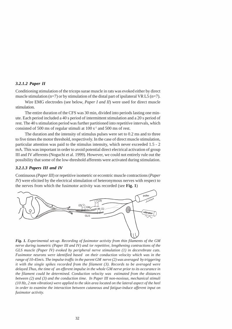
32
3.2.1.2 Paper II
Conditioning stimulation of the triceps surae muscle in rats was evoked either by directmuscle stimulation (n=7) or by stimulation of the distal part of ipsilateral VR L5 (n=7).
Wire EMG electrodes (see below, Paper I and II) were used for direct musclestimulation.
The entire duration of the CFS was 30 min, divided into periods lasting one min-ute. Each period included a 40 s period of intermittent stimulation and a 20 s period ofrest. The 40 s stimulation period was further partitioned into repetitive intervals, whichconsisted of 500 ms of regular stimuli at 100 s-1 and 500 ms of rest.
The duration and the intensity of stimulus pulses were set to 0.2 ms and to threeto five times the motor threshold, respectively. In the case of direct muscle stimulation,particular attention was paid to the stimulus intensity, which never exceeded 1.5 - 2mA. This was important in order to avoid potential direct electrical activation of groupIII and IV afferents (Noguchi et al. 1999). However, we could not entirely rule out thepossibility that some of the low-threshold afferents were activated during stimulation.
3.2.1.3 Papers III and IV
Continuous (Paper III) or repetitive isometric or eccentric muscle contractions (PaperIV) were elicited by the electrical stimulation of heteronymous nerves with respect tothe nerves from which the fusimotor activity was recorded (see Fig. 1)
Fig. 1. Experimental set-up. Recording of fusimotor activity from thin filaments of the GMnerve during isometric (Paper III and IV) and /or repetitive, lengthening contractions of theGLS muscle (Paper IV) evoked by peripheral nerve stimulation (1) in decerebrate cats.Fusimotor neurons were identified based on their conduction velocity which was in therange of 10-45m/s. The impulse traffic in the parent GM nerve (2) was averaged by triggeringit with the single spikes recorded from the filament (3). Records to be averaged weredelayed.Thus, the time of an efferent impulse in the whole GM nerve prior to its occurance inthe filament could be determined. Conduction velocity was estimated from the distancesbetween (2) and (3) and the conduction time. In Paper III non-noxious, mechanical stimuli(10 Hz, 2 mm vibration) were applied to the skin area located on the lateral aspect of the heelin order to examine the interaction between cutaneous and fatigue-induce afferent input onfusimotor activity.
vibrator
(1)(2)
(3)
GLS
GM
puller
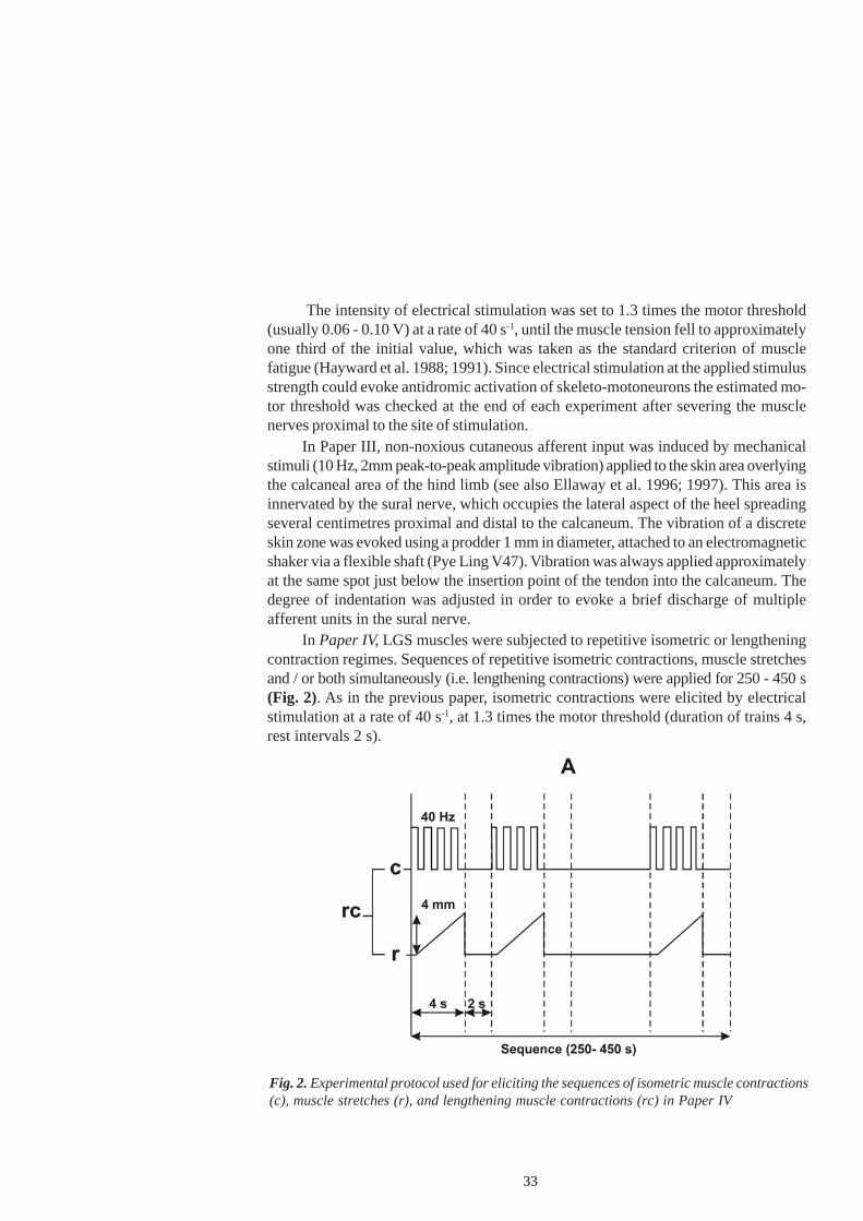
33
The intensity of electrical stimulation was set to 1.3 times the motor threshold(usually 0.06 - 0.10 V) at a rate of 40 s-1, until the muscle tension fell to approximatelyone third of the initial value, which was taken as the standard criterion of musclefatigue (Hayward et al. 1988; 1991). Since electrical stimulation at the applied stimulusstrength could evoke antidromic activation of skeleto-motoneurons the estimated mo-tor threshold was checked at the end of each experiment after severing the musclenerves proximal to the site of stimulation.
In Paper III, non-noxious cutaneous afferent input was induced by mechanicalstimuli (10 Hz, 2mm peak-to-peak amplitude vibration) applied to the skin area overlyingthe calcaneal area of the hind limb (see also Ellaway et al. 1996; 1997). This area isinnervated by the sural nerve, which occupies the lateral aspect of the heel spreadingseveral centimetres proximal and distal to the calcaneum. The vibration of a discreteskin zone was evoked using a prodder 1 mm in diameter, attached to an electromagneticshaker via a flexible shaft (Pye Ling V47). Vibration was always applied approximatelyat the same spot just below the insertion point of the tendon into the calcaneum. Thedegree of indentation was adjusted in order to evoke a brief discharge of multipleafferent units in the sural nerve.
In Paper IV, LGS muscles were subjected to repetitive isometric or lengtheningcontraction regimes. Sequences of repetitive isometric contractions, muscle stretchesand / or both simultaneously (i.e. lengthening contractions) were applied for 250 - 450 s(Fig. 2). As in the previous paper, isometric contractions were elicited by electricalstimulation at a rate of 40 s-1, at 1.3 times the motor threshold (duration of trains 4 s,rest intervals 2 s).
Fig. 2. Experimental protocol used for eliciting the sequences of isometric muscle contractions(c), muscle stretches (r), and lengthening muscle contractions (rc) in Paper IV
34
Repetitive 4-mm LGS muscle ramp stretches, at a velocity of 1 mm/s, wereproduced by a servo-controlled electromechanical puller (Pye Ling 706) and applied at6-s intervals, alone or simultaneously with muscle contractions. To provide adequatemuscle tension before lengthening, the stretches started slightly after the stimulation(Hesselink et al. 1996).
The described pattern of repetitive lengthening muscle contractions was chosenin order to simulate muscle activity in natural conditions. The amplitude of the stretches(but not their velocity) was kept in the physiological range, similar to that occurring inlocomotion (Bonasera and Nichols 1994). However, in these experiments, at the endof the fatiguing protocol, muscle tension fell to less than 30% of its initial value. Thus,according to the afore-mentioned criterion, muscles could not have been consideredfatigued, in spite of their long-lasting activity.
3.2.1.4 Paper V
Conditioning fatiguing contractions of the whole triceps surae muscle were evoked bylong-lasting intermittent stimulation of the distal part of ipsilateral VR S1. Stimulationperiods of 20 s, or in several experiments of 10 s duration, with repetitive regularstimuli occurring at a rate of 50 s-1, were separated by rest intervals of 10 s duration.The entire duration of the fatiguing stimulation (FST) was 10 or 12 minutes. The durationof stimulus pulses was 0.2 ms; their intensity was set at 1.3 times the motor threshold.The drop in tension during individual stimulation periods increased prominently overtime, reaching 25 - 30% of the initial value at the end of the stimulation sequence.
3.2.1.5 Paper VI
The experiments were performed on cats anaesthetized with α-chloraloze (Sigma, 50mgkg-1 i.p.). Capsaicin (5 mg, Sigma) dissolved in 1 ml of Tween 80 (7%) and sterileisotonic saline (93%) were injected in the treatment group of cats (n = 6), while thecontrol group (n=3) received only the vehicle (Tween 80 and saline). The intramuscularinjections (IMI) were made ipsilaterally, on the right side, through a thin needle insertedinto several spots over the rostral parts of the trapezius and splenius muscles (100 mlinto each of the 10 sites in the rostro-caudal extent).
3.2.2 Force and EMG recordingA force transducer, with a compliance of 20 - 50 mmN-1 and a resonance frequencyclose to 1.5 kHz was used to record muscle force (Papers I, II, III, IV and V).Additionally, in Paper IV, muscle length changes were recorded using a displacementtransducer (Tektronix 015-0168-00), the output of which also served as the feedbacksignal for the stretching system.
EMG signals were recorded separately from the GM and the GL, or the soleusmuscle (Paper I), or from the whole triceps surae muscle (Paper II) using pairs ofintramuscular wire electrodes. The electrodes were prepared from lacquered silverwires, 0.15 mm in diameter. The wires were introduced into a hypodermic injection
35
needle, their insulation was removed from a length of about 2 mm from the end and thebare ends were bent close to the needle end. Two such needles were inserted into themuscle belly to a depth of about 4 - 5 mm, whereupon the needles were withdrawn.The wires remained stuck and effectively fixed inside the muscle. The electrodeswere inserted in the middle part of the muscle, along the longitudinal axes at a distanceof about one third of the entire muscle length measured from the calcaneus. The wireelectrodes were connected to the isolated input units of Brownlee model 440 amplifiers(Brownlee Precision Co, USA). The amplifier frequency bandwidth was set to a rangeof 10 Hz - 7.5 kHz.
3.2.3 Recording of the fusimotor activity (Papers III and IV)Spike discharges of single fusimotor neurons were recorded from thin filaments, cutout from the intact GM nerve and dissected free from de-sheathed fascicles. Fusimotorneurons were identified based on the conduction velocity which was in the range of 10- 45 m/s. In order to estimate the conduction velocity the back-averaging methoddeveloped by Bostock and Sears (1976) was used in the following way: the impulsetraffic in the parent nerve was averaged (128 sweeps), by triggering it with the singlespikes recorded from the filament. Records to be averaged were usually delayed by 5ms. Thus, the time of occurrence of an efferent impulse in the whole GM nerve priorto its occurrence in the filament could be determined. Conduction velocity was estimatedfrom the distance between the two recording sites (one on the whole nerve and theother on the dissected filament) and the conduction time.
3.2.4 Recording the monosynaptic reflex (MR) and dorsal root potential(DRP) (Paper V)In order to record monosynaptic reflexes and to elicit recurrent inhibition, the VR L7was cut at the maximal possible distance from the point of its exit from the spinal cordand divided into five to seven filaments. From one of the filaments MSRs were recorded,while one of the other filaments was stimulated in order to evoke recurrent inhibition.It was possible to elicit MSRs with only one stimulus applied to the GS nerve, but suchrecords were usually unstable during the long-lasting test procedure. For that reason,on most occasions, MSRs were evoked by pairs of stimuli at 2 ms intervals. Theduration of the stimuli was 0.2 ms. Current intensity was adjusted to evoke MSRs inthe range of 60 - 70% of their maximal amplitude and not to exceed 1.3 - 1.4 times thethreshold for the appearance of an afferent wave in the cord dorsum potential (CDP)after single pulse stimulation.
To record dorsal root potentials (DRPs) a small filament was carefully dissectedfrom the most caudal rootlet of the L7 dorsal root up to its entry into the cord andtranssected 20 mm from the entry zone. The central end of the filament was placed ona bipolar hook electrode (distance between the leads was 10 mm). One lead waspositioned 1 mm from the cord and the other on the cut end of the root. The experi-mental set-up is presented in Fig. 3.
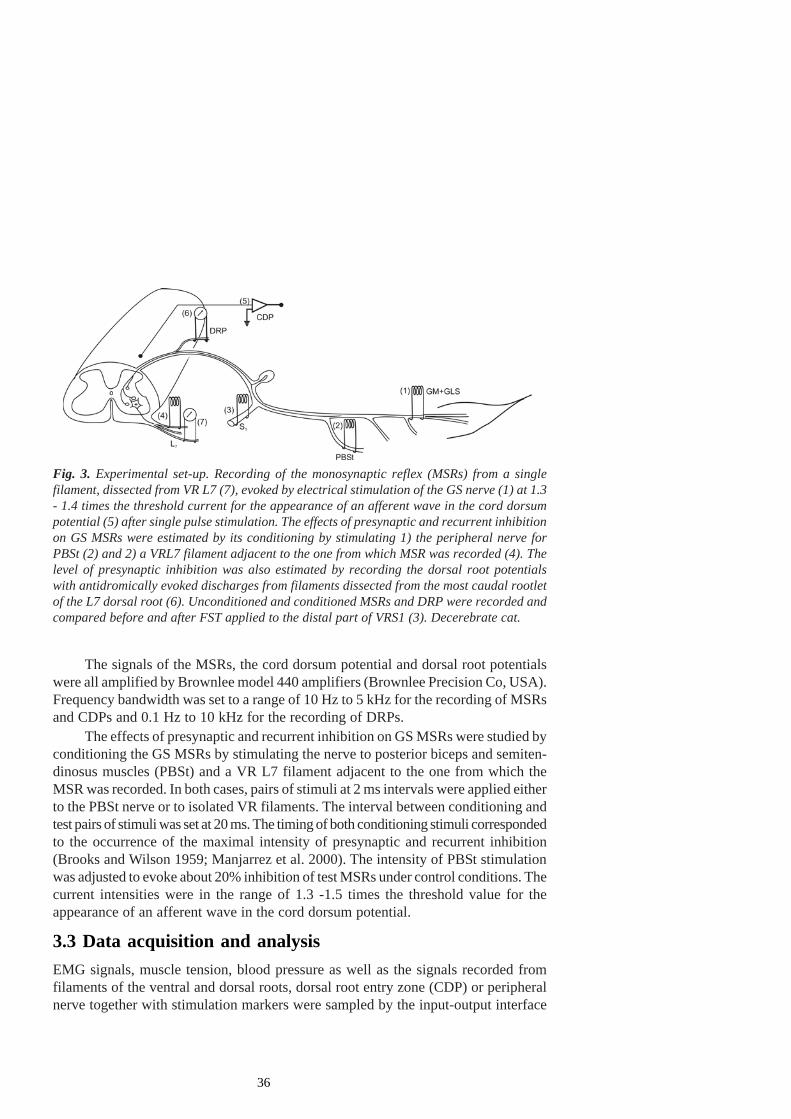
36
The signals of the MSRs, the cord dorsum potential and dorsal root potentialswere all amplified by Brownlee model 440 amplifiers (Brownlee Precision Co, USA).Frequency bandwidth was set to a range of 10 Hz to 5 kHz for the recording of MSRsand CDPs and 0.1 Hz to 10 kHz for the recording of DRPs.
The effects of presynaptic and recurrent inhibition on GS MSRs were studied byconditioning the GS MSRs by stimulating the nerve to posterior biceps and semiten-dinosus muscles (PBSt) and a VR L7 filament adjacent to the one from which theMSR was recorded. In both cases, pairs of stimuli at 2 ms intervals were applied eitherto the PBSt nerve or to isolated VR filaments. The interval between conditioning andtest pairs of stimuli was set at 20 ms. The timing of both conditioning stimuli correspondedto the occurrence of the maximal intensity of presynaptic and recurrent inhibition(Brooks and Wilson 1959; Manjarrez et al. 2000). The intensity of PBSt stimulationwas adjusted to evoke about 20% inhibition of test MSRs under control conditions. Thecurrent intensities were in the range of 1.3 -1.5 times the threshold value for theappearance of an afferent wave in the cord dorsum potential.
3.3 Data acquisition and analysisEMG signals, muscle tension, blood pressure as well as the signals recorded fromfilaments of the ventral and dorsal roots, dorsal root entry zone (CDP) or peripheralnerve together with stimulation markers were sampled by the input-output interface
Fig. 3. Experimental set-up. Recording of the monosynaptic reflex (MSRs) from a singlefilament, dissected from VR L7 (7), evoked by electrical stimulation of the GS nerve (1) at 1.3- 1.4 times the threshold current for the appearance of an afferent wave in the cord dorsumpotential (5) after single pulse stimulation. The effects of presynaptic and recurrent inhibitionon GS MSRs were estimated by its conditioning by stimulating 1) the peripheral nerve forPBSt (2) and 2) a VRL7 filament adjacent to the one from which MSR was recorded (4). Thelevel of presynaptic inhibition was also estimated by recording the dorsal root potentialswith antidromically evoked discharges from filaments dissected from the most caudal rootletof the L7 dorsal root (6). Unconditioned and conditioned MSRs and DRP were recorded andcompared before and after FST applied to the distal part of VRS1 (3). Decerebrate cat.
37
device CED Power 1401 (Cambridge Electronic Design, UK), while the Spike 2software (Cambridge Electronics Department, UK) was used for data acquisition andfurther processing. Input signals were digitised with 12-bit resolution at the rates of 1kHz for the tension and blood pressure (Paper I, II, III, I V), 10 kHz for the monosynapticreflex and cord dorsum potentials (CDP, Paper V and VII) and 15 kHz for the EMG(Paper I) and dorsal root potential with antidromic spikes (DRP, Paper V).
Fusimotor discharges were recorded before (50 - 100 s), during (250 - 450 s) andafter muscle contraction, and together with the tension recordings (Papers III and IV)stored on a magnetic tape and / or analysed on-line using a Hewlett-Packard 9817computer. The action potentials were converted to voltage steps, which were sampledat 20 ms intervals. The output signal from the tension transducer was amplified, storedon a magnetic tape and A/D converted (sampling interval 80 ms) simultaneously withfusimotor discharge.
Further analysis was performed off-line. Data analysis and diagram plotting wasperformed using the Origin 6.0 or 7.0 software (OriginLab Corporation, USA), whilethe statistical analyses were performed using the SPSS 10.0 for Windows (SPSS Inc.,USA).
3.3.1 Paper IQuantitative analysis of pre-fatigue and post-fatigue test responses (120 unfusedisometric contractions evoked by 10 s-1 regular stimulation in each test or control reaction)included the calculation of tension peak amplitudes and M-wave amplitudes. Meansand statistical deviations of peak tensions and M-wave amplitudes of post-fatigueresponses were normalized to the corresponding pre-fatigue mean values. Statisticallysignificant differences of both parameters compared to their resting values, as well astheir mutual difference, were examined using two-population Student’s t-test.
3.3.2 Papers III and IVQuantitative analysis of fusimotor discharges, recorded at distinct time intervals wasas follows: Mean value and standard deviation of the spontaneous activity was calculatedfrom the counts of spikes per second intervals from a 100-s period immediately precedingthe beginning of stimulation. In Paper IV the amount of change, when present, wasestimated by calculating the number of action potentials above the spontaneous firingrate during the whole stimulation period. The differences between the area underneaththe firing rate curve during the stimulation periodand the area underneath the meanspontaneous firing rate were taken as the responses to the applied stimuli. Statisticalsignificance of the increase in the number of spikes during sequences of contractions,as well as their mutual differences, were examined using Student’s paired t-test. Withsome modifications, a similar method was applied in Paper III.
38
3.3.3 Paper VStatistical significance of post-fatigue changes in MSR amplitude and in the intensityof presynaptic and recurrent inhibition was determined by repeated measures analysisof variance (ANOVA). Time interval of measurement (prefatigue - pre-test versuspost1 test versus post2 test etc.) was used as the within-subject factor. Provided thatthe changes of parameters under study were significant in successive time intervals,post hoc comparisons were performed using the Bonfferoni adjustment for multiplecomparisons.
3.4 Fos immunohistochemistry and NADPH-d histochemistry(Papers II and VI)
3.4.1PerfusionThe rats were perfused after the end of the fatiguing stimulation i.e. two hours afterdirect muscle stimulation or eight hours after lumbar laminectomy, when the fatigue oftriceps surae was evoked by VR stimulation (Paper II). The cats were perfused twohours after i.m. capsaicin injection (Paper VI). Perfusion protocol was the following:
At first, control and test group of animals were additionally anaesthetized withsodium-pentobarbital (Sigma 70 - 75 mg kg-1 i.p.) The animals were then perfusedthrough the ascending aorta, at first with phosphate-buffered saline (PBS, pH=7.4)containing heparin (25 000 units/l) and sodium nitrite (0.2%), and further with 4%paraformaldehyde in a 0.1 M phosphate buffer (pH 7.4). The spinal cord was isolated,lumbar L1-L6 segments of rats (Paper II) and cervical (C1-C8) and lumbar (L1-L7)segments of cats (Paper V) were post-fixed in the same fixative overnight andcryoprotected in phosphate-buffered 10% and 30% sucrose at 4°C for 48 h.
Frontal frozen sections of the spinal cord (30 µm thick in Paper II or 40 µm inPaper V) were cut. About 50 - 80 sections from each spinal segment were collected inwells, filled with PBS and subjected to further immunohystochemical and hystochemicalprocedure.
3.4.2 Fos-immunohistochemistryFor the visualisation of Fos-ir neurons in different spinal layers (laminas) of ipsi- andcontralateral sides to the stimulated (treated) one, the avidin-biotin-peroxidase techniquewas used (Hsu et al, 1981), using a rabbit polyclonal antibody directed against the c-Fos protein (Oncogene Research, Ab-5, USA), and a commercial kit (ABC; Vector,PK 4002, USA). After several rinses in 0.01 M PBS, the sections were incubated inPBS with 3% normal goat serum (NGS, Sigma) and 0.3% Triton-X-100 (Sigma) for 30minutes at room temperature. Free-floating sections were then placed in primary rabbitantiserum against the Fos protein, diluted (1:2000) in PBS containing 3% NGS and0.4% Triton X-100 for 48 h at 4°C. The sections were further incubated in biotinylated
39
goat anti-rabbit immunoglobulin G (1:200) and avidin-biotin-peroxidase complex (ABC)using the standard method. In order to obtain black-purple reaction product, the sectionswere reacted in 0.05 M Tris-buffer (pH = 7,4), containing chromogen 3,3’ diamino-benzidine tetrahydrochloride (DAB, Sigma), 0.2% nickel-ammonium-sulphate andsubstrate 0,005% hydrogen peroxide for 10 minutes. Fos-ir nuclei were detected asblack nuclei. The sections were washed in PBS to block staining reactions, mountedon gelatine-subbed slides, air-dried, cleared in xylene and cover-slipped with Entellan.
3.4.3 NADPH-d histochemistryTo evaluate the possibility of double-labelling of Fos-immunoreactivity and NADPH-dreactivity in the neurons, one half of all Fos-immuno-stained sections from each particularsegment were further incubated in a 0.1 M phosfate buffer (pH 7.4), containing 0.3%Triton X-100, 0.1 mg/ml nitroblue tetrazolium (Sigma) and 0.5 mg/ml β-NADPH te-tra-sodium salt, reduced form, at 37 °C for 30 - 60 min, according to the standardmethod of Vincent and Kimura (1992). All NADPH-d reactive neurons were detectedas light-blue cells with unstained nuclei.
Following the reaction, the sections were rinsed in phosphate buffer saline (pH7.4) and mounted on gelatine-subbed slides, air-dried, immersed in 100% alcohol, clearedin xylene and cover-slipped with Entellan.
3.4.4 Statistical analysisThe data reported here were obtained from every fifth section in the rostrocaudalextent. Up to 10 (rats) and 20 (cats) sections were taken from each particular spinalsegment and analysed. The mean number (± SEM) of Fos-ir neurons per section ofthe same group was counted in laminae I-X of the grey matter bilaterally, comparedand analysed using Student’s t-test for paired data (Paper II) or the ANOVA (PaperVI). The differences between the compared values were taken as statistically significantat p<0.01.
40
4. RESULTS
4.1 Paper IIn most mammalian muscles (including the cat GM muscle), muscle fibres of differentmotor units intermingle, so that the fibres belonging to different motor units may lieclose to each other. Such intramuscular architecture allows that muscle fibres of asingle motor unit do not function as isolated entities, but may actually influence eachother. Conditioning fatiguing stimulation (CFS) was applied to multiple motor units ofthe GM muscle of cats in order to assess whether their activation may affect neighbouring,inactive units. CFS was evoked by distributed stimulation of usually four to five fila-ments, originating from ipsilateral L7 or S1 ventral roots. For the test reaction, lowfrequency stimulation with a frequency of 10 s-1 and duration of 12 s was applied on asingle filament, dissected from the same ventral roots. To exclude somato-sympatheticreflexes during CFS, a full rhizotomy of the lumbo-sacral spinal cord was performed.
An example of the experimental protocol is presented in Fig. 4. Test EMG andtension were recorded from the GM, whereas control EMG and tension were recordedfrom the GL. The test (T) and control (C) stimulations were performed before theCFS (T1, C1) and following its cessation, at distinct time intervals (T2, C2; T3, C3) inorder to reveal recovery processes. During CFS the tetanic tension and the EMG ofthe GM muscle declined prominently. Test reactions changed markedly and a clear-cutdrop in tension records of the GM could be observed at distinct time intervals, e.g.from T1 to T2 and further on to T3. Twitch contractions evoked by a single stimulusdeclined from T1 to T2 and from T2 to T3. In the pre-fatigue test, the twitch thatfollowed the low-frequency train was potentiated compared to the twitch precedingthe stimulation. This potentiation effect disappeared immediately after the CFS (T2)and partly recovered later on (T3). At the same time, in T2 and T3, post-twitchessignificantly exceeded the force amplitudes at the end of repetitive 10 s-1 stimulation.Low-rate stimulation (10 s-1) usually evoked non-fused or partly fused contractions ofthe GM muscle. The steady-state amplitudes of the low-frequency contractions (afterthe initial fall of twitch amplitudes at the onset of stimulation) were prominently reducedafter the CFS (see T1 vs. T2 in Fig. 4B, lower row), followed by their gradual recoveryseveral minutes later (T3). EMGs of the test reactions (Fig. 4B, upper row) werereduced following the CFS (T2) and recovered almost completely after a rest period(T3).On the contrary, only minor changes in both the EMG and the tension occurred inthe GL muscle (compare C1, C2 and C3 in Fig. 4B).
4.1.1Quantitative description of changes in EMG and tensionQuantitative analysis of the changes in EMG and tension across different experimentsis presented in Fig. 5.
In the initial five experiments, the parameters of CFS that could evoke noticeabledepression in the test reactions were defined (Fig. 5, A-C). Neither the tension, nor
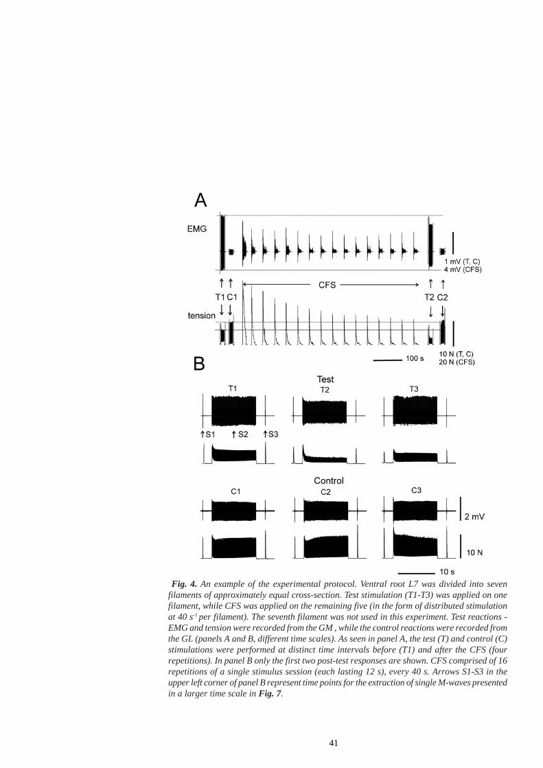
41
Fig. 4. An example of the experimental protocol. Ventral root L7 was divided into sevenfilaments of approximately equal cross-section. Test stimulation (T1-T3) was applied on onefilament, while CFS was applied on the remaining five (in the form of distributed stimulationat 40 s-1 per filament). The seventh filament was not used in this experiment. Test reactions -EMG and tension were recorded from the GM , while the control reactions were recorded fromthe GL (panels A and B, different time scales). As seen in panel A, the test (T) and control (C)stimulations were performed at distinct time intervals before (T1) and after the CFS (fourrepetitions). In panel B only the first two post-test responses are shown. CFS comprised of 16repetitions of a single stimulus session (each lasting 12 s), every 40 s. Arrows S1-S3 in theupper left corner of panel B represent time points for the extraction of single M-waves presentedin a larger time scale in Fig. 7.

42
Fig. 5. Quantitative changes in test (T1, T2…) and control (C1,C2…) reactions, recordedfrom the GM and GL muscles before (T1,C1) and after (T2,C2; T3,C3 etc) the fatiguing run. Inthe first series of experiments, only the test reactions of the GM were assessed, as presented inpanels A - C. Panels D - H display the data with control contractions. Data points andvertical bars represent the mean and the standard deviation of 120 contractions during teststimulation (12 s duration, 10 s-1). The tension peaks are indicated by squares and solid linesand the EMG amplitudes are shown as circles and dashed lines, respectively. The test andcontrol reactions are labelled using filled and unfilled markers, respectively. Changes inEMG and tension are normalized to estimated mean values before the CFS (pre-test values).The parameters of CFS for each particular experimental protocol were: A) 10 s-1, 4 filaments,17 repetitions B) 40 s-1, 4 filaments, 16 repetitions C) 40 s-1, 5 filaments, 16 repetitions D) 40s-1, 5 filaments, 16 repetitions E) 40 s-1, 5 filaments, 24 repetitions F) 40 s-1, 5 filaments, 17repetitions G) 40 s-1, five filaments, 15 repetitions H) 40 s-1, five filaments, 16 repetitions.
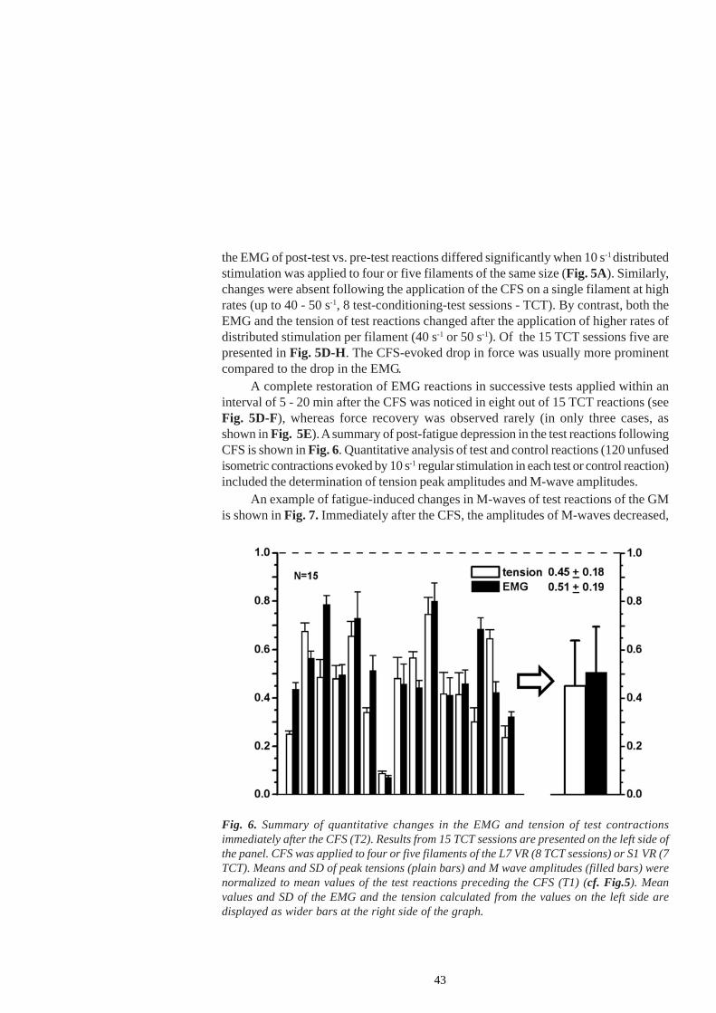
43
Fig. 6. Summary of quantitative changes in the EMG and tension of test contractionsimmediately after the CFS (T2). Results from 15 TCT sessions are presented on the left side ofthe panel. CFS was applied to four or five filaments of the L7 VR (8 TCT sessions) or S1 VR (7TCT). Means and SD of peak tensions (plain bars) and M wave amplitudes (filled bars) werenormalized to mean values of the test reactions preceding the CFS (T1) (cf. Fig.5). Meanvalues and SD of the EMG and the tension calculated from the values on the left side aredisplayed as wider bars at the right side of the graph.
the EMG of post-test vs. pre-test reactions differed significantly when 10 s-1 distributedstimulation was applied to four or five filaments of the same size (Fig. 5A). Similarly,changes were absent following the application of the CFS on a single filament at highrates (up to 40 - 50 s-1, 8 test-conditioning-test sessions - TCT). By contrast, both theEMG and the tension of test reactions changed after the application of higher rates ofdistributed stimulation per filament (40 s-1 or 50 s-1). Of the 15 TCT sessions five arepresented in Fig. 5D-H. The CFS-evoked drop in force was usually more prominentcompared to the drop in the EMG.
A complete restoration of EMG reactions in successive tests applied within aninterval of 5 - 20 min after the CFS was noticed in eight out of 15 TCT reactions (seeFig. 5D-F), whereas force recovery was observed rarely (in only three cases, asshown in Fig. 5E). A summary of post-fatigue depression in the test reactions followingCFS is shown in Fig. 6. Quantitative analysis of test and control reactions (120 unfusedisometric contractions evoked by 10 s-1 regular stimulation in each test or control reaction)included the determination of tension peak amplitudes and M-wave amplitudes.
An example of fatigue-induced changes in M-waves of test reactions of the GMis shown in Fig. 7. Immediately after the CFS, the amplitudes of M-waves decreased,
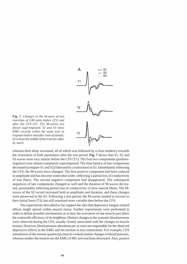
44
whereas their delay increased, all of which was followed by a clear tendency towardsthe restoration of both parameters after the rest period. Fig. 7 shows that S1, S2 andS3 waves were very similar before the CFS (T1). The first two components (positive-negative) were almost completely superimposed. The time latency of late componentsdecreased (compare S1 and S2) followed by a restoration in S3. Immediately followingthe CFS, the M-waves have changed. The first positive component had been reducedin amplitude and has become somewhat wider, reflecting a partial loss of conductivityof fast fibres. The second negative component had disappeared. The subsequentsequences of late components changed as well and the duration of M-waves decrea-sed, presumably reflecting partial loss of conductivity of slow muscle fibres. The M-waves of the S2 record increased both in amplitude and duration, and these changeswere preserved in the S3. Following a rest period, the M-waves tended to recover totheir initial form (T3), but still remained more variable then before the CFS.
The experiments described so far support the idea that depressive fatigue-relatedeffects might spread within muscle tissue. Further experiments were performed inorder to define possible mechanisms as to how the activation of one muscle part altersthe contractile efficiency of its neighbour. Distinct changes in the systemic blood pressurewere observed during the CFS, usually closely associated with the changes in muscletension. However, blood pressure alterations per se were not responsible for the observeddepressive effects in the EMG and the tension in test contractions. For example, CFSstimulation of the remote quadriceps muscle evoked similar changes in blood pressure,whereas neither the tension nor the EMG of MG test reactions decreased. Also, passive
Fig. 7. Changes in the M-wave of testreactions of GM units before (T1) andafter the CFS (T2, T3). M-waves areshown superimposed. S1 and S3 showEMG records within the same tests inresponse before and after train of stimuli;S2 is from the middle of the train (6 s afterits start).

45
squeezing of the belly of the GM muscle at a pressure which did not exceed thesystolic blood pressure of 120 mm Hg or the occlusion of the popliteal artery failed toinduce concomitant changes in EMG and tension, as observed by high-rate distributedCFS. Thus, an increase in intramuscular pressure during tetanic contractions and localhypoxia could not fully account for the obtained results and the involvement of otherpossible mechanisms underlying the observed effects was suggested (see original paper).
Subsequent experiments were devoted to the studies of spinal action of small-diameter afferent fibres elicited by muscle fatigue, and their role in the adjustment ofspinal motor output. Paper II described changes in the lamellar distribution of spinalinterneurons in the lumbar spinal segments of the rat, evoked by fatiguing long-lastingmuscle contractions. Possible contributions of selected spinal neuronal systems toskeleto-motoneuronal adaptation to muscle fatigue were then explored.Changes in theactivity of fusimotor neurons during muscle fatigue, with and without concomitant in-put from other afferent sources (e.g. skin)(Paper IV) and in different regimes ofmuscle activity (continuous, intermittent isometric vs. eccentric contractions)(PaperIII ) were estimated . In Paper V a quantitative estimate of the magnitude, time dynamicsand the direction of changes of the activity of skeleto-motoneurons and some of theinterneurons that mediate presynaptic inhibition or recurrent inhibition following long-lasting fatiguing contractions was performed.
4.2.Paper IIPaper II describes changes in the lamellar distribution of spinal interneurons in thelumbar spinal segments of the rat, activated by fatiguing long-lasting muscle contraction.
4.2.1 Dynamic of triceps surae muscle fatigue developmentIn order to quantify the development of fatigue of the triceps surae muscle evoked byhigh-rate intermittent stimulation (100 s-1), test stimuli (10 s-1 over 12s) were appliedbefore and after a 30-min long-lasting fatiguing run. After the fifth fatiguing session thetension declined to 50%, whereas after the 30th session it declined to 70% of its initialvalue. This drop in tension was followed by a corresponding drop in the EMG. Anexample of the fatiguing protocol is presented in Fig. 8.
4.2.2Fos-immunoreactivity and NADPH-d reactivity after fatiguing stimulationFollowing fatiguing muscle contraction induced by direct or VRL5 stimulation, thenumber of Fos-ir neurons and the pattern of their distribution have changed. Fatigue-associated c-fos gene expression was more extensive in the L2-L5 segments on thestimulated side. Immunoreactive neurons were mainly noticable in the dorsal half ofthe spinal grey matter and only occasionally recorded in the ventral horn.
After direct muscle stimulation the number of Fos-ir neurons increased ipsilaterallyin the outer part of layer 2 (2o), in the nucleus proprius (layers 3 and 4) and in the neckof the dorsal horn at the L4/L5 level. In the L2 and L3 segments, while Fos-ir was not
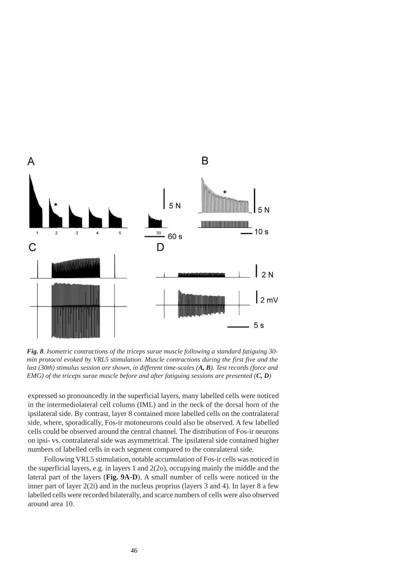
46
Fig. 8. Isometric contractions of the triceps surae muscle following a standard fatiguing 30-min protocol evoked by VRL5 stimulation. Muscle contractions during the first five and thelast (30th) stimulus session are shown, in different time-scales (A, B). Test records (force andEMG) of the triceps surae muscle before and after fatiguing sessions are presented (C, D)
expressed so pronouncedly in the superficial layers, many labelled cells were noticedin the intermediolateral cell column (IML) and in the neck of the dorsal horn of theipsilateral side. By contrast, layer 8 contained more labelled cells on the contralateralside, where, sporadically, Fos-ir motoneurons could also be observed. A few labelledcells could be observed around the central channel. The distribution of Fos-ir neuronson ipsi- vs. contralateral side was asymmetrical. The ipsilateral side contained highernumbers of labelled cells in each segment compared to the conralateral side.
Following VRL5 stimulation, notable accumulation of Fos-ir cells was noticed inthe superficial layers, e.g. in layers 1 and 2(2o), occupying mainly the middle and thelateral part of the layers (Fig. 9A-D). A small number of cells were noticed in theinner part of layer 2(2i) and in the nucleus proprius (layers 3 and 4). In layer 8 a fewlabelled cells were recorded bilaterally, and scarce numbers of cells were also observedaround area 10.
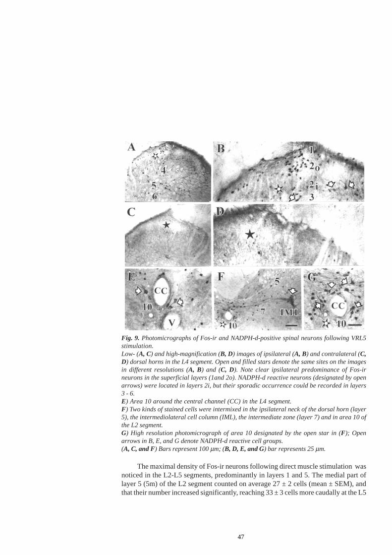
47
Fig. 9. Photomicrographs of Fos-ir and NADPH-d-positive spinal neurons following VRL5stimulation.Low- (A, C) and high-magnification (B, D) images of ipsilateral (A, B) and contralateral (C,D) dorsal horns in the L4 segment. Open and filled stars denote the same sites on the imagesin different resolutions (A, B) and (C, D). Note clear ipsilateral predominance of Fos-irneurons in the superficial layers (1and 2o). NADPH-d reactive neurons (designated by openarrows) were located in layers 2i, but their sporadic occurrence could be recorded in layers3 - 6.E) Area 10 around the central channel (CC) in the L4 segment.F) Two kinds of stained cells were intermixed in the ipsilateral neck of the dorsal horn (layer5), the intermediolateral cell column (IML), the intermediate zone (layer 7) and in area 10 ofthe L2 segment.G) High resolution photomicrograph of area 10 designated by the open star in (F); Openarrows in B, E, and G denote NADPH-d reactive cell groups.(A, C, and F) Bars represent 100 µm; (B, D, E, and G) bar represents 25 µm.
The maximal density of Fos-ir neurons following direct muscle stimulation wasnoticed in the L2-L5 segments, predominantly in layers 1 and 5. The medial part oflayer 5 (5m) of the L2 segment counted on average 27 ± 2 cells (mean ± SEM), andthat their number increased significantly, reaching 33 ± 3 cells more caudally at the L5
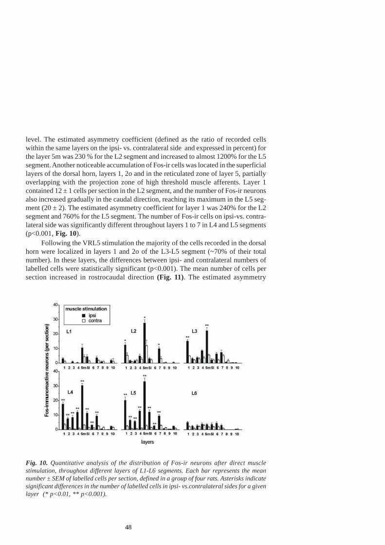
48
Fig. 10. Quantitative analysis of the distribution of Fos-ir neurons after direct musclestimulation, throughout different layers of L1-L6 segments. Each bar represents the meannumber ± SEM of labelled cells per section, defined in a group of four rats. Asterisks indicatesignificant differences in the number of labelled cells in ipsi- vs.contralateral sides for a givenlayer (* p<0.01, ** p<0.001).
level. The estimated asymmetry coefficient (defined as the ratio of recorded cellswithin the same layers on the ipsi- vs. contralateral side and expressed in percent) forthe layer 5m was 230 % for the L2 segment and increased to almost 1200% for the L5segment. Another noticeable accumulation of Fos-ir cells was located in the superficiallayers of the dorsal horn, layers 1, 2o and in the reticulated zone of layer 5, partiallyoverlapping with the projection zone of high threshold muscle afferents. Layer 1contained 12 ± 1 cells per section in the L2 segment, and the number of Fos-ir neuronsalso increased gradually in the caudal direction, reaching its maximum in the L5 seg-ment (20 ± 2). The estimated asymmetry coefficient for layer 1 was 240% for the L2segment and 760% for the L5 segment. The number of Fos-ir cells on ipsi-vs. contra-lateral side was significantly different throughout layers 1 to 7 in L4 and L5 segments(p<0.001, Fig. 10).
Following the VRL5 stimulation the majority of the cells recorded in the dorsalhorn were localized in layers 1 and 2o of the L3-L5 segment (~70% of their totalnumber). In these layers, the differences between ipsi- and contralateral numbers oflabelled cells were statistically significant (p<0.001). The mean number of cells persection increased in rostrocaudal direction (Fig. 11). The estimated asymmetry
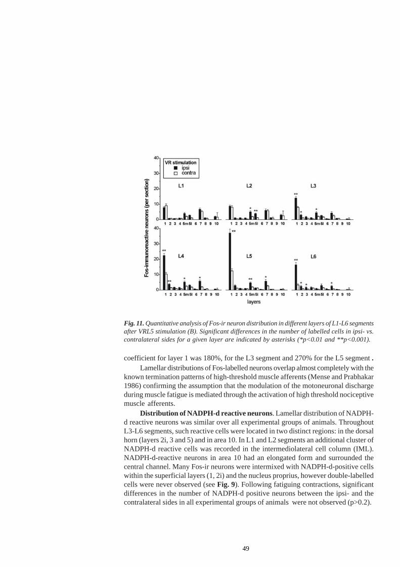
49
Fig. 11. Quantitative analysis of Fos-ir neuron distribution in different layers of L1-L6 segmentsafter VRL5 stimulation (B). Significant differences in the number of labelled cells in ipsi- vs.contralateral sides for a given layer are indicated by asterisks (*p<0.01 and **p<0.001).
coefficient for layer 1 was 180%, for the L3 segment and 270% for the L5 segment .Lamellar distributions of Fos-labelled neurons overlap almost completely with the
known termination patterns of high-threshold muscle afferents (Mense and Prabhakar1986) confirming the assumption that the modulation of the motoneuronal dischargeduring muscle fatigue is mediated through the activation of high threshold nociceptivemuscle afferents.
Distribution of NADPH-d reactive neurons. Lamellar distribution of NADPH-d reactive neurons was similar over all experimental groups of animals. ThroughoutL3-L6 segments, such reactive cells were located in two distinct regions: in the dorsalhorn (layers 2i, 3 and 5) and in area 10. In L1 and L2 segments an additional cluster ofNADPH-d reactive cells was recorded in the intermediolateral cell column (IML).NADPH-d-reactive neurons in area 10 had an elongated form and surrounded thecentral channel. Many Fos-ir neurons were intermixed with NADPH-d-positive cellswithin the superficial layers (1, 2i) and the nucleus proprius, however double-labelledcells were never observed (see Fig. 9). Following fatiguing contractions, significantdifferences in the number of NADPH-d positive neurons between the ipsi- and thecontralateral sides in all experimental groups of animals were not observed (p>0.2).
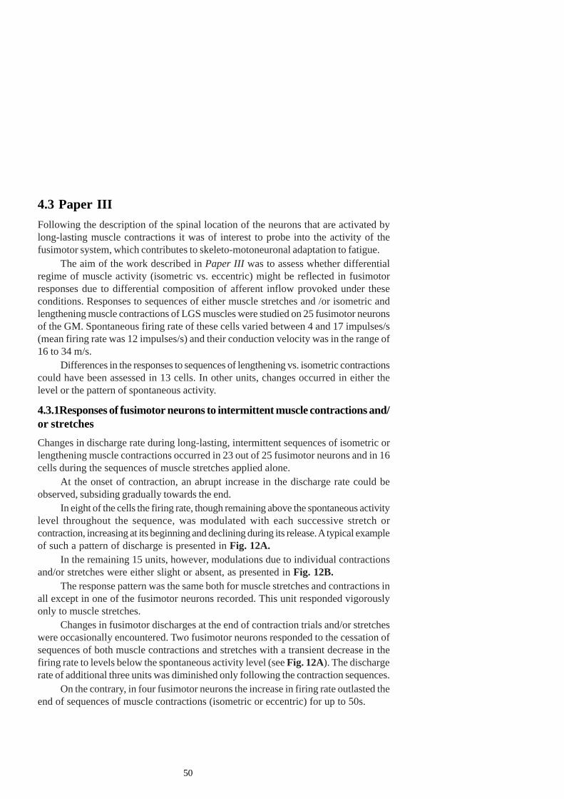
50
4.3 Paper IIIFollowing the description of the spinal location of the neurons that are activated bylong-lasting muscle contractions it was of interest to probe into the activity of thefusimotor system, which contributes to skeleto-motoneuronal adaptation to fatigue.
The aim of the work described in Paper III was to assess whether differentialregime of muscle activity (isometric vs. eccentric) might be reflected in fusimotorresponses due to differential composition of afferent inflow provoked under theseconditions. Responses to sequences of either muscle stretches and /or isometric andlengthening muscle contractions of LGS muscles were studied on 25 fusimotor neuronsof the GM. Spontaneous firing rate of these cells varied between 4 and 17 impulses/s(mean firing rate was 12 impulses/s) and their conduction velocity was in the range of16 to 34 m/s.
Differences in the responses to sequences of lengthening vs. isometric contractionscould have been assessed in 13 cells. In other units, changes occurred in either thelevel or the pattern of spontaneous activity.
4.3.1Responses of fusimotor neurons to intermittent muscle contractions and/or stretchesChanges in discharge rate during long-lasting, intermittent sequences of isometric orlengthening muscle contractions occurred in 23 out of 25 fusimotor neurons and in 16cells during the sequences of muscle stretches applied alone.
At the onset of contraction, an abrupt increase in the discharge rate could beobserved, subsiding gradually towards the end.
In eight of the cells the firing rate, though remaining above the spontaneous activitylevel throughout the sequence, was modulated with each successive stretch orcontraction, increasing at its beginning and declining during its release. A typical exampleof such a pattern of discharge is presented in Fig. 12A.
In the remaining 15 units, however, modulations due to individual contractionsand/or stretches were either slight or absent, as presented in Fig. 12B.
The response pattern was the same both for muscle stretches and contractions inall except in one of the fusimotor neurons recorded. This unit responded vigorouslyonly to muscle stretches.
Changes in fusimotor discharges at the end of contraction trials and/or stretcheswere occasionally encountered. Two fusimotor neurons responded to the cessation ofsequences of both muscle contractions and stretches with a transient decrease in thefiring rate to levels below the spontaneous activity level (see Fig. 12A). The dischargerate of additional three units was diminished only following the contraction sequences.
On the contrary, in four fusimotor neurons the increase in firing rate outlasted theend of sequences of muscle contractions (isometric or eccentric) for up to 50s.
51
Fig. 12. Two different fusimotor response patterns to sequences of isometric muscle contractions(c), stretches (r), and lengthening contractions (rc). Modulated and unmodulated changesin actual discharge rate are presented on panels A and B, respectively. Mean spontaneousfiring rate is represented by horizontal lines. Changes in muscle tension during sequences oflengthening contractions are shown on the left and right lowermost traces.
4.3.2 Responses of fusimotor neurons to muscle lengthening vs. isometriccontractionsIn order to compare the magnitudes of the responses of different patterns (with andwithout modulations in firing rate due to individual contractions and stretches within thesequence), the number of spikes during the whole stimulus session was calculated.The number of spikes during the sequence of both isometric and lengthening contractionswas significantly greater than that expected to occur due to spontaneous activity (p=0.007 and p= 0.002 for isometric and lengthening contractions, respectively, Fig. 13).Mean number of spikes during the sequences of isometric and lengthening musclecontractions, normalized to the mean number of those expected to occur spontaneou-sly during the same period of time, was 1.33 (range 1.01-2.25) and 1.42 (1.03-2.35),respectively. This increase in spike discharge above the spontaneous activity wassignificantly greater during the sequences of lengthening contractions than during theisometric ones (p=0.039). Fusimotor activity was also increased during the sequences
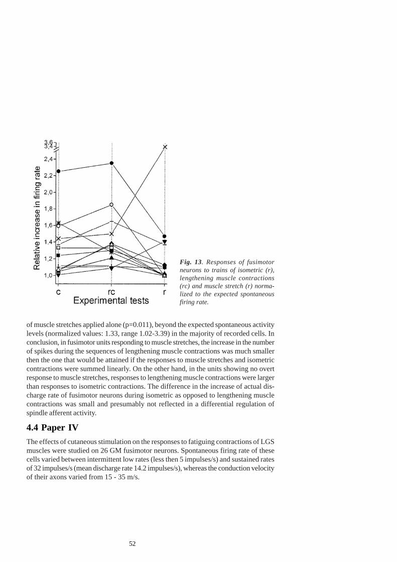
52
of muscle stretches applied alone (p=0.011), beyond the expected spontaneous activitylevels (normalized values: 1.33, range 1.02-3.39) in the majority of recorded cells. Inconclusion, in fusimotor units responding to muscle stretches, the increase in the numberof spikes during the sequences of lengthening muscle contractions was much smallerthen the one that would be attained if the responses to muscle stretches and isometriccontractions were summed linearly. On the other hand, in the units showing no overtresponse to muscle stretches, responses to lengthening muscle contractions were largerthan responses to isometric contractions. The difference in the increase of actual dis-charge rate of fusimotor neurons during isometric as opposed to lengthening musclecontractions was small and presumably not reflected in a differential regulation ofspindle afferent activity.
4.4 Paper IVThe effects of cutaneous stimulation on the responses to fatiguing contractions of LGSmuscles were studied on 26 GM fusimotor neurons. Spontaneous firing rate of thesecells varied between intermittent low rates (less then 5 impulses/s) and sustained ratesof 32 impulses/s (mean discharge rate 14.2 impulses/s), whereas the conduction velocityof their axons varied from 15 - 35 m/s.
Fig. 13. Responses of fusimotorneurons to trains of isometric (r),lengthening muscle contractions(rc) and muscle stretch (r) norma-lized to the expected spontaneousfiring rate.
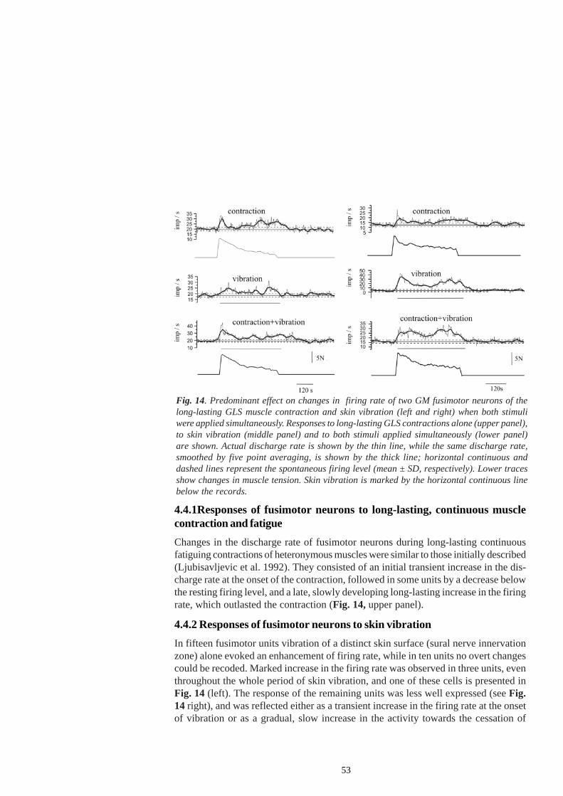
53
4.4.1Responses of fusimotor neurons to long-lasting, continuous musclecontraction and fatigueChanges in the discharge rate of fusimotor neurons during long-lasting continuousfatiguing contractions of heteronymous muscles were similar to those initially described(Ljubisavljevic et al. 1992). They consisted of an initial transient increase in the dis-charge rate at the onset of the contraction, followed in some units by a decrease belowthe resting firing level, and a late, slowly developing long-lasting increase in the firingrate, which outlasted the contraction (Fig. 14, upper panel).
4.4.2 Responses of fusimotor neurons to skin vibrationIn fifteen fusimotor units vibration of a distinct skin surface (sural nerve innervationzone) alone evoked an enhancement of firing rate, while in ten units no overt changescould be recoded. Marked increase in the firing rate was observed in three units, eventhroughout the whole period of skin vibration, and one of these cells is presented inFig. 14 (left). The response of the remaining units was less well expressed (see Fig.14 right), and was reflected either as a transient increase in the firing rate at the onsetof vibration or as a gradual, slow increase in the activity towards the cessation of
Fig. 14. Predominant effect on changes in firing rate of two GM fusimotor neurons of thelong-lasting GLS muscle contraction and skin vibration (left and right) when both stimuliwere applied simultaneously. Responses to long-lasting GLS contractions alone (upper panel),to skin vibration (middle panel) and to both stimuli applied simultaneously (lower panel)are shown. Actual discharge rate is shown by the thin line, while the same discharge rate,smoothed by five point averaging, is shown by the thick line; horizontal continuous anddashed lines represent the spontaneous firing level (mean ± SD, respectively). Lower tracesshow changes in muscle tension. Skin vibration is marked by the horizontal continuous linebelow the records.
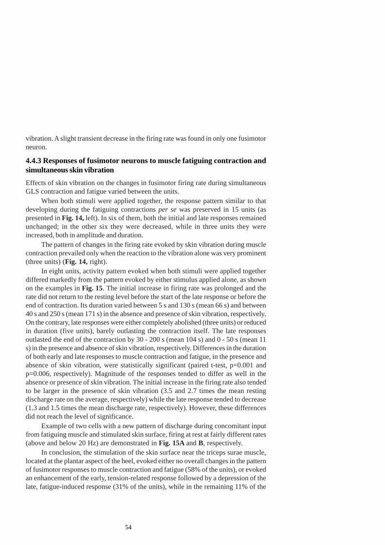
54
vibration. A slight transient decrease in the firing rate was found in only one fusimotorneuron.
4.4.3 Responses of fusimotor neurons to muscle fatiguing contraction andsimultaneous skin vibrationEffects of skin vibration on the changes in fusimotor firing rate during simultaneousGLS contraction and fatigue varied between the units.
When both stimuli were applied together, the response pattern similar to thatdeveloping during the fatiguing contractions per se was preserved in 15 units (aspresented in Fig. 14, left). In six of them, both the initial and late responses remainedunchanged; in the other six they were decreased, while in three units they wereincreased, both in amplitude and duration.
The pattern of changes in the firing rate evoked by skin vibration during musclecontraction prevailed only when the reaction to the vibration alone was very prominent(three units) (Fig. 14, right).
In eight units, activity pattern evoked when both stimuli were applied togetherdiffered markedly from the pattern evoked by either stimulus applied alone, as shownon the examples in Fig. 15. The initial increase in firing rate was prolonged and therate did not return to the resting level before the start of the late response or before theend of contraction. Its duration varied between 5 s and 130 s (mean 66 s) and between40 s and 250 s (mean 171 s) in the absence and presence of skin vibration, respectively.On the contrary, late responses were either completely abolished (three units) or reducedin duration (five units), barely outlasting the contraction itself. The late responsesoutlasted the end of the contraction by 30 - 200 s (mean 104 s) and 0 - 50 s (mean 11s) in the presence and absence of skin vibration, respectively. Differences in the durationof both early and late responses to muscle contraction and fatigue, in the presence andabsence of skin vibration, were statistically significant (paired t-test, p=0.001 andp=0.006, respectively). Magnitude of the responses tended to differ as well in theabsence or presence of skin vibration. The initial increase in the firing rate also tendedto be larger in the presence of skin vibration (3.5 and 2.7 times the mean restingdischarge rate on the average, respectively) while the late response tended to decrease(1.3 and 1.5 times the mean discharge rate, respectively). However, these differencesdid not reach the level of significance.
Example of two cells with a new pattern of discharge during concomitant inputfrom fatiguing muscle and stimulated skin surface, firing at rest at fairly different rates(above and below 20 Hz) are demonstrated in Fig. 15A and B, respectively.
In conclusion, the stimulation of the skin surface near the triceps surae muscle,located at the plantar aspect of the heel, evoked either no overall changes in the patternof fusimotor responses to muscle contraction and fatigue (58% of the units), or evokedan enhancement of the early, tension-related response followed by a depression of thelate, fatigue-induced response (31% of the units), while in the remaining 11% of the
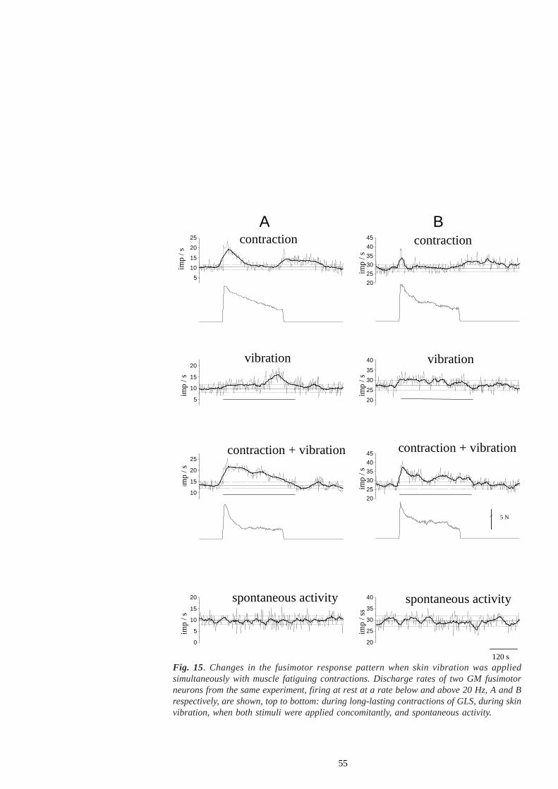
55
Fig. 15. Changes in the fusimotor response pattern when skin vibration was appliedsimultaneously with muscle fatiguing contractions. Discharge rates of two GM fusimotorneurons from the same experiment, firing at rest at a rate below and above 20 Hz, A and Brespectively, are shown, top to bottom: during long-lasting contractions of GLS, during skinvibration, when both stimuli were applied concomitantly, and spontaneous activity.
5 10 15 20 25
imp
/ s
20 25 30 35 40 45
B A
spontaneous activity spontaneous activity
contraction + vibration contraction + vibration
vibration vibration
contraction contraction
imp
/ s
5 10 15 20
imp
/ s
20 25 30 35 40
imp
/ s
10 15 20 25
imp
/ s
20 25 30 35 40 45
imp
/ s
5 N
0 5
10 15 20
imp
/ s
20 25 30 35 40
120 s
imp
/ ss
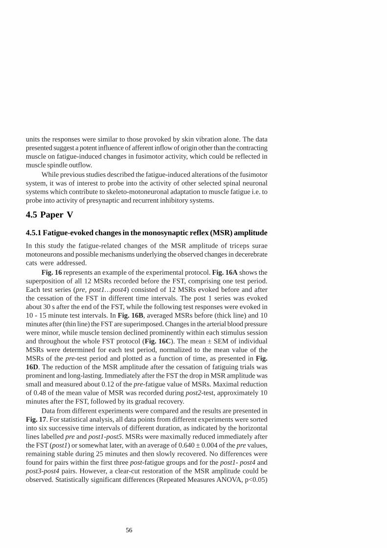
56
units the responses were similar to those provoked by skin vibration alone. The datapresented suggest a potent influence of afferent inflow of origin other than the contractingmuscle on fatigue-induced changes in fusimotor activity, which could be reflected inmuscle spindle outflow.
While previous studies described the fatigue-induced alterations of the fusimotorsystem, it was of interest to probe into the activity of other selected spinal neuronalsystems which contribute to skeleto-motoneuronal adaptation to muscle fatigue i.e. toprobe into activity of presynaptic and recurrent inhibitory systems.
4.5 Paper V
4.5.1 Fatigue-evoked changes in the monosynaptic reflex (MSR) amplitudeIn this study the fatigue-related changes of the MSR amplitude of triceps suraemotoneurons and possible mechanisms underlying the observed changes in decerebratecats were addressed.
Fig. 16 represents an example of the experimental protocol. Fig. 16A shows thesuperposition of all 12 MSRs recorded before the FST, comprising one test period.Each test series (pre, post1…post4) consisted of 12 MSRs evoked before and afterthe cessation of the FST in different time intervals. The post 1 series was evokedabout 30 s after the end of the FST, while the following test responses were evoked in10 - 15 minute test intervals. In Fig. 16B, averaged MSRs before (thick line) and 10minutes after (thin line) the FST are superimposed. Changes in the arterial blood pressurewere minor, while muscle tension declined prominently within each stimulus sessionand throughout the whole FST protocol (Fig. 16C). The mean ± SEM of individualMSRs were determined for each test period, normalized to the mean value of theMSRs of the pre-test period and plotted as a function of time, as presented in Fig.16D. The reduction of the MSR amplitude after the cessation of fatiguing trials wasprominent and long-lasting. Immediately after the FST the drop in MSR amplitude wassmall and measured about 0.12 of the pre-fatigue value of MSRs. Maximal reductionof 0.48 of the mean value of MSR was recorded during post2-test, approximately 10minutes after the FST, followed by its gradual recovery.
Data from different experiments were compared and the results are presented inFig. 17. For statistical analysis, all data points from different experiments were sortedinto six successive time intervals of different duration, as indicated by the horizontallines labelled pre and post1-post5. MSRs were maximally reduced immediately afterthe FST (post1) or somewhat later, with an average of 0.640 ± 0.004 of the pre values,remaining stable during 25 minutes and then slowly recovered. No differences werefound for pairs within the first three post-fatigue groups and for the post1- post4 andpost3-post4 pairs. However, a clear-cut restoration of the MSR amplitude could beobserved. Statistically significant differences (Repeated Measures ANOVA, p<0.05)
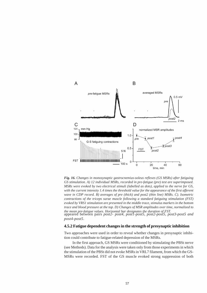
57
appeared between pairs post2- post4, post1-post5, post2-post5, post3-post5 andpost4-post5.
4.5.2 Fatigue dependent changes in the strength of presynaptic inhibitionTwo approaches were used in order to reveal whether changes in presynaptic inhibi-tion could contribute to fatigue-related depression of the MSRs.
In the first approach, GS MSRs were conditioned by stimulating the PBSt nerve(see Methods). Data for the analysis were taken only from those experiments in whichthe stimulation of the PBSt did not evoke MSRs in VRL7 filament, from which the GS-MSRs were recorded. FST of the GS muscle evoked strong suppression of both
Fig. 16. Changes in monosynaptic gastrocnemius-soleus reflexes (GS MSRs) after fatiguingGS stimulation. A) 12 individual MSRs, recorded in pre-fatigue (pre) test are superimposed.MSRs were evoked by two electrical stimuli (labelled as dots), applied to the nerve for GS,with the current intensity 1.4 times the threshold value for the appearance of the first afferentwave in CDP record. B) averages of pre (thick) and post2 (thin line) MSRs. C). Isometriccontractions of the triceps surae muscle following a standard fatiguing stimulation (FST)evoked by VRS1 stimulation are presented in the middle trace, stimulus markers in the bottomtrace and blood pressure at the top. D) Changes of MSR amplitudes over time, normalized tothe mean pre-fatigue values. Horizontal bar designates the duration of FST.
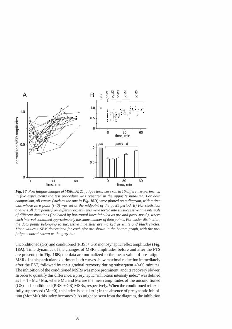
58
unconditioned (GS) and conditioned (PBSt + GS) monosynaptic reflex amplitudes (Fig.18A). Time dynamics of the changes of MSRs amplitudes before and after the FTSare presented in Fig. 18B; the data are normalized to the mean value of pre-fatigueMSRs. In this particular experiment both curves show maximal reduction immediatelyafter the FST, followed by their gradual recovery during subsequent 40-60 minutes.The inhibition of the conditioned MSRs was more prominent, and its recovery slower.In order to quantify this difference, a presynaptic ”inhibition intensity index” was definedas I = 1 - Mc / Mu, where Mu and Mc are the mean amplitudes of the unconditioned(GS) and conditioned (PBSt + GS) MSRs, respectively. When the conditioned reflex isfully suppressed (Mc=0), this index is equal to 1; in the absence of presynaptic inhibi-tion (Mc=Mu) this index becomes 0. As might be seen from the diagram, the inhibition
Fig. 17. Post fatigue changes of MSRs. A) 21 fatigue tests were run in 16 different experiments;in five experiments the test procedure was repeated in the opposite hindlimb. For datacomparison, all curves (such as the one in Fig. 16D) were plotted on a diagram, with a timeaxis whose zero point (t=0) was set at the midpoint of the post1 period. B) For statisticalanalysis all data points from different experiments were sorted into six successive time intervalsof different durations (indicated by horizontal lines labelled as pre and post1-post5), whereeach interval contained approximately the same number of data points. For easier distinction,the data points belonging to successive time slots are marked as white and black circles.Mean values ± SEM determined for each plot are shown in the bottom graph, with the pre-fatigue control shown as the grey bar.
59
intensity first increased to a maximum at around 40 minutes after the FST, followed byits recovery to the pre-fatigue value.
Statistical analysis of the data from nine experiments shows that the ’inhibitionintensity’ tended first to increase after the FST and then to decline. Statistically significantdifferences in the ”inhibition intensity” were shown for two pairs of test groups, namelyfor pre – post2 (increase of ”inhibition intensity”, p<0.01), and post2 – post4 (decreaseof ”inhibition intensity”, p<0.05).
Since PAD presents electrophysiological evidence of presynaptic inhibition, an-other assessment of presynaptic inhibition was performed by recording the DRPs andthe antidromic discharges from the filaments dissected from dorsal root L7. An exampleof fatigue-evoked changes of slow components of DRPs is presented in Fig.19. Afterthe procedures of smoothing and averaging, DRPs recorded in the pre and post1 -post4 periods were compared, and the pre and post1 averages were superimposed in
Fig. 18. Post-fatigue changes in the presynaptic inhibition of the GS MSRs induced byconditioning stimulation of the nerve to the PBSt. A) Fatigue-evoked changes in the GSMSRs, without (left column) and with (right column) preceding stimulation of the PBSt nerve(time interval between conditioning and test stimulation was 20 s). Averaged records, n=12.The intensity of PBSt stimulation was set so as to evoke approximately 20% inhibition of GSMSRs in the pre-fatigue tests. B) Changes of unconditioned (open circles, GS) and conditioned(filled circles, PBSt + GS) MSR amplitudes over time, normalized to pre-fatigue values of theMSRs. C) Presynaptic ‘inhibition intensity’ plotted as a function of time (for a definition seetext).
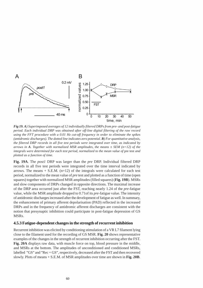
60
Fig. 19A. The post1 DRP was larger than the pre DRP. Individual filtered DRPrecords in all five test periods were integrated over the time interval indicated byarrows. The means + S.E.M. (n=12) of the integrals were calculated for each testperiod, normalized to the mean value of pre test and plotted as a function of time (opensquares) together with normalized MSR amplitudes (filled squares) (Fig. 19B). MSRsand slow components of DRPs changed in opposite directions. The maximal increaseof the DRP area occurred just after the FST, reaching nearly 1.24 of the pre-fatiguevalue, while the MSR amplitude dropped to 0.71of its pre-fatigue value. The intensityof antidromic discharges increased after the development of fatigue as well. In summary,the enhancement of primary afferent depolarisation (PAD) reflected in the increasedDRPs and in the frequency of antidromic afferent discharges are consistent with thenotion that presynaptic inhibition could participate in post-fatigue depression of GSMSRs.
4.5.3 Fatigue-dependent changes in the strength of recurrent inhibitionRecurrent inhibition was elicited by conditioning stimulation of a VR L7 filament lyingclose to the filament used for the recording of GS MSR. Fig. 20 shows representativeexamples of the changes in the strength of recurrent inhibition occurring after the FST.Fig. 20A displays raw data, with muscle force on top, blood pressure in the middle,and MSRs at the bottom. The amplitudes of unconditioned and conditioned MSRs,labelled ”GS” and ”Rec + GS”, respectively, decreased after the FST and then recoveredslowly. Plots of means + S.E.M. of MSR amplitudes over time are shown in Fig. 20B.
Fig 19. A) Superimposed averages of 12 individually filtered DRPs from pre- and post-fatigueperiod. Each individual DRP was obtained after off-line digital filtering of the raw recordusing the FFT procedure with a 0.01 Hz cut-off frequency in order to eliminate the spikes(antidromic discharges). The dotted line indicates zero potential. B) For quantitative analysis,the filtered DRP records in all five test periods were integrated over time, as indicated byarrows in A. Together with normalized MSR amplitudes, the means ± SEM (n=12) of theintegrals were determined for each test period, normalized to the mean value of pre test andplotted as a function of time.
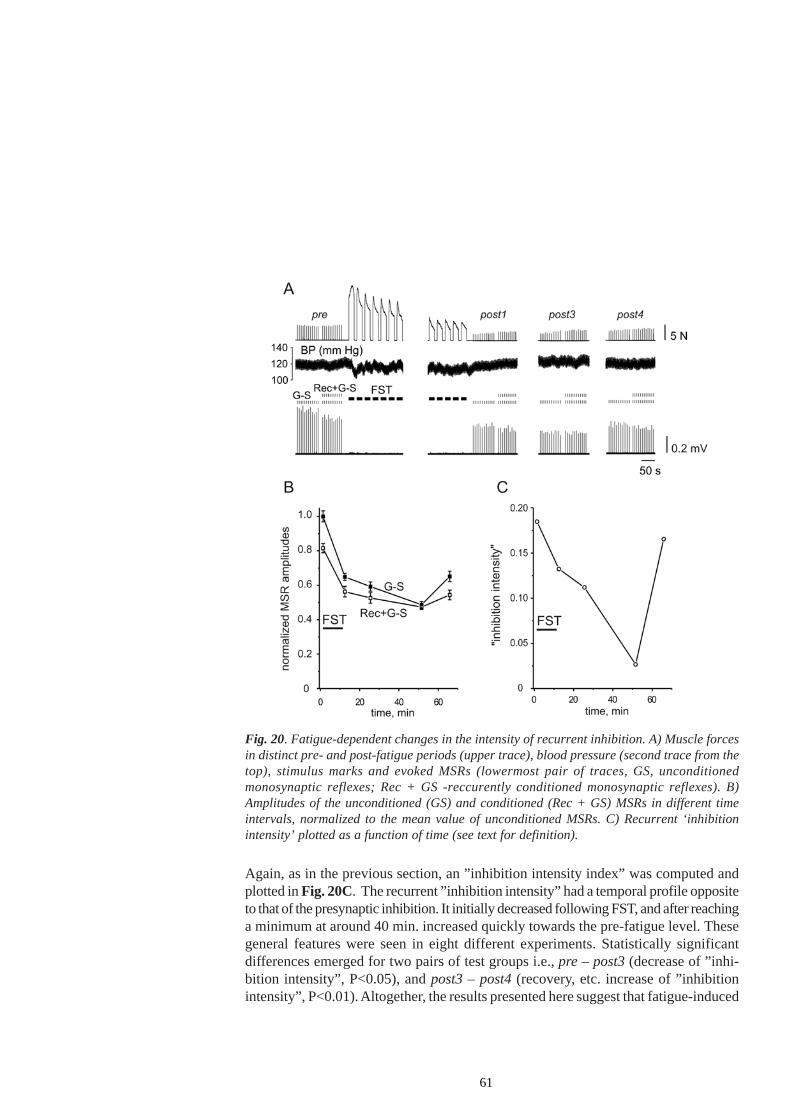
61
Again, as in the previous section, an ”inhibition intensity index” was computed andplotted in Fig. 20C. The recurrent ”inhibition intensity” had a temporal profile oppositeto that of the presynaptic inhibition. It initially decreased following FST, and after reachinga minimum at around 40 min. increased quickly towards the pre-fatigue level. Thesegeneral features were seen in eight different experiments. Statistically significantdifferences emerged for two pairs of test groups i.e., pre – post3 (decrease of ”inhi-bition intensity”, P<0.05), and post3 – post4 (recovery, etc. increase of ”inhibitionintensity”, P<0.01). Altogether, the results presented here suggest that fatigue-induced
Fig. 20. Fatigue-dependent changes in the intensity of recurrent inhibition. A) Muscle forcesin distinct pre- and post-fatigue periods (upper trace), blood pressure (second trace from thetop), stimulus marks and evoked MSRs (lowermost pair of traces, GS, unconditionedmonosynaptic reflexes; Rec + GS -reccurently conditioned monosynaptic reflexes). B)Amplitudes of the unconditioned (GS) and conditioned (Rec + GS) MSRs in different timeintervals, normalized to the mean value of unconditioned MSRs. C) Recurrent ‘inhibitionintensity’ plotted as a function of time (see text for definition).
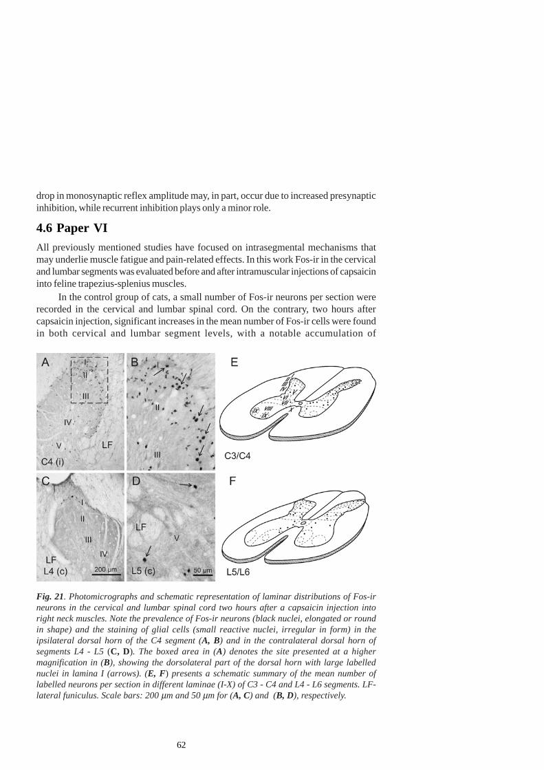
62
drop in monosynaptic reflex amplitude may, in part, occur due to increased presynapticinhibition, while recurrent inhibition plays only a minor role.
4.6 Paper VIAll previously mentioned studies have focused on intrasegmental mechanisms thatmay underlie muscle fatigue and pain-related effects. In this work Fos-ir in the cervicaland lumbar segments was evaluated before and after intramuscular injections of capsaicininto feline trapezius-splenius muscles.
In the control group of cats, a small number of Fos-ir neurons per section wererecorded in the cervical and lumbar spinal cord. On the contrary, two hours aftercapsaicin injection, significant increases in the mean number of Fos-ir cells were foundin both cervical and lumbar segment levels, with a notable accumulation of
Fig. 21. Photomicrographs and schematic representation of laminar distributions of Fos-irneurons in the cervical and lumbar spinal cord two hours after a capsaicin injection intoright neck muscles. Note the prevalence of Fos-ir neurons (black nuclei, elongated or roundin shape) and the staining of glial cells (small reactive nuclei, irregular in form) in theipsilateral dorsal horn of the C4 segment (A, B) and in the contralateral dorsal horn ofsegments L4 - L5 (C, D). The boxed area in (A) denotes the site presented at a highermagnification in (B), showing the dorsolateral part of the dorsal horn with large labellednuclei in lamina I (arrows). (E, F) presents a schematic summary of the mean number oflabelled neurons per section in different laminae (I-X) of C3 - C4 and L4 - L6 segments. LF-lateral funiculus. Scale bars: 200 µm and 50 µm for (A, C) and (B, D), respectively.
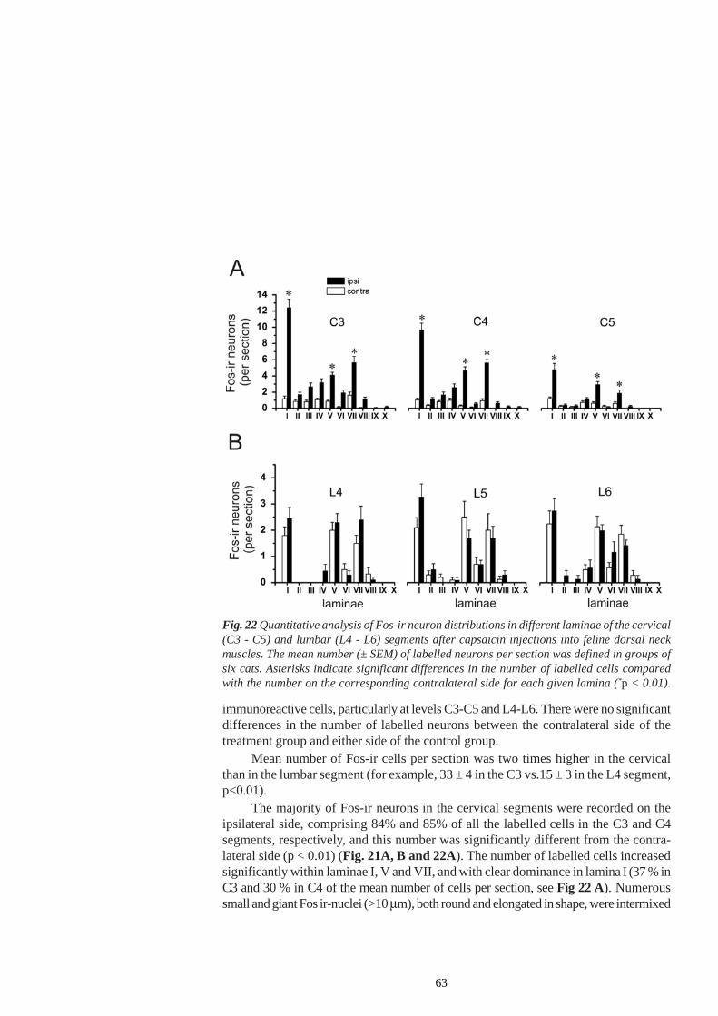
63
immunoreactive cells, particularly at levels C3-C5 and L4-L6. There were no significantdifferences in the number of labelled neurons between the contralateral side of thetreatment group and either side of the control group.
Mean number of Fos-ir cells per section was two times higher in the cervicalthan in the lumbar segment (for example, 33 ± 4 in the C3 vs.15 ± 3 in the L4 segment,p<0.01).
The majority of Fos-ir neurons in the cervical segments were recorded on theipsilateral side, comprising 84% and 85% of all the labelled cells in the C3 and C4segments, respectively, and this number was significantly different from the contra-lateral side (p < 0.01) (Fig. 21A, B and 22A). The number of labelled cells increasedsignificantly within laminae I, V and VII, and with clear dominance in lamina I (37 % inC3 and 30 % in C4 of the mean number of cells per section, see Fig 22 A). Numeroussmall and giant Fos ir-nuclei (>10 µm), both round and elongated in shape, were intermixed
Fig. 22 Quantitative analysis of Fos-ir neuron distributions in different laminae of the cervical(C3 - C5) and lumbar (L4 - L6) segments after capsaicin injections into feline dorsal neckmuscles. The mean number (± SEM) of labelled neurons per section was defined in groups ofsix cats. Asterisks indicate significant differences in the number of labelled cells comparedwith the number on the corresponding contralateral side for each given lamina (*p < 0.01).
64
with Fos-ir glial nuclei of irregular form (<5 µm).The number of labelled cells increased bilaterally in the lumbar segments, and
there were no significant difference between ipsi- (57%) and contralateral (43%) sides(Fig. 21C, D and 22 B).
65
5. DISCUSSIONThe work presented here reveals different interactive spinal mechanisms involved in,or related with muscle fatigue. The study of these mechanisms may provide importantinsights into the strategies and processes employed by the CNS in the optimisation ofperformance of its neuromuscular periphery. It might also unveil the processes thatmay become deficient in everyday occupational settings, during long-lasting repetitivefatiguing contractions, which are known to be associated with the development ofchronic muscle pain conditions.
5.1 Experimental evidence of spreading of fatigue effectsbetween different muscle partsThe results presented in Paper I show that the activation history of multiple motorunits of an individual muscle might affect the contractile ability of their neighbouringinactive units, thus promoting spatial spreading of fatigue between different muscleparts.
Fatiguing stimulation of a set of motor units evoked a noticeable decrease in thetension of test contractions of another, previously inactive set of motor units. Interestingly,maximal depressive effects of conditioning fatiguing stimulation were observed whendistributed high-rate (40 s-1) stimulation of four or five filaments of the supplying VRs(L7 or S1) was applied. Low-rate (10 s-1) stimulation of a single filament, or distributedstimulation of five filaments at this rate was usually insufficient to influence the testcontractions.
A tension drop in test muscle contractions following CFS was always accompaniedby a reduction in the amplitude of M-waves. The latency of the M-waves often increased,suggesting a failure of action potential propagation along the sarcolemmal membranesor motor axon terminals. Both tension and EMG demonstrated a tendency towardsrecovery after several minutes of rest and the recovery of the EMG was quicker thanthat of the tension. Yet, the amplitudes of M-waves were unstable following CFS andduring restoration.
The observed effects were probably caused by manifold processes such as: 1)the diffusion of metabolites from active to inactive muscle fibers 2) lowering of theefficiency of neuro-muscular transmission due to the squeezing of motor terminals anddue to the effects of metabolites on action potential propagation along motor axonterminals, and 3) the increase in intramuscular pressure which might evoke local hypoxia.
In particular, we assume that changes in interstitial K+ concentration might haveplayed a pivotal role. An increase in interstitial K+ might have led to a decrease inamplitude and propagation velocity of action potentials in active and inactive, neighbouringmuscle cells. Reduced action potentials would have further decreased the release ofCa2+ from the sarcoplasmatic reticulum, resulting in a diminished force output (Wester-blad et al. 1993; Fitts 1994). Slow continuous loss of intracellular K+ may take hours to
66
revert, possibly because at least a part of this K+ has been transferred to other tissuesand is not readily accessible for reuptake.
An increase in the K+ concentration in the interstitial fluid could also have reducedaction potential propagation along motor axon terminals, thus providing one of the possiblecauses of neuro-muscular propagation failure.
Apart from altered K+ dynamics, free radicals produced during muscle contractionmight have also effectively modified muscle contractile efficiency (Reid 1998; Marechaland Gailly 1999) of both active and inactive, neighbouring muscle fibres. It is knownthat endogenous NO limits the force developed in skeletal muscles (Kobzik et al. 1994).The isometric forces produced during twitch and submaximal tetanic contractions ofrat diaphragm fibre bundles were increased by NOS blockade or by NO scavengers,shifting the force-frequency relationship leftwards. Nitric oxide donors reversed thiseffect. Kobzik et al (1994) hypothesised that endogenous NO decreases submaximalforce by modulating the excitation-contraction coupling. On the other hand, there isincreasing evidence that endogenous NO is essential for optimal myofilament functionduring active shortening (Morrison et al. 1996). However, excessive production of NOmay diminish the sensitivity of myofibrilla to Ca2+ (Andrade et al. 1998). Also, NO andits derivative peroxynitrite may inhibit mitochondrial respiration (Brown and Borutaite1999). It might be expected that NO diffuses from active to inactive fibres in spite ofits short lifetime of 10 - 15 s and if not captured by scavengers.
Together with other vasoactive metabolites, NO could also modify regional bloodflow in skeletal muscles during their fatiguing activation (Porter et al. 2000) and thusindirectly influence the contractile efficiency of non-activated fibres. Vasodilatationper se increases nutrient and oxygen supply and facilitates the washing-out ofinflammatory substances, so it might improve the contractile abilities of both stimulatedand non-stimulated muscle parts. However, vasodilation can exert detrimental effectsif not balanced by sympathetic vasoconstriction, as a consequence of plasma fluid lossinto the extracellular space (Maspers et al. 1991).
CFS presumably evoked significant increments of intramuscular pressure.Increased intramuscular pressure could evoke the occlusion of intramuscular circulation.Occlusion of blood flow during high-rate intermittent stimulation of the gastrocnemiusmuscle in rat evoked a depression of both tension and EMG (De-Ruiter 1996). Simu-lation of impaired circulation by mechanical occlusion of the popliteal artery in ourexperiments evoked a reduction in the tension of test reactions, not paralleled, however,by a reduction in EMG. Passive squeezing of the muscle belly (which did not exceed120 mmHg) also failed to evoke changes in test reactions. It turns out that the changesin the intramuscular pressure, per se, could not have been the main underlyingmechanism for the depression of the test reactions. During CFS, though, local gradientsof intramuscular pressure could be much stronger, exceeding systolic blood pressureby more than ten times during strong tetanic stimulation (Ameredes and Provenzano1997) and evoking a harmful loss of the plasma fluid into the interstitium. Thus, regio-nal alterations of intramuscular pressure and local hypoxia might have also diminished
67
the contractile efficiency of the inactive set of motor units.Spreading of the effects of fatigue between different muscle parts may further
complicate motoneuronal adaptation to now peripherally altered contractile propertiesof their motor units once previously inactive neurons became active, thus disturbing theoptimal regulation of motoneuronal activity.
5.2 Experimental evidence of fatigue-induced activation ofsmall - diameter afferent fibresFatigue-induced appearance of metabolites in the interstitium could provoke excitationand/or sensitisation of high-threshold afferent fibres, with complex effects on the spi-nal premotoneuronal network involved in the modulation of motoneuronal output.Fatigue-related alterations of small-diameter afferent fibre discharge have not beenmonitored directly in our studies and the evidences concerning their activation are thusindirect. Namely, it would have been extremely difficult, if not impossible, to simultaneou-sly record from a representative sample of group III and IV afferents and in parallelrecord the variables of primary interest in the present studies.
Paper II provides functional anatomical evidence of spinal effects of fatigue-induced activation of group III and IV afferents and describes a fatigue-evoked patternof lamellar distribution of spinal interneurons in the lumbar spinal cord of rats. Followingfatiguing stimulation of the triceps surae muscle in rats, most of the Fos-immunoreactivecells were localized in the superficial layers 1, 2o and 5 of the dorsal horn. Theirnumber was increased in rostrocaudal direction, from L2 to L5 segments. Such lamellardistributions of Fos-labelled neurons overlap almost completely with the known termi-nation patterns of high-threshold muscle afferents (Mense and Prabhakar 1986), andtheir labelling can thus be attributed to the activation of these afferents, if only bycorrelation.
Activation of group III and IV muscle afferents during fatiguing musclecontractions is also suggested by timing arguments. Thus, significantly enhanced c-fosimmunoreactivity in the dorsal horn of the lumbar spinal cord in rats was noticed onlyfollowing a 30-min fatiguing protocol of the triceps surae. By contrast, five minutes ofcontraction only slightly increased the number of Fos-ir neurons. Furthermore, in ourexperimental conditions, only fatiguing contractions of long duration evoked observablechanges in spinal interneuronal and motoneuronal excitability. The strong depression ofthe monosynaptic reflexes (MSRs), as well as the alterations in presynaptic and recurrentinhibition (Paper V) were observed only after the application of a fatiguing protocol oflonger duration (15 - 20 min). Further, the recovery of MSR amplitude, as well as therecovery of presynaptic and recurrent inhibition intensity was rather slow, occurringwithin an hour after the fatiguing trials. Altogether, these delayed and long-lasting effectswould be expected under the assumption that they resulted from reflex effects ofsmall-diameter muscle afferents activated by the accumulation of metabolites andinflammatory substances in the muscle interstitium, with a possible involvement ofperipheral and/or central sensitisation.
68
5.3 Could neuromuscular propagation failure (NPF) contributeto fatiguing effects?To what extent could the effects of long-lasting muscle activity be ascribed exclusivelyto factors within the muscle and to what extent do they include a component ofneuromuscular transmission failure? Some authors reject the concept of substantialcontributions of the neuromuscular block to fatigue, even during maximal voluntarycontractions (Bigland-Ritchie et al. 1982; Bellemare and Bigland-Ritchie 1987), whileothers maintain the opposite (Stephens and Taylor 1972; Sieck and Prakash 1995). Ithas been shown that the effects of fatigue evoked by long-lasting distributed stimula-tion of ventral root filaments in decerebrate cats include neuromuscular components(Kostyukov et al. 2000a,b; Wise et al. 2001). In the experiments presented in this workNPF occurred, but its contribution to muscle fatigue was superseded by factors relatedto the disturbance of excitation-contraction coupling, muscle perfusion or the releaseof metabolites within the muscle tissue. Again, the described pattern of distribution ofFos-ir neurons together with the slow recovery of MSR amplitudes and presynapticand recurrent inhibition speak in favour of such an assumption.In the following sections fatigue-induced spinal reflex actions of large- and particularlysmall- diameter afferent fibres reaching α- and γ- motoneurons, as well as theirmodulation at the input (presynaptic inhibition) and output stage (recurrent inhibition)will be briefly discussed.
5.4 Does the gamma-spindle system act as a premotoneuronalintegrative system in fatiguing conditions related to differentregimes of muscle activity?In one part of this study the potential role of the fusimotor system in skeleto-motoneuronaladaptation to muscle fatigue was examined. The decline of the afferent inflow frommuscle spindles could contribute to the decrease in skeletomotor discharge rates duringfatiguing contractions. Indeed, Macefield et al (1993) demonstrated that during sub-maximal voluntary contractions of ankle dorsiflexors muscle spindles’ afferent dis-charge rate decreases.
On the other hand, voluntary contraction itself provokes an increase, rather thana decrease in muscle spindle outflow through α -γ coactivation (Vallbo 1971). A furtherincrease of, or compensation for eventual fatigue-evoked decrease in spindle outflowmight occur as a result of fatigue-induced reflex enhancement of fusimotor activity.This might be relevant because the precise adjustment of skeleto-motoneuronal activityto altered contractile properties of fatigued muscle fibres requires continuous informa-tion about the changes in muscle state. The requested information could be deliveredfrom muscle spindle sensory endings (Windhorst and Kokkoroyiannis 1991), providedtheir high sensitivity being maintained (Enoka and Stuart 1992; Gandevia et al. 1995;Gandevia 2001) by fusimotor influences in spite of spindle unloading during musclecontraction.
69
In decerebrate cats, long-lasting contractions of the LGS evoked early and lateincreases in the discharge rate of fusimotor neurons. The late increase, developed inparallel with the fatigue-dependent fall in muscle tension (Ljubisavljevic et al. 1992)was ascribed to muscle fatigue induced activation of chemosensitive fibres, to thesensitisation of mechanosensitive afferent fibres, or to both. Such an enhancement ofthe fusimotor activity was estimated to be sufficient to account for the rise in afferentfiring level of muscle spindles during the post-contraction period (Ljubisavljevic andAnastasijevic 1994). During the contraction the effects of intrafusal fibre slack andmuscle spindle unloading on the firing rate of its afferents were partly compensatedfor, so that the firing rate could be equal to that of the spontaneous activity, at the endof fatiguing trials. Changes in the responsiveness of spindle sensory endings, estimatedby the depth of modulation of their discharge rate in response to sinusoidal musclestretching at 1 Hz, 1-mm half-to-peak amplitude sinusoidal muscle stretching appliedbefore and after the fatiguing stimulation revealed persistent or enhanced influences ofboth static and dynamic fusimotor drives. However, in the presence of afferent inflowof different origin in the non-denervated hind limb, the late fatigue-induced increase inthe discharge rate of fusimotor neurons is less prominent in decerebrate and spinalcats (Ljubisavljevic et al. 1994). This study demonstrated that complex peripheral afferentinflow of origin other than the parental muscle may alter the fusimotor response tomuscle fatigue.
5.4.1 Interactive effects of cutaneous and muscle fatigue-induced afferentinflow on fusimotor neuronsPaper III further explores the aforementioned findings by examining whether definedafferent inflow from non-noxious mechanoreceptors in the skin (presumably the SA1type) may influence fusimotor responses to muscle contraction and fatigue. Such aninteraction might be relevant since an excitation of the receptors located in the skinareas overlying, or lying in the neighbourhood of the contracting muscles might occurdue to skin distortion during locomotion (Davey and Ellaway 1989a, b).
It was demonstrated that afferent discharges from cutaneous receptors(presumably from the SA1 type), could provoke both excitation and inhibition of fusimotorneurons to the triceps surae muscle (Ellaway et al. 1996; 1997), which is well inaccordance with the notion that each fusimotor neuron has its own receptive profile(Johansson and Sojka 1985; Ellaway et al. 1996; 1997). A precise topographicorganization of cutaneous reflexogenic territories in relation to the regulation of γ-motoneurone discharge was described (Ellaway et al. 1997).
The data in Paper III demonstrate that in 58% of all the fusimotor neuronsexamined, the patterns of contraction- and fatigue-induced changes in the firing ratewere preserved in the presence of skin vibration, although some alterations (an increaseor a decrease) of the discharge rate could be recorded. Such responses were ascribedto additional reflex inflow, excitatory or inhibitory, originating from skin receptors tofusimotor neurons.
70
However, in approximately one third of the units (31%), the observed changes inthe firing rate were more complex, i.e. the early, muscle tension-evoked response wasincreased while the fatigue-induced late response and the post-contraction part weredecreased. An enhancement of the early responses might be caused by the redistributionof activity among Renshaw interneurons inhibiting different γ-motoneuron pools. Sincemutual inhibition exists between Renshaw interneurons directed towards motoneuronsof different muscle groups, enhanced afferent discharges from cutaneous receptorsmay provoke an activation of the Renshaw interneurons projecting to motoneurons ofnon-contracting muscles (Piercey and Goldfarb 1974) and further inhibition of theRenshaw interneurons projecting to motoneurons of the triceps surae muscle. Such adecrease in recurrent inhibition intensity may evoke disinhibition of fusimotor neuronsto the triceps surae (Appelberg et al. 1983a) and may account for the prolonged, earlyincrease in fusimotor activity.
The same cutaneous afferent discharges might provoke the depression of thelate increase in fusimotor firing rate by stimulating the activity of inhibitory interneu-rons intercalated in reflex pathways of group III and IV muscle afferents.
Some other mechanisms might be relevant as well. Since early fusimotor responsesto muscle contraction are presumably evoked by increased muscle mechanosensitiveafferent discharge and since late responses are mainly due to the activation ofchemosensitive afferents (Ljubisavljevic et al. 1992), their reflex pathways might notbe the same. Furthermore, they could be differentially affected by the same cutaneousinput.
It is aso worthwhile to note that spino-bulbo-spinal pathways and propriospinalpathways might be activated in the decerebrate cat preparation and may additionallyinterfere with segmental reflex pathways.
Marked differences in reflex responses of fusimotor neurons in non-deneravatedvs. denervated preparations were taken as indicators of high susceptibility of fusimotorreflex responses to the changes in afferent and descending inflow (Ljubisavljevic et al.1994), as confirmed in the described study with evoked afferent discharges from SA1type skin receptors.
5.4.2 Changes in fusimotor activity during repetitive lengthening vs. isometricmuscle contractionsFusimotor activity might be altered in different regimes of muscle activity, partly due tovariations in the descending drive and partly due to the activation of a diffuse spectrumof afferent fibres and their reflex pathways in various conditions. It was of particularinterest to assess the alterations in the fusimotor drive during eccentric contractions,which occur frequently in everyday activities. They are characterized by the ability toachieve high muscle forces, but they are connected with a higher probability of tissuedamage associated with muscle soreness (Morgan and Proske 2004). The CNS engagesunique activation strategies in eccentric compared to isometric contractions (Enoka
71
1996), associated with differential recruitment order, discharge rate and recruitmentthreshold of motor units.
The majority of fusimotor neurons recorded in Paper IV responded to repetitiveisometric and lengthening contractions, and most of them to muscle stretch as well, byan increase in the spontaneous firing rate. This increase in the firing rate was greater,on the average, during the sequences of lengthening contractions than during theisometric ones. The increase in fusimotor firing rate was either smooth or modulatedwith each successive contraction and stretch. Two patterns of responses indicatedthat the distribution of afferent input and reflex connections to the MG fusimotor neu-ron pool were not homogeneous, which is again in agreement with the notion that eachfusimotor neuron has its own receptive profile (Johansson and Sojka 1985). Theenhancement of fusimotor firing rate during repetitive isometric contractions wasascribed primarily to the activation of group III (Ellaway et al. 1982) and II muscleafferents (Appelberg et al. 1983b). The observed increase in firing rate of fusimotorneurons during muscle stretches might occur due to the activation of group II and IIImechanosensitive muscle afferents, but reflex effects originating from Ia muscle spindleendings cannot be completely ruled out (Trott 1976; Anastasijevic and Vuco 1984),irrespective of their weak and sporadic autogenic reflex influences on fusimotor neurons(Appelberg et al. 1983c). The responses of some fusimotor neurons to lengtheningcontractions were larger than to the isometric contractions, despite the fact that thesequences of muscle stretches per se failed to evoke an alteration of their dischargerate, suggesting possible interactions of afferent discharges through reflex pathways,at the interneuronal level. However, the probability that the activity of the whole rangeof afferents responding to either stretch or isometric contraction would be equal totheir sum during lengthening contractions is low. Indeed, it turned out that the responsesof fusimotor neurons to lengthening contractions were, on the average, smaller thanthe sum of their responses to isometric contractions and stretches applied independently.The difference in the increase of fusimotor firing rate during lengthening, as opposedto isometric muscle contractions was rather small. This difference is presumably in-sufficient to change the firing rates of muscle spindle afferents.
It is well established that the activation of group III and IV muscle afferents,evoked by intra-arterially injected algesic substances, exerts potent excitatory reflexinfluences on fusimotor neurons (Jovanovic et al. 1990). It was also assumed(Ljubisavljevic and Anastasijevic 1996) that during fatiguing movements, the transmis-sion in reflex pathways originating from the fatiguing muscle may evoke an increase infusimotor activity strong enough to partially counteract or at least dampen, through thegamma loop, more direct inhibitory reflex influences on skeleto-motoneurons (Bigland-Ritchie et al. 1986) exerted by small-diameter afferent fibres. The authors suggestedthat the functional implication of the enhancement of fusimotor drive might be in theadjustment and maintenance of the discharge rate of skeletomotoneurons automatically,by muscle fatigue itself, in order to match the altered contractile properties of fatiguedmuscle fibres.
72
However, the results presented in this work demonstrate that the action of smalldiameter afferent fibres on the fusimotor drive could be altered in the presence ofafferent inflow of origin other than the parental muscle. Fusimotor neuronal activitymay vary (albeit only slightly in our experimental conditions) in dependence of theregime of muscle activity. For example, reflex pathways could be opened or shutdepending upon the current demands of the ongoing motor task. Therefore, involvementof an additional reflex and descending pathways may change the pattern of fusimotorneuron response during muscle fatigue and could account for the observed decline inmuscle spindle afferent discharge and α-motoneuron disfacilitation, as observed inhumans (Macefield et al. 1991), and recently in rats (Brunetti et al. 2003).
5.5 Muscle fatigue-induced alterations at the motoneuron in-put stage - the presynaptic inhibitory systemIn further work possible contributions of other selected spinal neuronal systems toskeleto-motoneuronal adaptation to muscle fatigue were explored. The activity ofparticular interneuronal networks and their influence on motor input (presynaptic inhi-bition system) and output (recurrent inhibition system) stages were addressed in PaperV.
Following fatiguing stimulation of the triceps surae muscle in decerebrate cats,the amplitudes of MSR of GS motoneurons were reduced to 0.64 ± 0.04 (mean ±SEM) of their pre-fatigue values. Potential contribution of presynaptic inhibition to theobserved fatigue-evoked reduction in MSRs (reflecting motoneuron adaptation) wasassessed using two approaches.
First, GS MSRs were conditioned by the stimulation of PBSt group Ia afferents,which preceded the stimuli to the GS nerve. Fatigue-induced depression of the GSmonosynaptic reflex, conditioned by PBSt stimulation, was more prominent andprolonged as compared to the depression of unconditioned reflexes. The intensity ofpresynaptic inhibition increased from 0.19 ± 0.02 pre-fatigue to 0.44 ± 0.04 post-fatigue,followed by a recovery. The frequency of antidromic discharges and area of slowDRP components recorded from small filaments of L7 dorsal root were enhancedfollowing the FST. The time course of fatigue-related changes in the presynaptic ”in-hibition intensity” and slow DRP components corresponded well with the time courseof post-fatigue MSR changes, indicating an underlying interaction. The maximal dropof MSR amplitudes, presynaptic ”inhibition intensity” and increase of area of slowcomponent of DRP were observed within 10 - 15 min after the FST, all being noticeablyaltered over longer time intervals.
The dynamics of changes in MSRs and presynaptic inhibition raised the possibilitythat group III and IV afferents exert their effects in fatigue by inhibiting group Iaterminals pre-synaptically, as already suggested by Pettorossi et al. (1999). By recordingmotoneuron-field potentials in spinalized rats, the authors provided evidence of theinvolvement of capsaicin-sensitive group III and IV afferents in the inhibition of themonosynaptic reflex during muscle fatigue, via a presynaptic mechanism. Such action
73
of small-diameter afferent fibres may reduce the gain in signal transmission from musclespindle afferents, and limit the input range of excitatory Ia signals to α-motoneurons. Itmay be assumed that this restriction would reduce the motor resolution in response toany input, which might lead to reduced accuracy of motor output as pointed out byWindhorst (2003).
Presynaptic inhibitory effects might be mediated by interneurons, or through thedirect influence of capsaicin sensitive afferent fibres on large-diameter afferents, sinceanatomical evidence of synaptic coupling between small and large afferent axons exists(Della Torre et al. 2002). However, during muscle ischaemia, the monosynaptic reflexis facilitated since both the size of early and late waves of field potentials and theexcitability of motoneurons increase (Della Torre et al. 2002), suggesting that thepresynaptic inhibition was not operative under these conditions.This is an interestingfinding since it might indicate that fatigue and ischaemia activate different subsets ofgroup III and IV muscle afferents with different reflex effects. This would also begenerally in line with the findings that muscle fatigue reflexly increases fusimotor dis-charge (Ljubisavljevic et al. 1992), wheras muscle ischaemia (Anastasijevic et al. 1987)and intramuscular inflammation evoke their decrease (Mense and Skeppar 1991)
It was demonstrated recently that the activation of high-threshold afferents arisingfrom the extensor digitorum brevis, elicited by muscle infiltration with levo-ascorbicacid, evokes long-lasting depression of Ia monosynaptic and Ib disynaptic pathways tosoleus motoneurons (Rossi et al. 1999). The interaction between muscle nociceptivedischarge and the presynaptic pathways could occur via segmental or supraspinalpathways, including cortical pathways or the muscle nociceptive volley could directlyfacilitate these presynaptic pathways or depress descending, tonic inhibition actingupon them. Such mechanisms need not be mutually exclusive, but could act together.The gating of sensory information from muscle spindle afferents by nociceptive inputmight be functionally valuable in adaptating the step cycle in the case of foot pain(Rossi et al. 1999). The observed presynaptic gating could also act to prevent automaticmotor actions, making the motor cortex dominant with respect to the spinal cord in thecontrol of locomotion (Armstrong and Drew 1985). Furthermore, volitional control oflocomotion could also be required in order to avoid limb movements that aggravatepain, e.g. pain could require skilled walking. In that sense, enhanced presynaptic inhi-bition from small-diameter afferent fibres on group Ia afferents might have positivefunctional implications.
Another possible mechanism underlying long-lasting MSR depression could bethe post-activation depression, described by Hultborn et al. (1996) and Wood et al.(1996). It was supposed that homosynaptic depression might contribute to the reductionof MSR amplitude during passive dorsiflexion of the ankle joint. In experiments ondecerebrate cats, Hultborn et al. (1996) showed that a similar long-lasting depressionof the triceps surae MSRs was evoked by previous conditioning stimulation of tricepssurae group Ia afferents. However, post-fatigue MSR depression described in thepresent study most probably results from different mechanisms because the activation
74
of low-threshold muscle afferents during FST was not enhanced.
5.6 Muscle fatigue induced alterations at the motoneuron out-put stage - the recurrent inhibition systemPossible contribution of recurrent inhibition to the observed fatigue-evoked reductionin MSR was estimated by conditioning the MSR with the stimulation of an isolatedfilament from VRL7, adjacent to the one from which the MSRs was recorded. Thetime course of fatigue-related changes in recurrent ”inhibition intensity” correspondedwell with the time course of post-fatigue MSR changes, indicating a functionalrelationship. Recurrent inhibition intensity decreased from 0.23 ± 0.02 pre-fatigue to0.15 ± 0.01 post-fatigue following FST, and then increased quickly towards the pre-fatigue level.
The described alterations in recurrent inhibition intensity are well in accordancewith the findings that Renshaw cells are less easily excited by motoneurons duringincreased activation of group III -IV afferents, thus contributing to motoneuron dis-inhibition (Windhorst et al. 1997b). This could reduce the gain in the negative feedbackbranch and increase the motoneuronal response to other inputs, disturbing the fineregulation of muscle activity (Windhorst 2003).
Modulation of recurrent inhibition by muscle fatigue and pain seems to be moreprone to changes than presynaptic inhibition. Renshaw cells receive their main excitatoryinput from motor axon collaterals, but the efficiency of these synaptic couplings couldbe altered by the descending drive (see Hultborn & Pierrot-Disselleigny 1979). Thismakes recurrent inhibition highly related to the motor task and dependent on the finebalance between the excitation from the motoneurons and the modulatory signals fromdescending and segmental afferent sources.
Using the paired H-reflex technique in humans, Rossi et al. (2003) havedemonstrated unchanged recurrent inhibition upon the activation of group III and IVmuscle afferents, elicited by an injection of levo-ascorbic acid into the soleus muscle atrest. On the contrary, during a steady weak voluntary contraction of the soleus (5 -10% of the MVC), the same nociceptive stimulation increased recurrent inhibition tohomonymous motoneurons, presumably because of the predominant effect of increasedexcitatory drive, which recruits new motoneurons that could potentially excite Ren-shaw cells (Hultborn and Pierrot-Deseilligny 1979). Increased excitability of the Ren-shaw cells during nociceptive muscle discharge may serve to limit the contraction in adamaged muscle or to limit excessively strong contractions that could activate groupIII-IV afferents.
In addition to segmental pathways, group III and IV afferents could act on Ren-shaw cells via a supraspinal loop. These afferents make synaptic contacts with cellslocated in the superficial layers and the neck of the dorsal horn and continue via brain-stem reticular formation pathways to the thalamus (Mense and Stanhke 1983), andfurther on to the somatosensory cortex and the frontal brain. It was assumed that
75
small-diameter muscle afferents might modify recurrent inhibition via frontal or pre-motor areas of the motor cortex (Mazzochio et al. 1994). Absence of supraspinal drivecould, at least partly, explain the difference between the results of Rossi (2003) andWindhorst (1998) on spinalized cats and our results.
The results of Paper V demonstrated that muscle fatigue induces a reduction inrecurrent inhibition, which may evoke motoneuron disinhibition, although this effectseems to be too weak to counteract the opposing increase in presynaptic inhibition.This is, to the best of our knowledge, the first study that investigated and comparedfatigue-induced alterations in presynaptic and recurrent inhibition intensity on α-moto-neuron excitability in the same preparation.
Both the reduced MSRs (Paper III) as well as scarce c-fos immunoreactivity inthe ventral horn (Paper II) suggest that the increase in fusimotor and spindle afferentactivity fails to compensate for inhibitory reflex influences to skeleto-motoneurons.Moreover, as mentioned before, even if enhanced, the afferent inflow to motoneuronsand the CNS from muscle spindles during the development of fatigue could be restrictedby presynaptic inhibition. Likely pre-synaptic mechanisms predominate over post-synaptic mechanisms when it comes to counteracting the potential effects of enhancedfusimotor drive on skeleto-motoneurons in fatiguing conditions.
5.7. c-fos expression in spinal motoneurons following musclefatigue and acute inflammationInterestingly, low Fos-ir in ventral horn motoneurons (Paper II) is somewhat indiscrepancy with the enhanced discharge rate of fusimotor neurons (Paper III andIV), usually observed at the end of fatiguing contractions, particularly in isometricconditions.
The reasons for such discrepancies are unknown and might be manifold.Continuous walking of non-anaesthetised rat for one hour evokes a high level of Fosexpression in the cervical and lumbar spinal cord, not only in those laminae whichoverlap with the projection zones of afferent fibres responding to non-noxious stimuli,but also in lamina VII and in the motoneurons (Jasmin et al. 1994). Thus, the durationof inducible stimuli may play a critical role for initiation of c-fos expression not only indorsal horn interneurons but in spinal motoneurons as well. It would be highly relevantto probe whether higher Fos-ir in ventral horn motoneurons could be ascribed to fusimotorneurons and whether it could be related to the duration of fatiguing stimulation. Beside,it seems that anaesthesia plays a role in preventing the initiation of c-fos expression inmotoneurons.
Furthermore, it should be emphasized that, neither the cellular factors that inducec-fos expression nor the consequences of the expression of Fos-protein are completelyelucidated. Alterations in ion channels and /or receptor activity owing to post-translationalprocessing and trafficking of receptors to the membrane, which preced the initiation ofc-fos expression might be different for fusimotor neurons and skeleto-motorneurons.
76
This assumption is also well in line with the data showing that the capability of initiationof c-fos expression may be dependant on neuronal phenotype. In the experimentsconducted by Hunt et al. (1987), even the noxious stimulation was not sufficient toevoke c-fos expression in dorsal root ganglion cells and in the neurons of the VPLnucleus of the thalamus, irrespective of the well-known role of this nucleus in nociception(Harris 1998).
Interestingly, scarce c-fos ir in ventral horn motoneurons, as opposed to theenhanced facilitation of the monosynaptic reflexes, was recently observed followingacute muscle inflammation, when presumably differential sets of group III and IVafferent fibres were activated compared to fatiguing conditions. After the infiltrationof the GS with carrageenan (1%) in anaemically decapitated high spinal cats, increasedc-fos immunoreactivity was concentrated in the main projection areas of afferent fibreterminals from the GS muscle (Schomburg et al. 2004). Monosynaptic reflexes offlexors and extensors showed a distinct increase and there was an increase of facilitationof flexors by group III and IV afferents (induced by an injection of bradykinin into thesural artery of the GS muscle) and inhibition from these afferents to extensors. It isconcluded that the input from acutely inflammed muscles may induce an increase ofthe reflex responsiveness of flexors and extensors which is not mediated via the γ-spindle loop.Scarce c-fos ir, as opposed to the facilitation of monosynaptic reflexes,was explained by low background activity of the motoneurons, being still far from thetonic activity. When inflammation was induced, interneurons located within the zonesof termination of high-threshold muscle afferents as well as last-order premotor interneu-rons could be activated tonically. This might evoke rising of the membrane potential ofthe motoneurons and lowering of their threshold, respectively. This led then to facilitatedmonosynaptic responses, compared to the pre-inflammation state. However, moto-neuron depolarization was still under the threshold for the initiation of c-fos expression.The facilitatory effect may be partly reduced by TTX, suggesting that group IV and IIIafferents are involved (personal communication).
In this work we did not probe into cellular transduction pathways occurring inspinal cord dorsal horn interneurons, which might be initiated by long-lasting musclecontractions. Although the Fos protein was used, first and above all, as a marker ofneuronal activity, we should not neglect the possibility that its expression may reflect,per se, transcription-dependent forms of central sensitisation. In that sense musclefatigue might be considered an antecedent of muscle pain.
5.8 NADPH-d reactivity in spinal neurons following musclefatigueFollowing the fatiguing stimulation of the triceps surea muscle lamellar distribution ofNOS reactive neurons was similar in all experimental groups of animals. Throughoutsegments L3-L6, such reactive cells were located in two distinct regions: in the dorsalhorn (layers 2i, 3 and 5) and in area 10. In segments L1 and L2 an additional cluster of
77
NADPH-d reactive cells was recorded in the intermediolateral cell column (IML).Following fatiguing contractions, no significant differences were observed in the numberof NADPH-d positive neurons between the ipsi- and the contralateral side in all expe-rimental groups of animals. Double-labelling of Fos immunoreactivity and NADPH-d-reactivity of spinal neurons was also absent (Paper II). This might indicate the absenceof changes related to up- or downregulation of nNOS at least during time of observa-tion in our experiments.
Thus, Herdegen et al. (1994) demonstrated a significant increase in the numberof NADPH-d neurons above basal values in the superficial dorsal horn of L3-L4 seg-ments in rats, 24 hours following a formaline injection in the hindpaw. Co-expression ofJunB, FosB, c-Fos and Krox-24 proteins was found in a small portion of NADPH-dreactive neurons of the superficial dorsal horn and in neurons of the deep dorsal horn.
Interestingly, bidirectional changes in nNOS expression in doral horn neuronswere reported following muscle infiltration with carageenen. It was assumed that NOSneurons behave like a sensor for peripheral lesions and signal chronic painful lesion bya decrease in NOS activity (Mense 2003). Contradictory data described in the litera-ture (for a review see Callsen-Cencic et al. 1999), investigating NOS immunoreactivityrelated to activation of group III and IV afferent fibres, including our data, might beexplained by the differential asctivation and prevalence of some of the multiplemechanisms underlying up- or downregulation of this enzyme. The changes in spinalnNOS expression may depend on the spectra of afferent fibres activated by musclepain or muscle loading, as well as on the intensity and duration of afferent inflow to thespinal cord.
5.9 Potential effects of the activation of preganglionicsympathetic neurons following long-lasting muscle contractionand fatigueIn the L1 and L2 segments an additional cluster of NADPH reactive cells togetherwith c-fos -immunoreactive neurons was found in the intermediolateral cell column(IML). This labelling might reflect, in part, the activation of interneuronal segmentalreflex pathways linking group III and IV afferents to preganglionic sympathetic neurons(Sato et al. 1997). However, the labelling might also be partly evoked by the activationof direct pathways connecting the rostroventrolateral reticular nucleus in the medullato the spinal intermediolateral cell column (Maisky and Doroshenko 1991; Maisky etal. 2002). These pathways, together with others (Johansson et al. 2003) may also beoperative in conditions related to occupational muscle pain and work related myalgia.It has been hypothesised that group III and IV provoke enhanced activity of preganglionicsympathetic neurons which activate the adrenal medulla releasing adrenaline/ andnoradrenaline, and postganglionic sympathetic neurons in the sympathetic chain, whichin turn constrict blood vessels, change muscle-fibre contractility and reduce sensitivityof muscle spindle afferents leading to deranged motor control and worsened Cinderella
78
syndrome. Indeed, such an assumption is supported by the experiments whichdemonstrated that stimulation of the peripheral stump of cervical sympathetic nervereduces static and dynamic sensitivity in both primary and secondary muscle spindleafferent fibres of the jaw-closing muscles in rabbits (Passatore et al. 1996; Roatta etal. 2002). A similar action of sympathetic activation was demonstrated on muscle spindleafferents located in the trapezius and splenius muscles of the cat (Hellström 2002).
5.10 Spreading of nociceptive signals from cervical to lumbarregion and vice-versaAforementioned studies have concentrated on intra-segmental mechanisms that mayunderlie muscle fatigue and pain-related effects. However, expansion of the areas ofreferred muscle pain (usually seen in chronic musculoskeletal pain patients) might berelated to the inter-segmental spread of central sensitisation. Thus, in study VI, c-fosexpression in the spinal cord was used to elucidate neuronal activation in conditionssimulating tonic neck pain, evoked by intramuscular injections of algogenic substancecapsaicin, which is known to stimulate vanilloid (Caterina et al. 1997) and possibly,ASIC type of receptors (Ji et al. 2003), and thereby evoke massive small-diameterafferent inflow, predominantly of group IV origin (Holzer 1988) into the spinal cord.Two hours after the injection of capsaicin into the trapezius and splenius muscles, thenumber of Fos-ir neurons in cervical as well as lumbar spinal segments significantlyincreased, particularly within the marginal zone (laminae I), the neck of the dorsal horn(lamina V), and the intermediate zone (lamina VII). Besides, bilateral c-fos expressionin the superficial dorsal horn and the intermediate zone supported the assumption thatmany interneurons in the lumbar cord could be activated during tonic neck musclepain.
Fos-immunoreactivity in the ventral horns of lumbar segments was rare, well inaccordance with the data showing that electrical stimulation of low- and high-thresholdafferents from the neck muscles evokes rather weak effects onto lumbar α-motoneurons(Kenins et al. 1978). This indicates that the reflex connections of small-diameter afferentfibres from the neck to lumbar α-motoneurons are rather weak. However, such aninterpretation should be taken cautiously (see section 5.6). As opposed to very rareFos-immunoreactivity in lumbar motoneurons, chemical or mechanical stimulation ofhigh-threshold muscle afferents has been shown to provoke both reflex excitation orinhibition of GS fusimotor neurons (Ellaway and Murthy 1984).
While this study provided anatomical evidence of spreading of nociceptive sign-als from cervical to lumbar region, the underlying pathways are unknown. These effectsmight be mediated by propriospinal connections. Particularly the long propriospinalneurons connecting cervical and lumbar enlargements deserve special attention becauseof their potentially important role in forelimb-hindlimb coordination. The bulk of fibresinterconnecting cervical and lumbar enlargements were found to terminate in theintermediate zone (lamina VII) and lamina VIII, whereas far fewer terminations were
79
found in the motor nucleus (Jankowska et al. 1974). The transmission of influencesfrom the neck nociceptors to the lumbar spinal cord might also be mediated by theactivation of spino-bulbo-spinal loops (Zimmermann and Herdegen 1996; Urban andGebhart 1999; Kalueff et al. 2001). In addition, the associated co-activation of glialcells (Watkins et al. 2001) could strengthen local nociceptive processing and may takepart in the spreading of nociceptive signals to adjacent or even distant regions along thecervico-lumbar extent (Zimmermann and Herdegen 1996; Urban and Gebhart 1999).Glial cells are now known to reciprocally communicate with neurons, and thus theymay play a more direct role in modulating neuronal transmission than previously imagined,being the sources of pro-nociceptive mediators.
In conclusion, this study suggests that the activation of neck muscle nociceptiveafferents may also be related to the neuronal activation within lumbar spinal segments,which might be relevant for the development of protective reflex contraction patternsin the cervico-lumbar extent (Irving 1980).
5.11 General conclusionMuscle fatigue is ubiquitous in everyday life. Nature has evolved a number ofmechanisms for the maintenance of force at the optimal level during fatigue in order tocounteract its effects. Study of these mechanisms provides an important insight intothe processes used by CNS in optimisation of performance of its neuromuscularperiphery. It may also help unveil some of the processes connected with developmentof chronic muscle pain, since some of them share the same neural substrate withfatigue.
The activation of multiple motor units of a single calf muscle, in decerebrate cats,may influence contractile properties of their neighbouring, otherwise inactive units,providing evidence of spatial spreading of fatigue between different muscle parts. Therelease of metabolites, their action on inactive muscle units and the effects of localhypoxia are among the most likely causes of this spreading. This may further complicatethe adjustment of motoneuron discharge to now peripherally altered contractile propertiesof their motor units once the previously inactive neurons become active. Fatigue-inducedappearance of metabolites in the interstitium could provoke excitation and sensitisationof high-threshold afferent fibres, with complex effects on the spinal premotoneuronalnetwork involved in the modulation of motoneuronal output. Spinal actions of group IIIand IV muscle afferents are not exerted at one point in the pathways responsible forforce production. Their activation evokes competing excitatory and inhibitory influenceson the motoneuron pool, some of them may act to counteract the inhibitory reflexeffects exerted by small-diameter afferent fibres while others may promote the declinein motor unit firing rate. The potency or gain of these effects might be small comparedwith the changes in the firing rate induced by intrinsic, activity-dependent properties ofthe motoneurone membrane. Spinal actions of group III and IV muscle afferents mightcome through the activation of fusimotor neurones and muscle spindle afferents.
80
However, an afferent inflow of origin other than the parent muscle, such as the onebeing provoked by mechanical activation of the skin, as presented in this work, altersthe fusimotor response to muscle fatigue. Thus, in natural conditions, or due to thealtered regime of muscle activity, the reflex pathways underlying fatigue-inducedincrease in fusimotor activity might be partly shut or dampened due to either additionalafferent or descending inflow to interneurons and fusimotorneurons. The results of thiswork also demonstrate that group III and IV afferents may exert their effects in fatigueby inhibiting group Ia terminals pre-synaptically. There is potentially even higherprobability for the pre-synaptic as opposed to post-synaptic (via recurrent inhibitorysystem) predominantly inhibitory influences on skeletomotoneurons to reduce the effectsof enhanced fusimotor drive in fatiguing conditions. Such interplay between differentspinal mechanisms might have a protective role, preventing the overuse of motorunits.
The results of this thesis confirm the pivotal role of small diameter muscle afferentsin the orchestration of segmental responses to fatigue and nociceptive stimulation andshow complex interactions that may lead to limited accuracy of motor output. Theyalso depict processes that may be related to, and even become precursors of chronicmuscle pain. Thus, these studies might contribute to a better understanding of motorcontrol of fatigued muscle in everyday activities, particularly in those occupationalsettings that involve long-lasting, low-intensity muscle activity.
81
ACKNOWLEDGEMENTSThe acknowledgments section is probably the most difficult part of the thesis to write.Sometimes one wonders whether it would have been possible at all to get to the pointof writing the thesis if it had not been for all the nice colleagues and friends.
First of all, I would like to express my gratitude to Håkan Johansson, Prof. at theCentre for Musculoskletal Research, who passed away this January. Many times Håkanwas the helping hand, something that I’ll never forget. I remember his lack of prejudice,amiable ways, openness, sincerity and great knowledge, both scientific and general. Iremember our first meeting at the cost of the Adriatic Sea during a congress, and theway I watched in amazement the Swedish habit of chewing tobacco. That was manyyears ago, and little did we know then that events, both global and local, would lead tomy coming to Umeå. I remember vividly his stories about history, philosophy, boats andmany, many other things…
Prof. Milos Ljubisavljevic, my supervisor, was also my first teacher, the one whorecruited a young and inexperienced graduate in molecular biology in Belgrade manyyears ago. He was well aware of all my doubts and interests...from the very beginning.Whether he was in Belgrade, Umeå or the United Arab Emirates, he promptly answeredall my questions. He was of particular help during this last period when the work had tobe finalised. Thank you, Milos, for all your time, patience and for your constant supportin the making of this thesis.
Prof. Alexander Kostyukov, my co-supervisor, was the guiding hand in an infinitenumber of situations after my arrival in Umeå. He always tolerated my mistakes in theexperiments - not rare at all, I must admit. For me, he was in many aspects the maindriving force… connecting two places, Umeå and Kiev.
I’m also grateful to Prof. Karin Henriksson-Larssen for her acceptance to be mysecond co-supervisor.
I would like to express my gratitude to Prof. Uwe Windhorst from Göettingen formany useful comments, suggestions, his critique and not least for reading and correctingthe thesis.
Stina, Monica and Maggan, thank you for your excellent technical assistancewhen it was the most difficult - during the experiments, early in the morning, late in theafternoon, in the evening… There were so many situations that we have been throughtogether and we were able to cope with all of them… I’ll never forget that. Stina,thank you for all your endless patience as well, during the days before the printing ofthe thesis.
Many thanks I owe to Göran Sandström for his excellent engineering help, hishelp with the laboratory equipment and his translation of my sometimes contradictorythoughts into Spike scripts. Many thanks also to the engineer Stanislav Kachavinskyfrom Kiev for his expertise on numerous quite unpredictable occasions.
Dr. Radmila Anastasijevic from Belgrade was also important in my work. She
82
taught me, or at least tried to teach me how to keep everything in order when working.Knowing me, this was certainly difficult… She has now retired, but I still enjoy ouroccasional meetings.
From the ‘’young gentlemen’’ from Kiev, Dr. Vladimir Maisky whose scientifictalent is only equal to his musical and Dr. Alexander Pilyavskii, who knows manylegends, I have learnt a lot and to both of you I am sincerely grateful. Together withyounger colleagues you made me feel very welcome during my stay in Kiev.
To Dr. Heinz Steffens from the Universty of Göttingen, I owe a lot for constantlysupporting me and answering my endless questions, even the most peculiar ones,particularly in the last period.
I sincerely appreciate all the help and encouragement I got from Prof. ZofiaZukowska of the Georgetown University in Washington. The results of our experimentswere not included in the thesis, but they have already found their right place.
Prof. Magda Passatore from Torino is also equally important. I am very gratefulto her for her sincere attitude and our stimulating talks.
I appreciate also the advices that I got from Dr Mats Djupsjöbacka and Prof.Svend-Erik Mathiassen during the past year.
Thanks are also due to my fellow PhD students, some of whom have alreadydefended their theses while some still have some way to go. Thank you, Sladjan andSuzana from Belgrade, Larisa, Sevda and Elena from Kiev for your friendship andcare while being in Kiev, Edward from Washington for your efficiency. Thanks also toEva, Gerd, Maria, Jenny, Yuka, Johan, Dmitry, Jonas, Ulrika, May and Inga-Märit fromUmeå for all the good times. A special place is reserved for Sasa Radovanovic, hiswife Dina and their family - it seems as we’ve known each other for ages and we’vebeen through quite a lot together, in Belgrade and in Umeå…
I would like particularly to thank to Vivi-Anne Sehlstedt for all her help from thevery beginning and Alice and Sten Sture Bergström for their hospitality and frienship.Last but no least, I appreciate all the help from Chicki, Agneta and Christina.
Thanks also to Fredrik for being in the lab in some critical moments and forreading the proof of the thesis, Marina (you might remember some peculiar questionsin statistics I had), Göran Axelsson for nice stories about minerals late in the afternoonwhen I was just a bit tired… I would like also to thank to Anders Östin, for allowing meto use the facilities in their laboratory in many situations, for his time and trials on themass spectrometer.
Thank you Slobodan and Jaanus, for your time and your advices…I would also like to express my gratitude to the senior researchers at the Centre
for Musculoskeletal Research- Albert Crenshaw, Margareta Barnekow Berqkvist,Eugene Lyskov and Minori Nakata. Thanks also to all the other people that werealways around - Lars, Per, Kerstin, Majken, Ninni, Agneta, Kjell, Kalle, Ulf, Nisse,Bosse for printing the thesis, Anders…

83
I hope I didn’t inadvertently miss anyone, I just realised what a trip around theworld this was, just naming you all.
I feel that this thesis is also a great tribute to my mother. Though in Belgrade, shewas always there to encourage me, stand by me and believe in me. I know that she willbe very glad just by browsing through these pages.
I‘m very well aware how lucky I was for getting a lot of understanding from mydaughter Irena. I sincerely hope that I’ll spend more time from now on with you, Irenaand share your curiosity about Harry Potter and the stories by Milorad Pavic. Andfinally, after all, I’m greatful to my husband Nebojsa for all his infinite tolerance andevery-day support throughout all these years, regardless of the place where we were…and to his large family, particularly to his brother Zoran (no one can make me laugh ashe can), Slobodanka, Mirko, Maja, Ivan, Sanja and Mateja and all the others.
I’ve had a lot of support from my friends in Belgrade, especially Ljilja, Marko,Jelena and Sonja, and I thank you for our outstanding friendship in spite of the longdistances between us.
To all of you good people - I owe a lot and I thank you a lot.
Official acknowledgements go to the economical contributors for making thisthesis possible.
84
6. REFERENCESAlmeida A, Cobos A, Tavares I, Lima D (2002) Brain afferents to the medullary dorsal reticular
nucleus: a retrograde and anterograde tracing study in the rat. European Journal of Neuro-science 16: 81-95
Ameredes BT, Provenzano MA (1997) Regional intramuscular pressure development and fatiguein the canine gastrocnemius muscle in situ. Journal of Applied Physiology: Respiratory,Environmental and Exercise Physiology 83: 1867-1876
Anastasijevic R, Vuco J (1978) The relative dependence of the activity of Renshaw cells onrecurrent pathways during contraction of the triceps muscle. Pflugers Archive 377: 255-261
Anastasijevic R, Vuco J (1984) Changes in fusimotor outflow during vibration-inducedcontraction of triceps surae muscles in decerebrate cats. Experimental Neurology 85: 523-532
Anastasijevic R, Jocic M, Vuco J (1987) Discharge rate and reflex responses of fusimotor neuronsduring muscle ischemia. Experimental Neurology 97: 340-344
Andrade FH, Reid MB, Allen DG, Westerblad H (1998) Effect of nitric oxide on single skeletalmuscle fibres from the mouse. Journal of Physiology 509 ( Pt 2): 577-586
Appelberg B, Hulliger M, Johansson H, Sojka P (1983a) Recurrent actions on gamma-motoneurones mediated via large and small ventral root fibres in the cat. Journal of Physiology335: 293-305
Appelberg B, Hulliger M, Johansson H, Sojka P (1983b) Actions on gamma-motoneuroneselicited by electrical stimulation of group II muscle afferent fibres in the hind limb of the cat.Journal of Physiology 335: 255-273
Appelberg B, Hulliger M, Johansson H, Sojka P (1983c) Actions on gamma-motoneuroneselicited by electrical stimulation of group I muscle afferent fibres in the hind limb of the cat.Journal of Physiology 335: 237-253
Arbogast S, Vassilakopoulos T, Darques JL, Duvauchelle JB, Jammes Y (2000) Influence ofoxygen supply on activation of group IV muscle afferents after low-frequency muscle stimu-lation. Muscle Nerve 23: 1187-1193
Arbogast S, Darques JL, Bregeon F, Jammes Y (2001) Effects of endogenous nitric oxide inactivation of group IV muscle afferents. Muscle Nerve 24: 247-253
Armstrong DM, Drew T (1985) Forelimb electromyographic responses to motor cortex stimula-tion during locomotion in the cat. Journal of Physiology 367: 327-351
Armstrong RB (1990) Initial events in exercise-induced muscular injury. Medical Science andSports Exercise 22: 429-435
Avela J, Kyrolainen H, Komi PV (2001) Neuromuscular changes after long-lasting mechanicallyand electrically elicited fatigue. European Journal of Applied Physiology and OccupationalPhysiology 85: 317-325
Balestra C, Duchateau J, Hainaut K (1992) Effects of fatigue on the stretch reflex in a humanmuscle. Electroencephalography and Clinical Neurophysiology 85: 46-52
Baron DH, Matthews BHC (1938) The interpretation of potential changes in the spinal cord.Journal of Physiology 92: 276-321
Bellemare F, Bigland-Ritchie B (1987) Central components of diaphragmatic fatigue assessed byphrenic nerve stimulation. Journal of Applied Physiology 62: 1307-1316
85
Berberich P, Hoheisel U, Mense S (1988) Effects of a carrageenan-induced myositis on thedischarge properties of group III and IV muscle receptors in the cat. Journal ofNeurophysiology 59: 1395-1409
Besson JM, Chaouch A (1987) Peripheral and spinal mechanisms of nociception. PhysiologicalReview 67: 67-186
Bigland-Ritchie B, Kukulka CG, Lippold OC, Woods JJ (1982) The absence of neuromusculartransmission failure in sustained maximal voluntary contractions. Journal of Physiology 330:265-278
Bigland-Ritchie B, Johansson R, Lippold OC, Smith S, Woods JJ (1983) Changes in motoneuronefiring rates during sustained maximal voluntary contractions. Journal of Physiology 340:335-346
Bigland-Ritchie B, Furbush F, SC G, Thomas CK (1992) Voluntary discharge frequencies ofhuman motoneurons at different muscle length. Muscle and Nerve 15: 130-137
Bigland-Ritchie B (1993) Regulation of motoneuron firing rate in fatigue. In: Sargeant AJ, KernellD (eds) Neuromuscular Fatigue. Royal Netherlands Academy of Arts and Sciences, Amster-dam, pp 147-155
Bigland-Ritchie BR, Dawson NJ, Johansson RS, Lippold OC (1986) Reflex origin for the slowingof motoneurone firing rates in fatigue of human voluntary contractions. Journal of Physiology379: 451-459
Bonasera SJ, Nichols TR (1994) Mechanical actions of heterogenic reflexes linking long toeflexors with ankle and knee extensors of the cat hindlimb. Journal of Neurophysiology 71:1096-1110
Bongiovanni LG, Hagbarth KE (1990) Tonic vibration reflexes elicited during fatigue from maxi-mal voluntary contractions in man. Journal of Physiology 423: 1-14
Bostock H, Sears TA (1976) Continuous conduction in demyelinated mammalian nerve fibers.Nature 263: 786-787
Brooks V, Wilson V (1959) Reccurent inhibition in the cat’s spinal cord. Journal of Physiology146: 380-391
Brown GC, Borutaite V (1999) Nitric oxide, cytochrome c and mitochondria. Biochemical SocietySymposium 66: 17-25
Brunetti O, Della Torre G, Lucchi ML, Chiocchetti R, Bortolami R, Pettorossi VE (2003) Inhibitionof muscle spindle afferent activity during masseter muscle fatigue in the rat. ExperimentalBrain Research 152: 251-262
Burke D, Hagbarth KE, Skuse NF (1979) Voluntary activation of spindle endings in humanmuscles temporarily paralysed by nerve pressure. Journal of Physiology 287: 329-336
Butler JE, Thomas CK (2003) Effects of sustained stimulation on the excitability of motoneuronsinnervating paralyzed and control muscles. Journal of Applied Physiology: Respiratory,Environmental and Exercise Physiology 94: 567-575
Callsen-Cencic P, Hoheisel U, Kaske A, Mense S, Tenschert S (1999) The controversy aboutspinal neuronal nitric oxide synthase: under which conditions is it up- or downregulated?Cell Tissue Research 295: 183-194
Capaday C, Stein RB (1986) Amplitude modulation of the soleus H-reflex in the human duringwalking and standing. Journal of Neurosciscence 6: 1308-1313
Caterina MJ, Schumacher MA, Tominaga M, Rosen TA, Levine JD, Julius D (1997) The capsaicin
86
receptor: a heat-activated ion channel in the pain pathway. Nature 389: 816-824Christakos CN, Windhorst U (1986) Spindle gain increase during muscle unit fatigue. Brain
Research 365: 388-392Curtis DR, Eccles JC (1960) Synaptic action during and after repetitive stimulation. Journal of
Physiology 150: 374-398Darques JL, Jammes Y (1997) Fatigue-induced changes in group IV muscle afferent activity:
differences between high- and low-frequency electrically induced fatigues. Brain Research750: 147-154
Darques JL, Decherchi P, Jammes Y (1998) Mechanisms of fatigue-induced activation of groupIV muscle afferents: the roles played by lactic acid and inflammatory mediators. Neuro-science Letters 257: 109-112
Davey NJ, Ellaway PH (1989a) Segmental influence of slowly-adapting cutaneousmechanoreceptors on gamma motoneurones revealed by cross-correlation of unit dischar-ges in the cat. Progress in Brain Research 80: 19-25
Davey NJ, Ellaway PH (1989b) Facilitation of individual gamma-motoneurones by the dischargeof single slowly adapting type 1 mechanoreceptors in cats. Journal of Physiology 411: 97-114
Decherchi P, Dousset E (2003) Role of metabosensitive afferent fibers in neuromuscular adaptivemechanisms. Canadian Journal of Neurological Sciences 30: 91-97
Decorte L, Emonet-Denand F, Harker DW, Jami L, Laporte Y (1984) Glycogen depletion elicitedin tenuissimus intrafusal muscle fibres by stimulation of static gamma-axons in the cat.Journal of Physiology 346: 341-352
Della Torre G, Brunetti O, Pettorossi VE (2002) Capsaicin-sensitive muscle afferents modulatethe monosynaptic reflex in response to muscle ischemia and fatigue in the rat. ArchivesItaliennes de Biologie 140: 51-65
De-Ruiter CJ (1996) Blood flow occlusion, maximal force production and EMG in two ratgastrocnemius muscle compartments. Pflugers Archiv 433: 166-173
Djupsjöbacka M, Johansson H, Bergenheim M, Sjölander P (1995a) Influences on the gamma-muscle-spindle system from contralateral muscle afferents stimulated by KCl and lactic acid.Neuroscience Research 21: 301-309
Djupsjöbacka M, Johansson H, Bergenheim M, Wenngren BI (1995b) Influences on the gamma-muscle spindle system from muscle afferents stimulated by increased intramuscularconcentrations of bradykinin and 5-HT. Neuroscience Research 22: 325-333
Djupsjöbacka M (2003) Effects of Physical Work Exposure on Proprioception. In: Johansson H,Windhorst U, Djupsjöbacka M, Passatore M (eds) Chronic Work Related Myalgia -Neuromuscular Mechanisms behind Work-Related Chronic Muscle Pain Syndromes. GävleUniversity Press, Gävle, pp 175-184
Draisci G, Iadarola MJ (1989) Temporal analysis of increases in c-fos, preprodynorphin andpreproenkephalin mRNAs in rat spinal cord. Molecular Brain Research 6: 31-37
Duchateau J, Hainaut K (1993) Behaviour of short and long latency reflexes in fatigued humanmuscles. Journal of Physiology 471: 787-799
Duchateau J, Balestra C, Carpentier A, Hainaut K (2002) Reflex regulation during sustained andintermittent submaximal contractions in humans. Journal of Physiology 541: 959-967
Eccles JC, Eccles RM, Magni F (1961) Central inhibitory action attributable to presynaptic
87
depolarization produced by muscle afferent volleys. Journal of Physiology (Paris) 159: 147-166
Eccles JC (1964) Presynaptic Inhibition in the Spinal Cord. Progres in Brain Research 12: 65-91Ellaway PH (1971) Recurrent inhibition of fusimotor neurones exhibiting background dischar-
ges in the decerebrate and the spinal cat. Journal of Physiology 216: 419-439Ellaway PH, Murphy PR (1980) Autogenetic effects of muscle contraction on extensor gamma
motoneurones in the cat. Experimental Brain Research 38: 305-312Ellaway PH, Murphy PR, Tripathi A (1982) Closely coupled excitation of gamma-motoneurones
by group III Muscle afferents with low mechanical threshold in the cat. Journal of Physiology331: 481-498
Ellaway PH, Murthy KS (1984) Reflex effects from high threshold neck muscle afferents on hindlimb extensor gamma motoneurones in the cat. Experimental Brain Research 54: 212-216
Ellaway PH, Davey NJ, Ferrell WR, Baxendale RH (1996) The action of knee joint afferents andthe concomitant influence of cutaneous (sural) afferents on the discharge of triceps suraegamma-motoneurones in the cat. Experimental Physiology 81: 45-66
Ellaway PH, Davey NJ, Ljubisavljevic M (1997) Organization of the sural cutaneous inputregulating the discharge of triceps surae gamma-motoneurones in the cat. ExperimentalPhysiology 82: 121-138
Ellrich J, Treede RD (1998) Convergence of nociceptive and non-nociceptive inputs onto spinalreflex pathways to the tibialis anterior muscle in humans. Acta Physiologica Scandinavica163: 391-401
Enoka RM, Hutton RS, Eldred E (1980) Changes in excitability of tendon tap and Hoffmannreflexes following voluntary contractions. Electroencephalography and ClinicalNeurophysiology 48: 664-672
Enoka RM, Stuart DG (1992) Neurobiology of muscle fatigue. Journal of Applied Physiology:Respiratory, Environmental and Exercise Physiology 72: 1631-1648
Enoka RM (1996) Eccentric contractions require unique activation strategies by the nervoussystem. Journal of Applied Physiology: Respiratory, Environmental and Exercise Physiology81: 2339-2346
Fallentin N (2003) Motor Unit Recruitment in Relation to Genesis of Muscle Pain (CinderellaHypothesis). In: Johansson H, Windhorst U, Djupsjöbacka M, Passatore M (eds) ChronicWork Related Myalgia - Neuromuscular Mechanisms behind Work-Related Chronic MusclePain Syndromes. Gävle University Press, Gävle, pp 133-140
Fitts RH (1994) Cellular mechanisms of muscle fatigue. Physiological Reviews 74: 49-94Frank K, Fuortes MGF (1957) Presynaptic and postsynaptic inhibition of monosynaptic reflexes.
Federal Procedeengs 16: 39-40Freude G, Ullsperger P (1987) Changes in Bereitschaftspotential during fatiguing and non-
fatiguing hand movements. European Journal of Applied Physiology and OccupationalPhysiology 56: 105-108
Gandevia SC (1993) Central and peripheral components to human isometric muscle fatigue. In:Sargeant AJ, Kernell D (eds) Neuromuscular Fatigue. Royal Netherlands Academy of Artsand Sciences, Amsterdam, pp 156-164
Gandevia SC, Enoka RM, McComas AJ, Stuart DG, Thomas CK (1995) Fatigue.Neural and muscularmechanisms. Plenum Press, New York, London

88
Gandevia SC (2001) Spinal and supraspinal factors in human muscle fatigue. PhysiologicalReviews 81: 1725-1789
Garland SJ, Garner SH, McComas AJ (1988) Reduced voluntary electromyographic activity afterfatiguing stimulation of human muscle. Journal of Physiology 401: 547-556
Garland SJ, McComas AJ (1990) Reflex inhibition of human soleus muscle during fatigue. Jour-nal of Physiology 429: 17-27
Garland SJ (1991) Role of small diameter afferents in reflex inhibition during human musclefatigue. Journal of Physiology 435: 547-558
Garland SJ, Kaufman MP (1995) Role of muscle afferents in the inhibition of motoneuronsduring fatigue. Advances in Experimental Medical Biology 384: 271-278
Gossard JP (1996) Control of transmission in muscle group IA afferents during fictive locomotionin the cat. Journal of Neurophysiology 76: 4104-4112
Greenberg ME, Ziff EB (1984) Stimulation of 3T3 cells induces transcription of the c-fos proto-oncogene. Nature 311: 433-438
Griffin L, Ivanova T, Garland SJ (2000) Role of limb movement in the modulation of motor unitdischarge rate during fatiguing contractions. Experimental Brain Reseach 130: 392-400
Grimby L, Hannerz J, Hedman B (1981) The fatigue and voluntary discharge properties of singlemotor units in man. Journal of Physiology 316: 545-554
Hägg G (2003) The Cinderella Hypothesis. In: Johansson H, Windhorst U, Djupsjöbacka M,Passatore M (eds) Chronic Work Related Myalgia - Neuromuscular Mechanisms behindWork-Related Chronic Muscle Pain Syndromes. Gävle University Press, Gävle, pp 127-132
Harris JA (1998) Using c-fos as a neural marker of pain. Brain Research Bulletin 45: 1-8Hayward L, Breitbach D, Rymer WZ (1988) Increased inhibitory effects on close synergists
during muscle fatigue in the decerebrate cat. Brain Research 440: 199-203Hayward L, Wesselmann U, Rymer WZ (1991) Effects of muscle fatigue on mechanically sensitive
afferents of slow conduction velocity in the cat triceps surae. Journal of Neurophysiology65: 360-370
Hellström F, Thunberg J, Bergenheim M, Sjölander P, Pedersen J, Johansson H (2000) Elevatedintramuscular concentration of bradykinin in jaw muscle increases the fusimotor drive toneck muscles in the cat. Journal of Dental Research 79: 1815-1822
Hellström F (2002) Effects of sympathetic stimulation and bradykinin injections on neck musclespindles. Possible mechanisms behind chronic muscle pain. In. Umeå University, Umeå
Herdegen T, Rudiger S, Mayer B, Bravo R, Zimmermann M (1994) Expression of nitric oxidesynthase and colocalisation with Jun, Fos and Krox transcription factors in spinal cordneurons following noxious stimulation of the rat hindpaw. Molecular Brain Research 22: 245-258
Hesselink MK, Kuipers H, Geurten P, Van-Straaten H (1996) Structural muscle damage andmuscle strength after incremental number of isometric and forced lengthening contractions.Journal of Muscle Research and Cell Motility 17: 335-341
Hoheisel U, Mense S (2000) The role of spinal nitric oxide in the control of spontaneous painfollowing nociceptive input. Progress in Brain Research 129: 163-172
Holzer P (1988) Local effector functions of capsaicin-sensitive sensory nerve endings:involvement of tachykinins, calcitonin gene-related peptide and other neuropeptides. Neuro-science 24: 739-768
89
Hultborn H, Pierrot-Deseilligny E (1979) Changes in recurrent inhibition during voluntary soleuscontractions in man studied by an H-reflex technique. Journal of Physiology 297: 229-251
Hultborn H, Meunier S, Pierrot-Deseilligny E, Shindo M (1986) Changes in polysynaptic Iaexcitation to quadriceps motoneurones during voluntary contraction in man. ExperimentalBrain Research 63: 436-438
Hultborn H, Katz R, Mackel R (1988) Distribution of recurrent inhibition within a motor nucleus.II. Amount of recurrent inhibition in motoneurones to fast and slow units. Acta PhysiologicaScandinavica 134: 363-374
Hultborn H, Illert M, Nielsen J, Paul A, Ballegaard M, Wiese H (1996) On the mechanism of thepost-activation depression of the H-reflex in human subjects. Experimental Brain Research108: 450-462
Hunt CC (1951) The reflex activity of mammalian small nerve fibres. Journal of Physiology 115:456-469
Hunt SP, Pini A, Evan G (1987) Induction of c-fos-like protein in spinal cord neurons followingsensory stimulation. Nature 328: 632-634
Hunter JC, Woodburn VL, Durieux C, Pettersson EK, Poat JA, Hughes J (1995) c-fos antisenseoligodeoxynucleotide increases formalin-induced nociception and regulates preprodynorphinexpression. Neuroscience 65: 485-492
Hutton RS, Nelson DL (1986) Stretch sensitivity of Golgi tendon organs in fatigued gastrocnemiusmuscle. Medical Science in Sports and Exercise 18: 69-74
Irving RE (1980) The tonic protective reflexes: a proposed nomenclature and classificationsystem. Journal of Manipulational Physiological Therapy 3: 244-252
Jankowska E, Lundberg A, Roberts WJ, Stuart D (1974) A long propriospinal system with directeffect on motoneurones and on interneurones in the cat lumbosacral cord. ExperimentalBrain Research 21: 169-194
Jankowska E (1992) Interneuronal relay in spinal pathways from proprioceptors. Progress inNeurobiology 38: 335-378
Jasmin L, Gogas KR, Ahlgren SC, Levine JD, Basbaum AI (1994) Walking evokes a distinctivepattern of Fos-like immunoreactivity in the caudal brainstem and spinal cord of the rat.Neuroscience 58: 275-286
Ji RR, Kohno T, Moore KA, Woolf CJ (2003) Central sensitization and LTP: do pain and memoryshare similar mechanisms? Trends in Neuroscience 26: 696-705
Johansson H, Sojka P (1985) Actions on gamma-motoneurones elicited by electrical stimulationof cutaneous afferent fibres in the hind limb of the cat. Journal of Physiology 366: 343-363
Johansson H, Sojka P (1991) Pathophysiological mechanisms involved in genesis and spread ofmuscular tension in occupational muscle pain and in chronic musculoskeletal pain syndromes:A hypothesis. Medical Hyopothesis 35: 196-203
Johansson H, Windhorst U, Djupsjöbacka M, Passatore M (2003) Chronic work-related myalgia.Gävle University Press
Jones DA, Bigland-Ritchie B, Edwards RH (1979) Excitation frequency and muscle fatigue:mechanical responses during voluntary and stimulated contractions. Experimental Neurology64: 401-413
Jovanovic K, Anastasijevic R, Vuco J (1990) Reflex effects on gamma fusimotor neurones ofchemically induced discharges in small-diameter muscle afferents in decerebrate cats. Brain
90
Research 521: 89-94Kalueff AV, Maisky VA, Pilyavskii AI, Makarchuk NE (2001) Persistent c-fos expression and
NADPH-d reactivity in the medulla and the lumbar spinal cord in rat with short-term peripheralanosmia. Neuroscience Letters 301: 131-134
Kaufman MP, Iwamoto GA, Longhurst JC, Mitchell JH (1982) Effects of capsaicin and bradykininon afferent fibers with ending in skeletal muscle. Circulation Research 50: 133-139
Kaufman MP, Longhurst JC, Rybicki KJ, Wallach JH, Mitchell JH (1983) Effects of static muscularcontraction on impulse activity of groups III and IV afferents in cats. Journal of AppliedPhysiology: Respiratory, Environmental and Exercise Physiology 55: 105-112
Kaufman MP, Waldrop TG, Rybicki KJ, Ordway GA, Mitchell JH (1984) Effects of static andrhythmic twitch contractions on the discharge of group III and IV muscle afferents.Cardiovascular Research 18: 663-668
Kaufman MP, Rybicki KJ (1987) Discharge properties of group III and IV muscle afferents: theirresponses to mechanical and metabolic stimuli. Circulation Research 61: I60-65
Kemm RE, Westbury DR (1978) Some properties of spinal gamma-motoneurones in the cat,determined by micro-electrode recording. Journal of Physiology 282: 59-71
Kenins P, Kikillus H, Schomburg ED (1978) Short- and long-latency reflex pathways from neckafferents to hindlimb motoneurones in the cat. Brain Research 149: 235-238
Kernell D (1965) Synaptic Influence on the Repetitive Activity Elicited in Cat LumbosacralMotoneurones by Long-Lasting Injected Currents. Acta Physiologica Scandinavica 63: 409-410
Kernell D, Monster AW (1982a) Motoneurone properties and motor fatigue. An intracellularstudy of gastrocnemius motoneurones of the cat. Experimental Brain Research 46: 197-204
Kernell D, Monster AW (1982b) Time course and properties of late adaptation in spinalmotoneurones of the cat. Experimental Brain Research 46: 191-196
Kernell D (1992) Organized variability in the neuromuscular system: a survey of task-relatedadaptations. Archives Italiennes de Biologie: 19-66
Kobzik L, Reid MB, Bredt DS, Stamler JS (1994) Nitric oxide in skeletal muscle. Nature 372: 546-548
Kostyuk PG, Maisky VA (1972) Propriospinal projections in the lumbar spinal cord of the cat.Brain Research 39: 530-535
Kostyukov AI, Cherkassky VL (1992) Movement-dependent after-effects in the firing of thespindle endings from the de-efferented muscles of the cat hindlimb. Neuroscience 46: 989-999
Kostyukov AI (1998) Muscle hysteresis and movement control: a theoretical study. Neuro-science 83: 303-320
Kostyukov AI, Day S, Hellstrom F, Radovanovic S, Ljubisavljevic M, Windhorst U, JohanssonH (2000a) Fatigue-related changes in electomyogram activity of the cat gastrocnemius duringfrequency-modulated efferent stimulation. Neuroscience 97: 801-809
Kostyukov AI, Hellstrom F, Korchak OE, Radovanovic S, Ljubisavljevic M, Windhorst U, Jo-hansson H (2000b) Fatigue effects in the cat gastrocnemius during frequency-modulatedefferent stimulation. Neuroscience 97: 789-799
Kukulka CG, Clamann HP (1981) Comparison of the recruitment and discharge properties ofmotor units in human brachial biceps and adductor pollicis during isometric contractions.Brain Research 219: 45-55
91
Kukulka CG, Russell AG, Moore MA (1986) Electrical and mechanical changes in human soleusmuscle during sustained maximum isometric contractions. Brain Research 362: 47-54
Le Bars D, Adam F (2002) Nociceptors and mediators in acute inflammatory pain. Annual ofFrench Anesthesiology and Reanimation 21: 315-335
Lee JH, Wilcox GL, Beitz AJ (1992) Nitric oxide mediates Fos expression in the spinal cordinduced by mechanical noxious stimulation. Neuroreport 3: 841-844
Li J, Hand GA, Potts JT, Wilson LB, Mitchell JH (1997) c-Fos expression in the medulla inducedby static muscle contraction in cats. The American Journal of Physiology 272: H48-56
Lima D, Almeida A (2002) The medullary dorsal reticular nucleus as a pronociceptive centre ofthe pain control system. Progress in Neurobiology 66: 81-108
Lin Q, Palecek J, Paleckova V, Peng YB, Wu J, Cui M, Willis WD (1999) Nitric oxide mediates thecentral sensitization of primate spinothalamic tract neurons. Journal of Neurophysiology 81:1075-1085
Ljubisavljevic M, Jovanovic K, Anastasijevic R (1992) Changes in discharge rate of fusimotorneurones provoked by fatiguing contractions of cat triceps surae muscles. Journal ofPhysiology 445: 499-513
Ljubisavljevic M, Anastasijevic R (1994) Fusimotor-induced changes in muscle spindle outflowand responsiveness in muscle fatigue in decerebrate cats. Neuroscience 63: 339-348
Ljubisavljevic M, Jovanovic K, Anastasijevic R (1994) Fusimotor responses to fatiguing musclecontractions in non-denervated hindlimb of decerebrate cats. Neuroscience 61: 683-689
Ljubisavljevic M, Anastasijevic R, Trifunjagic D (1995) Changes in fusimotor discharge rateprovoked by isotonic fatiguing muscle contractions in decerebrate cats. Brain Research 673:126-132
Ljubisavljevic M, Anastasijevic R (1996) Fusimotor system in muscle fatigue. Journal of thePeripheral Nervous System 1: 83-96
Lomeli J, Quevedo J, Linares P, Rudomin P (1998) Local control of information flow in segmentaland ascending collaterals of single afferents. Nature 395: 600-604
Löscher WN, Cresswell AG, Thorstensson A (1996a) Recurrent inhibition of soleus alpha-motoneurons during a sustained submaximal plantar flexion. Electroencephalography andClinical Neurophysiology 101: 334-338
Löscher WN, Cresswell AG, Thorstensson A (1996b) Excitatory drive to the alpha-motoneuronpool during a fatiguing submaximal contraction in man. Journal of Physiology 491 ( Pt 1): 271-280
Macefield G, Hagbarth KE, Gorman R, Gandevia SC, Burke D (1991) Decline in spindle supportto alpha-motoneurones during sustained voluntary contractions. Journal of Physiology440: 497-512
Macefield VG, Gandevia SC, Bigland-Ritchie B, Gorman RB, Burke D (1993) The firing rates ofhuman motoneurones voluntarily activated in the absence of muscle afferent feedback.Journal of Physiology 471: 429-443
Maisky VA, Doroshenko NZ (1991) Catecholamine projections to the spinal cord in the rat andtheir relationship to central cardiovascular neurons. Journal of the Autonomous NervousSystem 34: 119-128
Maisky VA, Pilyavskii AI, Kalezic I, Ljubisavljevic M, Kostyukov AI, Windhorst U, JohanssonH (2002) NADPH-diaphorase activity and c-fos expression in medullary neurons after fatiguing
92
stimulation of hindlimb muscles in the rat. Autonomic Neuroscience 101: 1-12Manjarrez E, Rojas-Piloni JG, Jimenez I, Rudomin P (2000) Modulation of synaptic transmission
from segmental afferents by spontaneous activity of dorsal horn spinal neurones in the cat.Journal of Physiology 529 Pt 2: 445-460
Manjarrez E, Rocha T, Rojas-Piloni G, Mendez I, Flores A (2001) Nitric oxide modulatesspontaneous cord dorsum potentials in the cat spinal cord. Neuroscience Letters 309: 5-8
Marechal G, Gailly P (1999) Effects of nitric oxide on the contraction of skeletal muscle. Cellularand Molecular Life Sciences 55: 1088-1102
Marsden CD, Meadows JC, Merton PA (1983) ”Muscular wisdom” that minimizes fatigue duringprolonged effort in man: peak rates of motoneuron discharge and slowing of dischargeduring fatigue. Advances in Neurology 39: 169-211
Maspers M, Ekelund U, Bjornberg J, Mellander S (1991) Protective role of sympathetic nerveactivity to exercising skeletal muscle in the regulation of capillary pressure and fluid filt-ration. Acta Physiologica Scandinavica 141: 351-361
Matre DA, Sinkjaer T, Svensson P, Arendt-Nielsen L (1998) Experimental muscle pain increasesthe human stretch reflex. Pain 75: 331-339
Meller ST, Gebhart GF (1993) Nitric oxide (NO) and nociceptive processing in the spinal cord.Pain 52: 127-136
Menetrey D, de-Pommery J, Roudier F (1985) Propriospinal fibers reaching the lumbarenlargement in the rat. Neuroscience Letters 58: 257-261
Mense S, Stahnke M (1983) Responses in muscle afferent fibres of slow conduction velocity tocontractions and ischaemia in the cat. Journal of Physiology 342: 383-397
Mense S, Prabhakar NR (1986) Spinal termination of nociceptive afferent fibres from deeptissues in the cat. Neuroscience Letters 66: 169-174
Mense S (1991) Considerations concerning the neurobiological basis of muscle pain. CanadianJournal of Physiology and Pharmacology 69: 610-616
Mense S, Skeppar P (1991) Discharge behaviour of feline gamma-motoneurones followinginduction of an artificial myositis. Pain 46: 201-210
Mense S (1993) Nociception from skeletal muscle in relation to clinical muscle pain. Pain 54: 241-289
Mense S (2003) The pathogenesis of muscle pain. Current Pain and Headache Reports 7: 419-425
Meunier S, Morin C (1989) Changes in presynaptic inhibition of Ia fibres to soleus motoneuronesduring voluntary dorsiflexion of the foot. Experimenal Brain Research 76: 510-518
Morgan DL, Proske U (2004) Popping sarcomere hypothesis explains stretch-induced muscledamage. Clinical Experimental Pharmacology and Physiology 31: 541-545
Morita H, Petersen N, Christensen LO, Sinkjaer T, Nielsen J (1998) Sensitivity of H-reflexes andstretch reflexes to presynaptic inhibition in humans. Journal of Neurophysiology 80: 610-620
Morrison RJ, Miller CC, 3rd, Reid MB (1996) Nitric oxide effects on shortening velocity andpower production in the rat diaphragm. Journal of Applied Physiology 80: 1065-1069
Murthy K, Gildenberg P, Marchand J (1978) Descending long-spinal excitation of lumbar alphaand gamma motoneurons evoked by stretch of dorsal neck muscles. Brain Research 140: 165-170
93
Nelson DL, Hutton RS (1985) Dynamic and static stretch responses in muscle spindle receptorsin fatigued muscle. Medical Science in Sports and Exercise 17: 445-450
Noguchi E, Ohsawa H, Kobayashi S, Shimura M, Uchida S, Sato Y (1999) The effect of electro-acupuncture stimulation on the muscle blood flow of the hindlimb in anesthetized rats.Journal of the Autonomic Nervous System 75: 78-86
Paintal AS (1960) Functional analysis of group III afferent fibres of mammalian muscles. Journalof Physiology 152: 250-270
Passatore M, Deriu F, Grassi C, Roatta S (1996) A comparative study of changes operated bysympathetic nervous system activation on spindle afferent discharge and on tonic vibrationreflex in rabbit jaw muscles. Journal of the Autonomic Nervous System 57: 163-167
Passatore M, Roatta S (2003) Sympathetic Nervous System: Interaction with Muscle Functionand Involvement in Motor Control. In: Johansson H, Windhorst U, Djupsjobacka M, Passa-tore M (eds) Chronic Work Related Myalgia - Neuromuscular Mechanisms behind Work-Related Chronic Muscle Pain Syndromes. Gävle University Press, Gävle, pp 243-263
Pedersen J, Ljubisavljevic M, Bergenheim M, Johansson H (1998) Alterations in informationtransmission in ensembles of primary muscle spindle afferents after muscle fatigue inheteronymous muscle. Neuroscience 84: 953-959
Pehl U, Schmid HA (1997) Electrophysiological responses of neurons in the rat spinal cord tonitric oxide. Neuroscience 77: 563-573
Peters EJ, Fuglevand AJ (1999) Cessation of human motor unit discharge during sustainedmaximal voluntary contraction. Neuroscience Letters 274: 66-70
Pettorossi VE, Della-Torre G, Bortolami R, Brunetti O (1999) The role of capsaicin-sensitivemuscle afferents in fatigue-induced modulation of the monosynaptic reflex in the rat. Journalof Physiology 515 ( Pt 2): 599-607
Piercey MF, Goldfarb J (1974) Discharge patterns of Renshaw cells evoked by volleys in ipsilateralcutaneous and high-threshold muscle afferents and their relationship to reflexes recorded inventral roots. Journal of Neurophysiology 37: 294-302
Porter LP, McNamee JE, Wolf MB (2000) Interaction of endothelin-1 and nitric oxide in endothelialbarrier failure in the cat hindlimb. Microcirculation 7: 347-356
Potts JT (2001) Exercise and sensory integration. Role of the nucleus tractus solitarius. Annalsof the New York Academy of Sciences 940: 221-236
Presley RW, Menetrey D, Levine JD, Basbaum AI (1990) Systemic morphine suppresses noxiousstimulus-evoked Fos protein-like immunoreactivity in the rat spinal cord. The Journal ofNeuroscience 10: 323-335
Pretel S, Piekut DT (1991) Enkephalin, substance P, and serotonin axonal input to c-fos-likeimmunoreactive neurons of the rat spinal cord. Peptides 12: 1243-1250
Proske U, Morgan DL, Gregory JE (1993) Thixotropy in skeletal muscle and in muscle spindles:a review. Progress in Neurobiology 41: 705-721
Reid MB (1998) Role of nitric oxide in skeletal muscle: synthesis, distribution and functionalimportance. Acta Physiologica Scandinavica 162: 401-409
Roatta S, Windhorst U, Ljubisavljevic M, Johansson H, Passatore M (2002) Sympathetic modu-lation of muscle spindle afferent sensitivity to stretch in rabbit jaw closing muscles. Journalof Physiology 540: 237-248
Rossi A, Decchi B, Ginanneschi F (1999) Presynaptic excitability changes of group Ia fibres to
94
muscle nociceptive stimulation in humans. Brain Research 818: 12-22Rossi A, Mazzocchio R, Decchi B (2003) Effect of chemically activated fine muscle afferents on
spinal recurrent inhibition in humans. Clinical Neurophysiology 114: 279-287Rudomin P, Jimenez I, Solodkin M, Duenas S (1983) Sites of action of segmental and descending
control of transmission on pathways mediating PAD of Ia- and Ib-afferent fibers in cat spinalcord. Journal of Neurophysiology 50: 743-769
Rudomin P (1999) Selectivity of presynaptic inhibition: a mechanism for independent control ofinformation flow through individual collaterals of single muscle spindle afferents. Progressin Brain Research 123: 109-117
Rudomin P, Schmidt RF (1999) Presynaptic inhibition in the vertebrate spinal cord revisited.Experimental Brain Research 129: 1-37
Rymer WZ, Houk JC, Crago PE (1979) Mechanisms of the clasp-knife reflex studied in an animalmodel. Experimental Brain Research 37: 93-113
Saito S, Kidd GJ, Trapp BD, Dawson TM, Bredt DS, Wilson DA, Traystman RJ, Snyder SH,Hanley DF (1994) Rat spinal cord neurons contain nitric oxide synthase. Neuroscience 59:447-456
Sato A, Sato Y, Schmidt RF (1997) The impact of somatosensory input on autonomic functions.Review of Physiology Biochemistry and Pharmacology 130: 1-328
Sawczuk A, Powers RK, Binder MD (1995) Intrinsic properties of motoneurons. Implications formuscle fatigue. Advances in Experimental Medical Biology 384: 123-134
Schmidt RF, Kniffki KD, Schomburg ED (1981) Der Einfluß kleinkalibriger Muskelafferenzen aufden Musckeltonus. In: Bauer HJ, Koella WP, Struppler A (eds) Therapie der Spastik. Verlagfür angewandte Wissenchaften, München, pp 71-84
Schomburg E, Pilyavskii A, Steffens H, Maisky V, Kostyukov A (2004) Increased Motor Reflexesand c-fos Immunoreactivity Induced by Acute Muscle Inflammation in the Feline SpinalCord. Acta Physiologica Scandinavica 181: A94-A95
Schomburg ED (1990) Spinal sensorimotor systems and their supraspinal control. NeuroscienceResearch 7: 265-340
Sieck G, Prakash Y (1995) Fatigue at the neuromuscular junction. In: Simon C. Gandevia, RogerM. Enoka, Alan J. McComas, Douglas G. Stuart, Thomas CK (eds) Fatigue-Neural andMuscular Mechanisms. Plenum Press, New York, pp 83-100
Sohn MK, Graven-Nielsen T, Arendt-Nielsen L, Svensson P (2000) Inhibition of motor unitfiring during experimental muscle pain in humans. Muscle Nerve 23: 1219-1226
Spielmann JM, Laouris Y, Nordstrom MA, Robinson GA, Reinking RM, Stuart DG (1993) Adap-tation of cat motoneurons to sustained and intermittent extracellular activation. Journal ofPhysiology 464: 75-120
Stephens JA, Taylor A (1972) Fatigue of maintained voluntary muscle contraction in man.Journal of Physiology 220: 1-18
Stuart GJ, Redman SJ (1992) The role of GABAA and GABAB receptors in presynaptic inhibi-tion of Ia EPSPs in cat spinal motoneurones. Journal of Physiology 447: 675-692
Svensson P, Graven-Nielsen T, Matre D, Arendt-Nielsen L (1998) Experimental muscle pain doesnot cause long-lasting increases in resting electromyographic activity. Muscle Nerve 21:1382-1389
Svensson P, Macaluso GM, De Laat A, Wang K (2001) Effects of local and remote muscle pain
95
on human jaw reflexes evoked by fast stretches at different clenching levels. ExperimentalBrain Research 139: 495-502
Thunberg J, Ljubisavljevic M, Djupsjöbacka M, Johansson H (2002) Effects on the fusimotor-muscle spindle system induced by intramuscular injections of hypertonic saline. Experimen-tal Brain Research 142: 319-326
Toney GM, Mifflin SW (2000) Sensory modalities conveyed in the hindlimb somatic afferentinput to nucleus tractus solitarius. Journal of Applied Physiology 88: 2062-2073
Trott JR (1976) The effect of low amplitude muscle vibration on the discharge of fusimotorneurones in the decerebrate cat. Journal of Physiology 255: 635-649
Urban MO, Gebhart GF (1999) Supraspinal contributions to hyperalgesia. Proceedings of theNational Academy of Sciences of the United States of America 96: 7687-7692
Vallbo AB (1971) Muscle spindle response at the onset of isometric voluntary contractions inman. Time difference between fusimotor and skeletomotor effects. Journal of Physiology218: 405-431
Vallbo AB (1974) Human muscle spindle discharge during isometric voluntary contractions.Amplitude relations between spindle frequency and torque. Acta Physiologica Scandinavica90: 319-336
Walmsley B, Graham B, Nicol MJ (1995) Serial E-M and simulation study of presynaptic inhibi-tion along a group Ia collateral in the spinal cord. Journal of Neurophysiology 74: 616-623
Valtschanoff JG, Weinberg RJ, Rustioni A (1992) NADPH diaphorase in the spinal cord of rats.Journal of Comparative Neurology 321: 209-222
van Dieen J, Visser B, Hermans V (2003) The Contribution of Task -Related BiomechanicalConstraints to the Development of Work-Related Myalgia. In: Johansson H, Windhorst U,Djupsjöbacka M, Passatore M (eds) Chronic Work Related Myalgia - NeuromuscularMechanisms behind Work-Related Chronic Muscle Pain Syndromes. Gävle University Press,Gävle, pp 83-93
Van Keulen L (1981) Autogenetic recurrent inhibition of individual spinal motoneurones of thecat. Neuroscience Letters 21: 297-300
Wang K, Svensson P, Arendt-Nielsen L (2000) Effect of tonic muscle pain on short-latency jaw-stretch reflexes in humans. Pain 88: 189-197
Wang K, Arendt-Nielsen L, Svensson P (2001) Excitatory actions of experimental muscle painon early and late components of human jaw stretch reflexes. Archives of Oral Biology 46:433-442
Wang K, Svensson P (2001) Influence of methodological parameters on human jaw-stretchreflexes. European Journal of Oral Sciences 109: 86-94
Vasilenko DA, Kostyukov AI (1976) Brain stem and primary afferent projections to the ventro-medial group of propriospinal neurones in the cat. Brain Research 117: 141-146
Watkins DN, Garlepp MJ, Thompson PJ (1997) Regulation of the inducible cyclo-oxygenasepathway in human cultured airway epithelial (A549) cells by nitric oxide. British Journal ofPharmacology 121: 1482-1488
Watkins LR, Milligan ED, Maier SF (2001) Spinal cord glia: new players in pain. Pain 93: 201-205Westerblad H, Duty S, Allen DG (1993) Intracellular calcium concentration during low-frequency
fatigue in isolated single fibers of mouse skeletal muscle. Journal of Applied Physiology:Respiratory, Environmental and Exercise Physiology 75: 382-388

96
Viersted K, RH W, P A (1993) Electromyographic evaluation of muscular work pattern as apredictor of trapezius myalgia. Scandinavian Journal of Work and Environmental Health: 15-20
Williams CA, Loyd SD, Hampton TA, Hoover DB (2000) Expression of c-fos-like immunoreactivityin the feline brainstem in response to isometric muscle contraction and baroreceptor reflexchanges in arterial pressure. Brain Research 852: 424-435
Wilson LB, Engbretson J, Crews AD (1999) Pressor reflex evoked by static muscle contraction:role of nitric oxide in the dorsal horn. The American Journal of Physiology 276: R1639-1646
Wilson LR, Gandevia SC, Burke D (1997) Discharge of human muscle spindle afferents innervatingankle dorsiflexors during target isometric contractions. Journal of Physiology 504 ( Pt 1): 221-232
Vincent SR, Kimura H (1992) Histochemical mapping of nitric oxide synthase in the rat brain.Neuroscience 46: 755-784
Windhorst U, Kokkoroyiannis T (1991) Interaction of recurrent inhibitory and muscle spindleafferent feedback during muscle fatigue. Neuroscience 43: 249-259
Windhorst U, Boorman G (1995) Overview: potential role of segmental motor circuitry in musclefatigue. Advances in Experimental Medicine and Biology 384: 241-258
Windhorst U (1996) On the role of recurrent inhibitory feedback in motor control. Progress inNeurobiology 49: 517-587
Windhorst U, Kirmayer D, Soibelman F, Misri A, Rose R (1997a) Effects of neurochemicallyexcited group III-IV muscle afferents on motoneuron afterhyperpolarization. Neuroscience76: 915-929
Windhorst U, Meyer-Lohmann J, Kirmayer D, Zochodne D (1997b) Renshaw cell responses tointra-arterial injection of muscle metabolites into cat calf muscles. Neuroscience Research27: 235-247
Windhorst U (2003) Short-term Effects on Group III-IV Muscle Afferent Nerve Fibres on Biasand Gain of Spinal Neurons. In: Johansson H, Windhorst U, Djupsjöbacka M, Passatore M(eds) Chronic Work Related Myalgia - Neuromuscular Mechanisms behind Work-RelatedChronic Muscle Pain Syndromes. Gävle University Press, Gävle, pp 191-206
Wise AK, Morgan DL, Gregory JE, Proske U (2001) Fatigue in mammalian skeletal musclestimulated under computer control. Journal of Applied Physiology: Respiratory, Environmentaland Exercise Physiology 90: 189-197
Wood SA, Gregory JE, Proske U (1996) The influence of muscle spindle discharge on the humanH reflex and the monosynaptic reflex in the cat. Journal of Physiology 497 ( Pt 1): 279-290
Woods JJ, Furbush F, Bigland-Ritchie B (1987) Evidence for a fatigue-induced reflex inhibitionof motoneuron firing rates. Journal of Neurophysiology 58: 125-137
Yezierski RP, Liu S, Ruenes GL, Busto R, Dietrich WD (1996) Neuronal damage followingintraspinal injection of a nitric oxide synthase inhibitor in the rat. Journal of Cerebral BloodFlow and Metabolism 16: 996-1004
Zanzinger J, Seller H (1997) Species differences in the distribution of nitric oxide synthase inbrain stem regions that regulate sympathetic activity. Brain Research 764: 265-268
Zedka M, Prochazka A, Knight B, Gillard D, Gauthier M (1999) Voluntary and reflex control ofhuman back muscles during induced pain. Journal of Physiology 520 Pt 2: 591-604
Zimmermann M, Herdegen T (1996) Plasticity of the nervous system at the systematic, cellularand molecular levels: a mechanism of chronic pain and hyperalgesia. Progress in BrainResearch 110: 233-259

97
Copyright © 2022 FDOKUMEN