Spinal cord blood flow and ischemic injury after experimental sacrifice of thoracic and abdominal...
9
Spinal cord blood flow and ischemic injury after experimental sacrifice of thoracic and abdominal segmental arteries § Christian D. Etz a, * , Tobias M. Homann a , Maximilian Luehr a , Fabian A. Kari a , Donald J. Weisz b,c , George Kleinman c , Konstadinos A. Plestis a , Randall B. Griepp a a Department of Cardiothoracic Surgery, Mount Sinai School of Medicine, New York, NY, USA b Department of Neurophysiology, Mount Sinai School of Medicine, New York, NY, USA c Department of Neuropathology, Mount Sinai School of Medicine, New York, NY, USA Received 4 September 2007; received in revised form 23 January 2008; accepted 28 January 2008; Available online 11 April 2008 Abstract Objective: Spinal cord blood flow (SCBF) after sacrifice of thoracoabdominal aortic segmental arteries (TAASA) during thoracoabdominal aortic aneurysm (TAAA) repair remains poorly understood. This study explored SCBF for 72h after sacrifice of all TAASA. Methods: Fourteen juvenile Yorkshire pigs underwent complete serial TAASA sacrifice (T4—L5). Six control pigs underwent anesthesia and cooling to 32 8C with no TAASA sacrifice. In the experimental animals, spinal cord function was continuously monitored using motor evoked potentials (MEPs) until 1 h after clamping the last TAASA. Fluorescent microspheres enabled segmental measurement of SCBF along the entire spinal cord before, and 5 min, 1 h, 5 h, 24 h and 72 h after complete TAASA sacrifice. A modified Tarlov score was obtained for 3 days after surgery. Results: All the pigs with complete TAASA sacrifice retained normal cord function (MEP) until 1 h after TAASA ligation. Seven pigs (50%) with complete TAASA sacrifice recovered after 72 h; seven pigs suffered paraparesis or paraplegia. Intraoperatively, and until 1 h postoperatively, SCBF was similar among the three groups along the entire cord. Postoperatively , SCBF did not decrease in any group, but significant hyperemia occurred at 5 h in controls and recovery animals, but did not occur in pigs that developed paraparesis or paraplegia in the T8—L2 segments ( p = 0.0002) and L3—S segments ( p = 0.0007). At 24 h, SCBF remained marginally lower from T8 caudally; at 72 h, SCBF was similar among all groups along the entire cord. SCBF in the segments T8—L2 at 5 h predicted functional recovery ( p = 0.003). Conclusions: This study suggests that critical spinal cord ischemia after complete TAASA sacrifice does not occur immediately (intraoperatively), but is delayed 1—5 h or longer after clamping, and represents failure to mount a hyperemic response to rewarming and awakening. The short duration of low SCBF associated with spinal cord injury suggests that hemodynamic and metabolic manipulation lasting only 24—72 h may allow routine preservation of normal cord function despite sacrifice of all TAASA secondary to surgical or endovascular repair of large TAAA. # 2008 European Association for Cardio-Thoracic Surgery. Published by Elsevier B.V. All rights reserved. Keywords: Spinal cord perfusion/protection; Paraparesis/paraplegia; Segmental artery sacrifice; Thoracoabdominal aortic aneurysm repair (TAA/A) 1. Introduction The mortality and morbidity of even extensive thora- coabdominal replacement has improved markedly in recent years [1]. However, postoperative paraplegia remains a devastating albeit rare complication often associated with significantly reduced long-term survival and its occurrence is still somewhat unpredictable [2—6]. Most often, neurologic injury becomes apparent immedi- ately postoperatively and is therefore assumed to be a consequence of ischemic injury during intraoperative aortic cross-clamping, or sacrifice of vessels critical to spinal cord function during operative repair. In recent years, various strategies to support the spinal cord circulation during aneurysm resection, often accompanied by intraoperative motor evoked potential (MEP) and somatosensory evoked potential (SSEP) monitoring, have become important fea- tures of aortic aneurysm surgery and have contributed to a reduction of permanent postoperative spinal cord injury to below 3% in experienced centers [7]. The studies described in this report were undertaken to try to gain a better understanding of the impact of extensive sacrifice of segmental arteries (SA) arising from the thoracic and abdominal aorta (TAASA) on spinal cord blood flow (SCBF) intraoperatively and in the early postoperative period under experimental conditions which approximate the circum- stances prevailing during clinical thoracoabdominal aortic surgery. Previous studies with this model have established the feasibility of routine extensive SA sacrifice without loss of function, revealing the presence of a dense and complex www.elsevier.com/locate/ejcts European Journal of Cardio-thoracic Surgery 33 (2008) 1030—1038 § Presented at the 21st Annual Meeting of the European Association for Cardio-thoracic Surgery, Geneva, Switzerland, September 16—19, 2007. * Corresponding author. Address: Mount Sinai School of Medicine, Depart- ment of Cardiothoracic Surgery, One Gustave L. Levy Place, PO Box 1028, New York, NY 10029, USA. Tel.: +1 212 659 6800; fax: +1 212 659 6818. E-mail address: [email protected] (C.D. Etz). 1010-7940/$ — see front matter # 2008 European Association for Cardio-Thoracic Surgery. Published by Elsevier B.V. All rights reserved. doi:10.1016/j.ejcts.2008.01.069
-
Upload
independent -
Category
Documents
-
view
2 -
download
0
Transcript of Spinal cord blood flow and ischemic injury after experimental sacrifice of thoracic and abdominal...
www.elsevier.com/locate/ejctsic Surgery 33 (2008) 1030—1038
European Journal of Cardio-thoracSpinal cord blood flow and ischemic injury after experimentalsacrifice of thoracic and abdominal segmental arteries§
Christian D. Etz a,*, Tobias M. Homann a, Maximilian Luehr a, Fabian A. Kari a,Donald J. Weisz b,c, George Kleinman c, Konstadinos A. Plestis a, Randall B. Griepp a
aDepartment of Cardiothoracic Surgery, Mount Sinai School of Medicine, New York, NY, USAbDepartment of Neurophysiology, Mount Sinai School of Medicine, New York, NY, USAcDepartment of Neuropathology, Mount Sinai School of Medicine, New York, NY, USA
Received 4 September 2007; received in revised form 23 January 2008; accepted 28 January 2008; Available online 11 April 2008
Abstract
Objective: Spinal cord blood flow (SCBF) after sacrifice of thoracoabdominal aortic segmental arteries (TAASA) during thoracoabdominal aorticaneurysm (TAAA) repair remains poorly understood. This study explored SCBF for 72 h after sacrifice of all TAASA. Methods: Fourteen juvenileYorkshire pigs underwent complete serial TAASA sacrifice (T4—L5). Six control pigs underwent anesthesia and cooling to 32 8C with no TAASAsacrifice. In the experimental animals, spinal cord function was continuously monitored using motor evoked potentials (MEPs) until 1 h afterclamping the last TAASA. Fluorescent microspheres enabled segmental measurement of SCBF along the entire spinal cord before, and 5 min, 1 h,5 h, 24 h and 72 h after complete TAASA sacrifice. Amodified Tarlov scorewas obtained for 3 days after surgery. Results: All the pigs with completeTAASA sacrifice retained normal cord function (MEP) until 1 h after TAASA ligation. Seven pigs (50%) with complete TAASA sacrifice recovered after72 h; seven pigs suffered paraparesis or paraplegia. Intraoperatively, and until 1 h postoperatively, SCBFwas similar among the three groups alongthe entire cord. Postoperatively, SCBF did not decrease in any group, but significant hyperemia occurred at 5 h in controls and recovery animals,but did not occur in pigs that developed paraparesis or paraplegia in the T8—L2 segments ( p = 0.0002) and L3—S segments ( p = 0.0007). At 24 h,SCBF remained marginally lower from T8 caudally; at 72 h, SCBF was similar among all groups along the entire cord. SCBF in the segments T8—L2at 5 h predicted functional recovery ( p = 0.003). Conclusions: This study suggests that critical spinal cord ischemia after complete TAASAsacrifice does not occur immediately (intraoperatively), but is delayed 1—5 h or longer after clamping, and represents failure to mount ahyperemic response to rewarming and awakening. The short duration of low SCBF associated with spinal cord injury suggests that hemodynamicand metabolic manipulation lasting only 24—72 h may allow routine preservation of normal cord function despite sacrifice of all TAASA secondaryto surgical or endovascular repair of large TAAA.# 2008 European Association for Cardio-Thoracic Surgery. Published by Elsevier B.V. All rights reserved.
Keywords: Spinal cord perfusion/protection; Paraparesis/paraplegia; Segmental artery sacrifice; Thoracoabdominal aortic aneurysm repair (TAA/A)
1. Introduction
The mortality and morbidity of even extensive thora-coabdominal replacement has improved markedly in recentyears [1]. However, postoperative paraplegia remains adevastating albeit rare complication often associated withsignificantly reduced long-term survival and its occurrence isstill somewhat unpredictable [2—6].
Most often, neurologic injury becomes apparent immedi-ately postoperatively and is therefore assumed to be aconsequence of ischemic injury during intraoperative aorticcross-clamping, or sacrifice of vessels critical to spinal cord
§ Presented at the 21st Annual Meeting of the European Association forCardio-thoracic Surgery, Geneva, Switzerland, September 16—19, 2007.* Corresponding author. Address: Mount Sinai School of Medicine, Depart-
ment of Cardiothoracic Surgery, One Gustave L. Levy Place, PO Box 1028,New York, NY 10029, USA. Tel.: +1 212 659 6800; fax: +1 212 659 6818.
E-mail address: [email protected] (C.D. Etz).
1010-7940/$ — see front matter # 2008 European Association for Cardio-Thoracic Sdoi:10.1016/j.ejcts.2008.01.069
function during operative repair. In recent years, variousstrategies to support the spinal cord circulation duringaneurysm resection, often accompanied by intraoperativemotor evoked potential (MEP) and somatosensory evokedpotential (SSEP) monitoring, have become important fea-tures of aortic aneurysm surgery and have contributed to areduction of permanent postoperative spinal cord injury tobelow 3% in experienced centers [7].
The studies described in this report were undertaken totry to gain a better understanding of the impact of extensivesacrifice of segmental arteries (SA) arising from the thoracicand abdominal aorta (TAASA) on spinal cord blood flow (SCBF)intraoperatively and in the early postoperative period underexperimental conditions which approximate the circum-stances prevailing during clinical thoracoabdominal aorticsurgery. Previous studies with this model have established thefeasibility of routine extensive SA sacrifice without lossof function, revealing the presence of a dense and complex
urgery. Published by Elsevier B.V. All rights reserved.

C.D. Etz et al. / European Journal of Cardio-thoracic Surgery 33 (2008) 1030—1038 1031
collateral arterial network feeding the spinal cord [8—10].Recent studies have revealed that spinal cord perfusionpressure (SCPP) is significantly but transiently reduced for atleast several hours after SA sacrifice in this model,potentially causing a critical, albeit temporary, reductionin spinal cord blood flow (SCBF) [11]. Further investigation ofthe physiological and functional response of the collateralspinal cord perfusion network to the sacrifice of importantcontributors to its blood supply, and especially of the timecourse of this response, should help to elucidate how best toprevent even the rare occurrence of paraplegia afterextensive SA sacrifice. Avoiding spinal cord injury despiteocclusion ofmost SAs is critical not only for surgical repair butfor the eventual successful endovascular treatment of largethoracoabdominal aneurysms.
2. Materials and methods
2.1. Study design
Twenty female juvenile Yorkshire pigs (Animal BiotechIndustries, Allentown, NJ, USA), 4—5months of age, weighing28—32 kg, were used for this experiment.
Fourteen pigs underwent complete serial TAASA sacrifice.In all animals, the descending thoracic and abdominal aortawere exposed, and all TAASA (T4—13, L1—4/5) carefullydissected. Thereafter, all thoracic and abdominal segmentalarteries were sequentially clamped along the craniocaudalaxis during mild hypothermia (32 8C), allowing a 3-mininterval between clamping of successive arteries. Spinal cordfunction was continuously monitored using MEPs until 1 hafter clamping the last TAASA. Six control pigs underwentanesthesia and cooling to 32 8C with exposure of thesegmental vessels but no TAASA sacrifice. Fluorescentmicrospheres enabled segmental measurement of SCBF alongthe entire spinal cord before, and 5 min, 1 h, 5 h, 24 h and72 h after complete aortic TAASA sacrifice. Functionalrecovery was evaluated for 5 days using a modification ofthe Tarlov score, and histopathological examination wascarried out after sacrifice. Pigs that died before completionof the protocol were replaced.
This experimental model closely simulates the procedureused for resection of descending thoracic and thoracoab-dominal aneurysms clinically at our institution. However, itshould be noted that the anatomy of the pig differs from thatof humans in having 13 thoracic (and 5 lumbar) segmentalarteries that arise together from the descending (abdominal)aorta and subsequently divide. Recent studies suggest thesubclavian arteries and the median sacral arteries play amajor role in the perfusion of the paraspinous collateralvascular network in both species, although the iliac arteriesmay provide a greater proportion of direct blood supply inhumans than in pigs.
This experimental model is simplified to allow anuncomplicated focus on the input to the collateral networkin the wake of segmental artery sacrifice. Previous experi-ments with this model have demonstrated that the spinalcord perfusion pressure and the collateral flow in the pigbehave in ways very similar to what is observed undercomparable circumstances clinically in humans.
2.2. Perioperative management and anesthesia
All animals received humane care in compliance with theguidelines of Principles of Laboratory Animal Care formulatedby the National Society for Medical Research and the Guidefor the Care and Use of Laboratory Animals published by theNational Institute of Health (NIH Publication No. 88-23,revised 1996). The Mount Sinai Institutional Animal Care andUse Committee approved the protocols for all experiments.
After pretreatment with intramuscular ketamine (15 mg/kg) and atropine (0.03 mg/kg), an endotracheal tube isplaced. The animals are then transferred to the operatingroom and are mechanically ventilated with a FiO2 of 0.5, anda minute volume adequate for maintenance of a normal pCO2
(35—40 mmHg).Anesthesia is induced via the bolus intravenous adminis-
tration of propofol (1 mg/kg) and fentanyl (50 mg/kg) and ismaintained with infusions of ketamine (15 mg/kg/h), pro-pofol (7 mg/kg/h) and fentanyl (5 mg/kg/h). This anestheticregimen has no major effect on MEP responses, and has beendescribed previously [12]. Paralysis for intubation is achievedwith intravenous pancuronium (0.1 mg/kg), but no furtherdoses are administered subsequently to avoid interferingwith measurement of MEPs.
The ventilator rate and the tidal volume are adjusted tomaintain the arterial carbon dioxide tension at 35—40 mmHg.End-expiratory carbon dioxide (PPG Biomedical Systems,Model 2010-200 R, Lenexa, KS, USA) is monitored continu-ously. Arterial oxygen tension is maintained >90 mmHg. Abladder catheter (Foley 8—10 F) is inserted for onlinemeasurement of urine output. Electrocardiographic mea-surements are recorded continuously. An arterial line isplaced in the right brachial artery for pressure monitoringand blood sampling (pH, oxygen tension, carbon dioxidetension, oxygen saturation, base excess, hematocrit, hemo-globin and glucose, lactate, Blood Gas Analyzer, Ciba Corning865, Chiron Diagnostics, Norwood, MA, USA).
2.3. Body temperature management
After inducing anesthesia, the pigs are cooled to 32 8Crectal temperature by covering them with packs of artificialrefrigerants for a period of 30 min. In addition, a coolingblanket is used even after the target temperature has beenreached to maintain hypothermia and prevent an upwardtemperature drift during the procedure. The operating roomtemperature is reduced to 14 8C. No local cooling of thevertebral column is undertaken.
The animals are subsequently warmed using a heatingblanket and a heating lamp, usually for 90—100 min, and byraising the operating room temperature to 24 8C. To preventany intraoperative temperature drift, the small left thor-acotomy in the fourth intercostal space is temporarily closedafter clamping the thoracic spinal arteries.
2.4. Monitoring of intra- and postoperative systemicperfusion pressure (MAP)
Two arterial pressure lines are placed: one in the rightbrachial artery to monitor intraoperatively and another in thedescending aorta, for direct postoperative aortic pressure

C.D. Etz et al. / European Journal of Cardio-thoracic Surgery 33 (2008) 1030—10381032
monitoring. These lines enable systemic perfusion pressuremonitoring, blood sampling (pH, oxygen tension, carbondioxide tension, oxygen saturation, base excess, hematocrit,hemoglobin and glucose, lactate; blood gas analyzer, CibaCorning 865, Chiron Diagnostics, Norwood, MA, USA) as well asreference sampling for microsphere measurements prior to,during, and after complete TAASA sacrifice.
2.5. Spinal cord blood flow (SCBF) measurements —microsphere injections
In all animals, the left pulmonary vein is surgicallycatheterized distally to the hilus of the left lung allowing fordirect microsphere injection into the left atrium while thedescending aortic catheter is used for reference sampling.
Fluorescent microspheres were injected to measure SCBF(immediately) prior to TAASA sacrifice (BASELINE, color 1),5 min after complete TAASA sacrifice (ENDCLAMPING, color2), at 1 h after complete TAASA sacrifice (1 h, color 3), 5 hcomplete TAASA sacrifice (5 h, color 4), at 24 h aftercomplete TAASA sacrifice (24 h, color 5) and finally at 72 hafter complete TAASA sacrifice (72 h, color 6).
2.6. Monitoring technique for motor evoked potentials(MEPs)
A 5 cm longitudinal incision is made in the scalp overlyingthe skull, and the periosteum is removed to expose thesagittal and coronal sutures of the calvarium. Four stainlesssteel screw electrodes with attached wire leads are screwedinto the skull 10 mm lateral to the sagittal suture. Two screwsare placed on the left side (8 mm anterior and 8 mm posteriorto the coronal suture), and two equally placed on the right.The wire leads are connected to an electrical stimulator(Digitimer Stimulator Model D 180A, Welwyn Garden City,United Kingdom). Electromyographic recordings are madefrom sterile stainless steel needle electrodes placed throughthe skin over the tibialis muscle in the hind leg and themuscles in the foreleg.
A stimulation train (three pulses, 200—300 V, 100 ms pulseduration, and 2 ms interstimulus interval) delivered to theskull electrodes is used to elicit MEPs. MEPs are amplified(gain 2000), bandpass filtered (10—1000 Hz), digitized, andstored on an optical disk for subsequent analysis by aSpectrum 32 neurophysiological recording system (CadwellLaboratories Inc., Kennewick, WA, USA). MEPs were recordedbefore clamping, during the 3-min interval after clamping ofeach segmental pair, and after clamping of all thoracic andabdominal segmental arteries for a period of 60 (�90) min.The baseline value was determined just prior to the start ofSA clamping. A lack of response to the stimulus is consideredevidence of acute spinal cord ischemia.
Data acquisition and analysis were performed on acomputer with an AD converter and software (LabVIEW,National Instruments, Austin, TX) as previously published.
2.7. Neurobehavioral assessment
All animals were videotaped at the same time daily, and aneuroscientist, blinded to the intraoperative course ofevents, utilized the coded videotapes to carry out neurolo-
gical scoring using a modification of the Tarlov score. Themodified Tarlov score is scaled as follows: no voluntarymovements (0); perceptible movements at joints (1); goodmovements at joints but inability to stand (2); ability to getup and standwith assistance<1 min (3); ability to get up withassistance and stand unassisted <1 min (4); ability to get upwith assistance and stand unassisted>1 min (5); ability to getup and stand unassisted >1 min (6); ability to walk <1 min(7); ability to walk >1 min (8); complete recovery (9).
2.8. Regional blood flow assessment (microspheres)
Our regional blood flow assessment strategy involves theuse of microspheres (15 mmpolystyrene beads) that fluoresceunder ultraviolet light. Use of different colors (yellow, pink,purple, and coral, each of which is available in low, medium,and high varieties), enables multiple measurements in thesame tissue; sampling technicalities limit this to about 8 pertissue.
The microsphere bolus consists of 2.5 million sphereswhich are administered centrally into a left heart chamber.At the same time as the injection is made, a referencesample of blood iswithdrawn froman artery ‘downstream’ inthe arterial tree from the injection site. This is withdrawnusing a specialized pump at a precise rate, in ourexperiments 2.91 ml/min. This basically allows a ratiocalculation: the ratio of microspheres in a given piece oftissue to those in the blood reference sample is the same asthe ratio of their blood flows. The reference samples areplaced in tubes with EDTA anticoagulant. Multiple injectionsof different colors aremade at relevant stages of the surgicalprotocol.
At the end of the experiment, the pig is sacrificed byexsanguination under anesthesia and the tissues areharvested for blood flow determination, histological analy-sis, or both. The spinal cord is removed from the animalthrough amidline dorsal incision which runs from the skull tothe sacrum. The paraspinal muscles are dissected off thevertebral column and the spinal canal is then entered viabilateral laminectomies down the length of the backexposing the spinal cord, which is then removed. Theanatomical levels can be identified through the origins of thespinal nerves and the appropriate divisions made foranalysis.
The blood and tissue samples are then analyzed atInteractive Medical technologies (IMT) Ltd., Irvine, Califor-nia. The samples are analyzed in a flow cytometer(fluorescent spectrophotometer) which measures the fluor-escence at the various wavelengths. Spinal cord blood flow(SCBF) is determined from the fluorescent intensities(counts) of the tissue and blood reference samples usingthe formula:
SCBF ðml=min=gÞ ¼�
R� ItIbr �Wt
�
where R = blood reference withdrawal rate (2.91 mls/min), Itand Ibr are the tissue and blood reference samples’ fluores-cent intensities or counts, and Wt is the weight of the tissuesample (g).

C.D. Etz et al. / European Journal of Cardio-thoracic Surgery 33 (2008) 1030—1038 1033
2.9. Histopathological evaluation
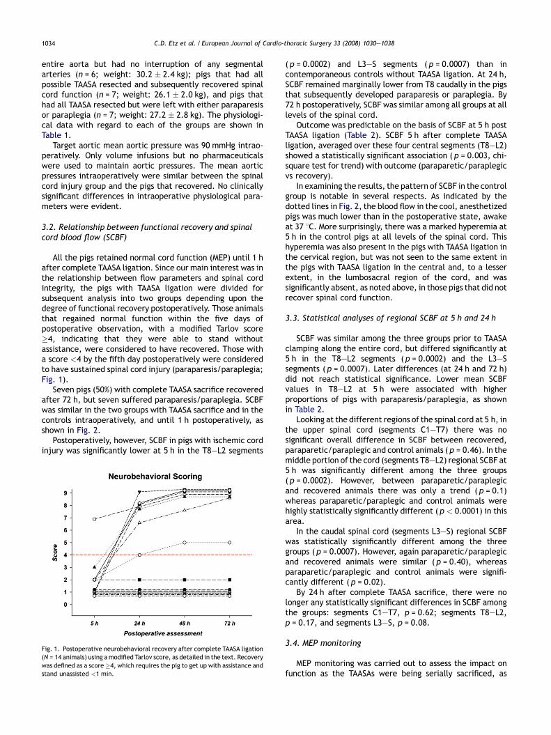
Portions of the spinal cord not used for microsphereanalysis were fixed in 10% formalin solution, embedded inparaffin, and then sectioned transverse to the craniocaudalaxis, with samples at 0.5 cm intervals. Sections 6 mm inthickness were stained with hematoxylin and eosin, exam-ined and scored blindly by an experienced neuropathologistaccording to a schematic grading system which wasdeveloped to classify the ischemic spinal cord damage (ISCD)at each segmental level: 1 = necrosis of single (motor)neurons only; 2 = necrosis of one posterior horn only;3 = necrosis of both posterior horns only; 4 = necrosis ofboth posterior horns + surrounding white matter; 5 = necrosisof both anterior horns only; 6 = central necrosis involvingposterior and anterior horns + parts of white matter;7 = complete necrosis of gray matter only; 8 = completenecrosis of the whole section (Fig. 3).
2.10. Data analysis
For the purposes of data analysis, pigs were classified intoone of three groups: controls that had no segmental arteriesresected, pigs that had all possible TAASA resected and
Table 1Intra- and postoperative data
Variable/group Baseline prior to ligation After TAASA
5 min
MAP (mmHg)Control group 90 � 2 90 � 1Paraparetic/paraplegic group 90 � 8 93 � 15Recovery group 91 � 9 91 � 8
pHControl group 7.52 � 0.04 7.51 � 0.05Paraparetic/paraplegic group 7.48 � 0.06 7.44 � 0.05Recovery group 7.50 � 0.04 7.48 � 0.05
PCO2
Control group 33.0 � 3.8 33.2 � 5.5Paraparetic/paraplegic group 40.0 � 7.9 41.6 � 6.3Recovery group 37.7 � 3.6 40.2 � 7.6
PO2
Control group 364 � 59 362 � 54Paraparetic/paraplegic group 390 � 54 388 � 47Recovery group 381 � 46 357 � 36
HbControl group 10.9 � 0.6 10.1 � 0.6Paraparetic/paraplegic group 10.4 � 1.4 10.8 � 2.1Recovery group 11.4 � 0.7 11.9 � 0.9
O2 — saturation (%)Control group 99.8 � 0.06 99.8 � 0.0Paraparetic/paraplegic group 99.8 � 0.05 99.8 � 0.04Recovery group 99.8 � 0.05 99.8 � 0.06
GlucoseControl group 89 � 42 78 � 35Paraparetic/paraplegic group 115 � 38 133 � 49Recovery group 97 � 32 92 � 42
LactateControl group 1.6 � 0.9 1.6 � 1.1Paraparetic/paraplegic group 1.4 � 0.7 1.5 � 1.1Recovery group 2.5 � 1.7 2.0 � 1.3
Mean aortic pressures (MAP) and blood gas analyses for control (N = 6), paraparetic
subsequently recovered spinal cord function, and pigs thathad all TAASA resected but were left with either paraplegia orparaparesis. All analyses were implemented with SAS soft-ware for the PC version 9.1.3, SAS Institute Inc., Cary, NC.Physiologic data are described by means and standarddeviations in each of these groups. Hierarchical linearmodels, using Proc Mixed, were used to analyze theassociation of flows with groups and segments. In separateanalyses of the three segment regions at the 5 h and 24 h timepoints, flows were modeled as a function of group, segments,and interaction, with repeated observations (over segments)and an unstructured or autoregressive covariance matrix.Pair wise comparisons between groups were obtained assolutions to the fixed effects. The chi-square test for trendwas used for predicting the binary outcome, recovery ofspinal cord function, on the basis of the 5 h flows.
3. Results
3.1. Comparability of experimental groups
The results of this experiment were analyzed by compar-ing three groups: pigs which were operated on to expose the
ligation
1 h 5 h 24 h 72 h
96 � 8 93 � 7 91 � 2 90 � 190 � 9 96 � 15 78 � 15 92 � 1689 � 7 96 � 16 99 � 21 108 � 17
7.44 � 0.05 7.38 � 0.03 7.53 � 0.01 7.49 � 0.027.40 � 0.05 7.39 � 0.07 7.45 � 0.05 7.47 � 0.047.50 � 0.04 7.36 � 0.08 7.47 � 0.04 7.47 � 0.03
39.7 � 8.0 43.7 � 3.9 35.8 � 2.0 38.0 � 2.844.0 � 7.4 40.4 � 5.1 33.3 � 2.0 33.9 � 4.137.4 � 3.5 37.5 � 4.7 32.4 � 2.9 33.6 � 1.9
367 � 23 76 � 18 84 � 5 85 � 8382 � 54 74 � 8 78 � 18 73 � 18364 � 58 72 � 18 87 � 14 76 � 8
9.9 � 0.8 10.3 � 0.8 9.8 � 1.2 9.9 � 1.39.7 � 1.3 10.5 � 1.7 10.8 � 2.6 9.6 � 2.2
11.3 � 1.5 13.7 � 1.1 11.1 � 1.6 9.3 � 1.5
99.8 � 0.0 94.6 � 4.7 97.3 � 0.5 97.0 � 0.899.8 � 0.1 94.7 � 0.7 94.9 � 2.5 94.7 � 3.199.8 � 0.1 92.3 � 5.5 93.8 � 8.0 93.4 � 6.6
83 � 21 119 � 18 94 � 12 94 � 12118 � 33 159 � 102 91 � 19 72 � 1280 � 39 103 � 32 110 � 36 94 � 12
1.3 � 0.8 2.3 � 2.1 0.6 � 0.2 0.7 � 0.51.4 � 0.6 2.7 � 1.3 3.0 � 1.3 1.9 � 1.81.6 � 0.9 5.7 � 2.4 4.1 � 5.0 2.2 � 1.9
/paraplegic (N = 7) and recovered (N = 7) animals.
C.D. Etz et al. / European Journal of Cardio-thoracic Surgery 33 (2008) 1030—10381034
entire aorta but had no interruption of any segmentalarteries (n = 6; weight: 30.2 � 2.4 kg); pigs that had allpossible TAASA resected and subsequently recovered spinalcord function (n = 7; weight: 26.1 � 2.0 kg), and pigs thathad all TAASA resected but were left with either paraparesisor paraplegia (n = 7; weight: 27.2 � 2.8 kg). The physiologi-cal data with regard to each of the groups are shown inTable 1.
Target aortic mean aortic pressure was 90 mmHg intrao-peratively. Only volume infusions but no pharmaceuticalswere used to maintain aortic pressures. The mean aorticpressures intraoperatively were similar between the spinalcord injury group and the pigs that recovered. No clinicallysignificant differences in intraoperative physiological para-meters were evident.
3.2. Relationship between functional recovery and spinalcord blood flow (SCBF)
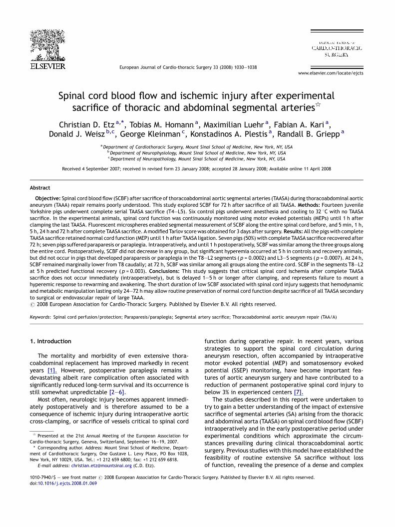
All the pigs retained normal cord function (MEP) until 1 hafter complete TAASA ligation. Since our main interest was inthe relationship between flow parameters and spinal cordintegrity, the pigs with TAASA ligation were divided forsubsequent analysis into two groups depending upon thedegree of functional recovery postoperatively. Those animalsthat regained normal function within the five days ofpostoperative observation, with a modified Tarlov score�4, indicating that they were able to stand withoutassistance, were considered to have recovered. Those witha score <4 by the fifth day postoperatively were consideredto have sustained spinal cord injury (paraparesis/paraplegia;Fig. 1).
Seven pigs (50%) with complete TAASA sacrifice recoveredafter 72 h, but seven suffered paraparesis/paraplegia. SCBFwas similar in the two groups with TAASA sacrifice and in thecontrols intraoperatively, and until 1 h postoperatively, asshown in Fig. 2.
Postoperatively, however, SCBF in pigs with ischemic cordinjury was significantly lower at 5 h in the T8—L2 segments
Fig. 1. Postoperative neurobehavioral recovery after complete TAASA ligation(N = 14 animals) using amodified Tarlov score, as detailed in the text. Recoverywas defined as a score�4, which requires the pig to get up with assistance andstand unassisted <1 min.
( p = 0.0002) and L3—S segments ( p = 0.0007) than incontemporaneous controls without TAASA ligation. At 24 h,SCBF remained marginally lower from T8 caudally in the pigsthat subsequently developed paraparesis or paraplegia. By72 h postoperatively, SCBF was similar among all groups at alllevels of the spinal cord.
Outcome was predictable on the basis of SCBF at 5 h postTAASA ligation (Table 2). SCBF 5 h after complete TAASAligation, averaged over these four central segments (T8—L2)showed a statistically significant association ( p = 0.003, chi-square test for trend) with outcome (paraparetic/paraplegicvs recovery).
In examining the results, the pattern of SCBF in the controlgroup is notable in several respects. As indicated by thedotted lines in Fig. 2, the blood flow in the cool, anesthetizedpigs was much lower than in the postoperative state, awakeat 37 8C. More surprisingly, there was a marked hyperemia at5 h in the control pigs at all levels of the spinal cord. Thishyperemia was also present in the pigs with TAASA ligation inthe cervical region, but was not seen to the same extent inthe pigs with TAASA ligation in the central and, to a lesserextent, in the lumbosacral region of the cord, and wassignificantly absent, as noted above, in those pigs that did notrecover spinal cord function.
3.3. Statistical analyses of regional SCBF at 5 h and 24 h
SCBF was similar among the three groups prior to TAASAclamping along the entire cord, but differed significantly at5 h in the T8—L2 segments ( p = 0.0002) and the L3—Ssegments ( p = 0.0007). Later differences (at 24 h and 72 h)did not reach statistical significance. Lower mean SCBFvalues in T8—L2 at 5 h were associated with higherproportions of pigs with paraparesis/paraplegia, as shownin Table 2.
Looking at the different regions of the spinal cord at 5 h, inthe upper spinal cord (segments C1—T7) there was nosignificant overall difference in SCBF between recovered,paraparetic/paraplegic and control animals ( p = 0.46). In themiddle portion of the cord (segments T8—L2) regional SCBF at5 h was significantly different among the three groups( p = 0.0002). However, between paraparetic/paraplegicand recovered animals there was only a trend ( p = 0.1)whereas paraparetic/paraplegic and control animals werehighly statistically significantly different ( p < 0.0001) in thisarea.
In the caudal spinal cord (segments L3—S) regional SCBFwas statistically significantly different among the threegroups ( p = 0.0007). However, again paraparetic/paraplegicand recovered animals were similar ( p = 0.40), whereasparaparetic/paraplegic and control animals were signifi-cantly different ( p = 0.02).
By 24 h after complete TAASA sacrifice, there were nolonger any statistically significant differences in SCBF amongthe groups: segments C1—T7, p = 0.62; segments T8—L2,p = 0.17, and segments L3—S, p = 0.08.
3.4. MEP monitoring
MEP monitoring was carried out to assess the impact onfunction as the TAASAs were being serially sacrificed, as

C.D. Etz et al. / European Journal of Cardio-thoracic Surgery 33 (2008) 1030—1038 1035
Fig. 2. Regional spinal cord blood flow (SCBF) was determined using fluorescentmicrospheres (as detailed in the text) at various time points following serial ligation ofall segmental arteries (TAASA). No segmental artery sacrifice was carried out in the controls. Pigs in the recovery group subsequently regained hind limb function, butthose in the paraparetic/paraplegia group suffered permanent spinal cord injury. The dashed lines represent baseline flows at 32 8C during anesthesia, and afterawakening, at 37 8C. The three graphs represent cervical and upper thoracic, lower thoracic and upper lumbar, and lumbosacral regions of the spinal cord, asindicated. The first baseline was recorded at 32 8C under anesthesia, and the second at 72 h. It is notable that there were no significant differences among the groupsin the cervical region of the cord. Below this level, however, there was a significant difference between the paraparetic animals and the controls at 5 h, with thecontrols exhibiting a marked hyperemia. There were no significant differences among the groups at the later time points.
described in detail previously, and until 1 h after the last ofthem had been sacrificed: later monitoring is not possiblewithout prolonging the duration of anesthesia. As was seen inearlier studies, MEPs after extensive SA sacrifice were intactat the end of the procedure even in those pigs thatsubsequently suffered a loss of spinal cord function. Thissuggests that critical ischemia occurred in these pigs afterthe last measurement of MEPs had been carried out (1 h afterthe last of the TAASAs had been occluded), at a time whenspinal cord perfusion pressure is still very low, but MEPs canno longer be recorded.
3.5. Histopathological findings
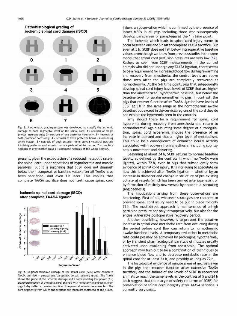
Despite complete TAASA sacrifice, there was no evidenceof necrosis in the cervical and higher thoracic segments ineither paraparetic/paraplegic or recovered animals to thelevel of T7 in all animals (and T9 in recovered animals; Fig. 4).In the lower thoracic and lumbar segments, however,
Table 2Predicting outcome on the basis of SCBF in T8—L2 @ 5 h after TAASA ligation
SCBF (ml/min/g) inT8—L2 @ 5 h aftercomplete TAASA ligation
No. ofanimals
Nos. withparaplegia
%Withparaplegia
<0.08 5 4 80%0.08—0.125 5 3 60%0.125—0.24 5 0 0�0.24 4 0 0
The table shows the segmental flows @ 5 h after complete TAASA ligation,averaged over the four segments (T8—L2) divided into equal size groups, andtheir relation to the chance of paraplegia. This association is statisticallysignificant (p = 0.003, chi-square test for trend).
paraparetic/paraplegic animals showed distinctive necrosisof the gray matter involving the posterior and anterior hornsand surrounding white matter, sparing a thin peripheral rimof white matter below the pia (Figs. 3 and 4) with the centerof ischemic damage between T8 and L2. Although thenecrosis had an abrupt onset between segments T8 and T9 inmost of the paraparetic/paraplegic animals, in otherrespects the areas in which necrosis was found correspondto the vascular territory of the anterior spinal artery.
In pigs that regained function, all segments showed anormal population of neurons and intact gray matter at alllevels at lower magnification. There was no evidence ofcomprehensive segmental necrosis, although higher powerevaluation showed rare minute areas with necrotic cells insections from the lower thoracic (T10—11) and lumbar levels(L1—S; see Fig. 4).
4. Discussion
The blood flow studies described in this report have led toseveral new observations which provide valuable insight intothe response of the spinal cord circulation to extensivesegmental artery sacrifice (TAASA). This has led to a betterunderstanding of the gradual adaptation of the spinal cord toloss of much of its usual vascular input, and an increase in thelikelihood that elimination of postoperative spinal cord injuryfollowing surgery for extensive thoracic and thoracoabdom-inal aneurysms is an achievable goal.
To begin with, SCBF under mild hypothermia andanesthesia, as seen in Fig. 2, is considerably less than theflow when the pig is warm and awake. This relatively reducedflow is not surprising, assuming that autoregulation is
C.D. Etz et al. / European Journal of Cardio-thoracic Surgery 33 (2008) 1030—10381036
Fig. 3. A schematic grading system was developed to classify the ischemicdamage at each segmental level of the spinal cord: 1 = necrosis of single(motor) neurons only; 2 = necrosis of one posterior horn only; 3 = necrosis ofboth posterior horns only; 4 = necrosis of both posterior horns + surroundingwhite matter; 5 = necrosis of both anterior horns only; 6 = central necrosisinvolving posterior and anterior horns + parts of white matter; 7 = completenecrosis of gray matter only; 8 = complete necrosis of the whole section.
present, given the expectation of a reducedmetabolic rate inthe spinal cord under conditions of hypothermia and muscleparalysis. But it is surprising that SCBF does not diminishbelow the intraoperative baseline value after all TAASA havebeen sacrificed, and even 1 h later. This implies thatcomplete TAASA sacrifice does not itself cause spinal cord
Fig. 4. Regional ischemic damage of the spinal cord (ISCD) after completeTAASA sacrifice — paraparetic/paraplegic versus recovery group. The Y-axisshows the grade of the ischemic damage and a corresponding low power (2�)transverse section of the spinal cord, stained with hematoxylin and eosin, frompigs 3 days after extensive sacrifice of segmental arteries as examples. Thecord segments from which the sections are taken are indicated at the X-axis.
injury, an observation which is confirmed by the presence ofintact MEPs in all pigs including those who subsequentlydevelop paraparesis or paraplegia at the 1 h time point.
The ischemia which leads to spinal cord injury seems tooccur between one and 5 h after complete TAASA sacrifice. Buteven at 5 h, SCBF does not fall below intraoperative baselinevalues, even thoughweknow fromprevious studies in the samemodel that spinal cord perfusion pressures are very low [12].Rather, as seen from SCBF measurements in the controlanimals who did not undergo any TAASA ligation, there seemsto be a requirement for increased bloodflowduring rewarmingand recovery from anesthesia: the control levels are abovethose seen after the pigs are completely recovered atnormothermia. At the 5-h time point, pigs that subsequentlydevelop spinal cord injury have levels of SCBF that are higherthan the anesthetized, hypothermic baseline, but below thebaseline level for awake normothermic pigs. In contrast, thepigs that recover function after TAASA ligation have levels ofSCBF at 5 h in the same range as the normothermic awakebaseline, but except in the cervical regions of the cord they donot exhibit the hyperemia seen in the controls.
Why should there be a requirement for spinal cordhyperemia during recovery from anesthesia and return tonormothermia? Again assuming some degree of autoregula-tion, spinal cord hyperemia implies the presence of anincrease in demand and thus a higher level of metabolism.This could be a consequence of enhanced neural activityassociated with recovery from anesthesia, including sponta-neous movement and shivering.
Beginning at about 24 h, SCBF returns to normal baselinelevels, as defined by the controls in whom no TAASA wereligated, within 72 h, even in pigs that subsequently showevidence of spinal cord injury. It is intriguing to speculate onhow this is achieved after TAASA ligation — whether by anincrease in diameter and change in structure of pre-existingcollateral vessels (which has been termed arteriogenesis), orby formation of entirely new vessels by endothelial sprouting(angiogenesis).
The implications arising from these observations areheartening. First of all, whatever strategies are required toprevent spinal cord injury need to be put in place for only72 h. The most direct approach is maintenance of a highperfusion pressure not only intraoperatively, but also for theentire vulnerable postoperative recovery period.
Another possibility, however, is to prevent the putativeincrease in spinal cord metabolic rate from occurring duringthe period before cord flow can return to normothermicawake baseline levels. A temporary reduction in metabolicrate could possibly be achieved by prolonging hypothermia,or by transient pharmacological paralysis of muscles usuallyactivated upon awakening from anesthesia. The optimalapproach may turn out to be a combination of techniques toenhance blood flow and to decrease metabolic rate in thespinal cord for at least 24 h, and possibly as long as 72 h.
The histological evidence of minute areas of necrosis evenin the pigs that recover function after extensive TAASAsacrifice, and the failure of the levels of SCBF in recoveredanimals to reach the same levels as the controls at 5 and 24 hboth suggest that the margin of safety (in terms of SCBF) forpreservation of spinal cord integrity after TAASA sacrifice iscurrently very small.

C.D. Etz et al. / European Journal of Cardio-thoracic Surgery 33 (2008) 1030—1038 1037
5. Conclusion
This study of spinal cord perfusion has documented thatextensive ligation of TAASA does not diminish SCBF belowintraoperative baseline values. What distinguishes pigs thatdevelop spinal cord injury from those who recover normalfunction is a failure to mount an adequate hyperemicresponse during awakening from anesthesia and rewarming.But even in animals with spinal cord injury, flow returns tonormal within 72 h, suggesting that a combination ofstrategies to optimize SCBF and decrease spinal cordmetabolism for 72 h postoperatively could reliably preventparaplegia following extensive TAASA sacrifice. This wouldenable endovascular treatment of TAA/A, offering therapy tomany patients too old and frail to be considered for opensurgical treatment.
References
[1] Svensson LG. Paralysis after aortic surgery: in search of lost cord function.Surgeon 2005;3:396—405.
[2] Coselli JS, LeMaire SA, Miller 3rd CC, Schmittling ZC, Koksoy C, Pagan J,Curling PE. Mortality and paraplegia after thoracoabdominal aorticaneurysm repair: a risk factor analysis. Ann Thorac Surg 2000;69:409—14.
[3] Bachet J, Guilmet D, Rosier J, Cron C, Dreyfus G, Goudot B, Piquois A,Brodaty D, Dubois C, de Lentdecker P. Protection of the spinal cord duringsurgery of thoraco-abdominal aortic aneurysms. Eur J Cardiothorac Surg1996;10:817—25.
[4] Kouchoukos NT, Rokkas CK. Hypothermic cardiopulmonary bypass forspinal cord protection: rationale and clinical results. Ann Thorac Surg1999;67:1940—2. discussion 1953—48.
[5] Svensson LG, Patel V, Robinson MF, Ueda T, Roehm Jr JO, Crawford ES.Influence of preservation or perfusion of intraoperatively identified spinalcord blood supply on spinal motor evoked potentials and paraplegia afteraortic surgery. J Vasc Surg 1991;13:355—65.
[6] Yamauchi T, Takano H, Nishimura M, Matsumiya G, Sawa Y. Paraplegia andparaparesis after descending thoracic aortic aneurysm repair: a riskfactor analysis. Ann Thorac Cardiovasc Surg 2006;12:179—83.
[7] Etz CD, Halstead JC, Spielvogel D, Shahani R, Lazala R, Homann TM, WeiszDJ, Plestis K, Griepp RB. Thoracic and thoracoabdominal aneurysmrepair: is reimplantation of spinal cord arteries a waste of time? AnnThorac Surg 2006;82:1670—7.
[8] Strauch JT, Spielvogel D, Lauten A, Zhang N, Shiang H,Weisz D, Bodian CA,Griepp RB. Importance of extrasegmental vessels for spinal cord bloodsupply in a chronic porcine model. Eur J Cardiothorac Surg 2003;24:817—24.
[9] Strauch JT, Spielvogel D, Lauten A, Zhang N, Shiang H,Weisz D, Bodian CA,Griepp RB. Importance of extrasegmental vessels for spinal cord bloodsupply in a chronic porcine model. Rev Port Cir Cardiothorac Vasc2003;10:185—91.
[10] Strauch JT, Lauten A, Spielvogel D, Rinke S, Zhang N, Weisz D, Bodian CA,Griepp RB. Mild hypothermia protects the spinal cord from ischemicinjury in a chronic porcine model. Eur J Cardiothorac Surg2004;25:708—15.
[11] Etz CD, Homann TM, Plestis KA, Zhang N, Luehr M, Weisz DJ, Kleinman G,Griepp RB. Spinal cord perfusion after extensive segmental artery sacri-fice: can paraplegia be prevented? Eur J Cardiothorac Surg 2007;31:643—8.
[12] Etz CD, Luehr M, Kari F, Bodian CA, Smego S, Plestis KA, Griepp RB.Paraplegia after extensive thoracic and thoracoabdominal aortic aneur-ysm repair: does critical spinal cord ischemia occur postoperatively? JThorac Cardiovasc Surg 2008;135:324—30.
Appendix A. Conference discussion
Dr J. Bachet (Paris, France): Thank you for sharing with us this veryimportant experimental work from the laboratory of Dr Randall Griepp atMount Sinai Hospital in New York.
It is in perfect continuity with several other published experimentalstudies from your group aimed at elucidating the patho-physiology of the spinalcord vascularization and the occurrence of spinal cord injury during surgery ofthe thoracic or thoracoabdominal aorta.
It is, in particular, the obvious and logical continuation of the study thatyou have presented last year in this very Association and that was publishedrecently in the EJCTS. It represents another piece of evidence that the‘Collateral Network Concept’ as developed by Dr Griepp’s group in New Yorkand Dr Biglioli also in Milan and published as early as 1996 is a reality whichmight in the near future drastically modify our surgical techniques andcertainly dramatically reduce, if not totally suppress, the dreadful perspectiveof postoperative paraplegia.
The animal model used in the present study was exactly the same as theone used in your previously published work, right?
Dr Etz: That’s correct.
Dr Bachet: All the animals were identical and underwent exactly the sameprotocol. However, seven, that is 50%, were paraplegic and seven were not.And apparently, the occurrence of spinal cord injury could not be predictedintraoperatively or before the fifth postoperative hour.
But, and this will be my first question, do you think that all animals wereactually identical? That is, could the spinal cord vascularization and, inparticular, the spinal anterior arterial axis or the collateral network comingfrom the subclavian and medial lumbar arteries have variations which couldexplain why some animals are more sensitive to the segmental arteries’exclusion than others?
In other words, do you think that any anatomical or radiological study ofthis vascularization with any kind of imaging technique could determinepreoperatively which animals are at higher risk?
It is quite interesting to note that 5 h after exclusion of the last segmentalarteries an important hyperhemic rebound of the spinal cord blood flow couldbe observed in the normal or recovering animals but not in the paraplegicanimals.
My second question is, then: is the spinal cord injury the consequence ofthis absence of hyperhemia as you infer, or on the contrary, is the absence ofhyperhemia the result of an already reduced metabolic demand due to thealready pathological status of the neurons? In other words, could the low flowduring and immediately after the segmental arteries’ exclusion trigger somedelayed phenomena such as excitotoxicity and/or apoptosis? In this regard,could the dosages of glutamate or TUNEL histological studies, for example,give any information about those processes?
Another obvious point of interrogation lies, as it is often the case, in thetranslation of those experimental results to the clinical setting, of course. Youstate indeed in your paper that this ‘experimental model closely simulates theprocedure for resection of descending thoracic and thoracoabdominalaneurysms clinically in your institution.’ Is it so?
The pig and human anatomies are not exactly comparable. In particular,the pig has more segmental arteries and only one at each level which thendivides in two, right?
Dr Etz: Correct.
Dr Bachet: In addition, your animals were perfectly healthy. They had novoluminous aneurysms, and dissection and exclusion of the segmental arterieswas certainly easy. This is not the case in many patients in whom the segmentalarteries are often very difficult to find and to exclude before opening theaneurysm. And for my personal information and teaching, how do you do that inpatients?
More importantly, most patients are operated on for huge atheromatouslesions or chronic dissections. Many segmental arteries are already occludedand the collateral network supplied by the subclavian and hypogastric systemsmay be jeopardized. Clamping the already occluded arteries can be a loss oftime, whereas clamping the few remaining might suppress an importantelement of an already fragile blood supply.
Again, do you think that in some selected patients with heavy atheroma orhuge extended lesions or previous aortic surgery a radiological assessment ofthe cord vascularization is worthless?
Similarly, it is noteworthy that in your experiments the animals werenot submitted to CSF drainage which is presently considered by mostgroups as part of the routine armamentarium in thoracic and thoracoab-dominal aortic surgery. I am aware that CSF drainage in pigs is difficultand could be troublesome in the postoperative aftermath. Nevertheless,don’t you think that absence of CSF drainage in your pigs could haveimpaired or worsened the spinal cord blood flow and precipitated the cordischemia?
C.D. Etz et al. / European Journal of Cardio-thoracic Surgery 33 (2008) 1030—10381038
Those questions might be irrelevant. Indeed, your group has published in2006 a clinical experience using the ‘Collateral Network Concept’ in 100consecutive patients operated between 2002 and 2004 with a paraplegia/paraparesis rate of 2%, which, to my knowledge, is among the lowest everpublished.
If I dare to use an old surgical joke, I would say that now that you haveproved that it works in human beings, you are trying to prove that it works inpigs as well.
Dr Etz: Let me first start with the anatomy in pig as compared to humans.That is themost obvious question we have to answer, and since wewere curiousas to how it actually compared, we did cast studies prior to these experiments.What you see on the left is a cast of the human vascular system focusing on theabdominal lower thoracic aorta and showing the segmental arteries, and onthe right side, a cast we produced in the pig.
You are absolutely right: the pig has a T13 segmental artery — one morethan in humans — and the majority of segmental arteries divide about 5 mmafter they take origin from the aorta. We believe that the anatomy, which weinvestigated with about 20 of these casts in pigs, has very little variability inthe pigs we are using, and that the anatomy is very similar to what we see inhumans. Here we can see the 13 segmental arteries. Not all of them have onesingle origin from the aorta. In the pig thoracic aorta, there are frequentlysome segmental arteries very similar to human arteries, with two origins. Wedo verify in all animals at post-mortem that all segmental arteries have beensacrificed.
We also do not think that there is a huge variability in the anatomy of thecollateral inflow, and it is also not very different in pigs and humans. Thehypogastric arteries are a little greater in caliber in pigs, but they are prettyclose to what one sees in the humans.
You are also right that we do not have access to atherosclerotic pigs, muchas we would love that. In humans, segmental arteries frequently are occludedprior to surgery. We are currently trying to investigate the remodelingactivities of the paraspinal and segmental artery network, and we can see thatchanges set in after experimental segmental artery clamping which may besimilar to what occurs after atherosclerotic occlusion of the segmentalarteries. We believe that when occlusion of the segmental arteries occurs, itmay provoke development of the collateral network, making it not moredangerous, but possibly even safer to clamp the segmental arteries whichremain.
As to the predictability of spinal cord blood flow recovery: this is the tablethat shows the predictability of outcome based on spinal cord blood flow. Wecan see that if the flow is below a certain threshold, 80% of animals developparaplegia, and that none have spinal cord injury if the flow is only about 50%higher. So it is critical to have an adequate blood flow from T8 to about L2. Wedo not think that reduced flow is secondary to earlier ischemia-inducednecrosis, since all animals have intact motor-evoked potentials until just
before warming and awakening. We think that variability of collateral flow,which is probably dependent upon minor anatomical variations, explainsdifferences in the ability of spinal cord hyperemia to occur.
With regard to clinical comparisons, one must bear in mind that althoughthe paraplegia rate was only 2%, the average number of sacrificed segmentalvessels in our patients is much smaller than in our experimental model. Ourultimate aim, however, is to learn how to manipulate the collateral network insuch a way that, in the clinical situation, cord viability can be assured routinelywith sacrifice of all segmental vessels. This is an absolute prerequisite for safeendovascular treatment of the entire thoracoabdominal aorta.
With regard to cerebrospinal fluid drainage, you are absolutely correct thatwe would expect to reach even lower paraplegia rates with its use: during theyear in question, we had 50% paraplegia without CSF drainage. In the last yearwe have had a recovery rate of 60—70%. We are convinced that if we could doCSF drainage, we would lower the paraplegia rate even further.
CSF drainage is not impossible in the pig; we have done it in the past. But itis very complicated to have the pigs survive for 5 days with yet anotherincision, and that is why we decided not to do it in these experiments.
Dr F. Beyersdorf (Freiburg, Germany): One of the most frequently askedquestion in the field of endovascular aortic surgery is why there is such a lowparaplegia rate in endovascular treatment? You and other members of DrGriepp’s group have shown that open conventional surgery may also result inlow paraplegia rates, if you ligate all segmental branches before opening thelumen of the aorta.
Now, in your present study you sacrificed the thoracic and lumbarsegmental arteries and wouldn’t you expect a low paraplegia rate and a highpercentage of recovery?
Dr Etz: Yes.
Dr Beyersdorf: So my question is, if the hypothesis is correct thatparaplegia results from a steal phenomenon (bleeding from these branchesduring open surgery), why is in your model the paraplegia rate still 30%, 40%,50%? Is it due to the fact, that you have not done extra neuroprotectivemeasures what Dr Bachet asked for, or is this related to the model you haveused?
Dr Etz: You have to consider one very important point here. We take allsegmental arteries arising from the aorta, so there is not a single one left. Thatis not the usual case if you do stenting, and it only occurs in perhaps 10% even ofopen thoracoabdominal aortic aneurysmal repairs.
We really take every single artery that is there, from T4 to L5: most of thepigs lose 15 segmental arteries. That is 80% of the inflow. We created theseextreme conditions deliberately, to better demonstrate differences betweenexperimental groups. We have no doubt that we could reach recovery rates of90—100% if we left the segmental arteries in this critical area in the lumbarpart of the aorta intact.