Immune-Mediated Mechanisms of Parasite Tissue Sequestration during Experimental Cerebral Malaria
12
of July 7, 2015. This information is current as Cerebral Malaria Tissue Sequestration during Experimental Immune-Mediated Mechanisms of Parasite Christian R. Engwerda Duke, Donald P. McManus, Alex Loukas, Geoff R. Hill and Good, Alberto Pinzon-Charry, Mark S. Pearson, Mary G. Tania F. de Koning-Ward, Rachel J. Lundie, Michael F. Wilson, Gladys Yeo, Christian Pieper, Brendan S. Crabb, Fabian de Labastida Rivera, Louise M. Randall, Yana A. Fiona H. Amante, Ashraful Haque, Amanda C. Stanley, http://www.jimmunol.org/content/185/6/3632 doi: 10.4049/jimmunol.1000944 August 2010; 2010; 185:3632-3642; Prepublished online 18 J Immunol Material Supplementary 4.DC1.html http://www.jimmunol.org/content/suppl/2010/08/18/jimmunol.100094 References http://www.jimmunol.org/content/185/6/3632.full#ref-list-1 , 28 of which you can access for free at: cites 71 articles This article Subscriptions http://jimmunol.org/subscriptions is online at: The Journal of Immunology Information about subscribing to Permissions http://www.aai.org/ji/copyright.html Submit copyright permission requests at: Email Alerts http://jimmunol.org/cgi/alerts/etoc Receive free email-alerts when new articles cite this article. Sign up at: Print ISSN: 0022-1767 Online ISSN: 1550-6606. Immunologists, Inc. All rights reserved. Copyright © 2010 by The American Association of 9650 Rockville Pike, Bethesda, MD 20814-3994. The American Association of Immunologists, Inc., is published twice each month by The Journal of Immunology by guest on July 7, 2015 http://www.jimmunol.org/ Downloaded from by guest on July 7, 2015 http://www.jimmunol.org/ Downloaded from
-
Upload
qimrberghofer -
Category
Documents
-
view
1 -
download
0
Transcript of Immune-Mediated Mechanisms of Parasite Tissue Sequestration during Experimental Cerebral Malaria

of July 7, 2015.This information is current as
Cerebral MalariaTissue Sequestration during Experimental Immune-Mediated Mechanisms of Parasite
Christian R. EngwerdaDuke, Donald P. McManus, Alex Loukas, Geoff R. Hill and Good, Alberto Pinzon-Charry, Mark S. Pearson, Mary G.Tania F. de Koning-Ward, Rachel J. Lundie, Michael F. Wilson, Gladys Yeo, Christian Pieper, Brendan S. Crabb,Fabian de Labastida Rivera, Louise M. Randall, Yana A. Fiona H. Amante, Ashraful Haque, Amanda C. Stanley,
http://www.jimmunol.org/content/185/6/3632doi: 10.4049/jimmunol.1000944August 2010;
2010; 185:3632-3642; Prepublished online 18J Immunol
MaterialSupplementary
4.DC1.htmlhttp://www.jimmunol.org/content/suppl/2010/08/18/jimmunol.100094
Referenceshttp://www.jimmunol.org/content/185/6/3632.full#ref-list-1
, 28 of which you can access for free at: cites 71 articlesThis article
Subscriptionshttp://jimmunol.org/subscriptions
is online at: The Journal of ImmunologyInformation about subscribing to
Permissionshttp://www.aai.org/ji/copyright.htmlSubmit copyright permission requests at:
Email Alertshttp://jimmunol.org/cgi/alerts/etocReceive free email-alerts when new articles cite this article. Sign up at:
Print ISSN: 0022-1767 Online ISSN: 1550-6606. Immunologists, Inc. All rights reserved.Copyright © 2010 by The American Association of9650 Rockville Pike, Bethesda, MD 20814-3994.The American Association of Immunologists, Inc.,
is published twice each month byThe Journal of Immunology
by guest on July 7, 2015http://w
ww
.jimm
unol.org/D
ownloaded from
by guest on July 7, 2015
http://ww
w.jim
munol.org/
Dow
nloaded from

The Journal of Immunology
Immune-Mediated Mechanisms of Parasite TissueSequestration during Experimental Cerebral Malaria
Fiona H. Amante,*,† Ashraful Haque,*,† Amanda C. Stanley,*,† Fabian de Labastida Rivera,*,†
Louise M. Randall,*,†,‡ Yana A. Wilson,*,† Gladys Yeo,*,†,‡ Christian Pieper,*,†,x
Brendan S. Crabb,{ Tania F. de Koning-Ward,‖ Rachel J. Lundie,# Michael F. Good,*,†
Alberto Pinzon-Charry,*,† Mark S. Pearson,*,† Mary G. Duke,*,† Donald P. McManus,*,†
Alex Loukas,*,† Geoff R. Hill,*,† and Christian R. Engwerda*
Cerebral malaria is a severe complication of malaria. Sequestration of parasitized RBCs in brain microvasculature is associatedwith disease pathogenesis, but our understanding of this process is incomplete. In this study, we examined parasite tissue seques-tration in an experimental model of cerebral malaria (ECM). We show that a rapid increase in parasite biomass is strongly as-sociated with the induction of ECM, mediated by IFN-g and lymphotoxin a, whereas TNF and IL-10 limit this process.Crucially, we discovered that host CD4+ and CD8+ T cells promote parasite accumulation in vital organs, including the brain.Modulation of CD4+ T cell responses by helminth coinfection amplified CD4+ T cell-mediated parasite sequestration, whereasvaccination could generate CD4+ T cells that reduced parasite biomass and prevented ECM. These findings provide novel insightsinto immune-mediated mechanisms of ECM pathogenesis and highlight the potential of T cells to both prevent and promoteinfectious diseases. The Journal of Immunology, 2010, 185: 3632–3642.
Cerebral malaria (CM) is a severe complication of blood-stage Plasmodium falciparum infection. Severe malariasyndromes, including cerebralmalaria, account for∼800,000
deaths worldwide each year, with 89% of mortalities occurring inAfrica, 88% being children under the age of 5 y (1). The pathogenesisof CM is poorly understood, but cerebral pathology is associated withthe sequestration of mature parasitized RBCs (pRBCs) in the micro-vasculature of tissues (2, 3). This characteristic feature ofP. falciparuminfection serves as an immune evasion strategy by the parasite thatprevents the removal of pRBCs in the spleen, thus facilitating parasitesurvival (3–5). Although advantageous for the parasite, this strategyconcentrates malaria parasites, and the metabolic and inflammatoryresponses triggered by them, in vital organs such as the brain (6).Parasite sequestrationmay lead tovascular obstruction, endothelial cellactivation, and the production of proinflammatory cytokines (reviewedinRefs. 6, 7). In addition to parasite-induced pathology, a large body ofwork indicates that host immune responses to parasites also play an
important role in CM pathogenesis (8). A robust proinflammatory re-sponse mediated by activated T cells and cytokines, as well as re-cruitment of activated leukocytes to the brain, have been associatedwith CM (9, 10).Experimental cerebral malaria (ECM) caused by Plasmodium
berghei ANKA (PbA) infection in mice displays key features ofhuman CM (reviewed in Refs. 6, 11). ECM-susceptible mice de-velop a neurologic condition characterized by paralysis, ataxia,convulsions, and coma 6–8 d after PbA infection, resulting in 80–100% mortality at relatively low peripheral blood parasitemia(11). Both CD4+ and CD8+ T cells have been implicated in ECMdevelopment (12–15), and the spleen is thought to play a centralrole in priming PbA-specific T cell responses (16, 17). The re-cruitment of activated malaria-specific CD8+ T cells to the brain isobserved in late-stage ECM (18, 19), and mice depleted of CD8+
T cells are protected from disease (14). Although the precisemechanisms by which CD8+ T cells mediate cerebral pathology isstill unclear, it has been suggested that they may damage cerebralmicrovascular endothelial cells in a perforin-dependent manner(14, 20, 21). Local production of proinflammatory cytokines suchas TNF (22), IFN-g (23), and lymphotoxin a (LTa) (24) is alsocritical for ECM pathogenesis. CD4+ T cell depletion studies havedemonstrated a causal role for this T cell subset in the induction ofECM by a mechanism thought to involve IFN-g production, whichamplifies local and systemic inflammatory cascades (12, 13, 15).The combined effects of CD4+ and CD8+ T cell activation arepostulated to lead to blood–brain barrier breakdown with vascularleakage and/or hemorrhaging into the brain (25).In contrast to ECM, findings from nonlethal mouse models of
malaria indicate that CD4+ T cells are required for the devel-opment of protective immunity and parasite control (26, 27). IFN-g plays a central role in limiting parasite growth through the ac-tivation of macrophages (28, 29) and their release of parasiticidalmediators and promoting the generation of cytophilic Abs (9, 30–33). Thus, CD4+ T cells are not only critical for the effectivecontrol of peripheral blood parasitemia, but they are also key
*Australian Centre for Vaccine Development; †Queensland Institute of MedicalResearch; ‡University of Queensland, Brisbane, Queensland; {Burnet Institute,Melbourne; ‖Deakin University School of Medicine, Geelong, Victoria, Australia;xWestfalische Wilhelms-Universitat, Munster, Germany; and #School of Biologi-cal Sciences, Edinburgh University, Edinburgh, United Kingdom
Received for publication March 23, 2010. Accepted for publication July 13, 2010.
This work was supported by grants from the Australian National Health and MedicalResearch Council.
Address correspondence and reprint requests to Dr. Christian R. Engwerda, Queens-land Institute of Medical Research, 300 Herston Road, Herston, Queensland 4006,Australia. E-mail address: [email protected]
The online version of this article contains supplemental material.
Abbreviations used in this paper: CM, cerebral malaria; ECM, experimental cerebralmalaria; GIT, gastrointestinal tract; LTa, lymphotoxin a; nRBC, normal RBC; PbA,Plasmodium berghei ANKA; PbAluc, PbA line (231c1l) expressing luciferase; PbG,transgenic PbA parasites expressing only GFP; PbTg, P. berghei parasites expressingMHC class II-restricted OVA epitopes fused to GFP under the control of the ef1-apromoter; p.i., postinfection; pRBC, parasitized RBC; SEA, Schistosoma mansoni eggAg; Treg, regulatory T cell.
Copyright! 2010 by TheAmericanAssociation of Immunologists, Inc. 0022-1767/10/$16.00
www.jimmunol.org/cgi/doi/10.4049/jimmunol.1000944
by guest on July 7, 2015http://w
ww
.jimm
unol.org/D
ownloaded from

mediators of ECM pathology, highlighting the delicate balancethat exists between host-mediated control of infection and devel-opment of disease. Defining the factors that distinguish patho-genic from protective CD4+ T cell responses is important to helpprevent severe malaria.Until recently, studying the relationship between peripheral blood
parasitemia, parasite tissue sequestration, and disease severity hasbeen difficult. Typically, peripheral blood parasitemia is a poorpredictor of parasite biomass and disease severity (34), because itonly measures circulating immature parasites, not tissue-sequesteredmature parasites (35). Although most studies on ECM focus onneurological complications, PbA infection causes multiorgan disease(36), particularly in the lung (37). We and others have employedtransgenic, luciferase-expressing PbA tomeasure parasite biomass inperipheral tissues (38–42). However, the contribution of host-derivedfactors to parasite sequestration in deep tissue sites during ECM iscompletely unknown. In this study, we show that PbA sequestrationin various organs, including the brain, is associated with the onset ofECM. Furthermore, we demonstrate that T cell-mediated immunemechanisms promote this process. Finally, using two models of im-mune modulation, we demonstrate that helminth coinfection exac-erbates CD4+ T cell-dependent parasite sequestration in deep tissuesites, whereas vaccine-induced CD4+ T cell responses can limit thisprocess and protect from ECM.
Materials and MethodsMice
Female C57BL/6 mice (6–8 wk of age) were purchased from the AnimalResources Centre (Canning Vale, Western Australia, Australia) andmaintained in the animal facility of the Queensland Institute of MedicalResearch (Herston, Queensland, Australia) under specific pathogen-free con-ditions. B6.RAG12/2 (43), B6.SJL.Ptprca 3 OT-II (44), B cell-deficient B6.mMT (45), B6.IFN-g2/2 (46), B6.LTa2/2 (47), and B6.TNF2/2mice (48) werebred and maintained in the Queensland Institute of Medical Researchanimal facility. All of the animal procedures were approved by theQueensland Institute of Medical Research Animal Ethics Committee.Splenectomized C57BL/6 mice were generated in the Queensland Instituteof Medical Research animal facility. Briefly, mice were anesthetized, andan incision was made in the skin and peritoneum of the left side of thebody. The splenic vascular pedicle was ligated and heat-cauterized, and thespleen was excised. The peritoneal cavity then was sutured, and the skinwas stapled. Sham-splenectomized mice underwent the same procedure,except that the spleen was not removed. Staples were removed 7 d aftersurgery, and mice were allowed 4 wk to recover prior to PbA infection.
Parasites and infections
A transgenic PbA line (231c1l) expressing luciferase (PbAluc) and GFPunder the control of the ef1-a promoter (38) was used for all of theexperiments unless otherwise stated. For adoptive transfer experiments,transgenic P. berghei parasites expressing MHC class II-restricted OVAepitopes fused to GFP under the control of the ef1-a promoter (PbTg) wereused (18). Transgenic PbA parasites expressing only GFP (PbG) were usedas control parasites. Mice infected with transgenic PbA parasites wereprovided with drinking water containing pyrimethamine (10 mg/ml) toselect for drug-resistant transfectants (49). All of the PbA infections wereestablished from parasites passaged in C57BL/6 mice once. All of the mice,unless otherwise stated, were challenged with 105 PbA pRBCs i.v. Pe-ripheral parasitemia was assessed from Diff-Quick (Lab Aids, Narrabeen,Australia) stained thin blood smears. Mice were examined daily for clinicalsigns of ECM and scored as described previously (38). Hemoglobin levelswere measured using the HemoCue Hb 201 analyzer according to manu-facturer’s instructions (HemoCue, Angelholm, Sweden). For experimentalinfection with Schistosoma mansoni, mice were infected percutaneouslywith either 80–100 (high dose) or 30–40 (low dose) S. mansoni cercariae asdescribed previously (50). Infections were allowed to establish in C57BL/6mice over an 8-wk period prior to infection with PbA.
Immunizations
PbA Ag was prepared as described previously (51, 52). Briefly, blood fromPbA-infected B6.LTa2/2 mice was collected when peripheral blood par-asitemia reached 35–40%. Blood was centrifuged, and the pellet was
subjected to two rounds of RBC lysis in distilled water. The parasite pelletwas then washed twice with PBS and subjected to three freeze–thaw cyclesat 270 and 37˚C. The parasite lysate was then disrupted further by passingthrough a syringe three times with a 26-gauge needle. An amount of PbAlysate Ag equivalent to106 PbA pRBCs in a volume of 50 ml was admixedwith an equal volume of aluminum hydroxide (Imject Alum; Pierce,Rockford, IL). Mice were immunized s.c. with 100 ml of this PbA lysatevaccine on the abdomen. For experiments modulating regulatory T cell(Treg) function, mice were administered 0.5 mg anti-CD25 mAb (PC61;American Type Culture Collection, Manassas, VA) i.p. 1 d prior to vaccinepriming. Three weeks after primary immunization, the mice were boostedi.p. twice, 2 wk apart, with the same amount of PbA lysate diluted in PBS.Two weeks later, mice were challenged i.v. with 105 PbAluc pRBCs.
In vivo bioluminescence imaging
Bioluminescence was detected with the In Vivo Imaging System 100 (Xeno-gen, Alameda, CA). At selected time points, PbAluc-infected mice wereanesthetized with isoflurane and injected s.c. with 150 mg/kg D-luciferin(Xenogen) 6 min before imaging. Mice were then killed by CO2 asphyxiationand subjected to intracardial perfusion with 20 ml ice-cold PBS to removecirculating blood and allow only the measurement of pRBCs sequestered intissue microvasculature. Bioluminescence was calculated as photons persecond per square centimeter per steridian using Living Image (Xenogen)and IGOR Pro software (WaveMetrics, Lake Oswego, OR).
Histology
Lung and brain tissue were removed from mice with or without cardiacperfusion and processed for wax embedding and preparing tissue sectionsfor H&E staining, as described previously (24).
Immunofluorescence and confocal microscopy
After cardiac perfusion, lungs and brains were removed frommice and fixedby immersion in 4% (w/v) paraformaldehyde for 1–1.5 h. Organs were thenincubated in a 30% (w/v) sucrose solution overnight and frozen in Tissue-Tek OCT (ProSciTech, Thuringowa, Queensland, Australia) on dry ice.Seven-micrometer sections were cut and stored in the dark at 280˚C untilrequired. Slides were brought to room temperature, and membranes werepermeabilized with 0.5% Triton X-100 (Sigma-Aldrich, Castle Hill, NewSouth Wales, Australia) for 10 min at room temperature in a humidifyingchamber. Slides were washed three times for 5 min in PBS. Slides wereblocked with 5% donkey serum for 30 min at room temperature and thenwashed once with PBS. The following primary and secondary Abs wereused for staining: anti–VCAM-1 (rat) (Biolegend, San Diego, CA), anti-GFP (rabbit), anti-rat Alexa Fluor 594 (donkey), and anti-rabbit Alexa 488(donkey) (Invitrogen, Mount Waverley, Victoria, Australia). After 1 h ofincubation with primary Abs, slides were washed three times for 5 minwith PBS. Sections were then incubated with secondary Abs, washed, andmounted with ProLong Gold antifade reagent with DAPI (Invitrogen).Confocal microscopy was performed using the TCS SP2 Confocal System(Leica Microsystems, Deerfield, IL) equipped with ArKr/HeNe lasers andLeica Confocal Software (version 2.61; Leica Microsystems).
Proliferation assays
CD4+ T cells were purified from splenocytes taken from PbA-infected miceby MACS (.90% purity) (Miltenyi Biotec, Bergisch Gladbach, Germany).CD4+ T cells resuspended in RPMI 1640 medium, 10% (v/v) FCS, andantibiotics were seeded in 96-well round-bottom plates (5 3 105 cells permilliliter). Irradiated (25 Gy) naive syngeneic splenocytes were added (1 3107 cells per milliliter) as a source of APCs. Cells were stimulated with PbApRBCs (5 3 106 cells per milliliter), S. mansoni egg Ag (SEA) (15 mg/ml),or Con A (2.5 mg/ml). Medium alone and an equivalent number of normalRBCs (nRBCs) served as negative controls. Cells were incubated at 37˚C in5% (v/v) CO2 for 3 d. Supernatants were collected and stored at270˚C untilassayed for cytokine levels. Proliferation was measured by [3H]thymidineincorporation during the final 16–18 h of a 4-d culture.
Measurement of serum and culture supernatant cytokine levels
Serum cytokine levels were measured using BD Cytometric Bead ArrayFlex Sets and a FACSArray plate reader (BD Biosciences, Franklin Lakes,NJ) according to the manufacturer’s instructions.
In vivo Ab-mediated cell depletion and cytokine blockade
Micewere administered 0.5 mg anti-CD4mAb (clone YTS191.1) or controlrat IgG (Sigma-Aldrich) i.p. 1 d prior to PbAluc infection and 4 d post-infection (p.i.). CD8+ T cell depletion was performed by administering
The Journal of Immunology 3633
by guest on July 7, 2015http://w
ww
.jimm
unol.org/D
ownloaded from

0.5 mg anti-CD8b mAb (clone 53-5.8) i.p. on the day of PbAluc infectionand every second day thereafter. Control animals received the same amountof rat IgG. For IL-10 blockade, a single injection of anti–IL-10R mAb(1 mg; clone 1B1.3a) or control rat IgG was administered i.p. 1 d prior toPbAluc infection. TNF and LTa blockade was performed by administeringTNFR2-Ig (Enbrel; Amgen, Thousand Oaks, CA), a fusion protein that hasbeen shown previously to inhibit the functional activity of both murine TNFand soluble LTa homotrimers (53). A total of 0.2 mg TNFR2-Ig was ad-ministered i.p. on the day of PbAluc infection and every second daythereafter. Control mice received control human IgG (Intragam; Com-monwealth Serum Laboratories, Parkville, Victoria, Australia).
Adoptive transfer of OT-II cells
CD4+ T cells were purified from the spleens of CD45.1+ transgenic OT-II miceby MACS purification (.90% purity) and were labeled with CFSE (MolecularProbes, Eugene, OR) at a final concentration of 2 mM as described previously(54, 55). Briefly, purified OVA-specific CD4+ OT-II cells resuspended inPBS were incubated for 7 min at room temperature with CFSE and thenwashed twice in PBS. Recipient CD45.2+ mice were injected i.v. with 13 106
CFSE-labeled CD4+ OT-II cells and challenged i.v. 2 h later with1 3 106
PbTg or PbG cells. Transferred CD4+ OT-II cells were identified by flowcytometry using CD45.1, TCRb, and CD4+ mAb staining. The proliferationof transferred cells was calculated based on CFSE dilution by these cells.
Flow cytometry
ForFACSanalysis, the followingAbswereused: allophycocyanin-conjugatedanti-TCRb-chain, PE-Cy5–conjugated anti-CD4, and PE-conjugated anti-CD45.1. FACS was performed using a FACSCalibur or FACSCanto II (BDBiosciences) instrument and analyzed using FlowJo software (Tree Star,Ashland, OR).
Statistical analysis
Differences in the survival of treatment groups were analyzed using theKaplan-Meier log-rank test with Prism version 4.03 for Windows(GraphPad Software, San Diego, CA). Differences in parasitemia, cytokinelevels, and bioluminescence were determined using the Mann-WhitneyU test using Prism. For all of the statistical tests, p , 0.05 was consid-ered significant.
ResultsECM induction is associated with a dramatic increase in totalbody PbA biomass
Sequestration of P. falciparum-infected erythrocytes in the micro-vasculature of vital organs is associated with severe malaria disease(10, 56). To establish if this important feature of human malariapathology could be modeled following PbA infection, we firstcompared peripheral blood parasitemia with total parasite biomassin the same mice over the course of PbA infection. To visualizeparasite distribution throughout the bodies of live mice, ECM-susceptible C57BL/6 mice were infected with PbAluc (38). Pe-ripheral blood parasitemia and bioluminescence (total body parasitebiomass) were measured daily from day 3 p.i. (patency) to the timeof severe ECM symptoms (day 6 p.i.). A linear increase in para-sitemia during the course of PbA infection was observed (Fig. 1A),with mice succumbing with cerebral symptoms 6 d p.i. when bloodparasitemia was,10%. In contrast, parasite biomass measured overthe same time period showed an exponential increase (Fig. 1B, 1C).Significantly, parasite biomass increased to a much greater extent(10-fold) than blood parasitemia (2-fold) from the onset of cerebralsymptoms (day 5 p.i.) to time of sacrifice (day 6 p.i.), indicating thatparasites were accumulating in tissue microvasculature and that thiswas associated with the onset of ECM. These findings indicate thattotal body parasite burden, not peripheral blood parasitemia, is mostpredictive of disease induction and severity.
Organ-specific sequestration of PbA pRBCs
We next examined where parasites accumulated in PbA-infectedmice when they developed ECM. A comparison of organ distri-bution of parasites wasmadewith andwithout intracardial perfusion.
Perfusion was performed immediately after sacrifice to remove cir-culating blood, allowing the measurement of only pRBCs trapped intissuemicrovasculature (hereafter referred to as sequestration). In theabsence of perfusion, PbA pRBCs were found in all of the organsstudied,with themajority residing in heart, lung, spleen, liver, kidney,and gastrointestinal tract (GIT) (Fig. 2A). After perfusion, the ma-jority of PbA pRBCswere observed in the lung, spleen, liver, kidney,and GIT (Fig. 2A, 2B). Interestingly, the brain represented onlya small proportion of parasite tissue accumulation, highlighting themultiorgan distribution of parasites in ECM. Interestingly, the bio-luminescence observed in the GIT was confined to the mesentericlymph nodes following perfusion, indicating that the majority ofparasites in theGITwere freely associatedwith circulating blood andnot trapped in the microvasculature of this tissue. Together, these re-sults show that at the time of ECM PbA pRBCs accumulate in mostvital organs, including in the brain.
PbA pRBCs adhere to tissue vasculature in mice with ECM
Given that PbA infection causes both brain and lung pathology (36,37), we examined the interaction between pRBCs and tissue vas-culature in brain and lung tissue sections taken from mice withECM (Fig. 2C). In the absence of perfusion, RBCs were observedin lung venules of naive mice and mice with ECM. However,venules were clear of RBCs in lungs taken from perfused mice
FIGURE 1. Increased PbA accumulation in tissues coincides with thedevelopment of ECM. Mice were infected with PbAluc. The course ofperipheral blood parasitemia was determined by microscopic examinationof blood smears (A). At the times indicated, mice were injected with lu-ciferin, and whole-body pseudocolor images (B) and total body bio-luminescence (C) were recorded for 1 min, 6 min after luciferin injection.Images are one representative of five mice per group. Data are from onerepresentative experiment of two performed. Each bar represents the mean6 SEM.
3634 PARASITE SEQUESTRATION IN ECM
by guest on July 7, 2015http://w
ww
.jimm
unol.org/D
ownloaded from

FIGURE 2. Organ-specific accumulation of PbA parasites. Bioluminescence imaging of organs from nonperfused (A, upper panel) or perfused (A, lowerpanel, B) mice following infection with PbAluc. Mice (n = 5) were infected with 105 PbAluc pRBCs, and organs were imaged when severe ECM symptomswere observed (day 6 p.i.; B). Histological examination of H&E-stained lung and brain tissue sections from naive mice and mice with ECM with or withoutperfusion via the heart, as indicated (C). Thin arrows indicate lung venules in sections taken at low magnification (350). Higher magnification (31000)photographs show infected and uninfected RBCs in lung and brain tissue. Thick arrow (nonperfused brain; lower middle panel) indicates infected RBCsdistinguishable from uninfected RBCs by the presence of hemozoin. Asterisks indicate cerebral hemorrhages. Data are from one representative experimentof two performed. Each bar represents the mean 6 SEM.
The Journal of Immunology 3635
by guest on July 7, 2015http://w
ww
.jimm
unol.org/D
ownloaded from

(see thin arrows in low-magnification panels), again showing theeffectiveness of perfusion at removing the majority of circulatingRBCs from the vasculature. Close examination of lung alveoliat high magnification showed the presence of RBCs in intimateassociation with parenchyma cells lining alveoli, regardless ofwhether tissue had been perfused. Similarly, both uninfected andinfected RBCs (revealed by the presence of hemozoin; see thickarrow in lower middle panel) were observed in close associationwith vascular endothelial cells in brain tissue taken from micewith ECM, regardless of whether they had been perfused. In ad-dition, uninfected and infected RBCs were also found in cerebralhemorrhages (indicated by an asterisk in panels) and occasionallyassociated with leukocytes in the vascular lumen (data not shown).We also were able to identify GFP+ PbA pRBCs in close associ-ation with VCAM+ endothelial cells in perfused lung and braintissue (Supplemental Fig. 1). Together, these observations indicatethat tissue-sequestered pRBCs were the source of the biolumi-nescent signal detected from live parasites in tissues from perfusedmice with ECM.
CD4+ and CD8+ T cells mediate PbA tissue sequestration
The spleen is thought to be a site for the development of protectiveand pathological immune responses following PbA infection, butthe requirement of this organ for ECM onset caused by PbA has notbeen demonstrated formally (4). To evaluate the importance of thespleen in PbA tissue sequestration and ECM induction, sham-operated and splenectomized C57BL/6 mice were challengedwith PbAluc and monitored. Splenectomized mice failed to de-velop ECM and had significantly reduced total parasite biomasscompared with that of sham-operated mice, which succumbed toECM by day 7 p.i. (Fig. 3A). Thus, the spleen contributes to PbAtissue sequestration and the development of ECM.The host immune response to PbA plays a major role in disease
pathogenesis (8). Therefore, we next investigated the contributionof T and B cells to PbA tissue sequestration. T and B cell-deficientB6.RAG12/2 mice failed to develop ECM, as previously reportedfor B6.RAG22/2 mice (14), and had significantly lower parasiteburdens comparedwith those of ECM-susceptibleC57BL/6 controls(Fig. 3B). In contrast, B cell-deficient B6.mMT mice had signifi-cantly higher parasite burdens compared with those of controlC57BL/6mice at the time of ECM, and all of themice succumbed toECM by day 8 p.i. (Fig. 3C). These data indicate that B cells do notcontribute to PbA tissue sequestration but may help to limit thisprocess. Depletion of either CD4+ or CD8+ T cells prevented thedevelopment of ECM, as expected (12, 13, 57), and, critically,resulted in significant reductions in parasite biomass (Fig. 3D, 3E).Thus, both CD4+ and CD8+ T cells contribute to PbA tissue se-questration during ECM.
PbA tissue sequestration is mediated by IFN-g and LTa
Studies in mouse and human malaria indicate an important role forcytokines in disease pathogenesis (7, 11). Therefore, we next in-vestigated whether key inflammatory cytokines known to playa role in ECM development contributed to PbA tissue sequestra-tion. C57BL/6 mice deficient in IFN-g, TNF, or LTawere infectedwith PbAluc, and parasite biomass was measured at the time con-trol C57BL/6 mice developed ECM (Fig. 4). As expected, both B6.LTa2/2 and B6.IFN-g2/2mice failed to develop ECM and showedprolonged survival compared with that of C57BL/6 controls (23,24). Importantly, total PbA biomass was significantly reduced inmice lacking LTaor IFN-g compared with that of ECM-susceptiblecontrols (Fig. 4A, 4B). These results indicate that both LTa andIFN-g play key roles in PbA tissue sequestration. In contrast, B6.TNF2/2 mice exhibited a small delay in ECM onset, but with
significantly higher parasite biomass than that of C57BL/6 controls(Fig. 4C), showing that TNF plays a role in limiting parasite growthand/or tissue sequestration.Given the opposing roles for TNF and LTa in mediating PbA
tissue sequestration and their shared receptor usage, we investi-gated the effect of blockade of both cytokines simultaneouslywith a soluble human TNFR2 fusion protein (Enbrel). Mice re-ceiving combined TNF and LTa blockade throughout the courseof PbA infection had comparable PbA biomass to that of controlmice receiving human Ig and succumbed to ECM (Fig. 4D).Thus, blockade of both of these cytokines by Enbrel had a neutraleffect on PbA tissue sequestration, indicating that despiteblockade of soluble LTa, TNF activity was required to protectmice from ECM, reinforcing the important antiparasitic roleplayed by TNF in malaria and the impact of parasite burden onECM susceptibility.
FIGURE 3. CD4+ and CD8+ T cells contribute to PbA tissue seques-tration during ECM. Whole-body bioluminescence (left panel) and sur-vival (right panel) of splenectomized (A), RAG12/2 (B), mMT (C), CD4+
T cell-depleted (D), and CD8+ T cell-depleted mice (E) relative to those ofimmunocompetent control mice following infection with PbAluc areshown. Bioluminescence imaging was performed when control mice de-veloped ECM (day 7 p.i.). Each bar represents the mean 6 SEM of five tosix mice. Data are from one representative experiment of at least twoperformed. pp , 0.05; ppp , 0.01; pppp , 0.001.
3636 PARASITE SEQUESTRATION IN ECM
by guest on July 7, 2015http://w
ww
.jimm
unol.org/D
ownloaded from

IL-10 can play an important role in protection against ECM (38).To assess if IL-10 might regulate PbA tissue sequestration, weadministered an anti–IL-10R blocking mAb prior to PbA infectionand measured parasite biomass at the time of ECM (Fig. 4E).Mice receiving IL-10R blockade succumbed to ECM at the sametime as control mice. However, these mice had significantly higherparasite biomass relative to that of control animals, indicatinga key role for IL-10 in the prevention of PbA tissue seques-tration. Together, these studies identify specific proinflammatoryand regulatory cytokines that either promote or prevent PbA tissuesequestration.
Modulation of malaria-specific CD4+ T cell response by S.mansoni coinfection and the impact on PbA tissuesequestration
Our results above indicate that the host T cell response during PbAinfection is unable to control parasite growth but instead promotesPbA tissue sequestration and contributes to ECM onset. Therefore,we next examined whether this pathogenic T cell response could bemodulated to control early parasite growth and prevent PbA tissue
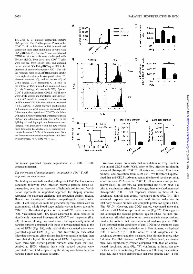
sequestration and ECM development. S. mansoni infectionmodulates host CD4+ T cell immune responses to third-partyAgs by generating a potent CD4+ T cell response that includesthe production of several regulatory cytokines (SupplementalFig. 2) (58, 59). S. mansoni-infected mice and age-matchednaive controls were infected with PbAluc and monitored forperipheral blood parasitemia, PbA biomass, and ECM onset.Coinfected mice had higher peripheral blood parasitemia thanmice infected with PbA alone from days 5–7 p.i. (Fig. 5A) andsuccumbed to ECM around the same time as these animals (Fig.5B). Significantly, coinfected mice also had a 2- to 3-fold highertotal body, but not brain, PbA biomass than mice infected withPbA alone (Fig. 5C, 5D).We also evaluated the impact of coinfection on malaria-specific
CD4+ T cell responses. CD4+ T cell recall responses to PbA bloodstage Ags in S. mansoni-coinfected micewere significantly reduced,whereas a strong S. mansoni-specific CD4+ T cell response againstSEAwas observed (Fig. 6A). To further studymalaria-specific CD4+
T cell responses, we used a transgenic OVA-expressing PbA line(PbTg) to measure the expansion of OVA-specific CD4+ (OT-II)T cells in mice with and without S. mansoni coinfection. Pro-liferation of transferred CFSE-labeled OT-II cells was measured4 d after PbTg challenge in the spleen (Fig. 6B). Control S. mansoni-infected mice injected with OT-II cells in the absence of PbTgchallenge showed no OT-II cell proliferation (Fig. 6B), as was thecase in mice infected with non-OVA–expressing PbA (PbG; data notshown). Robust OT-II cell expansion was observed in mice infectedwith OVA-transgenic PbA (Fig. 6B–D). In contrast, the absolutenumber and expansion of OT-II cells in the spleens of coinfectedmice were significantly reduced (Fig. 6B–D). Thus, CD4+ T cellresponses generated against PbA-expressed Ags could be detectedin S. mansoni-coinfected mice, but these responses were signifi-cantly impaired. After depletion of CD4+ T cells, the majority ofcoinfectedmice failed to develop ECM (Fig. 6E), and protectionwasassociated with significantly decreased total body and brain PbAbiomass (Fig. 6F, 6G). Thus, S. mansoni coinfection did not preventECM, despite modulating malaria-specific CD4+ T cells responses,
FIGURE 4. LTa and IFN-g mediate PbA tissue sequestration duringECM. Whole-body bioluminescence (left panel) and survival (right panel) ofB6.IFN-g2/2mice (A), B6.LTa2/2mice (B), B6.TNF2/2mice (C), C57BL/6mice in which both LTa and TNF were blocked throughout the course ofinfection by administration of TNFR2-Ig (D), and mice receiving IL-10Rblockade (E). Mice were infected with PbAluc, and bioluminescence imagingwas performed when control mice developed ECM (days 6–10 p.i.). Each barrepresents the mean 6 SEM of five to six mice. Data are from one repre-sentative experiment of at least two performed. pp , 0.05; ppp , 0.01
FIGURE 5. S. mansoni coinfection promotes increased PbA tissue se-questration and fails to prevent ECM. Peripheral blood parasitemia, asdetermined by microscopic examination of blood smears (A) and survival(B) of S. mansoni-infected mice following infection with PbAluc. Bio-luminescence imaging was performed when PbA-infected control micedeveloped ECM (day 7 p.i.). Total body (C) and brain (D) bioluminescenceare shown. Each bar represents the mean 6 SEM of four to five mice. AData are from one representative experiment of two performed. pp , 0.05.
The Journal of Immunology 3637
by guest on July 7, 2015http://w
ww
.jimm
unol.org/D
ownloaded from
but instead promoted parasite sequestration in a CD4+ T cell-dependent manner.
The generation of nonpathogenic, antiparasitic CD4+ T cellresponses by vaccination
Our findings above indicate that pathogenic CD4+ T cell responsesgenerated following PbA infection promote parasite tissue se-questration, even in the presence of helminth coinfection. Vacci-nation represents an important approach for shaping immuneresponses for pathogen challenge and protection against disease.Hence, we investigated whether nonpathogenic, antiparasiticCD4+ T cell responses could be generated by vaccination with anexperimental, whole blood stage malaria vaccine known to conferCD4+ T cell-mediated protection in non-ECM malaria models(52). Vaccination with PbA lysate adsorbed to alum resulted insignificantly increased PbA-specific CD4+ T cell responses (Fig.7A). However, although vaccinated mice had significantly reducedparasite burdens compared with those of unvaccinated mice at thetime of ECM (Fig. 7B), only half of the vaccinated mice wereprotected against ECM (Fig. 7C, 7D). Interestingly, vaccinatedmice that showed no clinical signs had lower parasite burdens thanthose that displayed clinical signs of ECM. Importantly, vacci-nated mice with higher parasite burdens were those that suc-cumbed to ECM, whereas those with reduced burdens wereprotected from ECM, emphasizing the strong correlation betweenparasite burden and disease severity.
We have shown previously that modulation of Treg functionwith an anti-CD25 mAb (PC61) prior to PbA infection resulted inenhanced PbA-specific CD4+ T cell activation, reduced PbA tissuebiomass, and protection from ECM (38). We therefore hypothe-sized that anti-CD25 mAb treatment at the time of vaccine primingwould increase PbA-specific CD4+ T cell responses and protectagainst ECM. To test this, we administered anti-CD25 mAb 1 dprior to vaccination. After PbA challenge, these mice had increasedPbA-specific CD4+ T cell responses relative to those of un-vaccinated controls and control vaccinated mice (Fig. 7A). Thisenhanced response was associated with further reductions intotal body parasite biomass and complete protection against ECM(Fig. 7B–D). However, anti-CD25–treated, vaccinated mice thathad survived ECM developed acute anemia (Fig. 7E). This suggeststhat although the vaccine protected against ECM, no such pro-tection was afforded against other severe malaria complications.Finally, to confirm that vaccine-induced malaria-specific CD4+
T cells primed under conditions of anti-CD25 mAb treatment wereresponsible for the observed reduction in PbA biomass, we depletedCD4+ T cells 5 d p.i. (at the onset of ECM symptoms in un-vaccinated controls) and then measured PbA biomass in these mice5 d later. The PbA biomass in CD4+ T cell-depleted, vaccinatedmice was significantly greater compared with that of control-treated, vaccinated mice (Fig. 7F), confirming an important rolefor these cells in controlling PbA biomass following vaccination.Together, these results demonstrate that PbA-specific CD4+ T cell
FIGURE 6. S. mansoni coinfection impairsPbA-specificCD4+T cell responses. PbA-specificCD4+ T cell proliferation in PbA-infected andcoinfected mice after stimulation in vitro withPbA pRBC Ag (A). Naive or S. mansoni-infectedC57BL/6 mice (n = 4) were challenged withPbAluc pRBCs. Four days later, CD4+ T cellswere purified from spleen cells and culturedinvitrowith nRBCs, PbApRBCAg, orSEA in thepresence of irradiated syngeneic APCs. The val-ues representmean6SEM [3H]thymidine uptakefrom triplicate cultures. In vivo proliferation (B),absolute numbers (C), and expansion (D) ofCFSE-labeled CD4+ transgenic OT-II cells inthe spleens of PbA-infected and coinfected mice(n = 4) following infection with PbTg. SplenicCD4+ T cells purified from CD45.1+ OT-II micewere CFSE-labeled and transferred into CD45.2+
recipient PbA-infected or coinfectedmice. Invivoproliferation of CFSE-labeled cells wasmeasured4 d p.i. Survival (E), total body (F), and brain (G)bioluminescence of S. mansoni-coinfected micefollowing in vivo depletion of CD4+ T cells. Micewith acuteS.mansoni infectionwere infectedwithPbAluc and administered anti-CD4 mAb or ratIgG day 21 and day 4 p.i., and bioluminescenceimaging was performed when rat IgG controlmice developed ECM (day 7 p.i.). Each bar rep-resents themean6 SEM of four to sixmice. Dataare from one representative experiment of at leasttwo performed. pp, 0.05; ppp, 0.01.
3638 PARASITE SEQUESTRATION IN ECM
by guest on July 7, 2015http://w
ww
.jimm
unol.org/D
ownloaded from

responses can be modulated by vaccination to reduce parasite se-questration and avoid ECM.
DiscussionThe sequestration of P. falciparum to various tissues has beenacknowledged as a major risk factor for severe malaria syndromes,including CM (6, 10). In this study, we have employed an ECMmodel to study parasite tissue sequestration and have identifiedhost cells and cytokines that contribute to or inhibit this process.Furthermore, we have shown that helminth coinfection can have amajor influence on parasite tissue sequestration, again via mech-anisms dependent on the host immune response. Critically, wewere able to demonstrate that the host immune response could bemodified by vaccination to protect against ECM rather than con-tribute to disease.The spleen is a major lymphoid organ and blood filtration tissue
during malaria (4). However, we found that splenectomized micesurvived ECM and had significantly reduced PbA biomass, in-dicating that removal of pRBCs by the spleen during PbA infectionis not a critical function for prevention of ECM and furthermore that
immune responses generated in this tissue site contribute to ECMpathogenesis. We identified both CD4+ and CD8+ T cells asmediators of PbA tissue sequestration. The importance of theseT cell subsets for ECM development has long been known (12–14,57), but their impact on parasite growth and/or survival has not beenappreciated. In the absence of either T cell subset, there wasa markedly reduced parasite biomass, indicating that CD4+ andCD8+ T cells are critical for either promoting PbA tissue seques-tration and/or suppressing the removal of PbA pRBCs from thecirculation by phagocytic cells. We believe that the latter possibilityis unlikely because blood parasitemia continues to increase in micedepleted of either CD4+ or CD8+ T cells and these animals die withhyperparasitemia (12–15). Therefore, a more likely explanation isthat T cells and/or their products condition tissues to allow parasitesequestration.Cytokines are important immune mediators and regulators with
both protective and pathogenic functions in malaria (7, 11). Weidentified IFN-g and LTa as critical mediators of PbA tissue se-questration. Both cytokines can activate microvascular endothelialcells (6, 60), and hence, one possibility is that changes to the ad-herent properties of these cells by IFN-g and LTa may result ingreater PbA adherence. However, this remains speculative becausethe nature of interactions between PbA and endothelial cells areunclear and to date few parasite molecules or host receptors thatmediate these interactions in vivo have been identified. Further-more, our studies on perfused tissues do not exclude the possibilitythat at least some of the PbA tissue sequestration observed couldarise from blockages in microvasculature caused by pRBCs or theaccumulation of pRBCs in tissue hemorrhages. Nevertheless, futurestudies on the roles of IFN-g and LTa in promoting adherence ofP. falciparum and endothelial cells, where parasite molecules andhost receptors are better characterized, may be warranted, becausethis may lead to new opportunities to suppress this pathogenicprocess in humans.Our efforts to block soluble LTa using soluble human TNFR2-
Fc failed to prevent ECM in PbA-infected mice, despite micedeficient in this cytokine being resistant to ECM. Blockade withhuman TNFR2-Fc had no effect on PbA biomass, but this is likelyexplained by TNF activity also being blocked, because our resultsshow that this cytokine plays an important antiparasitic role in theECM model. Thus, the positive effects of blocking pathogenicLTa were countered by the negative effects of neutralizing anti-parasitic TNF. The further delineation of LTa and TNF activitiesin ECM and CM must await the development of reagents withgreater selective actions on TNF and TNFR family memberinteractions. These studies are clearly important because of theopposing effects that this family of cytokines and receptors has onparasite growth and/or malaria pathogenesis.We also identified an important role for IL-10 in the regulation
of PbA tissue sequestration. Previous studies have shown that IL-10 is important for preventing pathology associated with differentexperimental malaria models (61) but also can cause parasitepersistence and thus contribute to the establishment of somechronic infectious diseases (62). We recently showed that mod-ulation of Treg function with an anti-CD25 mAb resulted inreduced parasite biomass and protection from ECM, but thisprotection was lost in IL-10–deficient mice (38). These datasupport an important regulatory role for IL-10 in malaria andalso highlight the dangers of blocking IL-10 activity to improveantiparasitic immune responses. Clearly, there is a fine balancebetween the tissue-protective role of IL-10 during infectiousdisease and the unwanted action of this cytokine in suppressingantiparasitic immunity. A greater understanding of how thisbalance is established and maintained is crucial for the design
FIGURE 7. Vaccination generates antiparasitic, nonpathogenic CD4+
T cell responses. PbA-specific CD4+ T cell proliferation in unvaccinated,vaccinated, or vaccinated mice in which Tregs weremodulated at priming (n= 4) following challenge with 105 PbAluc pRBCs (A). Four days afterPbAluc infection, CD4+ T cells were purified from spleen cells and culturedin vitro with nRBCs or PbA pRBC Ag in the presence of irradiated syn-geneic APCs. The values represent mean 6 SEM [3H]thymidine uptakefrom triplicate cultures. Total body bioluminescence (B), survival (C), ECMincidence (D), and hemoglobin levels (E) were measured following vacci-nation and PbAluc infection. Total body bioluminescence of PbA-infectedvaccinated mice, primed under conditions of Treg modulation, depleted ofCD4+ T cells at day 5 p.i., and imaged on day 10 p.i. (F). Each bar representsthe mean 6 SEM of four to six mice. Data are from one representativeexperiment of two performed. pp , 0.05; ppp , 0.01; ppp p , 0.001.
The Journal of Immunology 3639
by guest on July 7, 2015http://w
ww
.jimm
unol.org/D
ownloaded from

and implementation of strategies aiming to safely harness thetherapeutic potential of IL-10.Helminth infections can greatly influence the host immune
system (58), and importantly, these infections often occur in areasof endemic and seasonal malaria transmission (63). Therefore, it isimportant that we understand their impact on the development ofimmunity to malaria and any effects that they may have on malariapathogenesis. We found that an established S. mansoni infectionresulted in greater PbA tissue sequestration, but this was associ-ated with impaired development of PbA-specific CD4+ T cellresponses. These effects were only observed in mice with high-level helminth infections and not when lower numbers of S.mansoni cercariae were used to establish infections (SupplementalFig. 3 and data not shown). Therefore, helminth burden and/orthe inoculums used to establish infection is likely to be an im-portant factor in determining whether there is any impact on thegeneration of malaria-specific immunity and/or the likelihood ofdeveloping severe malaria syndromes. Nevertheless, it was in-triguing to note that despite suppressed PbA-specific CD4+ T cellresponses in mice with S. mansoni infections the enhanced PbAtissue sequestration in these animals was still dependent on CD4+
T cells. Although we cannot rule out the possibility that thesuppressed malaria-specific CD4+ T cell response was sufficientto mediate the enhanced PbA tissue sequestration and suscepti-bility to ECM, our data leave open the possibility that helminth-specific CD4+ T cell responses or even bystander CD4+ T cellactivation could contribute to the enhanced PbA biomass andsusceptibility to ECM in coinfected mice. Regardless, our datasupport previous suggestions (64) that the integration of hel-minth control programs into malaria control strategies will bebeneficial, particularly for individuals harboring high wormburdens, because this will help to improve the development ofmalaria-specific immunity and possibly reduce the risk of de-veloping severe malaria syndromes, such as CM, due to theaccumulation of high malaria parasite burdens.Our finding that concomitant S. mansoni and PbA infection failed
to protect against ECM contradicts a recent study in which a pro-tective effect was observed (65). This difference in susceptibility islikely to be due, in part at least, to differences in experimental design.For example, the previous study examined the impact of pre-existingS. mansoni infection on the development of ECM in an outbredmouse strain, as opposed to inbred C57BL/6 mice employed in thecurrent study. Furthermore, micewere challengedwith a 2-fold lowerPbA inoculum than that used here. Both mouse genetic backgroundand parasite inoculums will impact on the effect that helminths haveon susceptibility to malaria infection.Vaccination represents one of themost promising strategies to con-
trol malaria (66–68). Despite our incomplete understanding aboutthe types of immune responses that are necessary for safe andeffective control of infection, vaccine development and testing areproceeding rapidly due to an urgent need for this important publichealth tool (68). Nevertheless, it is important to recognize thatimmune responses generated by vaccination have the potential topromote disease rather than control infection (69). We found thatanti-CD25 mAb treatment at the time of vaccine priming resultedin the generation of a highly effective malaria-specific CD4+
T cell response that suppressed PbA tissue sequestration andprevented ECM development. Thus, the specific immune con-ditions established during vaccine priming had a major impact onthe quality of the subsequent malaria-specific CD4+ T cells re-sponse. It is important to note that despite preventing ECM the anti-CD25 mAb combined vaccine strategy did not result in ultimatecontrol of parasite growth and mice eventually succumbed tohyperparasitemia and anemia. We believe that this failure to control
parasite growth after protection from ECM could be attributed toeither inappropriate downregulation of malaria-specific CD4+
T cell responses, as previously described in this model (70, 71), ora failure to generate an effective antiparasitic Ab response, asreported in several different experimental malaria models (11, 72).These two possibilities are not mutually exclusive, and we arecurrently developing ways to improve our malaria vaccine formu-lation to enhance both aspects of our vaccine-induced immunity.In conclusion, we have identified specific components of the host
immune system that contribute to and inhibit parasite tissue se-questration in an experimental model of CM. Our findings suggestthat sequestration of parasites in vital organs, including the brain,marks the onset of ECM and that ECM susceptibility is stronglyassociated with the total parasite biomass. This observation is inkeeping with previous studies in acute P. falciparum malariashowing an association among total body parasite biomass, dis-ease severity, and clinical outcome (35). Importantly, the datashow that for ECM to be prevented, parasite sequestration needs tobe limited and parasite biomass significantly reduced early ininfection. This process appears to be at least partly dependent onTNF and IL-10. We also have been able to evaluate the impact ofhelminth coinfection on the generation of malaria-specific im-munity and ECM pathogenesis. Importantly, we were able to showthat vaccination could be used to generate potent antiparasiticCD4+ T cell responses without induction of pathology. Together,these advances in our understanding about the pathogenesis ofmalaria have important implications for developing therapeuticapproaches to treat severe malaria syndromes, the development ofsafe and effective malaria vaccines, and the implementation ofthese important public health tools.
AcknowledgmentsWe thank the Queensland Institute of Medical Research animal facility foranimal husbandry, Paula Hall and Grace Chojnowski for assistance with cellsorting, and Chris Janse, Maria Yazdanbakhsh, and Jeff Browning for help-ful discussions.
DisclosuresThe authors have no financial conflicts of interest.
References1. World Health Organization. 2009. World Malaria Report. World Health Orga-
nization, Geneva, Switzerland.2. MacPherson, G. G., M. J. Warrell, N. J. White, S. Looareesuwan, and
D. A. Warrell. 1985. Human cerebral malaria. A quantitative ultrastructuralanalysis of parasitized erythrocyte sequestration. Am. J. Pathol. 119: 385–401.
3. Berendt, A. R., G. D. Tumer, and C. I. Newbold. 1994. Cerebral malaria: thesequestration hypothesis. Parasitol. Today 10: 412–414.
4. Engwerda, C. R., L. Beattie, and F. H. Amante. 2005. The importance of thespleen in malaria. Trends Parasitol. 21: 75–80.
5. Newton, C. R., and S. Krishna. 1998. Severe falciparum malaria in children:current understanding of pathophysiology and supportive treatment. Pharmacol.Ther. 79: 1–53.
6. Schofield, L., and G. E. Grau. 2005. Immunological processes in malaria path-ogenesis. Nat. Rev. Immunol. 5: 722–735.
7. Good, M. F., H. Xu, M. Wykes, and C. R. Engwerda. 2005. Development andregulation of cell-mediated immune responses to the blood stages of malaria:implications for vaccine research. Annu. Rev. Immunol. 23: 69–99.
8. Renia, L., S. M. Potter, M. Mauduit, D. S. Rosa, M. Kayibanda, J. C. Deschemin,G. Snounou, and A. C. Gruner. 2006. Pathogenic T cells in cerebral malaria. Int.J. Parasitol. 36: 547–554.
9. Clark, I. A., and W. B. Cowden. 2003. The pathophysiology of falciparummalaria. Pharmacol. Ther. 99: 221–260.
10. van der Heyde, H. C., J. Nolan, V. Combes, I. Gramaglia, and G. E. Grau. 2006.A unified hypothesis for the genesis of cerebral malaria: sequestration, in-flammation and hemostasis leading to microcirculatory dysfunction. TrendsParasitol. 22: 503–508.
11. Engwerda, C., E. Belnoue, A. C. Gruner, and L. Renia. 2005. Experimentalmodels of cerebral malaria. Curr. Top. Microbiol. Immunol. 297: 103–143.
12. Hermsen, C., T. van de Wiel, E. Mommers, R. Sauerwein, and W. Eling. 1997.Depletion of CD4+ or CD8+ T-cells prevents Plasmodium berghei induced ce-rebral malaria in end-stage disease. Parasitology 114: 7–12.
3640 PARASITE SEQUESTRATION IN ECM
by guest on July 7, 2015http://w
ww
.jimm
unol.org/D
ownloaded from

13. Grau, G. E., P. F. Piguet, H. D. Engers, J. A. Louis, P. Vassalli, andP. H. Lambert. 1986. L3T4+ T lymphocytes play a major role in the pathogenesisof murine cerebral malaria. J. Immunol. 137: 2348–2354.
14. Nitcheu, J., O. Bonduelle, C. Combadiere, M. Tefit, D. Seilhean, D. Mazier, andB. Combadiere. 2003. Perforin-dependent brain-infiltrating cytotoxic CD8+T lymphocytes mediate experimental cerebral malaria pathogenesis. J. Immunol.170: 2221–2228.
15. Yanez, D. M., D. D. Manning, A. J. Cooley, W. P. Weidanz, and H. C. van derHeyde. 1996. Participation of lymphocyte subpopulations in the pathogenesis ofexperimental murine cerebral malaria. J. Immunol. 157: 1620–1624.
16. Curfs, J. H., T. P. Schetters, C. C. Hermsen, C. R. Jerusalem, A. A. van Zon, andW. M. Eling. 1989. Immunological aspects of cerebral lesions in murine malaria.Clin. Exp. Immunol. 75: 136–140.
17. Hermsen, C. C., E. Mommers, T. van de Wiel, R. W. Sauerwein, andW. M. Eling. 1998. Convulsions due to increased permeability of the blood-brainbarrier in experimental cerebral malaria can be prevented by splenectomy oranti-T cell treatment. J. Infect. Dis. 178: 1225–1227.
18. Lundie, R. J., T. F. de Koning-Ward, G. M. Davey, C. Q. Nie, D. S. Hansen,L. S. Lau, J. D. Mintern, G. T. Belz, L. Schofield, F. R. Carbone, et al. 2008.Blood-stage Plasmodium infection induces CD8+ T lymphocytes to parasite-expressed antigens, largely regulated by CD8alpha+ dendritic cells. Proc.Natl. Acad. Sci. USA 105: 14509–14514.
19. Miyakoda, M., D. Kimura, M. Yuda, Y. Chinzei, Y. Shibata, K. Honma, andK. Yui. 2008. Malaria-specific and nonspecific activation of CD8+ T cells duringblood stage of Plasmodium berghei infection. J. Immunol. 181: 1420–1428.
20. Potter, S., T. Chan-Ling, H. J. Ball, H. Mansour, A. Mitchell, L. Maluish, andN. H. Hunt. 2006. Perforin mediated apoptosis of cerebral microvascularendothelial cells during experimental cerebral malaria. Int. J. Parasitol. 36: 485–496.
21. Potter, S., G. Chaudhri, A. Hansen, and N. H. Hunt. 1999. Fas and perforincontribute to the pathogenesis of murine cerebral malaria. Redox Rep. 4: 333–335.
22. Grau, G. E., L. F. Fajardo, P. F. Piguet, B. Allet, P. H. Lambert, and P. Vassalli.1987. Tumor necrosis factor (cachectin) as an essential mediator in murine ce-rebral malaria. Science 237: 1210–1212.
23. Grau, G. E., H. Heremans, P. F. Piguet, P. Pointaire, P. H. Lambert, A. Billiau,and P. Vassalli. 1989. Monoclonal antibody against interferon gamma can pre-vent experimental cerebral malaria and its associated overproduction of tumornecrosis factor. Proc. Natl. Acad. Sci. USA 86: 5572–5574.
24. Engwerda, C. R., T. L. Mynott, S. Sawhney, J. B. De Souza, Q. D. Bickle, andP. M. Kaye. 2002. Locally up-regulated lymphotoxin alpha, not systemic tumornecrosis factor alpha, is the principle mediator of murine cerebral malaria. J.Exp. Med. 195: 1371–1377.
25. Hunt, N. H., J. Golenser, T. Chan-Ling, S. Parekh, C. Rae, S. Potter,I. M. Medana, J. Miu, and H. J. Ball. 2006. Immunopathogenesis of cerebralmalaria. Int. J. Parasitol. 36: 569–582.
26. Langhorne, J., B. Simon-Haarhaus, and S. J. Meding. 1990. The role of CD4+T cells in the protective immune response to Plasmodium chabaudi in vivo.Immunol. Lett. 25: 101–107.
27. Podoba, J. E., and M. M. Stevenson. 1991. CD4+ and CD8+ T lymphocytes bothcontribute to acquired immunity to blood-stage Plasmodium chabaudi AS. In-fect. Immun. 59: 51–58.
28. Ockenhouse, C. F., S. Schulman, and H. L. Shear. 1984. Induction of crisis formsin the human malaria parasite Plasmodium falciparum by gamma-interferon-activated, monocyte-derived macrophages. J. Immunol. 133: 1601–1608.
29. Shear, H. L., R. Srinivasan, T. Nolan, and C. Ng. 1989. Role of IFN-gamma inlethal and nonlethal malaria in susceptible and resistant murine hosts. J.Immunol. 143: 2038–2044.
30. Bouharoun-Tayoun, H., P. Attanath, A. Sabchareon, T. Chongsuphajaisiddhi, andP. Druilhe. 1990. Antibodies that protect humans against Plasmodium falciparumblood stages do not on their own inhibit parasite growth and invasion in vitro, butact in cooperation with monocytes. J. Exp. Med. 172: 1633–1641.
31. Mota, M. M., K. N. Brown, A. A. Holder, and W. Jarra. 1998. Acute Plasmodiumchabaudi chabaudi malaria infection induces antibodies which bind to the sur-faces of parasitized erythrocytes and promote their phagocytosis by macrophagesin vitro. Infect. Immun. 66: 4080–4086.
32. Shear, H. L., R. S. Nussenzweig, and C. Bianco. 1979. Immune phagocytosis inmurine malaria. J. Exp. Med. 149: 1288–1298.
33. Waki, S., S. Uehara, K. Kanbe, H. Nariuch, and M. Suzuki. 1995. Interferon-gamma and the induction of protective IgG2a antibodies in non-lethal Plasmo-dium berghei infections of mice. Parasite Immunol. 17: 503–508.
34. Silamut, K., N. H. Phu, C. Whitty, G. D. Turner, K. Louwrier, N. T. Mai,J. A. Simpson, T. T. Hien, and N. J. White. 1999. A quantitative analysis of themicrovascular sequestration of malaria parasites in the human brain. Am. J.Pathol. 155: 395–410.
35. Dondorp, A. M., V. Desakorn, W. Pongtavornpinyo, D. Sahassananda,K. Silamut, K. Chotivanich, P. N. Newton, P. Pitisuttithum, A. M. Smithyman,N. J. White, and N. P. Day. 2005. Estimation of the total parasite biomass inacute falciparum malaria from plasma PfHRP2. PLoS Med. 2: e204.
36. Chang, W. L., S. P. Jones, D. J. Lefer, T. Welbourne, G. Sun, L. Yin, H. Suzuki,J. Huang, D. N. Granger, and H. C. van der Heyde. 2001. CD8(+)-T-cell de-pletion ameliorates circulatory shock in Plasmodium berghei-infected mice.Infect. Immun. 69: 7341–7348.
37. Lovegrove, F. E., S. A. Gharib, L. Pena-Castillo, S. N. Patel, J. T. Ruzinski,T. R. Hughes, W. C. Liles, and K. C. Kain. 2008. Parasite burden and CD36-mediated sequestration are determinants of acute lung injury in an experimentalmalaria model. PLoS Pathog. 4: e1000068.
38. Amante, F. H., A. C. Stanley, L. M. Randall, Y. Zhou, A. Haque, K. McSweeney,A. P. Waters, C. J. Janse, M. F. Good, G. R. Hill, and C. R. Engwerda. 2007. Arole for natural regulatory T cells in the pathogenesis of experimental cerebralmalaria. Am. J. Pathol. 171: 548–559.
39. Franke-Fayard, B., C. J. Janse, M. Cunha-Rodrigues, J. Ramesar, P. Buscher, I. Que,C. Lowik, P. J. Voshol, M. A. den Boer, S. G. van Duinen, et al. 2005. Murinemalaria parasite sequestration: CD36 is the major receptor, but cerebral pathology isunlinked to sequestration. Proc. Natl. Acad. Sci. USA 102: 11468–11473.
40. Nie, C. Q., N. J. Bernard, M. U. Norman, F. H. Amante, R. J. Lundie, B. S. Crabb,W. R. Heath, C. R. Engwerda, M. J. Hickey, L. Schofield, and D. S. Hansen. 2009.IP-10-mediated T cell homing promotes cerebral inflammation over splenic im-munity to malaria infection. PLoS Pathog. 5: e1000369.
41. Randall, L. M., F. H. Amante, K. A. McSweeney, Y. Zhou, A. C. Stanley,A. Haque, M. K. Jones, G. R. Hill, G. M. Boyle, and C. R. Engwerda. 2008.Common strategies to prevent and modulate experimental cerebral malaria inmouse strains with different susceptibilities. Infect. Immun. 76: 3312–3320.
42. Randall, L. M., F. H. Amante, Y. Zhou, A. C. Stanley, A. Haque, F. Rivera,K. Pfeffer, S. Scheu, G. R. Hill, K. Tamada, and C. R. Engwerda. 2008. Cuttingedge: selective blockade of LIGHT-lymphotoxin beta receptor signaling protectsmice from experimental cerebral malaria caused by Plasmodium berghei ANKA.J. Immunol. 181: 7458–7462.
43. Mombaerts, P., J. Iacomini, R. S. Johnson, K. Herrup, S. Tonegawa, andV. E. Papaioannou. 1992. RAG-1-deficient mice have no mature B andT lymphocytes. Cell 68: 869–877.
44. Barnden, M. J., J. Allison, W. R. Heath, and F. R. Carbone. 1998. Defective TCRexpression in transgenic mice constructed using cDNA-based alpha- and beta-chain genes under the control of heterologous regulatory elements. Immunol.Cell Biol. 76: 34–40.
45. Kitamura, D., J. Roes, R. Kuhn, and K. Rajewsky. 1991. A B cell-deficientmouse by targeted disruption of the membrane exon of the immunoglobulinmu chain gene. Nature 350: 423–426.
46. Dalton, D. K., S. Pitts-Meek, S. Keshav, I. S. Figari, A. Bradley, andT. A. Stewart. 1993. Multiple defects of immune cell function in mice withdisrupted interferon-gamma genes. Science 259: 1739–1742.
47. Sean Riminton, D., H. Korner, D. H. Strickland, F. A. Lemckert, J. D. Pollard,and J. D. Sedgwick. 1998. Challenging cytokine redundancy: inflammatory cellmovement and clinical course of experimental autoimmune encephalomyelitisare normal in lymphotoxin-deficient, but not tumor necrosis factor-deficient,mice. J. Exp. Med. 187: 1517–1528.
48. Korner, H., M. Cook, D. S. Riminton, F. A. Lemckert, R. M. Hoek,B. Ledermann, F. Kontgen, B. Fazekas de St Groth, and J. D. Sedgwick. 1997.Distinct roles for lymphotoxin-alpha and tumor necrosis factor in organogenesisand spatial organization of lymphoid tissue. Eur. J. Immunol. 27: 2600–2609.
49. Janse, C. J., J. Ramesar, and A. P. Waters. 2006. High-efficiency transfection anddrug selection of genetically transformed blood stages of the rodent malariaparasite Plasmodium berghei. Nat. Protoc. 1: 346–356.
50. Tran, M. H., M. S. Pearson, J. M. Bethony, D. J. Smyth, M. K. Jones, M. Duke,T. A. Don, D. P. McManus, R. Correa-Oliveira, and A. Loukas. 2006. Tetra-spanins on the surface of Schistosoma mansoni are protective antigens againstschistosomiasis. Nat. Med. 12: 835–840.
51. Su, Z., M. Segura, K. Morgan, J. C. Loredo-Osti, and M. M. Stevenson. 2005.Impairment of protective immunity to blood-stage malaria by concurrent nem-atode infection. Infect. Immun. 73: 3531–3539.
52. Su, Z., M. F. Tam, D. Jankovic, and M. M. Stevenson. 2003. Vaccination withnovel immunostimulatory adjuvants against blood-stage malaria in mice. Infect.Immun. 71: 5178–5187.
53. Markey, K. A., A. C. Burman, T. Banovic, R. D. Kuns, N. C. Raffelt, V. Rowe,S. D. Olver, A. L. Don, E. S. Morris, A. R. Pettit, et al. 2009. Soluble lymphotoxinis an important effector molecule in GVHD and GVL. Blood 115: 122–132.
54. Mintern, J., M. Li, G. M. Davey, E. Blanas, C. Kurts, F. R. Carbone, andW. R. Heath. 1999. The use of carboxyfluorescein diacetate succinimidyl ester todetermine the site, duration and cell type responsible for antigen presentationin vivo. Immunol. Cell Biol. 77: 539–543.
55. Lyons, A. B., and C. R. Parish. 1994. Determination of lymphocyte division byflow cytometry. J. Immunol. Methods 171: 131–137.
56. Beeson, J. G., and G. V. Brown. 2002. Pathogenesis of Plasmodium falciparummalaria: the roles of parasite adhesion and antigenic variation. Cell. Mol. LifeSci. 59: 258–271.
57. Belnoue, E., M. Kayibanda, A. M. Vigario, J. C. Deschemin, N. van Rooijen,M. Viguier, G. Snounou, and L. Renia. 2002. On the pathogenic role of brain-sequestered alphabeta CD8+ T cells in experimental cerebral malaria. J.Immunol. 169: 6369–6375.
58. Hartgers, F. C., and M. Yazdanbakhsh. 2006. Co-infection of helminths andmalaria: modulation of the immune responses to malaria. Parasite Immunol. 28:497–506.
59. Pearce, E. J., and A. S. MacDonald. 2002. The immunobiology of schistoso-miasis. Nat. Rev. Immunol. 2: 499–511.
60. Wassmer, S. C., V. Combes, F. J. Candal, I. Juhan-Vague, and G. E. Grau. 2006.Platelets potentiate brain endothelial alterations induced by Plasmodium falci-parum. Infect. Immun. 74: 645–653.
61. Kossodo, S., C. Monso, P. Juillard, T. Velu, M. Goldman, and G. E. Grau. 1997.Interleukin-10 modulates susceptibility in experimental cerebral malaria. Im-munology 91: 536–540.
62. Belkaid, Y., K. F. Hoffmann, S. Mendez, S. Kamhawi, M. C. Udey, T. A. Wynn,and D. L. Sacks. 2001. The role of interleukin (IL)-10 in the persistence ofLeishmania major in the skin after healing and the therapeutic potential of anti-IL-10 receptor antibody for sterile cure. J. Exp. Med. 194: 1497–1506.
The Journal of Immunology 3641
by guest on July 7, 2015http://w
ww
.jimm
unol.org/D
ownloaded from

63. Mwangi, T. W., J. M. Bethony, and S. Brooker. 2006. Malaria and helminthinteractions in humans: an epidemiological viewpoint. Ann. Trop. Med. Para-sitol. 100: 551–570.
64. Druilhe, P., A. Tall, and C. Sokhna. 2005. Worms can worsen malaria: towardsa new means to roll back malaria? Trends Parasitol. 21: 359–362.
65. Waknine-Grinberg, J. H., D. Gold, A. Ohayon, E. Flescher, A. Heyfets,M. J. Doenhoff, G. Schramm, H. Haas, and J. Golenser. 2010. Schistosomamansoni infection reduces the incidence of murine cerebral malaria. Malar. J.9: 5.
66. Haque, A., and M. F. Good. 2009. Malaria vaccine research: lessons from 2008/9. Future Microbiol. 4: 649–654.
67. Good, M. F. 2009. The hope but challenge for developing a vaccine that mightcontrol malaria. Eur. J. Immunol. 39: 939–943.
68. Greenwood, B., and G. Targett. 2009. Do we still need a malaria vaccine?Parasite Immunol. 31: 582–586.
69. Schofield, L. 2007. Rational approaches to developing an anti-disease vaccineagainst malaria. Microbes Infect. 9: 784–791.
70. Hirunpetcharat, C., and M. F. Good. 1998. Deletion of Plasmodium berghei-specific CD4+ T cells adoptively transferred into recipient mice after challengewith homologous parasite. Proc. Natl. Acad. Sci. USA 95: 1715–1720.
71. Xu, H., J. Wipasa, H. Yan, M. Zeng, M. O. Makobongo, F. D. Finkelman, A. Kelso,and M. F. Good. 2002. The mechanism and significance of deletion of parasite-specific CD4(+) T cells in malaria infection. J. Exp. Med. 195: 881–892.
72. Achtman, A. H., P. C. Bull, R. Stephens, and J. Langhorne. 2005. Longevity ofthe immune response and memory to blood-stage malaria infection. Curr. Top.Microbiol. Immunol. 297: 71–102.
3642 PARASITE SEQUESTRATION IN ECM
by guest on July 7, 2015http://w
ww
.jimm
unol.org/D
ownloaded from